Astrocyte-targeted expression of IL6 protects the CNSagainst a focal brain injury
Upload
independentCategory
view
2download
0
RESEARCH ARTICLE
Effects of Astrocyte-Targeted Productionof Interleukin-6 in the Mouse on the Host
Response to Nerve Injury
Beatriz Almolda,1 N�adia Villacampa,1 Peter Manders,2 Juan Hidalgo,1 Iain L. Campbell,2
Berta Gonz�alez,1 and Bernardo Castellano1
Interleukin-6 (IL-6) is a pleiotropic cytokine with a key role in the control of inflammatory/immune responses. In the centralnervous system (CNS), an increase in IL-6 occurs in a wide range of pathological conditions such as excitotoxicity and trau-matic brain injury. We evaluated the effects of astrocyte-targeted production of IL-6 in the CNS in the sterile-nerve injurymodel of facial nerve axotomy. To accomplish this, facial nerve transection was performed in transgenic mice (glial fibrillaryacidic protein [GFAP]-IL6Tg) with IL-6 production under the GFAP promoter. Neuronal death, glial activation, lymphocyterecruitment, and integrin expression were evaluated by immunohistochemistry and flow cytometry from 3 to 28 days postin-jury. Our findings revealed an increase in motor neuron cell death in GFAP-IL6Tg mice correlating with changes in the micro-glial activation pattern, characterized principally by less attachment to neurons and reduced expression of both CD11b andCD18. We also found a higher CD41 T-lymphocyte recruitment in GFAP-IL6Tg mice. In addition, changes in the expressionpattern of different integrins and their receptors were observed in transgenic animals. Specifically, alterations in osteopontinexpression in motor neurons and its receptors CD44 and CD49e in lymphocytes and microglia, respectively, which mayaccount for the variations related to glial reactivity and lymphocyte infiltration. In conclusion, our results indicated that forcedlocal production of IL-6 has a direct impact on the outcome of nerve injury in the CNS inducing an increase in neurodegenera-tion, changes in glial response, and lymphocyte recruitment as well as in the expression of different integrins and theirreceptors.
GLIA 2014;62:1142–1161Key words: microglia, transgenic animal, lymphocytes, osteopontin, CD44, facial nerve axotomy
Introduction
Interleukin-6 (IL-6) is a pleiotropic and multifunctional
cytokine involved in the regulation of inflammatory and
immunological responses in the periphery (Kishimoto et al.,
1995; Taga and Kishimoto, 1997; Kishimoto, 2006). In the
central nervous system (CNS), IL-6 is low in normal condi-
tions but increased significantly in neurons, astrocytes, and
microglial cells, under acute and chronic injury and in certain
diseases including excitotoxicity, traumatic brain injury, ische-
mia, multiple sclerosis, and neurodegenerative diseases like
Alzheimer and Parkinson (Benveniste, 1998; Erta et al., 2012;
Gadient and Otten, 1997; Gruol and Nelson, 1997;
Nakamura et al., 2005; Spooren et al., 2011; Suzuki et al.,
2009; Van Wagoner and Benveniste, 1999). However, the
exact role played by this cytokine in these harmful circum-
stances is not yet well established, and both detrimental as
well as beneficial functions have been proposed in different
clinical as well as experimental neuropathologies.
The detrimental role attributed to IL-6 production is
based on evidence that, together with TNF-a and IL-1b, this
IL can be considered as a major modulator of the inflamma-
tory response in the CNS (Benveniste, 1998; Erta et al.,
2012; Gadient and Otten 1997). Although whether neuroin-
flammation is protective or detrimental remains the subject of
View this article online at wileyonlinelibrary.com. DOI: 10.1002/glia.22668
Published online April 2, 2014 in Wiley Online Library (wileyonlinelibrary.com). Received Dec 10, 2013, Accepted for publication Mar 14, 2014.
Address correspondence to Beatriz Almolda, Unitat d’Histologia, Torre M5, Universitat Aut�onoma de Barcelona, 08193 Bellaterra, Spain.
E-mail: [email protected]
From the 1Department of Cell Biology, Physiology and Immunology, Institute of Neuroscience, Autonomous University of Barcelona, Bellaterra, 08193, Spain;2School of Molecular Bioscience, University of Sydney, Sydney, NSW 2006, Australia.
Beatriz Almolda and N�adia Villacampa contributed equally to the work.
Additional Supporting Information may be found in the online version of this article.
1142 VC 2014 Wiley Periodicals, Inc.
intense discussion, classically it has been associated with glial
activation and the consequent production of deleterious reac-
tive oxygen species and inflammatory mediators, such as cyto-
kines, chemokines, and prostaglandins, which may, among
others, induce blood brain barrier disruption, lymphocyte
recruitment, and promote neurodegeneration. Studies in vitrosupport the view that IL-6 is able to induce glial activation
(Krady et al., 2008) and neuronal death (Conroy et al.,
2004). Moreover, in agreement with work in vivo showing
glial activation after IL-6 administration (Tilgner et al.,
2001), chronic production of IL-6 achieved in transgenic ani-
mals has been linked to neurodegeneration (Campbell et al.,
1993), increased astrocyte and microglial activation (Brunello
et al., 2000; Campbell et al., 1993; Chiang et al., 1994) and
impaired higher order brain function (Heyser et al., 1997).
Other studies showed a decrease in axonal regrowth in hyper-
IL-6-administered rats after spinal cord injury (Lacroix et al.,
2002). Furthermore, an improvement of neuronal survival
has been reported in IL-6 deficient mice after optic nerve
crush (Fisher et al., 2001) and blocking of IL-6 signaling by
anti-IL-6 receptor treatment induced a significant increase in
functional recovery together with decreased gliosis in spinal
cord-injured mice (Mukaino et al., 2010; Okada et al.,
2004).
In contrast, anti-inflammatory and neuroprotective func-
tions have been attributed to IL-6 due to the capacity of this
cytokine to increase axonal regeneration in spinal cord-injured
rats when administered intrathecally (Cao et al., 2006), the
decrease in lesion volume reported in ischemic rats after intra-
cerebroventricular IL-6 treatment (Loddick et al., 1998) and
the reduction of motor neuronal loss described after sciatic
nerve transection in intraperitoneally IL-6-treated rats (Ikeda
et al., 1996). Furthermore, some studies conducted in IL-6
deficient mice support a role for IL-6 in neuroprotection, as
these animals showed an increase in neuronal loss after both
cryolesion (Swartz et al., 2001) and sciatic nerve axotomy
(Murphy et al., 1999) and a reduction in functional recovery
in axotomized animals (Zhong et al., 1999).
Altogether, these studies clearly indicate that the effects
derived from IL-6 production are complex and may depend
on several factors including the time-point and the specific
microenvironment where this cytokine is produced or admin-
istered in the CNS. In this context, and in an attempt to bet-
ter understand the function of IL-6 in CNS pathologies, this
study focused on the analysis of the effects that local IL-6
production in the brain may exert following nerve injury in
the facial nerve axotomy paradigm. For this purpose, we used
the glial fibrillary acidic protein (GFAP)-IL6 transgenic (Tg)
mouse model in which production of this cytokine was selec-
tively targeted to astrocytes within the CNS of mice (Camp-
bell, 1998; Campbell et al., 1993).
Materials and Methods
Animals and Facial Nerve AxotomyA total of 47 transgenic animals with astrocyte-targeted production
of IL-6 (GFAP-IL6Tg) and their corresponding wild-type (WT) lit-
termates (n 5 36) were used in this study. Characterization of the
GFAP-IL6Tg mice was described previously (Campbell et al., 1993).
In these animals, astrocyte-targeted production of IL-6 has been
demonstrated under basal conditions in several CNS areas including
the brainstem (Giralt et al., 2013). Animals were maintained with
food and water ad libitum in a 12 h light/dark cycle during all the
experiment.
After being anaesthetized by an intraperitoneal injection of
ketamine 80 mg/kg and xylazine 20 mg/kg at dose of 0.01 mL/g,
the right facial nerve was resected at the level of the stylomastoid
foramen in both WT and GFAP-IL6Tg animals.
All experimental animal work was conducted according to
Spanish regulations (Ley 32/2007, Real Decreto 1201/2005, Ley 9/
2003, y Real Decreto 178/2004) in agreement with European Union
directives (86/609/CEE, 91/628/CEE i 92/65/CEE) and was
approved by the Ethical Committees of the Autonomous University
of Barcelona and the University of Sydney.
Experimental GroupsAxotomized animals were distributed in different groups and eutha-
nized at different days, ranging from 1 to 28 days postinjury (dpi),
and tissue samples appropriately processed for flow cytometry, histo-
chemistry, and immunohistochemistry (for details see later). A total
of 10 WT and 8 GFAP-IL6Tg animals were used for flow cytometry
studies; 23 WT and 36 GFAP-IL6Tg were processed for histochem-
istry and immunohistochemistry and 3 WT and 3 GFAP-IL6Tg ani-
mals were used for neuronal quantification.
Flow Cytometry AnalysisThe percentage of infiltrated CD31CD41 cells was analyzed by
flow cytometry at two specific time-points: 14 and 21 dpi following
the protocol previously described (Almolda et al., 2009, 2011). For
details of specific protocol, see Supporting Information 1.
Tissue Processing for Histochemistry andImmunohistochemistry AnalysisUnder deep anaesthesia (0.015 mL/g) of ketamine (80 mg/kg)/xyla-
zine (20 mg/kg) animals were perfused intracardially for 10 min with
either 4% paraformaldehyde in 0.1 M cacodylate buffer (pH 7.4) 1
5% sucrose for vibratome sectioning or 4% paraformaldehyde in
0.1M PBS (pH 7.4) for cryostat sections. Brainstems containing the
facial nucleus (FN) were immediately dissected out, postfixed for 4 h
at 4�C in the same fixative, and then a series of parallel sections were
obtained using a VT 1000S Leica vibratome (40-lm thick) or a CM
3050s Leica cryostat (30-lm thick). Vibratome and cryostat series
were stored at 220�C in Olmos antifreeze solution and used for
immunohistochemistry, with the exception of one of the vibratome
series that was processed for the histoenzymatic demonstration of
nucleoside diphosphatase (NDPase) as described later.
A set of axotomized WT (n 5 3) and GFAP-IL6Tg (n 5 3)
animals were perfused at 21 dpi as described earlier and coronal
Almolda et al.: Facial Nerve Axotomy in GFAP-IL6Tg Mice
July 2014 1143

sections (16 lm) of the entire brainstem were obtained using a CM
3050s Leica cryostat. The sections were mounted on gelatin-coated
slides and used for toluidine blue staining.
Toluidine Blue Staining and Neuronal SurvivalQuantificationToluidine blue staining was performed with a solution containing
0.1% toluidine blue diluted in Wallpole buffer (0.05 M, pH 4.5).
After staining, sections were dehydrated in graded alcohols, N-butyl
alcohol and after xylene treatment, coverslipped in DPX.
The nonlesioned (contralateral) and lesioned (ipsilateral) sides
of every section through the FN were examined, photographed using
a DXM1200F Nikon digital camera mounted on a Nikon Eclipse
80i brightfield microscope and photographs were analyzed using ana-
lySISVR software. In addition to the total number of neurons in each
side, maximum and minimum diameters of cell profiles were
recorded to calculate the mean diameter for each profile. To com-
pensate for double counting of neurons in adjacent sections, the
Abercrombie correction factor (Abercrombie et al., 1946) was
applied: N 5 n 3 T/T 1 D where N corresponds to the actual
number of neurons, n the total number of counted profiles, T the
sections thickness, and D the mean diameter of each profile.
Histoenzymatic Demonstration of NDPaseDetection of NDPase, a microglial marker (Murabe and Sano,
1981), was performed by collecting one of the vibratome series in
0.1 M cacodylate buffer (pH 7.4) (C0250; Sigma, St Louis) with
5% sucrose as described earlier (Almolda et al., 2013; Castellano
et al., 1991). See Supporting Information 1 for details of specific
protocol.
Single Stain ImmunohistochemistryParallel free-floating cryostat sections were processed for the visual-
ization of Iba1, CD11b (aM integrin), CD18 (b2 integrin), GFAP,
CD3, osteopontin (OPN) and its receptors CD44 and CD49e (a5
integrin) following the protocol previously described (Almolda et al.,
2010, 2011). For detail see Supporting Information 1.
Double and Triple Stain ImmunohistochemistryAs previously described parallel-free floating series were processed for
double and triple immunostaining, following the protocol previously
described (Almolda et al., 2010, 2011; Villacampa et al., 2013). For
detail see Supporting Information 1.
Densitometric AnalysisDensitometric analysis was performed on sections immunolabeled
with Iba1, CD11b, CD18, GFAP, OPN, CD44 and CD49e. For
each target protein, both the ipsilateral and the contralateral side of
at least three WT and three GFAP-IL6Tg animals per survival time
were analyzed. A minimum of three different sections from the
brainstem containing the central part of the FN were captured at
103 magnification with a DXM 1200F Nikon digital camera
mounted on a Nikon Eclipse 80i brightfield microscope using the
software ACT-1 2.20 (Nikon corporation). By means of NIH Image
JVR software (Wayne Rasband, National Institutes of Health), both
the percentage of area occupied by the immunolabeling as well as
the intensity of the immunoreaction (mean gray value) were
recorded for each photograph.
For each animal, the gray grade quotient (GGQ) was obtained
by dividing the mean gray value on the ipsilateral side by the mean
gray value on the contralateral side. The intensity grade (IG) was
calculated by multiplying the percentage of the immunolabeled area
by the GGQ. In the case of GFAP, CD44 and CD49e, in which
expression of these molecules was absent or very low in the contra-
lateral FN, the AI index (% area immunolabeled multiplied by the
mean gray value) was used.
Quantification of microglial clusters was performed, at 14 and
21 dpi, on sections stained for Iba1. At least three WT and three
GFAP-IL6Tg animals per survival time were analyzed. A minimum
of three different sections from the brainstem containing the central
part of the FN of each animal were captured at 103 magnification
with a DXM 1200F Nikon digital camera mounted on a Nikon
Eclipse 80i brightfield microscope using the software ACT-1 2.20
(Nikon corporation). The number of microglial clusters per section
was obtained using “cell counter” plug-in from NIH Image JVR soft-
ware (Wayne Rasband, National Institutes of Health).
Quantification of microglial cell density was performed, at 3
and 7 dpi, on sections stained with Iba1 and counterstained with
toluidine blue. At least, five WT and five GFAP-IL6Tg animals per
survival time were analyzed. A minimum of three different sections
from the brainstem containing the central part of the FN of each
animal were photographed at 403 magnification with a DXM
1200F Nikon digital camera mounted on a Nikon Eclipse 80i
brightfield microscope using the software ACT-1 2.20 (Nikon corpo-
ration). All the nucleated Iba11 cells were counted in each photo-
graph (0.0037 mm2 frame), averaged and converted to cells/mm2.
Statistical AnalysisAll results were expressed as mean 6 standard error. Statistics were
performed using the Graph Pad PrismVR software. Either standard
two-tailed or unpaired Student’s T-test were used to determine statis-
tically significant differences between WT and GFAP-IL6Tg animals,
and one-way ANOVA with Bonferroni’s analysis as a post hoc test to
determine differences among time-points postinjury.
Results
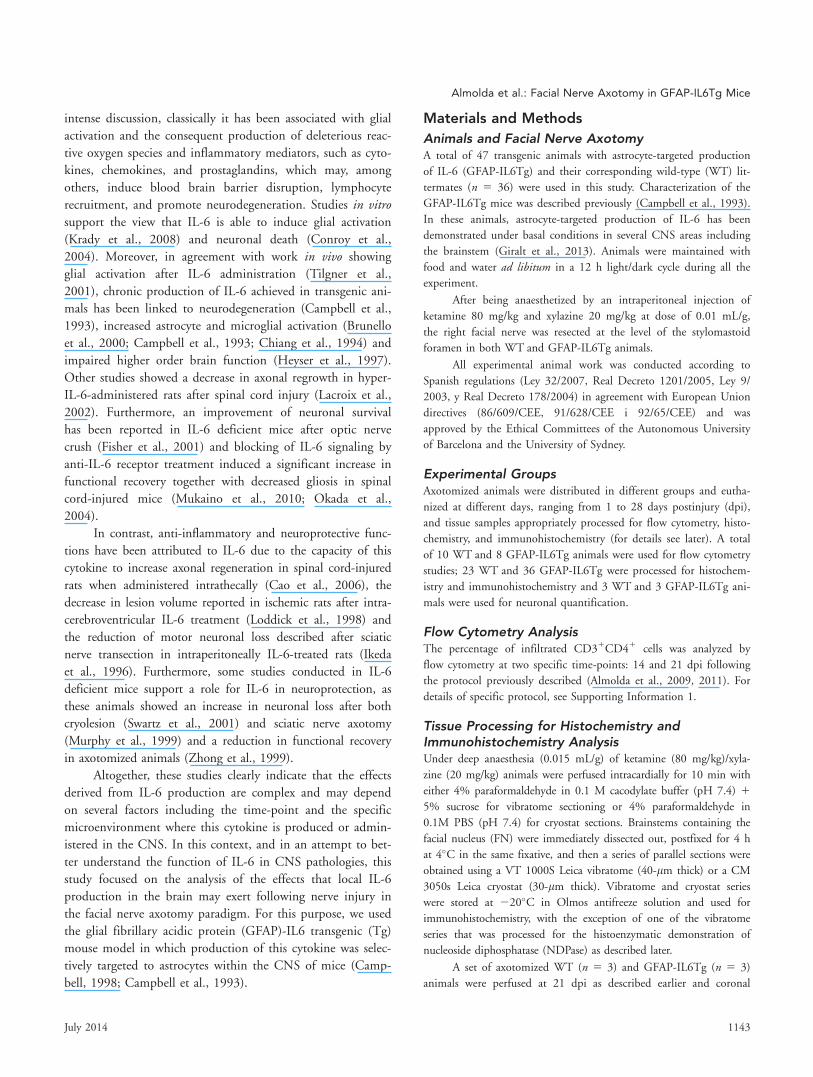
Neuronal SurvivalAfter facial nerve axotomy, there is a proportion of FN motor
neurons that degenerate (Dauer et al., 2011; Ha et al., 2008).
To determine if IL-6 alters the ratio of neuronal death, we
quantified the surviving neurons at 3 weeks postaxotomy.
Our analysis revealed that the total number of motor neurons
in the contralateral side of the FN of both WT (Fig. 1A) and
GFAP-IL6Tg (Fig. 1C) animals was similar being around
2,000 neurons. Facial nerve axotomy resulted in a loss of
30.51% of motor neurons in the ipsilateral FN of WT ani-
mals (1,291 6 10.54) while in the GFAP-IL6Tg animals, a
greater loss of motor neurons was observed (37.3%, 1,138 6
16.34; Fig. 1D,E).
1144 Volume 62, No. 7
Glial ReactivityFacial nerve axotomy provokes strong microglial activation in
the FN that was characterized by the microglial attachment
to motor neurons in the so-called mechanism of synaptic
stripping (Blinzinger and Kreutzberg, 1968; Kreutzberg,
1996). To characterize the effects that IL-6 induces in the
activation pattern of this glial population, microglial cells
were visualized using the histochemical demonstration of
NDPase, a purine-related enzyme located in the microglial
plasma membrane, commonly used for the study of “resting”
and reactive microglial cells (Castellano et al., 1991; Murabe
and Sano, 1981). This enzyme is also located in the blood
vessels, allowing the study of the relationship between micro-
glial cells and the vasculature. We also evaluated the expres-
sion of Iba1 and the CD11b and CD18 proteins, two
subunits of the heterodimeric integrin Mac-1, constitutively
expressed by microglial cells and whose upregulation has been
commonly associated with the activation of these cells. In
FIGURE 1: Neuronal death. Toluidine blue staining in the nonlesioned (contralateral) and lesioned (ipsilateral) FN of both WT (A and B)and GFAP-IL6Tg animals (C and D) at 21 dpi. La indicates the lateral area, whereas Me indicates the medial area of the brainstem. Notethe decrease in the number of motor neurons in the ipsilateral side of both groups of animals in comparison to their corresponding con-tralateral side. Scale bar 5 50 lm. (E) The histogram shows the quantification of the total number of motor neurons in the contralateral(black columns) and ipsilateral sides (white columns) of both experimental groups of animals (#P £ 0.05 with respect to the correspond-ing contralateral side; **P £ 0.0015). Note that motor neuron survival in GFAP-IL6Tg animals was reduced by a further 20% when com-pared with WT mice. [Color figure can be viewed in the online issue, which is available at wileyonlinelibrary.com.]
Almolda et al.: Facial Nerve Axotomy in GFAP-IL6Tg Mice
July 2014 1145

addition to the qualitative evaluation, a quantitative analysis
of Iba1, CD11b and CD18 staining was done by determining
the IG of each immunolabeling. As specified below, notable
differences in the microglial response were found in this study
with the use of different microglial markers.
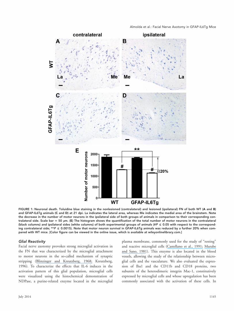
Analysis of NDPase histochemistry (Fig. 2) and Iba1
immunohistochemistry (Fig. 3) revealed that in the nonlesioned
FN of WT and GFAP-IL6Tg animals, microglial cells exhibited
a characteristic ramified morphology and were homogenously
distributed throughout the FN without any overlap between
them and without showing any specific relationship with the
neuronal cell bodies or blood vessels (Figs. 2A,B,O,P and
3A,B,P,Q). No significant differences in microglial NDPase,
Iba1, CD11b, and CD18 immunostaining were found in the
nonlesioned FN for GFAP-IL6Tg when compared with WT
animals (Figs. 2A,B, 3A,B, 4A,B and 5A,B). Nevertheless, a stat-
istically significant increase in the density of Iba11 cells was
observed in the nonlesioned FN of GFAP-IL6Tg animals (Fig.
3O). Throughout the course of the study, at the different sur-
vival times analyzed, no changes in either microglial cell mor-
phology or the spatial distribution or NDPase, Iba1, CD11b,
and CD18 expression were detected in the nonlesioned FN side
of neither WT nor GFAP-IL6Tg animals (not shown).
In the ipsilateral FN of both groups of animals, progres-
sive changes in microglial cell number and morphology, their
pattern of NDPase, Iba1, CD11b, and CD18 labeling and
their relationship with neuronal cell bodies were observed at
the different time-points studied. Marked differences between
WT and GFAP-IL6Tg mice were found. The first signs of
microglial reactivity were evident in both WT and GFAP-
IL6Tg animals at 1 dpi (Fig. 2C,D), when microglial cells
showed an enlargement of the cell body and a thickening of
the main branches, and began to migrate close to neuronal
cells bodies (Fig. 2Q,R).
At 3 dpi, in WT animals, reactive microglial cells dis-
played branched morphology but with coarser processes and
high levels of NDPase (Fig. 2E,S), Iba1 (Fig. 3C,R), CD11b
(Fig. 4C,P), and CD18 (Fig. 5C,P) staining and were wrapping
most neuronal cell bodies along the ipsilateral FN. Also the
density of Iba11 cells increased at this time-point in compari-
son with the nonlesioned FN (Fig. 3O). Although in GFAP-
IL6Tg animals, similar changes in microglial morphology and
both NDPase (Fig. 2F,T) and Iba1 (Fig. 3D,S) expression was
observed, the Iba11 cell density was higher (Fig. 3O) and the
motor neuron perikaryon surface wrapped by microglial cell
processes was lower than those observed in WT mice. Higher
CD11b (Fig. 4D,M,Q) and CD18 (Fig. 5D,M,Q) IG were
detected at this time-point in microglial cells of transgenic ani-
mals when compared with their WT littermates.
At 7 dpi, a marked increase in both the amount of
Iba11 cells and motor neuron perikaryon surface covered by
microglial cell projections was observed in both WT and
GFAP-IL6Tg mice (Figs. 2U,V and 3O). In both groups,
reactive microglia with high NDPase (Fig. 2G,H,U,V) and
high Iba1 IG (Fig. 3E,F,M) showed numerous projections dis-
playing, as well, high levels of both CD11b and CD18 (Figs.
4M and 5M, respectively) that, in addition to wrapping the
major part of neuronal cell bodies, were widely distributed
along the FN neuropil. No differences in the density of
Iba11 cells were detected at this time-point when WT and
GFAP-IL6Tg animals were compared (Fig. 3O).
Microglial reactivity remained high at 14 dpi in both
groups of animals and, in addition to elevated microglial
wrapping of neurons, characteristic clusters of highly
NDPase1 (Fig. 2W,X) and Iba11 (Fig. 3T,U) microglial cells
were also observed scattered along the ipsilateral FN.
Although, no significant differences in NDPase staining and
Iba1 (Fig. 3M) or CD18 (Fig. 5M) IG were detected between
WT and GFAP-IL6Tg animals, the neuronal wrapping was
more frequent in the ipsilateral FN of WT than in GFAP-
IL6Tg mice (Figs. 2W,X, 3T,U and 5R,S). In addition, the
number of microglial clusters found in the FN of GFAP-
IL6Tg mice was significantly lower than in WT animals (Fig.
3N). Noticeably at this time-point, significant changes in
CD11b were detected between WT and GFAP-IL6Tg ani-
mals: whereas in WT animals, the IG for CD11b showed the
highest level, in GFAP-IL6Tg animals, labeling of this integ-
rin abruptly decreased at this time-point dropping lower than
those observed at 3 dpi (Fig. 4R,S,M).
At 21 dpi, a reduction in the microglial expression of
NDPase (Fig. 2K), Iba1 (Fig. 3I), CD11b (Fig. 4I), and
CD18 (Fig. 5I) was observed in the ipsilateral FN of WT
mice, although microglial cells still exhibited high reactive
morphologies in specific FN subnucleus, such as the lateral
and the ventral intermediate subnucleus. Moreover, both the
number of neuronal cell bodies wrapped by microglial cells
and the clusters of microglia began to decrease at this time-
point in these animals. In contrast, NDPase (Fig. 2L) and
Iba1 (Fig. 3J,M) staining remained significantly elevated and
widely distributed along all the subnucleus of the ipsilateral
FN in GFAP-IL6Tg mice, and the amount of FN motor neu-
rons wrapped by microglial processes was comparatively
higher than in WT. Also the number of microglial clusters
found in the axotomized FN of transgenic animals was lower
than in WT mice at this time-point (Fig. 3N). Similar to 14
dpi, the IG of both CD11b (Fig. 4J,M), and CD18 (Fig.
5J,M) in activated microglia was significantly lower in GFAP-
IL6Tg animals than in their WT littermates.
At 28 dpi, there was a slight decrease in both NDPase
(Fig. 2M,N) and Iba1 (Fig. 3K,L,M) staining in the FN of both
WT and GFAP-IL6Tg animals although some areas of the FN
neuropil still remained totally covered by highly reactive
1146 Volume 62, No. 7
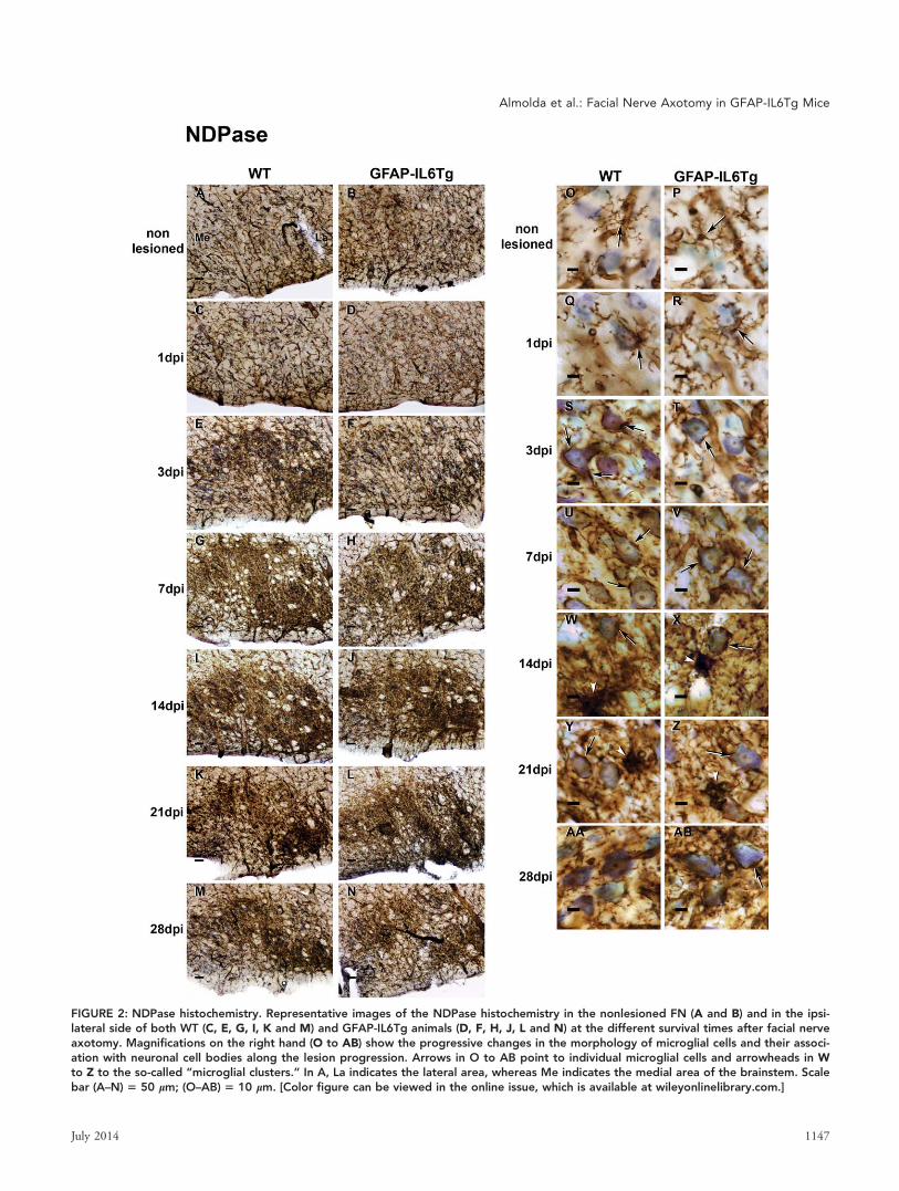
FIGURE 2: NDPase histochemistry. Representative images of the NDPase histochemistry in the nonlesioned FN (A and B) and in the ipsi-lateral side of both WT (C, E, G, I, K and M) and GFAP-IL6Tg animals (D, F, H, J, L and N) at the different survival times after facial nerveaxotomy. Magnifications on the right hand (O to AB) show the progressive changes in the morphology of microglial cells and their associ-ation with neuronal cell bodies along the lesion progression. Arrows in O to AB point to individual microglial cells and arrowheads in Wto Z to the so-called “microglial clusters.” In A, La indicates the lateral area, whereas Me indicates the medial area of the brainstem. Scalebar (A–N) 5 50 lm; (O–AB) 5 10 lm. [Color figure can be viewed in the online issue, which is available at wileyonlinelibrary.com.]
Almolda et al.: Facial Nerve Axotomy in GFAP-IL6Tg Mice
July 2014 1147
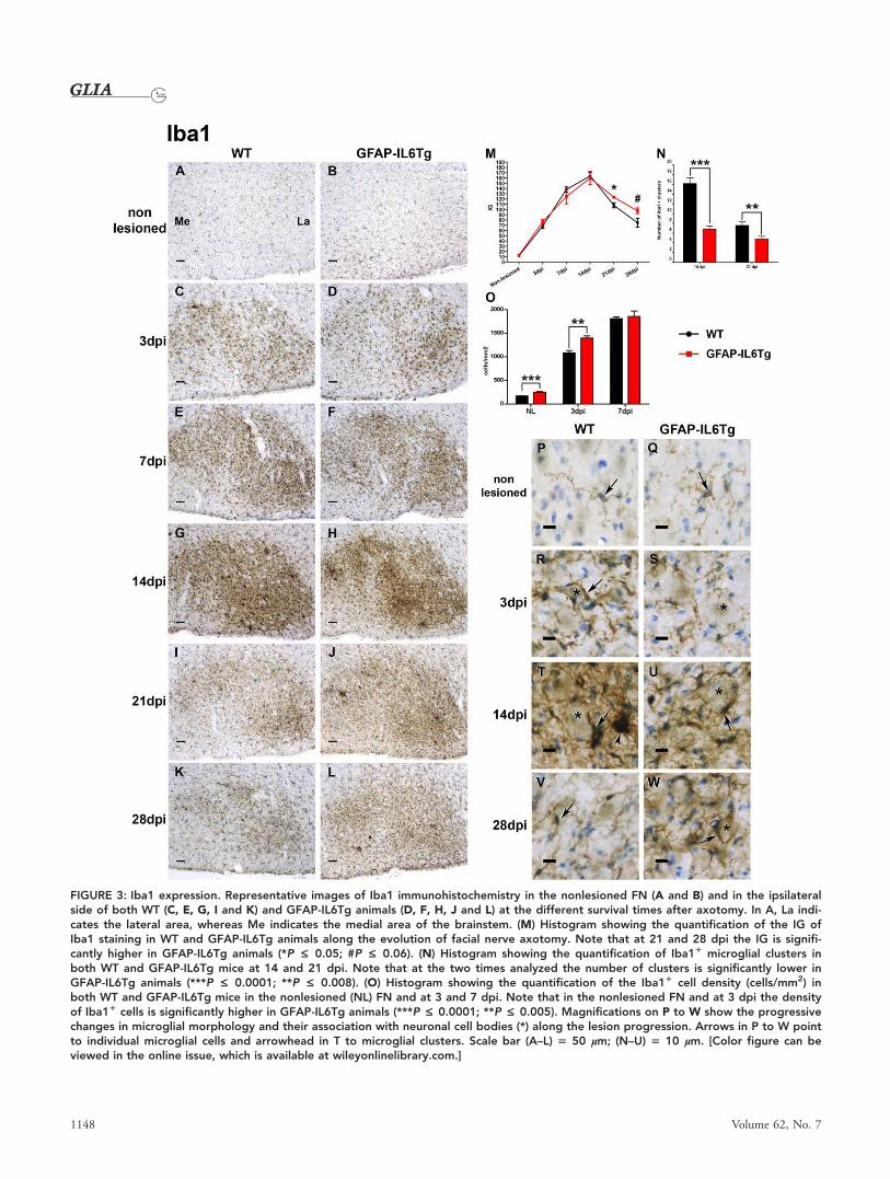
FIGURE 3: Iba1 expression. Representative images of Iba1 immunohistochemistry in the nonlesioned FN (A and B) and in the ipsilateralside of both WT (C, E, G, I and K) and GFAP-IL6Tg animals (D, F, H, J and L) at the different survival times after axotomy. In A, La indi-cates the lateral area, whereas Me indicates the medial area of the brainstem. (M) Histogram showing the quantification of the IG ofIba1 staining in WT and GFAP-IL6Tg animals along the evolution of facial nerve axotomy. Note that at 21 and 28 dpi the IG is signifi-cantly higher in GFAP-IL6Tg animals (*P £ 0.05; #P £ 0.06). (N) Histogram showing the quantification of Iba11 microglial clusters inboth WT and GFAP-IL6Tg mice at 14 and 21 dpi. Note that at the two times analyzed the number of clusters is significantly lower inGFAP-IL6Tg animals (***P £ 0.0001; **P £ 0.008). (O) Histogram showing the quantification of the Iba11 cell density (cells/mm2) inboth WT and GFAP-IL6Tg mice in the nonlesioned (NL) FN and at 3 and 7 dpi. Note that in the nonlesioned FN and at 3 dpi the densityof Iba11 cells is significantly higher in GFAP-IL6Tg animals (***P £ 0.0001; **P £ 0.005). Magnifications on P to W show the progressivechanges in microglial morphology and their association with neuronal cell bodies (*) along the lesion progression. Arrows in P to W pointto individual microglial cells and arrowhead in T to microglial clusters. Scale bar (A–L) 5 50 lm; (N–U) 5 10 lm. [Color figure can beviewed in the online issue, which is available at wileyonlinelibrary.com.]
1148 Volume 62, No. 7
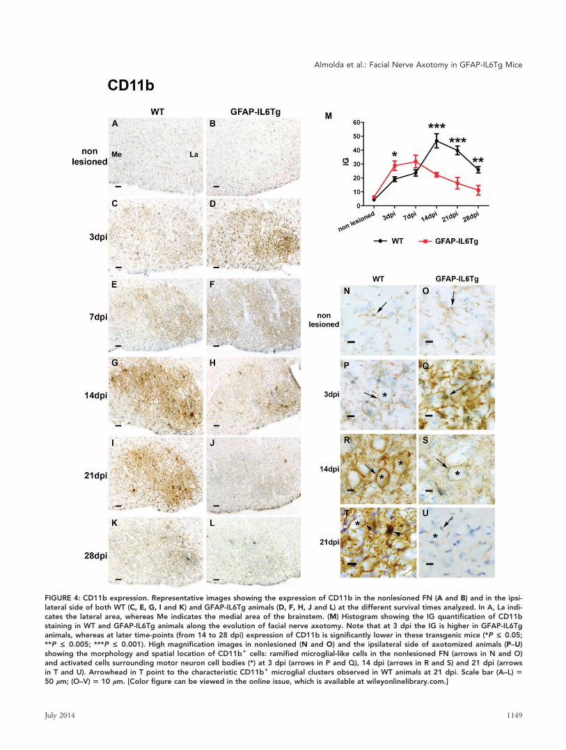
FIGURE 4: CD11b expression. Representative images showing the expression of CD11b in the nonlesioned FN (A and B) and in the ipsi-lateral side of both WT (C, E, G, I and K) and GFAP-IL6Tg animals (D, F, H, J and L) at the different survival times analyzed. In A, La indi-cates the lateral area, whereas Me indicates the medial area of the brainstem. (M) Histogram showing the IG quantification of CD11bstaining in WT and GFAP-IL6Tg animals along the evolution of facial nerve axotomy. Note that at 3 dpi the IG is higher in GFAP-IL6Tganimals, whereas at later time-points (from 14 to 28 dpi) expression of CD11b is significantly lower in these transgenic mice (*P £ 0.05;**P £ 0.005; ***P £ 0.001). High magnification images in nonlesioned (N and O) and the ipsilateral side of axotomized animals (P–U)showing the morphology and spatial location of CD11b1 cells: ramified microglial-like cells in the nonlesioned FN (arrows in N and O)and activated cells surrounding motor neuron cell bodies (*) at 3 dpi (arrows in P and Q), 14 dpi (arrows in R and S) and 21 dpi (arrowsin T and U). Arrowhead in T point to the characteristic CD11b1 microglial clusters observed in WT animals at 21 dpi. Scale bar (A–L) 550 lm; (O–V) 5 10 lm. [Color figure can be viewed in the online issue, which is available at wileyonlinelibrary.com.]
Almolda et al.: Facial Nerve Axotomy in GFAP-IL6Tg Mice
July 2014 1149
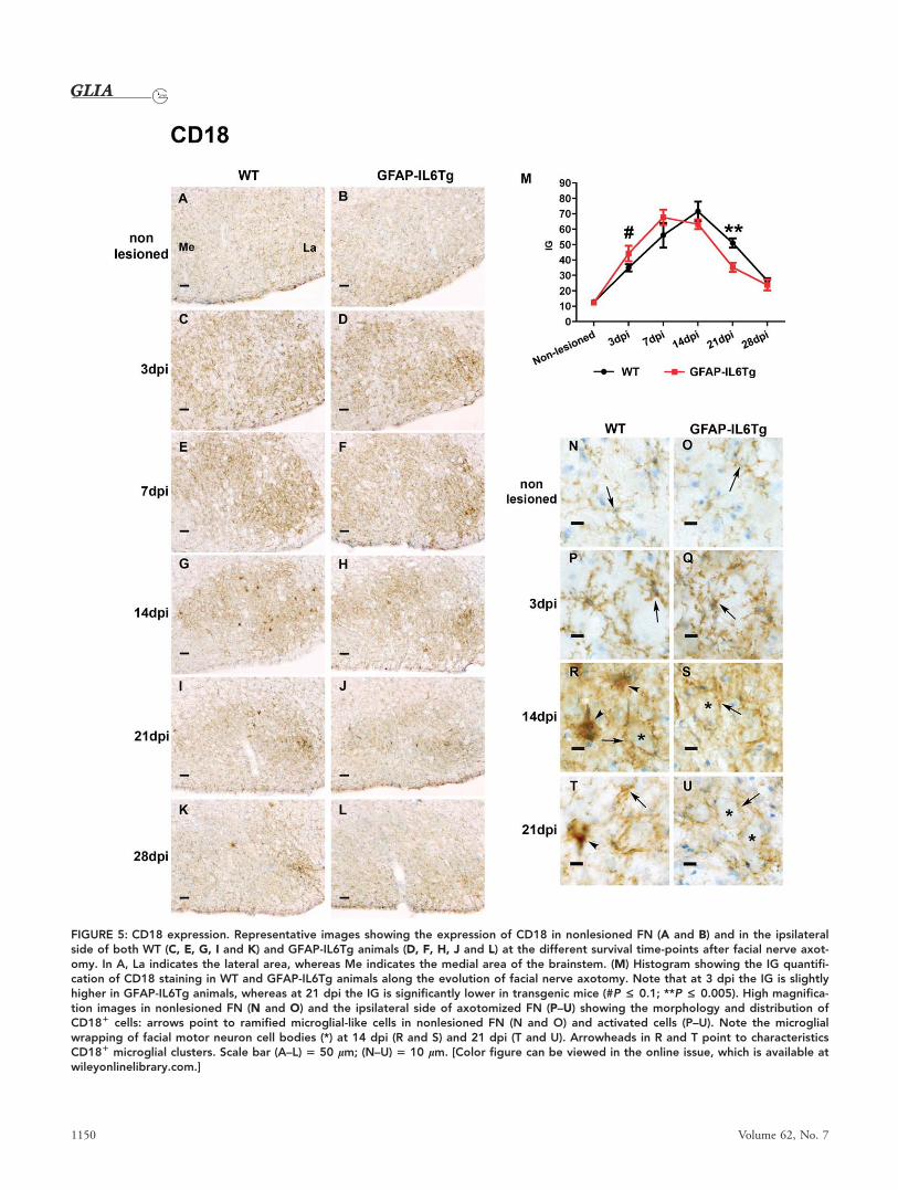
FIGURE 5: CD18 expression. Representative images showing the expression of CD18 in nonlesioned FN (A and B) and in the ipsilateralside of both WT (C, E, G, I and K) and GFAP-IL6Tg animals (D, F, H, J and L) at the different survival time-points after facial nerve axot-omy. In A, La indicates the lateral area, whereas Me indicates the medial area of the brainstem. (M) Histogram showing the IG quantifi-cation of CD18 staining in WT and GFAP-IL6Tg animals along the evolution of facial nerve axotomy. Note that at 3 dpi the IG is slightlyhigher in GFAP-IL6Tg animals, whereas at 21 dpi the IG is significantly lower in transgenic mice (#P £ 0.1; **P £ 0.005). High magnifica-tion images in nonlesioned FN (N and O) and the ipsilateral side of axotomized FN (P–U) showing the morphology and distribution ofCD181 cells: arrows point to ramified microglial-like cells in nonlesioned FN (N and O) and activated cells (P–U). Note the microglialwrapping of facial motor neuron cell bodies (*) at 14 dpi (R and S) and 21 dpi (T and U). Arrowheads in R and T point to characteristicsCD181 microglial clusters. Scale bar (A–L) 5 50 lm; (N–U) 5 10 lm. [Color figure can be viewed in the online issue, which is available atwileyonlinelibrary.com.]
1150 Volume 62, No. 7

NDPase1 or Iba11 microglial cells. In comparison with 21 dpi,
a reduction in both the number of motor neurons wrapped by
microglial processes and the number of microglial clusters was
found at this time-point in WT animals (Figs. 2AA and 3V),
whereas, noticeably, in GFAP-IL6Tg mice, the number of neu-
rons remaining covered by microglial processes (Figs. 2AB and
3W) was similar to that observed at 21 dpi. The IG of both
CD11b (Fig. 4M) and CD18 (Fig. 5M) decreased at 28 dpi in
WT and GFAP-IL6Tg animals. Remarkably, in the case of
CD11b this downregulation was significantly more pronounced
in GFAP-IL6Tg mice that in WT (Fig. 4M).
In addition to microglial reactivity, astrogliosis, charac-
terized by a progressive increase in GFAP expression, was also
observed from 3 to 28 dpi in the ipsilateral FN of both WT
and GFAP-IL6Tg animals (Supp. Info. Fig. 1A–D). Although
from 3 to 14 dpi, the increase in GFAP expression was simi-
lar in both WT and GFAP-IL6Tg mice, at 21 and 28 dpi,
the astrogliosis was less intense in GFAP-IL6Tg mice than in
WT (Supp. Info. Fig. 1E).
Overall, these findings showed that there were changes in
the microglial response in GFAP-IL6Tg animals compared with
WT, characterized principally by less attachment to motor neu-
rons at the early time-points following nerve injury. Notably, at
later time-points, 21 and 28 dpi, microglial cells in transgenic
animals had higher neuronal attachment and lower CD11b and
CD18 integrin staining, whereas astrogliosis monitored by
GFAP immunostaining was less pronounced than in WT.
Lymphocyte InfiltrationIn addition to microglial cell activation, facial nerve axotomy
induces the infiltration of lymphocytes (Raivich et al., 1998)
that has been commonly associated with a protective effect
(Jones et al., 2005). To determine if the lymphocyte infiltra-
tion was altered in GFAP-IL6Tg animals, both immunohisto-
chemistry and flow cytometry analysis was performed. As
determined by immunohistochemical detection for CD3, the
infiltration of T-lymphocytes was more pronounced in
GFAP-IL6Tg animals when compared with their WT litter-
mates (Fig. 6A,B). The quantification of T-cell infiltration in
the FN at the different time-points after facial nerve axotomy
was assessed by determining the proportion of CD31/CD41
cells by flow cytometry. As shown in Fig. 6C, a significant
increase in the proportion of CD31/CD41 cells was detected
in the ipsilateral FN of both WT and GFAP-IL6Tg animals.
Although, in both groups of animals the temporal pattern of
T-cell infiltration was similar, with the proportion of cells
peaking at 14 dpi and decreasing thereafter at 21 dpi, the
amount of CD31/CD41 cells detected in GFAP-IL6Tg ani-
mals was always significantly higher than observed in WT
animals (Fig. 6C). Notably, a small and unchanging propor-
tion of these CD31/CD41 cells was also found on the con-
tralateral side of both WT and GFAP-IL6Tg animals at the
two time-points studied, although this proportion was slightly
higher in GFAP-IL6Tg than in WT mice.
In summary, IL-6 production in the CNS alters the
amount of infiltrating T-cells in the FN parenchyma after axot-
omy inducing an increase in CD31 T-cells at 14 and 21 dpi.
OPN ExpressionOPN is upregulated in numerous pathological situations and
in response to injury (Carecchio and Comi, 2011). OPN pro-
motes cell adhesion to facilitate cell migration or survival via
interaction with integrins and CD44 (Kazanecki et al., 2007).
The changes we observed in the pattern of microglial attach-
ment to motor neurons in GFAP-IL6Tg animals suggested a
possible alteration in the OPN expression and its receptors.
The study of sections immunolabeled for OPN revealed that
this protein was constitutively expressed in the neuronal cell
bodies of motor neurons of the nonlesioned FN in both WT
and GFAP-IL6Tg animals (Fig. 7A,B,N,O). No change in
OPN staining was observed in the nonlesioned side of the
FN at the different time-points studied (not shown).
FIGURE 6: T-cell infiltration. Representative images showing theCD31 cells (arrows in inserts) found in the ipsilateral FN of bothWT (A) and GFAP-IL6Tg animals (B) at 14 dpi. Scale bar 5 50lm. (C) Histogram showing the relative percentage of CD31/CD41 cells detected in WT and GFAP-IL6Tg animals at 14 and21 dpi. Note that the percentage of this population of lympho-cytes is always higher in the ipsilateral side (ipsi) of GFAP-IL6Tgthan in WT animals (#P £ 0.2; *P £ 0.05; **P £ 0.01). [Color fig-ure can be viewed in the online issue, which is available atwileyonlinelibrary.com.]
Almolda et al.: Facial Nerve Axotomy in GFAP-IL6Tg Mice
July 2014 1151
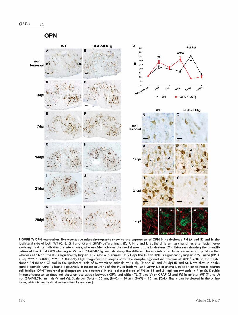
FIGURE 7: OPN expression. Representative microphotographs showing the expression of OPN in nonlesioned FN (A and B) and in theipsilateral side of both WT (C, E, G, I and K) and GFAP-IL6Tg animals (D, F, H, J and L) at the different survival times after facial nerveaxotomy. In A, La indicates the lateral area, whereas Me indicates the medial area of the brainstem. (M) Histogram showing the quantifi-cation of the IG of OPN staining in WT and GFAP-IL6Tg animals along the different time-points after facial nerve axotomy. Note thatwhereas at 14 dpi the IG is significantly higher in GFAP-IL6Tg animals, at 21 dpi the IG for OPN is significantly higher in WT mice (#P £0.06; ***P £ 0.0005; ****P £ 0.0001). High magnification images show the morphology and distribution of OPN1 cells in the nonle-sioned FN (N and O) and in the ipsilateral side of axotomized animals at 14 dpi (P and Q) and 21 dpi (R and S). Note that, in nonle-sioned animals, OPN is found exclusively in motor neurons of the FN in both WT and GFAP-IL6Tg animals. In addition to motor neuroncell bodies, OPN1 neuronal prolongations are observed in the ipsilateral side of FN at 14 and 21 dpi (arrowheads in P to S). Doubleimmunofluorescence does not show co-localization between OPN and either TL (T and V) or GFAP (U and W) in neither WT (T and U)nor GFAP-IL6Tg animals (V and W). Scale bar (A–L) 5 50 lm; (N–Q) 5 30 lm; (T–W) 5 10 lm. [Color figure can be viewed in the onlineissue, which is available at wileyonlinelibrary.com.]
1152 Volume 62, No. 7
However, an increase in OPN was observed at 3 dpi in the
axotomized FN side of both WT and GFAP-IL6Tg mice
(Fig. 7C,D,M). The IG for OPN slightly decreased at 7 dpi
in both groups of animals and from 14 dpi, marked differen-
ces in the OPN staining were found between WT and GFAP-
IL6Tg animals (Fig. 7M). Expression of this protein in WT
animals at 14 dpi showed similar levels to those found at 7
dpi and peaked thereafter at 21 dpi. In contrast, a significant
increase in OPN was found in GFAP-IL6Tg mice at 14 dpi
that decreased abruptly at 21 dpi. This rapid decrease in the
transgenic mice was due not only to a reduction in the num-
ber of motor neurons but also noticeably to a large decrease
in the intensity of OPN staining in comparison with WT
(Fig. 7R,S). At 28 dpi, no difference in OPN IG of staining
was observed between WT and GFAP-IL6Tg animals (Fig.
7K,L,M).
It is important to note that, in addition to expression in
neuronal cell bodies, OPN was also observed in some neuro-
nal projections along the ipsilateral FN side of both WT and
GFAP-IL6Tg mice at the different time-points analyzed, espe-
cially at 14 and 21 dpi (Fig. 7P–S).
Double immunofluorescence which combined OPN
with either tomato lectin (TL) for microglia or GFAP for
astrocyte labeling, demonstrated no co-localization of this
molecule with these two markers at any survival time in
either WT or GFAP-IL6Tg animals (Fig. 7T–W).
In summary, GFAP-IL6Tg animals had an altered pat-
tern of neuronal OPN expression characterized by higher lev-
els at 14 dpi and lowers at 21 dpi.
OPN-Receptors ExpressionExpression of two of the main OPN-receptors, CD44 and
CD49e, was analyzed throughout the evolution of facial nerve
injury. Our results demonstrated that there was no staining of
these two OPN-receptors in the nonlesioned FN side of both
WT and GFAP-IL6Tg at the different time-points studied
(Figs. 8A,B and 9A,B). After facial nerve axotomy, de novo
expression of both CD44 and CD49e receptors was detected
in the ipsilateral FN side of both WT and GFAP-IL6Tg ani-
mals along the course of the lesion, and their expression was
different between these two groups of mice, as specified below.
CD44 Expression. CD44 staining was detected initially at 3
dpi in the ipsilateral FN side of both WT and GFAP-IL6Tg
animals, where a weak immunolabeling was observed mainly
in the soma of motor neurons (Fig. 8C,D,N,O). In WT ani-
mals, the AI of CD44 staining increased progressively at 7
and 14 dpi, reaching maximum at 21 dpi (Fig. 8M). At these
time-points, CD44 immunolabeling was found scattered in
neuropil of the ipsilateral FN (Fig. 8P) and in some cells
identified, by double immunolabeling, as T-lymphocytes
(CD31), most of them in close relationship with the soma of
motor neurons (Fig. 8R). At 28 dpi, an abrupt reduction in
the CD44 AI was seen (Fig. 8M), and only a weak CD44
staining was observed in few small areas of the ipsilateral FN
of WT animals (Fig. 8K). No co-localization of CD44 with
TL was found at any survival time. Rather, CD44 staining
was located in the periphery of OPN1 motor neurons, just
underneath TL1 microglial processes (Fig. 8T).
Although in GFAP-IL6Tg animals CD44 followed a
similar temporal pattern of staining, increasing at 7 and 14
dpi and peaking at 21 dpi, the AI index of labeling of this
molecule, from 7 to 28 dpi, was significantly lower than that
observed in WT animals (Fig. 8M). Similar to WT, in
GFAP-IL6Tg animals, CD44 immunolabeling was observed
scattered in the neuropil (Fig. 8Q), and, noticeably and in
contrast to what was observed in WT animals, few CD441/
CD31 T-lymphocytes were found (Fig. 8S). Again, CD44
staining detected in the neuropil did not co-localize with TL,
but was located between OPN1 motor neurons and TL1
microglial processes (Fig. 8U).
CD49e Expression. Immunostaining for CD49e, which was
absent in the nonlesioned FN (Fig. 9A,B), was first detected
in the ipsilateral FN of both WT and GFAP-IL6Tg animals
at 3 dpi (Fig. 9C,D). Although in both groups of animals
this integrin markedly increased at 7 dpi (Fig. 9E,F), the AI
index of staining was significantly lower in GFAP-IL6Tg than
in WT animals (Fig. 9M). From 14 to 28 dpi, the CD49e AI
index slightly decreased in both WT and transgenic animals
(Fig. 9G–L,M). At 3 and 7 dpi, CD49e immunolabeling was
found in processes wrapping the soma of FN motor neurons
(Fig. 9N,O). From 14 dpi, in addition, characteristic clusters
of cells with higher CD49e staining were found in both
groups of animals (Fig. 9P,Q).
Double immunofluorescence demonstrated co-
localization of CD49e and Iba11 microglial cells in all the
time-points analyzed in both WT and GFAP-IL6Tg mice and
especially in the clusters of microglial cells (Fig. 9R,S).
In summary, CD44 and CD49e immunolabeling were
significantly lower in GFAP-IL6Tg animals than in WT from
7 to 28 dpi. Notably, T-cells and motor neurons but not
microglia are the predominant CD441 cells, whereas CD49e
was predominantly found in association with microglial
clusters.
Discussion
In this study, we demonstrated that the astrocyte-targeted pro-
duction of IL-6 within the CNS had a direct impact on the
development of the nerve injury response to facial nerve axot-
omy. Axotomized GFAP-IL6Tg animals had higher neuronal
death in the lesioned FN of the brainstem than WT and this
Almolda et al.: Facial Nerve Axotomy in GFAP-IL6Tg Mice
July 2014 1153

FIGURE 8: CD44 expression. Representative images showing CD44 expression in the nonlesioned FN (A and B) and in the ipsilateral side ofboth WT (C, E, G, I and K) and GFAP-IL6Tg animals (D, F, H, J and L) at the different survival times after facial nerve axotomy. In A, La indi-cates the lateral area, whereas Me indicates the medial area of the brainstem. (M) Histogram showing the quantification of AI index forCD44 in WT and GFAP-IL6Tg animals along the different time-points after facial nerve axotomy. Note that, although the dynamics of CD44AI index is similar in both groups of animals, from 7 to 28 dpi the AI index is significantly lower in GFAP-IL6Tg animals than in WT (*P £0.05; **P £ 0.005; #P £ 0.08). High magnification images show the morphology and spatial location of CD441 cells in the ipsilateral side ofaxotomized animals at 3 dpi (N and O) and 21 dpi (P and Q). Note that at early time-points CD44 is found within neuronal cell bodies (* in Nand O), whereas at later survival times (21 dpi), CD441 prolongations (arrows) as well as little-round cells (arrowheads) around motor neu-rons (* in P and Q) are also detected. Double and triple immunofluorescence combining CD44 with CD3 (R and S) and with OPN and TL (Tand U), demonstrate co-localization of CD44 with CD3 (arrows in R and S), but not with either OPN or TL (T and U) in both WT (R and T) andGFAP-IL6Tg animals (S and U). Note that, some CD441 processes are found surrounding neuronal OPN and covered by TL1 ramifications inboth WT and GFAP-IL6Tg animals (arrows in T and U). Scale bar (A–L) 5 50 lm; (N–U) 5 10 lm. [Color figure can be viewed in the onlineissue, which is available at wileyonlinelibrary.com.]
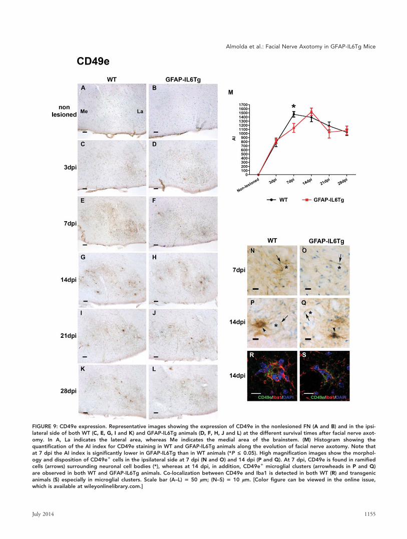
FIGURE 9: CD49e expression. Representative images showing the expression of CD49e in the nonlesioned FN (A and B) and in the ipsi-lateral side of both WT (C, E, G, I and K) and GFAP-IL6Tg animals (D, F, H, J and L) at the different survival times after facial nerve axot-omy. In A, La indicates the lateral area, whereas Me indicates the medial area of the brainstem. (M) Histogram showing thequantification of the AI index for CD49e staining in WT and GFAP-IL6Tg animals along the evolution of facial nerve axotomy. Note thatat 7 dpi the AI index is significantly lower in GFAP-IL6Tg than in WT animals (*P £ 0.05). High magnification images show the morphol-ogy and disposition of CD49e1 cells in the ipsilateral side at 7 dpi (N and O) and 14 dpi (P and Q). At 7 dpi, CD49e is found in ramifiedcells (arrows) surrounding neuronal cell bodies (*), whereas at 14 dpi, in addition, CD49e1 microglial clusters (arrowheads in P and Q)are observed in both WT and GFAP-IL6Tg animals. Co-localization between CD49e and Iba1 is detected in both WT (R) and transgenicanimals (S) especially in microglial clusters. Scale bar (A–L) 5 50 lm; (N–S) 5 10 lm. [Color figure can be viewed in the online issue,which is available at wileyonlinelibrary.com.]
Almolda et al.: Facial Nerve Axotomy in GFAP-IL6Tg Mice
July 2014 1155

difference was associated with significant changes in the tem-
poral activation pattern of glial cells and lymphocyte infiltra-
tion. Moreover, we found variations in the pattern of OPN
expression in neurons and its receptors CD44 and CD49e in
lymphocytes and microglia, respectively, that, as we will dis-
cuss, can play a role in the process of neuronal wrapping and,
therefore, exert an influence on the survival of axotomized
motor neurons.
Increased Neuronal DeathFacial nerve axotomy is one of the most well-characterized
models of peripheral nerve injury widely used for the study
of retrograde neuronal degeneration (Moran and Graeber,
2004). After facial nerve axotomy in mice, a proportion of
motor neurons located in the FN of the brainstem die over a
period of several days, by a mechanism of apoptosis that is
dependent on the type of axotomy (Ha et al., 2008), the
mouse strain used (Ha et al., 2006) and the age of the ani-
mals (Dauer et al., 2011). In parallel with this neurodegener-
ation, a regenerative process of the axotomized axons is also
induced in surviving neurons, leading to a partial functional
recovery (Moran and Graeber, 2004). Although the exact
mechanisms mediating neuronal survival remains unclear,
both microglia and lymphocytes have been suggested to play
a role in the maintenance of motor neuron viability (Byram
et al., 2004; Jones et al., 2005; Serpe et al., 200). Our results
revealed an increase in neuronal death in GFAP-IL6Tg ani-
mals suggesting that the proinflammatory environment cre-
ated by IL-6 production may exert a negative influence in
some of the key events involved in the processes of neurode-
generation, neuroregeneration, or in both.
IL-6 could have a direct neurotoxic effect on FN motor
neurons as it has been shown that they express IL-6 receptor
(IL-6R) (Klein et al., 1997) and the deficiency of IL-6 in KO
animals has been associated with increased motor neuron sur-
vival (Galiano et al., 2001). However, our results clearly
showed significant changes between WT and GFAP-IL6Tg
animals in microglial reaction, lymphocyte infiltration, and
the expression pattern of certain molecules that can be
derived from higher neuronal death, although we cannot dis-
card the possibility that these changes might be due to an
effect of IL-6 through these cell types.
A point to take into account here is the fact that expres-
sion of IL-6 in the GFAP-IL6Tg mice is chronic and, there-
fore, we cannot evaluate the different effects if any, derived
from early versus late IL-6 expression. In this way, further
studies using conditional animals (Quintana et al., 2013) or
animals lacking the intracellular signaling pathways of IL-6,
such as mice with inducible gp130 deletion in myeloid cells
(Sander et al., 2008) or in neurons (Stanke et al., 2006), may
be useful to clarify the role of IL6 in this paradigm.
Altered Pattern of Glial Reactivity and LymphocyteInfiltrationFollowing facial nerve axotomy, it has been reported that
microglial cells become activated, proliferate, attach to axo-
tomized motor neurons wrapping them, and participate in
the so-called phenomenon known as “synaptic stripping”
(Blinzinger and Kreutzberg, 1968; Kreutzberg, 1996). During
synaptic stripping, microglial cells interpose between the
healthy presynaptic and postsynaptic elements (Blinzinger and
Kreutzberg, 1968; Kreutzberg, 1996) to disconnect excitatory
inputs to motor neurons allowing axonal regeneration and
functional recovery. Synaptic stripping has also been described
in other circumstances such as inflammation in the cerebral
cortex (Trapp et al., 2007) or after hypoglossal nerve injury
(Svensson and Aldskogius, 1993). However, in these kinds of
lesions, microglia appear to be involved in the active phago-
cytic removal of synaptic contacts rather than in disconnec-
tion of inputs. Our analysis of microglial reactivity in WT
animals showed microglial processes wrapping motor neuron
cell bodies from 3 to 21 dpi, decreasing thereafter at 28 dpi.
Interestingly, in GFAP-IL6Tg animals, coinciding with a
higher neuronal death at 21 dpi, the number of motor neu-
rons wrapped by microglia was less during the previous time-
points (3 to 14 dpi), supporting the idea that neuronal wrap-
ping and synaptic stripping is a protective phenomenon in
this paradigm (Kreutzberg, 1996). In agreement with this,
some evidence suggests that defects in the microglia-neuron
attachment after facial nerve axotomy, as occurs in microglial
cathepsin deficient mice (Hao et al., 2007) and TGF-b1 defi-
cient animals (Makwana et al., 2007) might lead to a higher
neuronal death. Defects in motor neuron cell wrapping after
facial nerve axotomy have also been described in other cyto-
kine mutants, such as the IL-6 KO mice (Galiano et al.,
2001) and the MCSF deficient mice (Kalla et al., 2001) sug-
gesting that the cytokine microenvironment may regulate this
phenomenon.
Our observations also showed that motor neuron wrap-
ping in transgenic animals was higher than in WT at later
time-points after facial nerve axotomy, being still evident at
28 dpi but almost absent in WT animals. Is unclear what, if
any, consequences there might be of this delay. However, alto-
gether, these results indicate that IL-6 production modifies
the microglial response to nerve injury in the FN that
impacts the wrapping of motor neurons that might be
responsible for the increased neuronal death.
Microglial proliferation is another key event in the
microglial activation pattern associated with a wide range of
CNS injuries (Raivich et al., 1999) including facial nerve
axotomy (Raivich et al., 1994). This proliferative capacity has
been shown to be altered by different cytokines and other sig-
naling molecules. Indeed, treatment of microglial-astroglial
1156 Volume 62, No. 7
co-cultures with IL-6 leads to a slight stimulatory effect on
microglial proliferative capacity (Kloss et al., 1997), whereas a
reduction of microglial proliferation was observed in IL-6
deficient mice after facial nerve axotomy (Klein et al., 1997).
In agreement with these studies, our results revealed that in
GFAP-IL6Tg animals, at 3 dpi, the number of microglial
cells was higher than in WT, suggesting that IL-6 may play a
role in the proliferative capacity of microglia.
In addition to changes in microglial cell number, mor-
phology, and neuronal wrapping, our observations also
showed, in agreement with other studies (Kloss et al., 1999;
Raivich et al., 1999), a significant upregulation of CD11b
and CD18 staining during lesion evolution, that is, indicative
of microglial activation (Kettenmann et al., 2011). The
upregulation of both markers was higher in GFAP-IL6Tg ani-
mals at 3 dpi suggesting a more rapid response of microglial
cells to axotomy due to IL-6 production. In this sense, some
studies have shown that IL-6 is able to induce microglial acti-
vation both in vitro (Krady et al., 2008) and after its acute
administration in vivo (Tilgner et al., 2001). Moreover, strong
microglial activation occurs in the brain of GFAP-IL6Tg ani-
mals, the extent of which overlaps with the level of transgene
encoded IL-6 production (Campbell et al., 1993; Chiang
et al., 1994; Heyser et al., 1997).
Strikingly, at later time-points, we found a marked
reduction of CD18 IG, and especially of CD11b, in the
GFAP-IL6Tg animals. Although the role played by these mol-
ecules in CNS pathology has not yet been well determined,
particularly in the facial nerve axotomy model, expression of
its principal counter-receptor ICAM-1 was demonstrated in
lymphocytes (Werner et al., 1998). Consequently, the
decrease in CD11b and CD18 expression observed in GFAP-
IL6Tg animals, at later time-points, could be a signal of
reduced microglial activation or even may involve a deficient
attachment between microglia and lymphocytes. These obser-
vations in conjunction with the decrease in the neuronal
wrapping described earlier in GFAP-IL6Tg animals, lead us
to speculate that CD11b and perhaps also CD18, may be key
molecules involved in the control of the mechanisms of neu-
ronal wrapping and lymphocyte function in this paradigm.
It is important to point out here, the differences found
in our study in terms of the microglial response depending
on the microglial marker used for the analysis. This fact dem-
onstrated the tremendous plasticity of microglia and the
importance of studying the complex phenomenon of their
activation using different markers.
Another interesting difference observed in GFAP-IL-
6Tg animals in terms of microglial activation was the lower
number of microglial clusters found. Despite not being exten-
sively described in the literature, the few articles reporting the
formation of microglial clusters following facial nerve
axotomy suggest that this phenomenon is associated with
neuronal death (Dauer et al., 2011; Petito et al., 2003; Rai-
vich et al., 1998). In some of these studies, the amount of
cluster formation was used as a measure of motor neuron cell
death (Dauer et al., 2011; Petito et al., 2003). Notably, and
in contrast to these previous studies, our observations indi-
cated that in GFAP-IL6Tg mice, where there was a higher
neuronal death, there were less microglial clusters. This
decrease in the number of clusters in transgenic mice could
be related with a faster or more effective microglial phagocy-
tosis, although we cannot rule out the possibility that micro-
glial clustering may play other, yet to be identified functions.
In this sense, recent studies in multiple sclerosis showed the
presence of microglial cell clusters in preactive lesions with
high expression of IL-10, suggesting that they may play a role
as regulators of inflammation (van Horssen et al., 2012). Fur-
ther studies must be explored to better characterize, (1) the
factors controlling their formation, (2) the molecules
expressed within these microglial clusters, and (3) the interac-
tion established, if any, with infiltrating lymphocytes.
Our findings showed that after facial nerve axotomy, the
astroglial response monitored by GFAP staining was altered
in GFAP-IL6Tg animals. As GFAP expression is regulated by
IL-6 (reviewed in Pekny et al., 2014), we could expect a
greater upregulation of GFAP in axotomized transgenic mice
versus axotomized WT. In agreement with this hypothesis,
some authors showed that after facial nerve axotomy, IL-6
deficient mice showed less GFAP than WT (Klein et al.,
1997). However, our results revealed that, at later time-points
after lesion, the transgenic animals displayed a less intense
expression of GFAP than WT. We have not a single explana-
tion to this phenomenon, although we cannot discard the
possibility that chronic IL-6 expression produced an altered
state of microglia and/or factors produced by T-cells that
might downregulate GFAP expression.
Noticeably, the changes we have reported in glial activa-
tion correlates with an increase in the proportion of T-helper
lymphocytes in the FN of transgenic animals at the two time-
points analyzed, 14 and 21 dpi. It is well accepted that infil-
tration of lymphocytes in the FN after axotomy in the mouse
occurs in two different waves, one as early as 3 dpi and a sec-
ond at later time-points (14 dpi) (Raivich et al., 1998).
Although not too much is known about the role played by
these lymphocytes, it has been suggested that they may have a
protective role because the lack of functional mature T and B
cells, in either the SCID mutant mice or the RAG2-KO
mice, have been correlated with a dramatic increase in neuro-
nal death in this lesion paradigm (Serpe et al., 2000). More-
over, this higher neuronal death was prevented by
reconstitution of these mice with functional T and B cells
(Serpe et al., 1999). In our study, however, the higher
Almolda et al.: Facial Nerve Axotomy in GFAP-IL6Tg Mice
July 2014 1157

neuronal death correlated with an increase in lymphocyte
infiltration. There are two plausible explanations for these
contradictory results: the first may be that local production of
IL-6 shifts the phenotype of T-cells infiltrating the FN paren-
chyma in GFAP-IL6Tg animals toward a proinflammatory
Th17 phenotype, as it has been described in other neuroin-
flammatory conditions (Kimura and Kishimoto, 2010),
instead of the protective Th2 described after facial nerve axot-
omy (Deboy et al., 2006), inducing a higher death of motor
neurons. In this sense, it will be of considerable interest to
identify, in a future study, the specific lymphocyte subpopula-
tions entering the FN in GFAP-IL6Tg mice.
The second possibility is that production of IL-6 in
transgenic animals can change the pattern of chemokine or
adhesion molecules involved in lymphocyte recruitment in
the FN after axotomy, inducing a higher infiltration of these
cells. In support of this, increased lymphocyte infiltration has
already been described in specific areas of the CNS of GFAP-
IL6Tg mice concomitant with upregulation of VCAM-1 and
ICAM-1 expression (Campbell et al., 1993; Milner and
Campbell, 2006) as well as specific chemokines such as
CCL5 and CCL12 (Quintana et al., 2009) important mole-
cules involved in lymphocyte transmigration across blood
brain barrier.
Changes in OPN Expression and its ReceptorsOur study also demonstrated differences between WT and
GFAP-IL6Tg animals, in the pattern of OPN expression in
motor neurons and its receptors CD44 and CD49e in lym-
phocytes or microglia.
In contrast to previous studies reporting expression of
this glycoprotein in microglia and astrocytes after different
types of lesions such as spinal root avulsion (Fu et al., 2004),
traumatic spinal cord injury (Hashimoto et al., 2003), ische-
mia (Choi et al., 2007), LPS (Iczkiewicz et al., 2005) and
kainic acid injection (Kim et al., 2002), cuprizone-mediated
demyelination (Selvaraju et al., 2004), scrapie infection (Jin
et al., 2006), cryolesion (Shin et al., 2005) and Theiler’s
murine encephalitis (Shin and Koh, 2004), in our study,
OPN expression was not found in either microglial cells or
astrocytes. In agreement with previous studies in adult rat
(Shin et al., 1999; Suzuki et al., 2012), we found OPN con-
stitutively present in motor neurons of the nonlesioned FN of
both WT and GFAP-IL6Tg animals. After facial nerve axot-
omy, we observed a similar increase in neuronal OPN at 3
dpi that drop at 7 dpi in both WT and transgenic animals.
At 14 and 21 dpi, OPN staining in motor neurons drastically
differed in these two groups of animals peaking at 21 dpi in
WT and at 14 dpi in GFAP-IL6Tg animals. Increased OPN
has been described in affected neurons in diverse types of
CNS pathology such as spinal root avulsion (Fu et al., 2004),
epilepsy (Borges et al., 2008), scrapie infection (Jin et al.,
2006), cryolesion (Shin et al., 2005), oxygen-glucose-deprived
hippocampal slices (Lee et al., 2010), Alzheimer’s disease
(Wung et al., 2007), multiple sclerosis (Sinclair et al., 2005),
and different experimental autoimmune encephalomyelitis
(EAE) models (Chabas et al., 2001). The exact role played by
OPN in neurons is, however, still not completely known and
both detrimental as well as neuroprotective roles (Carecchio
and Comi, 2011) have been attributed to this glycoprotein in
this cell type. We do not know what role is played by OPN
in our paradigm because the increased neuronal death
observed in GFAP-IL6Tg mice could be associated with
either the increased OPN at 14 dpi or the lower expression at
21 dpi.
In addition to changes in OPN, differences in the level
of the OPN receptors CD44 and CD49e were also observed
in our study. Whereas, in agreement with other studies (Jones
et al., 1997; Kloss et al., 1999), the IG of both CD44 and
CD49e integrin in WT animals increased from 7 to 21 dpi,
in GFAP-IL6Tg animals we found less CD44 at all these
time-points and less CD49e specifically at 7 dpi. In WT ani-
mals, the simultaneous upregulation of OPN in motor neu-
rons, CD44 in lymphocytes and CD49e in surrounding
microglia could be an important factor for the adhesion of
these lymphocytes and microglial cells to neurons during the
synaptic stripping process and may regulate the survival or
death of motor neurons. Indeed, it is well known that
through binding with specific receptors, such as CD44, OPN
favors the recruitment of innate cells by inducing migration
and adhesion of dendritic cells, neutrophils, and macrophages
(Giachelli et al., 1998; Weber et al., 1996). Our observation
of CD44 expression in the periphery of motor neurons is in
agreement with previous studies showing its presence in the
plasma membrane of neurons (Jones et al., 1997). Although
the function played by CD44 in motor neurons is not clear,
some authors suggest that its presence in regenerative axons
may indicate a putative role in regulation of axonal outgrowth
(Makwana et al., 2010). The fact that GFAP-IL6Tg animals
showed less expression of both CD44 and CD49e receptors
at the time-points where the neuronal wrapping and synaptic
stripping has been described, lead us to speculate that the
increase in neuronal death observed in GFAP-IL6Tg animals
could be due to a deficient attachment of microglia and/or
lymphocytes to neurons. This deficiency may be due to an
alteration in the expression of OPN, CD44 and CD49e,
pointing to this molecular signaling mechanism as a key event
in driving the cross talk among these three cell types and the
subsequent further fate of axotomized motor neurons.
In conclusion, we have established that transgenic pro-
duction of IL-6 in the CNS induces a major impact on the
three main cellular components commonly related to the
1158 Volume 62, No. 7
neuro-degenerative and/or -regenerative processes after facial
nerve axotomy: neurons, microglia, and lymphocytes. We
demonstrated an increase in neuronal death in the FN of
transgenic animals that was accompanied by changes in the
association established between activated microglial cells and
neurons and a decrease in the expression of microglial
CD11b and CD18 integrins at the different time-points ana-
lyzed. Moreover, GFAP-IL6Tg animals showed an increase in
the number of lymphocytes in the FN and a lower level of
OPN in neurons, CD44 in lymphocytes and CD49e in
microglial cells. Altogether, these results suggest that IL-6
mediates alterations in the pattern of microglial activation
and the lymphocyte infiltration that impact negatively on the
outcome of neuronal death after a lesion in the CNS.
Acknowledgment
Grant sponsor: Spanish Ministry of Science and Innovation;
Grant numbers: BFU2011-27400 (to BC); Grant sponsor:
SAF; Grant numbers: 2008-00435 and 2011-23272 (to JH);
Grant sponsor: NHMRC project; Grant number: 632754 (to
ILC).
The authors would like to thank Miguel A. Martil and Isa-
bella Appiah for their outstanding technical help.
ReferencesAbercrombie M. 1946. Estimation of nuclear population from microtome sec-tions. Anat Rec 94:239–247.
Almolda B, Costa M, Montoya M, Gonzalez B, Castellano B. 2009. CD4microglial expression correlates with spontaneous clinical improvement in theacute Lewis rat EAE model. J Neuroimmunol 209:65–80.
Almolda B, Costa M, Montoya M, Gonzalez B, Castellano B. 2011. Increase inTh17 and T-reg lymphocytes and decrease of IL22 correlate with the recoveryphase of acute EAE in rat. PLoS One 6:e27473.
Almolda B, Gonzalez B, Castellano B. 2010. Activated microglial cells acquirean immature dendritic cell phenotype and may terminate the immuneresponse in an acute model of EAE. J Neuroimmunol 223:39–54.
Almolda B, Gonzalez B, Castellano B. 2013. Microglia detection by enzymatichistochemistry. Methods Mol Biol 1041:243–259.
Benveniste EN. 1998. Cytokine actions in the central nervous system. Cyto-kine Growth Factor Rev 9:259–275.
Blinzinger K, Kreutzberg G. 1968. Displacement of synaptic terminals fromregenerating motoneurons by microglial cells. Z Zellforsch Mikrosk Anat 85:145–157.
Borges K, Gearing M, Rittling S, Sorensen ES, Kotloski R, Denhardt DT,Dingledine R. 2008. Characterization of osteopontin expression and functionafter status epilepticus. Epilepsia 49:1675–1685.
Brunello AG, Weissenberger J, Kappeler A, Vallan C, Peters M, Rose-John S,Weis J. 2000. Astrocytic alterations in interleukin-6/Soluble interleukin-6receptor alpha double-transgenic mice. Am J Pathol 157:1485–1493.
Byram SC, Carson MJ, DeBoy CA, Serpe CJ, Sanders VM, Jones KJ. 2004.CD4-positive T cell-mediated neuroprotection requires dual compartmentantigen presentation. J Neurosci 24:4333–4339.
Campbell IL. 1998. Transgenic mice and cytokine actions in the brain: Bridg-ing the gap between structural and functional neuropathology. Brain ResBrain Res Rev 26:327–336.
Campbell IL, Abraham CR, Masliah E, Kemper P, Inglis JD, Oldstone MB,Mucke L. 1993. Neurologic disease induced in transgenic mice by cerebraloverexpression of interleukin 6. Proc Natl Acad Sci USA 90:10061–10065.
Cao Z, Gao Y, Bryson JB, Hou J, Chaudhry N, Siddiq M, Martinez J, SpencerT, Carmel J, Hart RB, Filbin MT. 2006. The cytokine interleukin-6 is sufficientbut not necessary to mimic the peripheral conditioning lesion effect on axo-nal growth. J Neurosci 26:5565–5573.
Carecchio M, Comi C. 2011. The role of osteopontin in neurodegenerativediseases. J Alzheimers Dis 25:179–185.
Castellano B, Gonzalez B, Dalmau I, Vela JM. 1991. Identification and distri-bution of microglial cells in the cerebral cortex of the lizard: A histochemicalstudy. J Comp Neurol 311:434–444.
Chabas D, Baranzini SE, Mitchell D, Bernard CC, Rittling SR, Denhardt DT,Sobel RA, Lock C, Karpuj M, Pedotti R, Heller R, Oksenberg JR, Steinman L.2001. The influence of the proinflammatory cytokine, osteopontin, on autoim-mune demyelinating disease. Science 294:1731–1735.
Chiang CS, Stalder A, Samimi A, Campbell IL. 1994. Reactive gliosis as aconsequence of interleukin-6 expression in the brain: Studies in transgenicmice. Dev Neurosci 16:212–221.
Choi JS, Kim HY, Cha JH, Choi JY, Lee MY. 2007. Transient microglial andprolonged astroglial upregulation of osteopontin following transient forebrainischemia in rats. Brain Res 1151:195–202.
Conroy SM, Nguyen V, Quina LA, Blakely-Gonzales P, Ur C, Netzeband JG,Prieto AL, Gruol DL. 2004. Interleukin-6 produces neuronal loss in developingcerebellar granule neuron cultures. J Neuroimmunol 155:43–54.
Dauer DJ, Huang Z, Ha GK, Kim J, Khosrowzadeh D, Petitto JM. 2011. Ageand facial nerve axotomy-induced T cell trafficking: Relation to microglial andmotor neuron status. Brain Behav Immun 25:77–82.
Deboy CA, Xin J, Byram SC, Serpe CJ, Sanders VM, Jones KJ. 2006.Immune-mediated neuroprotection of axotomized mouse facial motoneuronsis dependent on the IL-4/STAT6 signaling pathway in CD4(1) T cells. ExpNeurol 201:212–224.
Erta M, Quintana A, Hidalgo J. 2012. Interleukin-6, a major cytokine in thecentral nervous system. Int J Biol Sci 8:1254–1266.
Fisher J, Mizrahi T, Schori H, Yoles E, Levkovitch-Verbin H, Haggiag S, Revel M,Schwartz M. 2001. Increased post-traumatic survival of neurons in IL-6-knockoutmice on a background of EAE susceptibility. J Neuroimmunol 119:1–9.
Fu Y, Hashimoto M, Ino H, Murakami M, Yamazaki M, Moriya H. 2004. Spinalroot avulsion-induced upregulation of osteopontin expression in the adult ratspinal cord. Acta Neuropathol 107:8–16.
Gadient RA, Otten UH. 1997. Interleukin-6 (IL-6)––a molecule with both bene-ficial and destructive potentials. Prog Neurobiol 52:379–390.
Galiano M, Liu ZQ, Kalla R, Bohatschek M, Koppius A, Gschwendtner A, XuS, Werner A, Kloss CU, Jones LL, Bluethmann H, Raivich G. 2001. Interleukin-6 (IL6) and cellular response to facial nerve injury: Effects on lymphocyterecruitment, early microglial activation and axonal outgrowth in IL6-deficientmice. Eur J Neurosci 14:327–341.
Giachelli CM, Lombardi D, Johnson RJ, Murry CE, Almeida M. 1998. Evi-dence for a role of osteopontin in macrophage infiltration in response topathological stimuli in vivo. Am J Pathol 152:353–358.
Giralt M, Ramos R, Quintana A, Ferrer B, Erta M, Castro-Freire M, Comes G,Sanz E, Unzeta M, Pifarre P, Garcia A, Campbell IL, Hidalgo J. 2013. Induc-tion of atypical EAE mediated by transgenic production of IL-6 in astrocytesin the absence of systemic IL-6. Glia 61:587–600.
Gruol DL, Nelson TE. 1997. Physiological and pathological roles ofinterleukin-6 in the central nervous system. Mol Neurobiol 15:307–339.
Ha GK, Huang Z, Streit WJ, Petitto JM. 2006. Endogenous T lymphocytesand microglial reactivity in the axotomized facial motor nucleus of mice:Effect of genetic background and the RAG2 gene. J Neuroimmunol 172:1–8.
Ha GK, Parikh S, Huang Z, Petitto JM. 2008. Influence of injury severity onthe rate and magnitude of the T lymphocyte and neuronal response to facialnerve axotomy. J Neuroimmunol 199:18–23.
Almolda et al.: Facial Nerve Axotomy in GFAP-IL6Tg Mice
July 2014 1159
Hao HP, Doh-Ura K, Nakanishi H. 2007. Impairment of microglial responsesto facial nerve axotomy in cathepsin S-deficient mice. J Neurosci Res 85:2196–2206.
Hashimoto M, Koda M, Ino H, Murakami M, Yamazaki M, Moriya H. 2003.Upregulation of osteopontin expression in rat spinal cord microglia after trau-matic injury. J Neurotrauma 20:287–296.
Heyser CJ, Masliah E, Samimi A, Campbell IL, Gold LH. 1997. Progressivedecline in avoidance learning paralleled by inflammatory neurodegenerationin transgenic mice expressing interleukin 6 in the brain. Proc Natl Acad SciUSA 94:1500–1505.
Iczkiewicz J, Rose S, Jenner P. 2005. Increased osteopontin expression fol-lowing intranigral lipopolysaccharide injection in the rat. Eur J Neurosci 21:1911–1920.
Ikeda K, Iwasaki Y, Shiojima T, Kinoshita M. 1996. Neuroprotective effect ofvarious cytokines on developing spinal motoneurons following axotomy. JNeurol Sci 135:109–113.
Jin JK, Na YJ, Moon C, Kim H, Ahn M, Kim YS, Shin T. 2006. Increasedexpression of osteopontin in the brain with scrapie infection. Brain Res 1072:227–233.
Jones KJ, Serpe CJ, Byram SC, Deboy CA, Sanders VM. 2005. Role of theimmune system in the maintenance of mouse facial motoneuron viability afternerve injury. Brain Behav Immun 19:12–9.
Jones LL, Kreutzberg GW, Raivich G. 1997. Regulation of CD44 in the regen-erating mouse facial motor nucleus. Eur J Neurosci 9:1854–1863.
Kalla R, Liu Z, Xu S, Koppius A, Imai Y, Kloss CU, Kohsaka S, GschwendtnerA, Moller JC, Werner A, Raivich G. 2001. Microglia and the early phase ofimmune surveillance in the axotomized facial motor nucleus: Impaired micro-glial activation and lymphocyte recruitment but no effect on neuronal survivalor axonal regeneration in macrophage-colony stimulating factor-deficientmice. J Comp Neurol 436:182–201.
Kazanecki CC, Uzwiak DJ, Denhardt DT. 2007. Control of osteopontin signal-ing and function by post-translational phosphorylation and protein folding. JCell Biochem 102:912–924.
Kettenmann H, Hanisch UK, Noda M, Verkhratsky A. 2011. Physiology ofmicroglia. Physiol Rev 91:461–553.
Kim SY, Choi YS, Choi JS, Cha JH, Kim ON, Lee SB, Chung JW, Chun MH,Lee MY. 2002. Osteopontin in kainic acid-induced microglial reactions in therat brain. Mol Cells 13:429–435.
Kimura A, Kishimoto T. 2010. IL-6: Regulator of Treg/Th17 balance. Eur JImmunol 40:1830–1835.
Kishimoto T. 2006. Interleukin-6: Discovery of a pleiotropic cytokine. ArthritisRes Ther 8 (Suppl 2):S2.
Kishimoto T, Akira S, Narazaki M, Taga T. 1995. Interleukin-6 family of cyto-kines and gp130. Blood 86:1243–1254.
Klein MA, Moller JC, Jones LL, Bluethmann H, Kreutzberg GW, Raivich G.1997. Impaired neuroglial activation in interleukin-6 deficient mice. Glia 19:227–233.
Kloss CU, Kreutzberg GW, Raivich G. 1997. Proliferation of ramified microgliaon an astrocyte monolayer: Characterization of stimulatory and inhibitorycytokines. J Neurosci Res 49:248–254.
Kloss CU, Werner A, Klein MA, Shen J, Menuz K, Probst JC, Kreutzberg GW,Raivich G. 1999. Integrin family of cell adhesion molecules in the injuredbrain: Regulation and cellular localization in the normal and regeneratingmouse facial motor nucleus. J Comp Neurol 411:162–178.
Krady JK, Lin HW, Liberto CM, Basu A, Kremlev SG, Levison SW. 2008. Cili-ary neurotrophic factor and interleukin-6 differentially activate microglia. JNeurosci Res 86:1538–1547.
Kreutzberg GW. 1996. Microglia: A sensor for pathological events in theCNS. Trends Neurosci 19:312–318.
Lacroix S, Chang L, Rose-John S, Tuszynski MH. 2002. Delivery of hyper-interleukin-6 to the injured spinal cord increases neutrophil and macrophageinfiltration and inhibits axonal growth. J Comp Neurol 454:213–228.
Lee JY, Choi JS, Choi JY, Shin YJ, Yun H, Cha JH, Chun MH, Lee MY. 2010.Spatial and temporal changes of osteopontin in oxygen-glucose-deprivedhippocampal slice cultures. Acta Neurobiol Exp (Wars) 70:1–12.
Loddick SA, Turnbull AV, Rothwell NJ. 1998. Cerebral interleukin-6 is neuro-protective during permanent focal cerebral ischemia in the rat. J Cereb BloodFlow Metab 18:176–179.
Makwana M, Jones LL, Cuthill D, Heuer H, Bohatschek M, Hristova M,Friedrichsen S, Ormsby I, Bueringer D, Koppius A, Bauer K, Doetschman T,Raivich G. 2007. Endogenous transforming growth factor beta 1 suppressesinflammation and promotes survival in adult CNS. J Neurosci 27:11201–11213.
Makwana M, Werner A, Acosta-Saltos A, Gonitel R, Pararajasingham A, RuffC, Rumajogee P, Cuthill D, Galiano M, Bohatschek M, Wallace AS, AndersonPN, Mayer U, Behrens A, Raivich G. 2010. Peripheral facial nerve axotomy inmice causes sprouting of motor axons into perineuronal central white matter:Time course and molecular characterization. J Comp Neurol 518:699–721.
Milner R, Campbell IL. 2006. Increased expression of the beta4 and alpha5integrin subunits in cerebral blood vessels of transgenic mice chronically pro-ducing the pro-inflammatory cytokines IL-6 or IFN-alpha in the central nerv-ous system. Mol Cell Neurosci 33:429–440.
Moran LB, Graeber MB. 2004. The facial nerve axotomy model. Brain ResBrain Res Rev 44:154–178.
Mukaino M, Nakamura M, Yamada O, Okada S, Morikawa S, Renault-Mihara F,Iwanami A, Ikegami T, Ohsugi Y, Tsuji O, Katoh H, Matsuzaki Y, Toyama Y, LiuM, Okano H. 2010. Anti-IL-6-receptor antibody promotes repair of spinal cordinjury by inducing microglia-dominant inflammation. Exp Neurol 224:403–414.
Murabe Y, Sano Y. 1981. Morphological studies on neuroglia. I. Electronmicroscopic identification of silver-impregnated glial cells. Cell Tissue Res216:557–568.
Murphy PG, Borthwick LS, Johnston RS, Kuchel G, Richardson PM. 1999.Nature of the retrograde signal from injured nerves that induces interleukin-6mRNA in neurons. J Neurosci 19:3791–3800.
Nakamura M, Okada S, Toyama Y, Okano H. 2005. Role of IL-6 in spinal cordinjury in a mouse model. Clin Rev Allergy Immunol 28:197–204.
Okada S, Nakamura M, Mikami Y, Shimazaki T, Mihara M, Ohsugi Y, IwamotoY, Yoshizaki K, Kishimoto T, Toyama Y, Okano H. 2004. Blockade ofinterleukin-6 receptor suppresses reactive astrogliosis and ameliorates func-tional recovery in experimental spinal cord injury. J Neurosci Res 76:265–276.
Pekny M, Wilhelmsson U, Pekna M. 2014. The dual role of astrocyte activa-tion and reactive gliosis. Neurosci Lett S0304–3940(14):00008–00001.
Petito CK, Adkins B, McCarthy M, Roberts B, Khamis I. 2003. CD41 andCD81 cells accumulate in the brains of acquired immunodeficiency syndromepatients with human immunodeficiency virus encephalitis. J Neurovirol 9:36–44.
Quintana A, Erta M, Ferrer B, Comes G, Giralt M, Hidalgo J. 2013. Astrocyte-specific deficiency of interleukin-6 and its receptor reveal specific roles in sur-vival, body weight and behavior. Brain Behav Immun 27:162–173.
Quintana A, Muller M, Frausto RF, Ramos R, Getts DR, Sanz E, Hofer MJ,Krauthausen M, King NJ, Hidalgo J, Campbell IL. 2009. Site-specific produc-tion of IL-6 in the central nervous system retargets and enhances the inflam-matory response in experimental autoimmune encephalomyelitis. J Immunol183:2079–2088.
Raivich G, Bohatschek M, Kloss CU, Werner A, Jones LL, Kreutzberg GW.1999. Neuroglial activation repertoire in the injured brain: Graded response,molecular mechanisms and cues to physiological function. Brain Res BrainRes Rev 30:77–105.
Raivich G, Jones LL, Kloss CU, Werner A, Neumann H, Kreutzberg GW. 1998.Immune surveillance in the injured nervous system: T-lymphocytes invade theaxotomized mouse facial motor nucleus and aggregate around sites of neuro-nal degeneration. J Neurosci 18:5804–5816.
Raivich G, Moreno-Flores MT, Moller JC, Kreutzberg GW. 1994. Inhibition ofposttraumatic microglial proliferation in a genetic model of macrophagecolony-stimulating factor deficiency in the mouse. Eur J Neurosci 6:1615–1618.
1160 Volume 62, No. 7
Sander LE, Obermeier F, Dierssen U, Kroy DC, Singh AK, Seidler U, StreetzKL, Lutz HH, Muller W, Tacke F, Trautwein C. 2008. Gp130 signaling pro-motes development of acute experimental colitis by facilitating early neutro-phil/macrophage recruitment and activation. J Immunol 181:3586–3594.
Selvaraju R, Bernasconi L, Losberger C, Graber P, Kadi L, Avellana-Adalid V,Picard-Riera N, Baron Van Evercooren A, Cirillo R, Kosco-Vilbois M, Feger G,Papoian R, Boschert U. 2004. Osteopontin is upregulated during in vivodemyelination and remyelination and enhances myelin formation in vitro. MolCell Neurosci 25:707–721.
Serpe CJ, Kohm AP, Huppenbauer CB, Sanders VM, Jones KJ. 1999. Exacer-bation of facial motoneuron loss after facial nerve transection in severe com-bined immunodeficient (scid) mice. J Neurosci 19:RC7.
Serpe CJ, Sanders VM, Jones KJ. 2000. Kinetics of facial motoneuron loss fol-lowing facial nerve transection in severe combined immunodeficient mice. JNeurosci Res 62:273–278.
Shin SL, Cha JH, Chun MH, Chung JW, Lee MY. 1999. Expression of osteo-pontin mRNA in the adult rat brain. Neurosci Lett 273:73–76.
Shin T, Ahn M, Kim H, Moon C, Kang TY, Lee JM, Sim KB, Hyun JW. 2005.Temporal expression of osteopontin and CD44 in rat brains with experimen-tal cryolesions. Brain Res 1041:95–101.
Shin T, Koh CS. 2004. Immunohistochemical detection of osteopontin in thespinal cords of mice with Theiler’s murine encephalomyelitis virus-induceddemyelinating disease. Neurosci Lett 356:72–74.
Sinclair C, Mirakhur M, Kirk J, Farrell M, McQuaid S. 2005. Up-regulation ofosteopontin and alphaBeta-crystallin in the normal-appearing white matter ofmultiple sclerosis: An immunohistochemical study utilizing tissue microarrays.Neuropathol Appl Neurobiol 31:292–303.
Spooren A, Kolmus K, Laureys G, Clinckers R, De Keyser J, Haegeman G,Gerlo S. 2011. Interleukin-6, a mental cytokine. Brain Res Rev 67:157–183.
Stanke M, Duong CV, Pape M, Geissen M, Burbach G, Deller T, Gascan H,Otto C, Parlato R, Schutz G, Rohrer H. 2006. Target-dependent specificationof the neurotransmitter phenotype: Cholinergic differentiation of sympatheticneurons is mediated in vivo by gp 130 signaling. Development 133:141–150.
Suzuki S, Tanaka K, Suzuki N. 2009. Ambivalent aspects of interleukin-6 incerebral ischemia: Inflammatory versus neurotrophic aspects. J Cereb BloodFlow Metab 29:464–479.
Suzuki T, Sato T, Ichikawa H. 2012. Osteocalcin- and osteopontin-containingneurons in the rat hind brain. Cell Mol Neurobiol 32:1265–1273.
Svensson M, Aldskogius H. 1993. Synaptic density of axotomized hypoglossalmotorneurons following pharmacological blockade of the microglial cell pro-liferation. Exp Neurol 120:123–131.
Swartz KR, Liu F, Sewell D, Schochet T, Campbell I, Sandor M, Fabry Z. 2001.Interleukin-6 promotes post-traumatic healing in the central nervous system.Brain Res 896:86–95.
Taga T, Kishimoto T. 1997. Gp130 and the interleukin-6 family of cytokines.Annu Rev Immunol 15:797–819.
Tilgner J, Volk B, Kaltschmidt C. 2001. Continuous interleukin-6 application invivo via macroencapsulation of interleukin-6-expressing COS-7 cells inducesmassive gliosis. Glia 35:234–245.
Trapp BD, Wujek JR, Criste GA, Jalabi W, Yin X, Kidd GJ, Stohlman S,Ransohoff R. 2007. Evidence for synaptic stripping by cortical microglia. Glia55:360–368.
van Horssen J, Singh S, van der Pol S, Kipp M, Lim JL, Peferoen L, GerritsenW, Kooi EJ, Witte ME, Geurts JJ, de Vries HE, Peferoen-Baert R, van denElsen PJ, van der Valk P, Amor S. 2012. Clusters of activated microglia innormal-appearing white matter show signs of innate immune activation. JNeuroinflammation 9:156.
Van Wagoner NJ, Benveniste EN. 1999. Interleukin-6 expression and regula-tion in astrocytes. J Neuroimmunol 100:124–139.
Villacampa N, Almolda B, Gonzalez B, Castellano B. 2013. Tomato lectin his-tochemistry for microglial visualization. Methods Mol Biol 1041:261–279.
Weber GF, Ashkar S, Glimcher MJ, Cantor H. 1996. Receptor-ligand interac-tion between CD44 and osteopontin (Eta-1). Science 271:509–512.
Werner A, Kloss CU, Walter J, Kreutzberg GW, Raivich G. 1998. Intercellularadhesion molecule-1 (ICAM-1) in the mouse facial motor nucleus after axonalinjury and during regeneration. J Neurocytol 27:219–232.
Wung JK, Perry G, Kowalski A, Harris PL, Bishop GM, Trivedi MA, JohnsonSC, Smith MA, Denhardt DT, Atwood CS. 2007. Increased expression of theremodeling- and tumorigenic-associated factor osteopontin in pyramidal neu-rons of the Alzheimer’s disease brain. Curr Alzheimer Res 4:67–72.
Zhong J, Dietzel ID, Wahle P, Kopf M, Heumann R. 1999. Sensory impair-ments and delayed regeneration of sensory axons in interleukin-6-deficientmice. J Neurosci 19:4305–4313.
Almolda et al.: Facial Nerve Axotomy in GFAP-IL6Tg Mice
July 2014 1161
Copyright © 2022 FDOKUMEN