Surface Segregation Assessment In Poly( ε -caprolactone)-poly(ethylene glycol) Multiblock Copolymer...
11
Surface Segregation Assessment in Poly(e- caprolactone)-poly(ethylene glycol) Multiblock Copolymer Films Stefania Cometa, Federica Chiellini, Irene Bartolozzi, Emo Chiellini,* Elvira De Giglio, Luigia Sabbatini Introduction Although most of the degradable polymers proposed as biomaterials are well established in the biomedical and pharmaceutical fields, multiblock copolymers are often preferred since biocompatibility and efficacy of the relevant homopolymers could be optimized by refining and/or combining their structural and functional features. Poly- (ethylene glycol) (PEG) has been widely recognized as a bioinert polymer, mainly employed for its hydrophilic nature, which allows a poor protein adsorption and consequent limited cell adhesion. [1–5] Poly(e-caprolactone) (PCL) is a semi-crystalline bioresorbable aliphatic polyester, with slightly hydrophobic characteristics that make it slowly hydro-biodegradable under physiological condi- tions. [6–8] When PCL is copolymerized with PEG, its hydrophilicity and biodegradability can be improved, covering a wider field of applications formerly impeded by the inherent hydrophobic character of PCL homopoly- mer. [9–11] The biological response and long term performances of polymeric biomaterials are hugely influenced by their top layer chemical composition, the presence of contaminants, segregation of elements or blocks, functional group orientation etc. Surface dynamics of polymeric biomater- ials can be much more complex than those of other rigid biomaterials (i.e., metals, ceramics, etc.), due to the high mobility of macromolecules on the surface. Many block Full Paper S. Cometa, F. Chiellini, I. Bartolozzi, E. Chiellini Laboratory of Bioactive Polymeric Materials for Biomedical and Environmental Applications (BIOlab) UdR-INSTM, Department of Chemistry and Industrial Chemistry, University of Pisa, Via Vecchia Livornese 1291, 56010, S. Piero a Grado (Pisa), Italy Fax: þ39 50 221 0332; E-mail: [email protected] E. De Giglio, L. Sabbatini Department of Chemistry, University of Bari, Via E. Orabona 4, 70126 Bari, Italy The ability to predict the in vivo performance of multiblock-copolymer-based biomaterials is crucial for their applicability in the biomedical field. In this work, XPS analysis of PCL-PEG copolymers was carried out, as well as morphological and wettability evaluations by SEM and CA measurements, respectively. XPS analysis on films equilibrated in PBS demonstrated a further enrichment in the PEG component on the surface. Copolymer films obtained by casting using different solvents showed a dependence in segregation according to the solvent employed. Cell adhesion tests demonstrated the importance of copolymer segregation and rearrangement in a wet environ- ment, with a dependence of these phenomena on the copolymer molecular weight. Macromol. Biosci. 2010, 10, 317–327 ß 2010 WILEY-VCH Verlag GmbH & Co. KGaA, Weinheim DOI: 10.1002/mabi.200900284 317
Transcript of Surface Segregation Assessment In Poly( ε -caprolactone)-poly(ethylene glycol) Multiblock Copolymer...

Full Paper
Surface Segregation Assessment in Poly(e-caprolactone)-poly(ethylene glycol) MultiblockCopolymer Films
Stefania Cometa, Federica Chiellini, Irene Bartolozzi, Emo Chiellini,*Elvira De Giglio, Luigia Sabbatini
The ability to predict the in vivo performance of multiblock-copolymer-based biomaterials iscrucial for their applicability in the biomedical field. In this work, XPS analysis of PCL-PEGcopolymers was carried out, as well as morphological and wettability evaluations by SEM andCA measurements, respectively. XPS analysis onfilms equilibrated in PBS demonstrated a furtherenrichment in the PEG component on the surface.Copolymer films obtained by casting using differentsolvents showed a dependence in segregationaccording to the solvent employed. Cell adhesiontests demonstrated the importance of copolymersegregation and rearrangement in a wet environ-ment, with a dependence of these phenomena onthe copolymer molecular weight.
Introduction
Although most of the degradable polymers proposed as
biomaterials are well established in the biomedical and
pharmaceutical fields, multiblock copolymers are often
preferredsincebiocompatibilityandefficacyof the relevant
homopolymers could be optimized by refining and/or
combining their structural and functional features. Poly-
(ethylene glycol) (PEG) has been widely recognized as a
S. Cometa, F. Chiellini, I. Bartolozzi, E. ChielliniLaboratory of Bioactive Polymeric Materials for Biomedical andEnvironmental Applications (BIOlab) UdR-INSTM, Department ofChemistry and Industrial Chemistry, University of Pisa, ViaVecchia Livornese 1291, 56010, S. Piero a Grado (Pisa), ItalyFax: þ39 50 221 0332; E-mail: [email protected]. De Giglio, L. SabbatiniDepartment of Chemistry, University of Bari, Via E. Orabona 4,70126 Bari, Italy
Macromol. Biosci. 2010, 10, 317–327
� 2010 WILEY-VCH Verlag GmbH & Co. KGaA, Weinheim
bioinert polymer, mainly employed for its hydrophilic
nature, which allows a poor protein adsorption and
consequent limited cell adhesion.[1–5] Poly(e-caprolactone)(PCL) is a semi-crystalline bioresorbable aliphatic polyester,
with slightly hydrophobic characteristics that make it
slowly hydro-biodegradable under physiological condi-
tions.[6–8] When PCL is copolymerized with PEG, its
hydrophilicity and biodegradability can be improved,
covering a wider field of applications formerly impeded
by the inherent hydrophobic character of PCL homopoly-
mer.[9–11]
The biological response and long term performances of
polymeric biomaterials are hugely influenced by their top
layer chemical composition, the presence of contaminants,
segregation of elements or blocks, functional group
orientation etc. Surface dynamics of polymeric biomater-
ials can be much more complex than those of other rigid
biomaterials (i.e., metals, ceramics, etc.), due to the high
mobility of macromolecules on the surface. Many block
DOI: 10.1002/mabi.200900284 317

S. Cometa, F. Chiellini, I. Bartolozzi, E. Chiellini, E. De Giglio, L. Sabbatini
318
copolymers spontaneously undergo phase separation
during processing.[12,13] Copolymer components having a
lower surface energy always tend to enrich the surface in
order to minimize the free energy of the whole system: for
instance, amphiphilic multiblock copolymers tend to
rearrange so that the hydrophilic portion will dominate
the interface in polar hydrophilic media, whereas the
hydrophobic portionwill dominate in air. In themajority of
cases, in going froma specificmedium to an antagonist one
(i.e., from a dry environment to a wet one), a structural
rearrangementof theexposed surfaceoccurs. Ina fewcases,
a locking of the surface can be achieved thanks to a
segmental self-assembly thermodynamically impervious
to restructuring.[14,15]
Surface properties of biomedical polymers should be
properly characterized and optimized. In this respect, thin
films obtained by solvent casting methods (phase immer-
sion, spin coating or solvent evaporation) are often used to
determine the effect of surface chemical composition and
morphology on cell adhesion and proliferation. Solvent
casting of polymers on a Petri dish can introduce large
differences in terms of molecular conformation,
surface chemistry and topography.[16] The instrumental
surfaceanalysis techniquesdevelopedover the last30years
are being increasingly applied for the study of copolymers
surfaces and interfaces. Great efforts have been made in
order to elucidate the surface features of polymers in terms
of their elemental/molecular composition and physical
morphology, as well as in terms of evaluation of their
cellular response, by performing in vitro and in vivo
experiments.[17–21] However, only a limited number of
research works have been carried out in order to highlight
the role of surface science in understanding the complex
dynamics of materials occurring in physiological environ-
ments.
In this work, X-ray photoelectron spectroscopy (XPS),
scanning electron microscopy (SEM) and static and
dynamic contact angle (SCA and DCA) measurements have
been employed to characterize the surface chemistry/
morphology/wettability of PCL-PEG hydro-biodegradable
copolymers (named PEGCL copolymers). In particular, XPS
representsavaluable tool forprobingsurfacesegregation in
common multi-phase amphiphilic polymer systems.[22–26]
It is widely accepted that the polymer surface is indeed
stable and the results of the analysis obtained under high
vacuum conditions can also be applied to non-vacuum
environments. Since conventional XPS probes the top
10nm, whereas cells only probe the outermost molecular
layer, angle-resolvedXPS (AR-XPS)hasbeenutilized inorder
to investigate more surface layers. In vitro cell adhesion
tests, performedonPEGCL chloroformcast copolymerfilms,
have indeedshownnodependenceonthePCL/PEGnominal
ratio, whereas different molecular weights elicited differ-
ent cell adhesion. In this respect, here we present an XPS
Macromol. Biosci. 2010, 10, 317–327
� 2010 WILEY-VCH Verlag GmbH & Co. KGaA, Weinheim
investigation on surface rearrangements of copolymer
films after equilibration in an aqueous environment, in
correlation to their molecular weight. XPS analysis, as well
as SEM, SCA andDCA characterizations, have substantiated
the evidence for surface segregation of PEGCL copolymers,
providing a satisfactory explanation of the biological
response to the investigated systems.
Experimental Part
Materials
All reagents were purchased from Sigma Aldrich. Two different
PCL/PEG molar ratios have been employed: PCL/PEG 50/50 (coded
as PEGCL50) and 75/25 (coded as PEGCL25). Hexamethylene 1,6-
diisocyanate (HMDI) was used as a chain extender. A polymer
obtainedusingHMDIandPCL, asauniquerepeatingblock (codedas
PCL-HMDI), was synthesized as a control. Moreover, a higher
molecularweight copolymer having PCL/PEG¼75/25was synthe-
sized (coded as high MW-PEGCL25), in order to gain information
about a possible influence of the molecular weight parameter on
surface segregation phenomena. Briefly, the synthesis was
achieved by dissolving the previously dried dihydroxylated
prepolymers (PEG and PCL diols – molecular weight 2 000) in bulk
under a dry nitrogen atmosphere. A slight excess of HMDI and
0.5wt.-% stannous octoate [Sn(Oct)2] as a catalyst were added and
the reactionmixturewas allowed to react at 70 8C and the reaction
wasstoppedwhentheviscositywastoohightoallowstirringof the
mixture. The copolymers were obtained in yields > 85% by
precipitating the reaction mixture in excess of cold methanol. The
isolatedcopolymerswerepurifiedbydissolution in chloroformand
reprecipitation indiethylether, andfinallydriedunderavacuumat
room temperature for at least 48 h. A slight excess of HMDI (1.1:1
ratio)wasused inorder tohaveaprecisecontrolover themultiblock
copolymer’s molecular weight. When higher molecular weight
copolymers were desired, the reaction was carried out under
rigorously anhydrous conditions to prevent the eventual hydro-
lysis of HMDI.
Bulk Characterization
1H NMR spectra of PEGCL copolymers were acquired on a Varian
Gemini 200 spectrometer in CDCl3. Size exclusion chromatography
(SEC)wasperformedataflowrateof 1.0mL �min�1 byusinga Jasco
PU-1580 high-performance liquid chromatograph (HPLC) con-
nected to Jasco 830-RI and Perkin-Elmer LC-75 spectrophotometric
(l¼260nm) detectors, equippedwith twoMixed-D PLgel columns
(300�7.5mm2). Chloroform was used as the eluent and the
calibration curve was established using mono-dispersed polystyr-
ene standards.
Biological Investigation
Materials
Cell line BALB/3T3 Clone A31 mouse embryo fibroblasts (CCL163)
were obtained from American Type Culture Collection (ATCC) and
DOI: 10.1002/mabi.200900284

Surface Segregation Assessment In Poly(e-caprolactone)-poly(ethylene glycol) . . .
propagatedas indicatedbythesupplier.Dulbecco’sModifiedEagles
Medium (DMEM), 0.01M pH¼7.4 phosphate-buffered saline
without Ca2þ and Mg2þ (PBS 1X), calf serum (CS), trypsin/
ethylenediamine tetraacetate (EDTA), glutamine and antibiotics
(penicillin/streptomycin) were purchased from GIBCO Brl. The cell
proliferation reagentWST-1waspurchased fromRocheDiagnostic.
Tissue culture grade disposable plastics were obtained from
Corning Costar.
Cytotoxicity Tests on PEGCL Copolymers Extracts
To assess the cytotoxicity of possible substances that could leach
from the prepared PEGCL copolymers, 0.2 g of each polymer were
immersed in 1mL of DMEM supplemented with 10% Calf Serum,
4�10�3M L-Glutamine and 100U �mL�1:100mg �mL�1 penicillin:
streptomycin for 48h at 37 8C in an enriched 5% CO2 atmosphere.
The medium containing the extract was tested undiluted and
diluted at a volume ratio of 1:1 and 1:4 using the complete culture
medium. Balb/c 3T3 Clone A31 cells were seeded at a density of
1�103/well in a 96 well plate and allowed to proliferate for 24h.
Then the culturemediumwas changedwith theDMEMcontaining
the extracts and cellswere allowed to proliferate for further 24h at
37 8C in an 5% CO2 enriched atmosphere. Cells incubated
with complete DMEM and wells containing only complete DMEM
were used as controls. At the endof the exposure time, cell viability
was measured using WST-1 tetrazolium salt as previously
described.[27] Absorbance was measured at 450nm and values
relative to a controlwere reported. Experimentswere performed in
triplicate.
Cell Adhesion and Proliferation onto PEGCL Cast Films
To investigate the ability of the prepared PEGCL cast films to
support cell adhesion and proliferation, films were placed into 12
well culture plates and sterilized for 10min under UV light. BALB/
3T3 Clone A31 mouse embryo fibroblasts cells were seeded onto
the film at a concentration of 1�104 per well and allowed to
proliferate for 5 d at 37 8C in a 5% CO2 enriched atmosphere. The
complete culture media was renewed every 48h. At the end of the
experiment, cells were washed with PBS 1X, fixed with 3.8%
paraformaldehyde in PBS 1X for 1 h at room temperature and
stained with 1% solution of toluidine blue in PBS 1X. The cell
morphologywas investigatedbymeansof lightmicroscopyusinga
Nikon Eclipse TE2000 inverted microscope.
Surface Characterization
SCA measurements were performed on films spin coated from
chloroform solutions using HPLC-grade water and a sessile drop
method at 25�0.1 8C with a DSA 10 drop shape analysis system
(Kruss, Hamburg, Germany). In particular, 1wt.-% copolymer
solution in chloroform was filtered on a 0.45mm nylon filter,
dropped on the surface of amicroscope glass plate and spin coated
at 6 200 rpm for 60 s. The film was kept in a dry atmosphere for
further analysis. Data were averaged over at least nine measure-
ments.
Dynamic contact angles (DCA) were measured using a DCAT11
instrument (Dataphysics, Filderstadt, Germany) onglass slides dip-
Macromol. Biosci. 2010, 10, 317–327
� 2010 WILEY-VCH Verlag GmbH & Co. KGaA, Weinheim
coated in chloroform polymeric solution. Three cycles of immer-
sion/withdrawal andHPLC-gradewaterwere used.Measurements
were averaged over six samples.
Copolymer films for XPS and SEM analysis were prepared by a
solution-castingmethod from a 5% chloroform (in some cases also
inacetoneor ethyl acetate) solution. The solutionwas stirred for 3h
at room temperature and then filtered with a 0.2mm poly(tetra-
fluoroethylene) (PTFE) filter. The filtratewas cast into a Petri dish to
formapurifiedpolymerfilm. The solventwas allowed to evaporate
slowly at room temperature for approximately 1week and
subsequently the filmswere dried in a vacuum for 48h. In general,
all the films were easily removed from the Petri dishes since
detaching from the glass occurred consequently to the solvent
evaporation.
SEM analysis was performed using a JEOL LSM5600LV (Tokyo,
Japan) scanning electron microscope. Before analysis, samples
were gold sputtered under a high vacuum.
XPS spectra of dry films and PBS-equilibrated films
were obtained using a ThermoVG Theta Probe spectrometer
(Thermo Fisher Scientific, Inc., Waltham, MA, USA), equipped with
a microspot monochromatized Al Ka source and a flood
gun combined with an argon gun for compensation of sample
electrostatic charging. The Al Ka line (1 486.6 eV) was used
throughout the work and the base pressure of the
instrument was 10�9mbar. Survey scans (binding energy
range 0–1200 eV, FAT mode, pass energy¼150eV) and detailed
spectra (FAT mode, pass energy¼50 eV) were recorded for each
sample. The stability of the samples during X-ray exposure
was examined, and no degradation was observed within the
acquisition time. Data analysiswas performed using the Avantage
software package, which consists of a non-linear least
squares fitting program. The surface composition was determined
using the manufacturer’s sensitivity factors. The fractional
concentration of a particular element X (% X) was computed using
Equation (1):
X% ¼ Ix=fxð ÞP
Ii=fið Þ � 100 (1)
where Ii and fi are the integrated peak areas of each of the n
detected elements and their sensitivity factors, respectively. The
values of binding energies (BE, eV) were taken relative to the
binding energy of the C1s electrons of hydrocarbon contaminants
on the sample surface (from an adventitious carbon), which is
accepted to be equal to 285.0 eV. The curve fitting process was
done by imposing the peak full width at half maximum (FWHM)
of the most resolved peak (i.e., the peak at the highest BE in C1s
spectrum) to the other peaks. For AR-XPS, the Theta Probe
spectrometer was equipped with a radian lens and a two-
dimensional detector that collects the electrons, which are
discriminated for signal intensities in both photoelectron energy
and photoemission angle. Three simultaneous emission angles
were acquired ranging from 30.5 to 60.58. The higher the emission
angle, the smaller the sampling depth, resulting in a more surface
sensitive measurement. All data were averaged over at least three
replicates (i.e., analyzing three randomly selected portions of the
polymeric surface) and the mean� standard deviation was
reported for each value.
www.mbs-journal.de 319

S. Cometa, F. Chiellini, I. Bartolozzi, E. Chiellini, E. De Giglio, L. Sabbatini
Figure 1. 3T3/balb Clone A31 mouse embryo fibroblasts cell linegrown onto a) tissue culture polystyrene (control), b) PEGCL50,c) PEGCL25 and d) high MW-PEGCL25.
Table 1.Molecular weights of copolymers with different PCL/PEGratios, obtained using SEC analysis.
Copolymer PCL2000/PEG2000 Mn Mw=Mn
PCL-HMDI 100/0 42 000 1.7
PEGCL50 50/50 72 000 3.3
PEGCL25 75/25 62 000 2.6
highMW-PEGCL25 75/25 331 000 1.9
320
Results and Discussion
Bulk Characterization
Examination of copolymers by 1H NMR spectroscopy
confirmed all the peak characteristics of the two compo-
nents. The ratio of the PEG and PCL blocks was calculated
from the relative intensities of the peak at d¼ 3.6 for the
(CH2CH2O) of PEG and from the peak at d¼ 4.1 for the
(CH2�OC¼O) of PCL,which is in agreementwith the ratio of
the feed mixture.
Molecular weights and molecular weight distributions,
obtained by SEC analysis, are reported in Table 1.
Biological Evaluation
Solvent cast films could not be representative of the
ultimate manufactured biomaterial, since the copolymers
have to be processed in different manners to obtain three-
dimensional (3D) polymeric scaffolds (electrospinning,
rapid prototyping, etc.). On the other hand, biocompat-
ibility evaluation of novel synthesized materials is
imperative in biomaterial research, therefore cast films
are generally employed for this purpose.
Materials intended to be used in biomedical applications
should not release any cytotoxic agent susceptible to being
harmful for the cells. As reported in the literature,[28–29] PCL
homopolymer has been widely investigated in vitro as
material forpotentialbiomedicalapplications,demonstrat-
ing an interesting capacity to sustain cell adhesion and
proliferation. To know whether the prepared PEGCL
copolymers extracts might be harmful to cells, the
fibroblast cell line balb/c 3T3 Clone A31 was cultured in
the presence of the extractables of these materials for 24h
at 37 8C. A WST-1 assay was carried out to evaluate the
potential cytotoxicity of the PEGCL extracts. Aqueous
extracts of the investigated samples, undiluted, anddiluted
1:1 and 1:4were used. The result of this study revealed that
no significant amount of cytotoxic compounds were
released from the prepared copolymers. Moreover, cells
not only remainedviable but alsoproliferated similar to the
control, even in the case of the undiluted extract.
Macromol. Biosci. 2010, 10, 317–327
� 2010 WILEY-VCH Verlag GmbH & Co. KGaA, Weinheim
Investigations regarding PEGCL50 and 25 cast film
bioactivity in terms of cell adhesion and proliferation
revealeda limited cell adhesion, as shown in Figure1(b) and
1(c), probably correlated to the PEG and PCL component
phase segregation (higher surface PEG content on both the
chloroform cast films). Interestingly, in the case of high
MW-PEGCL25, a significantly higher cell adhesion was
observed, as reported in Figure 1(d), suggesting that, at least
in the investigated specimens, an improvement in the
biological response seems to be attained by the employ-
mentofhighermolecularweight copolymers. Cell adhesion
onto tissue culture polystyrene (TCPS) as a control is shown
in Figure 1(a). No significant morphological differences
were found between cells grown onto TCPS [Figure 1(a)],
whichwas used as a control and cells cultured onhighMW-
PEGCL25 [Figure 1(d)].
These biological tests pointed out that cell adhesion on
these samples cannot be predicted on the basis of the
copolymer’s composition, but appears to be related to the
molecular weight of the copolymers (i.e., PEGCL50 and
PEGCL25, despite their different bulk composition, gave a
similar biological response while PEGCL25 and high MW-
PEGCL25, despite having the same bulk composition,
provided very different biological responses). These unex-
pected results could be explained by investigating the
surface features of the copolymers, in terms of surface
wettability, morphology and chemical composition.
Morphological Investigation
PEGCLfilmsforbiological andsurface characterizationwere
cast from the solutions onto glass Petri dishes. Copolymer
solutionswere dried slowly in an enclosed space to provide
DOI: 10.1002/mabi.200900284
Surface Segregation Assessment In Poly(e-caprolactone)-poly(ethylene glycol) . . .
Figure 2. SEM images of air- (on the left) and glass- (on the right) exposed sides ofPEGCL25 film cast by chloroform. The scale bar shown in each micrograph is 10mm inlength.
an environment saturated with solvent vapor and the
resulting cast films appeared as whitish films having
thickness of about 150–170mm, with an opaque air-
exposed side and a translucent glass-exposed side.
The surface morphology of PEGCL50, PEGCL25 and high
MW-PEGCL25 chloroform cast films was investigated by
SEM. In particular, SEM micrographs of PEGCL25 - air and
glass sides - are illustrated in Figure 2 (PEGCL50, having a
morphology very similar to PEGCL25, is not reported).
The air side, on the left side of Figure 2, appeared rough
with visible pores and was characterized by discrete,
spherule-like aggregations (diameters ranging between 85
and 100mm) partially fused together. In contrast, the glass
side of PEGCL25 (on the right side of Figure 2) appeared
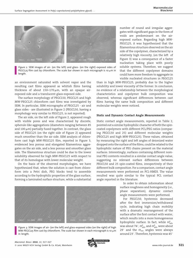
much smoother than the air side. SEM analysis of chloro-
form cast high MW-PEGCL25 film, reported in Figure 3,
evidenced less porous and elongated filamentous aggre-
gates on the air side, and a less porous and smoother glass
side. The filamentous structure could be due to the lower
solubility observed for high MW-PEGCL25 with respect to
that of its homologue with lower molecular weight.
On the basis of the observed morphologies, we have
hypothesized that, when the solution is cast from chloro-
form into a Petri dish, PEG blocks tend to assemble
according to the hydrophilic properties of the glass surface,
forming a laminated surface structure, while a substantial
Figure 3. SEM images of air- (on the left) and glass-exposed sides (on the right) of highMW-PEGCL25 film cast by chloroform. The scale bar shown in each micrograph is 10mmin length.
Macromol. Biosci. 2010, 10, 317–327
� 2010 WILEY-VCH Verlag GmbH & Co. KGaA, Weinheim
number of round and irregular aggre-
gateswith significant gaps in the form of
voids are predominant on the air-
exposed surface. Regarding high MW-
PEGCL25, it was hypothesized that the
filamentous structure observed on the air
side of the copolymer, characterized by a
relatively high viscosity, (on the left of
Figure 3) was a consequence of a faster
nucleation taking place with poorly
soluble systems. Therefore, we suggest
that the different copolymer domains
could havemore freedom to aggregate in
visible nucleated structures in PEGCL25
than in high MW-PEGCL25, probably due to the higher
solubility and lower viscosity of the former. In conclusion,
no evidence of a relationship between the morphological
characteristics and copolymer bulk composition was
observed, whereas significant differences between cast
films having the same bulk composition and different
molecular weights were noticed.
Static and Dynamic Contact Angle Measurements
Static contact angle measurements, reported in Table 2,
pointedout a similarhydrophilic character of both the spin-
coated copolymers with different PCL/PEG ratios (compar-
ing PEGCL50 and 25) and different molecular weights
(PEGCL25 and high MW-PEGCL25). These values, obtained
by measuring the right and left angles of deionized water
droppedonto the surface of thefilms, could be related to the
hydrophilic nature of PEG chains present on the material
surfaces. Interestingly, surfaces containing different nom-
inal PEG contents resulted in a similar contact angle value,
suggesting no relevant surface differences between
PEGCL50 and 25 spin-coated films, irrespectively of their
different bulk composition. For a comparison, contact angle
measurements were performed on PCL-HMDI. The value
resulted was quite similar to the typical PCL contact
angle reported in the literature.
In order to obtain information about
surface roughness and homogeneity (i.e.,
phase separation), dynamic contact
angle measurements were performed.
For PEGCL50, hysteresis decreased
after the first immersion/withdrawal
cycle, indicating high chain mobility
with a dramatic rearrangement of the
surface after the first contact withwater,
which results into a more homogeneous
hydrophilic surface. In fact, while Q1adv
was about 748,Q2adv andQ3
adv were about
298 and the Qrec angles were always
around238. Therefore, hysteresiswas 458
www.mbs-journal.de 321

S. Cometa, F. Chiellini, I. Bartolozzi, E. Chiellini, E. De Giglio, L. Sabbatini
Table 2. Static contact angles of PEGCL50, PEGCL25 and PCL-HMDIspin-coated films (data averaged over nine measurements).
Copolymer Contact angle
-
PEGCL50 72.0� 2.0
PEGCL25 71.2� 1.2
high MW-PEGCL25 74.0� 3.0
PCL-HMDI 82.0� 3.0
322
in the first cycle and only 68 in the second and third cycles.
Similar observations were reported by Crisci et al.[30] This
phenomenoncouldbe related to several contributions, such
as the surface rearrangement and the swelling of the PEG
component.
PEGCL25 exhibited a slightly more stable and hydro-
phobic surface thanPEGCL50. In fact,Q1adv resulted758,Q
2adv
and Q3adv were about 478, while receding angles were
constant around208and thereforehysteresiswasabout558for the first cycle and 278 for the second and third ones. As
far as high MW-PEGCL25 is concerned, the film exhibited a
fairly stable and more hydrophobic surface respect to
PEGCL25: Q1adv resulted equal to 818, while Q2
adv and Q3adv
were around 758, while receding angles resulted of about
208 for all the cycles. Therefore hysteresis ranged between
61 and 558, thus indicating lower chain mobility than for
PEGCL50 and 25.
PCL-HMDI exhibited constant values for both the
advancing contact angles (958) and the receding contact
angles (268). Thus, the surface was very hydrophobic and
hysteresis was constant at around 708. This similar
behavior can be explained by considering that PCL itself
can possibly have fast rearrangements which result in the
exposition of either the hydrophobic or hydrophilic part of
the molecules.
DCA results clearly showed a surface rearrangement of
PEGCL50 and PEGCL25, with enrichment of the hydrophilic
moieties, while this phenomenon was significantly less
pronounced in high MW-PEGCL25. This molecular weight-
dependent surface rearrangement was
investigated by XPS analysis of water-
equilibrated films in order to have a
better insight and correlation with
results obtained from the biological
characterization.
Figure 4. Survey spectra of PEGCL50 (a) and PEGCL25 (b) cast films from chloroform(air side).
Surface Chemistry Assessmentby XPS
Surface segregation phenomena can be
theoretically understood in terms of sur-
Macromol. Biosci. 2010, 10, 317–327
� 2010 WILEY-VCH Verlag GmbH & Co. KGaA, Weinheim
face energy and chemical potential balance arguments: the
driving force for surface segregation is the difference in
surface tension between different domains of a copolymer
system. Domains with low surface energywill move to the
surface to minimize the overall surface energy of the
system. At the same time, this surface migration could
increase the chemical potential due to a demixing process.
The balance of these two factors supplies the final surface
composition.
XPSanalysiswas carriedout inorder to assess the surface
chemistry of PEGCL50 and 25 cast films, devoting particular
attention to eventual differences existing between the
surface exposed to air and that exposed to the Petri dish
glass. Survey spectra of PEGCL50 and 25 films, cast from
chloroform (air side), are reported in Figure 4(a) and 4(b)
respectively. Typical elements (C1s andO1s) of PEG and PCL
samples were detected. In addition, Si2p signals were
recorded, probably due to the silicon grease contamination,
as well as a N1s signal, due to HMDI. It is important to note
the absence of an Sn2p signal, related to the catalyst used,
thus demonstrating the suitability of the purification steps
employed.
In order to evaluate the surface composition of the two
copolymers, curve fittings of high resolution C1s spectra
were performed. For comparison, the C1s signal curve
fitting of the air/polymer interface of PCL-HMDI is reported
in Figure 5.
Theobtainedpeaksdeconvolution is inagreementwitha
typical PCL film reported in the literature,[31,32] with the
exceptionofpeakD.This contribution isdue to theCH2�NH
groups relevant toHMDImolecules, and its peak’s areawas
well comparable to the N1s corrected signal area. The
C1s spectrum of PCL-HMDI showed an A:B:C:E ratio
[i.e., CH2: CH2C(¼O)O:CH2OC(¼O):C(¼O)O ratio] equal
to (5.0� 0.2):(1.61� 0.05):(1.58� 0.09):(1.04� 0.01) (data
averaged over three sampled points) with respect to the
expected 3:1:1:1 one. This finding is in keepingwith a slight
enrichment of the non-polar segments of PCL repeating
units on the air-exposed surface, as reported by Tang et al.
for PCL cast films.[33] However, peak A resulted larger than
expected from the caprolactone formula, also due to some
CHx -type surface contamination; hence this peak was not
DOI: 10.1002/mabi.200900284

Surface Segregation Assessment In Poly(e-caprolactone)-poly(ethylene glycol) . . .
Figure 5. C1s high-resolution spectrum of air-exposed surface ofPCL-HMDI film cast from chloroform. Peaks attributions arereported in the text.
used in further calculations. As far as PEG-based polymers
are concerned, XPS literature data[31,34] report a single C1s
contribution falling at 286.6� 0.1 eV (in addition a possible
minor component at 285.0 eV, due to adventitious con-
tamination, could be present). Therefore, it can be observed
that, in PEGCL copolymers, the presence of PEG moieties
does not add peculiar signals to the XPS spectra but simply
adds contributions to carbon and oxygen spectral regions.
In Figure 6, curve-fittings of C1s high resolution spectra
relevant to PEGCL50 and PEGCL25 – air and glass sides – are
reported. Peak attributions are illustrated in Scheme 1,
which allowed us to provide a detailed analysis of
quantitative information obtainable by relating the
normalized peak areas. Peaks binding energies, as well as
their atomic percentage, are reported in Table 3.
In particular, in the C1s signal of PEGCL copolymers can
be noticed a significant increase of peak C (relevant to the
PEG COH functionality, falling at the same binding energy
as thatof theCH2OC(¼O)ofPCLmoieties)withrespect to the
Figure 6. C1s high-resolution spectra of chloroform cast films: PEGCLexposed surfaces (a and b, respectively) and PEGCL25 glass- and air-eand d, respectively).
Macromol. Biosci. 2010, 10, 317–327
� 2010 WILEY-VCH Verlag GmbH & Co. KGaA, Weinheim
C1s signal ofPCLalone (seeFigure5), due to the introduction
of ethylene glycol components.
The equations used for the quantitative evaluation of
surface PEG and PCL content are reported in the following
system of Equation (2):
50 glaxposed
Area peak E� Area N1s ¼ 16xArea peak C ¼ 20xþ 90y
(2)
where x is the number of PCL2000 macromers in which 20
C�O and 16 C¼O functionalities were present, and y
represents the number of PEG2000 macromers, which
contain 90 C�O groups each.
It should be underlined that, when peak E was used to
estimate PCL content in PEGCL copolymers, a slight
underestimation of this value was implicitly linked to
the lower surface presence of polar C(¼O)O groups (see
peaks area ratio in C1s PCL/HMDI curve fitting). In any case,
using the reported equations, a surface PEGenrichment can
be detected on both the air-polymer and glass-polymer
interfaceswith respect to thebulk composition: 63.3� 1.6%
and 55.9� 1.1% of PEG content in PEGCL50 glass and air
sides, respectively, vs. 50% nominal value, and 58.1� 0.6%
and56.3� 1.4% in PEGCL25 glass and air sides, respectively,
vs. 25%. These findings suggest a PEG component enrich-
ment in both PEGCL50 and 25 glass-exposed copolymer
films, as expected, but an almost similar PEGCL50 and 25
surface composition, in disagreementwith their significant
difference in terms of bulk composition. Indeed, the PEG
surface content which is higher than the nominal one,
observed on both air and glass sides of the cast films, could
be responsible for the low serumprotein adhesion onto the
surface of the films resulting in poor cell attachment.[35]
ss- and air-surfaces (c
Moreover, results of XPS analysis on dry
chloroform high MW-PEGCL25 cast film
revealed a surface composition very
similar to the homologue copolymer
having lower molecular weight. This
result did not allow us to understand
the different biological performances of
higher and lower molecular weight
PEGCL25 copolymer films, thus convin-
cing us to exploit othermodalities of XPS
analysis.
In-Depth Composition AnalysisUsing AR-XPS
In order to better assess the real surface
chemistry experimented by cells, we
have carried out AR-XPS measurements,
which can supply information about the
outermost molecular layers.
www.mbs-journal.de 323

S. Cometa, F. Chiellini, I. Bartolozzi, E. Chiellini, E. De Giglio, L. Sabbatini
Scheme 1. Peak attributions of C1s curve fittings reported in Figure 6. Circles are used to highlight the carbon(s) whose capital letter refers topeak labels in Figure 6 and in Table 3.
Table 3. Binding energies and atomic percentages of the C1s fitting components of spectra reported in Figure 6.
Peak Peak BE Carbon content
eV at.-%
PEGCL50 (glass) PEGCL50 (air) PEGCL25 (glass) PEGCL25 (air)
A 285.0� 0.1 33.7� 0.4 55.29� 0.09 39.7� 0.4 48.9� 1.2
B 285.5� 0.1 10.8� 1.1 8.4� 0.5 8.9� 0.8 10.21� 0.02
C 286.7� 0.1 47.2� 1.9 28.8� 0.2 43.0� 0.6 33.0� 0.9
D 287.3� 0.1 2.3� 0.9 2.8� 0.3 2.16� 0.17 2.87� 0.03
E 289.4� 0.1 5.9� 0.3 4.64� 0.11 6.12� 0.03 5.0� 0.3
Table 4. PEG content on PEGCL50 and PEGCL25 (air and glass sides), at different takeoff angles, calculated from Equation (2) on C1s signalscurve fittings.
Takeoff angle PEG content
- %
PEGCL50 air PEGCL50 glass PEGCL25 air PEGCL25 glass
30.5 54� 2 60.7� 1.5 53� 2 59� 2
45.5 53.7� 1.2 62.0� 1.7 50.7� 1.5 59� 3
60.5 53� 3 62.8� 1.1 53� 3 60.0� 1.7
324
AR-XPS data for different takeoff angles showed that no
significant variations in PEG and PCL percentages were
observedbyvarying the samplingdepth (seeTable 4). These
results clearly show that surface analysis of the topmost
Macromol. Biosci. 2010, 10, 317–327
� 2010 WILEY-VCH Verlag GmbH & Co. KGaA, Weinheim
10nm of PEGCL50 and 25 chloroform cast films resulted in
quite homogeneously distributed layers, thus conventional
XPS analysis seems to be adequate to give information
about the real surface compositionofmultiblock copolymer
DOI: 10.1002/mabi.200900284

Surface Segregation Assessment In Poly(e-caprolactone)-poly(ethylene glycol) . . .
films experimented by cells. Similar
results have been obtained by perform-
ing AR-XPS on high MW-PEGCL25. In this
respect, after verifying this quite homo-
geneous in-depth composition, further
analyses have been acquired in the
conventional XPS mode.
Figure 7. C1s signals relevant to PEGCL25 (on the left) and high MW-PEGCL25 (on theright), before (solid line) and after (dotted line) 48h incubation in PBS.
XPS Analysis of PBS-EquilibratedFilms and Interpretation ofBiological Results
The analysis of polymer films after interaction with an
aqueous environment canbevery important tounderstand
if contactwithwater canmodify their surface composition.
In this respect, PEGCL chloroform cast films were analyzed
after immersion in PBS for 48hand successive dryingunder
a vacuum, in order to ascertain if the copolymer films
underwent reconstruction of their block composition. A
similarprocedurehasalreadybeenadopted in the literature
byother authors inorder to characterizewater-equilibrated
polymer films.[36–38] Since XPS data on ‘‘wet’’ samples was
in good agreement with DCA and cell adhesion results, we
have assumed that the ultrahigh vacuum used in the XPS
analysis did not substantially alter the copolymer surface
assembly. Therefore, XPS data on ‘‘wet’’ surfaces could be
considered indicative of the real chemical composition
experimented by the cells in aqueous environment.
Relative peak abundances, obtained by performing C1s
curve fitting on PEGCL50 and25 ‘‘wet’’ films, are reported in
Table 5.
These results demonstrated that, independently of the
glass and air sides, a further enrichment in PEG moieties
occurred, both for PEGCL50 and 25 samples; the PEG
percentage, calculated using Equation (2), reached
88.0� 0.9% and 84.3� 0.6% for PEGCL50 and 25, respec-
tively.
Table 5. Binding energies and atomic percentages of C1s peakscurve fitting relevant to PEGCL50 and 25 films after 48h immer-sion in PBS.
Peak Peak BE Carbon content
eV at.-%
PEGCL50 ‘‘wet’’ PEGCL25 ‘‘wet’’
A 285.0� 0.1 28.0� 0.6 38.3� 0.6
B 285.5� 0.1 15.2� 0.5 12.79� 0.16
C 286.7� 0.1 49.7� 0.9 41.6� 0.3
D 287.3� 0.1 3.1� 0.5 2.9� 0.3
E 289.4� 0.1 4.0� 0.3 4.7� 0.3
Macromol. Biosci. 2010, 10, 317–327
� 2010 WILEY-VCH Verlag GmbH & Co. KGaA, Weinheim
In Figure 7(a), a comparison between the PEGCL25 C1s
shape (glass side) before and after incubation in PBS has
beenreported, inorder tobetter clarify the incrementofPEG
contribution, as obviously expected by incubating in an
aqueousmedium a copolymer containing both hydrophilic
and hydrophobic components.
On the other hand, comparison between the high MW-
PEGCL25 C1s shape (glass side) before and after incubation
in PBS, reported in Figure 7(b), shows that quite similar C1s
shapeswere recorded. We hypothesized that themigration
of PEG domains and phase separation that resulted was
hindered by the high molecular weight of the copolymer.
Therefore, a surface rearrangement of high MW-PEGCL25
may be highly influenced by its low freedom in motion.
These results are in good agreement with DCA measure-
ments, nevertheless in XPS solvent cast films whereas in
DCA dip-coated glass slides have been analyzed. Anyway,
both XPS and DCA analysis results were useful to conclude
that the less pronounced phase segregation after immer-
sion in wet environment observed in high MW-PEGCL25
film allowed for a significant cell adhesion and prolifera-
tion, as shown in Figure 1(d). Additional detailed research is
necessary to fully delineate the complex mechanisms that
govern the surface segregation in these systems. In
particular, further investigations are in progress by
synthesizing a series of polymers with different molecular
weights and blockiness distribution. However, our main
interest is to analyze possible surface segregation phenom-
ena related to the processing methods chosen to obtain 3D
scaffolds by using these copolymers. In this respect, the
ongoing morphological, chemical and biological character-
ization of PEGCL 3D scaffolds obtained by electrospinning
and/or rapid prototyping appears of great interest for a
better understanding of their effective potential utilization
in biomedical fields.
XPS Analysis of Films Cast from Different SolventSystems
Finally, PEGCL50and25havebeenalso cast using twoother
different solvent systems, in order to investigate to what
www.mbs-journal.de 325

S. Cometa, F. Chiellini, I. Bartolozzi, E. Chiellini, E. De Giglio, L. Sabbatini
Figure 8. C1s high-resolution spectra of air-exposed PEGCL50 films cast from acetone (a)and ethyl acetate (b) and PEGCL25 films cast from acetone (c) and ethyl acetate (d).
326
extent their surface composition could be
influenced by solvent properties. The
solvents used were: acetone (hydrophilic,
Teb 56 8C) and ethyl acetate (hydrophobic,
Teb 77 8C), compared to previously ana-
lyzed chloroform (hydrophobic, Teb 61 8C)cast films. In this respect, we report
only XPS results about the air-exposed
surface, since, during the casting process,
itwas in contactwith thegasphase,made
by a mixture of air and residual solvent
vapors. When the solution was cast on
a Petri dish, indeed, the copolymer
systems assemble according to the high
hydrophilic glass properties (contact
angle� 258), forming a laminated surface
enriched with PEG hydrophilic domains,
while the air/polymer interface will
depend, in turn, on the hydrophilicity of
the solvent used. In our investigations,
XPS analysis pointed out a clear relationship
between surface copolymer composition and solvents
used, in particular with their relative hydrophilicity and
volatility.
In Figure 8, C1s curve fittings relevant to air-exposed
PEGCL50 and 25, fromacetone and ethyl acetate, have been
reported for comparison with air-exposed PEGCL50 and 25
cast from chloroform [see Figure 6(b) and 6(d)]. In Table 6,
binding energies and relative peak abundances, the
attribution of which is based on Scheme 1, have been
summarized.
Using Equation (2), the following PEG percentages in
films cast from the examined solvents resulted:
1. 8
Ma
� 2
2.3� 1.3% PEG in PEGCL50 and 83.1� 1.1% PEG in
PEGCL25 films cast from acetone, denoting higher
surface PEG enrichment than in chloroform cast films,
Table 6. Binding energies and atomic percentages of the C1s peaks in s
Peak Peak BE
eV
PEGCL50
(acetone)
PE
(ethy
A 285.0� 0.1 31.8� 1.8 34.
B 285.5� 0.1 10.5� 0.9 16.
C 286.7� 0.1 50.3� 0.9 37.
D 287.3� 0.1 3.7� 0.6 2.
E 289.4� 0.1 3.7� 0.3 8.3
cromol. Biosci. 2010, 10, 317–327
010 WILEY-VCH Verlag GmbH & Co. KGaA, Weinheim
due to the high hydrophilic character and the low
boiling point of acetone;
2. 4
3.2� 0.5% PEG in PEGCL50 and 26.3� 0.9% PEG inPEGCL25 films cast from ethyl acetate, resulting in a
surface PEG percentage more similar to the nominal
one, indicating that this more highly boiling and
hydrophobic solvent enabled PCL to reach the surface.
In conclusion, by comparing the surface PEG percentage
estimated by XPS investigation on films cast by different
solvents, the trend was that surface composition was
enriched by PCL when: (1) the casting solvent was more
hydrophobic and/or (2) it evaporated slowly.
Work is in progress to gain information about the
morphological features and biological performances of
acetone and ethyl acetate cast films, in order to correlate
these results to the obtained XPS data.
pectra reported in Figure 8.
Carbon content
at.-%
GCL50
l acetate)
PEGCL25
(acetone)
PEGCL25
(ethyl acetate)
8� 0.7 22.7� 0.6 40.0� 1.5
3� 1.8 8.0� 0.8 20.8� 1.4
9� 0.4 59.1� 0.5 27.4� 0.6
5� 0.5 5.1� 0.8 2.2� 0.5
8� 0.08 5.0� 0.4 9.6� 0.2
DOI: 10.1002/mabi.200900284

Surface Segregation Assessment In Poly(e-caprolactone)-poly(ethylene glycol) . . .
Conclusion
We focused our attention on the assessment of the
influence of multiblock copolymer film surface composi-
tion on their biological performance.
XPS investigation, supported by morphological and
wettability analysis, evidenced that cast films were
strongly affected by phase segregation phenomena, which
were found to depend on the solvent used in the casting
procedure, as well as on the molecular weight of the
copolymer. These segregation effects were enhanced after
equilibration in aqueous media and were also reflected in
cell behavior, observed in cell adhesion and proliferation
assays, using the balb/c 3T3 clone A31 mouse embryo
fibroblast cell line.
These results suggest that a systematic surface char-
acterization of random multiblock or triblock copolymers,
paying particular attention to their possible phase segrega-
tion as bound to primary structural parameters (molecular
weight, blockiness length and distribution) and to proces-
sing methods (i.e., solvent casting, spin coating, electro-
spinning, rapid prototyping)might provide useful hints for
thedevelopmentofpolymeric scaffoldswith successful and
optimized biocompatibility.
Acknowledgements: This work was performed within the frame-work of the projects FIRB-2005 - prot. RBIN043BCP, PRIN-2006 -prot. 2006038548 t and NoE Expertissues CT - 2004 - 500328. Theauthors would like to thank Mr. P. Narducci for the recording ofSEM images.
Received: August 3, 2009; Revised: September 25, 2009; Publishedonline: December 2, 2009; DOI: 10.1002/mabi.200900284
Keywords: biocompatibility; block copolymers; supports; sur-faces; X-ray
[1] J. A. Hubbell, Biotechnology 1995, 13, 565.[2] M. Zhang, T. Desai, M. Ferrari, Biomaterials 1998, 19, 953.[3] N. A. Alcantar, E. S. Aydil, J. N. Israelachvili, J. Biomed. Mater.
Res. 2000, 51, 343.[4] S. Sharma, K. C. Popat, T. A. Desai, Langmuir 2002, 18, 8728.[5] G. Altankov, V. Thom, T. Groth, K. Jankova, G. Jonsson, M.
Ulbricht, J. Biomed. Mater. Res. 2000, 52, 219.[6] K. H. Yong, Y. M. Kyong, P. I. Kyu, K. T. Hee, L. H. Chul, L.
Hyun-Sook, O. Jong-Suk, A. Toshihiro, C. C. Su, Biomaterials2003, 24, 801.
Macromol. Biosci. 2010, 10, 317–327
� 2010 WILEY-VCH Verlag GmbH & Co. KGaA, Weinheim
[7] M. C. Serrano, R. Pagani, M. Vallet-Regi, J. Pena, A. Ramila, I.Izquierdo, M. T. Portoles, Biomaterials 2004, 25, 5603.
[8] V. R. Sinha, K. Bansal, R. Kaushik, R. Kumria, A. Trehan, Int. J.Pharm. 2004, 278, 1.
[9] T. G. Kim, D. S. Lee, T. G. Park, Int. J. Pharm. 2007, 338,276.
[10] J. Rieger, P. Dubois, R. Jerome, C. Jerome, Langmuir 2006, 22,7471.
[11] Z. Gan, T. F. Jim,M. Li, Z. Yuer, S. Wang, C.Wu,Macromolecules1999, 32, 590.
[12] S.-H. Hsu, C.-M. Tang, C.-C. Lin, Biomaterials 2004, 25,5593.
[13] A. Rasmont, Ph. Leclere, C. Doneux, G. Lambin, J. D. Tong, R.Jerome, J. L. Bredas, R. Lazzaroni, Colloids Surf. B 2000, 19, 381.
[14] L. Andruzzi, F. D’Apollo, G. Galli, B. Gallot, Macromolecules2001, 34, 7707.
[15] M. Bertolucci, G. Galli, E. Chiellini, K. J. Wynne, Macromol-ecules 2004, 37, 3666.
[16] C. Biver, G. de Crevoisier, S. Girault, A. Mourran, R. Pirri, J. C.Razet, L. Leibler, Macromolecules 2002, 35, 2552.
[17] B. Kasemo, Surf. Sci. 2002, 500, 656.[18] J. A. Gardella, N. L. Hernandez de Gatica, J. Electron. Spectrosc.
Relat. Phenom. 1996, 81, 227.[19] G. Speranza, G. Gottardi, C. Pederzolli, L. Lunelli, R. Canteri, L.
Pasquardini, E. Carli, A. Lui, D. Maniglio, M. Brugnara, M.Anderle, Biomaterials 2004, 25, 2029.
[20] M. Tirrell, E. Kokkoli, M. Biesalski, Surf. Sci. 2002, 500, 61.[21] D. G. Castner, B. D. Ratner, Surf. Sci. 2002, 500, 28.[22] S. C. Yoon, B. D. Ratner, Macromolecules 1988, 21, 2392.[23] M. A. Helfand, J. B. Mazzanti, M. Fone, R. H. Reamey, Langmuir
1996, 12, 1296.[24] F. A. Bottino, G. Di Pasquale, A. Pollicino, Macromolecules
1998, 31, 7814.[25] L. Andruzzi, A. Hexemer, X. F. Li, C. K. Ober, E. J. Kramer, G.
Galli, E. Chiellini, D. A. Fischer, Langmuir 2004, 20, 1049.[26] C.-S. Ha, J. A. Gardella, Jr., J. Macromol. Sci., Polym. Rev. 2005,
45, 1.[27] F. Chiellini, A. M. Piras, M. Gazzarri, C. Bartoli, M. Ferri, L.
Paolini, E. Chiellini, Macromol. Biosci. 2008, 8, 516.[28] M. C. Serrano, R. Pagani, M. Vallet-Regı, J. Pena, A. Ramila, I.
Izquierdo, M. T. Portoles, Biomaterials 2004, 25, 5603.[29] P. S. Tan, S. H. Teoh, Mater. Sci. Engin. C 2007, 27, 304.[30] L. Crisci, C. Della Volpe, G. Maglio, G. Nese, R. Palombo, G. P.
Rachiero, Macromol. Biosci. 2003, 3, 749.[31] D. Briggs, G. Beamson, Anal. Chem. 1992, 64, 1729.[32] P. Viville, R. Lazzaroni, Langmuir 2003, 19, 9425.[33] Z. G. Tang, R. A. Black, J. M. Curran, J. A. Hunt, N. P. Rhodes, D. F.
Williams, Biomaterials 2004, 25, 4741.[34] G. Beamson, B. T. Pickup, W. Li, S.-M. Mai, J. Phys. Chem. B
2000, 104, 2656.[35] A. T. Hubbard, in: Encyclopedia of Surface and Colloid Science,
Volume 1, CRC Press, Boca Raton 2002, p. 1243.[36] E. Martinelli, S. Agostini, G. Galli, E. Chiellini, A. Glisenti, M. E.
Pettitt, M. E. Callow, J. A. Callow, K. Graf, F. W. Bartels,Langmuir 2008, 24, 13138.
[37] J. Wen, G. Somorjai, F. Lim, R. Ward,Macromolecules 1997, 30,7206.
[38] J.-H. Chen, E. Ruckenstein, J. Colloid Interface Sci. 1990, 135,496.
www.mbs-journal.de 327


![Microcalorimetric Investigation of DNA, poly(dA)poly(dT) and poly[d(AC)]poly[d(GT)] Melting in the Presence of Water Soluble (Meso tetra (4 N oxyethylpyridyl) Porphyrin) and its Zn](https://static.fdokumen.com/doc/165x107/631f222063ac2c35640aaab6/microcalorimetric-investigation-of-dna-polydapolydt-and-polydacpolydgt.jpg)