Thermodynamics of Interactions of TAlPyP4 and AgTAlPyP4 Porphyrins with Poly(rA)poly(rU) and...
9
This article was downloaded by: [New York University] On: 02 May 2013, At: 08:26 Publisher: Taylor & Francis Informa Ltd Registered in England and Wales Registered Number: 1072954 Registered office: Mortimer House, 37-41 Mortimer Street, London W1T 3JH, UK Journal of Biomolecular Structure and Dynamics Publication details, including instructions for authors and subscription information: http://www.tandfonline.com/loi/tbsd20 Thermodynamics of Interactions of TAlPyP4 and AgTAlPyP4 Porphyrins with Poly(rA)poly(rU) and Poly(rI)poly(rC) Duplexes A. A. Ghazaryan a , Y. B. Dalyan a , S. G. Haroutiunian a , V. I. Vardanyan a , R. K. Ghazaryan b & T. V. Chalikian c a Department of Molecular Physics Faculty of Physics, Yerevan State University, 1 Alex Manoogian Street, Yerevan, 375025, Armenia b Department of Pharmaceutical Chemistry, Yerevan State Medical University, 2 Korjun Street, Yerevan, 375025, Armenia c Department of Pharmaceutical Sciences Leslie Dan Faculty of Pharmacy, University of Toronto, 19 Russell Street, Toronto, Ontario, M5S 2S2, Canada Published online: 15 May 2012. To cite this article: A. A. Ghazaryan , Y. B. Dalyan , S. G. Haroutiunian , V. I. Vardanyan , R. K. Ghazaryan & T. V. Chalikian (2006): Thermodynamics of Interactions of TAlPyP4 and AgTAlPyP4 Porphyrins with Poly(rA)poly(rU) and Poly(rI)poly(rC) Duplexes, Journal of Biomolecular Structure and Dynamics, 24:1, 67-74 To link to this article: http://dx.doi.org/10.1080/07391102.2006.10507100 PLEASE SCROLL DOWN FOR ARTICLE Full terms and conditions of use: http://www.tandfonline.com/page/terms-and-conditions This article may be used for research, teaching, and private study purposes. Any substantial or systematic reproduction, redistribution, reselling, loan, sub-licensing, systematic supply, or distribution in any form to anyone is expressly forbidden. The publisher does not give any warranty express or implied or make any representation that the contents will be complete or accurate or up to date. The accuracy of any instructions, formulae, and drug doses should be independently verified with primary sources. The publisher shall not be liable for any loss, actions, claims, proceedings, demand, or costs or damages whatsoever or howsoever caused arising directly or indirectly in connection with or arising out of the use of this material.
Transcript of Thermodynamics of Interactions of TAlPyP4 and AgTAlPyP4 Porphyrins with Poly(rA)poly(rU) and...

This article was downloaded by: [New York University]On: 02 May 2013, At: 08:26Publisher: Taylor & FrancisInforma Ltd Registered in England and Wales Registered Number: 1072954 Registered office: Mortimer House,37-41 Mortimer Street, London W1T 3JH, UK
Journal of Biomolecular Structure and DynamicsPublication details, including instructions for authors and subscription information:http://www.tandfonline.com/loi/tbsd20
Thermodynamics of Interactions of TAlPyP4 andAgTAlPyP4 Porphyrins with Poly(rA)poly(rU) andPoly(rI)poly(rC) DuplexesA. A. Ghazaryan a , Y. B. Dalyan a , S. G. Haroutiunian a , V. I. Vardanyan a , R. K. Ghazaryanb & T. V. Chalikian ca Department of Molecular Physics Faculty of Physics, Yerevan State University, 1 AlexManoogian Street, Yerevan, 375025, Armeniab Department of Pharmaceutical Chemistry, Yerevan State Medical University, 2 KorjunStreet, Yerevan, 375025, Armeniac Department of Pharmaceutical Sciences Leslie Dan Faculty of Pharmacy, University ofToronto, 19 Russell Street, Toronto, Ontario, M5S 2S2, CanadaPublished online: 15 May 2012.
To cite this article: A. A. Ghazaryan , Y. B. Dalyan , S. G. Haroutiunian , V. I. Vardanyan , R. K. Ghazaryan & T. V. Chalikian(2006): Thermodynamics of Interactions of TAlPyP4 and AgTAlPyP4 Porphyrins with Poly(rA)poly(rU) and Poly(rI)poly(rC)Duplexes, Journal of Biomolecular Structure and Dynamics, 24:1, 67-74
To link to this article: http://dx.doi.org/10.1080/07391102.2006.10507100
PLEASE SCROLL DOWN FOR ARTICLE
Full terms and conditions of use: http://www.tandfonline.com/page/terms-and-conditions
This article may be used for research, teaching, and private study purposes. Any substantial or systematicreproduction, redistribution, reselling, loan, sub-licensing, systematic supply, or distribution in any form toanyone is expressly forbidden.
The publisher does not give any warranty express or implied or make any representation that the contentswill be complete or accurate or up to date. The accuracy of any instructions, formulae, and drug doses shouldbe independently verified with primary sources. The publisher shall not be liable for any loss, actions, claims,proceedings, demand, or costs or damages whatsoever or howsoever caused arising directly or indirectly inconnection with or arising out of the use of this material.

Thermodynamics of Interactions of TAlPyP4 and AgTAlPyP4 Porphyrins with Poly(rA)poly(rU) and
Poly(rI)poly(rC) Duplexes
http://www.jbsdonline.com
Abstract
We employed UV light absorption and circular dichroism (CD) spectroscopic measurements to study the binding of novel water-soluble porphyrins meso-tetra-(4N-allylpyridyl)porphyrin [TAlPyP4], and its Ag containing derivative to the poly(rA)poly(rU) and poly(rI)poly(rC) RNA duplexes. Our results suggest that TAlPyP4 associate with the duplexes via intercala-tion, whereas the conservative CD spectra indicates that AgTAlPyP4 preferably binds via outside self-stacking mode. We used our determined binding isotherms for each ligand-RNA binding event to calculate the binding constant, Kb, and binding free energy, ΔGb = -RTlnKb. By performing these experiments as a function of temperature, we evaluated the van’t Hoff binding enthalpies, ΔH. The binding entropies, ΔSb, were calculated as ΔSb = (ΔHb - ΔGb)/T. We interpret our data in terms of specific interactions that stabilize/destabilize each ligand-RNA complex studied in this work. Taken together, our data provide important new informa-tion about the thermodynamics of interactions of porphyrins with nucleic acids.
Introduction
The water-soluble porphyrins and their derivatives continue to be a subject of con-siderable interest in biochemistry due to their binding affinity to synthetic or natural nucleic acids and the ability to selectively cleave DNA (1-3). They have been ex-cellent probes of nucleic acid structure and dynamics since they are sensitive to the environmental changes and are monitored conveniently (4, 5). Cationic porphyrins proved to be promising yet relatively untapped class of DNA-binding molecules with potential applications in biology and medicine, in particular, as potent anti-vi-ral and anti-tumor therapeutic agents (3, 6). The water-soluble porphyrins are read-ily modificated, and some of them have been reported to show nuclease activity.
As DNA binding ligands, porphyrins are quite unusual, since they may associate with DNA in three distinct binding modes that include intercalation, groove binding, and outside binding with self-stacking along the DNA helix (3, 7-12). The binding mode of a specific porphyrin-nucleic acid system depends on the chemical features of the porphyrin and the structure and composition of the host DNA and RNA. A study of cationic porphyrin – DNA interaction in solution shows that intercalation of porphyrin into DNA requires at least temporary existence of planar conformation of porphyrin molecule (i.e., it must have a limited effective thickness). External (groove) binding is typical for porphyrins that cannot fit between nucleobases due to sterical factors, i.e., porphyrins bearing bulky peripheral substituents or axial ligands on the central ion (11, 13). In this case the side of the porphyrin ring fits into the minor groove of the helix or locates in the major groove by electrostatic in-teraction between the negatively charged phosphate group and the positive charged pyridinium rings of porphyrin periphery. The majority of poprphyrin-DNA stud-ies have focused on meso-tetrakis(N-methylpyridinium-4-yl)porphyrin (H2TMPyP)
Journal of Biomolecular Structure &Dynamics, ISSN 0739-1102Volume 24, Issue Number 1, (2006)©Adenine Press (2006)
A. A. Ghazaryan1
Y. B. Dalyan1,*
S. G. Haroutiunian1
V. I. Vardanyan1
R. K. Ghazaryan2
T. V. Chalikian3
1Department of Molecular PhysicsFaculty of PhysicsYerevan State University1 Alex Manoogian StreetYerevan 375025, Armenia2Department of Pharmaceutical ChemistryYerevan State Medical University2 Korjun StreetYerevan 375025, Armenia3Department of Pharmaceutical SciencesLeslie Dan Faculty of PharmacyUniversity of Toronto19 Russell StreetToronto, Ontario M5S 2S2, Canada
67
Phone: (37410) 554341Fax: (37410) 577-689Email: [email protected]
Dow
nloa
ded
by [
New
Yor
k U
nive
rsity
] at
08:
26 0
2 M
ay 2
013
68
Ghazaryan et al.
and its metal derivatives (3, 7-17). These studies have provided us with valuable information about the sequence specific and ligand-specific binding preferences of H2TMPyP4 and its metaloderivatives as well as the binding constants and the bind-ing stoichiometry of porphyrin-DNA interactions. Numerous studies showed that this porphyrin discriminates between GC-rich and AT-rich regions, tending to inter-calate into 5ʹCGʹ3 site of the first and bind externally to the second (11, 18). Long range stacks of planar porphyrins assembled along the polynucleotide chain and repeating its chiral structure are also evidenced.
In spite of the extensive studies on the interactions of water-soluble porphyrins with DNA, there is a lack of studies of binding properties for RNA thus far. In this work we investigated and classified the binding of the novel water-soluble meso-tetra-(4N-allylpyridyl)porphyrin [TAlPyP4], and its Ag containing derivative to synthetic double stranded poly(rA)poly(rU) and poly(rI)poly(rC). Homopolymers were chosen to simplify the model and reduce the number of possible binding modes to a single. We expect that the results obtained here can be extrapolated to the native heterogeneous RNA/DNA.
Materials and Methods
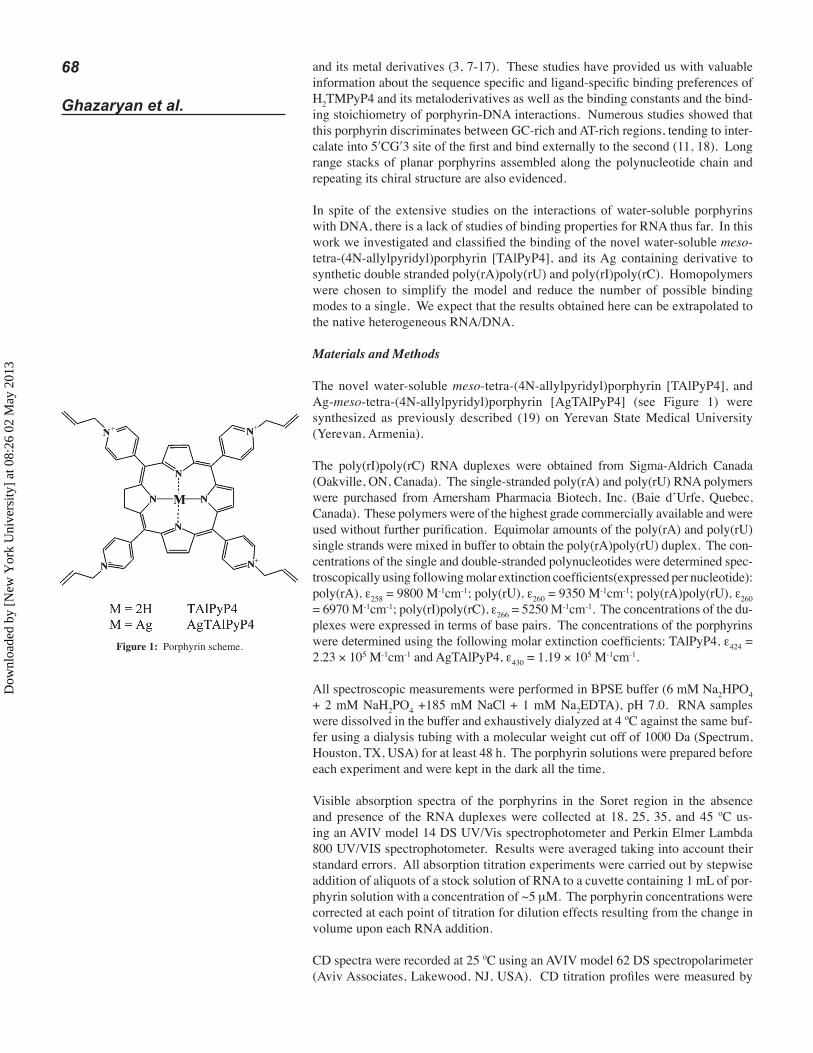
The novel water-soluble meso-tetra-(4N-allylpyridyl)porphyrin [TAlPyP4], and Ag-meso-tetra-(4N-allylpyridyl)porphyrin [AgTAlPyP4] (see Figure 1) were synthesized as previously described (19) on Yerevan State Medical University (Yerevan, Armenia).
The poly(rI)poly(rC) RNA duplexes were obtained from Sigma-Aldrich Canada (Oakville, ON, Canada). The single-stranded poly(rA) and poly(rU) RNA polymers were purchased from Amersham Pharmacia Biotech, Inc. (Baie d’Urfe, Quebec, Canada). These polymers were of the highest grade commercially available and were used without further purification. Equimolar amounts of the poly(rA) and poly(rU) single strands were mixed in buffer to obtain the poly(rA)poly(rU) duplex. The con-centrations of the single and double-stranded polynucleotides were determined spec-troscopically using following molar extinction coefficients(expressed per nucleotide): poly(rA), ε258 = 9800 M-1cm-1; poly(rU), ε260 = 9350 M-1cm-1; poly(rA)poly(rU), ε260 = 6970 M-1cm-1; poly(rI)poly(rC), ε266 = 5250 M-1cm-1. The concentrations of the du-plexes were expressed in terms of base pairs. The concentrations of the porphyrins were determined using the following molar extinction coefficients: TAlPyP4, ε424 = 2.23 × 10⁵ M-1cm-1 and AgTAlPyP4, ε430 = 1.19 × 10⁵ M-1cm-1.
All spectroscopic measurements were performed in BPSE buffer (6 mM Na2HPO4 + 2 mM NaH2PO4 +185 mM NaCl + 1 mM Na2EDTA), pH 7.0. RNA samples were dissolved in the buffer and exhaustively dialyzed at 4 ºC against the same buf-fer using a dialysis tubing with a molecular weight cut off of 1000 Da (Spectrum, Houston, TX, USA) for at least 48 h. The porphyrin solutions were prepared before each experiment and were kept in the dark all the time.
Visible absorption spectra of the porphyrins in the Soret region in the absence and presence of the RNA duplexes were collected at 18, 25, 35, and 45 ºC us-ing an AVIV model 14 DS UV/Vis spectrophotometer and Perkin Elmer Lambda 800 UV/VIS spectrophotometer. Results were averaged taking into account their standard errors. All absorption titration experiments were carried out by stepwise addition of aliquots of a stock solution of RNA to a cuvette containing 1 mL of por-phyrin solution with a concentration of ~5 μM. The porphyrin concentrations were corrected at each point of titration for dilution effects resulting from the change in volume upon each RNA addition.
CD spectra were recorded at 25 ºC using an AVIV model 62 DS spectropolarimeter (Aviv Associates, Lakewood, NJ, USA). CD titration profiles were measured by
Figure 1: Porphyrin scheme.
Dow
nloa
ded
by [
New
Yor
k U
nive
rsity
] at
08:
26 0
2 M
ay 2
013

69The Interaction of
Porphyrins with RNA
adding aliquots of a porphyrin to a known amount of RNA solution. The initial RNA concentrations were in the range of 50 to 100 μM.
Results
Circular Dichroism
To determine the binding modes of the studied porphyrins, the CD spectra of com-plexes were measured. The aliquots of porphyrin were added on fixed concentration of RNA solution and the CD spectra from 220 nm to 550 nm was recorded. The porphyrins considered here do not display visible range CD spectra (from 390 nm to 500 nm) in the absence of nucleic acids, but such spectra are induced from interac-tion with RNA and DNA. The spectra magnitude changed almost linearly conse-quently reaching it’s saturation. The ellipticity spectra of porphyrin/polynucleotide complexes are displayed in Figure 2. Here in all four cases the porphyrin and poly-nucleotide concentration is the same ([porphyrin] = 7.2 μM, [RNA] = 21 μM BP).
The apparent difference between CD spectra of TAlPyP4/polynucleotide and Ag-TAlPyP4/polynucleotide complexes is the overwhelming negative peak of the first in contrast to nearly conservative spectra of the second. It reaches saturation upon addition of porphyrin at μ = 0.3 (μ = [porphyrin]/[base pair]).
Absorbance
The interaction of studied porphyrins with RNA duplexes was monitored with changes in Soret region of absorbance spectra. The change in the absorption spec-trum of the studied porphyrins upon addition of poly(rI)poly(rC) at 25 ºC is shown in Figure 2 as an example.
The addition of aliquots of RNA stock solution to the fixed concentration of porphy-rins leads to the gradual red shift and large hypochromicity of the Soret maximum. The values for the hypochromicity and the bathochromic shift appeared to be larger for TAlPyP4 (Δλ ~ +15 nm, Δh ~ 40%) then the ones obtained for AgTAlPyP4 (Δλ ~ +8 nm, Δh ~ 30%). The larger values for these parameters found for TAlPyP4
, nm400 440 480-2
-1
0
1
ellipticity
[] ,
10
5de
g cm
2 dmol
-1
a)
400 440 480
b)
Figure 2: Induced CD spectra of TAlPyP (4) (solid line) and AgTAlPyP4 (dashed line) complexes with polynu-cleotides polynucleotides: (a) poly(rA)poly(rU) and (b) poly(rI)poly(rC) at 25 ºC in phosphate buffer (pH 6.8, μ = 0.2 M). [porphyrin] = 7.2μM; [RNA] = 21μM.
1
2
0.5
1
, n400 425 450 475
a)b)
400 425 450 475, n
Figure 2: Visible absorption spectra of TAlPyP4 (a) and AgTAlPyP4 (b) in the absence and presence of poly(rI)poly(rC) at 25 ºC. The initial concentrations of the porphyrins were ~8 μM. The final RNA-to-ligand ratios were 140 and 120 for TAlPyP4 and AgTAlPyP4, respectively. The individual values of the final RNA-to-ligand ratio for each titration were selected to ensure saturation of the binding profiles.
Dow
nloa
ded
by [
New
Yor
k U
nive
rsity
] at
08:
26 0
2 M
ay 2
013

70
Ghazaryan et al.
suggest that π electrons of this porphyrin were more perturbed upon binding then the ones of AgTAlPyP4. The titration experiments were continued until no further change in spectra was observed, which indicated that total saturation was reached. It was observed that when the total base-pair concentration was 15-20 times greater than the porphyrin concentration, no further changes in the Soret band occurred indicating that all porphyrin was already bound. One set of isosbestic points was observed in each case, and the binding was found to proceed apparently in a single step. This allowed us to use a non-cooperative neighbor exclusion model proposed by McGhee and von Hippel (20) to analyze the titration data.
Binding Thermodynamics
The porphyrin-RNA binding was monitored by the absorption at 422 nm (TAlPyP4) and 430 nm (AgTAlPyP4). The maximum deviation of Spectra of bound and free porphyrin was observed at these wavelengths. The fraction of bound porphyrin, α, was determined from α = (Afree - A) / (Afree - Asat), where A is the absorption at any given point in the course of titration, and Afree and Asat are the absorptions for the free and fully bound (beyond the point of saturation) ligands, respectively. The concentration of free ligand, Cf, is given by Cf = (1 - α)Ct, where Ct is the total por-phyrin concentration. The binding ratio, r, can be calculated using the definition r = (Ct - Cf)/[RNA], where [RNA] is the total RNA concentration in the solution. These data were further analyzed to derive binding parameters. The McGhee-von Hippel approach derived for the binding of drugs to one-dimensional polynucleotide lat-tices was used (20). Theoretical computations lead to the expression [1]:
Here Kb is the binding constant and n is the number of consecutive lattice residues made inaccessible when a single ligand binds to polymer.
In this work we found more appropriate to use alternate form of [1], suggested by Correia et al. (21). Equation [1] is transcendental and it cannot be solved in terms of r. It can, however, be readily rearranged into
The symbols are defined exactly as in Eq. [1]. The form [2] is usefull for at least two reasons. First, although both the dependent (r) and independent (Cf) variable in Eq. [2] still contain experimental error, however the error is not propagated by tak-ing the quotient r/Cf. Further, the form [2] is amenable for use in parameter estima-tion algorithms appropriate for the case in which both dependent and independent variables contain experimental uncertainties.
By fitting the theoretical curve to experimentally derived data we calculated Kb and n for each porphyrin/polynucleotide complex. These values obtained at 25 ºC are summarized in Table I.
rCf
= Kb(1 – nr) [ ]n – 11 – nr1– (n – 1)r
[1]
Cf = – [ ]-n(nr – r – 1)rKb
nr – 1nr – r – 1
[2]
Table I Thermodynamic parameters of interactions of porphyrins with RNA duplexes.a
Porphyrin RNA na Kb(105 -1)
Gb(Kcal M-1)
Hb(Kcal M-1)
Sb(cal M-1K-1)
poly(rA)poly(rU) 4.1±0.5 3.2±0.1 -7.5±0.1 -1.9±0.3 18.8±1.1 TAlPyP4 poly(rI)poly(rC) 2.5±0.2 0.72±0.01 -6.6±0.1 -3.0±0.4 12.1±1.4 poly(rA)poly(rU) 1.06±0.02 7.3±0.2 -8.0±0.1 9.6±0.5 58.9±1.8 AgTAlPyP4 poly(rI)poly(rC) 1.1±0.04 2.7±0.2 -7.4±0.04 8.1±0.5 52.1±1.9
a Kb is the apparent binding constant of the porphyrin-RNA interaction. n is the number of base pairs made inaccessible when single porphyrin is bound (n is averaged over 18, 25, 35, and 45 ºC). These parameters are obtained and averaged from absorption titration experiments using model proposed by Correia et al.
Dow
nloa
ded
by [
New
Yor
k U
nive
rsity
] at
08:
26 0
2 M
ay 2
013

71The Interaction of
Porphyrins with RNA
Data in Table I reveal that our determined porphyrin-RNA binding constants range between 7.2 × 10⁴ to 7.3 × 10⁵ M-1, a range typical for porphyrin-nucleic acid interac-tions (11, 16, 17). It appears that independent of the binding mode, both porphyrins studied here associate approximately three times more tightly with poly(rA)poly(rU) than with poly(rI)poly(rC). Molecular origins of the observed difference in affinity of the two RNA duplexes for the porphyrins are yet to be uncovered.
The thermodynamics of the interaction of porphyrins with RNA duplexes was inves-tigated in terms of the difference in Kb values determined at various temperatures. This approach provides a good means to indirectly determine the thermodynamic parameters by the van’t Hoff plot over a certain temperature range. This method also seems to be the only possible way to determine the thermodynamic parameters of the binding of this porphyrins to RNA and DNA duplexes because all high concentration experiments (namely ITC or DSC techniques) fail due to the fast aggregation and polymer fibril formation upon addition of porphyrins and thus eliminate the possibil-ity of deriving the data (parameters) from direct calorimetric measurements. Figure 3 plots the logarithms of the binding constants, lnKb, against reciprocal tempera-ture, 1/T, for the association of the porphyrins with poly(rA)poly(rU) (panel A) and poly(rI)poly(rC) (panel B), respectively. Our linear van’t Hoff plots indicated that no significant change in heat capacity occurred over the temperature range 18-45 ºC.
These experimental dependences were approximated by linear functions; the slopes of these functions yield the binding enthalpies, ΔHb = -R[∂lnKb/∂(1/T)]P. The bind-ing free energies, ΔGb, and entropies, ΔSb, were calculated using ΔGb = -RTlnKb and ΔSb = (ΔHb - ΔGb)/T. Table I presents our evaluated thermodynamic parameters for the association of the porphyrins with poly(rA)poly(rU) and poly(rI)poly(rC).
Discusion
Modes of Binding
The three modes of binding of porphyrins to nucleic acids are reflected in and can be discriminated based on characteristic changes in visible absorbance and induced CD spectra in the Soret region (3, 11, 15, 22, 23). Intercalation is char-acterized by a large bathochromic effect and hypochromicity of the Soret band while groove binding exhibits moderate bathochromic shift and hipochromicity of the Soret band. The most distinctive diagnostic property of outside binding with porphyrin stacking is a strong conservative CD band in the Soret region, while changes in the Soret absorption band are less conclusive.
Our results obtained from absorbance spectroscopy show that binding of TAlPyP4 to both polynucleotides is accompanied by a significant red shift (~ +15 nm) and hy-pochromicity (40%) of the Soret absorption band (see the Figure 2 as an example). In contrast, AgTAlPyP4 association with poly(rA)poly(rU) and poly(rI)poly(rC) is accompanied by a rather modest red shift (~ +8 nm) and smaller hypochromicity (25 to 30%) of the Soret band. We propose that the observed spectrophotometric changes may correspond to different binding modes.
12
13
14
15
Ln(K
b)
3.1 3.2 3.3 3.4 3.5
1000/T ( oK)
A)
11
12
13 B)
3.1 3.2 3.3 3.4 3.5
1000/T (oK)
Figure 3: van’t Hoff plots for TAlPyP4 (full cir-cles) and AgTAlPyP4 (open squares) binding with poly(rA)poly(rU) (a) and poly(rI)poly(rC) (b). Binding constants were determined from absorbance titration at 18 ºC, 25 ºC, (30 ºC), 35 ºC, and 45 ºC.
Dow
nloa
ded
by [
New
Yor
k U
nive
rsity
] at
08:
26 0
2 M
ay 2
013

72
Ghazaryan et al.
The induced CD spectra of porphyrins bound to nucleic acids provides more convenient signature for the mode of binding (11, 23). Generally, the shape of induced CD spectra depend on a number of factors, including the proximity, magnitudes, and relative orientations of the transition moments of the chromo-phore and helically aligned bases of DNA or RNA. Since, the orientation of the porphyrin dipole moments is relatively unaffected by the peripheral substituents, generalization concerning CD spectral patterns for this class of substances inter-acting with DNA or RNA could be anticipated: a positive induced CD band is indicative of external (minor) groove binding, and a negative induced CD band is present upon intercalation. It is shown also that the helical alignment of the externally bound porphyrin transition dipoles gives rise to large, conservative CD signals (24, 25). In this context, inspection of Figure 2 reveals that the com-plex of TAlPyP4 with the poly(rA)poly(rU) and poly(rI)poly(rC) host duplexes are characterized by negative induced CD bands in the Soret region. This ob-servation is consistent with the picture in which TAlPyP4 intercalate between bases of poly(rA)poly(rU) and poly(rI)poly(rC). In contrast, the complexes of AgTAlPyP4 with the two host RNA duplexes exhibit strong conservative CD bands in the Soret region in which a negative and positive peaks are almost equal in magnitudes. This observation is consistent with AgTAlPyP4 forming external stacks along the poly(rA)poly(rU) and poly(rI)poly(rC) duplexes.
Based on this observations we propose that TAlPyP4 intercalate into poly(rA) poly(rU) and poly(rI)poly(rC), while AgTAlPyP4 forms an outside type, self-stacked complexes with these duplexes.
The inspection of obtained neighbor exclusion parameter, n, provides additional evidences for the proposed model. As it is seen from Table I, AgTAlPyP4 binds to both polynucleotides with n � 1, which indicates that the binding event does not affect on the flanking binding sites. This makes the intercalative mode of bind-ing for this porphyrin unfeasible as it is shown that the insertion of big molecules into nucleotide helix alter subsequent base pairs the way that they become inac-cessible for other molecules, resulting in n ≥ 2 (25). In contrast, for both TAl-PyP4/poly(rA)poly(rU) and TAlPyP4/poly(rI)poly(rC) complexes the obtained n is greater then 2 (n = 4.1 and 2.5, respectively).
The following assumption can be made explaining the observed differences of binding preferences of TAlPyP4 and its Ag containing derivative. It is known that some metal ions, such as Zn, Co, Mn, extend their coordination number to five or six by cation interaction with H2O or other molecules as axial ligands (Fuji-wara and Tasumi, 1986; Bardwell and Dignam, 1987). If this is the case with Ag, the presence of the axial ligands would not favor porphyrin intercalation into the polynucleotide base pairs (because of the steric blockage), and complex formation would be limited to the outside binding mode.
Thermodynamics
On a practical level, it is the aim of most thermodynamic studies of bimolecular association to dissect the free energy of binding into enthalpic and entropic com-ponents in order to reveal the overall nature of the forces that drive the binding reaction. The nature of the thermodynamic profile provides essential clues as to the binding mode of the ligand.
Inspection of data in Table I reveals a correlation between the sign of ΔHb and the propsed mode of binding. The results show that the binding of TAlPyP4 to both RNA duplexes is an exothermic interaction, whereas binding of AgTAlPyP4 to the same RNA’s is clearly entropically driven endothermic reaction. The intercalation of TAlPyP4 is accompanied by favorable changes in enthalpy, ΔHb values (-1.9 and -3.0 kcal mol-1 for poly(rA)poly(rU) and poly(rI)poly(rC), respectively), while the
Dow
nloa
ded
by [
New
Yor
k U
nive
rsity
] at
08:
26 0
2 M
ay 2
013

73The Interaction of
Porphyrins with RNA
outside binding of AgTAlPyP4 results in highly unfavorable ΔHb values (9.6 and 8.1 kcal mol-1 for poly(rA)poly(rU) and poly(rI)poly(rC), respectively).
Previous results obtained for H2TMPyP4 and its Cu and Zn containing metalo-derivatives are consistent with this observation. The intercalation of both H2T-MPyP4 and CuTMPyP4 into calf thymus DNA is accompanied by favorable changes in enthalpy (-4.6 and -5.9 kcal mol-1, respectively), whereas the binding of ZnTMPyP4, an outside binder, to the same DNA causes an unfavorable change in enthalpy of 1.5 kcal mol-1 (26). Intercalation of H2TMPyP4 into three oligo-meric G-tetraplexes differing in length and composition is also accompanied by favorable changes in enthalpy (14).
Thermodynamic studies involving drugs binding to duplex sequences show that for intercalators and groove binders the overall nature of the forces driving the binding reaction is enthalpic, whereas for outside edge binding drugs and those that partially insert into the double helix, binding is predominately entropic (27-28). A favor-able change in enthalpy upon poprhyrin intercalation is consistent with a picture in which the bound porphyrin forms strong intermolecular interactions with the host DNA. It is shown that in this case complexes are stabilized by the of strong electrostatic interactions between the positively charged pyridinium groups of the porphyrin and DNA phosphates as well as π-stacking interactions (29).
The observed unfavorable changes in enthalpy, ΔHb, accompanying the binding of AgTAlPyP4 to poly(rA)poly(rU) and poly(rI)poly(rC) may, in part, reflect dehydra-tion of some polar and charged groups of DNA without adequately supplying them by hydrogen bonds from the external porphyrin stacks.
Further inspection of Table I reveals that all porphyrin-RNA association events stud-ied here, independent of their binding mode, are accompanied by favorable changes in entropy, ΔSb, ranging from 12.1 to 58.9 cal mol-1 K-1. However, external self-stacking of AgTAlPyP4 is characterized by much larger values of ΔSb than intercala-tion of TAlPyP4. Clearly, external stacking of AgTAlPyP4 along the duplex helices should be accompanied by more extensive dehydration and release of surface coun-terions compared to intercalation of TAlPyP4. Thus, analogous to binding enthalpy, ΔHb, binding entropy, ΔSb, appears to correlate with the binding mode, although the correlation of latter involves the absolute magnitude rather than the sign.
Concluding Remarks
We employed UV/Vis absorption and CD spectroscopic measurements to study the interactions of meso-tetrakis(4N-allylpyridinium-4-yl) porphyrin (TAlPyP4), and its Ag containing derivative with the poly(rA)poly(rU) and poly(rI)poly(rC) homopolymeric RNA duplexes. Our combined CD and absorbance results suggest that TAlPyP4 intercalate into poly(rA)poly(rU) and poly(rI)poly(rC), while AgTAlPyP4 forms outside-bound, self-stacked aggregates along the RNA helices. We used the equation proposed by Correia et al. to analyze obtained binding isotherms and determine the binding constants and stoichiometry for each porphyrin-RNA binding event studied in the present work. It appeared that independent of the binding mode, each porphyrin studied here associates ~3 times more tightly with poly(rA)poly(rU) than with poly(rI)poly(rC). From temperature-dependent data of the binding constants, we evaluated the binding free energies, ΔGb, enthalpies, ΔHb, and entropies, ΔSb. The intercalation reac-tions of TAlPyP4 result in negative (favorable) changes in enthalpy, ΔHb, while the external binding of AgTAlPyP4 is accompanied by positive (unfavorable) ΔHb values. The binding entropies, ΔSb, are positive (favorable) for all the stud-ied porphyrine-RNA association events. However, the outside binding of Ag-TAlPyP4 to poly(rA)poly(rU) and poly(rI)poly(rC) causes significantly larger values of ΔSb than the intercalation of TAlPyP4.
Dow
nloa
ded
by [
New
Yor
k U
nive
rsity
] at
08:
26 0
2 M
ay 2
013

74
Ghazaryan et al.
Taken together, our results suggest that the thermodynamic profile of the porphy-rin-RNA binding correlates with the binding mode. This correlation reflects the differential nature of the molecular forces that stabilize/destabilize the two modes of binding – intercalation versus external self-stacking along the host duplex. In general, thermodynamic results presented in this work, in conjunction with previ-ous thermodynamic data on porphyrin-nucleic acid interactions, may be useful for rational design of porphyrin-based ligands targeted with predictable affinity and specificity to selected nucleic acid sites.
Acknowledgement
This work was supported by grants ISTC #A-301.2, NFSAT #MB-078/CRDF #12027 and NATO LST. CLG. # 979777.
Reference and Footnotes1.2.
3.4.5.6.
7.
8.
9.10.
11.12.13.14.15.16.17.
18.19.20.21.22.
23.24.25.
26.
27.28.29.
Fiel, R. J., Howard, J. C., Mark, E. H., Datta Gupta N. Nucl. Acids Res. 6, 3093-3118 (1979).Kochevar, I. E., Dunn, D. A. Bioorganic Photochemistry, Photochemistry and the Nucleic Acids. New York (1990).Marzilli, L. G. New J. Chem. 14, 409-420 (1990).Huang, X., Nakanishi, K., Berova, N. Chirality 12, 237 (2003).Celander, D. W., Nussbaum, J. M. Biochemistry 35, 12061-12069 (1996).Villanueva, A., Juarranz, A., Diaz, V., Gomez, J., Canete, M. Anti-Cancer Drug Des. 7, 297-303 (1992).Haroutiunian, S. G., Ananyan, G. V, Vardanyan, V.I., Lando, D. Yu., Madakyan, V. N., Kazaryan, R. K., Messori, L., Orioli, P., Benight, A. S. J. Biomol. Struct. & Dyn. 18, 677-687 (2001).Dalyan, Y. B. Biophysics 47, 253-258 (2002). Translated from Biofizika, Russian Academy of Sciences.Ghazaryan, A. A., Dalyan, Y. B., Haroutiunian, S. G. Scientific Reports of YSU 3, 73-78 (2005).Ghazaryan, A. A., Dalyan, Y. B., Haroutiunian S. G., Tikhomirova, A., Taulier, N., Wells, J. W., Chalikian, T. V. J. Amer. Chem. Soc. 128, 1914-1921 (2006).Pasternack, R. F., Gibbs, E. J., Villafranca, J. J. Biochemistry 22, 2406-2414 (1983)Pasternack, R. F., Gibbs, E. J. Metal Ions in Biological Systems, pp. 367-377, New York (1996).Carvlin, M. J., Fiel, R. J. Nucl. Acids Res. 11, 6121-6139 (1983).Haq, I., Trent, J. O., Chowdhry, B. Z., Jenkins, T. C. J. Am. Chem. Soc. 121, 1768-1779 (1999).Pasternack, R. F., Gibbs, E. J., Villafranca, J. J Biochemistry 22, 5409-5417 (1983).Uno, T., Hamasaki, K., Tanigawa, M., Shimabayashi, S. Inorg. Chem. 36, 1676-1683 (1997).Uno, T., Aoki, K., Shikimi, T., Hiranuma, Y., Tomisugi, Y., Ishikawa, Y. Biochemistry 41, 13059-13066 (2002).Kuroda, R., Takahashi, E., Austin, C. A., Fisher, L. M. FEBS Lett. 262, 293-298 (1990).Diamond I., Granelli, S. G., McDonagh, A. F. Biochem. Med. 17, 121 (1977).Mcghee, J. D., von Hippel, P. H. J. Mol. Biol. 86, 469-489 (1974).Correia, J. J., Chaires, J. B. Methods Enzymol. 240, 593 (1994).Kelly, J. M., Murphy, M. J., Mcconnell, D. J., Ohuigin, C. A. Nucl. Acids Res. 13, 167-184 (1985).Pasternack, R. F. Chirality 15, 329-332 (2003).Pasternack, R. F., Gianetto, A., Pagano, P., Gibbs, E. J. J. Am.Chem.Soc. 113, 7799 (1991).Pasternack, R. F., Brigandi, R. F., Abrams, M. J., Williams, A. P., Gibbs, E. J. Inorg. Chem. 29, 4483-4486 (1990).Pasternack, R. F., Garrity, P., Ehrlich, B., Davis, C. B., Gibbs, E. J., Orloff, G., Giartosio, A., Turano, C. Nucl. Acids Res. 14, 5919-5931 (1986).Chaires, J. B. Biopolymers 44, 201-215 (1998a).Haq, I. Ladbury, J. J. Mol. Recognit.13, 188-197 (2000).Lee, S., Lee, Y. A., Lee, H. M., Lee, J. Y., Kim, D. H., Kim S. K. Biophys J. 83, 371-381 (2002).
Date Received: April 18, 2006
Communicated by the Editor Valery Ivanov
Dow
nloa
ded
by [
New
Yor
k U
nive
rsity
] at
08:
26 0
2 M
ay 2
013


![Microcalorimetric Investigation of DNA, poly(dA)poly(dT) and poly[d(AC)]poly[d(GT)] Melting in the Presence of Water Soluble (Meso tetra (4 N oxyethylpyridyl) Porphyrin) and its Zn](https://static.fdokumen.com/doc/165x107/631f222063ac2c35640aaab6/microcalorimetric-investigation-of-dna-polydapolydt-and-polydacpolydgt.jpg)