Sole and Stable RNA Duplexes of G-Rich Sequences Located in the 5′-Untranslated Region of...
12
pubs.acs.org/Biochemistry Published on Web 07/20/2010 r 2010 American Chemical Society 7190 Biochemistry 2010, 49, 7190–7201 DOI: 10.1021/bi101093a Sole and Stable RNA Duplexes of G-Rich Sequences Located in the 5 0 -Untranslated Region of Protooncogenes † Sarika Saxena, ‡ Daisuke Miyoshi,* ,‡,§ and Naoki Sugimoto* ,‡,§ ‡ Frontier Institute for Biomolecular Engineering Research (FIBER) and § Faculty of Frontiers of Innovative Research in Science and Technology (FIRST), 7-1-20 Minatojima-Minamimachi, Chuo-ku, Kobe 650-0047, Japan Received July 9, 2010 ABSTRACT: Guanine- (G-) rich nucleic acid sequences can form four-stranded structures called G-quad- ruplexes. It is widely held that the formation of a G-quadruplex in RNA is more feasible than in DNA because of the lack of a complementary strand in mRNA. Here, we analyzed sequences of 5 0 -untranslated regions of protooncogenes and surprisingly found that these regions showed an enrichment of not only guanine (G) but also cytosine (C) nucleotides. Since neighboring cytosine- (C-) rich regions can affect the formation and stability of a G-quadruplex structure, we further investigated the properties of DNA and RNA structures of G-rich and GC-rich regions. We selected typical GC-rich RNA sequences from protooncogenes and corres- ponding DNA sequences and investigated their structures. It was found that the GC-rich RNA sequences formed stable A-form duplexes as their major structure independent of the surrounding conditions, including the presence of different cations (Na þ ,K þ , or Li þ ) or molecular crowding with 40 wt % poly(ethylene glycol) with an average molecular mass of 200 Da although there are a few exceptions in which only a combination of K þ and molecular crowding induced a G-quadruplex structure of an extremely G-rich RNA sequence. In contrast, structural polymorphisms involving duplexes, G-quadruplexes, and i-motifs were observed for GC-rich DNA sequences depending on the surrounding factors. These results demonstrate the considerable structural and functional differences in GC-rich sequences of the genome (DNA) and transcriptosome (mRNA) with respect to the nucleic acid backbone. Moreover, it was suggested that structural study for a G-rich RNA sequence should be carried out under cell-mimicking condition where K þ and crowding cosolutes exist. DNA is known to form duplexes with Watson-Crick base pairs (1). However, polymorphic structures of DNA have attrac- ted great attention over the past 2 decades (2, 3). In particular, it is well-known that G-rich and C-rich nucleic acid sequences can form four-stranded structures, the G-quadruplex, and i-motif, respectively (4, 5). Sequences with high potential to form DNA G-quadruplexes have been found in many regions of the genome such as telomeres (6), promoter regions of protooncogenes (7), growth factors (8), immunoglobulin switch regions (3), insulin regulatory sequences (9), and the region responsible for fragile X syndrome (10). Recent publications have demonstrated the presence of G-quadruplex-forming sequences throughout the human genome and their enrichment adjacent to transcription start sites, thus emphasizing the biological significance of this structure (11-20). Structural studies using X-ray (21-23) crys- tallography and NMR (24-27) have further revealed that naturally occurring G-rich DNA sequences form G-quadru- plexes with various combinations of strand directions. Moreover, it has been demonstrated that since genomic DNA, except for its telomeres, is double stranded, G-quadruplex formation by G-rich sequences requires the canonical Watson-Crick base pairs to open to form Hoogsteen base pairs (28, 29). These structural transitions among secondary structures modulating gene func- tion are influenced by pH, temperature, cations, and molecu- lar crowding, which are essential as chemical stimuli inside cells (30-33). Not only for DNA, but also for RNA, bioinformatic analysis has revealed numerous G-quadruplex-forming sequences in trans- cribed mRNAs, especially in their 5 0 -untranslated regions (UTR) 1 (11). For example, RNA G-quadruplex formation has been pro- posed in insulin-like growth factor-II (IGF-II) (34), fragile X mental retardation protein (FMR1) (35), fibroblast growth fac- tor 2 (FGF-2) (36), matrix metalloproteinase (MT3MMP) (37), human neuroblastoma RAS viral oncogene (v-ras) (38), and zinc- finger protein (Zic-1) (39). Moreover, RNA G-quadruplex struc- tures are thought to be involved in the regulation of translation both in vitro and in vivo (40-42) and in various other biological functions such as structural roles (43), intron splicing (44), and protein binding (37, 45). Recently, the thermodynamic stability of the RNA G-quad- ruplex at its natural position within the 5 0 -UTR of NRAS has † This work was supported in part by Grants-in-Aid for Scientific Research, the “Core Research” project (2009-2014), and the “Academic Frontier” project (2004-2009) from the Ministry of Education, Culture, Sports, Science, and Technology, Japan, the Hirao Taro Foundation of the Konan University Association for Academic Research, and the Long- Range Research Initiative Project of Japan Chemical Industry Association. *To whom correspondence should be addressed. D.M.: phone, þ81-78- 303-1426; fax, þ81-78-303-1495; e-mail, [email protected], N.S.: phone, þ81-78-303-1416; fax, þ81-78-303-1495; e-mail, sugimoto@ konan-u.ac.jp. 1 Abbreviations: UTR, untranslated region; CD, circular dichroism; PAGE, polyacrylamide gel electrophoresis; T m , melting temperature; PEG, poly(ethylene glycol); PAGE, polyacrylamide gel electrophoresis; UV, ultravisible.
-
Upload
independent -
Category
Documents
-
view
2 -
download
0
Transcript of Sole and Stable RNA Duplexes of G-Rich Sequences Located in the 5′-Untranslated Region of...

pubs.acs.org/Biochemistry Published on Web 07/20/2010 r 2010 American Chemical Society
7190 Biochemistry 2010, 49, 7190–7201
DOI: 10.1021/bi101093a
Sole and Stable RNA Duplexes of G-Rich Sequences Located in the 50-UntranslatedRegion of Protooncogenes†
Sarika Saxena,‡ Daisuke Miyoshi,*,‡,§ and Naoki Sugimoto*,‡,§
‡Frontier Institute for Biomolecular Engineering Research (FIBER) and §Faculty of Frontiers of InnovativeResearch in Science and Technology (FIRST), 7-1-20 Minatojima-Minamimachi, Chuo-ku, Kobe 650-0047, Japan
Received July 9, 2010
ABSTRACT: Guanine- (G-) rich nucleic acid sequences can form four-stranded structures called G-quad-ruplexes. It is widely held that the formation of aG-quadruplex in RNA ismore feasible than inDNAbecauseof the lack of a complementary strand in mRNA. Here, we analyzed sequences of 50-untranslated regions ofprotooncogenes and surprisingly found that these regions showed an enrichment of not only guanine (G) butalso cytosine (C) nucleotides. Since neighboring cytosine- (C-) rich regions can affect the formation andstability of a G-quadruplex structure, we further investigated the properties of DNA and RNA structures ofG-rich and GC-rich regions. We selected typical GC-rich RNA sequences from protooncogenes and corres-ponding DNA sequences and investigated their structures. It was found that the GC-rich RNA sequencesformed stable A-form duplexes as their major structure independent of the surrounding conditions, includingthe presence of different cations (Naþ, Kþ, or Liþ) or molecular crowding with 40 wt% poly(ethylene glycol)with an average molecular mass of 200 Da although there are a few exceptions in which only a combination ofKþ and molecular crowding induced a G-quadruplex structure of an extremely G-rich RNA sequence.In contrast, structural polymorphisms involving duplexes, G-quadruplexes, and i-motifs were observed forGC-rich DNA sequences depending on the surrounding factors. These results demonstrate the considerablestructural and functional differences in GC-rich sequences of the genome (DNA) and transcriptosome(mRNA) with respect to the nucleic acid backbone. Moreover, it was suggested that structural study for aG-rich RNA sequence should be carried out under cell-mimicking condition where Kþ and crowdingcosolutes exist.
DNA is known to form duplexes with Watson-Crick basepairs (1). However, polymorphic structures of DNA have attrac-ted great attention over the past 2 decades (2, 3). In particular, it iswell-known that G-rich and C-rich nucleic acid sequences canform four-stranded structures, the G-quadruplex, and i-motif,respectively (4, 5). Sequences with high potential to form DNAG-quadruplexes have been found in many regions of the genomesuch as telomeres (6), promoter regions of protooncogenes (7),growth factors (8), immunoglobulin switch regions (3), insulinregulatory sequences (9), and the region responsible for fragile Xsyndrome (10). Recent publications have demonstrated thepresence of G-quadruplex-forming sequences throughout thehuman genome and their enrichment adjacent to transcriptionstart sites, thus emphasizing the biological significance of thisstructure (11-20). Structural studies using X-ray (21-23) crys-tallography and NMR (24-27) have further revealed thatnaturally occurring G-rich DNA sequences form G-quadru-plexes with various combinations of strand directions.Moreover,
it has been demonstrated that since genomic DNA, except for itstelomeres, is double stranded,G-quadruplex formation byG-richsequences requires the canonical Watson-Crick base pairs toopen to form Hoogsteen base pairs (28, 29). These structuraltransitions among secondary structures modulating gene func-tion are influenced by pH, temperature, cations, and molecu-lar crowding, which are essential as chemical stimuli insidecells (30-33).
Not only for DNA, but also for RNA, bioinformatic analysishas revealed numerous G-quadruplex-forming sequences in trans-cribed mRNAs, especially in their 50-untranslated regions (UTR)1
(11). For example, RNA G-quadruplex formation has been pro-posed in insulin-like growth factor-II (IGF-II) (34), fragile Xmental retardation protein (FMR1) (35), fibroblast growth fac-tor 2 (FGF-2) (36), matrix metalloproteinase (MT3MMP) (37),human neuroblastomaRAS viral oncogene (v-ras) (38), and zinc-finger protein (Zic-1) (39). Moreover, RNAG-quadruplex struc-tures are thought to be involved in the regulation of translationboth in vitro and in vivo (40-42) and in various other biologicalfunctions such as structural roles (43), intron splicing (44), andprotein binding (37, 45).
Recently, the thermodynamic stability of the RNA G-quad-ruplex at its natural position within the 50-UTR of NRAS has
†This work was supported in part by Grants-in-Aid for ScientificResearch, the “Core Research” project (2009-2014), and the “AcademicFrontier” project (2004-2009) from the Ministry of Education, Culture,Sports, Science, andTechnology, Japan, theHiraoTaroFoundation of theKonan University Association for Academic Research, and the Long-RangeResearch Initiative Project of JapanChemical IndustryAssociation.*To whom correspondence should be addressed. D.M.: phone,þ81-78-
303-1426; fax, þ81-78-303-1495; e-mail, [email protected],N.S.: phone, þ81-78-303-1416; fax, þ81-78-303-1495; e-mail, [email protected].
1Abbreviations: UTR, untranslated region; CD, circular dichroism;PAGE, polyacrylamide gel electrophoresis; Tm, melting temperature;PEG, poly(ethylene glycol); PAGE, polyacrylamide gel electrophoresis;UV, ultravisible.

Article Biochemistry, Vol. 49, No. 33, 2010 7191
been reported as an important factor to repress translation (46).Systematic studies of RNAG-quadruplexes have shown recently(47-51) that naturally occurring sequences around G-rich re-gions are heterogeneous in their arrangement such that they canaffect the structure and stability of G-quadruplexes (52). Single-stranded mRNA occurs naturally without a complementarystrand, indicating that G-quadruplex formation has no competi-tion from duplex structures. However, the G-rich sequences inmRNA can form a variety of structures with their neighboringregions, e.g., hairpin loop structures with C- (cytosine-) richsequences, which result in strong competition against G-quad-ruplex formation. Therefore, the neighboring sequences of theG-rich sequences should be taken into account when studying thestructural functions of mRNA.
Here, we focused on G-rich mRNA sequences located in the50-UTR of human protooncogenes to test for structures otherthan G-quadruplexes. Interestingly, sequence analysis revealedthat regulatory regions of human protooncogenes were enrichedin G and C, generating GC-rich regions in which C-rich regionsare located either adjacent to ormixedwith aG-rich regions. Thissequence analysis indicated that we have to expand the investiga-tion of DNA and RNA structures from G-rich regions to GC-rich regions.We investigated the structural features of two typicalGC-richRNA sequences found inmRNAsaswell as their corres-ponding DNA sequences as references. We found that these GC-rich RNA sequences formed a stable A-form duplex as a majorstructure in all of the conditions tested, evenmoreG-rich and lessC-rich RNA sequence also forms the A-form duplex, althoughonly the combination of Kþ and molecular crowding can induceG-quadruplex structure for a more G-rich and less C-rich RNAsequence. In contrast, corresponding GC-rich DNA sequencesshowed structural polymorphisms including duplexes, G-quad-ruplexes, and i-motifs, depending on the surrounding conditions.The results obtained here demonstratedmonomorphism ofRNAstructures and polymorphism of DNA structures of biologicallyimportant GC-rich sequences. The role of the 20-OH group andthe biological significance of the monomorphic RNA and poly-morphic DNA structures are discussed.
MATERIALS AND METHODS
Sample Preparation. High-performance liquid chromato-graphy (HPLC) purification grade unlabeled and fluorescent-labeled RNA sequences and DNA sequences (Table 1) werepurchased from Hokkaido System Science (Sapporo, Japan).Single-strand concentrations of the RNA and DNA sequenceswere determined by measuring absorbance at 260 nm at a high
temperature using a spectrophotometer (1700; Shimadzu,Kyoto,Japan) connected to a thermoprogammer. Single-strand extinctioncoefficients were calculated from mononucleotide and dinucleo-tide data using the nearest neighbor approximation (53, 54).Circular Dichroism (CD)Measurements.CDexperiments
utilizing a spectropolarimeter (J-820; Jasco, Hachioji, Japan)were measured at 4 �C in a 0.1 cm path length cuvette for 1, 2, or4 μMtotal strand concentration ofRNAandDNA. Samples wereprepared in 30 mMMES buffer (pH 7.0) or Tris-acetate buffer(pH 4.5) containing 100 mM NaCl, KCl, or LiCl and 0.5 mMNa2EDTA, K2EDTA, or Li2EDTA with and without 40 wt %poly(ethylene glycol) with an average molecular mass of 200 Da(PEG 200). The CD spectrum was the average of at least threescans from 200 to 350 nm. The temperature of the cell holder wasregulated by a temperature controller (PTC-348, Jasco), and thecuvette-holding chamber was flushed with a constant stream ofdryN2 gas to avoid condensation ofwater on the cuvette exterior.Before measurement, the sample was heated to 95 �C, gentlycooled at a rate of 0.5 �Cmin-1, and incubated at 4 �C overnight.UV Melting Analysis. UV absorbance was measured with
the spectrophotometer equipped with the temperature controller.Melting curves of RNA structures were obtained by measuringthe UV absorbance at 260 or 295 nm. Samples were prepared in30 mMMES buffer (pH 7.0) containing 100 mMNaCl, KCl, orLiCl and 0.5 mMNa2EDTA, K2EDTA, or Li2EDTA at 0 wt%PEG 200 or 40 wt % PEG 200. Before measurement, the samplewas heated to 95 �C, gently cooled at a rate of 0.5 �Cmin-1, andincubated at 4 �C for several hours.Measurement was performedusing a 1 cm path length cuvette, and for higher concentrations, a0.1 cm path length cuvette was also used. The melting tempera-ture (Tm) values for RNA structures were obtained from the UVmelting curves as described previously (53, 54). The heating ratewas 0.5 �C min-1.Nondenaturing Gel Electrophoresis. Nondenaturing gel
electrophoresis was carried out with 15% polyacrylamide gels(19:1 acrylamide/bisacrylamide). RNA and DNA samples of6 μM were mixed with ice-cold loading buffer. Samples wereprepared in 30 mM MES buffer (pH 7.0) containing 100 mMNaCl, KCl, or LiCl and 0.5 mM Na2EDTA, K2EDTA, orLi2EDTA with 40 wt % PEG 200. A 4 μL aliquot of the mixedsolution was loaded and run at 5 V cm-1 at 4 �C. The gel wasstained using SYBR Gold (Invitrogen, Ltd., U.K.) and imagedusing an FLA-5100 (Fujifilm Co., Ltd., Tokyo, Japan).RNaseT1Footprinting.The 50-fluorescence-labeled sequence
ofR2was used forRNaseT1 (AppliedBiosystems/Ambion, Texas)footprinting experiments. Before RNase T1 digestion, samples
Table 1: Sequences of DNA and RNA Used in This Studya
abbreviation sequence gene length
RNA Sequences
R1 50-rCCGGGGCUCCGGGCCCUCCCUGCCGGCGGCC-30 bcl-2 31-mer
R2 50-rCCCACCGCCCGCCCCCCUUGGGGCGCAGGGCAUGGUGUGAAAGG-30 THRA 44-mer
R2-G 50-rGGGGCGCAGGGCAUGGUGUGAAAGG-30 THRA 25-mer
R3 50-rCCGCUGGGCGGCGGGCGCCGGGGGCCGGAGGGGC-30 CRK-II 34-mer
DNA Sequences
D1 50-dCCGGGGCTCCGGGCCCTCCCTGCCGGCGGCC-30 bcl-2 31-mer
D2 50-dCCCACCGCCCGCCCCCCTTGGGGCGCAGGGCATGGUGTGAAAGG-30 THRA 44-mer
aSequences are located in the 50-untranslated region (UTR) of THRA, bcl-2, and CRK-II genes. DNA and RNA sequences differ with respect to the back-bone and the presence of base (U ribonucleotide in RNA and T deoxynucleotide in DNA). R2-G is the truncated G-rich fragment of R2.
7192 Biochemistry, Vol. 49, No. 33, 2010 Saxena et al.
were prepared in 30 mMMES buffer (pH 7.0) in the presence of100mMNaCl, KCl, or LiCl at 0 wt%PEG200 or 40 wt%PEG200. Before cleavage, samples were heated at 95 �C for 5 min andthen allowed to cool slowly at a rate of 0.5 �C min-1. Afterannealing, RNA samples were incubated for 1 h at 4 �C. RNaseT1 footprinting was performed by incubating the RNA sampleswith 0.20 unit of RNase T1 at 37 �C for 15 min. To assign eachcleaved base, alkaline hydrolysis was performed by incubation at95 �C for 5min in alkaline hydrolysis buffer (AppliedBiosystems/Ambion) containing 50 mM sodium carbonate (pH 9.2) and1 mM Na2EDTA. In both native and alkaline hydrolysis condi-tions, reactions were stopped by adding gel loading buffer (AppliedBiosystems/Ambion). Samples were heated at 95 �C for 2 minbefore denaturing polyacrylamide gel electrophoresis. An equalvolume from each sample was loaded on a 15% polyacrylamidegel containing 7 M urea. The gel was run at 23 V cm-1 at roomtemperature for 4 h. The gel image was visualized using an FLA-5100 (Fujifilm Co., Ltd., Tokyo, Japan).
RESULTS AND DISCUSSION
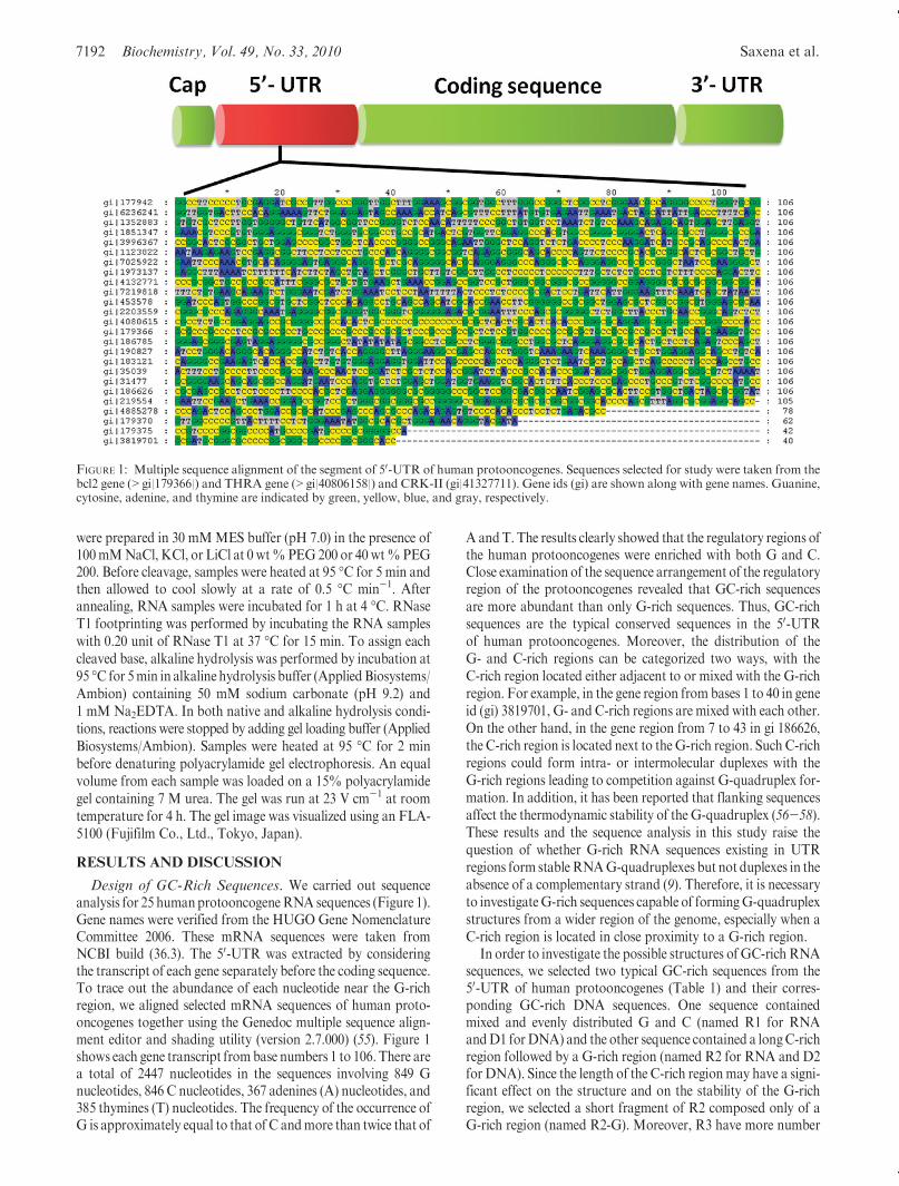
Design of GC-Rich Sequences. We carried out sequenceanalysis for 25 humanprotooncogeneRNA sequences (Figure 1).Gene names were verified from the HUGO Gene NomenclatureCommittee 2006. These mRNA sequences were taken fromNCBI build (36.3). The 50-UTR was extracted by consideringthe transcript of each gene separately before the coding sequence.To trace out the abundance of each nucleotide near the G-richregion, we aligned selected mRNA sequences of human proto-oncogenes together using the Genedoc multiple sequence align-ment editor and shading utility (version 2.7.000) (55). Figure 1shows each gene transcript frombase numbers 1 to 106. There area total of 2447 nucleotides in the sequences involving 849 Gnucleotides, 846 C nucleotides, 367 adenines (A) nucleotides, and385 thymines (T) nucleotides. The frequency of the occurrence ofG is approximately equal to that of C andmore than twice that of
A andT. The results clearly showed that the regulatory regions ofthe human protooncogenes were enriched with both G and C.Close examination of the sequence arrangement of the regulatoryregion of the protooncogenes revealed that GC-rich sequencesare more abundant than only G-rich sequences. Thus, GC-richsequences are the typical conserved sequences in the 50-UTRof human protooncogenes. Moreover, the distribution of theG- and C-rich regions can be categorized two ways, with theC-rich region located either adjacent to or mixed with the G-richregion. For example, in the gene region from bases 1 to 40 in geneid (gi) 3819701, G- and C-rich regions are mixed with each other.On the other hand, in the gene region from 7 to 43 in gi 186626,the C-rich region is located next to theG-rich region. Such C-richregions could form intra- or intermolecular duplexes with theG-rich regions leading to competition against G-quadruplex for-mation. In addition, it has been reported that flanking sequencesaffect the thermodynamic stability of the G-quadruplex (56-58).These results and the sequence analysis in this study raise thequestion of whether G-rich RNA sequences existing in UTRregions form stableRNAG-quadruplexes but not duplexes in theabsence of a complementary strand (9). Therefore, it is necessaryto investigateG-rich sequences capable of formingG-quadruplexstructures from a wider region of the genome, especially when aC-rich region is located in close proximity to a G-rich region.
In order to investigate the possible structures of GC-rich RNAsequences, we selected two typical GC-rich sequences from the50-UTR of human protooncogenes (Table 1) and their corres-ponding GC-rich DNA sequences. One sequence containedmixed and evenly distributed G and C (named R1 for RNAandD1 forDNA) and the other sequence contained a longC-richregion followed by a G-rich region (named R2 for RNA and D2for DNA). Since the length of the C-rich regionmay have a signi-ficant effect on the structure and on the stability of the G-richregion, we selected a short fragment of R2 composed only of aG-rich region (named R2-G). Moreover, R3 have more number
FIGURE 1: Multiple sequence alignment of the segment of 50-UTR of human protooncogenes. Sequences selected for study were taken from thebcl2 gene (>gi|179366|) and THRA gene (>gi|40806158|) and CRK-II (gi|41327711). Gene ids (gi) are shown along with gene names. Guanine,cytosine, adenine, and thymine are indicated by green, yellow, blue, and gray, respectively.
Article Biochemistry, Vol. 49, No. 33, 2010 7193
of Gs and less number of Cs as compared with R1 and R2forming a typical G-quadruplex forming sequence. Thus, R3 canbe useful to confirm whether the results with the other sequencesare general in the typical putativeG-quadruplex forming sequences.Structural Analysis of GC-RichRNAandDNASequences.
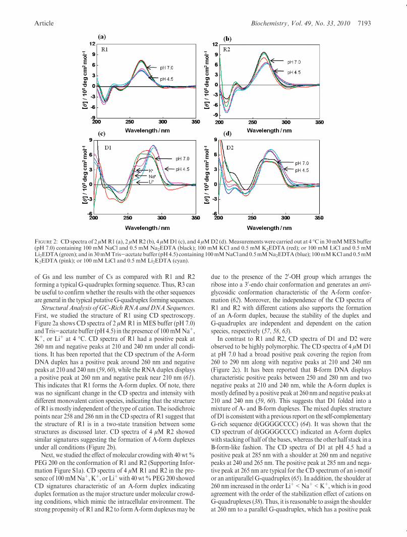
First, we studied the structure of R1 using CD spectroscopy.Figure 2a shows CD spectra of 2 μMR1 inMES buffer (pH 7.0)andTris-acetate buffer (pH 4.5) in the presence of 100mMNaþ,Kþ, or Liþ at 4 �C. CD spectra of R1 had a positive peak at260 nm and negative peaks at 210 and 240 nm under all condi-tions. It has been reported that the CD spectrum of the A-formDNA duplex has a positive peak around 260 nm and negativepeaks at 210 and 240 nm (59, 60), while the RNA duplex displaysa positive peak at 260 nm and negative peak near 210 nm (61).This indicates that R1 forms the A-form duplex. Of note, therewas no significant change in the CD spectra and intensity withdifferent monovalent cation species, indicating that the structureofR1 ismostly independent of the type of cation. The isodichroicpoints near 258 and 286 nm in the CD spectra of R1 suggest thatthe structure of R1 is in a two-state transition between somestructures as discussed later. CD spectra of 4 μM R2 showedsimilar signatures suggesting the formation of A-form duplexesunder all conditions (Figure 2b).
Next, we studied the effect of molecular crowding with 40 wt%PEG 200 on the conformation of R1 and R2 (Supporting Infor-mation Figure S1a). CD spectra of 4 μM R1 and R2 in the pre-sence of 100mMNaþ, Kþ, or Liþwith 40 wt%PEG200 showedCD signatures characteristic of an A-form duplex indicatingduplex formation as the major structure under molecular crowd-ing conditions, which mimic the intracellular environment. Thestrong propensity of R1 andR2 to formA-form duplexes may be
due to the presence of the 20-OH group which arranges theribose into a 30-endo chair conformation and generates an anti-glycosidic conformation characteristic of the A-form confor-mation (62). Moreover, the independence of the CD spectra ofR1 and R2 with different cations also supports the formationof an A-form duplex, because the stability of the duplex andG-quadruplex are independent and dependent on the cationspecies, respectively (57, 58, 63).
In contrast to R1 and R2, CD spectra of D1 and D2 wereobserved to be highly polymorphic. The CD spectra of 4 μMD1at pH 7.0 had a broad positive peak covering the region from260 to 290 nm along with negative peaks at 210 and 240 nm(Figure 2c). It has been reported that B-form DNA displayscharacteristic positive peaks between 250 and 280 nm and twonegative peaks at 210 and 240 nm, while the A-form duplex ismostly defined by a positive peak at 260 nm and negative peaks at210 and 240 nm (59, 60). This suggests that D1 folded into amixture of A- and B-form duplexes. The mixed duplex structureofD1 is consistentwith aprevious report on the self-complementaryG-rich sequence d(GGGGCCCC) (64). It was shown that theCD spectrum of d(GGGGCCCC) indicated an A-form duplexwith stacking of half of the bases, whereas the other half stack in aB-form-like fashion. The CD spectra of D1 at pH 4.5 had apositive peak at 285 nm with a shoulder at 260 nm and negativepeaks at 240 and 265 nm. The positive peak at 285 nm and nega-tive peak at 265 nm are typical for the CD spectrum of an i-motifor an antiparallel G-quadruplex (65). In addition, the shoulder at260 nm increased in the order Liþ<Naþ<Kþ, which is in goodagreement with the order of the stabilization effect of cations onG-quadruplexes (38). Thus, it is reasonable to assign the shoulderat 260 nm to a parallel G-quadruplex, which has a positive peak
FIGURE 2: CD spectra of 2 μMR1 (a), 2 μMR2 (b), 4 μMD1 (c), and 4 μMD2 (d).Measurements were carried out at 4 �C in 30mMMESbuffer(pH 7.0) containing 100 mM NaCl and 0.5 mM Na2EDTA (black); 100 mM KCl and 0.5 mM K2EDTA (red); or 100 mM LiCl and 0.5 mMLi2EDTA(green); and in 30mMTris-acetate buffer (pH4.5) containing 100mMNaCl and 0.5mMNa2EDTA(blue); 100mMKCl and 0.5mMK2EDTA (pink); or 100 mM LiCl and 0.5 mM Li2EDTA (cyan).
7194 Biochemistry, Vol. 49, No. 33, 2010 Saxena et al.
around 260 nm (66). Similar toD1, CD spectra ofD2 at pH7.0 alsoshowed broad positive peaks covering the region from 260 to286 nm along with negative peaks at 210 and 240 nm, indicating amixture ofA- andB-formduplexes under all conditions (Figure 2d).At pH 4.5, the presence of a positive peak at 276 nm indicatedpredominance of the B-form duplex. Therefore, it may be possiblethat the structures of D1 andD2 are in equilibrium between A- andB-form duplexes, i-motifs, and G-quadruplexes. The fact that therewas no isodichroic point in the CD spectra of D1 and D2 furthersupported the coexistence of several structures.
Next, we examined the effect of cosolutes on the conformationof D1 and D2 in the presence of 100 mM Naþ, Kþ, or Liþ with40 wt % PEG 200 (Supporting Information Figure S1b). CDspectra of D1 and D2 showed polymorphic structures characte-rized by positive peaks at 286 and 260 nm and a negative peak at240 nm under molecular crowding conditions. Similar to theirCD spectra under the dilute conditions, the CDspectra ofD1 andD2maybe due to antiparallel, parallelG-quadruplexes, and i-motifformations. This polymorphic character inD1 andD2 can be attri-buted to the deoxyribonucleotide backbone that can adopt bothanti and syn conformations dependingon the solution conditions,which is in contrast to the all anti conformation of RNA.Molecularity of R1 and R2. In order to confirm the struc-
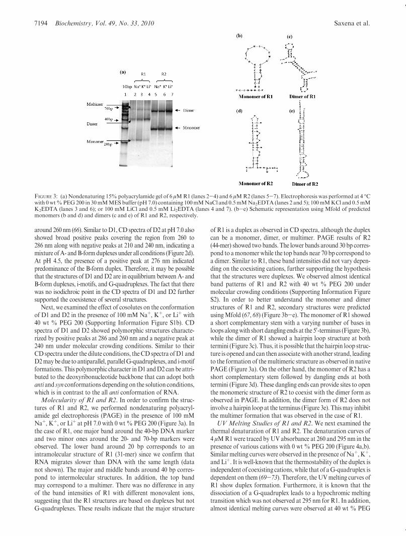
tures of R1 and R2, we performed nondenaturing polyacryl-amide gel electrophoresis (PAGE) in the presence of 100 mMNaþ, Kþ, or Liþ at pH 7.0 with 0 wt% PEG 200 (Figure 3a). Inthe case of R1, one major band around the 40-bp DNA markerand two minor ones around the 20- and 70-bp markers wereobserved. The lower band around 20 bp corresponds to anintramolecular structure of R1 (31-mer) since we confirm thatRNA migrates slower than DNA with the same length (datanot shown). The major and middle bands around 40 bp corres-pond to intermolecular structures. In addition, the top bandmay correspond to a multimer. There was no difference in anyof the band intensities of R1 with different monovalent ions,suggesting that the R1 structures are based on duplexes but notG-quadruplexes. These results indicate that the major structure
of R1 is a duplex as observed in CD spectra, although the duplexcan be a monomer, dimer, or multimer. PAGE results of R2(44-mer) showed two bands. The lower bands around 30 bp corres-pond to amonomerwhile the top bands near 70 bp correspond toa dimer. Similar to R1, these band intensities did not vary depen-ding on the coexisting cations, further supporting the hypothesisthat the structures were duplexes. We observed almost identicalband patterns of R1 and R2 with 40 wt % PEG 200 undermolecular crowding conditions (Supporting Information FigureS2). In order to better understand the monomer and dimerstructures of R1 and R2, secondary structures were predictedusingMfold (67, 68) (Figure 3b-e). Themonomer of R1 showeda short complementary stem with a varying number of bases inloops alongwith short dangling ends at the 50-terminus (Figure 3b),while the dimer of R1 showed a hairpin loop structure at bothtermini (Figure 3c). Thus, it is possible that the hairpin loop struc-ture is opened and can then associatewith another strand, leadingto the formation of the multimeric structure as observed in nativePAGE (Figure 3a). On the other hand, the monomer of R2 has ashort complementary stem followed by dangling ends at bothtermini (Figure 3d). These dangling ends can provide sites to openthe monomeric structure of R2 to coexist with the dimer form asobserved in PAGE. In addition, the dimer form of R2 does notinvolve a hairpin loop at the terminus (Figure 3e). Thismay inhibitthe multimer formation that was observed in the case of R1.UV Melting Studies of R1 and R2. We next examined the
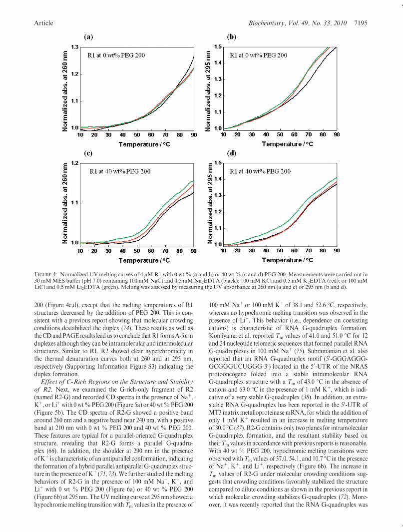
thermal denaturation of R1 and R2. The denaturation curves of4 μMR1were traced byUV absorbance at 260 and 295 nm in thepresence of various cations with 0 wt % PEG 200 (Figure 4a,b).Similarmelting curves were observed in the presence ofNaþ, Kþ,and Liþ. It is well-known that the thermostability of the duplex isindependent of coexisting cations,while that of aG-quadruplex isdependent on them (69-73). Therefore, theUVmelting curves ofR1 show duplex formation. Furthermore, it is known that thedissociation of a G-quadruplex leads to a hypochromic meltingtransition which was not observed at 295 nm for R1. In addition,almost identical melting curves were observed at 40 wt % PEG
FIGURE 3: (a) Nondenaturing 15%polyacrylamide gel of 6 μMR1 (lanes 2-4) and 6 μMR2 (lanes 5-7). Electrophoresis was performed at 4 �Cwith 0wt%PEG200 in 30mMMESbuffer (pH7.0) containing 100mMNaCl and 0.5mMNa2EDTA (lanes 2 and 5); 100mMKCl and 0.5mMK2EDTA (lanes 3 and 6); or 100 mM LiCl and 0.5 mM Li2EDTA (lanes 4 and 7). (b-e) Schematic representation using Mfold of predictedmonomers (b and d) and dimers (c and e) of R1 and R2, respectively.
Article Biochemistry, Vol. 49, No. 33, 2010 7195
200 (Figure 4c,d), except that the melting temperatures of R1structures decreased by the addition of PEG 200. This is con-sistent with a previous report showing that molecular crowdingconditions destabilized the duplex (74). These results as well astheCDandPAGEresults lead us to conclude thatR1 formsA-formduplexes although they can be intramolecular and intermolecularstructures. Similar to R1, R2 showed clear hyperchromicity inthe thermal denaturation curves both at 260 and at 295 nm,respectively (Supporting Information Figure S3) indicating theduplex formation.Effect of C-Rich Regions on the Structure and Stability
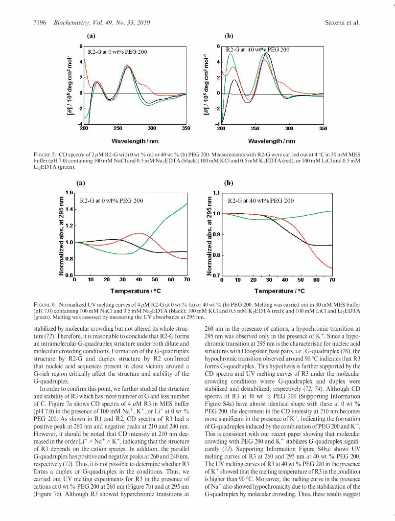
of R2. Next, we examined the G-rich-only fragment of R2(named R2-G) and recorded CD spectra in the presence of Naþ,Kþ, or Liþwith 0wt%PEG200 (Figure 5a) or 40wt%PEG200(Figure 5b). The CD spectra of R2-G showed a positive bandaround 260 nm and a negative band near 240 nm, with a positiveband at 210 nm with 0 wt % PEG 200 and 40 wt % PEG 200.These features are typical for a parallel-oriented G-quadruplexstructure, revealing that R2-G forms a parallel G-quadru-plex (66). In addition, the shoulder at 290 nm in the presenceofKþ is characteristic of an antiparallel conformation, indicatingthe formation of a hybrid parallel/antiparallel G-quadruplex struc-ture in the presence ofKþ (71, 73).We further studied themeltingbehaviors of R2-G in the presence of 100 mM Naþ, Kþ, andLiþ with 0 wt % PEG 200 (Figure 6a) or 40 wt % PEG 200(Figure 6b) at 295 nm. TheUVmelting curve at 295 nm showed ahypochromicmelting transition withTm values in the presence of
100 mM Naþ or 100 mM Kþ of 38.1 and 52.6 �C, respectively,whereas no hypochromic melting transition was observed in thepresence of Liþ. This behavior (i.e., dependence on coexistingcations) is characteristic of RNA G-quadruplex formation.Komiyama et al. reported Tm values of 41.0 and 51.0 �C for 12and 24 nucleotide telomeric sequences that formed parallel RNAG-quadruplexes in 100 mM Naþ (75). Subramanian et al. alsoreported that an RNA G-quadruplex motif (50-GGGAGGG-GCGGGUCUGGG-30) located in the 50-UTR of the NRASprotooncogene folded into a stable intramolecular RNAG-quadruplex structure with a Tm of 43.0 �C in the absence ofcations and 63.0 �C in the presence of 1 mM Kþ, which is indi-cative of a very stable G-quadruplex (38). In addition, an extra-stable RNA G-quadruplex has been reported in the 50-UTR ofMT3matrix metalloproteinase mRNA, for which the addition ofonly 1 mM Kþ resulted in an increase in melting temperatureof 30.0 �C (37). R2-G contains only two planes for intramolecularG-quadruplex formation, and the resultant stability based ontheirTm values in accordancewith previous reports is reasonable.With 40 wt % PEG 200, hypochromic melting transitions wereobservedwithTm values of 37.0, 54.1, and 10.7 �C in the presenceof Naþ, Kþ, and Liþ, respectively (Figure 6b). The increase inTm values of R2-G under molecular crowding conditions sug-gests that crowding conditions favorably stabilized the structurecompared to dilute conditions as shown in the previous report inwhich molecular crowding stabilizes G-quadruplex (72). More-over, it was recently reported that the RNA G-quadruplex was
FIGURE 4: Normalized UVmelting curves of 4 μMR1 with 0 wt % (a and b) or 40 wt % (c and d) PEG 200. Measurements were carried out in30 mMMES buffer (pH 7.0) containing 100 mMNaCl and 0.5 mMNa2EDTA (black); 100 mMKCl and 0.5 mMK2EDTA (red); or 100 mMLiCl and 0.5 mM Li2EDTA (green). Melting was assessed by measuring the UV absorbance at 260 nm (a and c) or 295 nm (b and d).
7196 Biochemistry, Vol. 49, No. 33, 2010 Saxena et al.
stabilized by molecular crowding but not altered its whole struc-ture (72). Therefore, it is reasonable to conclude that R2-G formsan intramolecular G-quadruplex structure under both dilute andmolecular crowding conditions. Formation of the G-quadruplexstructure by R2-G and duplex structure by R2 confirmedthat nucleic acid sequences present in close vicinity around aG-rich region critically affect the structure and stability of theG-quadruplex.
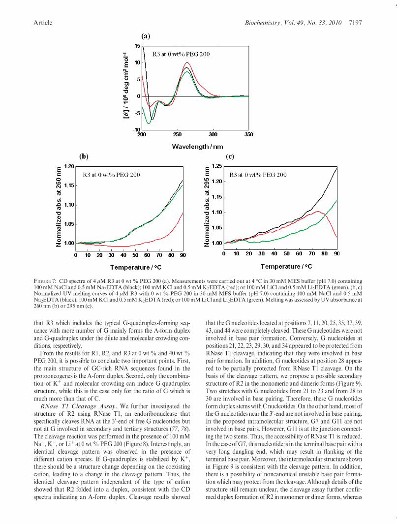
In order to confirm this point, we further studied the structureand stability of R3which has more number of G and less numberof C. Figure 7a shows CD spectra of 4 μM R3 in MES buffer(pH 7.0) in the presence of 100 mM Naþ, Kþ, or Liþ at 0 wt %PEG 200. As shown in R1 and R2, CD spectra of R3 had apositive peak at 260 nm and negative peaks at 210 and 240 nm.However, it should be noted that CD intensity at 210 nm dec-reased in the order Liþ>Naþ>Kþ, indicating that the structureof R3 depends on the cation species. In addition, the parallelG-quadruplex has positive and negative peaks at 260 and 240 nm,respectively (72). Thus, it is not possible to determine whether R3forms a duplex or G-quadruplex in the conditions. Thus, wecarried out UV melting experiments for R3 in the presence ofcations at 0 wt % PEG 200 at 260 nm (Figure 7b) and at 295 nm(Figure 7c). Although R3 showed hyperchromic transitions at
260 nm in the presence of cations, a hypochromic transition at295 nm was observed only in the presence of Kþ. Since a hypo-chromic transition at 295 nm is the characteristic for nucleic acidstructures withHoogsteen base pairs, i.e., G-quadruplex (76), thehypochromic transition observed around 90 �C indicates that R3forms G-quadruplex. This hypothesis is further supported by theCD spectra and UV melting curves of R3 under the molecularcrowding conditions where G-quadruplex and duplex werestabilized and destabilized, respectively (72, 74). Although CDspectra of R3 at 40 wt % PEG 200 (Supporting InformationFigure S4a) have almost identical shape with these at 0 wt %PEG 200, the decrement in the CD intensity at 210 nm becomesmore significant in the presence of Kþ, indicating the formationofG-quadruplex induced by the combination of PEG200 andKþ.This is consistent with our recent paper showing that molecularcrowding with PEG 200 and Kþ stabilizes G-quadruplex signifi-cantly (72). Supporting Information Figure S4b,c shows UVmelting curves of R3 at 260 and 295 nm at 40 wt % PEG 200.The UVmelting curves of R3 at 40 wt% PEG 200 in the presenceof Kþ showed that the melting temperature of R3 in the conditionis higher than 90 �C. Moreover, the melting curve in the presenceof Naþ also showed hypochromicity due to the stabilization of theG-quadruplex by molecular crowding. Thus, these results suggest
FIGURE 5: CD spectra of 2 μMR2-Gwith 0 wt% (a) or 40wt% (b) PEG 200.Measurements withR2-Gwere carried out at 4 �C in 30mMMESbuffer (pH7.0) containing 100mMNaCl and 0.5mMNa2EDTA(black); 100mMKCl and 0.5mMK2EDTA(red); or 100mMLiCl and 0.5mMLi2EDTA (green).
FIGURE 6: Normalized UVmelting curves of 4 μMR2-G at 0 wt% (a) or 40 wt% (b) PEG 200.Melting was carried out in 30 mMMES buffer(pH 7.0) containing 100 mMNaCl and 0.5 mMNa2EDTA (black); 100 mMKCl and 0.5 mMK2EDTA (red); and 100 mMLiCl and Li2EDTA(green). Melting was assessed by measuring the UV absorbance at 295 nm.
Article Biochemistry, Vol. 49, No. 33, 2010 7197
that R3 which includes the typical G-quadruplex-forming seq-uence with more number of G mainly forms the A-form duplexand G-quadruplex under the dilute and molecular crowding con-ditions, respectively.
From the results for R1, R2, and R3 at 0 wt % and 40 wt %PEG 200, it is possible to conclude two important points. First,the main structure of GC-rich RNA sequences found in theprotooncogenes is the A-form duplex. Second, only the combina-tion of Kþ and molecular crowding can induce G-quadruplexstructure, while this is the case only for the ratio of G which ismuch more than that of C.RNase T1 Cleavage Assay. We further investigated the
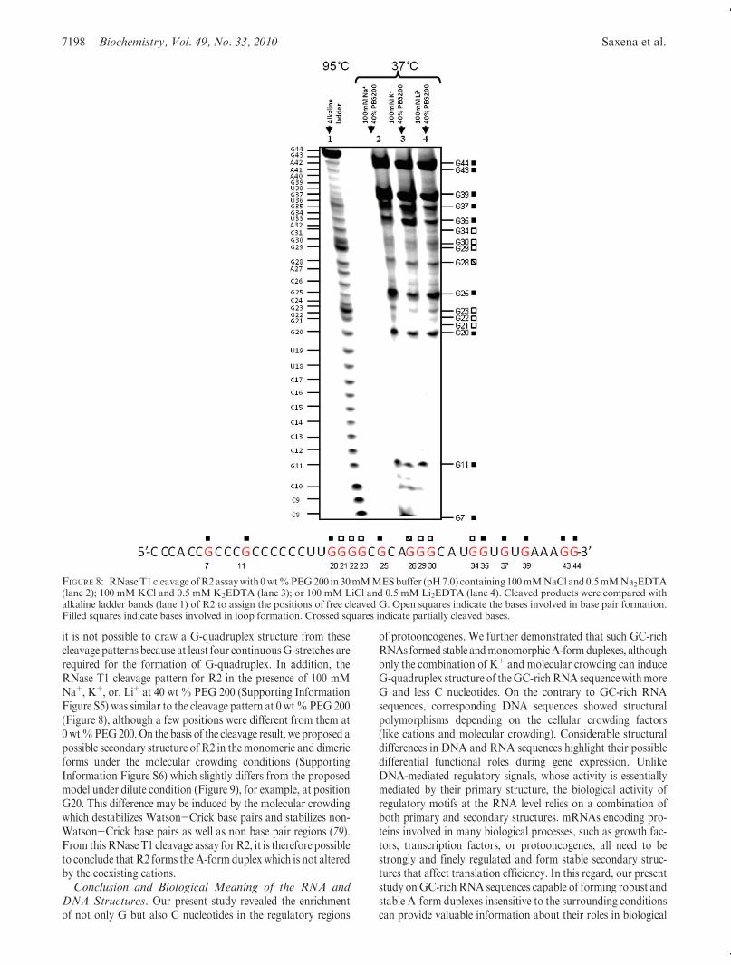
structure of R2 using RNase T1, an endoribonuclease thatspecifically cleaves RNA at the 30-end of free G nucleotides butnot at G involved in secondary and tertiary structures (77, 78).The cleavage reaction was performed in the presence of 100 mMNaþ, Kþ, or Liþ at 0 wt% PEG 200 (Figure 8). Interestingly, anidentical cleavage pattern was observed in the presence ofdifferent cation species. If G-quadruplex is stabilized by Kþ,there should be a structure change depending on the coexistingcation, leading to a change in the cleavage pattern. Thus, theidentical cleavage pattern independent of the type of cationshowed that R2 folded into a duplex, consistent with the CDspectra indicating an A-form duplex. Cleavage results showed
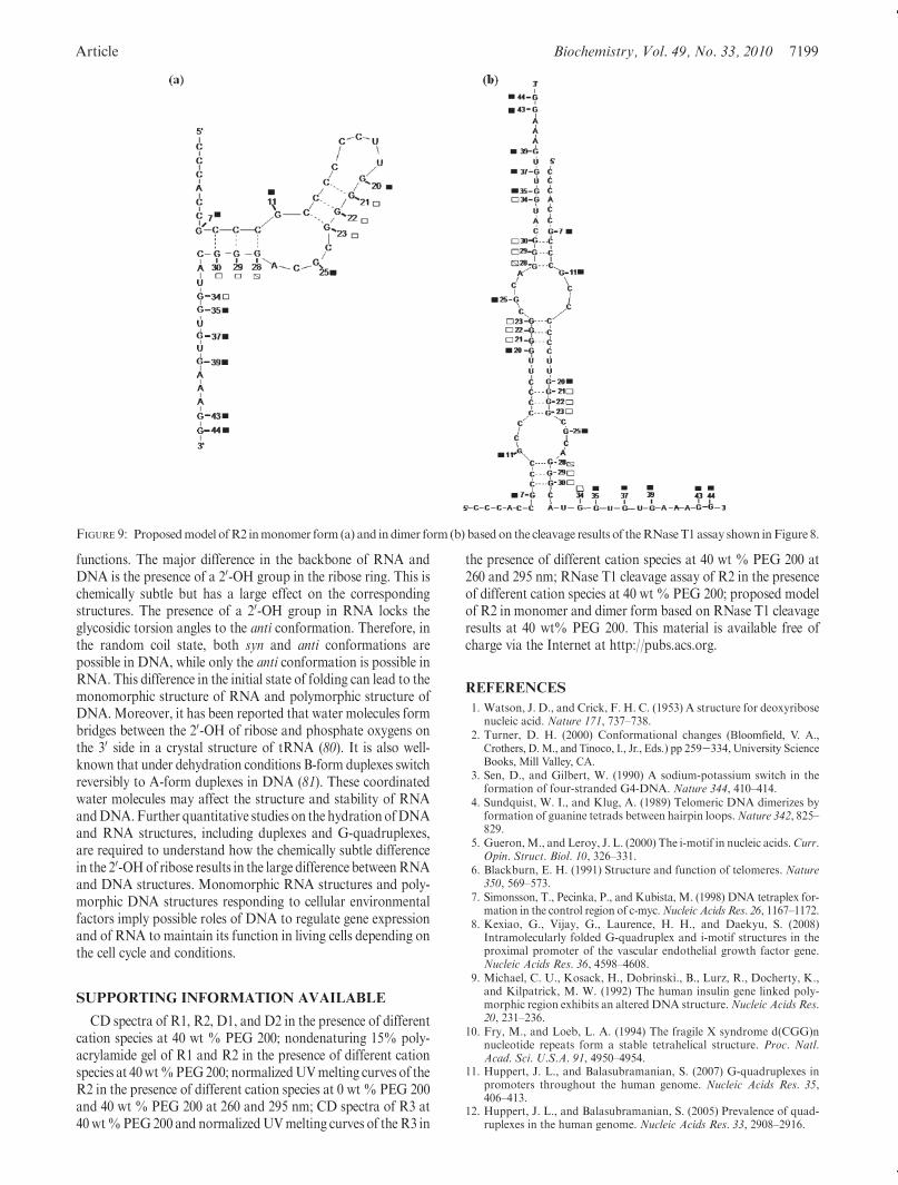
that theG nucleotides located at positions 7, 11, 20, 25, 35, 37, 39,43, and 44were completely cleaved. TheseGnucleotides were notinvolved in base pair formation. Conversely, G nucleotides atpositions 21, 22, 23, 29, 30, and 34 appeared to be protected fromRNase T1 cleavage, indicating that they were involved in basepair formation. In addition, G nucleotides at position 28 appea-red to be partially protected from RNase T1 cleavage. On thebasis of the cleavage pattern, we propose a possible secondarystructure of R2 in the monomeric and dimeric forms (Figure 9).Two stretches with G nucleotides from 21 to 23 and from 28 to30 are involved in base pairing. Therefore, these G nucleotidesformduplex stemswithCnucleotides.On the other hand,most oftheG nucleotides near the 30-end are not involved in base pairing.In the proposed intramolecular structure, G7 and G11 are notinvolved in base pairs. However, G11 is at the junction connect-ing the two stems. Thus, the accessibility of RNase T1 is reduced.In the case ofG7, this nucleotide is in the terminal base pairwith avery long dangling end, which may result in flanking of theterminal base pair.Moreover, the intermolecular structure shownin Figure 9 is consistent with the cleavage pattern. In addition,there is a possibility of noncanonical unstable base pair forma-tionwhichmayprotect from the cleavage. Although details of thestructure still remain unclear, the cleavage assay further confir-med duplex formation ofR2 inmonomer or dimer forms, whereas
FIGURE 7: CD spectra of 4 μMR3 at 0 wt % PEG 200 (a). Measurements were carried out at 4 �C in 30 mMMES buffer (pH 7.0) containing100 mMNaCl and 0.5 mMNa2EDTA (black); 100 mMKCl and 0.5 mMK2EDTA (red); or 100mMLiCl and 0.5 mMLi2EDTA (green). (b, c)Normalized UV melting curves of 4 μM R3 with 0 wt % PEG 200 in 30 mM MES buffer (pH 7.0) containing 100 mM NaCl and 0.5 mMNa2EDTA (black); 100mMKCl and 0.5mMK2EDTA (red); or 100mMLiCl andLi2EDTA (green).Meltingwas assessed byUVabsorbance at260 nm (b) or 295 nm (c).
7198 Biochemistry, Vol. 49, No. 33, 2010 Saxena et al.
it is not possible to draw a G-quadruplex structure from thesecleavage patterns because at least four continuousG-stretches arerequired for the formation of G-quadruplex. In addition, theRNase T1 cleavage pattern for R2 in the presence of 100 mMNaþ, Kþ, or, Liþ at 40 wt % PEG 200 (Supporting InformationFigure S5) was similar to the cleavage pattern at 0 wt%PEG 200(Figure 8), although a few positions were different from them at0wt%PEG200.On the basis of the cleavage result, we proposed apossible secondary structure of R2 in themonomeric and dimericforms under the molecular crowding conditions (SupportingInformation Figure S6) which slightly differs from the proposedmodel under dilute condition (Figure 9), for example, at positionG20. This difference may be induced by the molecular crowdingwhich destabilizes Watson-Crick base pairs and stabilizes non-Watson-Crick base pairs as well as non base pair regions (79).From thisRNase T1 cleavage assay forR2, it is therefore possibleto conclude that R2 forms the A-form duplex which is not alteredby the coexisting cations.Conclusion and Biological Meaning of the RNA and
DNA Structures. Our present study revealed the enrichmentof not only G but also C nucleotides in the regulatory regions
of protooncogenes. We further demonstrated that such GC-richRNAs formed stable andmonomorphicA-formduplexes, althoughonly the combination of Kþ and molecular crowding can induceG-quadruplex structure of theGC-richRNA sequencewithmoreG and less C nucleotides. On the contrary to GC-rich RNAsequences, corresponding DNA sequences showed structuralpolymorphisms depending on the cellular crowding factors(like cations and molecular crowding). Considerable structuraldifferences in DNA and RNA sequences highlight their possibledifferential functional roles during gene expression. UnlikeDNA-mediated regulatory signals, whose activity is essentiallymediated by their primary structure, the biological activity ofregulatory motifs at the RNA level relies on a combination ofboth primary and secondary structures. mRNAs encoding pro-teins involved in many biological processes, such as growth fac-tors, transcription factors, or protooncogenes, all need to bestrongly and finely regulated and form stable secondary struc-tures that affect translation efficiency. In this regard, our presentstudy onGC-richRNA sequences capable of forming robust andstable A-form duplexes insensitive to the surrounding conditionscan provide valuable information about their roles in biological
FIGURE 8: RNaseT1 cleavage ofR2assaywith 0wt%PEG200 in 30mMMESbuffer (pH7.0) containing100mMNaCl and0.5mMNa2EDTA(lane 2); 100 mMKCl and 0.5 mMK2EDTA (lane 3); or 100 mM LiCl and 0.5 mM Li2EDTA (lane 4). Cleaved products were compared withalkaline ladder bands (lane 1) of R2 to assign the positions of free cleaved G. Open squares indicate the bases involved in base pair formation.Filled squares indicate bases involved in loop formation. Crossed squares indicate partially cleaved bases.
Article Biochemistry, Vol. 49, No. 33, 2010 7199
functions. The major difference in the backbone of RNA andDNA is the presence of a 20-OH group in the ribose ring. This ischemically subtle but has a large effect on the correspondingstructures. The presence of a 20-OH group in RNA locks theglycosidic torsion angles to the anti conformation. Therefore, inthe random coil state, both syn and anti conformations arepossible in DNA, while only the anti conformation is possible inRNA. This difference in the initial state of folding can lead to themonomorphic structure of RNA and polymorphic structure ofDNA.Moreover, it has been reported that water molecules formbridges between the 20-OH of ribose and phosphate oxygens onthe 30 side in a crystal structure of tRNA (80). It is also well-known that under dehydration conditions B-form duplexes switchreversibly to A-form duplexes in DNA (81). These coordinatedwater molecules may affect the structure and stability of RNAandDNA.Further quantitative studies on the hydration ofDNAand RNA structures, including duplexes and G-quadruplexes,are required to understand how the chemically subtle differencein the 20-OHof ribose results in the large difference betweenRNAand DNA structures. Monomorphic RNA structures and poly-morphic DNA structures responding to cellular environmentalfactors imply possible roles of DNA to regulate gene expressionand of RNA to maintain its function in living cells depending onthe cell cycle and conditions.
SUPPORTING INFORMATION AVAILABLE
CD spectra of R1, R2, D1, and D2 in the presence of differentcation species at 40 wt % PEG 200; nondenaturing 15% poly-acrylamide gel of R1 and R2 in the presence of different cationspecies at 40wt%PEG200; normalizedUVmelting curves of theR2 in the presence of different cation species at 0 wt % PEG 200and 40 wt % PEG 200 at 260 and 295 nm; CD spectra of R3 at40wt%PEG200 and normalizedUVmelting curves of theR3 in
the presence of different cation species at 40 wt % PEG 200 at260 and 295 nm; RNase T1 cleavage assay of R2 in the presenceof different cation species at 40 wt % PEG 200; proposed modelof R2 in monomer and dimer form based on RNase T1 cleavageresults at 40 wt% PEG 200. This material is available free ofcharge via the Internet at http://pubs.acs.org.
REFERENCES
1. Watson, J. D., and Crick, F. H. C. (1953) A structure for deoxyribosenucleic acid. Nature 171, 737–738.
2. Turner, D. H. (2000) Conformational changes (Bloomfield, V. A.,Crothers, D. M., and Tinoco, I., Jr., Eds.) pp 259-334, University ScienceBooks, Mill Valley, CA.
3. Sen, D., and Gilbert, W. (1990) A sodium-potassium switch in theformation of four-stranded G4-DNA. Nature 344, 410–414.
4. Sundquist, W. I., and Klug, A. (1989) Telomeric DNA dimerizes byformation of guanine tetrads between hairpin loops.Nature 342, 825–829.
5. Gueron,M., and Leroy, J. L. (2000) The i-motif in nucleic acids.Curr.Opin. Struct. Biol. 10, 326–331.
6. Blackburn, E. H. (1991) Structure and function of telomeres. Nature350, 569–573.
7. Simonsson, T., Pecinka, P., and Kubista, M. (1998) DNA tetraplex for-mation in the control region of c-myc.Nucleic Acids Res. 26, 1167–1172.
8. Kexiao, G., Vijay, G., Laurence, H. H., and Daekyu, S. (2008)Intramolecularly folded G-quadruplex and i-motif structures in theproximal promoter of the vascular endothelial growth factor gene.Nucleic Acids Res. 36, 4598–4608.
9. Michael, C. U., Kosack, H., Dobrinski., B., Lurz, R., Docherty, K.,and Kilpatrick, M. W. (1992) The human insulin gene linked poly-morphic region exhibits an altered DNA structure.Nucleic Acids Res.20, 231–236.
10. Fry, M., and Loeb, L. A. (1994) The fragile X syndrome d(CGG)nnucleotide repeats form a stable tetrahelical structure. Proc. Natl.Acad. Sci. U.S.A. 91, 4950–4954.
11. Huppert, J. L., and Balasubramanian, S. (2007) G-quadruplexes inpromoters throughout the human genome. Nucleic Acids Res. 35,406–413.
12. Huppert, J. L., and Balasubramanian, S. (2005) Prevalence of quad-ruplexes in the human genome. Nucleic Acids Res. 33, 2908–2916.
FIGURE 9: Proposedmodel ofR2 inmonomer form (a) and indimer form (b) based on the cleavage results of theRNaseT1 assay shown inFigure 8.
7200 Biochemistry, Vol. 49, No. 33, 2010 Saxena et al.
13. Zhang, R., Lin, Y., and Zhang, C. T. (2008) Greglist: a databaselisting potential G-quadruplex regulated genes.Nucleic Acids Res. 36,D372–eD376.
14. Todd, A. K., Johnston, M., and Neidle, S. (2005) Highly prevalentputative quadruplex sequence motifs in human DNA. Nucleic AcidsRes. 33, 2901–2907.
15. Hershman, S. G., Chen, Q., Lee, J. Y., Kozak, M. L., Yue, P., Wang,L.-S., and Johnson, F. B. (2008) Genomic distribution and functionalanalyses of potential G-quadruplex-forming sequences in Saccharomycescerevisiae. Nucleic Acids Res. 36, 144–156.
16. Kostadinov, R., Malhotra, N., Viotti, M., Shine, R., D’Antonio, L.,and Bagga, P. S. (2006) GRSDB: a database of quadruplex formingG-rich sequences in alternatively processed mammalian pre-mRNAsequences. Nucleic Acids Res. 34, D119–D124.
17. D’Antonio, L., Bagga, P. S. (2004) IEEE Computational SystemsBioinformatics Conference, Computational methods for predictingintramolecular G-quadruplexes in nucleotide sequences, pp 561-562.
18. Kikin, O., D’Antonio, L., and Bagga, P. S. (2006) A web-based serverfor predicting G-quadruplexes in nucleotide sequences. Nucleic AcidsRes. 34, W676–W682.
19. Kikin, O., Zappala, Z., D’Antonio, L., and Bagga, P. S. (2008)GRSDB2 and GRS_UTRdb: databases of quadruplex formingG-rich sequences in pre-mRNAs and mRNAs. Nucleic Acids Res.36, D141–D148.
20. Eddy, J., and Maizels, N. (2006) Gene function correlates withpotential for G4DNA formation in the human genome.Nucleic AcidsRes. 34, 3887–3896.
21. Phillips, K., Dauter, Z., Murchie, A. I., Lilley, D. M. J., and Luisi, B.(1997) The crystal structure of a parallel-stranded guanine tetraplex at0.95 angstrom resolution. J. Mol. Biol. 273, 171–182.
22. Parkinson, G. N., Lee, M. P. H., and Neidle, S. (2002) Crystalstructure of parallel quadruplexes from human telomeric DNA.Nature 417, 876–880.
23. Haider, S., Parkinson, G. N., and Neidle, S. (2002) Crystal structureof the potassium form of an Oxytricha nova G-quadruplex. J. Mol.Biol. 320, 189–200.
24. Keniry, M. A., Strahan, G. D., Owen, E. A., and Shafer, R. H. (1995)Solution structure of the Naþ form of the dimeric quadruplex. Eur. J.Biochem. 233, 631–643.
25. Macaya, R. F., Schultze, P., Smith, F. W., Roe, J. A., and Feigon, J.(1993) Thrombin-binding DNA aptamer forms a unimolecular quad-ruplex structure in solution. Proc. Natl. Acad. Sci. U.S.A. 90, 3745–3749.
26. Wang, Y., and Patel, D. J. (1993) Solution structure of the humantelomeric repeat d[AG3(T2AG3)3] G-tetraplex. Structure 1, 76–94.
27. Wang, Y., and Patel, D. J. (1995) Solution structure of the Oxytrichatelomeric repeat d G-tetraplex. J. Mol. Biol. 251, 263–282.
28. Phan, A. T., and Mergny, J. L. (2002) Human telomeric DNA:G-quadruplex, i-motif and Watson-Crick double helix. Nucleic AcidsRes. 30, 4618–4625.
29. Li, W., Miyoshi, D., Nakano, S., and Sugimoto, N. (2003) Structuralcompetition involving G-quadruplex DNA and its complement.Biochemistry 42, 11736–11744.
30. Miyoshi, D., Nakano, A., Toda, T., and Sugimoto, N. (2001) Effect ofdivalent cations on antiparallel G-quartet structure of d(G4T4G4).FEBS Lett. 496, 128–133.
31. Miyoshi, D., Matsumura, S., Li, W., and Sugimoto, N. (2003)Structural polymorphism of telomeric DNA regulated by pH anddivalent cation. Nucleosides, Nucleotides, Nucleic Acids 22, 203–221.
32. Risitano, A., and Fox, K. R. (2003) Stability of intramolecular DNAquadruplexes: comparison with DNA duplexes. Biochemistry 42,6507–6513.
33. Miyoshi, D., Nakano, A., and Sugimoto, N. (2002) Molecularcrowding regulates the structural switch of the DNA G-quadruplex.Biochemistry 41, 15017–15024.
34. Christiansen, J., Kofod, M., and Nielsen, F. C. (1994) A guanosinequadruplex and two stable hairpins flank a major cleavage site ininsulin-like growth factor II mRNA. Nucleic Acids Res. 22, 5709–5716.
35. Darnell, J. C., Jensen, K. B., Jin, P., Brown, V., Warren, S. T., andDarnell, R. B. (2001) Fragile X mental retardation protein targets Gquartet mRNAs important for neuronal function. Cell 107, 489–499.
36. Bonnal, S., Schaeffer, C., Creancier, L., Clamens, S., Moine, H.,Prats, A. C., and Vagner, S. (2003) A single internal ribosome entrysite containing a G quartet RNA structure drives fibroblast growthfactor 2 gene expression at four alternative translation initiationcodons. J. Biol. Chem. 278, 39330–39336.
37. Morris, M. J., and Basu, S. (2009) An unusually stable G-quadruplexwithin the 50-UTR of the MT3 matrix metalloproteinase
mRNA represses translation in eukaryotic cells. Biochemistry 48,5313–5319.
38. Kumari, S., Bugaut, A., Huppert, J. L., and Balasubramanian, S.(2007) An RNA G-quadruplex in the 50 UTR of the NRAS proto-oncogene modulates translation. Nat. Chem. Biol. 4, 218–221.
39. Arora, A., Dutkiewicz, M., Scaria, V., Hariharan, M., Maiti, S., andKurreck, J. (2008) Inhibition of translation in living eukaryotic cellsby an RNA G-quadruplex motif. RNA 14, 1290–1296.
40. Wieland, M., and Hartig, J. S. (2007) RNA quadruplex-based modu-lation of gene expression. Chem. Biol. 14, 757–763.
41. Khateb, S.,Weisman, S. P., Hershco, S. I., Ludwig, A. L., andFry,M.(2007) The tetraplex (CGG)n destabilizing proteins hnRNP A2 andCBF-A enhance the in vivo translation of fragile X premutationmRNA. Nucleic Acids Res. 35, 5775–5788.
42. Awang, G., and Sen, D. (1993) Mode of dimerization of HIV-1genomic RNA. Biochemistry 32, 11453–11457.
43. Bonnal, S., Schaeffer, C., Cr�eancier, L., Clamens, S., Moine, H.,Prats, A. C., and Vagner, S. (2003) A single internal ribosome entrysite containing a G quartet RNA structure drives fibroblast growthfactor 2 gene expression at four alternative translation initiationcodons. J. Biol. Chem. 278, 39330–39336.
44. Gomez, D., Lemarteleur, T., Lacroix, L., Mailliet, P., Mergny, J. L.,and Riou, J. F. (2004) Telomerase downregulation induced by theG-quadruplex ligand 12459 inA549 cells is mediated by hTERTRNAalternative splicing. Nucleic Acids Res. 32, 371–379.
45. Zanotti, K. J., Lackey, P. E., Evans, G. L., and Mihailescu, M. R.(2006) Thermodynamics of the fragile X mental retardation proteinRGGbox interactionswithGquartet formingRNA.Biochemistry 45,8319–8330.
46. Kumari, S., Bugaut, A., and Balasubramanian, S. (2008) Position andstability are determining factors for translation repression by anRNAG-quadruplex-forming sequence within the 50 UTR of the NRASproto-oncogene. Biochemistry 47, 12664–12669.
47. Jianying, Q. I., and Shafer, R. H. (2007) Human telomere quadruplex:refolding and selection of individual conformers via RNA/DNAchimeric editing. Biochemistry 46, 7599–7606.
48. Joachimi, A., Benz,A., andHartig, J. S. (2009)A comparison ofDNAand RNA quadruplex structures and stabilities. Bioorg. Med. Chem.17, 6811–6815.
49. Olsen, C. M., and Marky, L. A. (2009) Energetic and hydrationcontributions of the removal of methyl groups from thymine to formuracil in G-quadruplexes. J. Phys. Chem. B. 113, 9–11.
50. Olsen, C. M., Lee, H. T., and Marky, L. A. (2009) Unfoldingthermodynamics of intramolecular G-quadruplexes: base sequencecontributions of the loops. J. Phys. Chem. B 113, 2587–2595.
51. Arora, A., andMaiti, S. (2009) Stability and molecular recognition ofquadruplexes with different loop length in absence and presence ofmolecular crowding agent. J. Phys. Chem. B 113, 10515–20.
52. Kumar, N., and Maiti, S. (2008) A thermodynamic overview ofnaturally occurring intramolecularDNAquadruplexes.Nucleic AcidsRes. 36, 5610–5622.
53. Sugimoto,N., Nakano, S., Kotah, S.,Matsumura, A., Nakamuta,H.,Ohmichi, T., Yoneyama, M., and Sasaki, M. (1995) Thermodynamicparameters to predict stability of RNA/DNA hybrid duplexes. Bio-chemistry 34, 11211–11216.
54. Nakano, S., Kanzaki, T., and Sugimoto, N. (2004) Influences ofribonucleotide on a duplex conformation and its thermal stability:study with the chimeric RNA-DNA strands. J. Am. Chem. Soc. 126,1088–1095.
55. Nicholas, K. B., Nicholas, H. B., Jr., and Deerfield, D. W., II (1997)GeneDoc: analysis and visualization of genetic variation. EMBNEW.NEWS 4, 14.
56. Guo, Q., Lu,M., andKallenbach,N.R. (1993) Effect of thymine tractlength on the structure and stability of model telomeric sequences.Biochemistry 32, 3596–3603.
57. Lu,M., Guo, Q., andKallenbach, N. R. (1992) Structure and stabilityof sodium and potassium complexes of dT4G4 and dT4G4T. Bio-chemistry 31, 2455–2459.
58. Merkina, E. E., and Fox, K. R. (2005) Kinetic stability of intermo-lecular DNA quadruplexes. Biophys. J. 89, 365–373.
59. Johnson, W. C. (1994) Circular Dichroism: Principles and Applica-tions, pp 523-540, VCH, New York, NY.
60. Fedor, S., Monique, M., Aurelie, M., Claude, M., and Serge, F.(1995) The high stability of the triple helices formed betweenshort purine oligonucleotides and SIV/HIV-2 vpx genes is deter-mined by the targeted DNA structure. Nucleic Acids Res. 23, 3831–3836.
61. Hung, S. H., Yu, Q., Gray, D. M., and Ratliff, R. L. (1994) EvidencefromCD spectra that d(purine)-r(pyrimidine) and r(purine)-d(pyrimidine)
Article Biochemistry, Vol. 49, No. 33, 2010 7201
hybrids are in different structural classes. Nucleic Acids Res. 22, 4326–4334.
62. Markus, C. W., and Sundaralingam, M. (2000) B-form to A-formconversion by a 30-terminal ribose: crystal structure of the chimerad(CCACTAGTG)r(G). Nucleic Acids Res. 28, 4356–4363.
63. Yu, H. Q., Miyoshi, D., and Sugimoto, N. (2006) Characterization ofstructure and stability of long telomeric DNAG-quadruplexes. J. Am.Chem. Soc. 128, 15461–15468.
64. Stefl, R., Trantirek, L., Vorlickova, M., Koca, J., Skelenar, V., andKypr, J. (2001) A-like guanine-guanine stacking in the aqeuous DNAduplex of d(GGGGCCCC). J. Mol. Biol. 307, 513–524.
65. Pataskar, S. S., Dash, D., and Brahmachari, S. K. (2001) Intramole-cular i-motif structure at acidic pH for progressivemyoclonus epilepsy(EPM1) repeat d(CCCCGCCCCGCG)n. J. Biomol. Struct. Dyn. 2,307–313.
66. Balagurumoorthy, P., Brahmachari, S. K., Mohanty, D., Bansal, M.,and Sasisekharan, V. (1992) Hairpin and parallel quartet structuresfor telomeric sequences. Nucleic Acids Res. 20, 4061–4067.
67. Zuker, M. (2003) Mfold web server for nucleic acid folding andhybridization prediction. Nucleic Acids Res. 31, 3406–3415.
68. Mathews, D. H., Disney,M. D., Childs, J. L., Schroeder, S. J., Zuker,M., and Turner, D. H. (2004) Incorporating chemical modificationconstraints into a dynamic programming algorithm for prediction ofRNA secondary structure. Proc. Natl. Acad. Sci. U.S.A. 101, 7287–7292.
69. Manning., G. S. (1972) On the application of polyelectrolyte “limitinglaws” to the helix-coil transition of DNA. I. Excess univalent cations.Biopolymers 11, 937–949.
70. Record, M. T., Jr., Anderson, C. F., and Lohman, T. M. (1978)Thermodynamic analysis of ion effects on the binding and conforma-tional equilibria of proteins and nucleic acids: the roles of ion asso-ciation or release, screening, and ion effects on water activity.Q. Rev.Biophys. 11, 103–178.
71. Miyoshi, D., Karimata, H., and Sugimoto, N. (2006) Characteriza-tion of structure and stability of long telomericDNAG-quadruplexes.J. Am. Chem. Soc. 128, 7957–7963.
72. Zhang, D. H., Fujimoto, T., Saxena, S., Yu, H. Q., Miyoshi, D., andSugimoto, N. (2010) Monomorphic RNA G-quadruplex and poly-morphic DNA G-quadruplex structures responding to cellular environ-mental factors. Biochemistry 49, 4554–63.
73. Ambrus, A., Chen, D., Dai, J., Bialis, T., Jones, R. A., and Yang, D.(2006)Human telomeric sequence forms a hybrid-type intramolecularG-quadruplex structure with mixed parallel/antiparallel strands inpotassium solution. Nucleic Acids Res. 34, 2723–2735.
74. Miyoshi, D., Matsumura, S., Nakano, S., and Sugimoto, N. (2004)Duplex dissociation of telomere DNAs induced by molecular crowding.J. Am. Chem. Soc. 126, 165–169.
75. Xu, Y., Kaminaga, K., and Komiyama, M. (2008) G-Quadruplexformation by human telomeric repeats-containing RNA in Naþ solu-tion. J. Am. Chem. Soc. 130, 11179–11184.
76. Mergny, J. L., Phan, A. T., and Lacroix, L. (1998) FollowingG-quartet formation by UV-spectroscopy. FEBS Lett. 435, 74–78.
77. Darnell, J. C., Jensen, K. B., Jin, P., Brown, V., Warren, S. T., andDarnell, R. B. (2001) Fragile X mental retardation protein targetsG quartet mRNAs important for neuronal function.Cell 107, 489–499.
78. Christiansen, J., Kofod, M., and Nielsen, F. C. (1994) A guanosinequadruplex and two stable hairpins flank amajor cleavage site in insulin-like growth factor II mRNA. Nucleic Acids Res. 22, 5709–5716.
79. Miyoshi, D.,Karimata,H., and Sugimoto, N. (2007)Hydration regulatesthe thermodynamic stability of DNA structures under molecular crowd-ing conditions. Nucleosides, Nucleotides, Nucleic Acids 26, 589–595.
80. Jovine, L., Djordjevic, S., and Rhodes., D. (2000) The crystalstructure of yeast phenylalanine tRNA at 2.0 A resolution: cleavageby Mg(2þ) in 15-year old crystals. J. Mol. Biol. 301, 401–414.
81. Paster, N. (2005) The B- to A-DNA transition and the reorganizationof solvent at the DNA surface. Biophys. J. 88, 3262–3275.