Reproductive risks for carriers of complex chromosome rearrangements: Analysis of 25 families
Upload
independentCategory
view
0download
0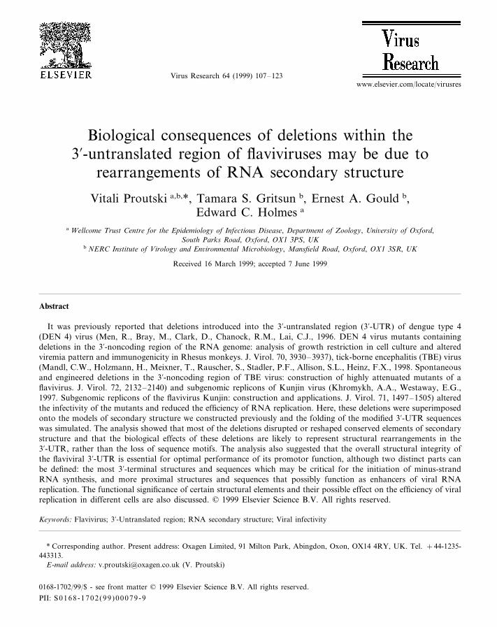
Virus Research 64 (1999) 107–123
Biological consequences of deletions within the3%-untranslated region of flaviviruses may be due to
rearrangements of RNA secondary structure
Vitali Proutski a,b,*, Tamara S. Gritsun b, Ernest A. Gould b,Edward C. Holmes a
a Wellcome Trust Centre for the Epidemiology of Infectious Disease, Department of Zoology, Uni6ersity of Oxford,South Parks Road, Oxford, OX1 3PS, UK
b NERC Institute of Virology and En6ironmental Microbiology, Mansfield Road, Oxford, OX1 3SR, UK
Received 16 March 1999; accepted 7 June 1999
Abstract
It was previously reported that deletions introduced into the 3%-untranslated region (3%-UTR) of dengue type 4(DEN 4) virus (Men, R., Bray, M., Clark, D., Chanock, R.M., Lai, C.J., 1996. DEN 4 virus mutants containingdeletions in the 3%-noncoding region of the RNA genome: analysis of growth restriction in cell culture and alteredviremia pattern and immunogenicity in Rhesus monkeys. J. Virol. 70, 3930–3937), tick-borne encephalitis (TBE) virus(Mandl, C.W., Holzmann, H., Meixner, T., Rauscher, S., Stadler, P.F., Allison, S.L., Heinz, F.X., 1998. Spontaneousand engineered deletions in the 3%-noncoding region of TBE virus: construction of highly attenuated mutants of aflavivirus. J. Virol. 72, 2132–2140) and subgenomic replicons of Kunjin virus (Khromykh, A.A., Westaway, E.G.,1997. Subgenomic replicons of the flavivirus Kunjin: construction and applications. J. Virol. 71, 1497–1505) alteredthe infectivity of the mutants and reduced the efficiency of RNA replication. Here, these deletions were superimposedonto the models of secondary structure we constructed previously and the folding of the modified 3%-UTR sequenceswas simulated. The analysis showed that most of the deletions disrupted or reshaped conserved elements of secondarystructure and that the biological effects of these deletions are likely to represent structural rearrangements in the3%-UTR, rather than the loss of sequence motifs. The analysis also suggested that the overall structural integrity ofthe flaviviral 3%-UTR is essential for optimal performance of its promotor function, although two distinct parts canbe defined: the most 3%-terminal structures and sequences which may be critical for the initiation of minus-strandRNA synthesis, and more proximal structures and sequences that possibly function as enhancers of viral RNAreplication. The functional significance of certain structural elements and their possible effect on the efficiency of viralreplication in different cells are also discussed. © 1999 Elsevier Science B.V. All rights reserved.
Keywords: Flavivirus; 3%-Untranslated region; RNA secondary structure; Viral infectivity
www.elsevier.com/locate/virusres
* Corresponding author. Present address: Oxagen Limited, 91 Milton Park, Abingdon, Oxon, OX14 4RY, UK. Tel. +44-1235-443313.
E-mail address: [email protected] (V. Proutski)
0168-1702/99/$ - see front matter © 1999 Elsevier Science B.V. All rights reserved.
PII: S 0168 -1702 (99 )00079 -9

V. Proutski et al. / Virus Research 64 (1999) 107–123108
1. Introduction
The genus Fla6i6irus includes more than 70viral species, some of which are of major medicalimportance since they cause epidemic and en-demic diseases world-wide (Rice, 1996). Allflaviviruses share a common genome organisa-tion: a single-stranded RNA molecule about 11kb in length consisting of an �10 kb long cod-ing part flanked by 5%- and 3%-untranslated re-gions (5%- and 3%-UTRs). These UTRs arebelieved to play a crucial role in the initiationand regulation of translation, replication andvirion assembly.
The 3%-UTRs of various flaviviruses demon-strate extensive heterogeneity in size and se-quence of their proximal parts where longdeletions, insertions, sequence repeats and evenpoly(A) stretches have been observed (Mandl etal., 1993; Wallner et al., 1995; Wang et al., 1996;Gritsun et al., 1997). In contrast, the distal partof the 3%-UTR [�330–400 nucleotides (nt) inlength] exhibits relatively high sequence identityand was previously defined as the functional‘core’ element of the flaviviral 3%-UTR (Wallneret al., 1995).
The functions of the 3%-UTR can be mediatedthrough signal sequences and elements of RNAsecondary structure. Indeed, a number of con-served sequence motifs have been identified inthe 3%-UTR of flaviviruses (Hahn et al., 1987;Mandl et al., 1993), although there is no directevidence for their function. On the other hand, aregion of �100 nt at the 3%-terminus of allflaviviruses is known to form a long stable hair-pin structure (3%-LSH) which shows conforma-tional conservation despite differences insequence suggesting that it is under selective con-straint and, therefore, of functional importance(Grange et al., 1985; Brinton et al., 1986; Hahnet al., 1987; Mohan and Padmanabhan, 1991;Mandl et al., 1993; Shi et al., 1996). The 3%-LSHwas also shown to exhibit a specific interactionwith host cellular proteins (Blackwell and Brin-ton, 1995, 1997) and viral polymerase (Chen etal., 1997), which points to the direct involvementof this structure in the initiation of viral minus-strand RNA synthesis.
We have recently proposed models for the sec-ondary structure of various flaviviruses whichcovered the whole ‘core’ element of the flaviviral3%-UTR (Gritsun et al., 1997; Proutski et al.,1997a,b). These models revealed a number ofconformationally conserved structural elements,supported by compensatory mutations, whichsuggests that they are of functional importance.In several cases these structures expose conservedsequence motifs in their loop regions. More re-cently, Rauscher et al. (1997) applied a partitionfunction algorithm (McCaskill, 1990) to explorethe secondary structure of flaviviral 3%-UTRs andreported structures that agreed very well withour models. The ‘core’ element of the flaviviral3%-UTR was shown to fold independently of theproximal sequences suggesting that the variablepart of the 3%-UTR may serve as a spacer tosecure autonomous folding of the conservedregion.
Several attempts have been made to determinethe functional importance of the 3%-UTR of someflaviviruses by genetic modifications of this re-gion (Men et al., 1996; Khromykh and West-away, 1997; Mandl et al., 1998). In the mostcomprehensive study, Men et al. (1996) demon-strated that the removal of particular parts ofdengue type 4 (DEN 4) virus 3%-UTR reducedthe replication activity of the mutants, althoughthe extent of this reduction depended on whatfragment was removed (Table 1). While provid-ing valuable information about what effects thedeletions in the 3%-UTR had on the biologicalproperties of the mutants, Men et al. (1996) in-terpreted these effects mainly as consequences ofthe loss of conserved sequence motifs or possiblestructural rearrangements within the 3%-terminalstem-loop structure. However, these interpreta-tions do not satisfactorily explain the effects ofall the deletions made. In particular, such in-triguing observations as a discrepancy betweenthe length of deletions and their consequences, aswell as an increased reduction in infectivity fol-lowing the extension of deletions upstream of the3%-LSH that did not include additional conservedsequences, remain to be interpreted.
Another study performed on the subgenomicreplicons of Kunjin virus (Khromykh and West-

V.
Proutski
etal./
Virus
Research
64(1999)
107–
123109
Table 1Summary of the results obtained for the wild dengue type 4 virus and the 3%-untranslated region (UTR) deletion mutants (Men et al., 1996)
Deletion DEN4-neutraliz-Growth on One-step growthPlaque size onInfectious centre Viremiab Dengue E, NS1Viabilitysimian cellsmosquito cells ing antibodiesassay analysis, 24 h, antibodies
on the sixth day simian cells(mm)
Efficient growth,20–30 plaques+Wild virus 11 of 12 mon-By sixth to sev-11 Detected 2 Titer 1:300–virus titer �5.8 1:370keys, 1–5 daysenth day weeks after in-lg (pfu/ml) fection, titer
1:304 after 6–8weeks
9 Ninth to tenthOne to ten faint+ Delayed appear-3%d172–143 Titer 1:83No Delayed detec-tion, titer 1:83ance of plaques,day, faintplaques
titer �3.7 lgplaques after 6–8 weeks(pfu/ml)
One to ten faint+3%d172–113 Delayed appear- Titer 1:115Delayed detec-10 Two of fourNinth to tenthance of plaques, monkeys, 3–4plaques tion, titer 1:115day, faint
plaques after 6–8 weeksdaystiter �4.8 lg(pfu/ml)
Not done Not done Not done Not doneNo plaques, no Not done3%d172–107 and Not done−3%d172–83 virus detected by
IFANo data No data No data No data No data+3%d243–183a Ninth to tenth9
day, faintplaquesBy sixth to sev-+ 20–30 faint One of four Detected 2Efficient growth3%d303–183 6 Titer 1:334
plaques virus titer �5.5 monkeysenth day weeks after in-fection, titerlg (pfu/ml)1:334 after 6–8weeks
Titer 1:52Delayed appear-Ninth to tenth5 No Delayed detec-No plaques virus+3%d333–183ance of plaques,detected by IFA day, faint tion, titer 52 af-
plaques ter 6–8 weekslower virus titer+ Titer 1:28Delayed appear-3%d384–183 Ninth to tenth Two of four3 Delayed detec-No plaques virus
ance of plaques,day, faintdetected by IFA monkeys, 1 day tion, titer 1:28titer �3.3 lgplaques after 6–8 weeks(pfu/ml)Not done Not done3%d384–183/172 Not done+ Not doneNo data 3 No growth
–113
a Progeny virus contained an additional deletion in the 3%-UTR (Men et al., 1996).b The significance of the results of this study is unclear because the wild virus also caused low viremia (Men et al., 1996).

V. Proutski et al. / Virus Research 64 (1999) 107–123110
away, 1997) demonstrated that the deletion ofpart of the 3%-UTR led to a significant reductionin replication. Such an effect was also attributedto the loss of sequence motifs. The authors sug-gested that their results were difficult to comparewith those obtained for DEN 4 (Men et al., 1996)because of the differences in size and sequencebetween the 3%-UTRs of two viruses (Khromykhand Westaway, 1997).
Most recently, the biological effects of deletionsintroduced into the 3%-UTR of tick-borne en-cephalitis (TBE) virus were analysed (Mandl etal., 1998). Although the deletions did not preciselyremove structural elements, rearrangements inRNA secondary structure were suggested to bethe major cause of a reduced virulence and infec-tivity in the mutants.
In this study we have superimposed the dele-tions made by Men et al. (1996) and Khromykhand Westaway (1997) onto our model of sec-ondary structure for the flaviviral 3%-UTRs(Proutski et al., 1997a) and using a genetic al-gorithm (GA) (Gultyaev et al., 1995) simulatedfolding of the modified sequences of the DEN 4virus 3%-UTR to determine whether or not thedeletions led to structural rearrangements and, ifso, to reveal the functional significance of thestructures. We also attempted to determinewhether it was the structural changes or the lossof the conserved sequence motifs that was primar-ily responsible for the reduced infectivity of themutants and whether it is possible to compare theresults obtained for DEN 4 and Kunjin virusesand extrapolate these findings to otherflaviviruses. Finally, we compared the deletionsintroduced into the 3%-UTR of TBE and DEN 4viruses and revealed an interesting discrepancybetween the biological effects of similar deletions.The GA simulation of RNA folding of the 3%-ter-minal sequences of TBE virus allowed us to pro-pose a possible explanation for a discrepancyobserved.
2. Materials and methods
The models of secondary structure for the wild-type 3%-UTR sequence of DEN 4 virus and Kunjinvirus (GenBank accession numbers M14931 andL24512, respectively) were constructed as previ-ously described (Proutski et al., 1997a) using aGA (Gultyaev et al., 1995) for a secondary struc-ture prediction along with the comparative analy-sis of all available 3%-UTR sequences of DENviruses and viruses from the Japanese encephalitis(JE) serological group. The comparative analysiswas aimed at the selection of conformationallyconserved structures, especially those containingcompensatory mutations (covariations) in thestem regions. By using the ‘CovarSearch’ moduleof program ‘SWAN’ (Proutski and Holmes, 1998)this analysis revealed a high number of compensa-tory mutations (Figs. 1–4 and 7) demonstratingselective constraint for most of the structuresincluded in our models thus supporting their exis-tence in vivo.
The GA was also used to simulate foldings ofthe 3%-UTR sequences containing the deletionsintroduced in DEN 4 virus by Men et al. (1996)and in TBE virus by Mandl et al. (1998). It shouldbe noted that our previous application of the GAfor the simulation of RNA folding in the 3%-UTRsof flaviviruses confirmed its high accuracy: moststructural elements predicted, including pseudo-knots and intermediate metastable structures,were subsequently confirmed by comparativeanalysis.
3. Results and discussion
3.1. Deletions in the 3 %-UTR of DEN 4 6irus
The 3%-UTRs were previously shown to behighly structured regions of flavivirus genomes,containing a number of complex structural ele-
Fig. 1. Structural rearrangements caused by the nonlethal downstream deletions (A) 3%d172–143 and (B) 3%d172–113, introducedinto the 3%-untranslated region (UTR) of dengue type 4 (DEN 4) virus. The sequence regions removed by the deletions from the3%-UTR of wild DEN 4 virus (left part of the figure) are numbered as in Men et al. (1996) and shadowed. The structural elementsare numbered in the 5%- to 3%-direction. The covariant nucleotides are underlined. The conserved sequence motifs (CS2, RCS2, TL1,TL2) are denoted and outlined. The putative cyclization domain (PCD) is boxed.

V. Proutski et al. / Virus Research 64 (1999) 107–123 111
Fig. 1.

V. Proutski et al. / Virus Research 64 (1999) 107–123112
Fig. 2.

V. Proutski et al. / Virus Research 64 (1999) 107–123 113
ments exposing conserved sequence motifs in theirloop regions (Gritsun et al., 1997; Proutski et al.,1997a; Rauscher et al., 1997). Mapping the dele-tions introduced into the 3%-UTR of DEN 4 virusonto the pattern of secondary structure we haveproposed (Proutski et al., 1997a) reveals that inall cases these deletions seriously affected elementsof secondary structure, although they did notdestroy the 3%-LSH typical of all flaviviruses.Some deletions also removed previously definedconserved sequence motifs [CS1, putative cycliza-tion domain (PCD), CS2, RCS2, TL1, TL2] ordisrupted secondary structures that exposed them,and altered the spatial orientation of these motifs(Figs. 1–4 and 6).
In the models of secondary structure for the3%-UTRs of DEN viruses and viruses from the JEserological group we defined two very conservedstructural elements supported by a number ofcompensatory mutations (structures 4 and 5 onFigs. 1–4 and 7). All deletions described by Menet al. (1996) involved these symmetrical structures.Some deletions destroyed only one structure (4 or5), which was associated with relatively mild bio-logical consequences. In particular, upstream(3%d243–183) and downstream (3%d172–143 and3%d172–113) deletions [using the nomenclature ofMen et al. (1996)] individually destroyed symmet-rical structures 4 (Fig. 2A) and 5 (Fig. 1A, B) andhad similar, nonsevere consequences for viralreplication (Table 1). This suggests that becausethey have a similar shape and expose the samesequence motifs (TL1/TL2, RCS2/CS2) thesestructures perform similar functions and that theloss of one can be compensated by presence of theother. The only deletion which eliminates bothstructures 4 and 5 [3%d384–183/172–113 (Fig. 6)]leads to the inability of the mutant to replicate inmammalian cells (Table 1), implying that the pres-ence of at least one of these structures is essentialfor viral replication in this system.
One interesting observation is that the conse-quences of the deletions do not necessarily corre-late with the length of the deleted sequence. Intwo instances longer deletions resulted in asmaller reduction of infectivity than shorter dele-tions. Among two downstream deletions, 3%d172–113 and 3%d172–143, the larger deletion(3%d172–113), although completely disruptingstructure 5 and removing both the TL2 and CS2(Fig. 1B), consistently had less impact on infectiv-ity than deletion 3%d172–143, which retained thebottom part of the structure and CS2 (Fig. 1A,Table 1). The observed discordance between thelength of deletions and their consequences sug-gests that structural changes, rather than loss ofthe sequence motifs, are responsible for the al-tered infectivity of the mutants and supports theview that structures 4 and 5 normally function inan independent yet similar manner. We presumethat in the case of deletion 3%d172–143, the puta-tive function of structure 5 (i.e. interaction withhost cellular and/or viral proteins and possiblywith other parts of the flaviviral genome) is abol-ished since the top part incorporating TL2 isremoved and CS2 is not properly exposed (Fig.1A). However, CS2 may partially retain its recep-tor capability, compete somehow with theanalogous motif RCS2 of structure 4 and henceinterfere with its function. In contrast, deletion3%d172–113 completely removes structure 5 (in-cluding CS2) but retains the proper function ofstructure 4, which results in a higher infectivity ofthis mutant compared to 3%d172–143. Somemosquito-borne flaviviruses, such as yellow fevervirus (YF), do not have a duplication of CS2,which suggests that a single copy of this con-served sequence may be sufficient for normal viralreplication.
The deletion mutant 3%d303–183 had nearly ashigh infectivity in simian cells and in monkeys asthe wild-type virus, while growth of the mutantwith the shorter deletion, 3%d243–183, was de-
Fig. 2. Structural rearrangements caused by the upstream deletions (A) 3%d243–183 and (B) 3%d303–183, introduced into the3%-untranslated region (UTR) of dengue type 4 (DEN 4) virus. Two variants of folding with a small difference in thermodynamicstability (0.3 kcal/mol) are presented for the 3%-UTR of deletion mutant 3%d243–183 (A-I and A-II). The sequence regions removedby the deletions are numbered as in Men et al. (1996) and shadowed on the model of secondary structure of wild DEN 4 virus. Thecovariant nucleotides are underlined. The structural elements and conserved sequence motifs are marked as in Fig. 1.

V. Proutski et al. / Virus Research 64 (1999) 107–123114
Fig. 3. Further structural rearrangements caused by the longer upstream deletions (A) 3%d333–183 and (B) 3%d384–183, introducedinto the 3%-untranslated region (UTR) of dengue type 4 (DEN 4) virus. The sequence regions removed by the deletions, structuralelements and conserved sequence motifs are marked as in Figs. 1 and 2.

V. Proutski et al. / Virus Research 64 (1999) 107–123 115
Fig. 4. Structural rearrangements caused by the lethal downstream deletions (A) 3%d172–107 and (B) 3%d172–83, introduced into the3%-untranslated region (UTR) of dengue type 4 (DEN 4) virus. The sequence regions removed by the deletions, structural elementsand conserved sequence motifs are marked as in Figs. 1–3.

V. Proutski et al. / Virus Research 64 (1999) 107–123116
layed in simian cells (Table 1). Computer predic-tion suggests that the longer deletion 3%d303–183introduces fewer perturbations into the structuralpattern of the 3%-UTR: it eliminates structures 3and 4 but structures 1, 2 and 5 are unchanged(Fig. 2A), while the shorter deletion 3%d243–183(Fig. 2B), though retaining sequence TL1, de-stroys structure 4, truncates structure 2 and islikely to affect the folding or orientation of struc-ture 3 (Fig. 2A-I, A-II). The significant reductionin plaque size produced by mutant 3%d303–183 inmosquito cells (plaque size 6 vs. 11 mm for wildvirus) can be interpreted as due to the loss ofstructure 3. The shorter deletion 3%d 243–183generally leads to greater structural changes andto the delayed growth of the mutant virus inmammalian cells. However, this deletion may re-tain structure 3 (Fig. 2A-I), which correlates withthe relatively efficient growth of the mutant inmosquito cells (plaque size 9 vs. 11 mm for wildvirus) and suggests that structure 3 may functionas an enhancer of viral replication in those cells.This conclusion is supported by the observationthat a similar structure in the same position islikely to be formed in the 3%-UTR of all mosquito-borne, but not of tick-borne, flaviviruses (Proutskiet al., 1997a).
The tropism of flaviviruses for different tissuesis conventionally attributed to properties of thestructural proteins (Pletnev et al., 1993; Gritsun etal., 1995; McMinn, 1997; Bray et al., 1998;Gualano et al., 1998). However, for polioviruses itis well documented that the internal ribosomeentry site (IRES), a complex structural elementformed by the 5%-UTR, and its interaction withcell factors, controls the capacity to express viralgenes in certain cells (Gromeier et al., 1996). Thespecific interaction of the 3%-LSH with the hostcellular proteins, believed to be components of theviral replication complex, has also been shown fora number of flaviviruses (Blackwell and Brinton,1995, 1997; Chen et al., 1997). The different con-sequences of some deletions on the infectivity ofDEN 4 virus mutants in different cell cultures,and their association with structural rearrange-ments in the 3%-UTR, suggests that RNA struc-tures upstream of the 3%-LSH may also beinvolved in interactions with cell specific factors
and so determine the efficiency of viral replicationin certain cells.
The increasing reduction of infectivity accom-panying the longer upstream deletions 3%d333–183and 3%d384–183 (Table 1) is likely to be associ-ated with the disruption and elimination of struc-ture 2, particularly its top part (Fig. 3A, B). Thesedeletions are characterised by a much greaterreduction of infectivity than deletion 3%d303–183,which preserves structure 2 (Fig. 2B), or deletion3%d243–183, which affects the rooting part ofstructure 2 but which retains its top part unal-tered (Fig. 2A). Notably, neither deletion 3%d333–183 nor 3%d384–183 remove any additionalconserved sequence motifs to those removed bydownstream or shorter upstream deletions. Thefunctional importance of structure 2 is supportedby the previous finding of an association betweenthe conformation of the corresponding structurein the 3%-UTR of YF virus and viral virulence(Proutski et al., 1997b). The slightly reduced in-fectivity of mutant 3%d384–183 compared to3%d333–183 may be explained by the additionaldisruption of the rooting part of structure 1(Fig. 3B).
3.2. Interpretation of the lethal deletions in the3 %-untranslated region of dengue type 4 6irus
The downstream deletions 3%d172–83 and3%d172–107 (Fig. 4A, B) are lethal for the mutants(Table 1). The longer deletion 3%d172–83 removespart of the 3%-UTR containing conserved motifsCS1 and PCD. According to our model of fold-ing, this deletion also removes a sequence whichparticipates in pairing in the proximity of the3%-LSH and may therefore alter conformation ofthe 3%-terminal structural domain (Fig. 4B). Wehave recently proposed a model suggesting thatthe intermediate metastable folding of the 3%-ter-minus is of functional importance for allflaviviruses (Proutski et al., 1997a,b). Deletion3%d172–83 abolishes this folding for the 3%-UTRof DEN 4 virus and the lethal nature of thisdeletion supports our model.
Another lethal deletion, 3%d172–107, does notinclude the conserved sequences CS1 and PCD,and is not likely to alter any pairing near the
V. Proutski et al. / Virus Research 64 (1999) 107–123 117
3%-LSH (Fig. 4A). This mutation differs from theviable deletion 3%d172–113 (Fig. 1B) only in thatit includes the additional 6 nt motif CAAAAA.The most likely explanations for the severe conse-quences of this deletion would be either that thedeleted CAAAAA motif normally functions as asignal sequence or that it is somehow involved inan important secondary structure. This motif isadjacent to the 3%-terminal structural domain ofgenomic RNA (Figs. 1–4 and 6). Interestingly, wefound a similar A-rich sequence motif (5%-CAAA-CAAA-3% for DEN 4 virus) immediately upstreamof a long conserved stem-loop structure formedby the 3%-terminus of DEN virus minus-strandRNA (Fig. 5). The complete deletion of this se-quence was previously reported to be lethal whilstpartial deletion resulted in a significant growthrestriction of the mutants (Cahour et al., 1995).These observations indicate that both sequencemotifs may act in cooperation with the 3%-terminalstructures and may be specifically recognised byproteins involved in viral RNA replication. How-ever, to perform a signal function one wouldexpect such short sequences to be conserved,which is not the case among flaviviruses and evenamong different DEN viruses. In DEN 1 virus,for example, the homologous sequence at the3%-terminus of genomic RNA is CAAUAA (Figs.2A, 7). This 6 nt stretch itself is also very unlikelyto participate in any stable canonical pairing. Toexamine the possible influence of this sequence onthe downstream 3%-terminal structural domain wesimulated, step-by-step, the RNA folding pathwayand found that the kinetics of folding were verysimilar and the conformation of the final structureidentical for the wild-type sequence and those ofboth mutants 3%d172–113 and 3%d172–107.
Another explanation for the different outcomesof deletions 3%d172–107 and 3%d172–113 is that,according to our model, all major structural do-mains in the 3%-UTR of flaviviruses are separatedby relatively long single-stranded spacer se-quences. These unpaired fragments of RNA maybe necessary to enable the structures to be inde-pendently folded, to orientate themselves correctlyin space, and prevent unnecessary interferencebetween structural domains. In particular, in the
3%-UTRs of DEN viruses a 9–10 nt single-stranded spacer precedes the 3%-terminal structuraldomain and separates it from the upstream struc-tures. Both deletions 3%d172–113 and 3%d172–107remove structure 5 and enable a new pairing atthe bottom of structure 4. However, deletion3%d172–113 retains the same 9 nt distance betweenstructure 4 and the 3%-terminal structures (Fig.1B), whereas deletion 3%d172–107 shortens it to 4nt (Fig. 4A). It is possible that this 4 nt spacerdoes not possess the necessary rotational capacityand flexibility to enable correct spacial orientationof the 3%-terminal structural domain thereby pro-ducing the lethal consequences of deletion3%d172–107. A critical point here is that the viablemutant 3%d384–183/172–113 also has a short 5 ntspacer preceding the 3%-terminal structural domain(Fig. 6). However, this mutant has a very lowviability and structure separation is less importanthere because the 3%-terminal structural domainjoins truncated structure 1, rather than the largestructural domain 4 as in the case of mutant3%d172–107. Although the spacer mechanismseems a likely explanation for the lethality ofdeletion 3%d172–107, only direct experiments re-placing sequence CAAAAA by heterogeneous se-quences and three-dimensional modelling of RNAfolding can resolve this issue.
3.3. Deletions in the subgenomic replicons ofKunjin 6irus
Recently another analysis of the role of the3%-UTR in flavivirus replication was performed onthe subgenomic replicons of Kunjin virus, a mem-ber of the JE serological group (Khromykh andWestaway, 1997). The deletion ME/352 whichcovered 352 nt of the proximal half of the 3%-UTRwas introduced and resulted in a 5–10-fold reduc-tion in replication. When superimposed onto ourmodel of secondary structure of Kunjin virus3%-UTR (Fig. 7) this deletion eliminates a numberof structural elements (structures 0, 1, 2, 3, X),some of which are conformationally conservedamong flaviviruses (structures 1, 2, 3) and/orformed by conserved sequence motifs CS3 (struc-ture 3) and RCS3 (structure 0) that are character-istic of viruses from the JE serocomplex (Hahn et

V. Proutski et al. / Virus Research 64 (1999) 107–123118
al., 1987). The deletion does not include RCS2,but disrupts the conserved structure 4 which nor-mally exposes this sequence motif. In view of theextensive structural rearrangements produced by
deletion ME/352 it is very likely that it is the lossand disruption of these structures, accompaniedby a loss of the motifs CS3, RCS3 and CS4, thatcauses a reduction in RNA replication.
Fig. 5. Structural alignment and proposed models of secondary structure for the 3%-untranslated region (UTR) of minus-strand RNAof dengue viruses. The A-rich sequence is boxed in the secondary structure models and shadowed in the alignment. The covariantnucleotides are underlined in the structure models and marked in bold in the alignment.

V. Proutski et al. / Virus Research 64 (1999) 107–123 119
Fig. 6. Structural rearrangements caused by the longest deletion, 3%d384–183/172–113, introduced into the 3%-untranslated region(UTR) of dengue type 4 (DEN 4) virus. The sequence regions removed by the deletion, structural elements and conserved sequencemotifs are marked as in Figs. 1–4.
Structural similarities between the 3%-UTRs ofDEN viruses and members of the JE serocomplex(Proutski et al., 1997a) suggest that upstreamdeletions in the 3%-UTR of DEN 4 virus (Figs. 2and 3) compromise and eliminate structures corre-sponding to those removed by deletion ME/352 inthe 3%-UTR of the Kunjin virus subgenomic repli-con (Fig. 7). Taking into account that ME/352affected replication of RNA it is likely that thecorresponding structures of DEN 3%-UTR are alsoimportant for viral RNA replication.
3.4. Deletions in the 3 %-untranslated region oftick-borne encephalitis 6irus
In the most recent study of the 3%-UTR offlaviviruses a series of deletions was introduced in
this region of TBE virus, strain Neudoerfl (Mandlet al., 1998). The shortest deletion covered avariable part of the 3%-UTR which is also oftenmissing in naturally occurring TBE virus deletionmutants (Wallner et al., 1995; Mandl et al., 1998).This deletion mutant was found to be indistin-guishable in its biological properties from theprototype Neudoerfl strain, suggesting that thevariable part of the 3%-UTR is indeed unnecessaryfor the replication of TBE viruses.
Longer deletions which involved the conserva-tive part of the 3%-UTR resulted in a steadilyincreasing loss of virulence as judged from analy-ses of the growth properties in cell cultures andviral infectivity in mice (Mandl et al., 1998). Al-though the deletions were not designed to pre-cisely remove or modify structural elements, their

V. Proutski et al. / Virus Research 64 (1999) 107–123120
effects on viral replication were interpreted as dueto rearrangements in RNA secondary structure,which agrees well with our interpretation of theconsequences of deletions in the 3%-UTR of DEN4 and Kunjin viruses.
Comparison of the deletions introduced intothe 3%-UTR of TBE and DEN 4 viruses allowedus to observe an interesting discrepancy betweentheir consequences. Specifically, while the longestdeletion introduced in TBE virus (D10994) waslethal (Mandl et al., 1998), a much longer deletionin DEN 4 virus (3%d384–183/172–113 Fig. 6),which virtually removed the entire ‘core’ element
of the 3%-UTR, still produced a viable virus (Menet al., 1996). According to the GA simulations ofRNA folding we have performed, deletion3%d384–183/172–113 removed or affected moststructural elements in the DEN 4 3-UTR butpreserved a normal folding of the 3%-terminal sec-ondary structure and the A-rich sequence up-stream of it (Fig. 6). Prediction of a minimum freeenergy structure for the deletion mutant pTNd/3%D10994 of TBE virus (Mandl et al., 1998) sug-gested that it also retained a correct 3%-terminalsecondary structure along with the upstream 47 nttail (Fig. 8A). Indeed, according to our calcula-
Fig. 7. Proposed secondary structure for the distal part of Kunjin virus 3%-untranslated region (UTR). The sequence used forconstruction of the secondary structure model was the MRM 61C strain (GenBank accession number L24512). The sequenceremoved by deletion ME/352 is shadowed. The covariant nucleotides are underlined. The conserved sequence motifs, CS2, RCS2and TL2, are denoted and shown in bold. The conserved sequence motifs, CS3, RCS3 and CS4, removed by the deletion aredenoted, shown in bold and outlined.

V. Proutski et al. / Virus Research 64 (1999) 107–123 121
Fig. 8. Proposed models of RNA folding for the 3%-terminus of tick-borne encephalitis (TBE) virus deletion mutant pTNd/3%D10994.(A) Minimum free energy structure according to Mandl et al. (1998). (B) Putative metastable structure formed due to the possiblepairing between the 5%-CCTCCT-3% sequence (shadowed) and the sequence normally involved in the 3%-long stable hairpin structure(LSH).
tion this structure indeed has the highest thermo-dynamic stability (−37.0 kcal/mol). However,the GA simulation also reveals that the 3%-termi-nal sequence of this mutant is likely to fold in analternative structure (Fig. 8B) due to the pairingbetween the upstream 5%-CCTCCT-3% sequence(shadowed in Fig. 8) and the sequence normallyinvolved in the 3%-LSH. Although the alternativefolding is significantly less stable (−31.5 kcal/mol), the energetic barrier necessary to disruptthis structure and form the correct one (with the3%-LSH) may be high enough to either completelyprevent folding of the correct structure, or atleast delay its formation. It should be noted thatthis type of alternative folding is not likely in anormal sequence of the TBE virus 3%-UTR assequence 5%-CCTCCT-3% (shadowed in Fig. 8) be-comes stably paired by an upstream sequence(this sequence is removed by deletion D10994)
and participates in a stable stem at the earlierstages or RNA synthesis (not shown). These datasuggest that deletion D10994 may in fact affectfolding of the 3%-terminal secondary structure ofTBE virus and that the structural changes in-curred may be responsible for the lethal conse-quences of this deletion. On the other hand, thedata support the idea that a conformationallyand kinetically correct folding of the flaviviral3%-terminal sequences is a sufficient requirementfor their promoter activity and for the minimumlevel of viral replication.
4. Conclusion
Previously, the functional significance of theflaviviral 3%-UTR was mainly attributed to the3%-LSH, a conserved element of secondary struc-

V. Proutski et al. / Virus Research 64 (1999) 107–123122
ture at the 3%-terminus of the region, and to theprimary sequence motifs upstream of the 3%-LSH(Brinton et al., 1986; Hahn et al., 1987; Mohanand Padmanabhan, 1991; Mandl et al., 1993;Blackwell and Brinton, 1995; Shi et al., 1996;Blackwell and Brinton, 1997; Chen et al., 1997).However, as we have shown, most of the dele-tions introduced by Men et al. (1996) andKhromykh and Westaway (1997), while affectingreplication of the mutants, in fact involved RNAstructures upstream of the 3%-LSH (Figs. 1–5 and7). This observation, along with the high confor-mational conservation of these structures, sug-gests that the entire structural composition of theflaviviral 3%-UTR, and not just the most 3%-termi-nal structures and sequences, is essential for theoptimal function of this region. We thereforepropose that two parts can be defined within the‘core’ element of the flaviviral 3%-UTR: the most3%-terminal structures and sequences acting as a6iral promotor critical for the initiation of minus-strand RNA synthesis, and more proximal struc-tures and sequences that may function asenhancers of viral RNA replication and whichmay also modulate efficiency of replication indifferent cells. We hope that such theoretical pre-dictions will eventually be tested experimentally.
Analysis of the deletions introduced into theflaviviral 3%-UTR and their effects on replicationand infectivity of the mutants support the modelswe have proposed for the secondary structure ofthis region. Mapping these deletions onto thepattern of secondary structure leads us to theconclusion that in many cases their consequencescan be interpreted as being due to the disruptionor reshaping of conformationally conservedRNA structures and not simply because of lossof sequence motifs. Finally, the structural simi-larities shared by the 3%-UTRs of flaviviruses sug-gest that the results obtained for DEN virus 4and Kunjin can be extrapolated onto otherflaviviruses and hence guide the targeted modifi-cation of structural elements of the flaviviral 3%-UTR. This may eventually lead to the fineadjustment of important viral functions and soassist in the design of strains with desirable bio-logical properties.
Acknowledgements
This work was supported by grants from TheWellcome Trust and The Royal Society.
References
Blackwell, J.L., Brinton, M.A., 1995. BHK cell proteins thatbind to the 3% stem-loop structure of the West Nile Virusgenome RNA. J. Virol. 69, 5650–5658.
Blackwell, J.L., Brinton, M.A., 1997. Translation elongationfactor-1 alpha interacts with the 3% stem-loop region ofWest Nile virus genomic RNA. J. Virol. 71, 6433–6444.
Bray, M., Men, R., Tokimatsu, I., Lai, C.-J., 1998. Geneticdeterminants responsible for acquisition of dengue type 2virus mouse neurovirulence. J. Virol. 72, 1647–1651.
Brinton, M.A., Fernandez, A.V., Dispoto, J.H., 1986. The3%-nucleotides of flavivirus genomic RNA form a conservedsecondary structure. Virology 153, 113–121.
Cahour, A., Pletnev, A., Vazeille-Falcoz, M., Rosen, L., Lai,C.-J., 1995. Growth-restricted dengue virus mutant con-taining deletions in the 5% noncoding region in the RNAgenome. Virology 207, 68–76.
Chen, C.J., Kuo, M.D., Chien, L.J., Hsu, S.L., Wang, Y.M.,Lin, J.H., 1997. RNA-protein interactions: involvement ofNS3, NS5, and 3% noncoding regions of Japanese En-cephalitis virus genomic RNA. J. Virol. 71, 3466–3473.
Grange, T., Bouloy, M., Girard, M., 1985. Stable secondarystructures at the 3%-end of the genome of yellow fever virus(17 D vaccine strain). FEBS Letters 188, 159–163.
Gritsun, T.S., Holmes, E.C., Gould, E.A., 1995. Analysis offlavivirus envelope proteins reveals variable domains thatreflect their antigenicity and may determine their pathogen-esis. Virus Res. 35, 307–321.
Gritsun, T.S., Venugopal, K., Zanotto, deA., Mikhailov, M.,Sall, E., Holmes, E.C., Polkinghorne, I., Frolova, T.V.,Pogodina, V.V., Lashkevich, V.A., Gould, E.A., 1997.Complete sequence of two tick-borne flaviviruses isolatedfrom Siberia and the UK: analysis and significance of the5% and 3%-UTRs. Virus Res. 49, 27–39.
Gromeier, M., Alexander, L., Wimmer, E., 1996. Internalribosome entry site substitution eliminates neurovirulencein intergeneric poliovirus recombinants. Proc. Natl. Acad.Sci. USA 93, 2370–2375.
Gualano, R.C., Pryor, M.J., Cauchi, M.R., Wright, P.J.,Davidson, A.D., 1998. Identification of a major determi-nant of mouse neurovirulence of dengue virus type 2 usingstably cloned genomic-length cDNA. J. Gen. Virol. 78,437–446.
Gultyaev, A.P., van Batenburg, F.H.D., Pleij, C.W.A., 1995.The computer simulation of RNA folding pathways usinga genetic algorithm. J. Mol. Biol. 250, 37–51.
Hahn, C.S., Hahn, Y.S., Rice, C.M., Lee, E., Dalgarno, L.,Strauss, E.G., Strauss, J.H., 1987. Conserved elements inthe 3% untranslated region of flavivirus RNAs and potentialcyclization sequences. J. Mol. Biol. 198, 33–41.

V. Proutski et al. / Virus Research 64 (1999) 107–123 123
Khromykh, A.A., Westaway, E.G., 1997. Subgenomic repli-cons of the flavivirus Kunjin: construction and applica-tions. J. Virol. 71, 1497–1505.
Mandl, C.W., Holzmann, H., Kunz, K., Heinz, F.X., 1993.Complete genomic sequence of Powassan Virus: evaluationof genetic elements in tick-borne versus mosquito-borneflaviviruses. Virology 194, 173–184.
Mandl, C.W., Holzmann, H., Meixner, T., Rauscher, S.,Stadler, P.F., Allison, S.L., Heinz, F.X., 1998. Sponta-neous and engineered deletions in the 3% noncoding regionof tick-borne encephalitis virus: construction of highlyattenuated mutants of a flavivirus. J. Virol. 72, 2132–2140.
McCaskill, J.S., 1990. The equilibrium partition function andbase pair binding probabilities for RNA secondary struc-ture. Biopolymers 29, 1105–1119.
McMinn, P.C., 1997. The molecular basis of virulence of theencephalitogenic flaviviruses. J. Gen. Virol. 78, 2711–2722.
Men, R., Bray, M., Clark, D., Chanock, R.M., Lai, C.J., 1996.Dengue type 4 virus mutants containing deletions in the 3%noncoding region of the RNA genome: analysis of growthrestriction in cell culture and altered viremia pattern andimmunogenicity in Rhesus monkeys. J. Virol. 70, 3930–3937.
Mohan, P.M., Padmanabhan, R., 1991. Detection of stablesecondary structure at the 3% terminus of dengue virus type2 RNA. Gene 108, 837–838.
Pletnev, A.G., Bray, M., Lai, C.J., 1993. Chimeric tick-bornencephalitis virus and dengue type 4 viruses: effect ofmutations on neurovirulence in mice. J. Virol. 67, 4956–4963.
Proutski, V., Gould, E.A., Holmes, E.C., 1997a. Secondarystructure of the 3%-untranslated region of flaviviruses: simi-larities and differences. Nucleic Acids Res. 25, 1194–1202.
Proutski, V., Gaunt, M.W., Gould, E.A., Holmes, E.C.,1997b. Secondary structure of the 3%-untranslated region ofYellow Fever virus: implications for virulence, attenuationand vaccine development. J. Gen. Virol. 78, 1543–1549.
Proutski, V., Holmes, E.C., 1998. ‘SWAN’: sliding windowanalysis of nucleotide sequence variability. Bioinformatics14, 467–468.
Rauscher, S., Flamm, C., Mandl, C., Heinz, F., Stadler, P.,1997. Secondary structure of the 3%-noncoding region offlavivirus genomes: comparative analysis of base paringprobabilities. RNA 3, 779–791.
Rice, C.M., 1996. Flaviviridae: the viruses and their replica-tion. In: Fields, B.N., Knipe, D.M., Howley, P.M., How-ley, P.M., Chanock, R.M., Melnick, J.L., Monath, T.P.,Roizmann, B., Straus, S.E. (Eds.), Fields Virology, thirded. Lippincott-Raven, Philadelphia, pp. 931–959.
Shi, P.Y., Brinton, M.A., Veal, J.M., Zhong, Y.Y., Wilson,W.D., 1996. Evidence for existance of a pseudoknot struc-ture at the 3% terminus of the flavivirus genomic RNA.Biochemistry 35, 4222–4230.
Wallner, G., Mandl, C.W., Kunz, C., Heinz, F.X., 1995. Theflavivirus 3%-noncoding region: extensive size heterogeneityindependent of evolutionary relationships among strains ofTick-Borne Encephalitis Virus. Virology 213, 169–178.
Wang, E., Weaver, S.C., Shope, R.E., Tesh, R.B., Watts,D.M., Barrett, A.D., 1996. Genetic variation in yellowfever virus: duplication in the 3% noncoding region ofstrains from Africa. Virology 225, 274–281.
.
Copyright © 2022 FDOKUMEN