Poly(lactic acid) and poly(lactic-co-glycolic acid) particles as versatile carrier platforms for...
16
2703 Nanomedicine (Lond.) (2014) 9(17), 2703–2718 ISSN 1743-5889 part of Review 10.2217/NNM.14.156 © 2014 Future Medicine Ltd The development of safe and effective vaccines for cancer and infectious diseases remains a major goal in public health. Over the last two decades, controlled release of vaccine antigens and immunostimulant molecules has been achieved using nanometer or micron-sized delivery vehicles synthesized using biodegradable polymers. In addition to achieving a depot effect, enhanced vaccine efficacy using such delivery vehicles has been attributed to efficient targeting of antigen presenting cells such as dendritic cells. Biodegradable and biocompatible poly(lactic acid) and poly(lactic- co-glycolic acid) polymers belong to one such family of polymers that have been a popular choice of material used in the design of these delivery vehicles. This review summarizes research findings from ourselves and others highlighting the promise of poly(lactic acid)- and poly(lactic- co-glycolic acid)-based vaccine carriers in enhancing immune responses. Keywords: adjuvants • biodegradable • controlled release • immune response • particles • PLA • PLGA • vaccine delivery Over the last two decades, nanoparticle (NP) and microparticle (MP) based deliv- ery systems have received much attention for applications in the fields of biology and medicine and appear to show promise as vac- cine vehicles. Particulate materials, including aluminum salts (referred to as ‘alum’), cal- cium phosphate, emulsions and biodegrad- able polymeric particles, are the most widely used and studied vehicle adjuvants [1] . Despite the fact that aluminum com- pounds have been used with a large num- ber of antigens and are capable of increasing humoral responses to high titer in most cases, these adjuvants have shown limitations in the development of vaccines for viral infections, cancer or other intracellular infections where CD8 + T-cell responses are critical. The adju- vant activity of alum is generally thought to be improved when antigens are adsorbed to the aluminum particles, although this is not the case for all antigens [2,3] . Limitations of alum as adjuvant include lack of effectiveness for some antigens, requirement for booster doses, incapacity to elicit mucosal IgA anti- bodies (Abs) and local hypersensitivity reac- tion in some subjects [4] . This has led to the development of new research in the design of innovative adjuvants to improve existing vac- cines and to support the newer and often less immunogenic antigens under development. Biodegradable polymer particles, most commonly micro- or nano-particles, carry- ing peptide or protein antigens, have been studied for more than 20 years since the ability to control the release of antigens over time was recognized, potentially allowing the elimination of booster doses [5] . As antigen delivery systems, particles have demonstrated enormous potential for the development of improved vaccines. Their benefits are manifold, including the sus- tained delivery of antigens for extended peri- ods of time, the potential to reduce the num- ber of booster injections and passive or active targeting of antigen-presenting cells (APCs) through nonspecific or receptor-mediated phagocytosis, respectively. Poly(lactic acid) and poly(lactic- co-glycolic acid) particles as versatile carrier platforms for vaccine delivery Vincent Pavot 1 , Morgane Berthet 2 , Julien Rességuier 2 , Sophie Legaz 2 , Nadège Handké 2 , Sarah C Gilbert 1 , Stéphane Paul 3 & Bernard Verrier* ,2 1 The Jenner Institute, University of Oxford, Oxford, UK 2 Institut de Biologie & Chimie des Protéines - LBTI, UMR 5305 – CNRS/University of Lyon, Lyon, France 3 Groupe Immunité des Muqueuses & Agents Pathogènes – INSERM CIE3 Vaccinologie, Faculté de Médecine de Saint-Etienne, France *Author for correspondence: Tel.: +33 0 4 72 72 26 36 Fax: +33 0 4 72 72 26 04 [email protected] SPECIAL FOCUS y Nanotechnology for vaccine development For reprint orders, please contact: [email protected]
Transcript of Poly(lactic acid) and poly(lactic-co-glycolic acid) particles as versatile carrier platforms for...

2703Nanomedicine (Lond.) (2014) 9(17), 2703–2718 ISSN 1743-5889
part of
Review
10.2217/NNM.14.156 © 2014 Future Medicine Ltd
Nanomedicine (Lond.)
Review9
17
2014
The development of safe and effective vaccines for cancer and infectious diseases remains a major goal in public health. Over the last two decades, controlled release of vaccine antigens and immunostimulant molecules has been achieved using nanometer or micron-sized delivery vehicles synthesized using biodegradable polymers. In addition to achieving a depot effect, enhanced vaccine efficacy using such delivery vehicles has been attributed to efficient targeting of antigen presenting cells such as dendritic cells. Biodegradable and biocompatible poly(lactic acid) and poly(lactic-co-glycolic acid) polymers belong to one such family of polymers that have been a popular choice of material used in the design of these delivery vehicles. This review summarizes research findings from ourselves and others highlighting the promise of poly(lactic acid)- and poly(lactic-co-glycolic acid)-based vaccine carriers in enhancing immune responses.
Keywords: adjuvants • biodegradable • controlled release • immune response • particles • PLA • PLGA • vaccine delivery
Over the last two decades, nanoparticle (NP) and microparticle (MP) based deliv-ery systems have received much attention for applications in the fields of biology and medicine and appear to show promise as vac-cine vehicles. Particulate materials, including aluminum salts (referred to as ‘alum’), cal-cium phosphate, emulsions and biodegrad-able polymeric particles, are the most widely used and studied vehicle adjuvants [1].
Despite the fact that aluminum com-pounds have been used with a large num-ber of antigens and are capable of increasing humoral responses to high titer in most cases, these adjuvants have shown limitations in the development of vaccines for viral infections, cancer or other intracellular infections where CD8+ T-cell responses are critical. The adju-vant activity of alum is generally thought to be improved when antigens are adsorbed to the aluminum particles, although this is not the case for all antigens [2,3]. Limitations of alum as adjuvant include lack of effectiveness for some antigens, requirement for booster
doses, incapacity to elicit mucosal IgA anti-bodies (Abs) and local hypersensitivity reac-tion in some subjects [4]. This has led to the development of new research in the design of innovative adjuvants to improve existing vac-cines and to support the newer and often less immunogenic antigens under development.
Biodegradable polymer particles, most commonly micro- or nano-particles, carry-ing peptide or protein antigens, have been studied for more than 20 years since the ability to control the release of antigens over time was recognized, potentially allowing the elimination of booster doses [5].
As antigen delivery systems, particles have demonstrated enormous potential for the development of improved vaccines. Their benefits are manifold, including the sus-tained delivery of antigens for extended peri-ods of time, the potential to reduce the num-ber of booster injections and passive or active targeting of antigen-presenting cells (APCs) through nonspecific or receptor-mediated phagocytosis, respectively.
Poly(lactic acid) and poly(lactic-co-glycolic acid) particles as versatile carrier platforms for vaccine delivery
Vincent Pavot1, Morgane Berthet2, Julien Rességuier2, Sophie Legaz2, Nadège Handké2, Sarah C Gilbert1, Stéphane Paul3 & Bernard Verrier*,2
1The Jenner Institute, University of Oxford, Oxford, UK 2Institut de Biologie & Chimie des Protéines - LBTI, UMR 5305 – CNRS/University of Lyon, Lyon, France 3Groupe Immunité des Muqueuses & Agents Pathogènes – INSERM CIE3 Vaccinologie, Faculté de Médecine de Saint-Etienne, France *Author for correspondence: Tel.: +33 0 4 72 72 26 36 Fax: +33 0 4 72 72 26 04 [email protected]
SPECIAL FOCUS y Nanotechnology for vaccine development
For reprint orders, please contact: [email protected]
2704 Nanomedicine (Lond.) (2014) 9(17) future science group
Review Pavot, Berthet, Rességuier et al.
A variety of polymers exists from which NPs and MPs for drug delivery can be prepared. However, the most commonly studied polymers are poly(lactic-co-glycolic acid) (PLGA) and poly(lactic acid) (PLA) [6]. These biodegradable and biocompatible polymers have been licensed for human use (e.g., as sutures, bone implants and screws as well as implants for sustained drug delivery) and are approved by US FDA [7,8].
Furthermore, PLA and PLGA polymers have been extensively studied for their use in the formulation of vaccine antigens (proteins, peptides, DNA) [8–11] and there is a large body of literature demonstrating the advantages of such particles for antigen delivery [8,12–13]. These particles can be tailored to degrade over a range of rates and can act as a depot from which the loaded antigen/ligand is gradually released [14]. Additionally, these particles allow the encapsulation of hydrophobic molecules and may protect encapsulated antigens [11,15] and preclinical studies have shown that PLA NPs can induce systemic Ab titers comparable to those of alu-minum salts or MF59® (Novartis, Basel, Switzerland), a more recently licensed adjuvant [10,16–17]. The objec-tive of this review is to provide an overview of the use of PLA/PLGA particles in vaccination.
Desired immune responses to vaccinesVaccines provoke immunity by imitating an infection (e.g., attenuated or viral vector-based vaccines) or by simply inducing antigen-specific immune responses (e.g., subunit vaccines). Vaccination does not cause ill-ness, but results in the production of T-lymphocytes and Abs. Long-term immunity is conferred by the maintenance of antigen-specific immune effectors and/or by the induction of immune memory cells that may be sufficiently efficient and rapidly reactivated into immune effectors in case of subsequent pathogen exposure.
The majority of vaccines result in the production of Abs, produced by plasma cells and capable of binding specifically to a toxin or a pathogen. This is referred to as ‘humoral immunity’ [18]. Whereas specific Abs are able to neutralize extracellular pathogens, specific cellular immune responses are important for the con-trol of intracellular pathogens after infection, or for the control of cancer. Therefore, induction by vacci-nation of cytotoxic CD8+ T lymphocytes (CTL) that may limit the spread of infectious agents by recogniz-ing and killing infected cells or tumor cells, or secret-ing specific antiviral cytokines is of interest. The gen-eration and maintenance of both B and CD8+ T-cell responses are supported by growth factors and signals provided by CD4+ T helper (Th) lymphocytes, which are commonly subdivided into T helper 1 (Th1) and T helper 2 (Th2) subtypes (Figure 1).
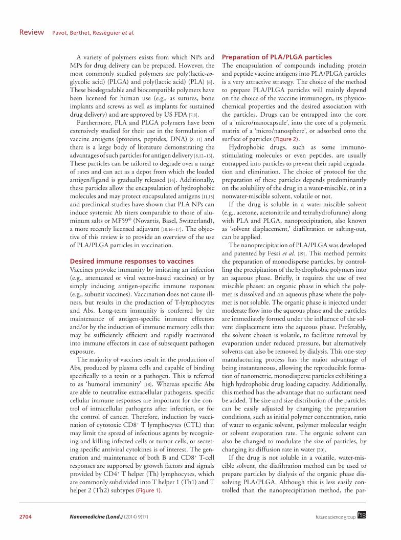
Preparation of PLA/PLGA particlesThe encapsulation of compounds including protein and peptide vaccine antigens into PLA/PLGA particles is a very attractive strategy. The choice of the method to prepare PLA/PLGA particles will mainly depend on the choice of the vaccine immunogen, its physico-chemical properties and the desired association with the particles. Drugs can be entrapped into the core of a ‘micro/nanocapsule’, into the core of a polymeric matrix of a ‘micro/nanosphere’, or adsorbed onto the surface of particles (Figure 2).
Hydrophobic drugs, such as some immuno-stimulating molecules or even peptides, are usually entrapped into particles to prevent their rapid degrada-tion and elimination. The choice of protocol for the preparation of these particles depends predominantly on the solubility of the drug in a water-miscible, or in a nonwater-miscible solvent, volatile or not.
If the drug is soluble in a water-miscible solvent (e.g., acetone, acetonitrile and tetrahydrofurane) along with PLA and PLGA, nanoprecipitation, also known as ‘solvent displacement,’ diafiltration or salting-out, can be applied.
The nanoprecipitation of PLA/PLGA was developed and patented by Fessi et al. [19]. This method permits the preparation of monodisperse particles, by control-ling the precipitation of the hydrophobic polymers into an aqueous phase. Briefly, it requires the use of two miscible phases: an organic phase in which the poly-mer is dissolved and an aqueous phase where the poly-mer is not soluble. The organic phase is injected under moderate flow into the aqueous phase and the particles are immediately formed under the influence of the sol-vent displacement into the aqueous phase. Preferably, the solvent chosen is volatile, to facilitate removal by evaporation under reduced pressure, but alternatively solvents can also be removed by dialysis. This one-step manufacturing process has the major advantage of being instantaneous, allowing the reproducible forma-tion of nanometric, monodisperse particles exhibiting a high hydrophobic drug loading capacity. Additionally, this method has the advantage that no surfactant need be added. The size and size distribution of the particles can be easily adjusted by changing the preparation conditions, such as initial polymer concentration, ratio of water to organic solvent, polymer molecular weight or solvent evaporation rate. The organic solvent can also be changed to modulate the size of particles, by changing its diffusion rate in water [20].
If the drug is not soluble in a volatile, water-mis-cible solvent, the diafiltration method can be used to prepare particles by dialysis of the organic phase dis-solving PLA/PLGA. Although this is less easily con-trolled than the nanoprecipitation method, the par-

www.futuremedicine.com 2705
Figure 1. Desired immune responses elicited by prophylactic vaccines. Vaccines against extra- or intra-cellular pathogens must target antigens and danger signals to immature DCs at the administration site and/or into the draining lymph nodes. DCs then present antigen through MHC class I or II complexes to CD8+ or CD4+ T cells, respectively. Danger signals stimulate DCs, which consequently express costimulatory molecules and secrete cytokines leading to T-cell stimulation and Th1 or Th2 orientation. Activated Th2 cells, produce B cell-stimulating cytokines that support differentiation of antigen-primed B cells. This facilitates/induces B-cell differentiation into memory B cells and plasma cells that produce large amounts of antibodies that prevent future virus infection. CD8+ lymphocytes acquire a cytotoxic phenotype, known as CTL, which is able to cause infected cell lysis directly. APC: Antigen-presenting cell; CTL: Cytotoxic CD8+ T lymphocyte; DC: Dendritic cell.
Cytotoxic immune response
Humoral immune response
B cell
IL-4
IL-13
Th2cell
Th1cell
IL-12
NaiveCD4+
APC
CD8+
Antibody production
CTL Effector CTLInfected cell
IL-4
Plasma cell
IL-2, IFN-γ
Vaccine antigen and ‘danger signal’
Th0 cell
future science group
PLA & PLGA particles as versatile carrier platforms for vaccine delivery Review
ticle sizes can be better controlled when the solution is precipitated prior to use.
The salting-out process was developed by Ibra-him [21]. Briefly, an emulsion of an organic phase in an aqueous phase containing a stabilizer and a highly concentrated electrolyte (salt, sucrose), in which the organic solvent is not soluble, is prepared with vigor-ous stirring. Pure water is then added to allow the sol-vent to diffuse into the aqueous phase leading to the formation of particles. This method allows for better control of the particle size, by controlling the stabilizer concentration, the stirring rate and the polymer con-centration [22,23]. However, an additional purification step is necessary to remove the electrolyte used for the saturation of the aqueous phase. The other compo-nents (active molecule and active surfactant) must also
be compatible with the electrolyte, which must also be nontoxic.
When the antigen or the active molecule is pref-erentially soluble in a nonwater-miscible solvent like dichloromethane (DCM), single emulsion solvent evaporation can be performed. The single emulsifica-tion process involves an oil-in-water (o/w) emulsion. The first step consists in dissolving the polymer in a nonwater miscible organic phase (e.g., DCM). This polymer solution is then dispersed into an aqueous phase commonly containing a surfactant (e.g., poly-vinyl alcohol and Poloxamer), in order to create an o/w emulsion with larger droplets. The emulsion is then treated by homogenizers or ultrasound in order to obtain smaller droplets. To replace the chlorinated solvent commonly used for the single emulsion sol-
2706 Nanomedicine (Lond.) (2014) 9(17)
Figure 2. Illustrative representation of the architecture of different nanostructured poly(lactic acid)/poly(lactic-co-glycolic acid) particles. These include nanospheres, in which the molecule is dissolved, adsorbed or dispersed throughout the matrix, attached to the surface or attached to the polymer matrix; nanocapsules, in which the molecule is in solution and surrounded by a shell-like wall. PGA: Poly(glycolic acid); PLA: Poly(lactic acid); PLGA: Poly(lactic-co-glycolic acid).
H
O
O
OH
m
HO
O
OH
n
OH
HO
O
O
mO
PGA PLGA PLA
Nanosphere Nanocapsule
Vaccine compounds
adsorptionVaccine compounds encapsulation
n
future science group
Review Pavot, Berthet, Rességuier et al.
vent evaporation methods, emulsion-solvent diffusion using a partially water-miscible solvent less toxic than dichloromethane, such as ethyl acetate, can be used instead. The main drawback of these two methods is the required use of surfactant to stabilize emulsions, often leading to the requirement to remove their excess for further in vitro or in vivo trials.
As peptide or nucleic acid antigens are generally highly water soluble, the former methods often lead to poor yield as the drug becomes deposited on the particle surface and in the polymer matrix during the process, and rapidly diffuses into the aqueous phase.
Therefore the encapsulation of such compounds becomes quite challenging. Indeed, their successful encapsulation, as for hydrophobic drug encapsulation, requires high drug loading in the NPs/MPs, predict-able release of the drug compound from the particle but most of all requires the prevention of protein deg-radation during the encapsulation process. Currently, the most common technique used for the encapsula-
tion of water-soluble vaccine antigens in PLA/PLGA particles is the double emulsification solvent evapora-tion [24,25]. An aqueous phase containing the drug is added to an organic phase composed of PLGA or PLA in DCM with vigorous stirring to create the water-in-oil emulsion. The w/o/w emulsion is created by adding the previous w/o emulsion into a large volume of water with an emulsifier to stabilize the emulsified polyvinyl alcohol and create the w/o/w emulsion. The particles are obtained after the solvent has been removed by evaporation. As for o/w emulsion method, the disad-vantage of w/o/w emulsion processes is the presence of emulsifiers which if not removed, lead to toxicity. Furthermore, this strategy requires high energy to cre-ate the emulsions, which can lead to the denaturation of the protein antigen, the use of surfactants and the poor drug loading into particles.
One of the disadvantages of all of these encapsula-tion techniques is the poor encapsulation efficiencies of drugs and the initial rapid release of drug (burst effect)
www.futuremedicine.com 2707future science group
PLA & PLGA particles as versatile carrier platforms for vaccine delivery Review
due to the diffusion of the drug present at the interface of particles after dilution into media.
An alternative strategy for the efficient presentation of protein on particles is adsorption onto the surface of ‘empty particles.’ With this strategy, all the meth-ods previously described to encapsulate hydrophobic drugs can be used to prepare the particles of the desired size and with the appropriate surface charge for the adsorption of protein antigen.
Many studies have also been conducted to obtain particle formulation that could be sterile filtered, lyoph-ilized and resuspended to the initial size with excipients appropriate for use as a vaccine formulation [26,27].
In brief, molecules of interest (e.g., vaccine anti-gens and immunostimulant molecules) can be incor-porated into the particles during the particle produc-tion (encapsulation) or adsorbed/chemically bound after their production. These methods are dependent on the hydrophobic/hydrophilic nature of the active drug (Ag or ligand). In the event of a hydrophobic Ag, it will be encapsulated within the polymer matrix during the formation of particles following methods precipitation/evaporation of solvent, emulsion (o/w). In the event of a hydrophilic Ag, the double emulsion (w/o/w) is used to encapsulate in the core of particles. Finally, the adsorption of hydrophilic Ag (or ligands) on the particles surface is one possible approach.
PLA/PLGA particles as vaccine antigen carrier systemsMost licensed vaccines rely on multiple dose immu-nization schedules which can be difficult and costly to achieve. In such a scenario, biodegradable poly-mer particle based vaccine delivery systems provide a viable alternative to single-dose vaccines [28]. In par-ticular, PLA and PLGA polymers have been used suc-cessfully in preclinical studies as vaccine delivery sys-tems for many years and are particularly well-suited to vaccine design because of their known adjuvant activity [6,29,30].
Already used in many medical devices (drug deliv-ery implants, suture material and microspheres) [31,32], these biodegradable polymers are probably the most studied materials for parenteral and mucosal antigen delivery. The initial motivation for using PLA/PLGA particles for antigen delivery came from the desire to reduce the number of repeated administrations required for long-term protection, through prolonged antigen release [33]. Another important advantage would be the reduction in mass vaccination costs.
As PLA/PLGA particles are efficiently phagocytosed by APCs in vitro and in vivo [17,34,35] they have been studied mainly for the delivery of antigens to DCs and macrophages aiming at inducing CTL responses.
Despite the encouraging immunological perfor-mances of PLA/PLGA-based MPs and NPs, the harsh conditions such as organic solvent/water interfaces, shear or cavitation forces, excessive heat, freezing and drying involved in the encapsulation methods often causes significant aggregation or degradation of vac-cine antigens [30,36–38]. Moreover, the degradation of the polymers generates acidic monomers and acidifica-tion of the inner polymer environment can cause pro-tein degradation and consequent loss of tertiary struc-ture, which is a central issue in the development of these devices for drug delivery [36,39,40]. These problems have been partially solved by optimized manufacturing methods [41,42] or addition of stabilizing agents such as Mg(OH)
2, other proteins, surfactants or sugars [43–46].
To address the limitations of antigen encapsulation, another strategy based on surface adsorption of anti-gens on PLA/PLGA particles was developed to avoid exposure of the proteins to organic solvents and shear forces during the emulsification step [47,48]. Mecha-nistic studies determined that protein adsorption on PLA/PLGA surface is driven by electrostatic and hydrophobic interactions [48,49]. Therefore, adsorp-tion of the proteins can be optimized by modulating ionic strength and pH. However, surface adsorption of antigen does not allow controlled release of antigen for single immunization vaccines; PLA/PLGA particles are instead used as an adjuvant for particulate delivery of antigen to the immune cells similar to alum, but the ease of encapsulating or adsorbing single or multiple antigens or ligands in PLA/PLGA particles (compared with alum) explains the renewed interest in vaccine approaches using these particulate systems [40].
In the last 10 years, micro and nanoparticles have been used mainly to carry vaccine antigens for paren-teral administration (subcutaneous, intradermal and intramuscular). Major efforts are being made to develop new mucosal candidate vaccines by selecting appropri-ate antigen delivery systems with high immunogenicity, designing new mucosal routes of administration and selecting immune-stimulatory adjuvant molecules. The versatility for delivery of PLA/PLGA NPs has led to their study as potential vaccine delivery systems for mucosal administration [11,50]. Indeed, it has been shown that these particles were able to carry antigens to microfold cells (M cells) at the surface of Peyer’s patches into the intestinal mucosa, leading to their uptake and delivery to mucosal APCs making them efficient antigen carrier systems for mucosal immunization [35].
Immune responses to PLA/PLGA carried antigensMany different antigens encapsulated or adsorbed on PLA/PLGA particles have been shown to induce broad
2708 Nanomedicine (Lond.) (2014) 9(17) future science group
Review Pavot, Berthet, Rességuier et al.
and potent humoral immune responses (Table 1). The majority of published pre-clinical studies are on tetanus toxoid (TT) or focused on more model antigens such as ovalbumin (OVA) and bovine serum albumin (BSA). Other antigens of interest were of bacterial origin: Esch-erichia coli detached fimbriae, pertussis toxoid, diphthe-ria toxoid (DT), subunit antigens from Haemophilus influenzae type B, Yersinia pestis and Mycobacterium tuberculosis; viral origin: subunit antigens from human and simian immunodeficiency virus (HIV, SIV), hepa-titis B virus surface antigen (HBsAg), influenza A virus, measles virus, parainfluenza virus, respiratory syncytial virus; and parasitological origin: subunit antigens from Plasmodium species (malaria) and Leishmania.
TT represents one of the most frequently used microencapsulated antigen for parenteral or muco-sal immunization and in most preclinical studies, a single injection of TT-loaded particles induced high Ab titers maintained over several months, generally up to 1 year or more [51,52,56,57]. Encapsulated toxoid induced low but continuous levels of neutralizing Abs (NAbs), whereas those obtained with the control alum-TT decreased after reaching the maximum level at 14 weeks. Moreover, the administration of a mixture of encapsulated and adsorbed TT led to significantly higher and more prolonged NAb levels than those measured for the adsorbed toxoid. Analysis of the Ab isotypes revealed dominance of IgG1 (pro-Th2) over IgG2a (pro-Th1); this pattern was consistent with that observed in the group injected with alum-TT.
A study performed by Saini et al. aimed to compare the humoral and cell-mediated immune responses between recombinant HBsAg adsorbed PLA MPs vaccine (single shot) and alum-HBsAg vaccine (two doses) [54]. Specific humoral immune responses (IgM and IgG) and cell-mediated immune responses were determined. Based on these findings, it was concluded that a single injection (using the subcutaneous route – sc.) of the polymeric MPs produced a better immune response (both humoral and cell mediated) than two injections of a conventional alum-HBsAg vaccine, in the mouse model. These data demonstrate the high potential of polymeric MPs for their use as a carrier adjuvant for hepatitis B vaccine.
The rabbit model was used in a quantitative and qualitative comparison of the Ab responses induced by PLA NPs or by the emulsion adjuvant MF59 using three HIV-1 antigens: p24gag, wild-type (WT) Tat and a mutated, detoxified form of Tat [10]. All antigen and adjuvants led to the induction of similar level of IgG titers in sera when injected by subcutaneous route. The p24 antigen, but not Tat, also induced fecal IgG in rabbits when formulated with PLA particles or MF59. The nature of the adjuvant had consequences on the
spectrum of specificity induced, depending on the antigen: PLA adjuvant focused the anti-p24 response to an immunodominant domain when compared with MF59. With WT Tat, no difference between adjuvants was observed in the spectrum of specificity induced. However, detoxified Tat coated on PLA NPs increased the number of epitopes recognized by serum IgG compared with MF59. The impact of these quali-tative differences depending on the antigen/adjuvant associations is important to take into account for fur-ther designs of vaccine formulation using particulate adjuvants.
In summary, for the antigens studied, strong Ab responses were observed after a single administration. Responses were frequently comparable to that elic-ited by gold-standard formulations, such as antigen in alum, MF59 or Freund’s adjuvant, given once or twice [10,17,58–60]. Thus, the particle preparations proved to be highly suitable for a large variety of antigens, derived from various sources and having very differ-ent structural features. Besides inducing strong and functional humoral immune responses, PLA/PLGA particles are of particular interest in releasing antigens over extended periods of time and thereby providing prolonged stimulation of the immune system.
Whereas specific Abs appear necessary to block pathogen invasion, specific cellular immune responses are important for the control of virus replication and viral load after infection, and also for therapeutic can-cer vaccines. Particulate vaccines are recognized as extracellular antigens and are taken up by APCs by one of the endocytic mechanisms – receptor-dependent or receptor-independent endocytosis – and placed into a membrane-delimited compartment of the endocytic pathway [61]. This compartment undergoes a series of modifications and finally fuses with lysosomes or MHC class II compartments leading to peptide pre-sentation to antigen-specific CD4+ T cells but no CD8+ T cells. Hence, it appears that particulate for-mulations alone are poor inductors of CTL responses. To improve immune responses, the use of additional stimuli, such as pattern recognition receptors (PRRs) ligands, appears to be crucial; in particular to induce cross-presentation of extracellular Ags to CD8+ T cells [62].
Potentiating the immune response with PLA/PLGA particles carrying pattern recognition receptor ligandsMost successful vaccines induce persistent Ab responses that can last a lifetime. The mechanisms involved remain unclear, but emerging evidence indicates that they activate immune cells (especially DCs) via PRRs including Toll-like receptors (TLRs),

www.futuremedicine.com 2709future science group
PLA & PLGA particles as versatile carrier platforms for vaccine delivery Review
Table 1. Examples of studies demonstrating that carrying of antigens by poly(lactic acid)/poly(lactic-co-glycolic acid) particles potentiate specific immune responses in vivo.
Nanocarrier Antigen Model/immunization route
Immune response Ref.
PLA or PLGA micro- or nanoparticles
Encapsulated TT Wistar rats/im. – Particles encapsulating TT elicited higher anti-TT Ab titers than TT alone – Titers persisted for more than 5 months
[51]
PLGA NPs Encapsulated TT and QS Rabbits/in. – The serum IgG titer induced with PLGA(TT) was higher than TT solution
[52]
– IgG titers induced with PLGA(TT + QS) was higher than PLGA(TT)
PLA or PLGA MPs Encapsulated DT Guinea pig/sc. – PLGA(DT) made with the relatively hydrophilic PLGA 50:50 exhibited specific and sustained Ab responses over 40 weeks, comparable with the responses to alum(DT)
[53]
PLA MPs Adsorbed HBsAg Mouse/sc. – A single injection of the PLA-HBsAg produced better immune response (both humoral and cellular) than two injections of a conventional alum-HBsAg vaccine
[54]
PLA or PLGA NPs Adsorbed HBsAg Rat/pulmonary delivery
– Hydrophobic particles >500 nm elicited a more robust increase in secretary IgA, IL-2 and IFN-γ levels compared with hydrophilic particles <500 nm
[55]
PLA NPs Adsorbed HIV-1 antigens: p24gag, WT Tat and a mutated, detoxified form of Tat
Rabbit/sc. – PLA NPs and MF59® (Novartis, Basel, Switzerland) adjuvant led to the induction of similar level of IgG titers
[10]
– PLA adjuvant focused the anti-p24 response to an immunodominant domain when compared with MF59
[10]
PLA NPs Adsorbed HIV-1 p24 Mouse/sc. – After boosting PLA-p24 induced significantly higher IgG titers compared with the gold standard alum-p24
[17]
PLA NPs Adsorbed HIV-1 p24 Macaque, rabbit, mouse/sc.
– PLA-p24 induced high Ab titers (>106) in mice, rabbits and macaques
[16]
– PLA-p24 elicited strong CTL responses and a Th1-biased cytokine release in mice
– p24 protein generates a more Th1-oriented response when coated onto PLA NPs than adjuvanted with Freund’s adjuvant
– The ability of PLA-p24 particles to induce Th1 responses was confirmed in the macaque model
Ab: Antibody; Alum: Aluminium salts; CTL: Cytotoxic CD8+ T lymphocytes; DT: Diphtheria toxoid; HBsAg: Hepatitis B virus surface antigen; im.: Intramuscular; in.: intranasal; MP: Microparticle; NP: Nanoparticle; PLA: Poly(lactic acid); PLGA: Poly(lactic-co-glycolic acid); QS: Quillaja saponins; sc.: subcutaneous; TT: Tetanus toxoid; WT: Wild-type.
NOD-like receptors (NLRs), C-type lectin recep-tors (CLRs) and RIG-I-like receptors (RLRs) [63,64]. Triggering specific combinations of PRRs in DCs can induce synergistic production of cytokines, which can result in enhanced T-cell responses (Th1, Th2 and Th17) [65–67] and/or reversal of the immunosuppres-sive action of regulatory T cells (T
reg) [68–70]. The inclu-
sion of such adjuvants within vaccines can enhance vaccine-induced protection by providing strong and long-term immune responses, broadening protection or providing cross-protection against related patho-gens, improving immune responses in poorly respon-sive populations and potentially reducing the amount of antigen required. Reducing the dose enables an
2710 Nanomedicine (Lond.) (2014) 9(17) future science group
Review Pavot, Berthet, Rességuier et al.
increase in the manufacturing capacity and offers a potential for greater distribution of the given vaccine. This section describes the potential of targeting PRRs by PLA/PLGA particulate vectors to promote immune responses and outlines the progress being made toward the development of PRR-based adjuvants for use in clinical-grade vaccines (Figure 3).
A number of studies addressing this question have been performed in vitro and in vivo. As an example, Heit et al. determined in vitro that full maturation and cytokine secretion by APCs as well as cross-pre-sentation of antigen were enhanced when coencapsula-tion into PLGA particles of antigen with TLR7 and 9 ligands was used [71]. Wischke et al., as well as our group, recently showed that PLGA microencapsulated (∼5 μm) or PLA nanoencapsulated (∼200 nm) NOD ligands (respectively) induce an in vitro maturation of human monocyte-derived DCs to a proinflamma-tory phenotype with high levels of released cytokines compared with the soluble ligands [17,72].
Moreover, in vivo studies have been performed in different animal models in order to improve humoral and/or cellular immune responses (Table 2). In a study from Demento et al. [73]. PLGA NPs loaded with recombinant West Nile virus envelope protein and sur-face modified with CpG ODN (TLR9 ligand) were used to immunize mice subcutaneously. The animals showed robust humoral responses polarized toward Th1 immune responses compared with predominately Th2-biased responses with the alum adjuvant. Immu-nizations with PLGA-CpG ODN resulted in a greater number of circulating effector T cells and greater activ-ity of antigen-specific lymphocytes than unmodified NPs or alum. Last, compared with alum, this system offered superior protection in a mouse model of West Nile virus encephalitis.
PLGA NPs containing poly(I:C) or CpG ODN together with OVA have also been studied for the induction of CTL responses in a cancer vaccine study [75]. Immunization of mice (intravenous) with the PLGA(poly(I:C)) or (CpG ODN) together with OVA elicited potent OVA-specific CTL activity compared with particles containing OVA only. In accordance with these results, those formulations exerted potent antitumor activity in mice that were subcutaneously implanted with EG7.OVA tumor cells. These results show that encapsulation of poly(I:C) or CpG ODN together with antigen in biodegradable PLGA NPs is an effective approach for the induction of potent antigen-specific CTL responses in vivo.
In addition to improving parenteral immunizations, PLA/PLGA particles carrying PRR ligands have also been studied for the purpose of improving mucosal vaccine administration. Zhu et al. designed PLGA par-
ticles carrying PRR ligands and able to target the large intestine after oral delivery in the mouse model [11]. The particles were coated with methacrylate-based polymer Eudragit FS30D to bypass the harmful effects of diges-tive low pH and enzymatic destruction and to selectively deliver the particles to the lower GI tract. The PLGA particles containing the ligands macrophage-activating lipoprotein (MALP-2) for TLR2, polyinosine-polycyt-idylic acid (poly(I:C)) for TLR3 and the CpG ODN for TLR9, were able to induce colorectal immunity and protected against rectal or vaginal viral challenge with the vaccinia virus vPE-16, which expresses the HIV Env epitope used in the peptide vaccine.
However, despite the fact that numerous stud-ies have used codelivery systems (antigen and PRR ligand(s) in the same particle), others have shown that coadministration (antigen and PRR ligand(s) in different particles) has equivalent and even bet-ter effect. In this manner, Kazzaz et al. [76] observed that codelivery of HIV-1 gp120 and MPLA (TLR4 ligand) or RC529 (synthetic form of MPLA) encapsu-lated within the same particle and coadministration of PLGA(gp120) and PLGA(MPLA) (or RC529) within different particles have an equivalent immune potency for specific-IgG and IgG2a production in mice.
More recently and consistent with previous obser-vations [76,77], Kasturi et al. showed, in their supple-mentary data, that coadministration of PLGA(OVA) and PLGA(MPLA+R837(TLR7)) induced stron-ger IgG2c and IgG1 production than codelivery of PLGA(OVA+MPLA+R837) after subcutaneous administration in the mouse model [15]. Moreover, PLGA(OVA) plus PLGA(MPLA+R837) induced syn-ergistic increases in antigen-specific, NAbs compared with immunization with NPs containing antigens plus a single TLR ligand. In addition to OVA, they also used the protective antigen (PA) from Bacillus anthracis and hemagglutinin (HA) from avian influenza H5N1 virus. As observed with OVA, there was a synergis-tic enhancement in the antigen-specific Ab responses after immunization with PLGA(MPLA+R837) plus PLGA(PA) or PLGA(HA). These responses com-pletely protected mice against lethal avian and swine influenza virus strains, and induced robust immunity against pandemic H1N1 influenza in rhesus macaques.
Data supporting the idea that antigen and PRR ligand(s) can be delivered on separate particles have also been observed recently using ultrasmall Pluronic-stabilized poly(propylene sulfide) [78] or PLGA [79].
Despite all these observations, questions still remain concerning the codelivery or the coadministration strategies. This warrants further consideration and needs intensive investigations but probably depends

www.futuremedicine.com 2711
Figure 3. Potentiating the immune response with poly(lactic acid)/poly(lactic-co-glycolic acid) particles carrying pattern recognition receptor ligands. The actual concept comprises target dendritic cells with immunostimulant molecules and vaccine antigen carried by a poly(lactic acid)/poly(lactic-co-glycolic acid) particulate vector to trigger specific immune responses against the corresponding pathogen. CLR: C-type lectin receptor; NLR: NOD-like receptor; PRR: Pattern recognition receptor; TLR: Toll-like receptor.
Dendritic cell
• Actor 1: particulate vector• Actor 2: vaccine antigen• Actor 3: PRR ligand (immunomodulator)
PRRs: • TLRs• NLRs• CLRs
Costimulatory moleculesCD80/CD86
T cell
Activation and proliferation
Immunity
Cytokines(IL-4, IL-12, among others)
Maturation and antigen presentation
Internalize Ag in thepresence of danger signal
future science group
PLA & PLGA particles as versatile carrier platforms for vaccine delivery Review
on the type of particle system, antigen, administra-tion route and of course the immunostimulatory molecule(s) used.
Strategies to improve cell & tissue targeting by PLA/PLGA particlesAs discussed above, DCs are a key component in the induction of strong and long-lasting immune responses toward specific antigens. Being able to specifically deliver antigens and/or immunostimulant molecules to those cells by PLA or PLGA particles is a popular strategy [80–82]. However this delivery could be fur-ther improved by the presence of ligands included on the surface of particles to target specific tissues or cell surface antigens and reduce nonspecific distribu-
tion. In this context a large number of ligands are currently being assessed including proteins, peptides, Abs, polysaccharides, glycolipids, glycoproteins and lectins [8].
The use of lectins capable of targeting CLRs is a very attractive strategy to enhance the efficacy of vac-cines. These molecules are highly specific internaliza-tion receptors that facilitate antigen uptake, process-ing and loading on MHC class I and II molecules leading to antigen presentation toward Th1 or Th2 orientation [83].
This was the objective of the recent study from Gupta et al. who used DT- and TT-encapsulated PLA particles with adsorbed soybean agglutinin (SBagg) lectins that resulted in a better association of polymer

2712 Nanomedicine (Lond.) (2014) 9(17) future science group
Review Pavot, Berthet, Rességuier et al.
particles with DCs [84]. In the mouse model, immuni-zations (im.) with PLA-SBagg-antigens elicited a stron-ger Ab response than with plain particles entrapping antigens (also higher than alum adsorbed antigens). The improved immunogenicity of PLA-SBagg-anti-gens was attributed to the N-linked glycan-mediated targeting of polymer particles to DCs. Other in vivo studies using varied particulate vectors demonstrated how CLR targeting can be employed for cell-specific antigen delivery and can result in enhanced humoral and cellular immune responses [85–88].
Several studies have also demonstrated the success-ful use of antilectin antibody-grafted nanoparticles to increase the uptake of these carriers by DCs. As an example, Cruz et al. tested various Abs recognizing the DC-specific receptor DC-SIGN (type II CLR) to study the effects of PEG copolymer shielding and
Ab type on antibody–receptor interactions [89]. While they found that shielding can lessen the efficacy of the targeted delivery through Abs, they also showed that particles with a coat of Abs targeting the car-bohydrate recognition domain of the DC-SIGN were more efficiently taken up by DCs than those target-ing the receptor’s neck domain. However both for-mulations bound selectively to DCs and were able to induce a stronger cytokine production toward Th1 than Th2.
Mannosylation of particles is also a promising strat-egy to selectively target vaccine antigens to the man-nose receptor expressed on DCs. PLGA NPs decorated with mannan (Saccharomyces cerevisae), are able to simultaneously enhance antigen-specific CD4+ and CD8+ T-cell responses compared with nondecorated NPs [90]. Mannose-PEG(3)-NH(2) grafting can also
Table 2. Examples of studies demonstrating that carrying of antigens and pattern recognition receptor ligands by poly(lactic acid)/poly(lactic-co-glycolic acid) particles potentiate specific immune responses in vivo.
Nanocarrier Antigen/immunostimulant molecule(s)
Model/immunization route
Immune response Ref.
PLGA NPs Adsorbed rWNVE and CpG ODN (TLR9 ligand)
Mouse/sc. – Humoral responses polarized toward Th1 responses compared with predominately Th2-biased responses with alum adjuvant
[73]
– Compared with alum, this system offered superior protection against WN virus encephalitis
PLGA NPs – Adsorbed HA or OVA Mouse, rhesus macaques/sc.
– PLGA(MPLA+R837) + PLGA(Ag) induces synergistic increases in NAbs compared with immunization with NPs containing Ags + a single ligand
[15]
– Encapsulated MPLA (TLR4) and/or R837 (TLR7)
– Immunization protected completely against lethal influenza virus strains in mice
– Induced robust immunity against pandemic H1N1 influenza in macaques
PLGA NPs Encapsulated PeptAg/ MALP-2 (TLR2), poly(I:C) (TLR3) and CpG ODN (TLR9)
Mouse/oral – Colorectal immunity [11]
– Protection against rectal or vaginal viral challenge
PLA NPs – Adsorbed HIV-1 p24 –Encapsulated NOD1/NOD2 ligands
Mouse/sc. – Encapsulated NOD1/2 ligands increased anti-p24 IgG responses up to 100-times compared with the free ligands, alum-p24 or PLA-p24 alone
[17]
PLGA NPs Encapsulated HBcAg + MPLA (TLR4)
Mouse/sc. – PLGA(MPLA+HBcAg) induced a stronger Th1 cellular immune response than HBcAg alone or PLGA(HBcAg)
[74]
PLGA NPs Encapsulated OVA + poly(I:C) or CpG ODN
Mouse/iv. – PLGA(OVA+poly(I:C) or CpG ODN) elicited potent specific CTL activity compared with PLGA(OVA)
[75]
– PLGA(OVA+poly(I:C) or CpG ODN) exerted potent antitumor activity in mice
Ag: Antigen; CpG ODN: CpG oligodeoxynucleotides; CTL: Cytotoxic CD8+ T lymphocytes; HA: Hemagglutinin; HBcAg: Hepatitis B virus core antigen; iv.: intravenous; MPLA: Monophosphoryl lipid A; NP: Nanoparticle; OVA: Ovalbumin; PLA: Poly(lactic acid); PLGA: Poly(lactic-co-glycolic acid); poly(I:C): Polyinosinic-polycytidylic acid; rWNVE: West Nile virus envelope protein; sc.: subcutaneous; WN: West Nile.
www.futuremedicine.com 2713future science group
PLA & PLGA particles as versatile carrier platforms for vaccine delivery Review
enhance the uptake of PLGA MS, with a variable rela-tive contribution of specific and nonspecific uptake mechanism [91].
While targeting DCs during parenteral adminis-trations has shown to be effective, to achieve such a goal by mucosal administration (e.g., enteral or nasal route), seems to be more complex [92].
The ability of M cells to take up and transcytose diverse numbers of microorganisms to APCs have made M cells an ideal target for vaccine delivery to the mucosal immune system [93]. Those cells are found in the follicle-associated epithelium of the Peyer’s patches as well as in nasopharyngeal-associated lymphoid tis-sue (NALT) and bronchus-associated lymphoid tissue (BALT) [94]. It has been shown that the lectins Ulex europaeus 1 (UEA-1) and the Lotus tetragonolobus agglutinin (LTA) are able to selectively bind to α-L-fucose targets on mouse M cells and are rapidly endo-cytosed and transported to M cell basolateral mem-branes. Therefore, oral administration (mouse model) of UEA-1 or LTA grafted PLGA NPs encapsulating HBsAg result in higher mucosal and systemic responses compared with uncoated NPs [58,95]. However, UEA-1 selectively binds to mouse M cells but not human [96].
New PLGA formulations were then developed to target the β1 integrins expressed at the apical side of human M cells [97,98] via an RGD peptide that enhances the transport of those particles by 3.5-fold compared with particles without RGD peptides [99]. In vivo studies showed these NPs concentrate in Peyer’s patches and were able to induce the production of spe-cific IgG. Other M cell homing peptide (CKS9) or Abs (CA19.9) have also been studied [93,100].
Promising outcomes have been achieved with par-ticle-based vaccine targeting M cells through mucosal administration. By applying the knowledge gained from studies on DC particle targeting, further major advances should arise in the field of mucosal vaccines. The dual targeting of M cells and DCs with codelivery or coadministration of immunostimulants seems to be a promising approach.
Vaccination strategy not only benefits from major advances in antigen/ligand formulation and adminis-tration, but also a better comprehension of the rela-tionship between modulating particle physical proper-ties and immune cells uptake. It has been established that rendering PLGA nanoparticles mucoadherent, by shifting their charge to positive through glycol-chitosan coating, induces higher systemic and mucosal immune response after nasal immunization in mice than the naked PLGA counterpart, presumably linked to their higher bioavaibility due to a lower clearance [101].
It has also been demonstrated that modulating the size of the particles can influence the immune
response outcome [102–105]. Using PLGA particles sized at 300 nm, 1 μm, 7 μm and 17 μm formulated with OVA and CpG ODN in in vitro and in vivo studies, it has been observed that 300 nm sized particles were the most efficiently taken up by DCs leading to efficient antigen delivery and DC activation, compared with bigger particles [106]. The systemic antigen-specific immune response to vaccination was also measured in mice. PLGA particles sized 300 nm generated the highest antigen-specific cytotoxic T-cell responses. These mice also showed the highest antigen-specific IgG2a:IgG1 ratio. This study suggests that the smaller the PLGA particle used to deliver antigen and adju-vants the stronger the antigen-specific cytotoxic T-cell response generated.
However this study did not establish the lower size limit for efficient uptake. Will 50 nm particles be more efficient that those of 300 nm?
Conclusion & future perspectiveOver the last couple of decades, many groups have con-tributed to the development of PLA/PLGA controlled release NPs and MPs for antigens delivery to compen-sate their poor immunogenicity. These particle-based vaccines are now well established for their ability to provide persistent levels of Abs and to exhibit immu-nological memory. For designing new vaccines against viral infections and cancers, or intracellular pathogens, several strategies have been employed, from inclusion of different immunostimulatory molecules (essentially PRR ligands) to specific targeting of PLA/PLGA par-ticles to APCs. This has led to the induction of both humoral and cellular immune responses, in animal models ranging from rodents to nonhuman primates. The induction of both types of responses may be a prerequisite for future vaccination strategies based on these eco-compatible polymers. It is also worth not-ing both, their potential interest and drawbacks toward alternative delivery systems already on the market, such as virosomes, emulsions or aluminum salts. Indeed, PLA/PLGA particles have a very short history in clini-cal studies and in some cases, sophisticated design of vaccine formulations associated to complex regulatory requirements could be major limitations for their devel-opment as vaccine delivery systems for human use. It is therefore essential that further research on such multifunctional vaccine delivery platform addresses these questions to be envisioned as a viable alternative method for vaccine delivery.
First, the chemistry behind particle synthesis should be kept scalable, with simple design that will not impair cost, reproducibility and safety issues. Second, the choice of immunostimulatory molecules should be focused on well-studied candidates, with strong preclin-
2714 Nanomedicine (Lond.) (2014) 9(17) future science group
Review Pavot, Berthet, Rességuier et al.
ical data, as prototype PRR ligands could have varied effects, depending on dose and route of administration, among others. Last, more basic science is required to decipher the mechanisms of particle uptake by APCs, transport to the lymphoid system, biodistribution and degradation of particles in the organism. In this respect, the particle formulation process facilitates the labeling of PLA/PLGA particles with different probes (such as fluorescent or radioactive probes). These labeled particles will constitute effective tools to understand the molecular basis of vaccine immunity through live imaging studies and to fine tune vaccine formulations through a back and forth scientific approach.
As illustrated in this review, the huge amount of preclinical data gathered with PLA/PLGA vaccine
platform provides strong hope that these particles will overcome the current challenges, potentially becom-ing key components among the vaccine armada of the future.
Financial & competing interests disclosureThe authors have no relevant affiliations or financial in-
volvement with any organization or entity with a financial
interest in or financial conflict with the subject matter or
materials discussed in the manuscript. This includes employ-
ment, consultancies, honoraria, stock ownership or options,
expert testimony, grants or patents received or pending or
royalties.
No writing assistance was utilized in the production of this
manuscript.
Executive summary
Background• The development of safe and effective vaccines for cancer and infectious diseases remains a major goal in
global public health.• One of the most important areas of research is the design of new generation of adjuvants.• Particulate compounds including poly(lactic acid) (PLA) and poly(lactic-co-glycolic acid) (PLGA) nano- and
micro-particles have been shown to possess significant potential as antigen and immunostimulant molecule delivery systems to antigen-presenting cells (APCs).
PLA & PLGA particles as versatile carrier platforms for vaccine delivery• Ease of encapsulating or adsorbing single or combination of antigen and ligands in PLA/PLGA particles.• Potential of targeting pattern recognition receptors by PLA/PLGA particulate vectors to promote immune
responses and outline the progress being made toward the development of pattern recognition receptor-based adjuvants for use in clinical-grade vaccines.
• New selective targeting of PLA/PLGA particles to APCs has been successfully achieved, and elevated humoral and cellular immune responses have been attained in preclinical models.
Conclusion & future perspective• Nanoparticles can be optimized to improve their uptake and immunogenicity; understanding the role
of nanoparticle’s immunoproperties is important to fine tune these materials before their use in vaccine formulations.
• Thorough characterization and standardized preparation methods are critical to create an accurate knowledge base about PLA/PLGA particulate vector effects on the immune system.
• More basic science is required to decipher the mechanism of particle uptake by APCs, transport to the lymphoid system, biodistribution and toxicity.
References1 Mckee AS, Munks MW, Marrack P. How do adjuvants work?
Important considerations for new generation adjuvants. Immunity 27(5), 687–690 (2007).
2 Clapp T, Siebert P, Chen D, Jones Braun L. Vaccines with aluminum-containing adjuvants: optimizing vaccine efficacy and thermal stability. J. Pharm. Sci. 100(2), 388–401 (2011).
3 Reed SG, Orr MT, Fox CB. Key roles of adjuvants in modern vaccines. Nat. Med. 19(12), 1597–1608 (2013).
4 Kool M, Fierens K, Lambrecht BN. Alum adjuvant: some of the tricks of the oldest adjuvant. J. Med. Microbiol. 61(Pt 7), 927–934 (2012).
5 Leleux J, Roy K. Micro and nanoparticle-based delivery systems for vaccine immunotherapy: an immunological and materials perspective. Adv. Healthc. Mater. 2(1), 72–94 (2013).
6 De Temmerman ML, Rejman J, Demeester J, Irvine DJ, Gander B, De Smedt SC. Particulate vaccines: on the quest for optimal delivery and immune response. Drug Discov. Today 16(13–14), 569–582 (2011).
7 Athanasiou KA, Niederauer GG, Agrawal CM. Sterilization, toxicity, biocompatibility and clinical applications of polylactic acid/polyglycolic acid copolymers. Biomaterials 17(2), 93–102 (1996).
8 Silva JM, Videira M, Gaspar R, Preat V, Florindo HF. Immune system targeting by biodegradable nanoparticles for cancer vaccines. J. Control. Release 168(2), 179–199 (2013).
9 Lamalle-Bernard D, Munier S, Compagnon C et al. Coadsorption of HIV-1 p24 and gp120 proteins to surfactant-free anionic PLA nanoparticles preserves antigenicity and immunogenicity. J. Control. Release 115(1), 57–67 (2006).

www.futuremedicine.com 2715future science group
PLA & PLGA particles as versatile carrier platforms for vaccine delivery Review
10 Guillon C, Mayol K, Terrat C et al. Formulation of HIV-1 Tat and p24 antigens by PLA nanoparticles or MF59 impacts the breadth, but not the magnitude, of serum and faecal antibody responses in rabbits. Vaccine 25(43), 7491–7501 (2007).
11 Zhu Q, Talton J, Zhang G et al. Large intestine-targeted, nanoparticle-releasing oral vaccine to control genitorectal viral infection. Nat. Med. 18(8), 1291–1296 (2012).
12 Xu FH, Zhang Q. Recent advances in the preparation progress of protein/peptide drug loaded PLA/PLGA microspheres. Yao Xue Xue Bao 42(1), 1–7 (2007).
13 Wischke C, Schwendeman SP. Principles of encapsulating hydrophobic drugs in PLA/PLGA microparticles. Int. J. Pharm. 364(2), 298–327 (2008).
14 Panyam J, Labhasetwar V. Biodegradable nanoparticles for drug and gene delivery to cells and tissue. Adv. Drug Deliv. Rev. 55(3), 329–347 (2003).
15 Kasturi SP, Skountzou I, Albrecht RA et al. Programming the magnitude and persistence of antibody responses with innate immunity. Nature 470(7335), 543–547 (2011).
16 Ataman-Onal Y, Munier S, Ganee A et al. Surfactant-free anionic PLA nanoparticles coated with HIV-1 p24 protein induced enhanced cellular and humoral immune responses in various animal models. J. Control. Release 112(2), 175–185 (2006).
17 Pavot V, Rochereau N, Primard C et al. Encapsulation of Nod1 and Nod2 receptor ligands into poly(lactic acid) nanoparticles potentiates their immune properties. J. Control. Release 167(1), 60–67 (2013).
18 Cooper NR, Nemerow GR. The role of antibody and complement in the control of viral infections. J. Invest. Dermatol. 83(1 Suppl.), s121–s127 (1984).
19 Fessi H, Puisieux F, Devissaguet JP, Ammoury N, Benita S. Nanocapsule formation by interfacial polymer deposition following solvent displacement. Int. J. Pharm. 55(1), R1–R4 (1989).
20 Legrand P, Lesieur S, Bochot A et al. Influence of polymer behaviour in organic solution on the production of polylactide nanoparticles by nanoprecipitation. Int. J. Pharm. 344(1–2), 33–43 (2007).
21 Ibrahim H BC, Doelker E, Buri P, Gurny R. Aqueous nanodispersions prepared by a salting-out process. Int. J. Pharm. 87(1–3), 239–246 (1992).
22 Rivera PA, Martinez-Oharriz MC, Rubio M, Irache JM, Espuelas S. Fluconazole encapsulation in PLGA microspheres by spray-drying. J. Microencapsul. 21(2), 203–211 (2004).
23 Dinarvand R, Sepehri N, Manoochehri S, Rouhani H, Atyabi F. Polylactide-co-glycolide nanoparticles for controlled delivery of anticancer agents. Int. J. Nanomed. 6, 877–895 (2011).
24 Danhier F, Ansorena E, Silva JM, Coco R, Le Breton A, Preat V. PLGA-based nanoparticles: an overview of biomedical applications. J. Control. Release 161(2), 505–522 (2012).
25 Johansen P, Men Y, Merkle HP, Gander B. Revisiting PLA/PLGA microspheres: an analysis of their potential
in parenteral vaccination. Eur. J. Pharm. Biopharm 50(1), 129–146 (2000).
26 Wendorf J, Singh M, Chesko J et al. A practical approach to the use of nanoparticles for vaccine delivery. J. Pharm. Sci. 95(12), 2738–2750 (2006).
27 Holzer M, Vogel V, Mantele W, Schwartz D, Haase W, Langer K. Physico-chemical characterisation of PLGA nanoparticles after freeze-drying and storage. Eur. J. Pharm. Biopharm. 72(2), 428–437 (2009).
28 Cleland JL. Single-administration vaccines: controlled-release technology to mimic repeated immunizations. Trends Biotechnol. 17(1), 25–29 (1999).
29 O’Hagan DT, Singh M. Microparticles as vaccine adjuvants and delivery systems. Expert Rev. Vaccines 2(2), 269–283 (2003).
30 Jiang W, Gupta RK, Deshpande MC, Schwendeman SP. Biodegradable poly(lactic-co-glycolic acid) microparticles for injectable delivery of vaccine antigens. Adv. Drug Deliv. Rev. 57(3), 391–410 (2005).
31 Okada H, Toguchi H. Biodegradable microspheres in drug delivery. Crit. Rev. Ther. Drug Carrier Syst. 12(1), 1–99 (1995).
32 Putney SD, Burke PA. Improving protein therapeutics with sustained-release formulations. Nat. Biotechnol. 16(2), 153–157 (1998).
33 Aguado MT, Lambert PH. Controlled-release vaccines‐‐biodegradable polylactide/polyglycolide (PL/PG) microspheres as antigen vehicles. Immunobiology 184(2–3), 113–125 (1992).
34 Newman KD, Elamanchili P, Kwon GS, Samuel J. Uptake of poly(d,l-lactic-co-glycolic acid) microspheres by antigen-presenting cells in vivo. J. Biomed. Mater. Res. 60(3), 480–486 (2002).
35 Primard C, Rochereau N, Luciani E et al. Traffic of poly(lactic acid) nanoparticulate vaccine vehicle from intestinal mucus to sub-epithelial immune competent cells. Biomaterials 31(23), 6060–6068 (2010).
36 Schwendeman SP. Recent advances in the stabilization of proteins encapsulated in injectable PLGA delivery systems. Crit. Rev. Ther. Drug Carrier Syst. 19(1), 73–98 (2002).
37 Jiang W, Schwendeman SP. Stabilization of tetanus toxoid encapsulated in PLGA microspheres. Mol. Pharm. 5(5), 808–817 (2008).
38 Johansen P, Men Y, Audran R, Corradin G, Merkle HP, Gander B. Improving stability and release kinetics of microencapsulated tetanus toxoid by co-encapsulation of additives. Pharm. Res. 15(7), 1103–1110 (1998).
39 Fu K, Pack DW, Klibanov AM, Langer R. Visual evidence of acidic environment within degrading poly(lactic-co-glycolic acid) (PLGA) microspheres. Pharm. Res. 17(1), 100–106 (2000).
40 Jain S, O’Hagan DT, Singh M. The long-term potential of biodegradable poly(lactide-co-glycolide) microparticles as the next-generation vaccine adjuvant. Expert Rev. Vaccines 10(12), 1731–1742 (2011).
41 Almeida AJ, Alpar HO, Brown MR. Immune response to nasal delivery of antigenically intact tetanus toxoid

2716 Nanomedicine (Lond.) (2014) 9(17) future science group
Review Pavot, Berthet, Rességuier et al.
associated with poly(L-lactic acid) microspheres in rats, rabbits and guinea-pigs. J. Pharm. Pharmacol. 45(3), 198–203 (1993).
42 Butler SM, Tracy MA, Tilton RD. Adsorption of serum albumin to thin films of poly(lactide-co-glycolide). J. Control. Release 58(3), 335–347 (1999).
43 Audran R, Men Y, Johansen P, Gander B, Corradin G. Enhanced immunogenicity of microencapsulated tetanus toxoid with stabilizing agents. Pharm. Res. 15(7), 1111–1116 (1998).
44 Zhu G, Schwendeman SP. Stabilization of proteins encapsulated in cylindrical poly(lactide-co-glycolide) implants: mechanism of stabilization by basic additives. Pharm. Res. 17(3), 351–357 (2000).
45 Zhu G, Mallery SR, Schwendeman SP. Stabilization of proteins encapsulated in injectable poly (lactide- co-glycolide). Nat. Biotechnol. 18(1), 52–57 (2000).
46 Jiang W, Schwendeman SP. Stabilization and controlled release of bovine serum albumin encapsulated in poly(D, L-lactide) and poly(ethylene glycol) microsphere blends. Pharm. Res. 18(6), 878–885 (2001).
47 Kazzaz J, Neidleman J, Singh M, Ott G, O’Hagan DT. Novel anionic microparticles are a potent adjuvant for the induction of cytotoxic T lymphocytes against recombinant p55 gag from HIV-1. J. Control. Release 67(2–3), 347–356 (2000).
48 Singh M, Kazzaz J, Ugozzoli M, Malyala P, Chesko J, O’Hagan DT. Polylactide-co-glycolide microparticles with surface adsorbed antigens as vaccine delivery systems. Curr. Drug Deliv. 3(1), 115–120 (2006).
49 Singh M, Chesko J, Kazzaz J et al. Adsorption of a novel recombinant glycoprotein from HIV (Env gp120dV2 SF162) to anionic PLG microparticles retains the structural integrity of the protein, whereas encapsulation in PLG microparticles does not. Pharm. Res. 21(12), 2148–2152 (2004).
50 Slutter B, Bal S, Keijzer C et al. Nasal vaccination with N-trimethyl chitosan and PLGA based nanoparticles: nanoparticle characteristics determine quality and strength of the antibody response in mice against the encapsulated antigen. Vaccine 28(38), 6282–6291 (2010).
51 Raghuvanshi RS, Katare YK, Lalwani K, Ali MM, Singh O, Panda AK. Improved immune response from biodegradable polymer particles entrapping tetanus toxoid by use of different immunization protocol and adjuvants. Int. J. Pharm. 245(1–2), 109–121 (2002).
52 Mohaghegh M, Tafaghodi M. Dextran microspheres could enhance immune responses against PLGA nanospheres encapsulated with tetanus toxoid and Quillaja saponins after nasal immunization in rabbit. Pharm. Dev. Technol. 16(1), 36–43 (2011).
53 Johansen P, Moon L, Tamber H, Merkle HP, Gander B, Sesardic D. Immunogenicity of single-dose diphtheria vaccines based on PLA/PLGA microspheres in guinea pigs. Vaccine 18(3–4), 209–215 (1999).
54 Saini V, Jain V, Sudheesh MS et al. Humoral and cell-mediated immune-responses after administration of a single-shot recombinant hepatitis B surface antigen vaccine formulated with cationic poly(l-lactide) microspheres. J. Drug Target 18(3), 212–222 (2010).
55 Thomas C, Rawat A, Hope-Weeks L, Ahsan F. Aerosolized PLA and PLGA nanoparticles enhance humoral, mucosal and cytokine responses to hepatitis B vaccine. Mol. Pharm. 8(2), 405–415 (2011).
56 Quintilio W, Takata CS, Sant’anna OA, Da Costa MH, Raw I. Evaluation of a diphtheria and tetanus PLGA microencapsulated vaccine formulation without stabilizers. Curr. Drug Deliv. 6(3), 297–204 (2009).
57 Tobio M, Schwendeman SP, Guo Y, McIver J, Langer R, Alonso MJ. Improved immunogenicity of a core-coated tetanus toxoid delivery system. Vaccine 18(7–8), 618–622 (1999).
58 Gupta PN, Khatri K, Goyal AK, Mishra N, Vyas SP. M-cell targeted biodegradable PLGA nanoparticles for oral immunization against hepatitis B. J. Drug Target 15(10), 701–713 (2007).
59 Demento SL, Cui W, Criscione JM et al. Role of sustained antigen release from nanoparticle vaccines in shaping the T cell memory phenotype. Biomaterials 33(19), 4957–4964 (2012).
60 Salvador A, Igartua M, Hernandez RM, Pedraz JL. An overview on the field of micro- and nanotechnologies for synthetic peptide-based vaccines. J. Drug Deliv. 2011, 181646 (2011).
61 Hillaireau H, Couvreur P. Nanocarriers’ entry into the cell: relevance to drug delivery. Cell. Mol. life Sci. 66(17), 2873–2896 (2009).
62 Joffre OP, Segura E, Savina A, Amigorena S. Cross-presentation by dendritic cells. Nat. Rev. Immunol. 12(8), 557–569 (2012).
63 Geddes K, Magalhaes JG, Girardin SE. Unleashing the therapeutic potential of NOD-like receptors. Nat. Rev. Drug Discov. 8(6), 465–479 (2009).
64 Duthie MS, Windish HP, Fox CB, Reed SG. Use of defined TLR ligands as adjuvants within human vaccines. Immunol. Rev. 239(1), 178–196 (2011).
65 Timmermans K, Plantinga TS, Kox M et al. Blueprints of signaling interactions between pattern recognition receptors: implications for the design of vaccine adjuvants. Clin. Vaccine Immunol. 20(3), 427–432 (2013).
66 Napolitani G, Rinaldi A, Bertoni F, Sallusto F, Lanzavecchia A. Selected Toll-like receptor agonist combinations synergistically trigger a T helper type 1-polarizing program in dendritic cells. Nat. Immunol. 6(8), 769–776 (2005).
67 Mount A, Koernig S, Silva A, Drane D, Maraskovsky E, Morelli AB. Combination of adjuvants: the future of vaccine design. Expert Rev. Vaccines 12(7), 733–746 (2013).
68 Dai J, Liu B, Li Z. Regulatory T cells and Toll-like receptors: what is the missing link? Int. Immunopharmacol. 9(5), 528–533 (2009).
69 Liu G, Zhang L, Zhao Y. Modulation of immune responses through direct activation of Toll-like receptors to T cells. Clin. Exp. Immunol. 160(2), 168–175 (2010).
70 Warger T, Osterloh P, Rechtsteiner G et al. Synergistic activation of dendritic cells by combined Toll-like receptor ligation induces superior CTL responses in vivo. Blood 108(2), 544–550 (2006).

www.futuremedicine.com 2717future science group
PLA & PLGA particles as versatile carrier platforms for vaccine delivery Review
71 Heit A, Schmitz F, Haas T, Busch DH, Wagner H. Antigen co-encapsulated with adjuvants efficiently drive protective T cell immunity. Eur. J. Immunol. 37(8), 2063–2074 (2007).
72 Wischke C, Mathew S, Roch T, Frentsch M, Lendlein A. Potential of NOD receptor ligands as immunomodulators in particulate vaccine carriers. J. Control. Release 164(3), 299–306 (2012).
73 Demento SL, Bonafe N, Cui W et al. TLR9-targeted biodegradable nanoparticles as immunization vectors protect against West Nile encephalitis. J. Immunol. 185(5), 2989–2997 (2010).
74 Chong CS, Cao M, Wong WW et al. Enhancement of T helper type 1 immune responses against hepatitis B virus core antigen by PLGA nanoparticle vaccine delivery. J. Control. Release 102(1), 85–99 (2005).
75 Lee YR, Lee YH, Kim KH, Im SA, Lee CK. Induction of potent antigen-specific cytotoxic T cell response by PLGA-nanoparticles containing antigen and TLR agonist. Immune Netw. 13(1), 30–33 (2013).
76 Kazzaz J, Singh M, Ugozzoli M, Chesko J, Soenawan E, O’Hagan DT. Encapsulation of the immune potentiators MPL and RC529 in PLG microparticles enhances their potency. J. Control. Release 110(3), 566–573 (2006).
77 Singh M, Chakrapani A, O’Hagan D. Nanoparticles and microparticles as vaccine-delivery systems. Expert Rev. Vaccines 6(5), 797–808 (2007).
78 De Titta A, Ballester M, Julier Z et al. Nanoparticle conjugation of CpG enhances adjuvancy for cellular immunity and memory recall at low dose. Proc. Natl Acad. Sci. USA 110(49), 19902–19907 (2013).
79 Ilyinskii PO, Roy CJ, O’Neil CP et al. Adjuvant-carrying synthetic vaccine particles augment the immune response to encapsulated antigen and exhibit strong local immune activation without inducing systemic cytokine release. Vaccine 32(24), 2882–2895 (2014).
80 Brannon-Peppas L, Blanchette JO. Nanoparticle and targeted systems for cancer therapy. Adv. Drug Deliv. Rev. 56(11), 1649–1659 (2004).
81 Hu L ZY, Gao C. Influence of structures and properties of polymer nanoparticles on their cellular uptake and cell functions. Prog. Chem. 21, 1254–1267 (2009).
82 Lee YR, Lee YH, Im SA, Kim K, Lee CK. Formulation and characterization of antigen-loaded PLGA nanoparticles for efficient cross-priming of the antigen. Immune Netw. 11(3), 163–168 (2011).
83 Geijtenbeek TB, Gringhuis SI. Signalling through C-type lectin receptors: shaping immune responses. Nat. Rev. Immunol. 9(7), 465–479 (2009).
84 Gupta RK, Goswami DG, Singh RR, Surolia A, Panda AK. Soybean agglutinin coated PLA particles entrapping candidate vaccines induces enhanced primary and sustained secondary antibody response from single point immunization. Eur. J. Pharm. Sci. 45(3), 282–295 (2012).
85 Ramakrishna V, Treml JF, Vitale L et al. Mannose receptor targeting of tumor antigen pmel17 to human dendritic cells directs anti-melanoma T cell responses via multiple HLA molecules. J. Immunol. 172(5), 2845–2852 (2004).
86 He LZ, Crocker A, Lee J et al. Antigenic targeting of the human mannose receptor induces tumor immunity. J. Immunol. 178(10), 6259–6267 (2007).
87 Maglinao M, Eriksson M, Schlegel MK et al. A platform to screen for C-type lectin receptor-binding carbohydrates and their potential for cell-specific targeting and immune modulation. J. Control. Release 175, 36–42 (2014).
88 Lepenies B, Lee J, Sonkaria S. Targeting C-type lectin receptors with multivalent carbohydrate ligands. Adv. Drug Deliv. Rev. 65(9), 1271–1281 (2013).
89 Cruz LJ, Tacken PJ, Fokkink R, Figdor CG. The influence of PEG chain length and targeting moiety on antibody-mediated delivery of nanoparticle vaccines to human dendritic cells. Biomaterials 32(28), 6791–6803 (2011).
90 Hamdy S, Haddadi A, Shayeganpour A, Samuel J, Lavasanifar A. Activation of antigen-specific T cell-responses by mannan-decorated PLGA nanoparticles. Pharm. Res. 28(9), 2288–2301 (2011).
91 Brandhonneur N, Chevanne F, Vie V et al. Specific and non-specific phagocytosis of ligand-grafted PLGA microspheres by macrophages. Eur. J. Pharm. Sci. 36(4–5), 474–485 (2009).
92 Pavot V, Rochereau N, Genin C, Verrier B, Paul S. New insights in mucosal vaccine development. Vaccine 30(2), 142–154 (2012).
93 Azizi A, Kumar A, Diaz-Mitoma F, Mestecky J. Enhancing oral vaccine potency by targeting intestinal M cells. PLoS Pathog. 6(11), e1001147 (2010).
94 Gebert A, Pabst R. M cells at locations outside the gut. Semin. Immunol. 11(3), 165–170 (1999).
95 Mishra N, Tiwari S, Vaidya B, Agrawal GP, Vyas SP. Lectin anchored PLGA nanoparticles for oral mucosal immunization against hepatitis B. J. Drug Target 19(1), 67–78 (2011).
96 Clark MA, Jepson MA, Simmons NL, Hirst BH. Selective binding and transcytosis of Ulex europaeus 1 lectin by mouse Peyer’s patch M-cells in vivo. Cell Tissue Res. 282(3), 455–461 (1995).
97 Gullberg E, Leonard M, Karlsson J et al. Expression of specific markers and particle transport in a new human intestinal M-cell model. Biochem. Biophys. Res. Commun. 279(3), 808–813 (2000).
98 Gullberg E, Keita AV, Salim SY et al. Identification of cell adhesion molecules in the human follicle-associated epithelium that improve nanoparticle uptake into the Peyer’s patches. J. Pharmacol. Exp. Ther. 319(2), 632–639 (2006).
99 Garinot M, Fievez V, Pourcelle V et al. PEGylated PLGA-based nanoparticles targeting M cells for oral vaccination. J. Control. Release 120(3), 195–204 (2007).
100 Jiang T, Singh B, Li HS et al. Targeted oral delivery of BmpB vaccine using porous PLGA microparticles coated with M cell homing peptide-coupled chitosan. Biomaterials 35(7), 2365–2373 (2014).
101 Pawar D, Mangal S, Goswami R, Jaganathan KS. Development and characterization of surface modified PLGA nanoparticles for nasal vaccine delivery: effect of mucoadhesive coating on antigen uptake and immune adjuvant activity. Eur. J. Pharm. Biopharm. 85(3 Pt A), 550–559 (2013).
2718 Nanomedicine (Lond.) (2014) 9(17) future science group
Review Pavot, Berthet, Rességuier et al.
102 Audran R, Peter K, Dannull J et al. Encapsulation of peptides in biodegradable microspheres prolongs their MHC class-I presentation by dendritic cells and macrophages in vitro. Vaccine 21(11–12), 1250–1255 (2003).
103 Kanchan V, Panda AK. Interactions of antigen-loaded polylactide particles with macrophages and their correlation with the immune response. Biomaterials 28(35), 5344–5357 (2007).
104 Champion JA, Walker A, Mitragotri S. Role of particle size in phagocytosis of polymeric microspheres. Pharm. Res. 25(8), 1815–1821 (2008).
105 Petros RA, Desimone JM. Strategies in the design of nanoparticles for therapeutic applications. Nat. Rev. Drug Discov. 9(8), 615–627 (2010).
106 Joshi VB, Geary SM, Salem AK. Biodegradable particles as vaccine delivery systems: size matters. AAPS J. 15(1), 85–94 (2013).