Carbon dioxide, ethylene and water vapor sorption in poly(lactic acid)
Poly(e-caprolactone) and poly(D,L-lactic acid-co-glycolic acid)scaffolds used in bone tissue engineering prepared by meltcompression–particulate leaching method
Samuel H. Barbanti • Arnaldo R. Santos Jr •
Cecılia A. C. Zavaglia • Eliana A. R. Duek
Received: 27 January 2011 / Accepted: 11 July 2011 / Published online: 21 July 2011
� Springer Science+Business Media, LLC 2011
Abstract Porous bioresorbable polymers have been
widely used as scaffolds in tissue engineering. Most of the
bioresorbable scaffolds are aliphatic polyesters and the
methods employed to prepare the porous morphology may
vary. This work describes and evaluates the in vitro deg-
radation of porous and dense scaffolds of poly(e-capro-
lactone) (PCL) and poly(D,L-lactic acid-co-glycolic acid)
(50/50) (PLGA50) prepared by particulate leaching-melt
compression process. Biological evaluation was carried out
using osteoblast cell cultures. The results showed an
autocatalytic effect on the dense samples. Osteoblasts
presented intermediate adhesion and the cell morphology
on the surface of these materials was dispersed, which
indicated a good interaction of the cells with the surface
and the material.
1 Introduction
Tissue engineering has been proposed as a therapeutic
approach to create new tissues and treat patients suffering
from loss or failure of organs and tissues. A number of
tissue engineering strategies have been developed and
many involve the transplantation of cells on or within
bioresorbable polymeric scaffolds [1].
The most common of these bioresorbable scaffolds are
the synthetic aliphatic polyesters, such as poly(glycolic
acid) (PGA), poly(lactic acid) (PLA), poly(e-caprolactone)
(PCL) and their copolymers. These polymers have been
widely used in tissue engineering applications because they
undergo controllable hydrolytic degradation into natural
metabolites, which become incorporated in the tricarbox-
ylic acid cycle and are subsequently excreted as water and
carbon dioxide [2].
In addition to biodegradation and bioresorption pro-
cesses, certain properties of the scaffolds must be consid-
ered when designing a substrate to be used in tissue
engineering. Macroporous structures are desirable in many
cases to facilitate cell seeding, infiltration of fluids, vas-
cularization and tissue ingrowths. Thus, several techniques
have been developed to create porous scaffolds, including
solvent casting/particulate leaching, fiber bonding, gas
foaming, phase inversion, solvent removal by freeze-dry-
ing, etc. The morphologies and properties of the resultant
scaffolds largely depend on the fabrication process [3].
Usually, particulate leaching is a general procedure that
is utilized to obtain porous scaffolds in solvent casting
methods. The first step in the casting process is to dissolve
the polymer in chloroform or methylene chloride, which
usually includes the addition of sodium salts, with a con-
trolled particle size. After organic solvent evaporation, the
polymer/salt composite is immersed in water to remove the
salt (porogen), producing porous scaffolds in the range of
100–500 lm [4]. Particulate leaching is an efficient tech-
nique to create porous scaffolds; however, residues of
organic solvents used to dissolve the polymer, can remain
after casting process, damaging the seed cells and
S. H. Barbanti � C. A. C. Zavaglia � E. A. R. Duek
Department of Materials Engineering, Faculty of Mechanical
Engineering, State University of Campinas (UNICAMP),
Campinas, SP, Brazil
S. H. Barbanti (&) � E. A. R. Duek
Department of Physiological Sciences, Faculty of Biological
Sciences, Pontificial Catholic University of Sao Paulo (PUCSP),
Sorocaba, SP, Brazil
e-mail: [email protected]
A. R. Santos Jr
Natural and Human Sciences Center, Federal University of the
ABC (UFABC), Santo Andre, SP, Brazil
123
J Mater Sci: Mater Med (2011) 22:2377–2385
DOI 10.1007/s10856-011-4398-0
neighboring tissue. Additionally, many biological active
factors are inactivated by exposure to organic solvents [5].
Thus, from a manufacturing point of view, an alternative
is the thermal processing of the polymer, mixed with
leachable particles by extrusion and/or compression
molding. The technique combines the advantages of par-
ticulate leaching without the use of organic solvents. The
present paper investigates the melt compression process
with the particulate leaching method to produce porous and
dense scaffolds of PCL and PLGA, evaluated by in vitro
degradation process and osteoblast culture.
2 Materials and methods
2.1 Preparation of porous and dense scaffolds
Scaffolds were prepared using poly(D,L-acid lactic-co-gly-
colic acid) (50/50) (PLGA50) (Purac Biochem., The
Netherlands), Mw 60,000 g/mol and poly(e-caprolactone)
(PCL), Mw 100,000 g/mol (Sigma Co., USA), through the
melt compression process. Dense scaffolds were fabricated
by compression into a mold (4 mm 9 50 mm with a cen-
tral pin with 1.6 mm in diameter) using Mini Max Molder
(LMM-2017, USA) at 160�C and injection pressure
230 bar. The mold was cooled at room temperature. Porous
scaffolds were prepared as described above, including the
addition of citrate sodium (Fluka Chemicals, Switzerland)
sieved in particles of 180–250 lm in diameter, and a 2 salt/
1 polymer weight ratio. The salt was added during the
polymer melt compression and stirred to obtain paste-like
polymer-particulate mixture. The polymer-salt composites
were placed into demineralized water and stirred for 24 h
to leach out the salt. The samples were dried and stored in a
desiccator under vacuum until use.
2.2 Evaluation of in vitro degradation
The in vitro degradation of dense and porous scaffolds was
evaluated using phosphate buffer solution (PBS), pH 7.4,
maintained at 37 ± 0.5�C. PLGA50 samples were
removed from the solution after 1, 2 and 3 weeks, and PCL
after 8, 20, 32 and 52 weeks. The samples were washed
with distilled water and ethanol before vacuum drying. The
in vitro evaluation experiments were done in triplicate.
2.3 Scaffold characterization
2.3.1 Scanning electron microscopy (SEM)
The samples were fractured in liquid nitrogen and coated
with gold using a sputter coater SCD 050 Cool Sputter
System (Bal-Tec, Balzers, Switzerland). The fractured
surfaces of the samples were observed with a JXA 840
scanning electron microscope (Jeol, Peabody, USA) at a
tension of 10 kV. The porosity of the samples was deter-
mined using an image software analyzer, Scion Image�.
2.3.2 Thermogravimetric analysis (TGA)
TGA analysis was carried out in a STA 409C Netzsch
(Geratebau GmbH Thermal, Selb, Germany). The samples
(n = 3) were heated from 25�C up to 400�C at a heating
rate of 10�C/min under helium atmosphere.
2.3.3 Differential scanning calorimetry (DSC)
The DSC analysis was carried out in a STA 409C Netzsch
(Geratebau GmbH Thermal, Selb, Germany), and the
samples (n = 3) were submitted to heat from 25�C up to
200�C at a rate of 10�C/min. After cooling at the same rate,
each sample was re-heated. The changes in the glass
transition temperature (Tg), crystallization temperature
(Tc) and melting temperature (Tm) were evaluated during
the degradation process. The degree of crystallinity of the
PCL samples were calculated from the enthalpy change
(DH) using the equation below, where DH100% = 139.5 J/g
[6] (melting enthalpy for 100% crystalline polymer).
Degree of crystallinity (%) = 100 9 (DHmelt - DHcrystallization)/
DH100%.
2.3.4 Mechanical test
Samples of of dense and porous scaffolds, prepared as
described above in Sect. 2.1, were submitted to the tests of
three-point bending. Cylindrical segments (n = 8) were
carried out at room temperature using a MTS (TestStar II)
according to the ASTM D790-95a. Young’s modulus was
determined by the angular coefficient of the linear curve of
stress9strain.
2.3.5 Gel permeation chromatography
The average molar mass in weight (Mw), in number (Mn)
and the rate of polydispersity was determined by gel per-
meation chromatography (GPC) coupled to a refraction
index detector. Samples of 200 ml were dissolved in 10 ml
THF (Merck) and injected with chloroform used as an eluent
at a rate of 1 ml/min. Molar mass and rate of polydispersivity
were calculated using polystyrene as a standard.
2.4 Cell culture
Cells hFOB 1.19, obtained from the American Type Cul-
ture Collection (Manassas, VA, USA), were cultured in
Ham-F12 medium (Sigma, St. Louis, USA) supplemented
2378 J Mater Sci: Mater Med (2011) 22:2377–2385
123
with 15% fetal calf serum (FCS) (Nutricell Nutrientes
Celulares, Campinas, SP, Brazil) at 37�C.
2.4.1 Cell adhesion assay
Cell adhesion was studied using the method described in
Murakami et al. [7] with some modifications. Briefly, the
scaffolds (n = 6) were added to 96 well plates (Corning/
Costar Corporation, Cambridge, MA, USA) in Ham-F12 for
24 h at 37�C. After this incubation time, 200 ll of a cell
suspension (1.0 9 105 cells/ml) in Ham-F12 medium con-
taining 15% FCS were added to the wells. The cells were
cultured for 2 h at 37�C and then washed with 0.1 mmol/l PBS
in pH 7.4 at 37�C, before fixing in 10% formalin for 15 min.
After washing in PBS, the cells were stained with 100 ll
of fresh medium and 50 ll of yellow tetrazolium MTT (3-
(4,5-dimethylthiazolyl-2)-2,5-diphenyltetrazolium bromide)
(5 mg/ml) for more 4 h. After this time, 100 ll of isopropanol
acid were added in each well plate. The wells were read in a
Multiskan Biochromatic microplate reader (Labsystems,
Helsinki, Finland) at 540 nm. As a positive control, an empty
polypropylene culture plate was read. Teflon membranes were
used as a negative control. Absorbance of all wells was also
determined without cells as a control for dye staining. One-
way ANOVA was used to evaluate the results. Statistical
differences among groups were detected by Newman-Keuls
test. All experiments were done in triplicate.
2.4.2 Scanning electron microscopy
For morphological analysis, 1.0 9 105 cell/ml were incu-
bated with polymers in Ham-F12 medium supplemented
with 15% FCS. Cells cultured on glass cover slips under the
same conditions were used as control. After 24 h, the sam-
ples were fixed with Karnovisky solution (4% of parafor-
maldehyde in 0.1 mol/l PBS), pH 7.2, for 45 min at 4�C, and
postfixed with 1% OsO4 for 2 h at 4�C. The samples were
then dehydrated in an ethanol series, critical point dried
(Balzers CDT 030) and coated with gold in a sputter coater
(Balzers CDT 050). The coated specimens were viewed and
photographed with a JEOL JSM–5800 Low Vacuum scan-
ning electron microscope. Energy dispersive X-ray spec-
troscopy (EDS) (JEOL JXA-840A Electron Probe
Microanalyses) was used to characterize granules observed
during microscopy. All experiments were done in triplicate.
3 Results
3.1 Scaffolds morphological analysis
The results of scanning electronic microscopy of PCL and
PLGA50 samples prepared without the addition of salt
showed dense similar morphologic aspect. The analysis of
the dense undegraded samples (Figs. 1a, 2a) shows tubular
geometry with flat and regular surfaces with no pores due
to the contact with the mold surface during the process.
The morphology of the external surfaces of porous scaf-
folds (Figs. 1b, 2b) was similar to those of the dense
samples. The internal morphology was irregular with
concavities generated by the encapsulation of salt particles
during the melting process and pores caused by the pene-
tration of water due to salt leaching. During the degrada-
tion process, the comparison between PCL and PLGA50
scaffolds indicates a clear difference between degradation
rates. While samples of PLGA50 lose their geometry in
3 weeks (Fig. 2c, d), samples of PCL are stable morpho-
logically for about 1 year (52 weeks) (Fig. 1c, d).
Both dense scaffolds showed a marked morphologic
change compared with their porous equivalent. In PCL
scaffolds, the material maintains the initial tubular geom-
etry, but presents superficial erosion (Fig. 1c). PLGA50
dense samples lose their tubular geometry, becoming a
viscous material when manipulated and appear as a plane
film in SEM analysis (Fig. 2c).
Using an image analyzer software, the average pore size
(130–200 lm) of the undegraded samples were shown to
be smaller than the size of the particles of salt used
(180–250 lm). The pores sizes are homogeneous in rela-
tion to their distribution, and porosity was estimated to be
80% for both materials. During the degradation process,
decrease in the pores sizes of PLGA50 samples was
directly proportional to degradation time.
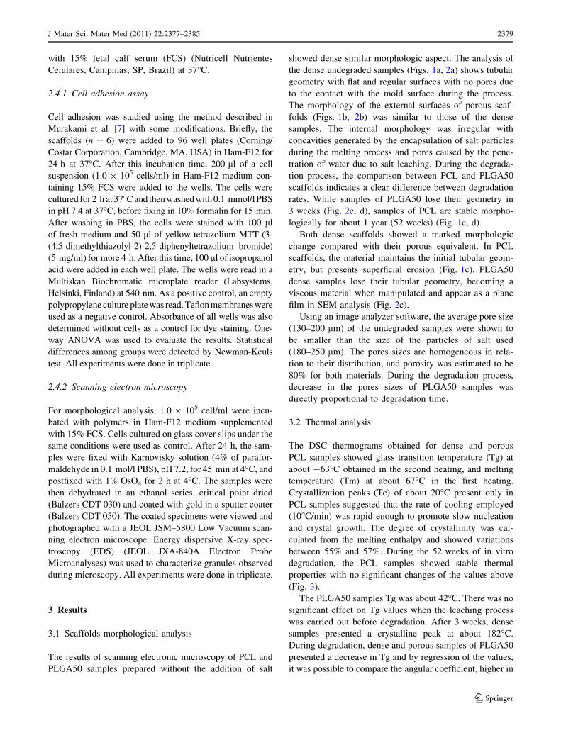
3.2 Thermal analysis
The DSC thermograms obtained for dense and porous
PCL samples showed glass transition temperature (Tg) at
about -63�C obtained in the second heating, and melting
temperature (Tm) at about 67�C in the first heating.
Crystallization peaks (Tc) of about 20�C present only in
PCL samples suggested that the rate of cooling employed
(10�C/min) was rapid enough to promote slow nucleation
and crystal growth. The degree of crystallinity was cal-
culated from the melting enthalpy and showed variations
between 55% and 57%. During the 52 weeks of in vitro
degradation, the PCL samples showed stable thermal
properties with no significant changes of the values above
(Fig. 3).
The PLGA50 samples Tg was about 42�C. There was no
significant effect on Tg values when the leaching process
was carried out before degradation. After 3 weeks, dense
samples presented a crystalline peak at about 182�C.
During degradation, dense and porous samples of PLGA50
presented a decrease in Tg and by regression of the values,
it was possible to compare the angular coefficient, higher in
J Mater Sci: Mater Med (2011) 22:2377–2385 2379
123

Fig. 1 Scanning electron micrographs of cross section of PCL samples. a, c Dense scaffolds. b, d Porous scaffolds. The numbers reported on the
top of each image indicate the degradation time in weeks
Fig. 2 Scanning electron micrographs of cross section of PLGA50 samples. a, c Dense scaffolds. b, d Porous scaffolds. The numbers reported
on the top of each image indicate the degradation time in weeks
2380 J Mater Sci: Mater Med (2011) 22:2377–2385
123
dense samples (9�C/week) when compared to porous
samples (7�C/week).
Tonset supplied by TGA analysis, showed differences in
temperature values (PCL at about 390�C and PLGA50 at
about 330�C) before and after degradation. During the
1-year degradation process, PCL was thermally stable,
whereas the PLGA50 Tonset decreased after few weeks of
degradation. Table 1.
3.3 Mechanical bending tests and gel permeation
chromatography (GPC)
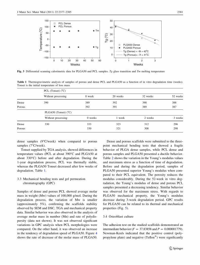
Samples of dense and porous PCL showed average molar
mass in weight (Mw) values of 100,000 g/mol. During the
degradation process, the variation of Mw is smaller
(approximately 5%), confirming the scaffolds stability
observed by SEM and DSC, TGA and mechanical property
data. Similar behavior was also observed in the analysis of
average molar mass in number (Mn) and rate of polydis-
persity (data not shown). It was not observed significant
variations in GPC analysis when PCL morphologies were
compared. On the other hand, it was observed an increase
in the tendency of degradation speed of PLGA50. Figure 4
shows the rate of decrease of the molar mass of PLGA50.
Dense and porous scaffolds were submitted to the three-
point mechanical bending tests that showed a fragile
behavior of PLGA dense samples, while PCL dense and
porous samples and PLGA50 presented a ductile behavior.
Table 2 shows the variation in the Young’s modulus values
and maximum stress as a function of time of degradation.
Before and during the degradation period, samples of
PLGA50 presented superior Young’s modulus when com-
pared to their PCL equivalent. The porosity reduces the
modulus considerably. During the 52-week in vitro deg-
radation, the Young’s modulus of dense and porous PCL
samples presented a decreasing tendency. Similar behavior
was observed for the maximum stress. With regards to
PLGA50 mechanical property, the Young’s modulus
decrease during 3-week degradation period. GPC results
for PLGA50 can be related to its thermal and mechanical
properties (Fig. 5).
3.4 Osteoblast culture
The adhesion test on the studied scaffolds demonstrated an
intermediate behavior (F = 37.83858 and P = 0.000001779).
Newman-Keuls indicated that the positive control (poly-
propylene plate) and negative (Teflon�) were significantly
0 10 20 30 40 50 60-150
-100
-50
0
50
100
150
Tg
Tm
Tem
pera
ture
(o C)
Weeks
PCL Dense PCL Porous
0 1 2 3
0
10
20
30
40
50
PLGA50 Dense PLGA50 Porous
Tg (Dense) = -9t + 42oC
Tg (Porous)= -7t + 41oC
Tg
(o C)
Weeks
Fig. 3 Differential scanning calorimetric data for PLGA50 and PCL samples. Tg glass transition and Tm melting temperature
Table 1 Thermogravimetric analysis of samples of porous and dense PCL and PLGA50 as a function of in vitro degradation time (weeks).
Tonset is the initial temperature of loss mass
PCL (Tonset) (oC)
Without processing 0 week 20 weeks 32 weeks 52 weeks
Dense 390 389 392 390 388
Porous 392 391 389 387
PLGA50 (Tonset) (oC)
Without processing 0 weeks 1 week 2 weeks 3 weeks
Dense 330 333 323 312 296
Porous 330 321 308 298
J Mater Sci: Mater Med (2011) 22:2377–2385 2381
123
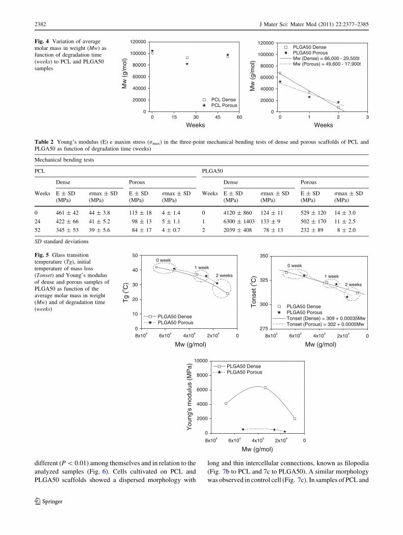
different (P \ 0.01) among themselves and in relation to the
analyzed samples (Fig. 6). Cells cultivated on PCL and
PLGA50 scaffolds showed a dispersed morphology with
long and thin intercellular connections, known as filopodia
(Fig. 7b to PCL and 7c to PLGA50). A similar morphology
was observed in control cell (Fig. 7c). In samples of PCL and
0 15 30 45 600
20000
40000
60000
80000
100000
120000
PCL Dense PCL Porous
Mw
(g/
mol
)Weeks
0 1 2 30
20000
40000
60000
80000
100000
120000 PLGA50 Dense PLGA50 Porous Mw (Dense) = 66,000 - 29,500t Mw (Porous) = 49,600 - 17,900t
Mw
(g/
mol
)
Weeks
Fig. 4 Variation of average
molar mass in weight (Mw) as
function of degradation time
(weeks) to PCL and PLGA50
samples
Table 2 Young’s modulus (E) e maxim stress (rmax) in the three-point mechanical bending tests of dense and porous scaffolds of PCL and
PLGA50 as function of degradation time (weeks)
Mechanical bending tests
PCL PLGA50
Dense Porous Dense Porous
Weeks E ± SD
(MPa)
rmax ± SD
(MPa)
E ± SD
(MPa)
rmax ± SD
(MPa)
Weeks E ± SD
(MPa)
rmax ± SD
(MPa)
E ± SD
(MPa)
rmax ± SD
(MPa)
0 461 ± 42 44 ± 3.8 115 ± 18 4 ± 1.4 0 4120 ± 860 124 ± 11 529 ± 120 14 ± 3.0
24 422 ± 66 41 ± 5.2 98 ± 13 5 ± 1.1 1 6300 ± 1403 133 ± 9 502 ± 170 11 ± 2.5
52 345 ± 53 39 ± 5.6 84 ± 17 4 ± 0.7 2 2039 ± 408 78 ± 13 232 ± 89 8 ± 2.0
SD standard deviations
8x10⁴ 6x10⁴ 4x10⁴ 2x10⁴ 00
10
20
30
40
500 week
2 weeks
1 week
PLGA50 Dense PLGA50 Porous
Tg
(o C)
Mw (g/mol)8x104 6x10⁴ 4x10⁴
2x10⁴
0
275
300
325
350
0 week
1 week
2 weeks
PLGA50 Dense PLGA50 Porous Tonset (Dense) = 309 + 0.00035Mw Tonset (Porous) = 302 + 0.0005Mw
Ton
set (
o C)
Mw (g/mol)
8x10⁴ 6x10⁴ 4x10⁴ 2x10⁴ 0
0
2000
4000
6000
8000
10000 PLGA50 Dense PLGA50 Porous
You
ng's
mod
ulus
(M
Pa)
Mw (g/mol)
Fig. 5 Glass transition
temperature (Tg), initial
temperature of mass loss
(Tonset) and Young’s modulus
of dense and porous samples of
PLGA50 as function of the
average molar mass in weight
(Mw) and of degradation time
(weeks)
2382 J Mater Sci: Mater Med (2011) 22:2377–2385
123
PLGA50, the cells were not as densely packed as in the glass
control surface; however, they showed a semi-confluent
monolayer disposition. During the electronic microscopy
analysis, it was observed crystalline granules on cells, sug-
gesting deposition of organic material. EDS was used to
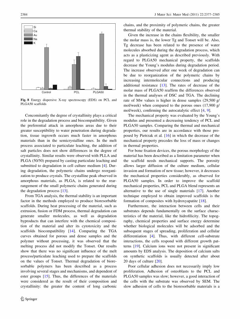
characterize the samples. Figure 8 presents the results of
EDS analysis of granules and shows different chemical
elements.
4 Discussion
A first analysis of morphology of the scaffolds showed that
the difference of degradation rate is attributed to the type of
material and their chemical composition. The hydrolysis
rate, and consequent mass loss, is directly proportional to
the number of ester bonds of the polymeric chain (greater
in the chains of PLGA50 when compared to the chains of
PCL). Several factors influence the degradation rate of the
bioresorbable scaffolds, such as chemical composition,
crystallinity, geometry and morphology of the scaffold,
surface energy and others parameters [8]. These factors are
interrelated and to treat them separately represents an
oversimplification of the degradation process.
Differences between dense and porous morphologies
exemplify the autocatalytic effect of the bioresorbable
polymers, previously presented and discussed [4]. The
smaller the area of diffusion of the degradation products,
the larger the effect of the acidic products will be [9].
The control of porosity of the polymeric supports can be
done by varying the amount of salt and the size of salt
particles. Widmer et al. [10] obtained samples of PLGA
(75/15) and porous PLLA using the extrusion process. The
authors evaluated the variation of the size of salt particles
and salt concentration (between 70 and 90%). The study
concluded that salt concentration is the most significant
parameter for the formation of scaffold porosity. Hence,
higher salt concentrations result in higher porosity. How-
ever, significant increases in the porosity reduce mechan-
ical resistance. When applied to tissue engineering, the
porosity allows a flow of nutrients and metabolic products
through the scaffolds, aiding in the process of local vas-
cularization, indispensable for tissue growth [11]. Tubular
samples similar to the ones obtained in our experiment with
regards to geometry and morphology have been developed
for applications in guided tissue regeneration, such as
peripheral nerves [12].
0,0
0,1
0,2
PLGA50PCL NegativeControl
PositiveControl
Scaffolds
Abs
orba
nce
(540
nm
)
Fig. 6 Human osteoblasts adhesion of PCL and PLGA50 scaffolds.
(P \ 0.01)
Fig. 7 Scanning electron micrographs of osteoblasts on porous PCL
and PLGA50 scaffolds. a Cells on PCL. b Cells on PLGA50. c Glass
control
J Mater Sci: Mater Med (2011) 22:2377–2385 2383
123
Concomitantly the degree of crystallinity plays a critical
role in the degradation process and biocompatibility. Given
the preferential attack in amorphous areas due to their
greater susceptibility to water penetration during degrada-
tion, tissue ingrowth occurs much faster in amorphous
materials than in the semicrystalline ones. In the melt
process associated to particulate leaching, the addition of
salt particles does not show differences in the degree of
crystallinity. Similar results were observed with PLLA and
PLGA (50/50) prepared by casting particulate leaching and
submitted to degradation in cell culture medium [4]. Dur-
ing degradation, the polymeric chains undergo reorgani-
zation to produce crystals. The crystalline peak observed in
amorphous materials, as PLGA, is related to the rear-
rangement of the small polymeric chains generated during
the degradation process [13].
From TGA analysis, the thermal stability is an important
factor in the methods employed to produce bioresorbable
scaffolds. During heat processing of the material, such as
extrusion, fusion or FDM process, thermal degradation can
generate smaller molecules, as well as degradation
byproducts that can interfere with the chemical composi-
tion of the material and alter its cytotoxicity and the
scaffolds biocompatibility [14]. Comparing the TGA
curves obtained for porous and dense samples and the
polymer without processing, it was observed that the
melting process did not modify the Tonset. Our results
show that there was no significant influence of the melt
process/particulate leaching used to prepare the scaffolds
on the values of Tonset. Thermal degradation of biore-
sorbable polymers has been described as a process
involving several stages and mechanisms, and dependent of
ester groups [15]. Thus, the differences of the materials
were considered as the result of their composition and
crystallinity: the greater the content of long carbonic
chains, and the proximity of polymeric chains, the greater
thermal stability of the material.
Given the increase in the chains flexibility, the smaller
the molar mass is, the lower Tg and Tonset will be. Also,
Tg decrease has been related to the presence of water
molecules absorbed during the degradation process, which
acts as a plasticizing agent as described previously. With
regard to PLGA50 mechanical property, the scaffolds
decrease the Young’s modulus during degradation period.
The increase observed after one week of degradation can
be due to reorganization of the polymeric chains by
increasing intermolecular connections and producing
additional resistance [13]. The rates of decrease of the
molar mass of PLGA50 reaffirm the differences observed
in the thermal analyses of DSC and TGA. The declining
rate of Mw values is higher in dense samples (29,500 g/
mol/week) when compared to the porous ones (17,900 g/
mol/week), confirming the autocatalytic effect [4, 9].
The mechanical property was evaluated by the Young’s
modulus and presented a decreasing tendency of PCL and
PLGA50 samples. Comparing the thermal and mechanical
properties, our results are in accordance with those pro-
posed by Pietrzak et al. [16] in which the decrease of the
mechanical property precedes the loss of mass or changes
in thermal properties.
For bone fixation devices, the porous morphology of the
material has been described as a limitation parameter when
the scaffold needs mechanical supports. The porosity
allows larger diffusion of the culture medium, cellular
invasion and formation of new tissue; however, it decreases
the mechanical properties considerably, as observed for
PLGA50 samples. In order to improve the scaffold
mechanical properties, PCL and PLGA blend represents an
alternative to the use of single materials [17]. Another
technique employed to obtain improved scaffolds is the
formation of composites with hydroxyapatite [18].
Furthermore, the interaction between cells and their
substrates depends fundamentally on the surface charac-
teristics of the material, like the hidrofilicity. The topog-
raphy, chemical properties and surface energy determine
whether biological molecules will be adsorbed and the
subsequent stages of spreading, proliferation and cellular
differentiation [4]. Thus, with different cell-substrate
interactions, the cells respond with different growth pat-
terns [19]. Calcium ions were not present in significant
amounts by EDS analysis. The deposition of calcium salts
on synthetic scaffolds is usually detected after about
20 days of culture [20].
Poor cellular adhesion does not necessarily imply low
proliferation. Adhesion of osteoblasts to the PCL and
PLGA50 samples was slow; however, a good interaction of
the cells with the substrate was observed by SEM. The
slow adhesion of cells to the bioresorbable materials is a
PLGA50PCLControl
Si Ca Au Na K
Fig. 8 Energy dispersive X-ray spectroscopy (EDS) on PCL and
PLGA50 scaffolds
2384 J Mater Sci: Mater Med (2011) 22:2377–2385
123
phenomenon that has already been described [19, 21], and
the present results suggest that this phenomenon happens
on porous PLGA50 and PCL samples. In protocols that
allowed longer time for adhesion evaluation, the cells
begin multiplication on the substrate [22]. In this sense,
systems based on bioresorbable polymers to which growth
factors are incorporated are thoroughly studied [23].
5 Conclusion
Different types of bioresorbable polymers were used in the
study. The degradation of the PLGA50 samples was rapid
when compared to PCL because PLGA50 is an amorphous
material and has a higher number of ester bonds. The
autocatalytic effect of poly(a-hydroxy acids) was seen in
dense PLGA50 samples as compared to the porous sam-
ples. The samples of PCL were stable morphologically for
1 year in phosphate buffer solution. Cultured osteoblasts
showed a favorable proliferation and differentiation on the
samples. These results show that scaffolds of PCL can be
used when a substrate with a prolonged degradation is
required to serve as a physical support for cell cultures,
before and after implantation. Scaffolds of PLGA degrade
quickly and may be useful for the formation of mature
tissue prior to implantation.
Acknowledgments This work was supported by the Brazilian
National Council for Scientific and Technological Development
(CNPq) (Grant 141582).
References
1. Gloria A, De Santis R, Ambrosio L. Polymer-based composite
scaffolds for tissue engineering. J Appl Biomater Biomech.
2010;8(2):57–67.
2. Vert M. Degradable and bioresorbable polymers in surgery and in
pharmacology: beliefs and facts. J Mater Sci Mater Med. 2009;
20(2):437–46.
3. Kretlow JD, Klouda L, Mikos AG. Injectable matrices and
scaffolds for drug delivery in tissue engineering. Adv Drug Deliv
Rev. 2007;59(4–5):263–73.
4. Barbanti SH, Santos AR Jr, Zavaglia CAC, Duek EAR. Porous
and dense poly(L-lactic acid) and poly(D,L-lactic acid-co-glycolic
acid) scaffolds: in vitro degradation in culture medium and
osteoblasts culture. J Mater Sci Mater Med. 2004;15:1315–21.
5. Mooney DJ, Baldwin DF, Suh NP, Vacanti JP, Langer R. Novel
approach to fabricate porous sponges of poly(D,L-lactic-co-gly-
colic acid) without the use of organic solvents. Biomaterials.
1996;17:1417–22.
6. Eldsater C, Erlandsson B, Renstad R, Albertsson AC, Karlsson S.
The biodegradation of amorphous and crystalline regions in film-
blown poly(e-caprolactone). Polymer. 2000;41:1297–304.
7. Murakami N, Fukuchi S, Takeuchi K, Hori T, Shibamoto S, Ito F.
Antagonistic regulation of cell migration by epidermal growth
factor and glucocorticoid in human gastric carcinoma cells. J Cell
Physiol. 1998;176(1):127–37.
8. Eglin D, Alini M. Degradable polymeric materials for osteo-
synthesis: tutorial. Eur Cell Mater. 2008;16:80–91.
9. Li S. Hydrolytic degradation characteristics of aliphatic polyes-
ters derived from lactic and glycolic acids. J Biomed Mater Res.
1999;48:342–53.
10. Widmer MS, Gupta PK, Lu L, Meszlenyi RK, Evans GRD,
Brandt K, Savel T, Gurlek A, Patrick CW, Mikos AG. Manu-
facture of porous biodegradable polymer conduits by an extrusion
process for guided tissue regeneration. Biomaterials. 1998;19:
1945–55.
11. Mikos A, Temenoff J. Formation of highly porous biodegradable
scaffolds for tissue engineering. Electron J Biotechnol. 2000;3:
114–9. http://www.ejbiotechnology.info/index.php/ejbiotechnology/
article/view/427. Accessed 24 June 2011.
12. Plikk P, Malberg S, Albertsson AC. Design of resorbable porous
tubular copolyester scaffolds for use in nerve regeneration. Bio-
macromolecules. 2009;10(5):1259–64.
13. Duek EAR, Zavaglia CAC, Belangero WD. In vitro study of
poly(lactic acid) pin degradation. Polymer. 1999;40:6465–73.
14. Wu L, Jing D, Ding J. A ‘‘room-temperature’’ injection molding/
particulate leaching approach for fabrication of biodegradable
three-dimensional porous scaffolds. Biomaterials. 2006;27(2):
185–91.
15. Penco M, Sartore L, Bignotti F, D’antone S, Landro L. Thermal
properties of a new class of block copolymers based on segments
of poly(D,L-lactic-glycolic acid) and poly(e-caprolactone).
J European Polym. 2000;36:901–8.
16. Pietrzak WS, Sarver DR, Verstynen ML. Bioabsorbable polymer
science for the practicing surgeon. J Craniofac Surg. 1997;8(2):
87–91.
17. Kim JY, Yoon JJ, Park EK, Kim DS, Kim SY, Cho DW. Cell
adhesion and proliferation evaluation of SFF-based biodegrad-
able scaffolds fabricated using a multi-head deposition system.
Biofabrication. 2009;1(1):015002.
18. Ren J, Zhao P, Ren T, Gu S, Pan K. Poly (D,L-lactide)/nano-
hydroxyapatite composite scaffolds for bone tissue engineering
and biocompatibility evaluation. J Mater Sci Mater Med. 2008;
19(3):1075–82.
19. Santos AR Jr, Barbanti SH, Duek EAR, Dolder H, Wada RS,
Wada MLF. Vero cell growth and differentiation on poly(L-lactic
acid) membranes of different pore diameters. Artif Organs. 2001;
25:7–13.
20. Moreira PL, An YH, Santos AR Jr, Genari SC. In vitro analysis of
anionic collagen scaffolds for bone repair. J Biomed Mater Res B
Appl Biomater. 2004;15:229–37.
21. Lombello CB, Santos AR Jr, Malmonge SM, Barbanti SH, Wada
MLF, Duek EAR. Adhesion and morphology of fibroblastic cells
cultured on different polymeric biomaterials. J Mater Sci Mater
Med. 2002;13:867–74.
22. van Eijk F, Saris DB, Creemers LB, Riesle J, Willems WJ, van
Blitterswijk CA, Verbout AJ, Dhert WJ. The effect of timing of
mechanical stimulation on proliferation and differentiation of
goat bone marrow stem cells cultured on braided PLGA scaf-
folds. Tissue Eng. 2008;14(8):1425–33.
23. Bessa PC, Casal M, Reis RL. Bone morphogenetic proteins in
tissue engineering: the road from laboratory to clinic, part II
(BMP delivery). J Tissue Eng Regen Med. 2008;2(2–3):81–96.
J Mater Sci: Mater Med (2011) 22:2377–2385 2385
123
Copyright © 2022 FDOKUMEN