RECOGNITION AND SELECTION OF SETTLEMENT SUBSTRATA DETERMINE POST-SETTLEMENT SURVIVAL IN CORALS
rXXXX American Chemical Society A dx.doi.org/10.1021/la203140a | Langmuir XXXX, XXX, 000–000
ARTICLE
pubs.acs.org/Langmuir
Evaluation of Substrata Effect on Cell Adhesion Properties UsingFreestanding Poly(L-lactic acid) NanosheetsToshinori Fujie,*,†,‡ Leonardo Ricotti,†,§ Andrea Desii,†,§ Arianna Menciassi,†,§ Paolo Dario,†,§ andVirgilio Mattoli*,†
†Center for MicroBioRobotics IIT@SSSA, Istituto Italiano di Tecnologia, Viale Rinaldo Piaggio 34 Pontedera, Pisa, 56025, Italy‡European Biomedical Science Institute (EBSI), Organization for European Studies, Waseda University, 2-2 Wakamtsu-cho, Shinjuku,Tokyo 162-8480, Japan§The BioRobotics Institute, Polo Sant’Anna Valdera, Scuola Superiore Sant’Anna, Viale Rinaldo Piaggio 34 Pontedera, Pisa, 56025, Italy
bS Supporting Information
’ INTRODUCTION
Investigation of the interactions between cells and materialsurfaces plays an important role in the development of smartbiomaterials. There have been several attempts to constructengineered scaffolds or matrices paying attention to their chemi-cal and physical surface properties, in order to obtain suitableinteractions with cells.1�3 These studies have been undertakenon solid substrates such as plastics, glass and SiO2. However, suchmicroenvironments are substantially different from those sur-rounding the living systems from the mechanical point of view(frequently called “mechanobiology”).4 For example, mechano-taxis is one of themost important aspects for cell mechanobiology,as cells sensematrix stiffness and preferentially adhere onto harderregions.5,6 Besides, the matrix stiffness was also unveiled to haveeffects on proliferation, migration and even differentiation. Theseapproaches have been undertaken by integrating soft materials,such as elastic gels,4 rubbers,7 and functional membranes,8 thanksto their mechanical tunability, achieved by changing the degree ofchemical cross-linking.9,10
Freestanding polymeric ultrathin films (nanosheets) can beraised as a new category of the quasi-two-dimensional soft materials.Typical features of the polymeric nanosheet are tens-of-nano-meters thickness, a huge size-aspect ratio (>106), and uniqueinterfacial and mechanical properties, such as tunable flexibility,noncovalent adhesiveness, and high transparency.11 From thestructural point of view, the quasi-two-dimensional structure ofthe polymeric nanosheet could represent an ideal interface tomimic a native extracellular matrix (ECM). Freestanding poly-meric nanosheets have been fabricated using different approaches,including simple spin-coating,12 layer-by-layer (LbL) method,13,14
Langmuir�Blodgett method with cross-linkable amphiphilic co-polymers,15 and sol�gel method with organic�inorganic inter-penetrating networks.16 Up to date, clinical benefits of the poly-meric nanosheets have been investigated for biomedical applications,
Received: August 11, 2011Revised: September 5, 2011
ABSTRACT: Investigation of the interactions between cells and materialsurfaces is important not only for the understanding of cell biology but alsofor the development of smart biomaterials. In this study, we investigatedthe substrate-related effects on the interaction between cell and polymericultrathin film (nanosheet) by modulating the mechanical properties of thenanosheet with a metal substrate or mesh. A freestanding polymericnanosheet with tens-of-nanometers thickness composed of poly(L-lacticacid) (PLLA nanosheet) was fabricated by combination of a spin-coatingtechnique and a water-soluble sacrificial layer. The freestanding PLLAnanosheet was collected on a stainless steel mesh (PLLA�mesh) andsubsequently used for cell adhesion studies, comparing the results to theones on a control SiO2 substrate coated with an ultrathin layer of PLLA(PLLA�substrate). The adhesion of rat cardiomyocytes (H9c2) wasevaluated on both samples after 24 h of culture. The PLLA�mesh with the tens-of-nanometers thick nanosheets induced ananisotropic adhesion of H9c2, while H9c2 on the PLLA�substrate showed an isotropic adhesion independent from the nanosheetthickness. Interestingly, an increment in the nanosheet thickness in the PLLA�mesh samples reduced the cellular anisotropy andled to a similar morphology to the PLLA�substrate. Considering the huge discrepancy of Young’s modulus between PLLAnanosheet (3.5�4.2GPa) andmetal substrate (hundreds of GPa), cell adhesion wasmechanically regulated by the Young’smodulusof the underlying substrate when the thickness of the PLLA nanosheet was tens of nanometers. Modulation of the stiffness of thepolymeric nanosheet by utilizing a rigid underlying material will allow the constitution of a unique cell culture environment.
B dx.doi.org/10.1021/la203140a |Langmuir XXXX, XXX, 000–000
Langmuir ARTICLE
such as tissue-defect repair without surgical adhesion,17,18 localdelivery of antibiotics by implantation,19 remote-controllable tissuesealing,20,21 and other applications in different fields.22 We cur-rently focused on the flexible structure of the polymeric na-nosheet and pursued biomedical research in the development oftissue engineering scaffold and biomedical devices.23�25 Thesestudies revealed the influence of the surface chemistry on celladhesion properties, including morphology, adhesion area, andelongation ratio. However, the entire complexity of cell activityon the polymeric nanosheet has not been fully understood becauseseveral factors other than surface chemistry could also influencethe cellular activity (e.g., surface topology and mechanical pro-perties). In particular, the substrate-related effect on the interac-tion between cells and a polymeric nanosheet is not negligible,because the mechanical flexibility of the nanosheet can beimpaired by the stiffness of the underlying material during cellculture.
In this study, we investigated the effect of an underlying sub-strate on the interaction between cells and nanosheet, by couplingthe flexible nanosheet with rigidmaterials, such as SiO2 substratesand metal meshes. A polymeric nanosheet composed of biocom-patible poly(L-lactic acid) (referred to as “PLLA nano-sheet”) was fabricated by combination of a spin-coating tech-nique and a water-soluble sacrificial layer method, which facili-tated the aqueous exfoliation of a freestanding PLLA nanosheetsshowing a thickness from tens to hundreds of nanometers. Thesenanosheets were collected on stainless steel meshes (i.e., PLLA�mesh) and subsequently used for cell adhesion analysis on thePLLA nanosheet. On the other hand, we prepared PLLA-coatedSiO2 substrates by directly spin-coating a PLLA solution (i.e.,PLLA�substrate) as a control sample. Then, cell activity andadhesion of rat cardiomyocytes (H9c2) were compared for bothsample typologies after 24 h of culture. H9c2 is a permanent cellline derived from rat cardiac tissue,26 sensitive to matrix topo-graphy and substrate stiffness. Therefore, adhesion, proliferation,and differentiation rate ofH9c2myoblasts are strongly affected bythe matrix properties.27,28 Furthermore, we characterized surfacemorphology and mechanical properties of the PLLA nanosheetsusing scanning electron microscope (SEM), atomic force micro-scopy (AFM), and strain-induced elastic buckling instability formechanical measurement (SIEBIMM) test. The role of the under-lying substrate on the biointerfacial properties was thus clarified,evaluating how it modulated the topological and mechanicalcharacteristics of the PLLA nanosheets.
’MATERIALS AND METHODS
Materials. SiO2 substrates (Si-Mat Silicon Materials, Kaufering,Germany), used as substrates for film deposition, were cut (2� 2 cm2),treated with an acid mixture [SPM 96% H2SO4:30% H2O2 = 4:1 (v/v)]at 120 �C for 10 min, and then thoroughly rinsed with deionized (DI)water (18 MΩ cm) in order to remove impurities from the surface.Poly(vinyl alcohol) (PVA), average Mw 13 000�23 000, 98% hydrolyzed,was purchased fromKantoChemical Co., Inc. (Tokyo, Japan). Poly(L-lacticacid) (PLLA), Mw 80 000�100 000, was obtained from Polysciences Inc.(Warrington, PA). A stainless steel mesh with a 0.5 mm � 0.5 mm latticecomposed of 150-μm-wide stainless steel wires was obtained from TokyuHands, Co. Ltd. (Tokyo, Japan). All other chemicals were purchasedfrom Sigma-Aldrich (St. Louis, MO) and used without further treatment,unless noted.Preparation of Freestanding PLLA Nanosheets. Freestand-
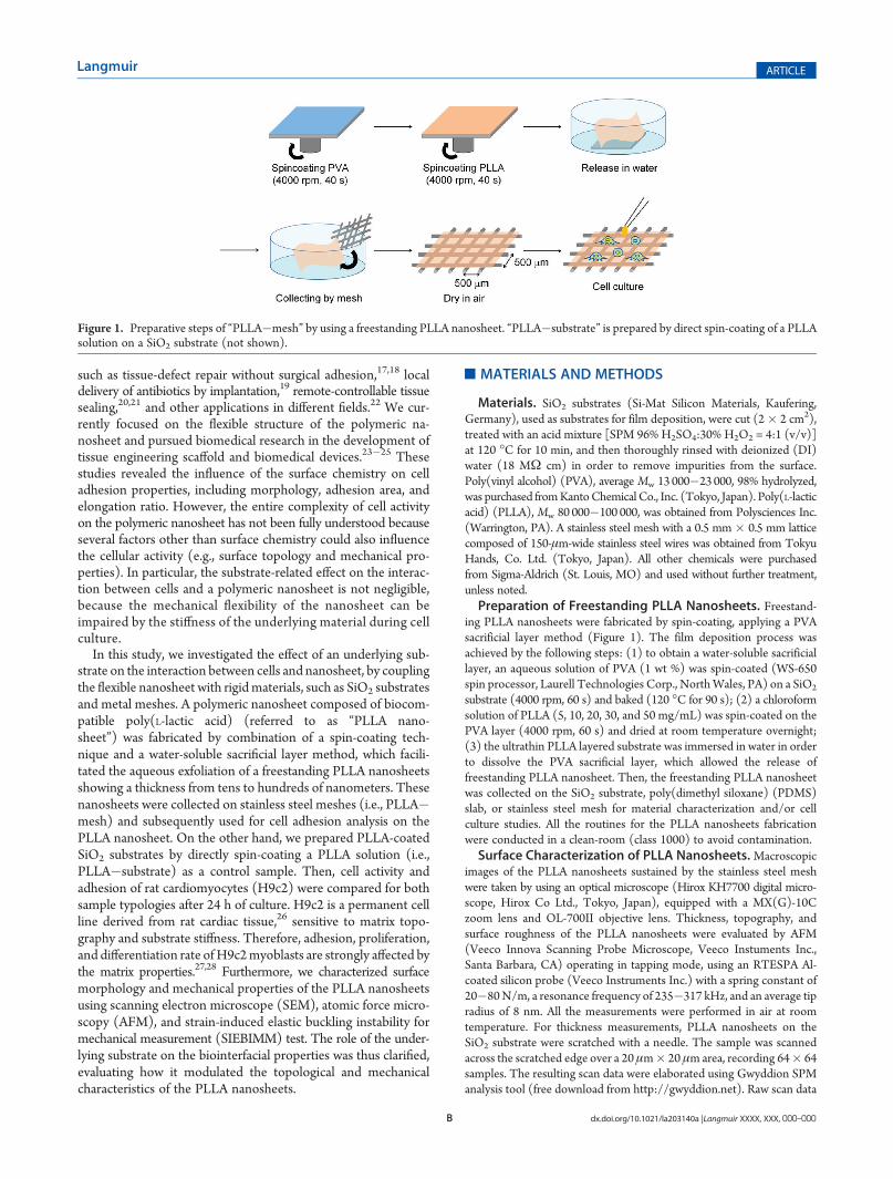
ing PLLA nanosheets were fabricated by spin-coating, applying a PVAsacrificial layer method (Figure 1). The film deposition process wasachieved by the following steps: (1) to obtain a water-soluble sacrificiallayer, an aqueous solution of PVA (1 wt %) was spin-coated (WS-650spin processor, Laurell Technologies Corp., NorthWales, PA) on a SiO2
substrate (4000 rpm, 60 s) and baked (120 �C for 90 s); (2) a chloroformsolution of PLLA (5, 10, 20, 30, and 50 mg/mL) was spin-coated on thePVA layer (4000 rpm, 60 s) and dried at room temperature overnight;(3) the ultrathin PLLA layered substrate was immersed in water in orderto dissolve the PVA sacrificial layer, which allowed the release offreestanding PLLA nanosheet. Then, the freestanding PLLA nanosheetwas collected on the SiO2 substrate, poly(dimethyl siloxane) (PDMS)slab, or stainless steel mesh for material characterization and/or cellculture studies. All the routines for the PLLA nanosheets fabricationwere conducted in a clean-room (class 1000) to avoid contamination.Surface Characterization of PLLA Nanosheets.Macroscopic
images of the PLLA nanosheets sustained by the stainless steel meshwere taken by using an optical microscope (Hirox KH7700 digital micro-scope, Hirox Co Ltd., Tokyo, Japan), equipped with a MX(G)-10Czoom lens and OL-700II objective lens. Thickness, topography, andsurface roughness of the PLLA nanosheets were evaluated by AFM(Veeco Innova Scanning Probe Microscope, Veeco Instuments Inc.,Santa Barbara, CA) operating in tapping mode, using an RTESPA Al-coated silicon probe (Veeco Instruments Inc.) with a spring constant of20�80N/m, a resonance frequency of 235�317 kHz, and an average tipradius of 8 nm. All the measurements were performed in air at roomtemperature. For thickness measurements, PLLA nanosheets on theSiO2 substrate were scratched with a needle. The sample was scannedacross the scratched edge over a 20 μm� 20 μm area, recording 64� 64samples. The resulting scan data were elaborated using Gwyddion SPManalysis tool (free download from http://gwyddion.net). Raw scan data
Figure 1. Preparative steps of “PLLA�mesh” by using a freestanding PLLA nanosheet. “PLLA�substrate” is prepared by direct spin-coating of a PLLAsolution on a SiO2 substrate (not shown).

C dx.doi.org/10.1021/la203140a |Langmuir XXXX, XXX, 000–000
Langmuir ARTICLE
were leveled with a facet level tool to remove sample tilt, and then thesample profile was extracted in a direction perpendicular to the scratchedge. Height data were averaged and the thickness was evaluated fromthe difference between horizontal lines fitting to the SiO2 substrate andthe PLLA nanosheet. For roughness (root mean square, rms) measure-ment, the surface of the PLLA nanosheets recollected on the SiO2
substrates was scanned in tapping mode over 2 μm� 2 μm area (512�512 samples). Then, the rms values were obtained from the topogra-phical images. Microscopic surface morphology of the PLLA�meshsamples was investigated by SEM (EVO/MA10, Carl Zeiss SMT,Oberkochen, Germany) without metal sputtering.Mechanical Characterization of PLLA Nanosheets. The
mechanical properties of the PLLA nanosheet were evaluated by meansof the SIEBIMM technique. The SIEBIMM test is based on the bucklingmetrology and allowed the calculation of Young’smodulus for polymericnanosheets. As reported previously in the literature,29�31 the elasticmodulus can be calculated by measuring the buckling wavelength of thenanosheet on a mechanically compressed or stretched matrix. A free-standing PLLA nanosheet was collected from water onto a prestretched(∼3% strain of the original size) PDMS slab (1.5 cm �4.0 cm). Theprepared sample was dried in a desiccator overnight prior to theSIEBIMM test. Then, the strain of the PDMS substrate was relaxed,producing a relative compression of the PLLA nanosheet and generatinga characteristic corrugation. Immediately, the wavelength (λ) of thebuckling pattern on the PLLA nanosheet was analyzed using an opticalmicroscope. Then, the Young’s modulus (EPLLA) of the PLLA na-nosheets with different thickness (h) was individually obtained using thefollowing formula 1
EPLLA ¼ 3EPDMSð1� vPLLA2Þ
1� vPDMS2
λ
2πh
� �3
ð1Þ
where E and v represented the Young’s modulus and Poisson’s ratio ofPLLA nanosheet and PDMS (i.e., EPDMS = 1.8 MPa). In this calculation,we assumed Poisson’s ratios for the PLLA nanosheets and for the PDMSequal to 0.33 and 0.50, respectively, in accordance with previousreports.30,31
In addition, the stiffness of the PLLA nanosheet was also evaluated byusing tapping mode AFM scans, where the phase images (15 μm� 15 μmrange) of the PLLA�mesh samples were collected with a scanningdirection perpendicular to a metal wire. The recorded data wereaveraged on columns and displayed as average profiles in order toevaluate the gradient of topographical and mechanical properties acrossthe metal wire border.Cell Culture. H9c2 cells (embryonic myocardium rat cell line,
ATCC, CLR-1446, Milan, Italy) were cultured in expansion medium,constituted by Dulbecco’s modified Eagle’s medium (DMEM), supple-mented with 10% fetal bovine serum (FBS), 100 μg/mL gentamycin,and 2 mM L-glutamine. Cells were maintained at 37 �C in a saturatedhumidity atmosphere (95% air, 5% CO2). Before reaching confluent,the cells were detached from a polystyrene culture plate by means of a0.05 wt % trypsin, 0.53 mmol/L EDTA-4Na solution with phenol red.The aliquot was purified by centrifugation and suspended in freshculture medium. Then, a small quantity of a cell suspension (200 μL)was gently deposited on the sample surface with a seeding density of1.5� 104 cells/cm2 and incubated for 30 min in the cell incubator. Thisinitial treatment facilitated the stable tethering of H9c2 on the PLLAsurface. Afterward, additional culture medium was added, and thesample was cultured under standard conditions for 24 h.Cell Activity Assay. Cell activity of H9c2 on PLLA�mesh and
PLLA�substrate was evaluated at 24 h after cell seeding using two tests.First, a qualitative viability assay was performed by using the LIVE/DEAD Viability/Cytotoxicity kit (Invitrogen Co., Carlsbad, CA). Thekit contains calcein AM (4 mM in anhydrous DMSO) and ethidium
homodimer-1 (EthD-1, 2mM inDMSO/H2O 1:4 v/v), which identifieslive versus dead cells on the basis of membrane integrity and esteraseactivity. After 24 h of cell culture, the culture medium was removed, andthe cellular layers on the sample surface were rinsed with phosphatebuffered saline (PBS), which was then treated for 10 min at 37 �C with2 μMcalcein AM and 4μMEthD-1. Cells were finally observed under aninverted fluorescent microscope (TE2000U, FITC-TRITC filters, Ni-kon Co., Tokyo, Japan) equipped with a cooled CCD camera (DS-5MCUSB2,NikonCo., Tokyo, Japan) andwithNISElements Imaging Software.
Second, the cell population was quantitatively evaluated, measuringthe DNA concentration for each type of sample after 24 h of culture. Thesamples were removed from the original cell culture wells and placed innew wells, which were treated with appropriate volumes of DI water.Cell lysates were then obtained by two freeze/thaw cycles of the samples—overnight freezing at �80 �C and 15 min thawing at 37 �C in anultrasonication bath—to enable the DNA to go into the aqueous media.The DNA content in the cell lysates was measured by using the PicoGreenkit (Invitrogen Co., Carlsbad, CA). The PicoGreen dye binds to DNA,and the resulting fluorescence intensity is directly proportional to theDNA concentration. Standard solutions of DNA in DI water at con-centrations from 0 up to 6 μg/mL were prepared and 50 μL of standardor sample were loaded for quantification in a 96-well black microplate.Working buffer and PicoGreen dye solution were prepared and addedaccording to the manufacturer’s instructions (100 and 150 μL/well,respectively). After 10 min of incubation in the dark at room tempera-ture, fluorescence intensity was measured on a microplate reader(Victor3, PerkinElmer Inc., Waltham, MA) using an excitation wave-length of 485 nm and an emission wavelength of 535 nm.Cell Adhesion Properties. The adhesion properties of H9c2 on
the samples were evaluated by measuring the adhesion area and elongationratio of a representative sample (more than five images for each material).First, cells were fixed with a 4 wt % paraformaldehyde solution after 24 hof culture in the expansion medium, and their membrane became per-meabilized by means of a 0.1% Triton-X solution in PBS. Second, thefixed cells were stained with Alexa Fluor 594 phalloidin and Hoechst(Invitrogen Co., Carlsbad, CA). Finally, the cell adhesion area and theelongation ratio were analyzed using ImageJ, a software for image analysis(free download from NIH, http://rsbweb.nih.gov/ij/). Individual cellswere identified by the fluorescent signals of nuclei, and the contour ofcells was distinguished by fluorescent contrast of actin filaments afterconverting the color image into gray scale mode. The cell elongationratio was defined by the following formula 223,32
ε ¼ rmax=rmin ð2Þwhere ε represents the cell elongation ratio, and rmax and rmin representthe maximum and minimum distance of adhered pseudopodia from themedian point of the cell. Cell adhesion was compared between PLLA�mesh and PLLA�substrate samples (excluding cell adhesive region onthe metal wire from the analytical region of the PLLA�mesh) andbetween the inside and outside regions of the individual mesh lattice inthe PLLA�mesh samples. According to these evaluations, the substrateeffect on cell adhesion properties could be investigated for differentmaterials: plain nanosheet, nanosheet supported by SiO2 substrate, andnanosheet supported by steel mesh.Statistical Analysis. All data are presented as mean values ( SD.
Statistical analysis of the cell adhesion area and the cell elongation ratiowas performed using the unpaired t test, with *p < 0.05 and **p < 0.01 setas the level of statistical significance.
’RESULTS AND DISCUSSIONS
Surface Properties of PLLA Nanosheets. The thickness ofthe PLLA nanosheets was easily controlled by changing thePLLA concentration and keeping the same spin-coating conditions
D dx.doi.org/10.1021/la203140a |Langmuir XXXX, XXX, 000–000
Langmuir ARTICLE
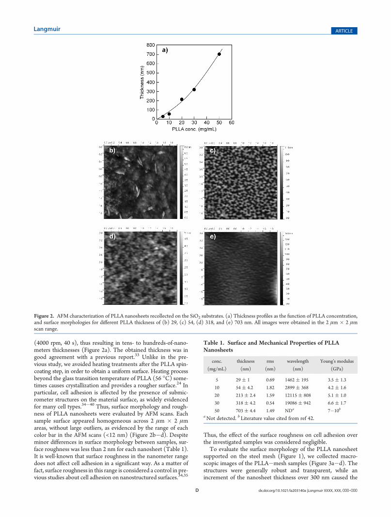
(4000 rpm, 40 s), thus resulting in tens- to hundreds-of-nano-meters thicknesses (Figure 2a). The obtained thickness was ingood agreement with a previous report.33 Unlike in the pre-vious study, we avoided heating treatments after the PLLA spin-coating step, in order to obtain a uniform surface. Heating processbeyond the glass transition temperature of PLLA (56 �C) some-times causes crystallization and provides a rougher surface.24 Inparticular, cell adhesion is affected by the presence of submic-rometer structures on the material surface, as widely evidencedfor many cell types.34�40 Thus, surface morphology and rough-ness of PLLA nanosheets were evaluated by AFM scans. Eachsample surface appeared homogeneous across 2 μm � 2 μmareas, without large outliers, as evidenced by the range of eachcolor bar in the AFM scans (<12 nm) (Figure 2b�d). Despiteminor differences in surface morphology between samples, sur-face roughness was less than 2 nm for each nanosheet (Table 1).It is well-known that surface roughness in the nanometer rangedoes not affect cell adhesion in a significant way. As a matter offact, surface roughness in this range is considered a control in pre-vious studies about cell adhesion on nanostructured surfaces.34,35
Thus, the effect of the surface roughness on cell adhesion overthe investigated samples was considered negligible.To evaluate the surface morphology of the PLLA nanosheet
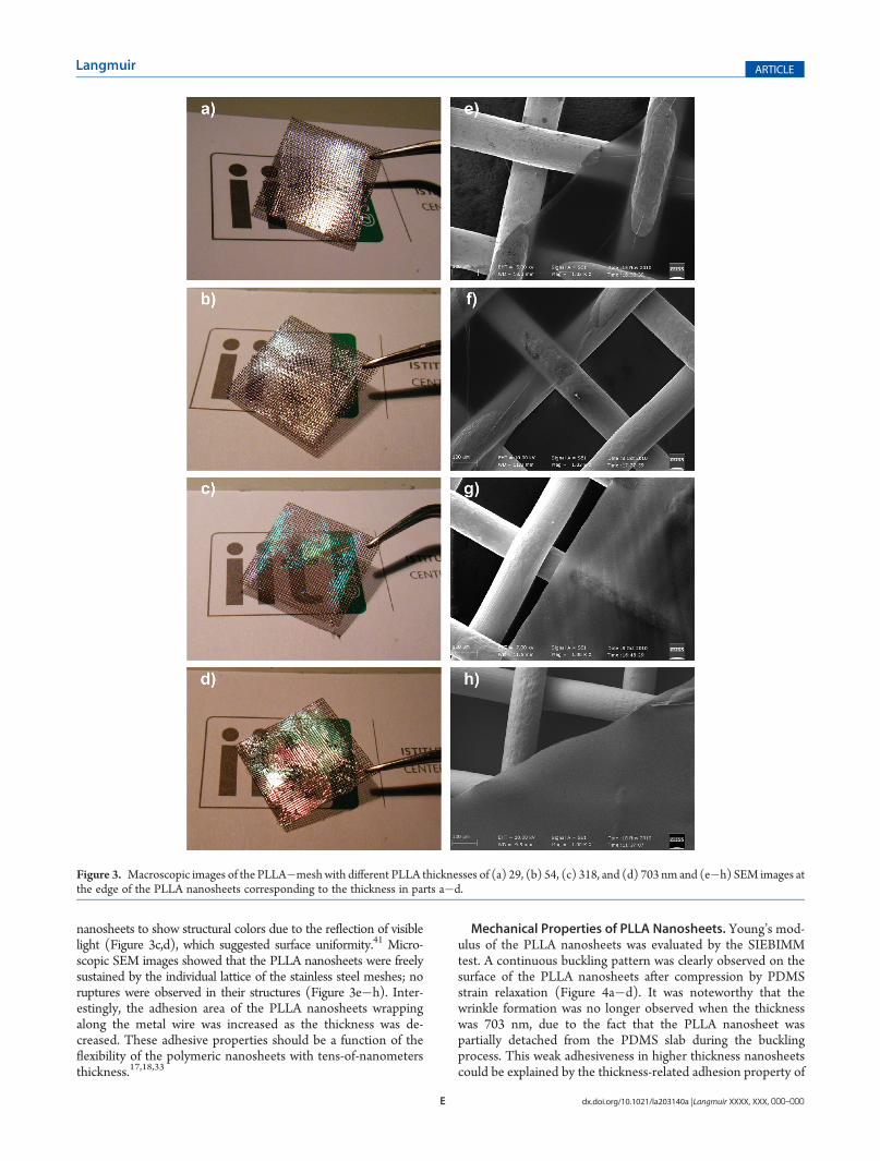
supported on the steel mesh (Figure 1), we collected macro-scopic images of the PLLA�mesh samples (Figure 3a�d). Thestructures were generally robust and transparent, while anincrement of the nanosheet thickness over 300 nm caused the
Figure 2. AFM characterization of PLLA nanosheets recollected on the SiO2 substrates. (a) Thickness profiles as the function of PLLA concentration,and surface morphologies for different PLLA thickness of (b) 29, (c) 54, (d) 318, and (e) 703 nm. All images were obtained in the 2 μm � 2 μmscan range.
Table 1. Surface and Mechanical Properties of PLLANanosheets
conc.
(mg/mL)
thickness
(nm)
rms
(nm)
wavelength
(nm)
Young’s modulus
(GPa)
5 29 ( 1 0.69 1462 ( 195 3.5 ( 1.3
10 54 ( 4.2 1.82 2899 ( 368 4.2 ( 1.6
20 213 ( 2.4 1.59 12115 ( 808 5.1 ( 1.0
30 318 ( 4.2 0.54 19086 ( 942 6.6 ( 1.7
50 703 ( 4.4 1.49 NDa 7�10b
aNot detected. b Literature value cited from ref 42.
E dx.doi.org/10.1021/la203140a |Langmuir XXXX, XXX, 000–000
Langmuir ARTICLE
nanosheets to show structural colors due to the reflection of visiblelight (Figure 3c,d), which suggested surface uniformity.41 Micro-scopic SEM images showed that the PLLA nanosheets were freelysustained by the individual lattice of the stainless steel meshes; noruptures were observed in their structures (Figure 3e�h). Inter-estingly, the adhesion area of the PLLA nanosheets wrappingalong the metal wire was increased as the thickness was de-creased. These adhesive properties should be a function of theflexibility of the polymeric nanosheets with tens-of-nanometersthickness.17,18,33
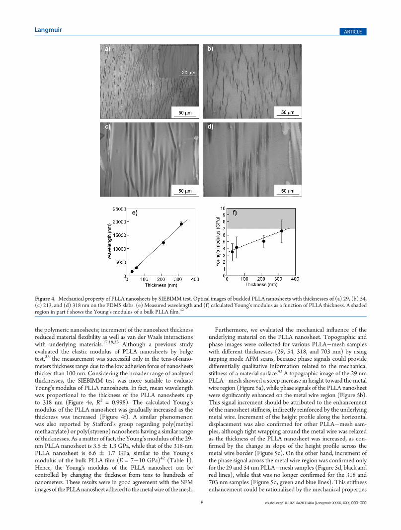
Mechanical Properties of PLLA Nanosheets. Young’s mod-ulus of the PLLA nanosheets was evaluated by the SIEBIMMtest. A continuous buckling pattern was clearly observed on thesurface of the PLLA nanosheets after compression by PDMSstrain relaxation (Figure 4a�d). It was noteworthy that thewrinkle formation was no longer observed when the thicknesswas 703 nm, due to the fact that the PLLA nanosheet waspartially detached from the PDMS slab during the bucklingprocess. This weak adhesiveness in higher thickness nanosheetscould be explained by the thickness-related adhesion property of
Figure 3. Macroscopic images of the PLLA�meshwith different PLLA thicknesses of (a) 29, (b) 54, (c) 318, and (d) 703 nm and (e�h) SEM images atthe edge of the PLLA nanosheets corresponding to the thickness in parts a�d.
F dx.doi.org/10.1021/la203140a |Langmuir XXXX, XXX, 000–000
Langmuir ARTICLE
the polymeric nanosheets; increment of the nanosheet thicknessreduced material flexibility as well as van der Waals interactionswith underlying materials.17,18,33 Although a previous studyevaluated the elastic modulus of PLLA nanosheets by bulgetest,33 the measurement was successful only in the tens-of-nano-meters thickness range due to the low adhesion force of nanosheetsthicker than 100 nm. Considering the broader range of analyzedthicknesses, the SIEBIMM test was more suitable to evaluateYoung’s modulus of PLLA nanosheets. In fact, mean wavelengthwas proportional to the thickness of the PLLA nanosheets upto 318 nm (Figure 4e, R2 = 0.998). The calculated Young’smodulus of the PLLA nanosheet was gradually increased as thethickness was increased (Figure 4f). A similar phenomenonwas also reported by Stafford’s group regarding poly(methylmethacrylate) or poly(styrene) nanosheets having a similar rangeof thicknesses. As a matter of fact, the Young’s modulus of the 29-nm PLLA nanosheet is 3.5 ( 1.3 GPa, while that of the 318-nmPLLA nanosheet is 6.6 ( 1.7 GPa, similar to the Young’smodulus of the bulk PLLA film (E = 7�10 GPa)42 (Table 1).Hence, the Young’s modulus of the PLLA nanosheet can becontrolled by changing the thickness from tens to hundreds ofnanometers. These results were in good agreement with the SEMimages of thePLLAnanosheet adhered to themetalwire of themesh.
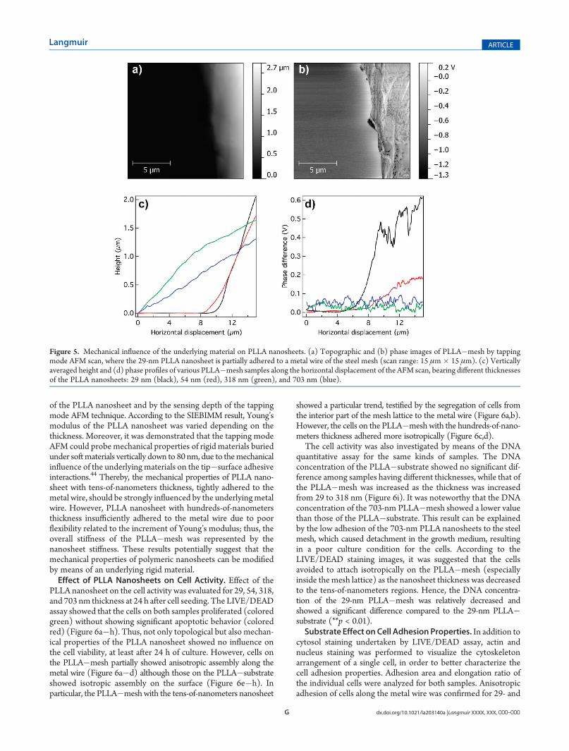
Furthermore, we evaluated the mechanical influence of theunderlying material on the PLLA nanosheet. Topographic andphase images were collected for various PLLA�mesh sampleswith different thicknesses (29, 54, 318, and 703 nm) by usingtapping mode AFM scans, because phase signals could providedifferentially qualitative information related to the mechanicalstiffness of a material surface.43 A topographic image of the 29-nmPLLA�mesh showed a steep increase in height toward the metalwire region (Figure 5a), while phase signals of the PLLA nanosheetwere significantly enhanced on the metal wire region (Figure 5b).This signal increment should be attributed to the enhancementof the nanosheet stiffness, indirectly reinforced by the underlyingmetal wire. Increment of the height profile along the horizontaldisplacement was also confirmed for other PLLA�mesh sam-ples, although tight wrapping around the metal wire was relaxedas the thickness of the PLLA nanosheet was increased, as con-firmed by the change in slope of the height profile across themetal wire border (Figure 5c). On the other hand, increment ofthe phase signal across the metal wire region was confirmed onlyfor the 29 and 54 nmPLLA�mesh samples (Figure 5d, black andred lines), while that was no longer confirmed for the 318 and703 nm samples (Figure 5d, green and blue lines). This stiffnessenhancement could be rationalized by the mechanical properties
Figure 4. Mechanical property of PLLA nanosheets by SIEBIMM test. Optical images of buckled PLLA nanosheets with thicknesses of (a) 29, (b) 54,(c) 213, and (d) 318 nm on the PDMS slabs. (e) Measured wavelength and (f) calculated Young’s modulus as a function of PLLA thickness. A shadedregion in part f shows the Young’s modulus of a bulk PLLA film.42
G dx.doi.org/10.1021/la203140a |Langmuir XXXX, XXX, 000–000
Langmuir ARTICLE
of the PLLA nanosheet and by the sensing depth of the tappingmode AFM technique. According to the SIEBIMM result, Young’smodulus of the PLLA nanosheet was varied depending on thethickness. Moreover, it was demonstrated that the tapping modeAFM could probe mechanical properties of rigid materials buriedunder softmaterials vertically down to 80 nm, due to themechanicalinfluence of the underlying materials on the tip�surface adhesiveinteractions.44 Thereby, the mechanical properties of PLLA nano-sheet with tens-of-nanometers thickness, tightly adhered to themetal wire, should be strongly influenced by the underlyingmetalwire. However, PLLA nanosheet with hundreds-of-nanometersthickness insufficiently adhered to the metal wire due to poorflexibility related to the increment of Young’s modulus; thus, theoverall stiffness of the PLLA�mesh was represented by thenanosheet stiffness. These results potentially suggest that themechanical properties of polymeric nanosheets can be modifiedby means of an underlying rigid material.Effect of PLLA Nanosheets on Cell Activity. Effect of the
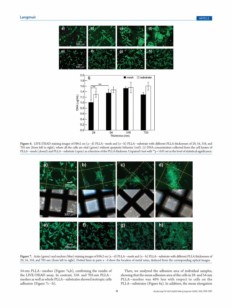
PLLA nanosheet on the cell activity was evaluated for 29, 54, 318,and 703 nm thickness at 24 h after cell seeding. The LIVE/DEADassay showed that the cells on both samples proliferated (coloredgreen) without showing significant apoptotic behavior (coloredred) (Figure 6a�h). Thus, not only topological but also mechan-ical properties of the PLLA nanosheet showed no influence onthe cell viability, at least after 24 h of culture. However, cells onthe PLLA�mesh partially showed anisotropic assembly along themetal wire (Figure 6a�d) although those on the PLLA�substrateshowed isotropic assembly on the surface (Figure 6e�h). Inparticular, the PLLA�meshwith the tens-of-nanometers nanosheet
showed a particular trend, testified by the segregation of cells fromthe interior part of the mesh lattice to the metal wire (Figure 6a,b).However, the cells on the PLLA�mesh with the hundreds-of-nano-meters thickness adhered more isotropically (Figure 6c,d).The cell activity was also investigated by means of the DNA
quantitative assay for the same kinds of samples. The DNAconcentration of the PLLA�substrate showed no significant dif-ference among samples having different thicknesses, while that ofthe PLLA�mesh was increased as the thickness was increasedfrom 29 to 318 nm (Figure 6i). It was noteworthy that the DNAconcentration of the 703-nm PLLA�mesh showed a lower valuethan those of the PLLA�substrate. This result can be explainedby the low adhesion of the 703-nm PLLA nanosheets to the steelmesh, which caused detachment in the growth medium, resultingin a poor culture condition for the cells. According to theLIVE/DEAD staining images, it was suggested that the cellsavoided to attach isotropically on the PLLA�mesh (especiallyinside the mesh lattice) as the nanosheet thickness was decreasedto the tens-of-nanometers regions. Hence, the DNA concentra-tion of the 29-nm PLLA�mesh was relatively decreased andshowed a significant difference compared to the 29-nm PLLA�substrate (**p < 0.01).Substrate Effect onCell Adhesion Properties. In addition to
cytosol staining undertaken by LIVE/DEAD assay, actin andnucleus staining was performed to visualize the cytoskeletonarrangement of a single cell, in order to better characterize thecell adhesion properties. Adhesion area and elongation ratio ofthe individual cells were analyzed for both samples. Anisotropicadhesion of cells along the metal wire was confirmed for 29- and
Figure 5. Mechanical influence of the underlying material on PLLA nanosheets. (a) Topographic and (b) phase images of PLLA�mesh by tappingmode AFM scan, where the 29-nm PLLA nanosheet is partially adhered to a metal wire of the steel mesh (scan range: 15 μm� 15 μm). (c) Verticallyaveraged height and (d) phase profiles of various PLLA�mesh samples along the horizontal displacement of the AFM scan, bearing different thicknessesof the PLLA nanosheets: 29 nm (black), 54 nm (red), 318 nm (green), and 703 nm (blue).
H dx.doi.org/10.1021/la203140a |Langmuir XXXX, XXX, 000–000
Langmuir ARTICLE
54-nm PLLA�meshes (Figure 7a,b), confirming the results ofthe LIVE/DEAD assay. In contrast, 318- and 703-nm PLLA�meshes as well as whole PLLA�substrates showed isotropic cellsadhesion (Figure 7c�h).
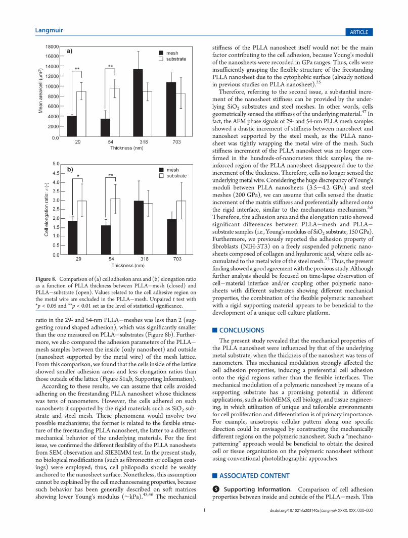
Then, we analyzed the adhesion area of individual samples,showing that themean adhesion area of the cells in 29- and 54-nmPLLA�meshes was 40% less with respect to cells on thePLLA�substrates (Figure 8a). In addition, the mean elongation
Figure 6. LIVE/DEAD staining images of H9c2 on (a�d) PLLA�mesh and (e�h) PLLA�substrate with different PLLA thicknesses of 29, 54, 318, and703 nm (from left to right), where all the cells are vital (green) without apoptotic behavior (red). (i) DNA concentration collected from the cell lysates ofPLLA�mesh (closed) and PLLA�substrate (open) as a function of the PLLA thickness. Unpaired t test with **p< 0.01 set as the level of statistical significance.
Figure 7. Actin (green) and nucleus (blue) staining images of H9c2 on (a�d) PLLA�mesh and (e�h) PLLA�substrate with different PLLA thicknesses of29, 54, 318, and 703 nm (from left to right). Dotted lines in parts a�d show the location of metal wires, deduced from the corresponding optical images.
I dx.doi.org/10.1021/la203140a |Langmuir XXXX, XXX, 000–000
Langmuir ARTICLE
ratio in the 29- and 54-nm PLLA�meshes was less than 2 (sug-gesting round shaped adhesion), which was significantly smallerthan the one measured on PLLA�substrates (Figure 8b). Further-more, we also compared the adhesion parameters of the PLLA�mesh samples between the inside (only nanosheet) and outside(nanosheet supported by the metal wire) of the mesh lattice.From this comparison, we found that the cells inside of the latticeshowed smaller adhesion areas and less elongation ratios thanthose outside of the lattice (Figure S1a,b, Supporting Information).According to these results, we can assume that cells avoided
adhering on the freestanding PLLA nanosheet whose thicknesswas tens of nanometers. However, the cells adhered on suchnanosheets if supported by the rigid materials such as SiO2 sub-strate and steel mesh. These phenomena would involve twopossible mechanisms; the former is related to the flexible struc-ture of the freestanding PLLA nanosheet, the latter to a differentmechanical behavior of the underlying materials. For the firstissue, we confirmed the different flexibility of the PLLA nanosheetsfrom SEM observation and SIEBIMM test. In the present study,no biological modifications (such as fibronectin or collagen coat-ings) were employed; thus, cell philopodia should be weaklyanchored to the nanosheet surface. Nonetheless, this assumptioncannot be explained by the cell mechanosensing properties, becausesuch behavior has been generally described on soft matricesshowing lower Young’s modulus (∼kPa).45,46 The mechanical
stiffness of the PLLA nanosheet itself would not be the mainfactor contributing to the cell adhesion, because Young’s moduliof the nanosheets were recorded in GPa ranges. Thus, cells wereinsufficiently grasping the flexible structure of the freestandingPLLA nanosheet due to the cytophobic surface (already noticedin previous studies on PLLA nanosheet).25
Therefore, referring to the second issue, a substantial incre-ment of the nanosheet stiffness can be provided by the under-lying SiO2 substrates and steel meshes. In other words, cellsgeometrically sensed the stiffness of the underlying material.47 Infact, the AFM phase signals of 29- and 54-nm PLLA mesh samplesshowed a drastic increment of stiffness between nanosheet andnanosheet supported by the steel mesh, as the PLLA nano-sheet was tightly wrapping the metal wire of the mesh. Suchstiffness increment of the PLLA nanosheet was no longer con-firmed in the hundreds-of-nanometers thick samples; the re-inforced region of the PLLA nanosheet disappeared due to theincrement of the thickness. Therefore, cells no longer sensed theunderlyingmetal wire. Considering the huge discrepancy of Young’smoduli between PLLA nanosheets (3.5�4.2 GPa) and steelmeshes (200 GPa), we can assume that cells sensed the drasticincrement of the matrix stiffness and preferentially adhered ontothe rigid interface, similar to the mechanotaxis mechanism.5,6
Therefore, the adhesion area and the elongation ratio showedsignificant differences between PLLA�mesh and PLLA�substrate samples (i.e., Young’smodulus of SiO2 substrate, 150GPa).Furthermore, we previously reported the adhesion property offibroblasts (NIH-3T3) on a freely suspended polymeric nano-sheets composed of collagen and hyaluronic acid, where cells ac-cumulated to themetal wire of the steel mesh.23 Thus, the presentfinding showed a good agreementwith the previous study. Althoughfurther analysis should be focused on time-lapse observation ofcell�material interface and/or coupling other polymeric nano-sheets with different substrates showing different mechanicalproperties, the combination of the flexible polymeric nanosheetwith a rigid supporting material appears to be beneficial to thedevelopment of a unique cell culture platform.
’CONCLUSIONS
The present study revealed that the mechanical properties ofthe PLLA nanosheet were influenced by that of the underlyingmetal substrate, when the thickness of the nanosheet was tens ofnanometers. This mechanical modulation strongly affected thecell adhesion properties, inducing a preferential cell adhesiononto the rigid regions rather than the flexible interfaces. Themechanical modulation of a polymeric nanosheet by means of asupporting substrate has a promising potential in differentapplications, such as bioMEMS, cell biology, and tissue engineer-ing, in which utilization of unique and tailorable environmentsfor cell proliferation and differentiation is of primary importance.For example, anisotropic cellular pattern along one specificdirection could be envisaged by constructing the mechanicallydifferent regions on the polymeric nanosheet. Such a “mechano-patterning” approach would be beneficial to obtain the desiredcell or tissue organization on the polymeric nanosheet withoutusing conventional photolithographic approaches.
’ASSOCIATED CONTENT
bS Supporting Information. Comparison of cell adhesionproperties between inside and outside of the PLLA�mesh. This
Figure 8. Comparison of (a) cell adhesion area and (b) elongation ratioas a function of PLLA thickness between PLLA�mesh (closed) andPLLA�substrate (open). Values related to the cell adhesive region onthe metal wire are excluded in the PLLA�mesh. Unpaired t test with*p < 0.05 and **p < 0.01 set as the level of statistical significance.
J dx.doi.org/10.1021/la203140a |Langmuir XXXX, XXX, 000–000
Langmuir ARTICLE
material is available free of charge via the Internet at http://pubs.acs.org.
’AUTHOR INFORMATION
Corresponding Author*Tel: +39-050-883414. Fax: +39-050-883101.E-mail: [email protected] (T.F.), [email protected] (V.M.).
’ACKNOWLEDGMENT
This work was supported in part by JFE (The Japanese Founda-tion for Research and Promotion of Endoscopy) Grant (T.F.)
’REFERENCES
(1) Tang, Z.;Wang, Y.; Podsiadlo, P.; Kotov, N. A.Adv. Mater. 2006,18, 3203–3224.(2) Bettinger, C. J.; Langer, R.; Borenstein, J. T. Angew. Chem., Int.
Ed. 2009, 48, 5406–5415.(3) Lutolf, M. P.; Hubbell, J. A. Nat. Biotechnol. 2005, 23, 47–55.(4) Discher, D. E.; Mooney, D. J.; Zandstra., P. W. Science 2009,
324, 1673–1677.(5) Lo, C.-M.; Wang, H.-B.; Dembo, M.; Wang, Y.-L. Biophys. J. 2000,
79, 141–152.(6) Kawano, T.; Kidoaki, S. Biomaterials 2011, 32, 2725–2733.(7) Zhu, X.; Mills, K. L.; Peters, P. R.; Bahng, J. H.; Liu, E. H.; Shim,
J.; Naruse, K.; Csete, M. E.; Thouless, M. D.; Takayama, S. Nat. Mater.2005, 4, 403–406.(8) Ren, K.; Crouzier, T.; Roy, C.; Picart, C. Adv. Funct. Mater. 2008,
18, 1378–1389.(9) Discher, D. E.; Janmey, P.; Wang, Y. L. Science 2005, 310,
1139–1143.(10) Moussallem, M. D.; Olenych, S. G.; Scott, S. L.; Keller, T. C.,
3rd.; Schlenoff, J. B. Biomacromolecules 2009, 10, 3062–3068.(11) Takeoka, S.; Okamura, Y.; Fujie, T.; Fukui., Y. Pure Appl. Chem.
2008, 80, 2259–2271.(12) Forrest, J. A.; Dalnoki-Veress, K.; Stevens, J. R.; Dutcher, J. R.
Phys. Rev. Lett. 1996, 77, 2002–2005.(13) Jiang, C.; Tsukruk, V. V. Adv. Mater. 2006, 18, 829–840.(14) Fujie, T.; Okamura, Y.; Takeoka, S. Adv. Mater. 2007,
19, 3549–3553.(15) Endo, H.; Kado, Y.; Mitsuishi, M.;Miyashita, T.Macromolecules
2006, 39, 5559–5563.(16) Vendamme, R.; Onoue, S.; Nakao, A.; Kunitake, T. Nat. Mater.
2006, 5, 494–501.(17) Fujie, T.; Kinosita, M.; Shono, S.; Saito, A.; Okamura, Y.;
Saitoh, D.; Takeoka, S. Surgery 2010, 148, 48–58.(18) Fujie, T.; Matsutani, N.; Kinoshtia, M.; Okamura, Y.; Saito, A.;
Takeoka, S. Adv. Funct. Mater. 2009, 19, 2560–2568.(19) Fujie, T.; Saito, A.; Kinoshita, M.; Miyazaki, H.; Ohtsubo, S.;
Saitoh, D.; Takeoka, S. Biomaterials 2010, 31, 6269–6278.(20) Mattoli, V.; Pensabene, V.; Fujie, T.; Taccola, S.; Menciassi, A.;
Takeoka, S.; Dario, P. Procedia Chem. 2009, 1, 28–31.(21) Taccola, S.; Desii, A.; Pensabene, V.; Fujie, T.; Saito, A.;
Takeoka, S.; Dario, P.; Menciassi, A.; Mattoli, V. Langmuir 2011,27, 5589–5595.(22) Fujie, T.; Okamura, Y.; Takeoka, S. In Functional Polymer Films;
Knoll, W., Advincula, R. C., Eds.; Wiley-VCH Verlag GmbH & Co.KGaA: Weinheim, 2011; Vol. 2, p 907.(23) Fujie, T.; Furutate, S.; Niwa, D. Soft Matter 2010, 6, 4672–4676.(24) Ricotti, L.; Taccola, S.; Pensabene, V.; Mattoli, V.; Fujie, T.;
Takeoka, S.; Menciassi, A.; Dario, P. Biomed. Microdevices 2010, 12,809–819.(25) Niwa, D.; Fujie, T.; Lang, T.; Goda, N.; Takeoka, S. J. Biomater.
Appl. 2011, DOI: 10.1177/0885328210394470.
(26) Hescheler, J.; Meyer, R.; Plant, S.; Krautwurst, D.; Rosenthal,W.; Schiltz, G. Circ. Res. 1991, 69, 1476–1486.
(27) Ciofani, G.; Ricotti, L.; Menciassi, A.; Mattoli, V. Biomed.Microdev. 2011, 13, 255–266.
(28) Prabhakaran, M. P.; Venugopal, J.; Kai, D.; Ramakrishna, S.Mater. Sci. Eng. C. 2011, 31, 503–513.
(29) Stafford, C.M.; Harrison, C.; Beers, K. L.; Karim, A.; Amis, E. J.;VanLandingham, M. R.; Kim, H.; Volksen, W.; Miller, R. D.; Simonyi,E. E. Nat. Mater. 2004, 3, 545–550.
(30) Nolte, A. J.; Cohen, R. E.; Rubner, M. F.Macromolecules 2006,39, 4841–4847.
(31) Stafford, C. M.; Vogt, B. D.; Harrison, C.; Julthongpiput, D.;Huang, R. Macromolecules 2006, 39, 5095–5099.
(32) Dalby, M. J.; Gadegaard, N.; Tare, R.; Andar, A.; Riehle, M. O.;Herzyk, P.; Wilkinson, C. D. W.; Oreffo, R. O. C. Nat. Mater. 2007,6, 997–1003.
(33) Okamura, Y.; Kabata, K.; Kinoshita, M.; Saitoh, D.; Takeoka, S.Adv. Mater. 2009, 21, 4388–4392.
(34) Chung, T. W.; Liu, D. Z.; Wang, S. Y.; Wang, S. S. Biomaterials2003, 24, 4655–4661.
(35) Richert, L.; Vetrone, F.; Yi, J.-H.; Zalzal, S. F.; Wuest, J. D.;Rosei, F.; Nanci, A. Adv. Mater. 2008, 20, 1488–1492.
(36) Xu, C.; Yang, F.; Wang, S.; Ramakrishna, S. J. Biomed. Mater.Res. A. 2004, 71A, 154–161.
(37) Miller, D. C.; Thapa, A.; Haberstroh, K. M.; Webster, T. J.Biomaterials 2004, 25, 53–61.
(38) Deligianni, D. D.; Katsala, N. D.; Koutsoukos, P. G.; Missirlis,Y. F. Biomaterials 2001, 22, 87–96.
(39) Lampin, M.; Warocquier-Cl�erout, R.; Legris, C.; Degrange, M.;Sigot-Luizard, M. F. J. Biom. Mat. Res. A. 1997, 36, 99–108.
(40) Fan, Y. W.; Cui, F. Z.; Chen, L. N.; Zhai, Y.; Xu, Q.; Lee, I.-S.Appl. Surf. Sci. 2002, 187, 313–318.
(41) Fujie, T.; Okamura, Y.; Takeoka, S. Colloids Surf., A. 2009,334, 28–33.
(42) Eling, B.; Gogolewski, S.; Pennings, A. J. Polymer 1982, 23,1587–1593.
(43) Magonov, S. N.; Elings, V.; Whangbo, M.-H. Surf. Sci. 1997,375, L385–L391.
(44) Bodiguel, H.; Montes, H.; Fretigny, C. Rev. Sci. Instrum. 2004,75, 2529–2535.
(45) Schneider, A.; Francius, G.; Obeid, R.; Schwint�e, P.; Hemmerl�e,J.; Frisch, B.; Schaaf, P.; Voegel, J. C.; Senger, B.; Picart, C. Langmuir2006, 22, 1193–1200.
(46) V�azquez, C. P.; Boudou, T.; Dulong, V.; Nicolas., C.; Picart, C.;Glinel, K. Langmuir 2009, 25, 3556–3563.
(47) Hur, S. S.; Zhao, Y.; Li, Y.-S.; Botvinick, E.; Chien, S. Cell. Mol.Bioeng. 2009, 2, 425–436.
Copyright © 2022 FDOKUMEN