Preservation of porcine blood quality by means of lactic acid bacteria
8
Preservation of porcine blood quality by means of lactic acid bacteria Eduard Da `vila * , Elena Saguer, Mo ` nica Toldra `, Carmen Carretero, Dolors Pare ´s Institut de Tecnologia Agroalimenta ` ria – CeRTA, Universitat de Girona, Escola Polite `cnica Superior, Av. Lluis Santalo ´ s/n, 17071 Girona, Spain Received 30 October 2005; received in revised form 28 December 2005; accepted 28 December 2005 Abstract The capacity of 12 lactic acid bacteria (LAB) strains to preserve porcine blood during storage was evaluated. A general ability of LAB to prevent blood’s hemolysis and to maintain the functional properties of plasma was observed. Two strains, PS99 (Enterococcus raff- inosus) and TA43 (Lactobacillus reuteri), were selected for studies at 5 °C according to their antibacterial activity in blood stored at 15 °C. After 144 h at 5 °C, lower counts of coliforms, Pseudomonas spp., proteolytic and hemolytic bacteria were obtained in blood containing either PS99 or TA43 as compared to the non-inoculated blood. When inulin (2%) was added to blood, higher inhibition values were obtained and Enterococcus raffinosus (PS99) showed the best abilities for blood preservation. On the basis of these results it seems worth- while to supplement blood with inulin and to inoculate it with an active LAB strain to avoid undesirable changes during chill storage, especially useful to prevent the effects of a cold-chain breakdown. Ó 2006 Elsevier Ltd. All rights reserved. Keywords: Porcine blood; Biopreservation; Lactic acid bacteria; Enterococcus raffinosus; Functional properties 1. Introduction In Spain, more than 38 million pigs are slaughtered every year (FAOSTAT, 2005), thus producing more than 100,000 tons of collected blood. Interesting blood indus- trial applications deriving from both its nutritional value and functional properties have been described (Ockerman & Hansen, 1988; Pearson & Dutson, 1988). Still, the fre- quent lack of appropriate handling facilities leads to a poor quality product with high microbial contamination. In many countries most animal blood, especially that from pigs, is collected using open collection systems and it can contain bacterial counts over 10 6 cfu/mL (Carretero & Pare ´s, 2000; Jensen & Hess, 1941; Swingler, 1982; Ock- erman & Hansen, 1988). The contamination of this blood, basically airborne bacteria or microorganisms present on the skin or in the stomach and intestines of the slaughtered animal (Ockerman & Hansen, 1988), cannot be avoided even when processed under strictly hygienic conditions. The quality of blood can be improved by the use of closed collection systems, incorporating a hollow sticking knife through which the blood is drawn into a sterile container (Swingler, 1982; Dill & Landmann, 1988). Even in this case, initial microbial counts are typically around 10 3 cfu/ mL (Carretero & Pare ´s, 2000; Nilsson, 1975; Ockerman & Hansen, 1988) and blood can easily reach counts up to 10,000-fold higher during storage under chill conditions (Erikson & Von Bockelmann, 1975). Biopreservation with lactic acid bacteria (LAB) is a promising practice in different areas of the food industry (Holzapfel, Geisen, & Schilinger, 1995; Ross, Morgan, & Hill, 2002). LAB have proved their ability to inhibit the growth of undesirable bacteria (Adams & Halls, 1988; De Vuyst & Vandamme, 1994), using a wide variety of mech- anisms (Cleveland, Montville, Nes, & Chikindas, 2001; Hugas, 1998; Lindgren & Dobrogosz, 1990; O’Sullivan, Ross, & Hill, 2002). LAB inoculation just after blood col- lection may provide a safer as well as a better quality prod- uct for both the processing industry and the end consumer. Spoilage microorganisms and foodborne pathogens, which can easily grow in such a nutritive medium as blood (Car- retero & Pare ´s, 2000), could be reduced without the need to 0309-1740/$ - see front matter Ó 2006 Elsevier Ltd. All rights reserved. doi:10.1016/j.meatsci.2005.12.017 * Corresponding author. Tel.: +34 972 41 89 41; fax: +34 972 41 83 99. E-mail address: [email protected] (E. Da `vila). www.elsevier.com/locate/meatsci Meat Science 73 (2006) 386–393 MEAT SCIENCE
Transcript of Preservation of porcine blood quality by means of lactic acid bacteria

www.elsevier.com/locate/meatsci
Meat Science 73 (2006) 386–393
MEATSCIENCE
Preservation of porcine blood quality by means of lactic acid bacteria
Eduard Davila *, Elena Saguer, Monica Toldra, Carmen Carretero, Dolors Pares
Institut de Tecnologia Agroalimentaria – CeRTA, Universitat de Girona, Escola Politecnica Superior, Av. Lluis Santalo s/n, 17071 Girona, Spain
Received 30 October 2005; received in revised form 28 December 2005; accepted 28 December 2005
Abstract
The capacity of 12 lactic acid bacteria (LAB) strains to preserve porcine blood during storage was evaluated. A general ability of LABto prevent blood’s hemolysis and to maintain the functional properties of plasma was observed. Two strains, PS99 (Enterococcus raff-
inosus) and TA43 (Lactobacillus reuteri), were selected for studies at 5 �C according to their antibacterial activity in blood stored at 15 �C.After 144 h at 5 �C, lower counts of coliforms, Pseudomonas spp., proteolytic and hemolytic bacteria were obtained in blood containingeither PS99 or TA43 as compared to the non-inoculated blood. When inulin (2%) was added to blood, higher inhibition values wereobtained and Enterococcus raffinosus (PS99) showed the best abilities for blood preservation. On the basis of these results it seems worth-while to supplement blood with inulin and to inoculate it with an active LAB strain to avoid undesirable changes during chill storage,especially useful to prevent the effects of a cold-chain breakdown.� 2006 Elsevier Ltd. All rights reserved.
Keywords: Porcine blood; Biopreservation; Lactic acid bacteria; Enterococcus raffinosus; Functional properties
1. Introduction
In Spain, more than 38 million pigs are slaughteredevery year (FAOSTAT, 2005), thus producing more than100,000 tons of collected blood. Interesting blood indus-trial applications deriving from both its nutritional valueand functional properties have been described (Ockerman& Hansen, 1988; Pearson & Dutson, 1988). Still, the fre-quent lack of appropriate handling facilities leads to a poorquality product with high microbial contamination.
In many countries most animal blood, especially thatfrom pigs, is collected using open collection systems andit can contain bacterial counts over 106 cfu/mL (Carretero& Pares, 2000; Jensen & Hess, 1941; Swingler, 1982; Ock-erman & Hansen, 1988). The contamination of this blood,basically airborne bacteria or microorganisms present onthe skin or in the stomach and intestines of the slaughteredanimal (Ockerman & Hansen, 1988), cannot be avoidedeven when processed under strictly hygienic conditions.
0309-1740/$ - see front matter � 2006 Elsevier Ltd. All rights reserved.
doi:10.1016/j.meatsci.2005.12.017
* Corresponding author. Tel.: +34 972 41 89 41; fax: +34 972 41 83 99.E-mail address: [email protected] (E. Davila).
The quality of blood can be improved by the use of closedcollection systems, incorporating a hollow sticking knifethrough which the blood is drawn into a sterile container(Swingler, 1982; Dill & Landmann, 1988). Even in thiscase, initial microbial counts are typically around 103 cfu/mL (Carretero & Pares, 2000; Nilsson, 1975; Ockerman& Hansen, 1988) and blood can easily reach counts up to10,000-fold higher during storage under chill conditions(Erikson & Von Bockelmann, 1975).
Biopreservation with lactic acid bacteria (LAB) is apromising practice in different areas of the food industry(Holzapfel, Geisen, & Schilinger, 1995; Ross, Morgan, &Hill, 2002). LAB have proved their ability to inhibit thegrowth of undesirable bacteria (Adams & Halls, 1988; DeVuyst & Vandamme, 1994), using a wide variety of mech-anisms (Cleveland, Montville, Nes, & Chikindas, 2001;Hugas, 1998; Lindgren & Dobrogosz, 1990; O’Sullivan,Ross, & Hill, 2002). LAB inoculation just after blood col-lection may provide a safer as well as a better quality prod-uct for both the processing industry and the end consumer.Spoilage microorganisms and foodborne pathogens, whichcan easily grow in such a nutritive medium as blood (Car-retero & Pares, 2000), could be reduced without the need to
E. Davila et al. / Meat Science 73 (2006) 386–393 387
change the slaughterhouses’ facilities or to increase thehygienic collection requirements.
Previous studies by Marland (1984) and Pinel (1985) pro-posed the use of LAB to preserve blood for feed purposes. Inthese studies, glucose was added to blood in order to favourLAB’s biological competition. Studies which were carriedout by our group also reported that the addition of glucoseenhances the antagonistic activity of LAB (Pares, Zamora,Saguer, & Carretero, 2004). However, glucose can be widelyused by microorganisms, therefore facilitating the growth ofundesirable bacteria as well. Conversely, fructooligosaccha-rides could be used as a selective energy source since it hasbeen proved that they can specifically favour LAB’s growth,without increasing the population of contaminant bacteria(Gibson, Beatty, Wang, & Cummings, 1995; Kaplan & Hut-kins, 2000; Wang & Gibson, 1993).
The aim of the present work is to study the preservationcapacity of LAB during the storage of blood regardingboth the inhibition of microbial spoilage and the retentionof functional properties and the potential role of inulin as aselective supplement to enhance LAB activity.
2. Materials and methods
2.1. Blood
Blood samples were obtained during the slaughter ofpigs in industrial abattoirs. They were collected in sterilecontainers (5 L) containing sodium citrate (1% w/v finalconcentration) as blood was draining from the animal dur-ing bleeding, in order to prevent coagulation. Containerswere maintained at chill temperatures until blood was pro-cessed. Blood samples were split into several fractions, oneto be used as a non-inoculated control and the others to beinoculated with LAB strains.
2.2. LAB cultures
Twelve strains, out of 144 lactic acid bacteria previouslyisolated from porcine blood, were investigated for theirblood preservation capacity. They had been selected asshowing the broadest antagonistic spectrum against fourindicator microorganisms (Escherichia coli, Staphylococcus
aureus, Bacillus spp. and Pseudomonas fluorescens) in agarplates as well as when growing in blood (Pares et al., 2004).They had been identified as Lactococcus garviae (PS14,PS22, PS23, PS48, PS95 and TA20), Enterococcus raffino-
sus (PS7 and PS99), Lactobacillus murinus (TA73, TA75and TA76) and Lactobacillus reuteri (TA43) (Davila,Zamora, Pla, Carretero, & Pares, in press). All LAB strainswere preserved under freezing at �80 �C with 20% glycerolin 2 mL vials.
2.3. Blood inoculation
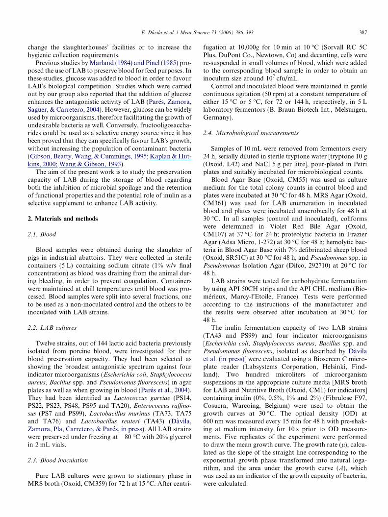
Pure LAB cultures were grown to stationary phase inMRS broth (Oxoid, CM359) for 72 h at 15 �C. After centri-
fugation at 10,000g for 10 min at 10 �C (Sorvall RC 5CPlus, DuPont Co., Newtown, Co) and decanting, cells werere-suspended in small volumes of blood, which were addedto the corresponding blood sample in order to obtain aninoculum size around 107 cfu/mL.
Control and inoculated blood were maintained in gentlecontinuous agitation (50 rpm) at a constant temperature ofeither 15 �C or 5 �C, for 72 or 144 h, respectively, in 5 Llaboratory fermentors (B. Braun Biotech Int., Melsungen,Germany).
2.4. Microbiological measurements
Samples of 10 mL were removed from fermentors every24 h, serially diluted in sterile tryptone water [tryptone 10 g(Oxoid, L42) and NaCl 5 g per litre], pour-plated in Petriplates and suitably incubated for microbiological counts.
Blood Agar Base (Oxoid, CM55) was used as culturemedium for the total colony counts in control blood andplates were incubated at 30 �C for 48 h. MRS Agar (Oxoid,CM361) was used for LAB enumeration in inoculatedblood and plates were incubated anaerobically for 48 h at30 �C. In all samples (control and inoculated), coliformswere determined in Violet Red Bile Agar (Oxoid,CM107) at 37 �C for 24 h; proteolytic bacteria in FrazierAgar (Adsa Micro, 1-272) at 30 �C for 48 h; hemolytic bac-teria in Blood Agar Base with 7% defibrinated sheep blood(Oxoid, SR51C) at 30 �C for 48 h; and Pseudomonas spp. inPseudomonas Isolation Agar (Difco, 292710) at 20 �C for48 h.
LAB strains were tested for carbohydrate fermentationby using API 50CH strips and the API CHL medium (Bio-merieux, Marcy-l’Etoile, France). Tests were performedaccording to the instructions of the manufacturer andthe results were observed after incubation at 30 �C for48 h.
The inulin fermentation capacity of two LAB strains(TA43 and PS99) and four indicator microorganisms[Escherichia coli, Staphylococcus aureus, Bacillus spp. andPseudomonas fluorescens, isolated as described by Davilaet al. (in press)] were evaluated using a Bioscreen C micro-plate reader (Labsystems Corporation, Helsinki, Find-land). Two hundred microliters of microorganismsuspensions in the appropriate culture media [MRS brothfor LAB and Nutritive Broth (Oxoid, CM1) for indicators]containing inulin (0%, 0.5%, 1% and 2%) (Fibrulose F97,Cosucra, Warcoing, Belgium) were used to obtain thegrowth curves at 30 �C. The optical density (OD) at600 nm was measured every 15 min for 48 h with pre-shak-ing at medium intensity for 10 s prior to OD measure-ments. Five replicates of the experiment were performedto draw the mean growth curve. The growth rate (l), calcu-lated as the slope of the straight line corresponding to theexponential growth phase transformed into natural loga-rithm, and the area under the growth curve (A), whichwas used as an indicator of the growth capacity of bacteria,were calculated.
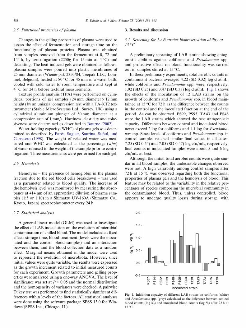
Fig. 1. Inhibition capacity of different LAB strains on coliforms (white)and Pseudomonas spp. (grey) calculated as the difference between controlblood counts (log Nc) and inoculated blood counts (log Ni) after 72 h at15 �C.
388 E. Davila et al. / Meat Science 73 (2006) 386–393
2.5. Functional properties of plasma
Changes in the gelling properties of plasma were used toassess the effect of fermentation and storage time on thefunctionality of plasma proteins. Plasma was obtainedfrom samples removed from the fermentors at 0, 72 and144 h, by centrifugation (2250g for 15 min at 4 �C) anddecanting. The heat-induced gels were obtained as follows:plasma samples were poured into plastic membranes of25 mm diameter (Wienie-pak 2350/84, Teepak LLC, Lom-mel, Belgium), heated at 80 �C for 45 min in a water bath,cooled with cold water to room temperature and kept at4 �C for 24 h before textural measurements.
Texture profile analysis (TPA) were performed on cylin-drical portions of gel samples (24 mm diameter · 12 mmheight) by an uniaxial compression test with a TA-XT2 tex-turometer (Stable MicroSystems Ltd., Surrey, UK) using acylindrical aluminium plunger of 50 mm diameter at acompression rate of 1 mm/s. Hardness, elasticity and cohe-siveness were determined as described in Bourne (2002).
Water-holding capacity (WHC) of plasma gels was deter-mined as described by Pares, Saguer, Saurina, Sunol, andCarretero (1998). The weight of released water was mea-sured and WHC was calculated as the percentage (w/w)of water released to the weight of the sample prior to centri-fugation. Three measurements were performed for each gel.
2.6. Hemolysis
Hemolysis – the presence of hemoglobin in the plasmafraction due to the red blood cells breakdown – was usedas a parameter related to blood quality. The increase ofthe hemolysis level was monitored by measuring the absor-bance at 414 nm of an appropriate dilution of plasma sam-ples (1:5 or 1:10) in a Shimatzu UV-160A (Shimatzu Co.,Kyoto, Japan) spectrophotometer every 24 h.
2.7. Statistical analysis
A general linear model (GLM) was used to investigatethe effect of LAB inoculation on the evolution of microbialcontamination of chilled blood. The model included as fixedeffects storage time, blood treatment (levels were the inocu-lated and the control blood samples) and an interactionbetween them, and the blood collection date as a randomeffect. Marginal means obtained in the model were usedto represent the evolution of microbiota. However, sinceinitial values were quite variable, the results were expressedas the growth increment related to initial measured countsfor each experiment. Growth parameters and gelling prop-erties were analyzed using a one-way ANOVA. The level ofsignificance was set at P < 0.05 and the normal distributionand the homogeneity of variances were checked. A pairwiseTukey test was performed to find statistically significant dif-ferences within levels of the factors. All statistical analyseswere done using the software package SPSS 13.0 for Win-dows (SPSS Inc., Chicago, IL).
3. Results and discussion
3.1. Screening for LAB strains biopreservation ability at
15 �C
A preliminary screening of LAB strains showing antag-onistic abilities against coliforms and Pseudomonas spp.and protective effects on blood functionality was carriedout in samples stored at 15 �C.
In these preliminary experiments, total aerobic counts ofcontaminant bacteria averaged 4.22 (SD 0.32) log cfu/mL,while coliforms and Pseudomonas spp. were, respectively,1.92 (SD 0.25) and 3.47 (SD 0.33) log cfu/mL. Fig. 1 showsthe effects of the inoculation of 12 LAB strains on thegrowth of coliforms and Pseudomonas spp. in blood main-tained at 15 �C for 72 h as the difference between the countsin the control and the inoculated fraction at the end of theperiod. As can be observed, PS99, PS95, TA43 and PS48were the LAB strains which showed the best antagonisticcapacity. Differences between control and inoculated bloodnever exceed 2 log for coliforms and 1.1 log for Pseudomo-
nas spp. Since levels of coliforms and Pseudomonas spp. incontrol samples reached similar final values in all cases,7.25 (SD 0.54) and 7.05 (SD 0.47) log cfu/mL, respectively,final counts in inoculated samples were about 5 and 6 logcfu/mL at best.
Although the initial total aerobic counts were quite sim-ilar in all blood samples, the undesirable changes observedwere not. A high variability among control samples after72 h at 15 �C was observed regarding both the functionalproperties of plasma gels and the hemolysis of blood. Thisfeature may be related to the variability in the relative per-centages of species composing the microbial community inthe contaminated blood. Thus, unless controlled, bloodappears to undergo quality losses during storage, with
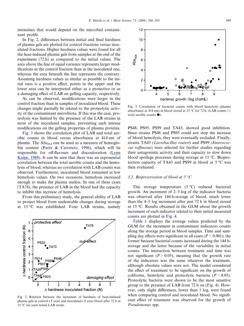
Fig. 3. Correlation of bacterial counts with blood hemolysis (plasmaabsorbance at 414 nm) in blood stored at 15 �C for 72 h. (LAB counts s;total aerobic counts d).
E. Davila et al. / Meat Science 73 (2006) 386–393 389
intensities that would depend on the microbial contami-nant profile.
In Fig. 2, differences between initial and final hardnessof plasma gels are plotted for control fractions versus inoc-ulated fractions. Higher hardness values were found for allthe heat-induced plasma gels from samples at the end of theexperiment (72 h) as compared to the initial values. Thearea above the line of equal variance represents larger mod-ifications in the control fraction than in the inoculated one,whereas the area beneath the line represents the contrary.Assuming hardness values as similar as possible to the ini-tial ones is a positive effect, points in the upper and thelower area can be interpreted either as a protective or asa damaging effect of LAB on gelling capacity, respectively.
As can be observed, modifications were larger in thecontrol fraction than in samples of inoculated blood. Thesechanges might partially be related to the proteolytic activ-ity of the contaminant microbiota. If this was the case, pro-teolysis was limited by the presence of the LAB strains inmost of the inoculated samples, preventing such intensemodifications on the gelling properties of plasma proteins.
Fig. 3 shows the correlation plot of LAB and total aer-obic counts in blood versus absorbance at 414 nm ofplasma. The Abs414 can be used as a measure of hemoglo-bin content (Pares & Carretero, 1996), which will beresponsible for off-flavours and discouloration (LynnKnipe, 1988). It can be seen that there was an exponentialcorrelation between the total aerobic counts and the hemo-lysis of blood, whereas no correlation with LAB counts wasobserved. Furthermore, inoculated blood remained at lowhemolysis values. On two occasions, hemolysis increasedenough to make the plasma useless. In one of these cases(TA76), the presence of LAB in the blood had the capacityto inhibit this increase of hemolysis.
From this preliminary study, the general ability of LABto protect blood from undesirable changes during storageat 15 �C was established. Four LAB strains, namely
Fig. 2. Relation between the increment of hardness of heat-inducedplasma gels in control (Y axis) and inoculated (X axis) blood after 72 h at15 �C for each tested LAB strain.
PS48, PS95, PS99 and TA43, showed good inhibition.Since strains PS48 and PS95 could not stop the increaseof blood hemolysis, they were eventually excluded. Finally,strains TA43 (Lactobacillus reuteri) and PS99 (Enterococ-cus raffinosus) were selected for further studies regardingtheir antagonistic activity and their capacity to slow downblood spoilage processes during storage at 15 �C. Biopro-tection capacity of TA43 and PS99 in blood at 5 �C wasthen evaluated.
3.2. Biopreservation of blood at 5 �C
This storage temperature (5 �C) reduced bacterialgrowth. An increment of 2–3 log of the indicator bacteriawas observed after 144 h-storage of blood, much lowerthan the 4–5 log increment after just 72 h in blood storedat 15 �C. Results obtained in the GLM about the growthincrement of each indicator related to their initial measuredcounts are plotted in Fig. 4.
Table 1 displays the average values predicted by theGLM for the increment in contaminant indicators countsalong the storage period in blood samples. Time and sam-pling day effects were significant in all cases (P < 0.001); theformer because bacterial counts increased during the 144 h-storage and the latter because of the variability in initialcounts. The interaction between treatment and time wasnot significant (P > 0.05), meaning that the growth rateof the indicators was the same whatever the treatment,although absolute values were not. The model consideredthe effect of treatment to be significant on the growth ofcoliforms, hemolytic and proteolytic bacteria (P < 0.05).Proteolytic bacteria were shown to be the most sensitivegroup to the presence of LAB from 72 h on (Fig. 4). How-ever, only slight differences, lower than 1 log, were foundwhen comparing control and inoculated blood. No signifi-cant effect of treatment was observed for the growth ofPseudomonas spp.
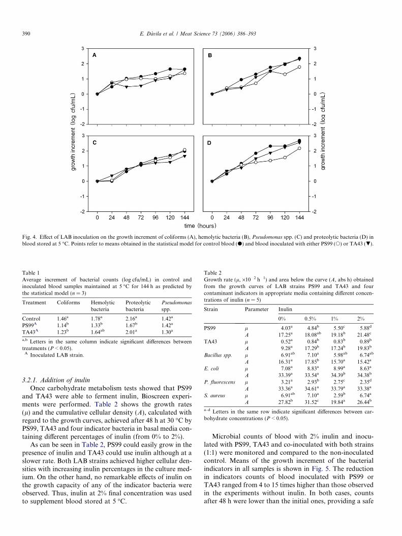
Fig. 4. Effect of LAB inoculation on the growth increment of coliforms (A), hemolytic bacteria (B), Pseudomonas spp. (C) and proteolytic bacteria (D) inblood stored at 5 �C. Points refer to means obtained in the statistical model for control blood (d) and blood inoculated with either PS99 (s) or TA43 (.).
Table 1Average increment of bacterial counts (log cfu/mL) in control andinoculated blood samples maintained at 5 �C for 144 h as predicted bythe statistical model (n = 3)
Treatment Coliforms Hemolyticbacteria
Proteolyticbacteria
Pseudomonas
spp.
Control 1.46a 1.78a 2.16a 1.42a
PS99A 1.14b 1.33b 1.67b 1.42a
TA43A 1.23b 1.64ab 2.01a 1.30a
a,b Letters in the same column indicate significant differences betweentreatments (P < 0.05).
A Inoculated LAB strain.
Table 2Growth rate (l, ·10�2 h�1) and area below the curve (A, abs h) obtainedfrom the growth curves of LAB strains PS99 and TA43 and fourcontaminant indicators in appropriate media containing different concen-trations of inulin (n = 5)
Strain Parameter Inulin
0% 0.5% 1% 2%
PS99 l 4.03a 4.84b 5.50c 5.88d
A 17.25a 18.08ab 19.18b 21.48c
TA43 l 0.52a 0.84b 0.83b 0.89b
A 9.28a 17.29b 17.24b 19.83b
Bacillus spp. l 6.91ab 7.10a 5.98ab 6.74ab
A 16.31a 17.85b 15.70a 15.42a
E. coli l 7.08a 8.83a 8.99a 8.63a
A 33.39a 33.54a 34.39b 34.38b
P. fluorescens l 3.21a 2.93b 2.75c 2.35d
A 33.36a 34.61a 33.79a 33.38a
S. aureus l 6.91ab 7.10a 2.59b 6.74a
A 27.82b 31.52c 19.84a 26.44b
a–d Letters in the same row indicate significant differences between car-bohydrate concentrations (P < 0.05).
390 E. Davila et al. / Meat Science 73 (2006) 386–393
3.2.1. Addition of inulin
Once carbohydrate metabolism tests showed that PS99and TA43 were able to ferment inulin, Bioscreen experi-ments were performed. Table 2 shows the growth rates(l) and the cumulative cellular density (A), calculated withregard to the growth curves, achieved after 48 h at 30 �C byPS99, TA43 and four indicator bacteria in basal media con-taining different percentages of inulin (from 0% to 2%).
As can be seen in Table 2, PS99 could easily grow in thepresence of inulin and TA43 could use inulin although at aslower rate. Both LAB strains achieved higher cellular den-sities with increasing inulin percentages in the culture med-ium. On the other hand, no remarkable effects of inulin onthe growth capacity of any of the indicator bacteria wereobserved. Thus, inulin at 2% final concentration was usedto supplement blood stored at 5 �C.
Microbial counts of blood with 2% inulin and inocu-lated with PS99, TA43 and co-inoculated with both strains(1:1) were monitored and compared to the non-inoculatedcontrol. Means of the growth increment of the bacterialindicators in all samples is shown in Fig. 5. The reductionin indicators counts of blood inoculated with PS99 orTA43 ranged from 4 to 15 times higher than those observedin the experiments without inulin. In both cases, countsafter 48 h were lower than the initial ones, providing a safe
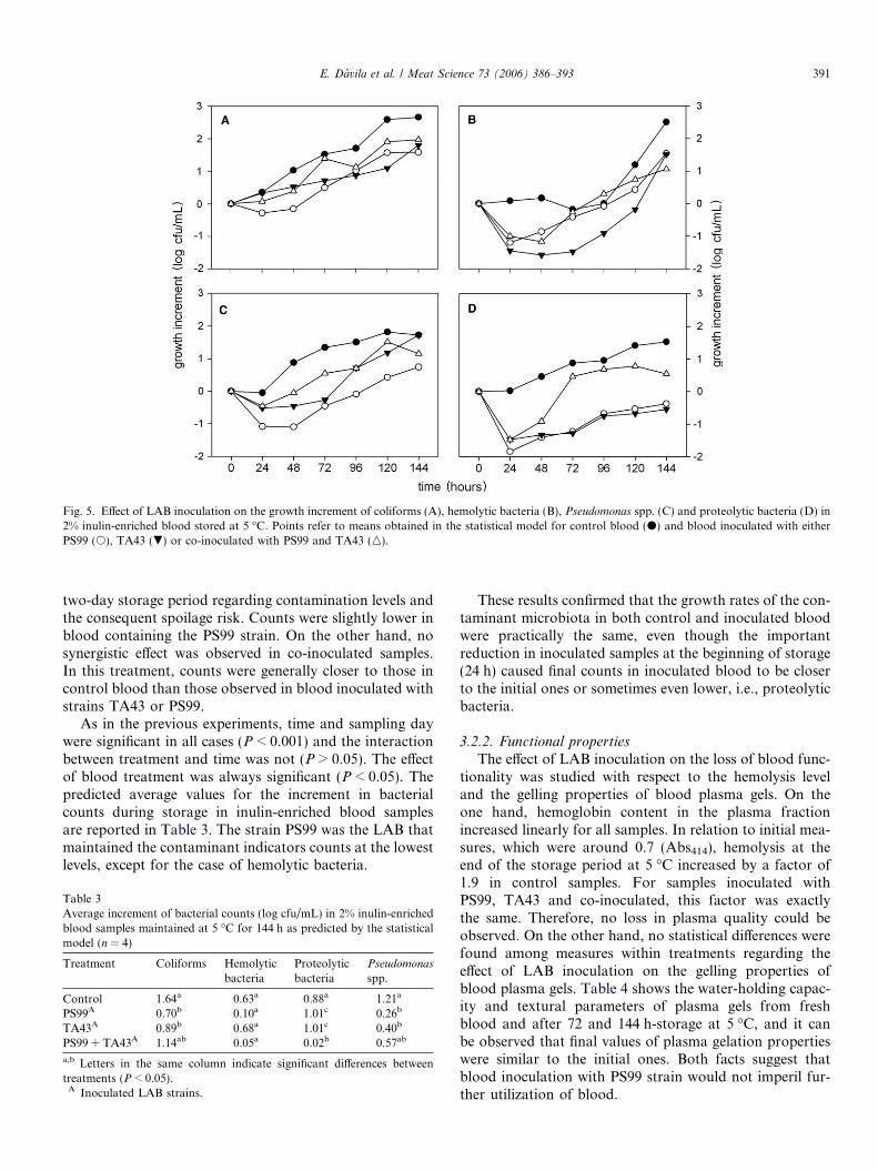
Fig. 5. Effect of LAB inoculation on the growth increment of coliforms (A), hemolytic bacteria (B), Pseudomonas spp. (C) and proteolytic bacteria (D) in2% inulin-enriched blood stored at 5 �C. Points refer to means obtained in the statistical model for control blood (d) and blood inoculated with eitherPS99 (s), TA43 (.) or co-inoculated with PS99 and TA43 (n).
E. Davila et al. / Meat Science 73 (2006) 386–393 391
two-day storage period regarding contamination levels andthe consequent spoilage risk. Counts were slightly lower inblood containing the PS99 strain. On the other hand, nosynergistic effect was observed in co-inoculated samples.In this treatment, counts were generally closer to those incontrol blood than those observed in blood inoculated withstrains TA43 or PS99.
As in the previous experiments, time and sampling daywere significant in all cases (P < 0.001) and the interactionbetween treatment and time was not (P > 0.05). The effectof blood treatment was always significant (P < 0.05). Thepredicted average values for the increment in bacterialcounts during storage in inulin-enriched blood samplesare reported in Table 3. The strain PS99 was the LAB thatmaintained the contaminant indicators counts at the lowestlevels, except for the case of hemolytic bacteria.
Table 3Average increment of bacterial counts (log cfu/mL) in 2% inulin-enrichedblood samples maintained at 5 �C for 144 h as predicted by the statisticalmodel (n = 4)
Treatment Coliforms Hemolyticbacteria
Proteolyticbacteria
Pseudomonas
spp.
Control 1.64a 0.63a 0.88a 1.21a
PS99A 0.70b �0.10a �1.01c �0.26b
TA43A 0.89b �0.68a �1.01c 0.40b
PS99 + TA43A 1.14ab �0.05a 0.02b 0.57ab
a,b Letters in the same column indicate significant differences betweentreatments (P < 0.05).
A Inoculated LAB strains.
These results confirmed that the growth rates of the con-taminant microbiota in both control and inoculated bloodwere practically the same, even though the importantreduction in inoculated samples at the beginning of storage(24 h) caused final counts in inoculated blood to be closerto the initial ones or sometimes even lower, i.e., proteolyticbacteria.
3.2.2. Functional properties
The effect of LAB inoculation on the loss of blood func-tionality was studied with respect to the hemolysis leveland the gelling properties of blood plasma gels. On theone hand, hemoglobin content in the plasma fractionincreased linearly for all samples. In relation to initial mea-sures, which were around 0.7 (Abs414), hemolysis at theend of the storage period at 5 �C increased by a factor of1.9 in control samples. For samples inoculated withPS99, TA43 and co-inoculated, this factor was exactlythe same. Therefore, no loss in plasma quality could beobserved. On the other hand, no statistical differences werefound among measures within treatments regarding theeffect of LAB inoculation on the gelling properties ofblood plasma gels. Table 4 shows the water-holding capac-ity and textural parameters of plasma gels from freshblood and after 72 and 144 h-storage at 5 �C, and it canbe observed that final values of plasma gelation propertieswere similar to the initial ones. Both facts suggest thatblood inoculation with PS99 strain would not imperil fur-ther utilization of blood.
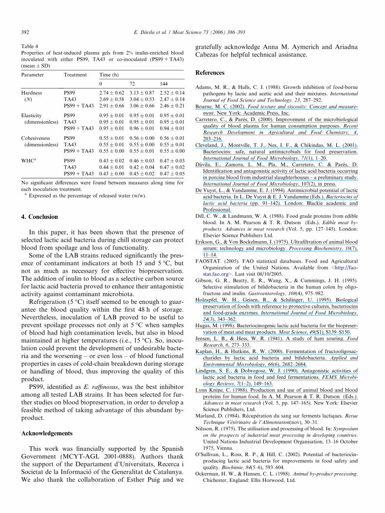
Table 4Properties of heat-induced plasma gels from 2% inulin-enriched bloodinoculated with either PS99, TA43 or co-inoculated (PS99 + TA43)(mean ± SD)
Parameter Treatment Time (h)
0 72 144
Hardness(N)
PS99 2.74 ± 0.62 3.13 ± 0.87 2.52 ± 0.14TA43 2.69 ± 0.58 3.04 ± 0.53 2.47 ± 0.14PS99 + TA43 2.91 ± 0.66 3.06 ± 0.66 2.46 ± 0.21
Elasticity(dimensionless)
PS99 0.95 ± 0.01 0.95 ± 0.01 0.95 ± 0.01TA43 0.95 ± 0.01 0.95 ± 0.01 0.95 ± 0.01PS99 + TA43 0.95 ± 0.01 0.96 ± 0.01 0.94 ± 0.01
Cohesiveness(dimensionless)
PS99 0.55 ± 0.01 0.56 ± 0.00 0.56 ± 0.01TA43 0.55 ± 0.01 0.55 ± 0.00 0.55 ± 0.01PS99 + TA43 0.55 ± 0.00 0.55 ± 0.01 0.55 ± 0.00
WHCa PS99 0.43 ± 0.02 0.46 ± 0.03 0.47 ± 0.03TA43 0.44 ± 0.01 0.42 ± 0.04 0.47 ± 0.02PS99 + TA43 0.43 ± 0.00 0.45 ± 0.02 0.47 ± 0.05
No significant differences were found between measures along time foreach inoculation treatment.
a Expressed as the percentage of released water (w/w).
392 E. Davila et al. / Meat Science 73 (2006) 386–393
4. Conclusion
In this paper, it has been shown that the presence ofselected lactic acid bacteria during chill storage can protectblood from spoilage and loss of functionality.
Some of the LAB strains reduced significantly the pres-ence of contaminant indicators at both 15 and 5 �C, butnot as much as necessary for effective biopreservation.The addition of inulin to blood as a selective carbon sourcefor lactic acid bacteria proved to enhance their antagonisticactivity against contaminant microbiota.
Refrigeration (5 �C) itself seemed to be enough to guar-antee the blood quality within the first 48 h of storage.Nevertheless, inoculation of LAB proved to be useful toprevent spoilage processes not only at 5 �C when samplesof blood had high contamination levels, but also in bloodmaintained at higher temperatures (i.e., 15 �C). So, inocu-lation could prevent the development of undesirable bacte-ria and the worsening – or even loss – of blood functionalproperties in cases of cold-chain breakdown during storageor handling of blood, thus improving the quality of thisproduct.
PS99, identified as E. raffinosus, was the best inhibitoramong all tested LAB strains. It has been selected for fur-ther studies on blood biopreservation, in order to develop afeasible method of taking advantage of this abundant by-product.
Acknowledgements
This work was financially supported by the SpanishGovernment (MCYT-AGL 2001-0888). Authors thankthe support of the Departament d’Universitats, Recerca iSocietat de la Informacio of the Generalitat de Catalunya.We also thank the collaboration of Esther Puig and we
gratefully acknowledge Anna M. Aymerich and AriadnaCabezas for helpful technical assistance.
References
Adams, M. R., & Halls, C. J. (1988). Growth inhibition of food-bornepathogens by lactic and acetic acid and their mixtures. International
Journal of Food Science and Technology, 23, 287–292.Bourne, M. C. (2002). Food texture and viscosity: Concept and measure-
ment. New York: Academic Press, Inc.Carretero, C., & Pares, D. (2000). Improvement of the microbiological
quality of blood plasma for human consumption purposes. Recent
Research Development in Agricultural and Food Chemistry, 4,203–216.
Cleveland, J., Montville, T. J., Nes, I. F., & Chikindas, M. L. (2001).Bacteriocins: safe, natural antimicrobials for food preservation.International Journal of Food Microbiology, 71(1), 1–20.
Davila, E., Zamora, L. M., Pla, M., Carretero, C. & Pares, D.Identification and antagonistic activity of lactic acid bacteria occurringin porcine blood from industrial slaughterhouses – a preliminary study.International Journal of Food Microbiology, 107(2), in press.
De Vuyst, L., & Vandamme, E. J. (1994). Antimicrobial potential of lacticacid bacteria. In L. De Vuyst & E. J. Vandamme (Eds.), Bacteriocins of
lactic acid bacteria (pp. 91–142). London: Blackie academic andProfessional.
Dill, C. W., & Landmann, W. A. (1988). Food grade proteins from edibleblood. In A. M. Pearson & T. R. Dutson (Eds.). Edible meat by-
products. Advances in meat research (Vol. 5, pp. 127–145). London:Elsevier Science Publishers Ltd.
Erikson, G., & Von Bockelmann, I. (1975). Ultrafiltration of animal bloodserum: technology and microbiology. Processing Biochemistry, 10(7),11–14.
FAOSTAT. (2005). FAO statistical databases. Food and AgriculturalOrganization of the United Nations. Available from <http://fao-stat.fao.org>. Last visit 08/10/2005.
Gibson, G. R., Beatty, E. R., Wang, X., & Cummings, J. H. (1995).Selective stimulation of bifidobacteria in the human colon by oligo-fructose and inulin. Gastroenterology, 108(4), 975–982.
Holzapfel, W. H., Geisen, R., & Schilinger, U. (1995). Biologicalpreservation of foods with reference to protective cultures, bacteriocinsand food-grade enzymes. International Journal of Food Microbiology,
24(3), 343–362.Hugas, M. (1998). Bacteriocinogenic lactic acid bacteria for the biopreser-
vation of meat and meat products. Meat Science, 49(S1), S139–S150.Jensen, L. B., & Hess, W. R. (1941). A study of ham souring. Food
Research, 6, 273–333.Kaplan, H., & Hutkins, R. W. (2000). Fermentation of fructooligosac-
charides by lactic acid bacteria and bifidobacteria. Applied and
Environmental Microbiology, 66(6), 2682–2684.Lindgren, S. E., & Dobrogosz, W. J. (1990). Antagonistic activities of
lactic acid bacteria in food and feed fermentations. FEMS Microbi-
ology Reviews, 7(1–2), 149–163.Lynn Knipe, C. (1988). Production and use of animal blood and blood
proteins for human food. In A. M. Pearson & T. R. Dutson (Eds.).Advances in meat research (Vol. 5, pp. 147–165). New York: ElsevierScience Publishers, Ltd.
Marland, D. (1984). Recuperation du sang sur ferments lactiques. Revue
Technique Veterinaire de l’Alimentation(nov), 30–31.Nilsson, R. (1975). The utilisation and processing of blood. In: Symposium
on the prospects of industrial meat processing in developing countries.United Nations Industrial Development Organisation, 13–16 October1975, Vienna.
O’Sullivan, L., Ross, R. P., & Hill, C. (2002). Potential of bacteriocin-producing lactic acid bacteria for improvements in food safety andquality. Biochimie, 84(5–6), 593–604.
Ockerman, H. W., & Hansen, C. L. (1988). Animal by-product processing.Chichester, England: Ellis Horwood, Ltd.

E. Davila et al. / Meat Science 73 (2006) 386–393 393
Pares, D., & Carretero, C. (1996). Influencia de algunos factores sobre elgrado de hemolisis de la sangre porcina de mataderos industriales.Alimentaria(April), 65–69.
Pares, D., Saguer, E., Saurina, J., Sunol, J. J., & Carretero, C. (1998).Functional properties of heat induced gels from liquid and spray-driedporcine blood plasma as influenced by pH. Journal of Food Science,
63(6), 958–961.Pares, D., Zamora, L., Saguer, E., & Carretero, C. (2004). Acid lactic
bacteria as biopreservative cultures in porcine blood for humanconsumption. Food Science Central. FoodInfo Online Features. Avail-able from<http://www.foodsciencecentral.com>.
Pearson, A. M., & Dutson, T. R. (1988). Edible meat by-products. London:Elsevier Applied Science.
Pinel, M. (1985). Etat des techniques de traitement du sang. Viandes et
Produits Carnes, 6(2), 53–57.Ross, R. P., Morgan, S., & Hill, C. (2002). Preservation and fermentation:
past, present and future. International Journal of Food Microbiology,
79(1–2), 3–16.Swingler, G. R. (1982). Microbiology of meat industry by-products. In M.
H. Brown (Ed.), Meat microbiology (pp. 179–224). New York: ElsevierScience.
Wang, X., & Gibson, G. R. (1993). Effects of the in-vitrofermentation of oligofructose and inulin by bacteria growing inthe human large-intestine. Journal of Applied Bacteriology, 75(4),373–380.