Natural and Lactic Acid Bacteria Fermentations of - Brage NMBU
Upload
khangminh22Category
view
1download
0
DISSERTATIONES MEDICINAE UNIVERSITATIS TARTUENSIS 187


DISSERTATIONES MEDICINAE UNIVERSITATIS TARTUENSIS 187
JELENA ŠTŠEPETOVA The characterisation of intestinal lactic
acid bacteria using bacteriological, biochemical and molecular approaches

Department of Microbiology, University of Tartu, Estonia Dissertation has been accepted for the commencement of the degree of Doctor of Medical Science on May 18, 2011, by the Council of the Faculty of Medi-cine, University of Tartu, Estonia Supervisor: Professor extraordinaria Marika Mikelsaar, MD, PhD
Department of Microbiology University of Tartu, Estonia
Co-supervisor: Epp Sepp, MD, PhD Senior Research Fellow at the Department of Microbiology University of Tartu Rewiewed by: Professor Pärt Peterson, PhD
Head of the Department of General and Molecular Pathology University of Tartu, Estonia
Meeme Utt, PhD Senior Research Fellow at the Department of General and Molecular Pathology, Chair of Immunology University of Tartu, Estonia
Opponent: Professor Gjalt W. Welling, PhD
Department of Medical Microbiology University of Groningen
Commencement: August 29th, 2011 Publication of this dissertation is granted by University of Tartu
ISSN 1024–395X ISBN 978–9949–19–775–0 (trükis) ISBN 978–9949–19–776–7(PDF)
Autoriõigus Jelena Štšepetova, 2011
Tartu Ülikooli Kirjastus www.tyk.ee Tellimus nr 400

5
CONTENTS
LIST OF ORIGINAL PUBLICATIONS ....................................................... 8
ABBREVIATIONS ........................................................................................ 9
1. GENERAL INTRODUCTION .................................................................. 11
2. LITERATURE REVIEW ........................................................................... 13 2.1. Human intestinal microbiota .............................................................. 13
2.1.1. Composition of intestinal microbiota ....................................... 13 2.1.2. The functions of the intestinal microbiota ................................ 14
2.2. Lactic acid bacteria ............................................................................. 15 2.2.1. Taxonomy of lactic acid bacteria .............................................. 15
2.2.1.1. The genus Lactobacillus .............................................. 16 2.2.1.2. The genus Bifidobacterium ......................................... 18
2.2.2. Metabolism of lactic acid bacteria ............................................ 19 2.2.2.1. Sugar metabolism of Lactobacillus spp. ..................... 19 2.2.2.2. Sugar metabolism of Bifidobacterium spp. ................. 22 2.2.2.3. Poly- and biogenic amines .......................................... 23
2.3. The composition and functions of the intestinal lactic acid bacteria . 26 2.3.1. The distribution of lactobacilli and bifidobacteria .................... 26
2.3.1.1. The role of short-chain fatty acids in the intestinal tract .............................................................. 28
2.3.2. Age related differences ............................................................ 29 2.3.2.1. Neonates and children ................................................. 29 2.3.2.2. Adults and elderly people ........................................... 30
2.3.3. Environmental differences ....................................................... 31 2.3.3.1. Geographical and social differences ........................... 31
2.3.4. Gut microbiota in diseases ........................................................ 31 2.3.4.1. Allergy ........................................................................ 31 2.3.4.2. Metabolic syndrome ................................................... 32
2.4. Methods for studying lactic acid bacteria .......................................... 33 2.4.1. Bacteriological and biochemical methods for identification
of lactic acid bacteria ............................................................... 34 2.4.2. Gas-liquid chromatography for studying metabolic activity
of lactic acid bacteria ............................................................... 34 2.4.2.1. Detection of organic acids .......................................... 35 2.4.2.2. Detection of poly- and biogenic amines ..................... 36
2.4.3. Molecular tools for analysing of lactic acid bacteria ............... 36 2.4.3.1. Qualitative molecular methods ................................... 36 2.4.3.2. Quantitative molecular methods ................................. 41
3. STUDY RATIONALE ............................................................................... 44
AIMS OF THE STUDY ................................................................................. 45
2

6
4. MATERIALS AND METHODS ............................................................... 46 4.1. Subjects and materials ....................................................................... 46 4.2. Sample collection .............................................................................. 46 4.3. The reference strains .......................................................................... 50 4.4. Bacteriological methods for identification of lactic acid bacteria .... 51 4.5. Metabolitic activity of lactic acid bacteria ......................................... 52
4.5.1. Extraction and detection of organic acids ................................ 52 4.5.2. Detection of poly-and biogenic amines.................................... 52
4.6. The molecular methods for the detection of lactic acid bacteria ...... 54 4.6.1. DNA extraction ........................................................................ 54 4.6.2. Primers and probes ................................................................... 54 4.6.3. PCR based methods ................................................................. 55
4.6.3.1. Identification of lactobacilli using ITS-PCR/RFLP analysis ....................................................................... 55
4.6.3.2. Assessment of lactobacilli in faecal samples .............. 55 4.6.3.3. Detection of Bifidobacterium spp. in faecal samples
using PCR-DGGE ....................................................... 55 4.6.3.3.1. PCR amplification ...................................... 55 4.6.3.3.2 DGGE analysis of PCR products ............... 56 4.6.3.3.3 Cloning of the PCR products ...................... 56 4.6.3.3.4. Sequence analysis ....................................... 56
4.6.3.4. Real-time PCR ............................................................ 56 4.6.4. Fluorescence in situ hybridization ........................................... 57 4.6.5. Pulse field gel electrophoresis ................................................. 58
4.7. Statistical analysis ............................................................................. 58
5. RESULTS ................................................................................................... 63 5.1. Comparison of bacteriological and molecular methods for the
identification of intestinal Lactobacillus isolates (Papers I, III) ........ 63 5.1.1. Biochemical and molecular identification of probiotic
L. plantarum strain Inducia DSM 21379 (Paper VI, present study) ......................................................... 67
5.2. Metabolic activity of Lactobacillus and Bifidobacterium spp. ......... 68 5.2.1. Production of organic acids and ethanol by lactobacilli and
bifidobacteria isolates and its relationship with their culture environment (Papers I, III, and present study) ........................ 68
5.2.2. Production of poly- and biogenic amines by Lactobacillus spp. strains (Paper VI, present study) ...................................... 71
5.3. The detection and prevalence of lactic acid bacteria determined using bacteriological and molecular methods (Papers II, VI) ..................... 74
5.4. Diversity of gut lactobacilli in healthy adults and the elderly (Paper IV) .............................................................................. 75

7
5.4.1. Association of Lactobacillus spp. with some anthropometrical and metabolic characteristics in healthy adults and elderly (Paper IV) ................................................................................ 77
5.5. Influence of some environmental factors on gut microbiota ............ 78 5.5.1. Diversity of gut lactobacilli in 1–2-year-old Estonian and
Swedish children (Paper III) .................................................... 78 5.6. The diversity of the bifidobacterial community in non-allergic and
allergic children (Paper V) ................................................................ 79
6. GENERAL DISCUSSION ......................................................................... 82
CONCLUSIONS ............................................................................................ 95
REFERENCES ............................................................................................... 97
SUMMARY IN ESTONIAN Soole piimhappebakterite iseloomustus bakterioloogiliste, biokeemiliste ja molekulaarsete meetoditega ........................................................................... 119
ACKNOWLEDGMENTS .............................................................................. 122
PUBLICATIONS ........................................................................................... 123
CURRICULUM VITAE ................................................................................ 219

8
LIST OF ORIGINAL PUBLICATIONS
I Annuk H, Shchepetova J, Kullisaar T, Songisepp E, Zilmer M, Mikelsaar M. Characterization of intestinal lactobacilli as putative probiotic candidates. Journal of Applied Microbiology 2003; 94: 403–412.
II Štšepetova J, Sepp E, Hütt P, Mikelsaar M. Estimation of lactobacilli, enterococci and bifidobacteria in faecal samples by quantitative bac-teriology and FISH. Mikroökologie and Therapie 2002; 29: 51–59.
III Mikelsaar M, Annuk H, Shchepetova J, Mändar R, Sepp E, Björksten B. Intestinal lactobacilli of Estonian and Swedish children. Microbial Ecology in Health and Disease 2002; 14: 75–80.
IV Štšepetova J, Sepp E, Kolk H, Lõivukene K, Songisepp E, Mikelsaar M. Diversity and metabolic impact of intestinal Lactobacillus spp. in healthy adults and elderly. British Journal of Nutrition 2011; 105: 1235–1244. DOI: 10.1017/S0007114510004770
V Štšepetova J, Sepp E, Julge K, Vaughan E, Mikelsaar M, de Vos WM. Molecularly assessed shifts of Bifidobacterium ssp. and less diverse microbial communities are characteristic of 5-year-old allergic children. FEMS Immunology and Medical Microbiology 2007; 51: 260–269.
VI Mikelsaar M; Songisepp E; Smidt I, Štšepetova J; Zilmer M, Hütt P, Truusalu K, Kilk K. Isolated microorganism strain Lactobacillus plantarum Inducia DSM 21379 as probiotic enhancing natural immunity of organism, food product and composition comprising said microorganism and use of said microorganism for production of medicine for enhancing of cellular immunity. (Priority number: P200800027). PCT/EE2009/000006, Estonian Patent EE05341 B1, 07.09.2010.
Jelena Štšepetova has contributed to the following original publications: Papers I, III: molecular analysis of lactobacilli, detection of metabolites by gas
chromatography, participation in data analysis and manuscript drafting.
Paper II: study design, study performance, molecular analysis, data analy-sis and manuscript drafting.
Paper IV: study design, study performance, molecular analysis, data analy-sis and manuscript drafting.
Paper V: study design, study performance, molecular analysis, data analy-sis and manuscript drafting.
Paper VI: characterisation of Lactobacillus plantarum Inducia DSM 21379 by molecular methods, detection of metabolites by gas chro-matography, data analysis.

9
ABBREVIATIONS
API 50CHL Analytical Profile Index according to fermentation pattern of 50 Carbohydrates by Lactobacillus (isolates)
ADP Adenosine Diphosphate ATCC American Type Culture Collection ATP Adenosine Triphosphate BMI Body mass index CAN Columbia Agar with Colistin and Nalidixic Acids CoA Coenzyme A CFU Colony Forming Units DAPI 4’6-diamino-2-phenylindole dihydrochloride DGGE Denaturing Gradient Gel Electrophoresis DNA Deoxyribonucleic Acid DSM Deutsche Sammlung von Mikroorganismen EMP Embden-Meyerhof-Parnas Pathway F6PPK Fructose-6-Phosphate Phosphoketolase FAO Food and Agriculture Organization FHEL Facultatively Heterofermentative Lactobacilli FISH Fluorescent In Situ Hybridization GALT Gut-associated Lymphoid Tissue GC Gas Chromatography GLC Gas Liquid Chromatography GI Gastrointestinal Tract HPLC High Pressure Liquid Chromatography ITS Internal-Transcribed Spacer ITS-PCR Internal-Transcribed Spacer Polymerase Chain Reaction LDH Lactate Dehydrogenase LAB Lactic Acid Bacteria LB Lactobacillus spp. MRS de Man-Rogosa-Sharpe NADH Reduced Nicotinamide-adeninenucleotide NAD Nicotinamide-adeninedinucleotide NO Nitric oxide NOS-NO Nitric oxide synthase-nitric oxide system NSP Non-starch polysaccharides ODS Ornithine decarboxylase OHEL Obligately Heterofermentative Lactobacilli
3

10
OHOL Obligately Homofermentative Lactobacilli PFGE Pulse Field Gel Electrophoresis PCR Polymerase Chain Reaction RT-PCR Real-Time Polymerase Chain Reaction RFLP Restriction Fragment Length Polymorphism SAM S-adenosylmethionine SCFA Short-Chain Fatty Acids TGGE Temperature Gradient Gel Electrophoresis WHO World Health Organization

11
1. GENERAL INTRODUCTION
Lactic acid bacteria (LAB), including Lactobacillus spp. and Bifidobacterium spp., are well known and safe components of the microbial ecosystem in the gastrointestinal tract. They colonise the digestive tract soon after birth and are present in high numbers in both infants and adults (Mikelsaar and Mändar, 1993; Guarner and Malagelada, 2003). The potential role bifidobacteria in infant digestion (Tissier, 1900; Moro, 1900a, 1900b) and the application of lactobacilli-fermented food against the effects of aging and atherosclerosis have attracted attention since early in the last century (Metchnikoff, 1908). In more recent times, several investigations have connected the loss of LAB in the host with the development of allergies (Björksten et al., 2001; Kalliomäki et al., 2001). In the last 15 years the level of knowledge regarding LAB in the human host and its impact on human health has been exapnded considerably at the Department of Microbiology of Tartu University (Mikelsaar, 1993; Mändar, 1996; Karki, 1996; Naaber, 1997; Sepp, 1998; Annuk, 2002; Songisepp, 2005). However, there remains a lack of data regarding the species composition of intestinal LAB and their secreted metabolites in healthy and allergic children, and also during the aging process.
Worldwide, there is a rising demand for functional food incorporating pro-biotic bacteria as live microorganisms which administered in adequate amounts provide a health benefit to the host (FAO-Food and Agriculture Organization and WHO-World Health Organization, 2002). However, the information about which species and strains of LAB could provide the best health effect under different environmental conditions and at different age scale are still scarce. Although, the necessity of fundamental molecular typing of LAB probiotic can-didates has been underlined (FAO; Vanckerhofen et al., 2008), the assessment of their molecular and metabolite profiles, which grant particular functional properties on the strains regarding health promotion, is often insufficient (Douglas and Sanders, 2008). The understanding of the complex genetic and metabolic properties of the microbiota is of the utmost importance for under-standing and identifying candidate strains for novel group-specific and indi-vidual healthy diets.
The present thesis integrates data that has been collected over several years using available molecular (PCR, ITS-PCR, FISH, DGGE, real-time PCR, 16S rRNA sequencing) and biochemical methods (enzymatic profile, gas-chro-matography) for characterisation of intestinal lactic acid bacteria from people in different age groups including people with allergies. Consequently, a well-characterised pool of lactobacilli strains has been created in the Department of Microbiology at the University of Tartu. The studies on metabolism of carbo-hydrates and polyamines have helped to reveal some novel functional properties of these collected lactobacilli. The author of the current PhD thesis participated in the charactertisation of the probiotic strain Lactobacillus plantarum Inducia (DSM 21379) which has been patented in Estonia (EE05341 B1, 07.09.2010)

12
and has an international patent application (PCT/EE2009/000006) filed. This probiotic aims to enhance natural immunity and the author has performed the molecular identification of the strain and evaluated its metabolic functional properties.

13
2. LITERATURE REVIEW
2.1. Human intestinal microbiota
2.1.1. Composition of intestinal microbiota
The human digestive tract harbours a wealth of different microbial ecosystems that vary according to their location within the intestinal tract. Culture-based studies have shown that faecal bacteria comprise between 400–500 distinct spe-cies and each individual human has a unique flora dominated by 30–40 species (Kimura et al., 1997). The application of molecular techniques has indicated that 60–80% of the organisms of the human microbiota have not been cultivated so far (Laugendijk et al., 1995; Saue et al., 1999; Eckburg et al., 2005). Based on molecular methods, current estimates indicate that intestinal microbiota con-sists of at least 1014 microbes and is dominated by anaerobic bacteria, com-prising over 1000 species, among which gram-positive bacteria predominate (Vaughan et al., 2000; Zoetendal et al., 2004; Eckburg et al., 2005; Bäckhed et al., 2004; Ley et al., 2006; Ventura et al., 2009). The microbial composition and their relative proportions vary, depending on the physiological conditions of the compartment (e.g. pH), with different parts of the gastrointestinal tract, be-coming richer and more diverse, ranging from the relatively germ-free stomach and the upper small intestine to the colon (Figure 1) (Kleessen et al., 1997). It has been demonstrated previously that the majority of faecal bacteria pre-dominantly belong to genera such as Bacteroides, Eubacterium, Clostridium, Ruminococcus, Fusobacterium, Bifidobacterium and Peptostreptococcus (Moore et al., 1974; Simon et al., 1984). Recent molecular studies have revealed that the gut microbiota of adults is largely dominated by the members of only two bacterial phyla, Bacteroidetes and Firmicutes, and a single member of the archaea, Methanobrevibacter smithii (Huys et al., 2008). More specifically, three bacterial groups predominate within these phyla: the Bacteroides-Prevotella group, the Clostridium coccoides group, and the Clostridium leptum group (Eckburg et al., 2005).
Lactobacillus spp. and Streptococcus spp. prevail in the microflora of small intestine (Finegold et al., 1983; Franks et al., 1998; Marteau et al., 2001; Zoetendal et al., 2006) whit a 10-fold difference in the ratio of anaerobes/ aerobes compared to a 1000-fold difference in caecum (Klessen et al., 2000; Macfarlane et al., 2000). The bacteria of the small intestine have the strongest influence on the immune system since the gut-associated-lymphoid-tissue (GALT) is situated mainly in the small intestinal and caecal mucosa (Schroff et al., 1995). Therefore, the composition and activity of the intestinal flora can have a profound influence on health and disease.
4

14
Figure 1. A bacterial distribution in human gastrointestinal tract (modified from Simon et al., 1984; Ouwehand et al., 2003).
2.1.2. The functions of the intestinal microbiota
The intestinal microbiota has important functions for the human host such as involvement in nutrition, immunomodulation and as a barrier against exogenous infections. Generally, these functions may be divided into three categories: structural, metabolic and protective (Guarner, 2006).
The gut microbiota is considered to represent a crucial line of defence against colonisation by exogenous or opportunistic bacteria that are present in the gut. This barrier effect involves several mechanisms, including displacement of pathogens by outcompeting them for the nutrients and epithelial-binding sites (Guarner, 2006), as well as production of antimicrobial factors such as lactic acid, hydrogen peroxide and bacteriocins, the latter inhibiting the growth of the invading bacteria (Shanahan, 2002). Its role in the development of a competent immune system cannot be neglected either (Guarner, 2006) and it is thought that the modulation of the immune system proceeds in a strain-dependent manner (Servin, 2004).
Gut bacteria also provide the metabolic functions involved in host nutrition. They have the ability to ferment non-digestible dietary substrates from the upper part of the gastrointestinal tract and endogenous mucus produced by the epithelia. The basic fermentative reaction in the human colon is hydrolysis of

15
polysaccharides, oligosaccharides, and disaccharides to their constituent sugars, resulting in an increased biomass. The fermentation of carbohydrates is the major source of energy in the colon for the bacterial growth, producing short chain fatty acids (SCFA) that can be absorbed by the host for structural com-pletion of gut mucosa (McFarlane et al., 1995). A comparison of germ-free mice with conventional mice has shown that gut bacteria play a key role in the gut wall fortification by influencing the proliferation and differentiation of epithelial cells (Hooper et al., 2001).
The microbiota is also able to metabolise proteins and protein degradation products, sulfur containing compounds, as well as endogenous and exogenous glycoproteins (Gibson et al., 1995). In addition, gut bacteria are responsible for the production of vitamins such as K, B12, biotin, folic acid, pantothenate, and in addition synthesis of amino acids from ammonia or urea (Hooper et al., 2002) as well as the inactivation of dietary carcinogens (Wollowski et al., 2001). However, few data are available on the metabolism of polyamines and biogenic amines by the intestinal microbiota (Arena et al., 2001; Matsumoto et al., 2007) or their specific roles in the aforementioned three functional categories of the microbiota in the host.
2.2. Lactic acid bacteria
LAB can be found in different nutrient-rich habitats, for example on the muco-sal membranes of humans and animals, as well as on plants (Holzapfel et al., 2001). Bifidobacteria mainly originate from the gastrointestinal tract and breast milk of humans (Tannock, 1997; 2010). Lactobacillus spp. are ubiquitous and can be retrieved from dairy, grains, meat, fish, beer, wine, sauerkraut, sour dough, mash products, fruits, pickled vegetables, water, sewage, silage, and the mucosa of different human and animal cavities (mouth, intestine and vagina) (Salminen et al., 2004).
2.2.1. Taxonomy of lactic acid bacteria
LAB are a genetically distinct group of bacteria that share the same biochemical properties, e.g. gram-positive, non-sporulating rods or cocci, catalase negative, acid tolerant, lactic acid production as a result of carbohydrate fermentation and which prefer growth under anaerobic conditions but are quite aerotolerant (Wessels et al., 2004).
The genus Lactobacillus belongs to the phylum Firmicutes, class Bacilli, order Lactobacillales, family Lactobacillaceae. Important members of Lacto-bacillaceae are the genera Aerococcus, Carnobacterium, Enterococcus, Lacto-coccus, Lactobacillus, Leuconostoc, Oenococcus, Pediococcus, Streptococcus, Tetragenococcus, Vagococcus and Weissella (Collins et al., 1987, 1993; Dicks et al., 1995; Adams and Moss, 2000; Felis et al., 2007). Its closest relative,

16
being grouped within the same family, is also the genus Paralactobacillus (Garrity et al., 2004). Nevertheless, after regrouping, the phylogenetically closest family appears to be the Leuconostocacea family (Hammes et al., 2003).
Genus Bifidobacterium has previously been considered in the same context as the genuine LAB while sharing some of their typical morphological and functional properties (Kandler and Lauer, 1974; Vaughan et al., 2002). How-ever, Pourand et al. (1973) first postulated a relation of the genus Bifido-bacterium to Actinomycetaceae group. Moreover, its place in the phylum Acti-nobacteria, order Bifidobacteriales and family Bifidobacteriaceae has been confirmed by analysis of 16S rRNA sequences (Woese, 1987; Garrity et al., 2004) and in addition, it has a specific pathway for sugar fermentation.
2.2.1.1. The genus Lactobacillus
Lactobacilli are gram-positive bacteria and vary in morphology from long, slen-der rods to short coccobacilli, which frequently form chains. They are fermen-tative, catalase-negative microaerophilic and chemo-organotrophic, producing lactic acid as the major product during the fermentation of carbohydrates (Axelsson, 1998). They have complex growth requirements supplied by carbo-hydrates, amino acids, peptides, fatty acid esters, salts, nucleic acid derivatives and vitamins. A low pH (between 4.5 and 6.2) is stimulatory. The genus Lacto-bacillus is one of the largest, comprising more than 90 described species as well as some subspecies (Robinson, 2002; http://www.bacterio.cict.fr), and approxi-mately 30% of them have been isolated from faecal sources (Table 2). The genus is very heterogeneous, encompassing species with a large variety of phe-notypic, biochemical, and physiological properties. With regards to the DNA base composition of their genome, lactobacilli usually have GC content lower than 54%.
Lactobacilli can be subdivided into three groups according to the type of sugar fermentation they demonstrate: two genera of homofermenters ‘Thermo-bacterium’, ‘Streptobacterium’, and a third genus of heterofermenters ‘Beta-bacterium’ (Table 1) (Kandler and Weiss, 1986; Pot et al., 1994; Hammes et al., 1995; Axelsson, 1998).
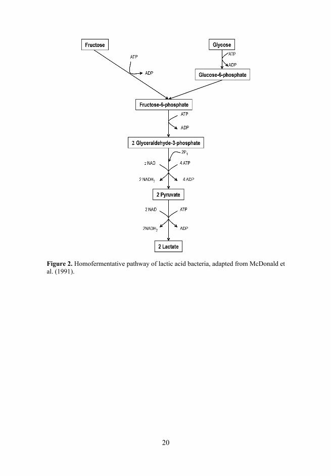
Group I – obligately homofermentative lactobacilli (OHOL) are able to con-vert the hexoses into lactic acid via the Embden-Meyerhof-Parnas (EMP) path-way (Figure 2) while the pentoses and the gluconate are not fermented as OHOL lack phosphoketolase;
Group II – facultatively heterofermentative lactobacilli (FHEL) degrade the hexoses to lactic acid by the EMP pathway and the pentoses to lactic acid and ethanol/acetic acid via phosphoketolase. In addition, gluconate is often fer-mented (Figure 3);
Group III – obligately heterofermentative lactobacilli (OHEL) ferment the hexoses to lactic acid, carbon dioxide and ethanol (or acetic acid in the presence of an alternative electron acceptor). Furthermore, pentoses are converted to lactic and acetic acids (Figure 3).

17
Table 1. The division of the most common Lactobacillus ssp. (adapted from Botazzi, 1983; Axelsson, 1993).
Fermentation pathway
Group I Obligately
homo-fermentative
Group II Facultatively
hetero-fermentative
Group III Obligately
hetero-fermentative
Growth at 45°C + + +/– Growth at 15°C –(+)# +(–)** +(–)** Hexose fermentation + + + Pentose fermentation – + + Fructose-diphosphate (FDP) aldolase
+ + –
Phosphoketolase (PK) – +* + Gas from glucose – – + Gas from gluconate – + + NH3 from arginine –(+)# – +(–)** Lactic acid modification
D-, L-, DL D-, L-, DL DL
L. delbrueckii L. acidophilus L. helveticus L. salivarius
L. casei L. curvatus L. paracasei L. plantarum L. peptosus L. sakei L. rhamnosus
L. brevis L. buchneri L. fermentum L. reuteri L. oris L. mucosae
* – inducible by pentose; # – mostly negative; ** – mostly positive, with a few exceptions The different lactobacilli species are able to use different pathways depending on the conditions and enzymatic capacity (Kandler et al., 1986; Axelsson, 1998). However, the division of lactobacilli into three main groups according to the type of fermentation is not in accordance with their phylogenetic classi-fication as revealed by analysis of their 16S rDNA sequences (Axelsson, 1998). Lactobacillus spp. with widely diverse DNA base ratios can be grouped into three clusters: L. delbrueckii group, L. casei-Pediococcus group and the Leu-conostoc group which also contains some lactobacilli (Collins, 1991). Never-theless, the L. delbrueckii group was renamed as the L. acidophilus group, according to the more typical representative of this group (Schleiefer et al., 1995). Furthermore, based on DNA-DNA hybridization and other phylogenetic methods they were grouped into 8 major groups: L. buchneri, L. delbrueckii, L. casei, L. plantarum, L. reuteri, L. sakei, L. salivarius and L. brevis group (Salminen et al., 1998; Felis et al., 2005). The L. casei group comprises the recently revised species L. zeae, L. casei, L. paracasei and L. rhamnosus (Dicks et al., 1996).
5

18
2.2.1.2. The genus Bifidobacterium
The 29 species belonging to the genus Bifidobacterium, share phenotypical features typical to LAB, such as organic acid production. Bifidobacteria are gram-positive, catalase-negative, polymorphic branched rods that occur singly, in chains or clumps. They are non-spore-forming, non-motile, and non-filamen-tous. Bifidobacterium spp. are chemoorganotrophs, growing in an anaerobic environment, having a fermentative type of metabolism, and producing organic acids but not gas from a variety of carbohydrates. Their genome GC content varies from 42 to 67% (Sebald et al., 1965; Scardovi et al., 1986; Biavati and Mattarelli, 2001).
The optimum temperature for the isolation of the bifidobacteria species from a human host is 36–38°C. In contrast, that for the species from animals is slightly higher, at about 41–43°C and may even reach above 46°C. Further-more, the initial optimum growth pH is between 6.5 and 7.0.
According to their phylogenetic analysis, the Bifidobacterium branch forms a coherent phylogenetic unit as their 16S rRNA sequences share over 93% simi-larity (Leblond-Bourget et al., 1996; Matsuki et al., 1998; Botaccini et al., 2010). For the phylogenetic differentiation between closely related bifidobac-terial species some other genes including the elongation factor Tu (tuf) gene (Ventura et al., 2004), recombinase A (recA) gene (Ventura et al., 2003), ATP synthase subunit B (atpD) gene (Ventura et al., 2004), pyruvate kinase (Vaugien et al., 2002) and xylose-5-phosphate/fructose-6-phosphate (xfp) (Yin et al., 2005) have been introduced. Multiple PCR-based methods support and com-plement the 16S rRNA-based division of bifidobacterial species. A phyloge-netic tree of Bifidobacterium spp. includes 5 major groups: B. adolescentis, B. pullorum, B. asteroides, B. boum and B. pseudolongum. Species B. breve and B. longum form a couple, as well as B. minimum and B. psychroaerophilum. How-ever, B. bifidum, B. magnum, B. scardovii and B. subtile form distinct branches.
Nevertheless, there are taxonomic issues concerning the genus Bifidobac-terium in particular regarding the species B. longum-B. infantis, recently united under the name B. longum and the recognition of their three biotypes (infantis, longum and suis types) using molecular methods (Sakata et al., 2002). In addi-tion, another debated issue is the relationship between B. animalis and B. lactis, the latter recently reclassified as B. animalis subsp. lactis. So far there are 29 recognised Bifidobacterium species with 11 being isolated only from human host (Table 2).

19
2.2.2. Metabolism of lactic acid bacteria
2.2.2.1. Sugar metabolism of Lactobacillus spp.
Lactobacilli are able to ferment various carbohydrates and adapt to the various environmental conditions by changing their metabolism accordingly. Two major sugar fermentation pathways are recognised in lactobacilli: glycolysis (the Embden-Meyerhof pathway) and the pentose-phosphate pathway (pento-sephosphoketolase or 6-phosphogluconate pathway) (Kandler, 1983).
Glycolysis is a sequence of reactions that converts glucose into pyruvate with the concomitant production of a relatively small amount of ATP (Figure 2). In these reactions, two substrate level phosphorylation reactions, involving phosphoglycerate kinase and pyruvate kinase, operate to yield energy. If the only end product of this pathway is lactic acid, the fermentation is referred to as homolactic fermentation. This pathway is used by all the lactic acid bacteria, with the exception of the leuconostocs and OHEL. Complete homolactic fer-mentation of glucose results in 2 moles of lactic acid and a net gain of 2 ATP per mole of glucose consumed.
The pentose-phosphate pathway is used in heterofermentative lactobacilli and the main difference from homolactic is the presence of enzyme phos-phoketolase and lack of fructose-diphosphate aldolase (Figure 3). Glyceral-dehyde-3-phosphate from the phosphoketolase reaction is metabolised to lactic acid similarly to glycolysis, and acetyl phosphate is converted to ethanol. Heterolactic fermentation of glucose gives 1 mole of each lactic acid, ethanol and CO2 and 1 mole ATP per glucose consumed. In aerobic conditions, NADH can be oxidized by oxygen and acetate is produced from acetyl phosphate, yielding additional ATP from substrate level phosphorylation.
Facultatively and obligatory heterofermentative lactobacilli also ferment pentose via the lower half of the pentose-phosphate pathway, starting from ribulose-5-phosphate or xylulose-5-phosphate (Figure 3). The fermentation of pentoses results in production of equimolar amounts of lactic and acetic acids, while no CO2 is formed (Axelsson, 1993). Metabolism of hexose is similar to that of glucose and begins from glucose-6-phosphate or fructose-6-phosphate (Figure 3).
Configuration of the lactic acid produced by lactobacilli depends on the presence of specific NAD+-dependent lactate dehydrogenases (nLDH) and their respective activities in the cell. If both D- and L- lactic acids are formed, there are generally one D-nLDH and one L-nLDH present. In these cultures, generally L-lactate is the major form produced in early growth phase and D-lactic acid in the late stationary phase (Axelsson, 1993).
Lactobacilli may change their metabolism in response to various conditions and this can be attributed to an alternative pyruvate metabolism or the use of external electron acceptors. Pyruvate, intermediately formed in both pathways, may undergo several conversions (Figure 4).
20
Figure 2. Homofermentative pathway of lactic acid bacteria, adapted from McDonald et al. (1991).

21
Figure 3. Heterofermentative pathway of lactic acid bacteria, adapted from McDonald et al. (1991).
6
22
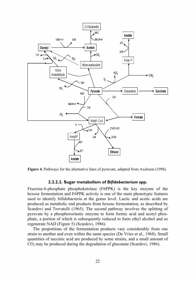
Figure 4. Pathways for the alternative fates of pyruvate, adapted from Axelsson (1998).
2.2.2.2. Sugar metabolism of Bifidobacterium spp.
Fructose-6-phosphate phosphoketolase (F6PPK) is the key enzyme of the hexose fermentation and F6PPK activity is one of the main phenotypic features used to identify bifidobacteria at the genus level. Lactic and acetic acids are produced as metabolic end products from hexose fermentation, as described by Scardovi and Trovatelli (1965). The second pathway involves the splitting of pyruvate by a phosphoroclastic enzyme to form formic acid and acetyl phos-phate, a portion of which is subsequently reduced to form ethyl alcohol and so regenerate NAD (Figure 5) (Scardovi, 1986).
The proportions of the fermentation products vary considerably from one strain to another and even within the same species (De Vries et al., 1968). Small quantities of succinic acid are produced by some strains, and a small amount of CO2 may be produced during the degradation of gluconate (Scardovi, 1986).

23
Figure 5. Metabolic pathway of Bifidobacterium spp. (Ballongue, 2004): 1– hexokinase and glucose-6-phosphate isomerase; 2 – fructose-6-phosphate phosphoketolase; 3 – transaldolase; 4 – transketolase; 5 – ribose-5-phosphate isomerase; 6 – ribulose-5-phos-phate epimerase; 7 – xylose-5-phosphate phosphocetolase; 8 – acetate kinase; 9 – ho-mofermentative pathway enzymes; 10 – (L+) lactate dehydrogenase; 11– phosphoro-clastic enzyme; 12 – formate dehydrogenase; 13– alcohol dehydrogenase.
2.2.2.3. Poly- and biogenic amines
Polyamines are aliphatic molecules with amine groups distributed along their structure. Polyamines are known classically by the names of putrescine [1,4-butane diamine or tetramethylenediamine], spermidine [N-(3-aminopropyl)-1,4-butane diamine or aminopropyl-tetramethylenediamine] and spermine [N,N’-bis(3-aminopropyl)-1,4-butane diamine or diaminopropyltetramethylene-diamine]. They are present in all human cells (Larque et al., 2007). Other oligoamines and diamines such as histamine, tyramine and cadaverine also occur naturally, but they are not included usually with the general term of polyamines (Wallace, 2003) and are referred as biogenic amines.

24
Polyamines are water-soluble, with pK (dissociation constant) values of about 10 when fully protonated at body pH and are bound by a strong inter-action with polyanionic macromolecules such as DNA and RNA. Only around 7–10% of the total cell content remains as free polyamines (Gugliucci, 2004; Moinard et al., 2005). The functions of polyamines depend on their electrical charges; their binding energy decrease from spermine to putrescine (spermine > spermidine > putrescine) (Yuan et al., 2001). The source of polyamines may be endogenous (intracellular de novo synthesis and interconversion pathways) or exogenous supplied in the diet.
Endogenously, polyamines can be synthesized from ornithine by a reaction catalysed by the enzyme ornithine-decarboxylase (ODS), which produces putre-scine (Figure 6). Spermidine derives from putrescine after the addition of a pro-pylamine group derived from the decarboxylated S-adenosylmethionine (SAM) by the action of spermidine synthase. Spermidine is similarly converted into spermine by the enzyme spermine synthase that adds a second propylamine group from SAM to the spermidine. Polyamine interconversion is performed by two coupled reactions: acetylation, mediated by the action of an acetyl coen-zyme A: polyamine N’-acetyl transferase, and cleavage through the action of an enzyme, polyamine oxidase. However, there is an alternative pathway in which arginine is firstly decarboxylated to yield agmatine, a compound that breaks down to urea and putrescine (Figure 6). However, evidence for this alternative pathway in LAB is lacking (Moreno-Arribas et al., 2003).
The main source of exogenous polyamines is dietary (especially from cheese, fruit, meat, and some vegetables) and human milk (Kalaš et al., 2005; Larque et al., 2006). Human milk is very high in polyamines where they may account for its growth promotion properties and protective effects against aller-gies. The substantial amounts of spermine and spermidine of breast milk poten-tially modulate the maturation of the infant’s intestines, enzyme activity and mucosal barrier functions (Wang et al., 1991; Buts et al., 1993; Capano et al., 1994; Deloyer et al., 2001). However, the larger amounts of putrescine can potentiate the effects of histamine by inhibiting the detoxifying enzymes dia-mine oxidase and hydroxymethyl transferase (Eerola et al., 1997; Guerrini et al., 2002).
The polyamines are involved in many physiological functions, including immunity. These chemical entities play also an important role in cell growth, proliferation and the synthesis of proteins and nucleic acids. Moreover, it has been reported that polyamines are involved in DNA transcription and RNA translation processes with their cellular storage in the cytosol and nucleus (Moinard et al., 2004). They are also involved in the repair of the extracellular matrix, cell adhesion and certain signaling processes. Polyamines depletion has been shown to inhibit cell proliferation and migration, or cause defective embryo development, whereas over-accumulation of polyamines induces apop-tosis and cell transformation. In sufficient amounts the polyamines, particularly putrescine, are important in maintaining the healthy structure and function of
25
intestinal mucosa, a function which seems to also require vitamin D (Shinki et al., 1991). The exogenous polyamines derived from food are absorbed mainly in the upper parts of the intestine (Milovic, 2001; Bardocz et al., 1998).
Although the increase of polyamine synthesis is necessary for all of the tis-sue reparative processes, it is not known to what extent dietary polyamines intake may have a relevant role in the recovery of damaged tissue, especially the bowel and the liver, as well as their functional properties in aiding host resis-tance against infection. Nevertheless, a long term polyamine deficient diet re-sults in the atrophy of the intestinal lining in both the small intestine and the colon of animals (Chamaillard et al., 1993; Loser et al., 1999).
The biogenic amines of microbial origin are formed from the decarboxy-lation of amino acids. An ability to form the biogenic amines has been described for several groups of microorganisms, mainly Enterobacteriaceae, Pseudo-monas spp., enterococci and lactic acid bacteria (Halász et al., 1994; Lavizarri et al., 2010). The amino acids such as phenylalanine, lysine, histidine, tyrosine and methionine are the precursors to the following biogenic amines, respectively: phenethylamine, cadaverine, histamine, and tyramine. However, the conditions under which tyramine and histamine are produced by LAB have not been elu-cidated yet.
Biogenic amine-producing obligately heterofermentative lactobacilli such as L. brevis, L. buchneri, L. divergens and L. hilgardii, as well as the facultatively heterofermentative L. carnis and L. curvatus, have been isolated from meat and meat products. Edwards et al. (1983) showed that tyramine formation is restricted to some species of lactobacilli, particularly L. divergens and L. carnis. Some other lactobacilli may be responsible for the build-up of amines present in cheese. Histidine decarboxylase activity appeared to be species and strain spe-cific. Some bacteria which are used as starter cultures in the dairy industry, such as Streptococcus lactis and L. helveticus, are identified as the histamine produc-ers (Stratton et al., 1991). Other histamine-producing organisms including L. buchneri, L. bulgaricus, L. plantarum, L. casei, L. acidophilus and L. arabinose have also been shown to possess histidine decarboxylase activity (Stratton et al., 1991).
7

26
Figure 6. Synthesis and interconversion of polyamines. DAO, diamine oxydase; GABA, γ-aminobutyric acid; ODS, ornithine-decarboxylase; ADS, arginine-decar-boxylase; PAO, polyamine-oxydase; SAM, S-adenosylmethionine; SAM-DC, S- adeno-sylmethionine decarboxylase; SAM-HC, S-adenosylmethionine homocysteamine (Larque et al., 2006).
2.3. The composition and functions of the intestinal lactic acid bacteria
2.3.1. The distribution of lactobacilli and bifidobacteria
Lactobacillus species are natural commensals of the gastrointestinal tract, oral cavity and female urogenital tract (Mikelsaar et al., 2004). Lactobacilli account no more than 1% of the faecal flora of adults (Franks et al., 1998; Sghir et al., 2000; Marteau et al., 2001; Zoetendal et al., 2006). In the stomach, at a pH of 3 (2.2–4.2), they are considered to be transient rather than resident species. In the large intestine, where the elementary sugars are rarely available, lactobacilli rely on the fermentation of the products from the other organisms able to degrade mucin and plant carbohydrates.
The microbiota of the small intestine increases from < 104 bacteria per ml of digesta in the duodenum up to 108 cells bacteria per g in the ileum. Total Lacto-

27
bacillus counts range from 3.98 x 10³ to 3.16 x 10¹² with a mean of 3.98 x 10⁹
CFU per g of faeces. They are retrieved from 78% of faecal samples analysed (Gorbach et al., 1967; Drasar and Hill, 1974; Finegold et al., 1983; Tannock, 1995; Reuteri, 2001). The microbiota becomes more complex in the ileum re-sembling that of the large intestine, and also the relative proportion of lacto-bacilli drops (Walter, 2008). Table 2. Lactobacillus spp. and Bifidobacterium spp. isolated from human faeces.
Lactobacillus spp. Reference Bifidobacterium spp. Reference
L. acidophilus Moro, 1900 B. bifidum Tissier, 1900 L. crispatus Finegold et al., 1977 B. longum Sakata et al., 2002 L. gasseri Lauer et al., 1980 B. infantis Sakata et al., 2002 L. johnsonii Fujisawa et al., 1992 B. breve Reuter, 1963 L. jensenii Carlsson et al., 1975 B. adolescentis Reuter, 1963 L. amylovorus Tannock, 1999 B. angulatum Skardovii et al., 1974 L. delbrueckii Finegold et al., 1974 B. catenulatum Scardovii et al., 1974 L. helveticus Finegold et al., 1977 B. pseudocatenulatum Skardovii et al., 1979 L. salivarius Moore et al., 1974 B. dentium Skardovii et al., 1974 L. ruminis Sharpe, 1977 B. thermophilum Finegold et al., 1974 L. casei Moore et al., 1974 B. gallicum Lauer, 1990 L. paracasei Dal Bello et al., 2006 L. sakei Heilig et al., 2002 L. curvatus Tannock, 1999 L. mucosae Decroos et al., 2005 L. rhamnosus Tannock, 1999 L. plantarum Finegold et al., 1974 L. reuteri Molin et al., 1993 L. fermentum Moore et al., 1974 L. brevis Tannock, 1999 L.buchneri Tannock, 1999
Total Bifidobacterium counts range from 7.94 x 10⁴ to 2.51 x 10¹³ CFU with a mean of 1.58 x 10¹⁰ cells per g of faeces. They can be detected from 74% of human stools (Finegold et al., 1983), comprising up to 10% of the total faecal microflora of adults (Langendijk et al., 1995; Franks et al., 1998; Sghir et al., 2000). They are more numerous in the infant gut where they form up to 91% of the total microbiota of breast-fed babies, and up to 75% in formula-fed infants (Harmsen et al., 2000). The most commonly detected lactobacilli and bifido-bacteria species in the intestine are depicted in the Table 2.

28
2.3.1.1. The role of short-chain fatty acids in the intestinal tract
SCFA produced by the fermentation of carbohydrates are groups of organic acids with the chain lengths of up to 6 carbon atoms and include, acetic, propionic, iso-butyric, butyric, iso-valeric, valeric, iso-caproic and caproic acids (Cummings et al., 1987; Axelsson, 1998).
Fermentation by LAB is characterised by the accumulation of organic acids and the accompanying reduction in pH level. The main fermentation products of LAB are acetic, lactic and succinic acids (Holdeman and Moore, 1975), the additional end products include formic, caproic, propionic, butyric, valeric acids and ethanol (Corsetti et al., 1998; Zalan et al., 2010). Succinic acid is created as a fermentation product of sugars with the carboxylate anion called succinate. Succinic acid is a dicarboxylic acid, produced in considerable amounts during fermentation of carbohydrates by FHEL and OHEL group. Succinate plays a biochemical role in the citric acid cycle and is capable of donating electrons to the electron transport chain leading to fumarate and ubiquinone. Also there are a few reports about the production of succinic acid from tartarate, malate and fumarate by some Lactobacillus strains (Radler, 1975; Whiting, 1975), which may play an important role in anti-oxidative processes. For instance, the pro-duction of succinate by L. fermentum ME-3 (DSM14241) seems to be one of the mechanisms for its antioxidative capacity (Mikelsaar and Zilmer, 2009). Succinic acid is a final product of the oxidation of putrescine in the small bowel of animals. 80% of putrescine is converted to succinate in fasting animals and may serve as a source of instantly metabolisible energy (Bardocz et al., 1998). Succinate lends to the fermented beverages such as wine and beer a common taste that is a combination of saltiness, bitterness and acidity (Whiting et al., 1975).
SCFA are readily absorbed from the human colon, and facilitate the absorp-tion of salt and water by the colon, moreover, they can stimulate mucosal growth in the gut. Colonic epithelium derives 60–70% of its energy from SCFA. The content and type of organic acids produced during the fermentation process depend on the species of lactobacilli, culture composition and growth conditions (Lindgren and Dobrogosz, 1990). However, there is limited infor-mation available about how the growth environment (microaerobic, anaerobic) facilitates the SCFA production. Moreover, there is always a necessity for evaluating the specific profile of SCFAs when assessing the functional proper-ties of the putative probiotic candidates of Lactobacillus sp. strains. The appli-cation of lactobacilli against particular pathogens in the different intestinal tract compartments requires this kind of fundamental data.

29
2.3.2. Age related differences
The composition of the human microbiota has been closely associated with age and the major changes in the composition occur during early life. Moreover, there are clear changes in the intestinal microbiota content which occurs between neonates (0–30 days), infants (1–12 months), children (6–12 years), adults (20–64 years) and the elderly (<65 years) which have been studied (de Onis et al., 1996; Wenzel, 2007).
2.3.2.1. Neonates and children
The formation of microbiota in parallel with the development of the immune system starts from the very first days of life, and this may be influenced by several factors such as mode of birth, composition of maternal microbiota, diet, environmental conditions and use of antibiotics (Wold et al., 1998; Grönlund et al., 1999, Strannegard et al., 2000; Harmsen et al., 2000). At birth, the intestine is sterile but within a few hours, bacteria start to appear in the faeces. As a result of the intestinal environment showing a positive oxidation/reduction potential at birth, the gastrointestinal tract is first colonised by facultative anaerobes, such as Escherichia coli, Streptococcus spp., Staphylococcus spp. and Enterococcus spp. Gradually, the consumption of oxygen by these bacteria changes the intes-tinal environment into a more-reduced one, permitting the subsequent growth of strict anaerobes, such as Bifidobacterium, Bacteroides spp. and Clostridium spp. (Bezirtzoglou, 1997). The bacteria colonising the infant gut during the first days of life originate mainly from the mother and the environment.
One of the first major determinants of the gut microbiota is the mode of delivery. A vaginally-born infant is exposed to bacteria from the mother (vagina and faeces) and the environment. Furthermore, the gut microbiota of infants delivered by caesarean section has been reported to differ from that of infants delivered vaginally, both in the timing of colonisation and in composition (Mändar et al., 1996; Gronlund et al., 1999; Penders et al., 2006). The cae-sarean-born infants are initially colonised by the strains from their mothers, the hospital environment and health care workers (Bezirtzoglou, 1997; Gronlund et al., 1999; Penders et al., 2008).
The second important factor that can influence the composition of the intes-tinal microbiota in infancy is the type of feeding used (Heavey et al., 1999; Penders et al., 2005). Breast-fed infants traditionally have a colonic population that are dominated by bifidobacteria and lactic acid bacteria, with very few bacteroides, clostridia and coliforms. More diversity occurs in the microbiota of the formula-fed infants that tend to contain large numbers of bacteroides, clos-tridia and enteric bacteria. However, during weaning, substantial temporal changes occur and the microbiota of young children only stabilises by the end of the second year. Consequently, at the age of 5 it resembles that of an adult in terms of composition (Rotimi and Duerden, 1981; Adlerberth et al., 1996) and metabolism (Midtvedt and Midvedt, 1992). The prevalence of bifidobacteria
8

30
and its metabolites decreases due to the changes of food in young children. Since the mid-1990s several research teams have been involved in elucidation of the impact of environmental factors on the well being of children, adults and elderly people (Adlerberth et al., 1991; Bennet et al., 1991; Sepp, 1998). Thus, country-specific differences have been postulated.
2.3.2.2. Adults and elderly people
Due to the altered physiological characteristics of elderly people, including a decreased intestinal motility, reduced secretion of gastric acid, and change of dietary habits and lifestyle, the microbiota of elderly persons differs from that of younger adults, despite expressing significant individual variations (Mitsuoka, 1992; Silvi et al., 2003; Hebuterne, 2003; Woodmansey et al., 2004; Claesson et al., 2010). The mechanisms underlying the observed age-dependent differences in microbiota composition are unknown. In elderly persons, it is common to have a reduction in the numbers and diversity of Bacteroides spp. and Bifido-bacterium spp., and also a reduced production of SCFA and amylolytic activity. In addition, increased numbers of facultative anaerobes, fusobacteria, clostridia, eubacteria and fungi have been reported in elderly people (Gorbach et al., 1967; Woodmansey, 2007). Moreover, the frequency of isolation of Clostridium diffi-cile is higher in the elderly. While this is partly a result of factors such as hos-pitalisation and nursing home care, clostridia in general have been found to occur in significantly higher numbers in healthy elderly volunteers compared with younger subjects (Ljungberg et al., 1990; Hopkins et al., 2001). Although there is a large disagreement in the findings between studies, probably due to individual differences and the use of different methodologies, the abundance and diversity of bifidobacteria is consistently reported to be decreased in elderly individuals (Mueller et al., 2006; Woodmansey, 2007).
In contrast an increased prevalence in the numbers of Lactobacillus spp. during aging has been described (Mitsuoka, 1992; Tiihonen, 2008), and often described as country-specific (Silvi, 2003; Mueller, 2006). Recently, some studies have reported an association between the presence of intestinal lacto-bacilli and an impact on metabolism and energy uptake in the host (Cani et al., 2009). Although, there are some comparative studies of Lactobacillus spp. diversity between adults and elderly groups, these are carried out using different methods, such as bacteriological methods (Woodmansey et al., 2004; Vassos, 2007) or denaturing gradient gel electrophoresis (DGGE) with Lactobacillus genus-specific primers (Song et al., 2000; Nielsen et al., 2003; Cagno et al., 2009).

31
2.3.3. Environmental differences
A change in microbial ecology prompted by Western diets, and/or differences in microecology between individuals living in these societies, may function as the “environmental” factors.
2.3.3.1. Geographical and social differences
In various geographical regions differences of diet emerge, usually playing an important role in the composition of intestinal microbiota (Finegold et al., 1974; Salminen et al., 1995). More anaerobic bacteria are found in the gut microbiota of Swedish children and American adults who are consuming a Western diet compared to that of Japanese, Chinese and Estonian subjects. However, Japa-nese who are living in Western countries for many years obtain a new type of gut microbiota (Reddy et al., 1973; Finegold et al., 1974; Sepp et al., 1997; 2006). On the other hand, in modern developed Western countries the environ-mental factors such as the higher hygienic life style may cause shifts in intesti-nal microbiota (Dunder et al., 2001; Alm et al., 2002). There are several studies on diversity of the human intestinal microbiota between different countries (Lay et al., 2005; Mueller et al., 2006; Fallani et al., 2010). Moreover, children living in countries with a lower level of industrialisation have a significantly higher number of LAB such as enterococci and lactobacilli (Bennet et al., 1991; Sepp et al., 1997).
It has been shown that there is a greater predominance of enterobacteria in Estonian neonates during their first week of life in comparison to Finnish new-borns (Mikelsaar, 1992). However, the increased industrialisation in late 1990s could have had an impact on the changes of gut microbiota in former socialist countries after regaining independence. This may be related to the changes in lifestyle and a more strict level of hygiene (Sepp et al., 1997, 2006).
2.3.4. Gut microbiota in diseases
2.3.4.1. Allergy
Potential explanations for the increased prevalence of eczema and other atopic diseases include reduced exposure to the microbial agents (so-called “hygiene hypothesis”) (Strachan, 1989) and/or changes in the gut microbiota in early life (Marticardi et al., 2001). The development of allergic disease could be asso-ciated with an imbalance of gut microbial ecosystem. Normally, due to coloni-sation resistance, the major groups of anaerobes such as bifidobacteria, eubac-teria, bacteroides and peptostreptococci suppress the potentially pathogenic micro-organisms such as aerobes and clostridia.
A few prospective studies have examined the relationship between the com-position of the gut microbiota in early life and the development of atopy (Kalliomaki et al., 2001; Adlerberth et al., 2007; Penders et al., 2007; Wang et

32
al., 2007). A culture-based study of stool samples of 324 European neonates followed from birth to the age of 18 months found that neither time of gut colo-nisation with 11 bacterial groups nor ratio of strict anaerobes to facultative anaerobes was associated with eczema or food allergy (Adlerberth et al., 2007). In contrast, a more advanced study of 957 Dutch infants showed that the presence of C. difficile in stool samples at the age of 1 month (assessed by quantitative real-time PCR) was associated with an increased risk of eczema, recurrent wheeze, and allergic sensitization at the age of 2 (Penders et al., 2007). Moreover, in that study, early colonisation with E. coli was associated with eczema by parental report but not with objectively diagnosed eczema.
There is an increasing prevalence of allergic multifactorial diseases in the industrialised countries. Moreover, some studies of children in the former Soviet countries have indicated a lower prevalence of allergic disease than in countries with a market economy (Björkstén, 1994; Riikjärv et al., 1995). According to several studies, bifidobacteria are less commonly detected in children and adults with allergic disease than in healthy persons (Björksten et al., 2001; Kalliomäki et al., 2001; Watanabe et al., 2001). In addition, children who developed allergy were significantly less colonised with L. rhamnosus, L. casei, L. paracasei, B. adolescentis and C. difficile during their first 2 months of life. Nevertheless, the infants colonised with several Bifidobacterium species had been exposed to the higher amounts of endotoxin and grew up in the larger families than infants harbouring a few species (Sjogren et al., 2009).
The deprivation of microbial abundance has been proposed as a reason for the development of allergy in genetically predisposed infants (Björkstén et al., 2001). However, molecular studies starting from infancy are needed to quantify LAB numbers and composition, comparing the different populations with and without developing allergy. It has not been established yet, how long the changed during infancy composition of intestinal microbiota is kept specific for allergy.
2.3.4.2. Metabolic syndrome
An obesity epidemic has spread all over the world during the past 30–40 years. Persons becoming clinically obese have a higher risk of developing dys-metabolism. This is characterised by ectopic fat accumulation resulting in increased triglyceride content and reduced HDL-cholesterol in the blood, arte-rial hypertension and type 2 diabetes. Today, this phenotype is designated metabolic syndrome (Beck-Nielsen et al., 2010). The term “metabolic” refers to the biochemical processes involved in the body’s normal function (Kalliomäki et al., 2008).
It is suggested that obesity is associated with the genome of the human micorbiota which encodes its metabolic capacities (Bäckhed et al., 2004; Gill et al., 2006; Vrieze et al., 2010). Studies have shown that the diabetics have a dif-ferent composition of bacteria living in their digestive tracts (Larsen et al., 2010). Reduced glucose tolerance is a key issue in diabetes and it has been

33
shown that the intestinal microbiota of persons with diabetes consist of lower levels of bacteria from the phylum Firmicutes and class Clostridia, and higher amounts of bacteria from the phylum Bacteroidetes (Ley et al., 2005). The researchers have also found a positive correlation between the ratios of Bacter-oidetes to Firmicutes and plasma glucose concentration (Larsen et al., 2010). Firmicutes enable hydrolysis of indigestible polysaccharides to easily ab-sorbable monosaccharides and activation of lipoprotein lipase by direct action on the villous epithelium. In addition, the ratio of the Bacteroides-Prevotella group to the Clostridium coccoides-Escherichia rectale group has been corre-lated positively and significantly with plasma glucose concentration. Further-more, a reduction in Firmicutes phyla such as Clostridium histolyticum and Eubacterium rectale-Clostridium coccoides has been significantly correlated with a reduction in weight and body mass index score (Nadal et al., 2009). Moreover, in a study of elderly people with high frailty scores, a significant decline in numbers of lactobacilli-enterococci has been shown (van Tongeren et al., 2005).
Furthermore, during pregnancy the microbiota composition has shown to be related to body weight, weight gain and metabolic biomarkers, which might be of relevance to the management of the health of women and infants. The com-parison of over-weight to normal-weight pregnant women showed that the for-mer had reduced numbers of Bifidobacterium spp. and Bacteroides spp. and increased numbers of Staphylococcus spp., Enterobacteriaceae and Escherichia coli (Santacruz et al., 2009). Moreover, the E. coli numbers were higher in women with excessive weight gain compared to women with normal weight gain during pregnancy, whilst Bifidobacterium and Akkermansia muciniphila showed an opposite trend. In addition, increased counts of total bacteria and Staphylococcus spp. numbers have been related to the increased plasma cho-lesterol levels (Santacruz et al., 2009). These recent studies suggest that Bifido-bacterium spp. and Bacteroides spp. may protect against the development of obesity.
It has also been shown that in infancy, the bifidobacterial numbers were higher and the number of Staphylococcus spp. were lower in faecal samples from children with normal weight compared to overweight children (Kalliomaki et al., 2008).
2.4. Methods for studying lactic acid bacteria
Classical techniques for analysing LAB in intestinal microbiota include culture-dependent and culture-independent approaches.
9

34
2.4.1. Bacteriological and biochemical methods for identification of lactic acid bacteria
Traditionally, analysis of the composition of the gut microbiota relied on the use of bacteriological methods such as cultivation on specific medium, microscopy and identification (O’Sullivan, 1999; Finegold et al., 1983; Moore and Holde-man, 1974). After isolation of colonies it is necessary to confirm the genus identity and characterise on the species (or strain) level. For Lactobacillus spp. this characterisation requires a battery of classical morphological and bio-chemical tests described in the Bergy’s Manual of Systematic Bacteriology (Bergey, 1986). In addition, a bacterial count in the original sample is deter-mined by multiplying the number of colonies that develop with the degree of dilution.
Phenotypic methods have been the most commonly used for the identi-fication of LAB. The phenotypic differentiation between species of lactobacilli relie on the carbohydrate fermentation pattern, configuration of lactic acid (L-, D-, LD-isomers) and hydrolysis of arginine, requirements for the growth at certain temperatures (Orla-Jensen, 1919; Sharpe, 1981; Botazzy, 1983). There is a detection kit developed by bioMerieux (France) for the identification of lacto-bacilli. The system is called an Analytical Profile Index and is based on the fermentation pattern of 50 carbohydrates by Lactobacillus (isolates) (API 50 CHL). The kit is mainly used for the identification to species level, and its pre-cision can be greatly improved by computerized application of Bayes’s theorem (Cox and Thomsen, 1990). The great advantage of cultivation is that the isolates can be recovered and further studied for their ability to utilise the different sub-stances and also other physiological parameters, including their antibiotic sus-ceptibility pattern.
However, cultivation suffers from several drawbacks. Firstly, only a small proportion (approximately 40%) of the fastidious bacterial community residing within intestinal tract can be cultivated with the approaches currently available (Tannock et al., 2000; Eckburg et al., 2005). Secondly, the labour intensity and finances necessary for classical bacteriology are remarkably high thus limiting the effectiveness for analysing a large number of individuals.
2.4.2. Gas-liquid chromatography for studying metabolic activity of lactic acid bacteria
Gas chromatography (GC) has been one of the most versatile and widely appli-cable techniques leading the field of analytical chemistry over the last 40 years. GC is a technique used to separate volatile and semi-volatile organic com-pounds in a mixture. In gas-liquid chromatography (GLC), the compounds move through a heated column in a mobile carrier phase (e.g. helium gas) and are separated by the different rates at which they move through the stationary phase (an inert support material coated with a liquid resin). At the end of the

35
column there is a detector that is sensitive to changes in temperature and in the flow of electrical current. As the compounds leave the column, the change in current is amplified and recorded on a strip chart recorder. The height of each peak produced is proportional to the amount of each compound present. Identi-fication of a peak is accomplished by comparing it with the retention time of standards or known solution of compounds.
2.4.2.1. Detection of organic acids
GLC has been used for almost 35 years as a method for the detection of organic acids produced by bacteria. Bacteria produce characteristic metabolic products, including volatile SCFA (formic, acetic, propionic, isobutyric, butyric, isovaleric, valeric, isocaproic and caproic acids), alcohols and non-volatile organic acids (lactic, fumaric and succinic acids) that are distinctive for the various groups or species. GLC analysis of metabolic end-products serves as a tool for classification of microorganisms to the genus level (Holdeman et al., 1972).
GLC of fatty acid methyl esters is widely used (Holdeman et al., 1972). Several methodologies using GLC have been elaborated. For example, Sutter et al. (1972) proposed to directly inject the acidificated culture medium in the column without prior treatment of the samples, despite the limited success to characterise anaerobic bacteria shown previously (Wiggins et al., 1985; Socolowsky et al., 1990). This method allowed rapid determination of volatile acids but did not succeed in identifying non-volatile acids. In addidtion, ghosting and tailing peaks appeared after a few injections, which promoted loss of sensitivity (Socolowsky et al., 1990). However, Lambert and Moss (1972) analysed non-volatile and volatile organic acids after obtaining their butyl esters. However, this method was relatively time-consuming and included sol-vent extraction and evaporation steps which could lead to the considerable loss of free acids in sample. In the same way, Holdeman et al. (1977) extracted acids and derivitized these to methyl esters, and also Carlier and Sellier (1987) studied methylated and butylated SCFA. Consequently, using GC and mass spectrometry, they succeeded to identify some SCFA, which are exclusive de-terminants for the identification of some particular strains of Fusobacterium spp. and Clostridium spp.
All species of lactobacilli produce primarily lactic acid; some also produce ethanol or detectable amounts of acetic, succinic, or formic acids. However, there are only limited data available regarding the variation of the fermentative reactions in the different oxidative environments (microaerobic, anaerobic) in which the cultures are grown.

36
2.4.2.2. Detection of poly- and biogenic amines
Although historically amines present difficulties in GLC analysis, it is possible to detect them if they are derivatized to become volatile prior to investigation (Kataoka, 1996). Different methods can be employed to derivatize amines such as acylation, silylation, and carbamate formation. For derivatization of primary and secondary amines the acylation reaction with the N-methyl-bis-trifluoroacetamide (MBTFA) has been used (Kataoka, 1996). Advantages of using acylation as a derivatization technique for amines are that the reaction occurs readily in mild conditions, and not only amines but other active sub-stituents like phenols, thiols, and hydroxyls can become derivatized (Kataoka, 1996).
For silylation reactions with amines two reagents such as N,O-bis(trimethylsilyl) trifluoroacetamide (BSTFA) and N-methyl-N-(tert-butyldi-methylsilyl) rifluoroacetamide (MTBSTFA) can be used (Drouett-Coassolo et al., 1989). BSTFA derivatizes hydroxyl and carboxyl groups, which is most effective under the anhydrous reaction conditions. Therefore, the aqueous samples are not recommended for such reaction. However, the primary amines are more readily derivatized than secondary amines. The advantage of using MTBSTFA is that generates derivatives that are orders of magnitude more sta-ble for hydrolysis (Kataoka, 1996).
The most practical methods used to derivatize amines for GLC is through carbatate formation. The derivatisation of amines by alkylchloroformates in an aqueous enviromnent was extensively studied in the early 1980s (Ahnfelt et al., 1980). Consequently, primary, secondary and tertiary amines are derivatized with alkyl chloroformate reagents (Husek et al., 1998) and organic solvents, buffered aqueous solution or a two-phase system of organic solvent and water could be used. Also, chloroformates have been widely used to convert amines into carbamates in biological materials (Ugland et al., 1997).
2.4.3. Molecular tools for analysing of lactic acid bacteria
Within the last decade the microbial ecology of gastro-intestinal (GI) tract has been revised due to the development of wide variety of molecular techniques. It has been estimated that the microbial world harboured within us is significantly larger than expected. Modern molecular methods mainly based on ribosomal RNA (rRNA) and the encoded genes have revealed many intestinal bacterial species not previously described or cultivated (Suau et al., 1999; Zoetendal et al., 2001; Eckburg et al., 2005; van der Waaij et al., 2005; Frank et al., 2007).
2.4.3.1. Qualitative molecular methods
The 16S rRNA sequence analysis The 16S rRNA has been the most widely employed molecule to develop the phylogeny of prokaryotes. The analysis of rRNA sequences has revealed sig-

37
nature sequences, short stretches of rRNA, that are unique to a certain group or groups of microorganisms enabling the phylogenetic identification of bacteria and detection of evolutionary relationships between species (Olsen et al., 1986; Amman et al., 1995).
The 16S rRNA is characterised by genetic stability in its domain structure with conserved and variable regions, and its high copy number (Woese, 1987). A comparison of sequences of different bacterial 16S rRNAs shows that the molecule contains segments with a different degree of variability. Currently, more than twenty thousands 16S rRNA sequences are available (Maidak et al., 2001). This allows the design of nucleotide-probes and primer sets that hybridize with a particular sequence in the 16S rRNA molecule.
The phylogenetic framework provided by the comparison of 16S rRNA gene sequences offers a conceptual approach to microbial identification and taxonomy. 16S rRNA gene sequences contain regions conserved across all bacterial species interspersed with regions (V1–V9) in which the nucleotide base sequences are variable among bacterial types (Stackebrandt and Goebel, 1994). Sometimes, the variable regions are highly species-specific. Comparison of 16S rDNA sequences can therefore be used in the identification of bacterial species and consequently, in the analysis of bacterial communities (Raskin et al., 1997). Universal or group-specific primers can be used in PCR to amplify 16S rDNA from bacterial cells in biological samples. The amplified 16S rDNA sequences can be cloned, screened and sequenced. Alignment of the sequence with those stored in databanks permits the recognition of the species represented in the habitat and detects those that cannot be cultivated using conventional bacteriological techniques.
From an rRNA sequence obtained using a PCR-cloning and sequencing approach, it is possible to determine the abundance of the corresponding micro-organism, its cell morphology and its spatial distribution using in situ hybridi-zation.
However, there are some deficiencies in the use of 16S rDNA for studies of biodiversity. One is that in terms of size, the genes of the 16S molecule are extremely constant with a total variation of about 200 bp for a mean length of 1550 bp (Rainey et al., 1994) and therefore different genes cannot be easily separated by size. Additionaly, the 16S sequence, in spite of having hyper-variable and extremely informative regions for close relationships, is often not divergent enough to give good separation in close relationships, e.g. species of the same genus (Normand et al., 1996). Among the strains of the genus Lacto-bacillus and Bifidobacterium, the rRNA sequence is highly conserved (Leblond-Bourget et al., 1996) and may not be sensitive enough for the desired level of comparative analysis that is likely to be needed for the selection of worthwhile strains.
Currently, the complete genomes of more than 20 Lactobacillus and 9 Bifi-dobacterium strains are available. The genomes of lactobacilli and bifido-
10
38
bacteria have sizes varying from 1.8 to 3.3 Mb and from 2.0–2.8 Mb, respec-tively (Kant et al., 2010; Bottacini et al., 2010). 16S–23S rRNA intergenic spacer region analysis In prokaryotes, the three genes coding for rRNA (16S, 23S and 5S rRNA) are separated by spacer region (Figure 7). Most known prokaryotes have genes coding for the different RNAs of an assembled ribosome organized into an operon as the functional transcription unit. The number of these operons for a given species largely depends on its growth rates and can range from 1 to 11 (Kostman et al., 1992; Gatcia-Martinez et al., 1996). Moreover, the spacer regions within a single strain may differ in respect of their sequence length.
The intergenic/internal transcribed spacer (ITS), also known as the intergenic spacer region (ISR) (Jensen et al., 1993; Toth et al., 2001; Shaver et al., 2002) between 16S and 23S rDNA genes, includes both conserved and highly variable sequence motifs, such as tRNA genes, ribonuclease III (RIII) enzymes and anti-terminators (box A) (Figure 7) (Garcia-Martinez et al., 1999; Abd-El Haleem et al., 2002). The size of the spacer may vary considerably for the different spe-cies, and even among the different operons within a single cell, in the case of multiple operons (Condon et al., 1995).
This region is extremely variable in size and sequence even within a closely related taxonomic group (Gürtler and Stanisich, 1996). Size patterns can be used to characterise different communities of bacteria, and the widely divergent sequence allows the detection of species-like units very precisely using PCR and oligo-probe hybridization (Jensen et al., 1993).
To complement the rRNA sequence approach, analysis of another molecule, which is not as conserved as 16S RNA but still retains the characteristics of a meaningful phylogenetic marker, is required. Two important criteria for such a molecule are that it is universally present in bacteria and it has high sequence conservation, where sequence changes are less influenced by temporary envi-ronmental changes. The region between the 16S and 23S rRNA genes, termed ITS, has been used for more detailed analysis of bifidobacteria and lactobacilli (Leblond-Bourget et al., 1996; Jacobsen et al., 1999). This molecule is univer-sally present in bacteria, but can exhibit very low sequence conservation (Barry et al., 1991), thus limiting its accuracy as a phylogenetic marker. In addition, the ITS regions within the same bacterial strain can exhibit heterogeneity (Garcia-Martinez et al., 1996; Christensen et al., 2000). However, the molecule is tech-nically easy to obtain, as PCR can be used to amplify it directly from colonies using primers directed at universally conserved regions within the bordering 16S and 23S rRNA genes. Leblond-Bourget et al. (1996) have evaluated the sequence analysis of this molecule to further characterise bifidobacteria and found it much more sensitive than the rRNA analysis. Furthermore, Tannock et al. (1999) demonstrated its usefulness for the identification of intestinal Lacto-bacillus spp.

39
Reverse transcriptase was used to generate DNA from rRNA, and this DNA was then sequenced. It is now possible to sequence 16S or 23S rDNA molecules by direct PCR, and this method has generated a large sequence database. Although the species-specific sequences are located in the first half of the 16S rRNA gene (V1–V3 region), identification is more accurate if the whole genome is sequenced (Stackebrandt et al., 1994). This requires the sequencing of about 1.5 kb of DNA.
Tannock et al. (1999) showed that comparison of the 16S–23S spacer region sequences of lactobacilli can be used in practical situations for the identification of strains. The 16S–23S spacer sequences of lactobacilli are small, only about 200 bp in length. These short sequences are easy to sequence on both strands and provide reliable information for comparative work.
Figure 7. Structure of ITS region. (5S, 16S, 23S rDNA-genes, includes such as tRNA genes, ribonuclease III (RIII) enzymes, box A-anti-terminators; adapted from Garcia-Martinaz et al., 1999). Denaturing Gradient Gel Electrophoresis and Temperature Gradient Gel Electrophoresis Denaturing gradient gel electrophoresis (DGGE) and temperature gradient gel electrophoresis (TGGE) are among the most common fingerprinting approaches to assess the structure of complex biological systems. The general principle of DGGE/TGGE is the separation of individual rRNA genes based on differences in chemical stability or melting temperature of genes. One end of the fragment is held together with a so-called GC-clamp, which is an approximately 30 to 50 bp long C+G-rich sequence that is attached to the 5’-end of one of the primers. These methods separate multitemplate PCR products as bands on gels according to GC content, dependent on melting behaviors of the amplicons as they migrate thorough the gels. Polyacrylamide gels consisting of a linear denaturing gra-dient, formed by urea and formamide are employed for DGGE, whereas a linear

40
temperature gradient is used during TGGE. The resulting banding patterns on gels can be compared visually on the gel or analysed by cluster analysis (Muyzer et al., 1993). The advantage of this approach is that bands of interest can be excised and sequenced to obtain information about the species that they represent. The limitation of DGGE/TGGE is that heterologous sequences may migrate similarly, and thus bands at the same position in the gel are not necces-sarily phylogenetically related (Muyzer et al., 1993; Muyzer et al., 1998).
PCR-DGGE/TGGE may be used for whole community analysis, or for the investigation of specific populations or groups within the sample. This method has been useful for analysing human faecal microbiota in health and disease (Walter et al., 2001; Zortendal et al., 2001; Favier et al., 2002), monitoring dynamic changes in mixed bacterial populations over time (Zoetendal et al., 1998; Heilig et al., 2002; Favier et al., 2002), assessing the effect of antibiotic therapy, probiotics and prebiotics on the faecal microbiota of patients and volunteers (Donskey et al., 2003). Moreover, it has been used to assess the microbial composition in mouth (Maukonen et al., 2008), stomach (Monstein et al., 2000) and breast milk (Delgado et al., 2008). The PCR-DGGE of 16S rRNA (or DNA) and 16S–23S ITS-region rRNA is widely used for selective moni-toring of lactic acid bacteria and bifidobacteria populations (Walter et al., 2001; Heilig et al., 2002; Murray et al., 2005; Vaughan et al., 2005). Satokari et al. (2001) studied the bifidobacteria microbiota in adults using DGGE, and showed that B. adolescentis is the most common species found in the faeces of human adults. Pulse field gel electrophoresis Pulse field gel electrophoresis (PFGE) employs an alternative fields of electro-phoresis to allow the separation of the large DNA fragments obtained from restriction digests with rare-cutting enzymes (ApaI, SmaI, NotI, SfiI, XbaI, SacII) with increasing pulse times throughout the run. The resulting fingerprint profiles can be explored for culture identification (O’Sullivan et al., 1999; Hol-zapfel et al., 2001). PFGE has been widely applied in the analysis of bacterial genomes. This tehnique has shown excellent discriminatory power in compari-son to other typing methods in the differentiation of lactobacilli strains (Kimura et al., 1997). Strain typing has been successfully achieved by PFGE for the L. acidophilus complex, L. casei, L. delbrueckii, and its three subspecies (bul-garicus, delbrueckii and lactis), L. fermentum, L. helveticus, L. plantarum, L. rhamnosus and L. sakei (Roussel et al., 1993; McCartney et al., 1996; Klein et al., 1998; Tynkkynen et al., 1999). Drawbacks of method are that it is time-con-suming and only a limited number of samples may be analysed.

41
2.4.3.2. Quantitative molecular methods
Fluorescence in situ hybridization The fluorescence in situ hybridization (FISH) technique allows nucleic acid sequences to be examined inside a cell without altering the cell’s morphology or the integrity of its various compartments (Amann et al., 1995). The most commonly used target molecule for FISH is 16S rRNA. In the FISH technique, the detection of rRNA sequences within morphologically intact cells is achieved using fluorescently labeled oligonucletide probes.
The procedure includes the following steps: the fixation of the specimen (e.g. using paraformaldehyde), to maintain the integrity of the cell; the per-meabilisation, which allows the probe to enter the cells, preparation of the samples, possibly including specific pretreatment steps; the hybridization with the respective probes for detecting the respective target sequence; washing steps to remove unbound probes; mounting, visualisation, counting and documen-tation of results (Amann et al., 2001). For some gram-positive bacteria, espe-cially lactobacilli, additional pretreatment including the use of cell wall lytic enzymes, e.g. lysozyme, mutanolysin, lyzostaphine, proteinase K or a mixture of these is needed (Harmsen et al., 1999). The multiple rRNA molecules present in bacterial cells capture the probe, the signal is intensified and the cells can be detected and enumerated by using fluorescence microscope or flow cytometry (Jansen et al., 1999; Ben Amor, 2004)
The highly conserved regions of 16S rRNA may be used to design domain-specific probes such as EUB338 which collectively targets most of the bacteria, whereas specific probes for each taxonomic level, down to genus-specific and strain specific probes can be designed according to the highly variable regions of the 16S rRNA (Amann et al., 1990, 1995, 2001). The number of cells con-taining enough rRNA to be detected by the probe and the total number of cells stained with DAPI (4’6-diamidino-2-phenylindole dihydrochloride) in the same microscopic field are counted.
The first probes were radioactively labeled and hybridization was conse-quently observed using microautoradiography and it was possible to identify single microbial cells (Giovannoni et al., 1988). Today the nucleotide probes used in FISH are generally between 15 and 30 nucleotides long and covalently linked at the 5’-end to a single fluorescent dye molecule. Common fluorophors include fluorescein, tetramethylrhodamine, Texas red and, increasingly, carbo-cyanine dyes like Cy3 or Cy5 (Southwick et al., 1990).
During recent years, hybridization with rRNA-targeted probes has provided a significant knowledge about the structure of gut microbiota. FISH of the 16S rRNA has shown that species composition is comprised of less than 20 domi-nant phylogenetic groups (Amann et al., 1990; Alm et al., 1996). Specific nucleotide probes targeting rDNA have been designed for the different species of Bifidobacterium and Lactobacillus that occur in human intestine. FISH is applied to morphologically intact cells and thus provides a quantitative measure of target organism without the limitation of culture-dependent methods (Amann
11

42
et al., 1995, 2001). Presently, probes are available for all Bifidobacterium spe-cies present in the human intestine. However, the probe panel for lactobacilli is incomplete and lacks specific probes for intestinal species. While lactobacilli are phylogenetically heterogenous, two group-specific probes (Lab 158 and Lab 722) have been designed that cover also related genera such as Enterococcus, Streptococcus, Vagococcus and Oenococcus (Harmsen et al., 1999).
The major advantage of this method is the capability to detect individual cells in a complex mixture of cells without the need to grow them. However, accessibility is the main problem of this method resulting in low signal inten-sity. The rRNA may not be fully denaturated, but proteins still interact with it and consequently, many regions may be weakly accessible or even inaccessible to the probe (Fuchs et al., 1998). This problem can be solved by unlabled helper probes which increase the accessibility for the fluorescent probe (Fuchs et al., 2000). There are studies where the composition of the intestinal Lactobacillus spp. has been compared using bacteriological, biochemical and molecular, par-ticularly FISH methods (Harmsen et al., 1999).
Real-time PCR Another way for direct quantification of the number of bacteria in a sample is to use quantitative real-time PCR (RT-PCR). This method uses a chemiluminscent fluorescent reaction to determine the kinetics of product accumulation during PCR amplification with specific primers for a specific group or species of bac-teria. It is then possible to use the product accumulation rate curves to back calculate to the number of original target molecules in a sample. There are dif-ferent approaches for RT-PCR. One involves using DNA binding dyes such as SYBR Green I which are easy to design and optimise in an assay. Compared to non specific chemistries for RT-PCR such as SYBR Green I, a higher level of detection specificity is provided by using an internal probe together with a primer set to detect the product of interest. In the absence of a specific target sequence in the reaction, the fluorescence probe does not hybridize and there-fore the fluorescence remains quenched. The level of fluorescence detected is directly related to the amount of amplified target in each PCR cycle (Wittwer et al., 2001).
This method is limited by the specificity of the primers used during PCR and has mainly been used to detect and quantify bacteria at the genus or phylum level. The 16S rRNA gene is commonly used as a target molecule however, due to heterogenous gene copy numbers in some species, quantification based on 16S rRNA gene copies can be misleading.
The advantages of this technique are high throughput, high specificity and sensitivity and the absence of requirement of any post-PCR manipulation. The disadvantages are the high sensitivity to contamination, inaccuracies due to differences in DNA extraction or reverse transcription efficiencies, bias intro-duced by inadequate normalization and the high cost.
43
Bacterial quantification by RT-PCR can be influenced by differences in the number of rRNA operons between the quantified species or groups, sequence heterogeneity between different operons in the same species, or differential amplification of different DNA molecules (Wintzingerode et al., 1997; Schmalenberger et al., 2001).
The value of real-time PCR has been demonstrated in several studies. Real-time PCR can be used to quantify bacteria from various samples including fae-ces (Requena et al., 2002; Malinen et al., 2003; Matsuki et al., 2004; Rinttilä et al., 2004, Maruo et al., 2006), dairy products and food (Kao et al., 2007). It can be used for processing, detecting and confirming pathogens in multiple samples at one time (Yanagihara et al., 2010). Recently, RT-PCR has also been applied for strain specific quantification in probiotic products (Vitali et al., 2003; Ahlroos et al., 2009; Kullisaar et al., 2010).
Thus, though several possibilities exist, the develoment of new methods such as microarrays or pyrosequencing hints variability across methods and will require more comparative evaluations.
44
3. STUDY RATIONALE
In human microbial ecology the species composition of LAB in lower parts of GI tract is still not well described in different age groups or in the case of some environmentally-related diseases such as allergy. This is despite the fact that the main metabolic impact of the microbiota takes place in these regions.
Usually, metabolic impact has been assessed using phenotypic and bio-chemical analysis. However, molecular analysis provides more precise data. Ideally, extensive phenotypic and biochemical analysis would complement the molecular approach. Nevertheless, a combined approach is thought to be a powerful tool for understanding the true phylogeny and impact of microbes and is emphasised in a review by Palleroni (1997). However, this strategy is too labour intensive for high throughput of organisms of the intestinal ecosystem.
Currently, researchers suggest using the combined analysis of the human genome and its microbiome with the application of functional metagenomics (Gill et al., 2006; Frank and Pace, 2008). Other available approaches are there-fore seldom explored. These include the association of variable components of the human microbiota with different clinical and biochemical indices of host (Ouwehand et al., 2009; Tiihonen et al., 2010). It is necessary to provide the level of analysis of the microbiota needed for successful strain selection in order for their application in functional food (de Vos et al., 2006). Although the Food and Agriculture organization (FAO, 2002) has determined a necessity for the data on strain identification, its functional properties and safety, the suggested methods are still voluntarily selected. It is notable that a significant amount of data has been collected on the carbohydrate and amino acid metabolism of lactic acid bacteria and their impact on health-related indices of host.

45
AIMS OF THE STUDY
The main aims of the study were to characterise the composition and the meta-bolic impact of intestinal lactic acid bacteria in diverse environments, in people from different age groups, and in the persons with different allergy status using various bacteriological, biochemical and molecular methods. The present study set the following specific objectives: 1. Comparison of some bacteriological, biochemical and molecular methods for
assessment of qualitative and quantitative composition of intestinal lactic acid bacteria and their metabolic properties.
2. Assessment of counts and composition of intestinal Lactobacillus spp. in Estonian and Swedish children, healthy Estonian adults and elderly persons.
3. Assessment of association of Lactobacillus spp. counts, fermentative groups and species distribution with body mass index and blood glucose content in adults and elderly.
4. Comparison of the intestinal Bifidobacterium spp. composition in healthy and allergic 5 years old children by application of genotyping of bi-fidobacteria.
5. Characterization of genotypic identification pattern and the profile of organic acids, poly- and biogenic amines of a selected probiotic strain Lactobacillus plantarum Inducia DSM 21379.
12

46
4. MATERIALS AND METHODS
4.1. Subjects and materials
Altogether 71 Estonian infants, 73 Swedish infants, 40 children at the age of 5 (20 allergic, 20 non-allergic), 24 healthy adults and 37 elderly persons were recruited in this study.
The number of Lactobacillus sp. strains investigated was 133 (including 35 randomly collected intestinal strains, 76 intestinal strains from 1–2 year old Estonian and Swedish children, 21 reference strains and 1 strain of L. plantarum Inducia).
The strain Lactobacillus plantarum Inducia DSM 21379, was isolated from a faecal sample of a healthy Estonian child during a comparative study of the microbiota of Estonian and Swedish children.
A summary of the materials and methods used in the study is presented in Table 3.
4.2. Sample collection
The selection of individuals included in our studies and their demographics have been described by coauthors in the aforementioned studies.
Paper I, III: Identification and characterisation of Lactobacillus isolates from the faecal samples of 1–2 year old Estonian and Swedish children. The collection of specimens was carried out in Tartu, Estonia, from October to December 1995 and in Linköping, Sweden, from March to December 1995 (Sepp et al., 1997).
Paper II, IV: The prevalence and counts of lactic acid bacteria; bacterio-logical and molecular methods used for samples from infants and adults: Altogether 64 faecal samples from the infants and adults. Faecal samples from the infants (n=40) were collected at the Linköping University hospital between December 1996 and August 1998. Faecal samples were collected at 5–6 days and at one, three, six and twelve months of age. The selection criteria included vaginal delivery at term, an uncomplicated perinatal period and the normal birth-weight (range 2965–4320 g, median 3380 g) (Sepp et al., 1997). Faecal samples (n=24) from healthy adults have been included from the study assessing the impact of a probiotic product (ISRCTN38739209). The inclusion criteria for adults were: considering themselves healthy, no gastrointestinal dis-orders, and no recent history of antibiotic treatment.

Tabl
e 3.
Stu
dy s
ubje
cts,
mic
robi
al s
trai
ns a
nd p
erfo
rmed
inve
stig
atio
ns.
Bact
eria
St
udy
subj
ects
/No.
of
per
sons
St
rain
s/Sa
mpl
es
Met
hods
A
ims
Pape
rs
Lac
toba
cillu
s sp
ecie
s fr
om th
e cu
lture
co
llect
ion
of D
ept.
of
Mic
robi
olog
y
35
inte
stin
al L
B
isol
ates
Id
entif
icat
ion
usin
g –
AP
I 50
CH
L
– I
TS-
PC
R
– G
LC
(or
gani
c ac
ids
in
mic
roae
robi
c an
d
an
aero
bic
mil
ieu)
Com
pari
son
usin
g di
ffer
ent m
etho
ds f
or
iden
tific
atio
n of
LB
Pap
er I
21 L
B r
efer
ence
st
rain
s
P
rese
nt s
tudy
71
ES
T (
40F
; 31M
) an
d 65
SW
E (
31F
; 34M
) 1–
2-ye
ar-o
ld c
hild
ren
76 in
test
inal
LB
is
olat
es
E
stim
atio
n of
geo
grap
hic
diff
eren
ces
in L
B
colo
nisa
tion
Pap
er I
II
L. p
lant
arum
Ind
ucia
D
SM
213
79 f
rom
the
cultu
re c
olle
ctio
n of
D
ept.
of
Mic
robi
olog
y,
lice
nsed
to B
io-
Com
pete
nce
Cen
tre
of H
ealth
y D
airy
P
rodu
ct L
LC
Est
onia
n he
alth
y ch
ild
Fae
cal s
ampl
e,
1 st
rain
Id
entif
icat
ion
usin
g –
AP
I 50
CH
L
– I
TS-
PC
R
– P
FG
E
– S
eque
ncin
g (1
6S r
RN
A)
– G
LC
(or
gani
c ac
ids,
po
lyam
ines
)
Gen
otyp
ic a
nd m
etab
olic
pr
oper
ties
of th
e pr
obio
tic
stra
in
L. p
lant
arum
Ind
ucia
Pap
er V
I
47

Tab
le 3
. Con
tinue
d
Bact
eria
St
udy
subj
ects
/No.
of
per
sons
St
rain
s/Sa
mpl
es
Met
hods
A
ims
Pape
rs
P
robi
otic
che
ese
Che
ese
sam
ple
D
etec
tion
of p
olya
min
es
in p
robi
otic
che
ese
Lac
toba
cillu
s sp
p.,
Bifi
doba
cter
ium
sp
p.,
Ent
eroc
occu
s sp
p.
8 in
fant
s 1-
year
-old
(6
M; 2
F)
40 f
aeca
l sam
ples
(1
w, 4
w, 1
2w, 2
4 w
, 48w
) 6
LB
is
olat
es
– C
ultiv
atio
n of
LA
B
– F
ISH
–
ITS-
PCR
Com
pari
son
usin
g di
ffer
ent m
etho
ds, t
he
prev
alen
ce a
nd th
e co
unts
of
inte
stin
al L
AB
Pap
er I
I
Lac
toba
cillu
s sp
p.
24 h
ealth
y ad
ults
(1
5F; 9
M)
24 f
aeca
l and
bl
ood
sam
ples
E
num
erat
ion
of L
B
– c
ultiv
atio
n –
FISH
Id
entif
icat
ion
usin
g
– R
T-P
CR
–
LB
spe
cies
-spe
cifi
c P
CR
– G
C (
poly
amin
es)
Pre
vale
nce
of L
B s
peci
es
in h
uman
gut
and
thei
r as
soci
atio
n w
ith B
MI
and
bloo
d gl
ucos
e
Pap
er I
V
24 u
rine
sam
ples
Det
ectio
n of
pol
yam
ines
in
uri
ne s
ampl
es in
pr
obio
tic tr
ial
Pap
er V
I
37
hea
lthy
elde
rly
(21F
; 16M
) 37
fae
cal a
nd
bloo
d sa
mpl
es
Bifi
doba
cter
ium
spp
.
40 5
-yea
r-ol
d ch
ildr
en
(23F
; 17M
; 20
alle
rgic
an
d 20
non
-alle
rgic
)
40 f
aeca
l sam
ples
Id
enti
fica
tion
usi
ng
– PC
R-D
GG
E
– C
loni
ng a
nd s
eque
ncin
g of
16S
rR
NA
gen
e am
plic
ons
Com
pari
son
of in
test
inal
m
icro
biot
a an
d th
e pr
evel
ence
of
bifi
doba
cter
ia in
alle
rgic
an
d no
n-al
lerg
ic c
hild
ren
Pap
er V
EST
– E
ston
ian;
SW
E –
Sw
edis
h; L
B –
lact
obac
illi
; F –
fem
ale;
M –
mal
e; w
– w
eek;
BM
I– b
ody
mas
s in
dex
48
49
Paper IV: Diversity and metabolic impact of intestinal Lactobacillus spp. in healthy adults and elderly. Faecal and blood samples from 61 persons (24 adults and 37 elderly persons) were collected for this study. The baseline values of the healthy adults have been included from the study assessing the impact of a probiotic product (ISRCTN38739209). The inclusion criteria for adults were: considering themselves healthy, no gastrointestinal disorders, and no recent history of antibiotic treatment.
The healthy elderly persons were selected from the registry of family doctors and orthopedists of Tartu University Hospital, Estonia, before performing the elective orthopedic surgery. The inclusion criteria for the volunteers were: the age 65 years or above, considered themselves generally healthy, no antibiotic treatment.
The blood samples were obtained in the early morning after 8h fasting. The samples were drawn from the antecubital vein with vacutainer into heparinised tubes and immediately stored at 4°C. Plasma glucose (mmol/l) was determined using standard laboratory methods a certified assay in the local clinical labo-ratory of the Tartu University hospital. Body mass index (BMI) was calculated as the weight (kg) divided by height squared (m2) (Wenzel, 2007).
Paper V: Investigation of the predominant bifidobacterial species of the intestinal microbiota in allergic and non-allergic 5–year-old children. The study group comprised of 40 Estonian children who were at the age of 5 years randomly selected from a larger group in which the immune responses to aller-gens and the development of allergy were studied starting from birth (Julge et al., 2001). The inclusion criteria were vaginal delivery, breastfeeding at least till the end of neonatal period, presence or absence of the diagnosis of allergy at the age of 5 years and availability of faecal samples. The diagnosis of allergy was based on the clinical examination of the children and on the data obtained from questionnaires (Julge et al., 2001). Nonallergic children had no signs of clinical allergy at any time during the first 5 years of life.
Paper VI: Detection of bio- and polyamines in urine samples of adults and in a cheese sample. Altogether 24 samples of morning urine (mid flow) of adults were collected to plastic cups by the staff of Bio-Competence Centre of Healthy Dairy Product LLC. The samples were stored frozen at –20°C until analysis.
Approximately 1–2 g of fresh human faecal sample was collected into the sterile plastic containers. The samples collected at home were kept in a domes-tic refrigerator at 4°C for no more than 2h before transportation to the labora-tory, where the plastic cups were stored frozen at –20°C or –70°C (which depended on time of storage) until use. The Swedish samples were frozen at –20°C immediately at home, delivered to the laboratory within 1 months and then transported on dry ice to Estonia for analysis.
13

50
4.3. The reference strains
Thirty one culture collection strains were purchased from both the American Type Culture Collection (ATCC) and the Deutsche Sammlung von Mikro-organismen und Zellkulturen (DSMZ) and used in the study (Table 4). Table 4. The reference strains used in the study.
Species Strain L. acidophilus ATCC 4356 L. delbrueckii ssp. delbrueckii ATCC 9649 L. delbrueckii ssp. lactis DSM20355 L. delbrueckii ssp. bulgaricus DSM20080 L. salivarius DSM 20555 L. crispatus DSM 20584 L. johnsonii DSM 10533 L. gasseri DSM 20077 L. curvatus ATCC 2560 L. helveticus DSM 20075 L. paracasei DSM 20020 L. paracasei DSM 5622 L. paracasei DSM 13434 L. casei NCDC 150 L. rhamnosus ATCC 53103 L. plantarum DSM 9843 L. plantarum DSM21380 Tensia L. plantarum ATCC 14917 L. sakei DSM 20100 L. fermentum ATCC 14931 L. reuteri DSM 20016 L. cellobiosus DSM 20055 L. brevis DSM 20054 L. buchneri DSM 5987 L. ruminis DSM 20403 B. longum DSM 14583 B. lactis Bb 12 Chr.Hansen, Hørsholm, Denmark E. coli ATCC 700336 E. coli ATCC 700414 E. coli ATCC 25922 P. aeruginosa ATCC 27853
51
4.4. Bacteriological methods for identification of lactic acid bacteria
Weighed faecal samples were serially diluted (10–2–10–9) in pre-reduced phos-phate buffer (pH=7.2) in an anaerobic glove chamber (Sheldon Manufacturing, Inc., Shel LAB, USA) with a gas mixture of CO2/H2/N2:5/5/90% and cultivated on freshly prepared media: de Man-Rogosa-Sharpe agar (MRS; Oxoid) for microaerophiles, such as lactobacilli; Columbia agar with colistin and nalidixic acids (CAN; BBL) for gram-positive anaerobes, such as bifidobacteria; Yeast extract agar was used for aerobes, particularly enterococci. Incubation of CAN was performed for 5–6 days in an anaerobic glove box. The MRS medium was incubated under microaerobic conditions in a CO2-thermostat (“Jouan” IG 150, France) for 72 hours; the yeast agar was incubated at 37°C for 48 hours. The colonies of different morphologies growing on the plate with the highest dilu-tion of bacteria were counted, Gram-stained and subjected to the light micros-copy.
The intestinal Lactobacillus isolates were identified based on a negative catalase reaction, in addition to their colony and cell morphology. The ability of isolates to grow in MRS broth for 24 h in a 10% CO2 environment at 15°C and at 37°C and to produce gas in MRS agar with 1% glucose was also assessed. The fermentation of glucose without gas, growth at 37°C and no growth at 15°C identifies OHOL; growth both at 15 and 37°C without gas production is characteristic of FHEL, whereas gas production at 37°C and variable growth at 15°C are characteristic of OHEL Lactobacillus species (Kandler et al., 1986; Lenzner et al., 1984; Sharpe et al., 1981). The isolated lactobacilli strains were further characterised by API 50CHL kit and analysed by API LAB Plus soft-ware version 4.0 database (bioMérieux, Marcy l’Etoile, France).
Bifidobacteria were identified to the genus level based on their growth on selective media (blood agar medium) in anaerobic conditions, colony and cell morphology and Gram-strain reactions.
Enterococci were identified based on the negative catalase rection and dif-ferentiated to the genus level by fermentation of aesculine (Facklam et al., 1991).
Subsequent to the identification of microorganisms (qualitative composi-tion), the quantitative composition, counts of microorganisms were determined (log10 CFU/g-colony forming units per gram). A detection level of various microorganisms was 3 log CFU/g.

52
4.5. Metabolitic activity of lactic acid bacteria
4.5.1. Extraction and detection of organic acids
The production of organic acids and ethanol (mmol/L) was estimated using GC as described by Holdeman et al. (1977). Analysis was performed after culti-vation of lactobacilli at 37°C in modified MRS broth for 24 and 48 hours in a 10% CO2 environment and also in an anaerobic glove chamber (Sheldon Manu-facturing, Inc., Shel LAB, USA) with a gas mixture of CO2/H2/N2:5/5/90%. The samples were then centrifuged at1500 rpm for 10 min.
For analysis of volatile organic acids an ether extract was prepared by adding 0.2 ml 50% sulfuric acid, 0.4 g sodium chloride and 1.0 ml tert- butyl-methyl ether to 1 ml culture broth. The sample was cenrtifugated at 1500 rpm for 3 min. The top layer was removed and drove by adding of calcium chloride. One microliter of the extracted ether phase was injected into the column.
Non volatile organic acids methylate samples were prepared by adding 0.4 g sodium chloride, 0.2 ml 50% sulfuric acid and 2 ml methanol in 1 ml prepared culture broth. The tubes were heated at 60 °C for 1.5h, after which 1 ml of water and 0.5 ml of chloroform were added. The emulsion formed was centrifuged at 3000 rpm for 3 min. One microliter of the chloroform sample was injected into the GC column.
The HP Chemical Station for GC system (A.06 revision) was used. The gas chromatograph (Hewlett-Packard 6890, USA) was equipped with a hydrogen flame ionization detector, an autosampler (model 7683) and a capillary GC column HP-INNOWax (Hewlett-Packard, USA) was 15 m X 0.25 mm (i.d.), coated with cross-linked polyethylene glycol (film thickness 0.15 mm). The detector temperature was 350°C and the injector temperature was 200°C, re-spectively. The oven temperature program was a gradient system; the initial temperature was 60°C for 1 min that was increased to 120°C at a rate of 20°C/min and held there for 10 min. Acetic, lactic, succinic acids and ethanol (Aldrich, USA) were used as a standards. The stock solutions were a different concentration of each acid and ethanol, namely 20, 50, 100, 1000 mg/L. Each standard solution was injected in duplicate to obtain its retention time and area under the curve. The chromatographic peak areas were integrated with a Hewlett-Packard networking integrator.
4.5.2 Detection of poly-and biogenic amines
GC analysis was performed with a Hewlett-Packard HP model 6890 gas chro-matograph (Hewlett Packard, USA) equipped with a split/splitless capillary inlet system and a flame ionization detector (FID). The GC column was a 30 m X 0.32 mm (i.d.) fused silica capillary coated with cross-linked 5% phenyl methyl silicone (film thickness 0.25 mm). The detector temperature was 350°C and the injector temperature was 200°C, respectively. The oven temperature
53
program was a gradient system; the initial temperature was 150°C that was increased to 280°C at a rate of 20°C/min and held there for 4.5 min. The chro-matographic peak areas were integrated with a Hewlett-Packard networking integrator.
A growth medium and cheese: Microbial strains were suspended in physiological saline according to McFarlandi turbidity (109 CFU/ml) and 0.5 ml of each strain suspension was seeded into decarboxylation medium (á 4.5 ml) with precursor amino acids (0.5%) (arginine, glutamine, lysine, ornitine and histidine) and incubated at 37ºC for 4 days (Bover-Cid et al., 1999).
Ten grams of cheese were weighed, chopped up into very small fragments and placed into plastic bottle, which contained 20 ml of 50% methanol solution (HPLC grade, Aldrich, USA). The solution was homogenized by vortex mixing for 5 minutes. The cheese mix was then incubated at 45ºC for one hour. There-after, the extract was cooled to 30 ºC and centrifuged at 4000 rpm for 15 minutes (Nackovich, 2003).
For detection of poly- and biogenic amines 200 µl of medium or supernate of cheese was derivatized for GC analysis using a modified method of Nakovich (Nakovich, 2003).
Carbamate derivatives of the biogenic amines listed above were created using propyl chloroformate (HPLC grade, Aldrich, USA) as the derivatizing reagent. To a 200 µl of medium (or cheese supernate), 200 µl of a 1:4 mixture of chloroform (HPLC grade; Reanal, Hungary) and iso-octane (HPLC grade, Aldrich, USA) was added to the plastic sample preparative vial. To ensure that the solution remained at a constant pH, 50 µl of pH 12.2 K
2CO
3-KHCO
3 buffer
was also added to the mix. Finally, 1 µl of propyl chloroformate (98% purity, Aldrich, USA) was added to the solution. The solution was homogenized using a vortex mixer (Heidolph Reax 2000, Germany) for 1 minute. Thereafter, a sample vial was placed in the centrifuge (Eppendorf 5415D, Fisher Scientific Inc., USA) for 5 minutes. Prior to GC analysis, 100μL of the organic layer on top was diluted by removing and depositing it into a glass sample vial, con-taining 400μL of the chloroform/iso-octane solvent mixture. One microliter of this solution was injected into the GC. Polyamine concentrations in medium and cheese were expressed in mg/L.
Urine: Urine samples (1 ml) were mixed using vortex with 1ml of dichloro-methane containing 5µL propylchloroformate. After the first mixing for 10 minutes, pH of the mixture was increased to above 12 with 5M sodium hydroxide solution and the sample was mixed for next 10 minutes. The aqueous phase was removed and the organic phase was centrifuged at 3000 rpm for 5 minutes and the remaining aqueous phase was removed. Subsequently, the organic phase was concentrated under stream of nitrogen, and 1 µL was injected to the GC system. The calibration graphs were prepared using polyamine stan-dard solutions at different concentrations (3–150 nmol). 1,6-diaminohexane was used as internal standard. Polyamine concentrations in urine were expressed as
14

54
ng/mol of creatinine. Creatinine was measured colorimetrically using Jaffe kinetic method (Burtis et al., 1998).
4.6. The molecular methods for the detection of lactic acid bacteria
4.6.1. DNA extraction
The DNA extraction from Lactobacillus isolates was performed as described by Alander et al. (1999). Bacterial cells were collected from 1 ml of an overnight culture by centrifugation, washed with 50 mM Tris buffer (pH 8.0), and sus-pended in 100 µl of 50 mM Tris-EDTA buffer (pH 8.0). Lysozyme (100 µl, 20 mg/ml) (Serva, Sweden) and mutanolysin (8 µl, 0.5 mg/ml) (Sigma, USA.) were added, and the mixture was incubated at 37°C for 1 h. The cells were lysed by addition 20 µl of 20% sodium dodecyl sulfate and 12 µl of proteinase K solution (14.6 mg/ml) (Fermentas, Lithuania) followed by a 10 min incubation at 65°C. The volume was adjusted to 500 µl by ultrapure water. Deproteini-zation was done by extraction with 1 volume of Tris-saturated phenol (Sigma, USA). The water phase was extracted once more with phenol-chloroform (1:1). Finally, DNA was precipitated by adding 0.1 volume of 3M sodium acetate to the water phase followed by 2 volumes of 94% ethanol and incubating the mixture in an ice bath for 30 min. DNA was collected by centrifugation at 13000 rpm for 15 min, and the pellet was washed with 70% ethanol and finally dissolved in 20 µl of sterile ultrapure water.
Bacterial DNA from faecal samples was extracted using at QIAamp DNA stool mini kit (QIAgen, Hilden, Germany) with some modifications. 0.22g of faeces was placed into the ion different bead-beating tubes filled with 0.3g of 0.1 mm zirconia/silica beads and then 1.4 ml of ASL solution from the stool mini kit was added. The tubes were then agitated for 3 min at a speed of 5000 rpm in a mini bead beater (GlenMills Inc., USA). The protocol was then con-tinued from step 3 as described by the manufacturer. The amount of DNA was determined visually after electrophoresis on a 1.2% agarose gel containing ethidium bromide (Zoetendal et al., 2001).
The amounts of DNA were quantified using spectrophotometry (QuibitTM, Invitrogen, USA).
4.6.2. Primers and probes
Primers and probes used in the study were targeted on the 16S rRNA, 23S rRNA or intergenic spacer region between 16S and 23S rRNA genes (Table 5).

55
4.6.3. PCR based methods
4.6.3.1. Identification of lactobacilli using ITS-PCR/RFLP analysis
ITS-PCR followed by enzymatic restriction was used to confirm the species identification. The DNA amplification was performed according to Jacobsen et al. (1999) in a reaction volume of 50 l containing 1xTaq polymerase buffer (Fermentas, Lithuania), 1.5U Taq polymerase (Fermentas), 0.5 M of each primer (16S–1500F and 23S–32R; DNA Tehnology AS, Denmark (Table 5) (Jacobsen et al. 1999), 200 M deoxynucleoside triphosphates, 2 M MgCl2 and 2l of extracted DNA. Subsequently, The PCR products were restricted as descrbed by Zhong et al., 1998 using a TaqI restriction enzyme (10U, 65ºC) (Fermentas).
4.6.3.2. Assessment of lactobacilli in faecal samples
The Lactobacillus species-specific qualitative PCR was carried out using primers listed in Table 5 targeted on the 16S and 16S–23S rRNA intergenic spacer region. The primer pair for L. ruminis subgroup was specific for L. rumi-nis, L. animalis, L. mali, L. salivarius and L. satsumensis. A reaction mixture (50µl) consisted of 10X reaction buffer, a 200 µM concentration of each de-oxynucleoside triphosphate, 1µM of each primers, 100 ng of bacterial DNA (extracted from faecal samples) and 1.5 U of HotStar Taq Plus DNA poly-merase (QIAgen, Germany). The amplification program consisted in pre-dena-turation at 94°C for 5 min, followed by 35 cycles of 94°C for 30 sec, 30 sec at the appropriate annealing temperature (Table 5), and finally 72°C for 30 sec. A cycle of 72°C for 10 min concluded the program. Amplification products were detected using agarose gel electrophoresis on 2% agarose gel, ethidium bromide staining, and UV transillumination.
The size of the PCR products was compared with that of the aforementioned Lactobacillus reference strains (Table 4).
4.6.3.3. Detection of Bifidobacterium spp. in faecal samples using PCR-DGGE
4.6.3.3.1. PCR amplification Bacterial PCR products were produced with the primers 968-GC-f and 1401-r (Lane, 1991). Bifidobacterium genus-specific PCR was performed with the primers Bif164-f and Bif662-GC-r (Langendijk et al., 1995; Satokari et al., 2001). Im-3-r and Im-26-f were used for construction of a clone library of 16S rRNA gene from Bifidobacterium ssp. (Kaufmann et al., 1997).
PCR was performed in a reaction volume of 50 l containing 10 mM deoxy-ribonucleotide triphosphate each, 1.25U of Taq polymerase (Invitrogen, USA), 10X reaction buffer, 10 mol of the each primer (Table 5) and 200 ng (1l) of DNA solution. The initial DNA denaturation and enzyme activation steps with the primers 968-GC-f and 1401-r were performed at 94°C for 5 min, followed

56
by 35 cycles of denaturation at 94°C for 30 s, annealing at 56°C for 20 s, and elongation at 68°C for 40 s, which was followed by final elongation at 68°C for 7 min. The annealing temperatures were set at 62°C with the primers Bif164-f and Bif662-GC-r, and at 57°C with the primers Im26-f and Im3-r.
4.6.3.3.2. DGGE analysis of PCR products
The DGGE analysis of PCR amplicons was performed using a DcodeTM System apparatus (Bio-Rad, Hercules, USA). Polyacrylamide gels 8% [wt/vol] acryla-mide-bisacrylamide [37.5:1] in 0.5X Tris-acetic acid-EDTA buffers with a de-naturing gradient were prepared with a gradient mixer and Econopump (Bio-Rad, USA). Gradients from 30 to 60% were employed for the separation of the products amplified with universal primers and from 45 to 60% for the products amplified with the primers specific for the Bifidobacterium spp.
4.6.3.3.3. Cloning of the PCR products
The PCR amplicons were purified and concentrated with the QIAquick PCR purification kit (Qiagen, Hilden, Germany) according to the manufacturer’s instructions and cloned in E.coli JM109 using the pGEM-T vector system (Promega, USA). Colonies for sequencing were selected according to the migration position of the PCR fragment of the clone in DGGE in comparison to the fragments in the original DGGE profile. The plasmid DNA of the selected transformants was isolated using the QIAprep spin miniprep kit (QIAgen, Germany).
4.6.3.3.4. Sequence analysis
Sequencing of the cloned PCR fragments was carried out using purified plasmid DNA and the sequencing primers SP6 and T7 (Promega, USA). The sequencing reactions were performed with a Sequenase sequencing kit (Amersham, Slough, UK) according to manufacturer’s instructions. The sequences were analysed with the automatic LI-COR DNA Sequencer 4000L (Lincoln, USA) and cor-rected manually. Sequence alignment of the complementary strands was carried out using the DNASTAR SeqMan program (Madison, Wis., USA). Similarity searches for the 16S rDNA sequences were performed in the GenBank database using the BLAST algorithm.
4.6.3.4. Real-time PCR RT-PCR was used to quantify lactobacilli in faecal samples. It was performed with the ABI PRISM 7500 HT Sequence Detection System (Applied Bio-systems, USA) using optical grade 96-well plates. The PCR reaction was per-formed in a total volume of 25 μL using the SYBR Green PCR Master mix (Applied Biosystems, USA). Each reaction included 150 ng of template DNA, 12.5 μL of SYBR Green master mix (Applied Biosystems, USA) and 0.2 μM of

57
each primer (Table 5). The PCR program consisted of an initial denaturation step 50°C for 2 min and 95°C for 10 min, amplification step followed by 40 cycles consisting of denaturation at 95°C for 15 s, annealing-elongation at 60°C for 1 min and a melting-curve determination step. Melting temperature analysis of PCR products was performed to determine the specificity of the PCR. The melting curves were obtained by slow heating at 0.2 °C/s increments from 60–96°C, with continuous fluorescence collection. Negative control was included on each plate.
Standard curves were constructed using a plasmid containing 16S rDNA fragments amplified with the corresponding primers (Rinttilä et al., 2004). The PCR amplicons for L. paracasei (DSM 13434) were individually inserted into a separate plasmid vector pGEM-T (Promega Corp., USA); the recombinant vector was transformed into chemically competent E. coli JM109 (Promega Corp., USA). The plasmids were purified with the QIAgen MaxiPrep kit (QIAgen, Germany). The purified plasmids were quantified using spectro-photometry (QuibitTM, Invitrogen, USA) of multiple dilutions. The copy num-bers of the DNA standards were calculated according to Sambrook and Russell et al. (2001). A standard curve for Lactobacillus spp. was generated. The target DNA was quantified using serial 10-fold dilutions from 103 to 109 plasmid copies of the previously quantified plasmid standards. The number of 16S rRNA gene copies that had been previously determined in Lactobacillus species (Dudez et al., 2002; Furet et al., 2004) was used to convert the copy numbers into a cell equivalent value (Margin et al., 2006). Plasmid standards and samples were run in triplicate and the average values were used to calculate the bacterial load. The conversion of Ct (cycle threshold) of DNA copy number was based on a linear regression equation of DNA standard curve obtained from each RT-PCR assay.
To check the reliability of the QIAamp DNA Stool Mini kit (Qiagen) and to determinate a detection limit of the RT-PCR method, a recovery experiment was performed. Faecal samples, in which Lactobacilli could not be detected, were dosed with aliquots of different dilutions of lactobacilli (ATCC 43597; 106–0 CFU/ml). DNA isolation was undertaken and RT-PCR analysis of the samples was performed to determine the amount of lactobacilli. Additionally, electrophoresis on agarose gel was performed to confirm the results.
Data analysis was conducted with the Sequence Detection Software version 1.6.3 (Applied Biosystems, USA). The functions describing the relationship between Ct (threshold number) and x (copy number) for lactobacilli were: Ct=–3.14x+49.88; R2=0.99.
4.6.4. Fluorescence in situ hybridization
The faecal samples were weighed and phosphate-buffered saline (PBS) added to make up a 1/10 solution (w/V). The sample was homogenized and a 5 ml por-tion removed and vortexed with glass beads for 30 s, then centrifuged at 300Xg
15

58
for 1 min. Bacterial cells were fixed in a 4% (v/v) paraformaldehyde solution overnight at 4 °C and FISH performed as described by Harmsen and associates (Harmsen et al., 1999, 2000). The cells were then washed twice in PBS and resuspended in 1 ml of PBS: ethanol (1:1, v/v). A portion of the cell suspension was hybridized overnight in hybridization buffer (20 mM Tris-HCl, 0.9% NaCl) with Cy3-labeled probe (Table 5). The cells were washed with hybridization buffer, filtered through a 0.2 m polycarbonate (Millipore Corporation) and mounted on a glass slide. Digital images of the slides, viewed with a «Zeiss Axio 2» (Germany) fluorescence microscope, were taken with «Sony 3 CCD» (Japan) device camera. Ten to twenty fields with a total of approximately 200–300 positive cells were counted for each sample.
4.6.5. Pulse field gel electrophoresis
The preparation of genomic DNA in situ in agarose was performed using slightly modified version of the method desribed by Simpson (2003). Lacto-bacillus strains were grown to an A620 of 0.530 in MRS broth. The cells were harvested from 1.5 ml of culture, washed with 75mM NaCl, 25mM EDTA (pH 7.4), and suspended in 150 μl of the same buffer. The suspension was heated to 50°C, and 150 μl of 2% agarose in 0.5XTBE buffer at the same temperature was added before solidifying the suspension in molds. The agarose plugs were incu-bated overnight at 37 °C in lysis buffer (50mM EDTA (pH 8.5), 0.5% Na-laurylsarcosine, 0.2% Na-deoxylate) containing 2mg of lysozyme (Sigma) per ml and 10U of mutanolysin (Sigma) per ml and then incubated overnight at 53 °C in solution containing 10 mM Tris, 0.5M EDTA (pH 8.5), 1% sodium do-decyl sulfate and 2 mg of proteinase K per ml. The agarose blocks were washed three times with 75mM NaCl, 25mM EDTA (pH 7.4). A restriction enzyme digestion with NotI was performed overnight at 37 °C. Electrophoresis was carried out with a CHEF DR II (Bio-Rad, Hercules, USA) with 0.5XTBE buffer. The pulse time was 1 to 15 s, the current was 6V/cm, the temperature was 14°C, and the running time was 18 hours. The agarose gel was stained with etidium bromide (0.5μg/ml) and visualised under UV light.
4.7. Statistical analysis The statistical analysis was performed using SIGMASTAT 2.0 (Jandel Scien-tific Corporation, USA) and SPSS 11.0 (SPSS Inc., Chicago, IL; USA) statis-tical software packages. Data were presented as the mean value ± SD or range and median depending on the normality distribution of data. Bacterial counts data were logarithmically transformed; the prevalence of the species was described as percentage (%).

Tabl
e 5.
Lis
t of
prob
es a
nd p
rim
ers
used
in th
e st
udy.
Prim
ers
Sequ
ence
(5’–
3’)
Sp
ecifi
city
of t
arge
t M
etho
ds
Ann
ealin
g te
mp
(°C
) R
efer
ence
Lab
158
G
GT
AT
TA
GC
A[C
/T]C
TG
TT
TC
CA
L
acto
baci
llus
16S
FIS
H
45
Har
mse
n et
al.,
199
9 B
if 1
64
CA
TC
CG
GC
AT
TA
CC
AC
CC
B
ifido
bact
eriu
m 1
6S
FIS
H
50
Lan
gend
ijk e
t al.,
199
5 96
8 G
C-F
C
GC
CC
GG
GG
CG
CG
CC
CC
GG
GC
GG
GG
CG
GG
GG
CA
CG
GG
GG
G-
AA
CG
CG
AG
AA
CC
TT
AC
Eub
acte
rial
16S
P
CR
56
N
ubel
et a
l., 1
996
1401
-R
GC
GA
AG
AA
CC
TT
AC
E
ubac
teri
al 1
6S
PC
R
56
Nub
el e
t al.,
199
6 C
O1-
F A
GT
TT
GA
TC
CT
GG
CT
CA
G
Eub
acte
rial
16S
S
eque
ncin
g 52
S
imps
on e
t al.,
200
3 C
O2-
R
TA
CC
TT
GT
TA
CG
AC
T
Eub
acte
rial
16S
S
eque
ncin
g 52
S
imps
on e
t al.,
200
3 T
7 G
TG
AA
GC
TT
AC
GG
T[C
/T]T
AC
CT
TG
TT
AC
GA
CT
T
pGE
M-T
Seq
uenc
ing
44
Pro
meg
a
SP
6 T
AA
TA
CG
AC
TC
AC
TA
TA
GG
pG
EM
-TS
eque
ncin
g 44
P
rom
ega
Bif
-164
-F
GA
TT
TA
GG
TG
AC
AC
TA
TA
G
Bifi
doba
cter
ium
16S
P
CR
/ nes
ted
62
Kok
et a
l., 1
996
Bif
-662
-GC
-R
CG
CC
CG
CC
GC
GC
GC
GG
CG
GG
CG
GG
GC
GG
GG
GC
AC
GG
GG
GG
- C
CA
CC
GT
TA
CA
CC
GG
GA
A
Bifi
doba
cter
ium
16S
P
CR
/ nes
ted
62
Kok
et a
l., 1
996
Im-3
-R
GG
GG
GG
-CC
AC
CG
TT
AC
AC
CG
GG
AA
B
ifido
bact
eriu
m 1
6S
PC
R/ n
este
d 57
K
aufm
ann
et a
., 19
97
Im-2
6-F
C
GG
GT
GC
TIC
CC
AC
TT
TC
AT
G
Bifi
doba
cter
ium
16S
P
CR
/ nes
ted
57
Kau
fman
n et
a.,
1997
15
00-F
G
AT
TC
TG
GC
TC
AG
GA
TG
AA
CG
L
acto
baci
llus
(16S
-23
S)
ITS
-PC
R
48
Jaco
bsen
et a
l. 19
99
32-R
A
GG
TC
CT
AA
CA
AG
GT
A
Lac
toba
cillu
s (1
6S-
23S
) IT
S-P
CR
48
Ja
cobs
en e
t al.
1999
F_a
cid_
IS
GA
AA
GA
GC
CC
AA
AC
CA
AG
TG
AT
T
L. a
cido
philu
s 16
S-
23S
PC
R
59
Haa
rman
et a
l., 2
006
59

Tab
le 5
. Con
tinue
d
Prim
ers
Sequ
ence
(5’–
3’)
Spec
ifici
ty o
f tar
get
Met
hods
A
nnea
ling
tem
p (°
C)
Ref
eren
ce
R_a
cid_
IS
CT
TC
CC
AG
AT
AA
TT
CA
AC
TA
TC
GC
TT
A
L. a
cido
philu
s 16
S–
23S
PC
R
59
Haa
rman
et a
l., 2
006
F_h
elv_
IS
CT
CT
TC
TC
GG
TC
GC
CT
TG
L
. hel
vetic
us 1
6–23
S PC
R
56
Til
sala
-Tim
isjä
rvi e
t al.
1997
R
_hel
v_IS
G
AA
GT
GA
TG
GA
GA
GT
AG
AG
AT
TA
L
. hel
vetic
us 1
6–23
S PC
R
56
Til
sala
-Tim
isjä
rvi e
t al.
1997
R
_del
b_IS
C
GA
AC
TC
TC
TC
GG
TC
TT
L
. del
brue
ckii
16–2
3SP
CR
58
H
aarm
an e
t al.,
200
6 F
_del
b_IS
C
AC
TT
GT
AC
GT
TG
AA
AA
CT
GA
AT
AT
CT
TA
A
L. d
elbr
ueck
ii 16
–23S
PC
R
58
Haa
rman
et a
l., 2
006
R_s
al_I
S G
TC
GT
AA
CA
AG
GT
AG
CC
GT
AG
GA
L
. sal
ivar
ius
16S
PC
R
61
Har
row
et a
l., 2
007
F_s
al_I
S T
AA
AC
AA
AG
TA
TT
CG
AT
AA
AT
GT
AC
AG
GT
T
L. s
aliv
ariu
s 16
S
PC
R
61
Har
row
et a
l., 2
007
R_c
ri_I
S G
TA
AT
GA
CG
TT
AG
GA
AA
GG
G
L. c
risp
atus
16–
23S
PC
R
57
Wal
ter
et a
l., 2
000
F_c
ri_I
S A
CT
AC
CA
GG
GT
AT
CT
AA
TG
G
L. c
risp
atus
16–
23S
PC
R
57
Wal
ter
et a
l., 2
000
R_j
oh_I
S G
AG
CT
TG
CC
TA
GT
AG
AT
TT
TA
L
. joh
nson
ii 16
–23S
P
CR
58
S
ong
et a
l., 2
000
F_j
oh_I
S A
CT
AC
CA
GG
GT
AT
CT
AA
TC
C
L. j
ohns
onii
16–2
3S
PC
R
58
Son
g et
al.,
200
0 R
_gas
_IS
AG
CG
AC
CG
AG
AA
GA
GA
GA
GA
L
. gas
seri
16–
23S
P
CR
57
S
ong
et a
l., 2
000
F_g
as_I
S T
GC
TA
TC
GC
TT
CA
AG
TG
CT
T
L. g
asse
ri 1
6–23
S
PC
R
57
Son
g et
al.,
200
0 F
_rum
_sub
C
AC
CG
AA
TG
CT
TG
CA
YT
CA
CC
L
. rum
inis
sub
grou
p P
CR
57
M
atsu
da e
t al.,
200
9 R
_rum
_sub
G
CC
GC
GG
GT
CC
AT
CC
AA
AA
L
. rum
inis
sub
grou
p P
CR
57
M
atsu
da e
t al.,
200
9 R
_fer
m_I
S
AC
TT
AA
CC
TT
AC
TG
AT
CG
TA
GA
TC
AG
TC
A
L. f
erm
entu
m 1
6–23
S
PC
R
58
Haa
rman
et a
l., 2
006
F_fe
rm_I
S A
AC
CG
AG
AA
CA
CC
GC
GT
TA
T
L. f
erm
entu
m 1
6–23
S P
CR
58
H
aarm
an e
t al.,
200
6 F_
reut
_IS
AC
CG
AG
AA
CA
CC
GC
GT
TA
TT
T
L. r
eute
ri 1
6–23
S
PC
R
59
Haa
rman
et a
l., 2
006
60

Tab
le 5
. Con
tinue
d
Prim
ers
Sequ
ence
(5’–
3’)
Sp
ecifi
city
of t
arge
t M
etho
ds
Ann
ealin
g te
mp
(°C
) R
efer
ence
R_r
eut_
IS
CA
TA
AC
TT
AA
CC
TA
AA
CA
AT
CA
AA
GA
TT
GT
CT
L
. reu
teri
16–
23S
P
CR
59
H
aarm
an e
t al.,
200
6
F_b
re_I
S A
TT
TT
GT
TT
GA
AA
GG
TG
GC
TT
CG
G
L. b
revi
s 16
–23S
PC
R
55
Wat
anab
e, 1
998
R_b
re_I
S
AC
CC
TT
GA
AC
AG
TT
AC
TC
TC
AA
AG
G
L. b
revi
s 16
–23S
PC
R
55
Wat
anab
e, 1
998
R_b
uc_I
S G
GA
CC
AA
TG
GA
GC
AA
CT
GA
A
L. b
uchn
eri 1
6S–2
3S
PC
R
55
Ste
vens
on e
t al.,
200
5 F
_buc
_IS
AG
AT
TA
CT
GA
CG
CA
TT
GG
TT
AC
CA
L
. buc
hner
i 16S
–23S
P
CR
55
S
teve
nson
et a
l., 2
005
R_c
asei
_IS
CT
TC
CT
GC
GG
GT
AC
TG
AG
AT
GT
L
. cas
ei 1
6–23
S
PC
R
59
Haa
rman
et a
l., 2
006
F_c
asei
_IS
CT
AT
AA
GT
AA
GC
TT
TG
AT
CC
GG
AG
AT
TT
L
. cas
ei 1
6–23
S
PC
R
59
Haa
rman
et a
l., 2
006
F_p
aca_
IS
AC
AT
CA
GT
GT
AT
TG
CT
TG
TC
AG
TG
AA
TA
C
L. p
arac
asei
16–
23S
P
CR
60
H
aarm
an e
t al.,
200
6 R
_pac
a_IS
C
CT
GC
GG
GT
AC
TG
AG
AT
GT
TT
C
L. p
arac
asei
16–
23S
P
CR
60
H
aarm
an e
t al.,
200
6 F
_pla
n_IS
T
GG
AT
CA
CC
TC
CT
TT
CT
AA
GG
AA
T
L. p
lant
arum
16–
23S
P
CR
60
H
aarm
an e
t al.,
200
6 R
_pla
n_IS
T
GT
TC
TC
GG
TT
TC
AT
TA
TG
AA
AA
AA
TA
L
. pla
ntar
um 1
6–23
S
PC
R
60
Haa
rman
et a
l., 2
006
F_r
ham
_IS
CG
GC
TG
GA
TC
AC
CT
CC
TT
T
L. r
ham
nosu
s 16
–23S
PC
R
59
Haa
rman
et a
l., 2
006
R_r
ham
_IS
G
CT
TG
AG
GG
TA
AT
CC
CC
TC
AA
L
. rha
mno
sus
16–2
3SP
CR
59
H
aarm
an e
t al.,
200
6 R
_cur
_IS
TT
GG
TA
CT
AT
TT
AA
TT
CT
TA
G
L. c
urva
tus
16–2
3S
PC
R
58
Aym
emer
ich
et a
l.,
2003
F
_cur
_IS
G
CT
GG
AT
CA
CC
TC
CT
TT
C
L. c
urva
tus
16–2
3S
PC
R
58
Aym
emer
ich
et a
l.,
2003
R
_sak
_IS
G
AG
CT
AA
TC
CC
CC
AT
AA
TG
AA
AC
TA
T
L. s
akei
16–
23S
P
CR
57
M
arti
n et
al.,
200
6 F
_sak
_IS
GA
TA
AG
CG
TG
AG
GT
CG
AT
GG
TT
L
. sak
ei 1
6–23
S
PC
R
57
Mar
tin
et a
l., 2
006
Lac
-F
AG
CA
GT
AG
GG
AA
TC
TT
CC
A
Lac
toba
cillu
s gr
oup
16S
Rea
l-ti
me
PC
R
58
Wal
ter
et a
l., 2
000
Lac
-R
CA
CC
GC
TA
CA
CA
TG
GA
G
Lac
toba
cillu
s gr
oup
16S
Rea
l-ti
me
PC
R
58
Wal
ter
et a
l., 2
000
6116

62
Other statistical tests was used, included the odds Ratio (OR) test with 95% confidence intervals (CI 95%), the X2 or Fisher exact test for the prevalence of LAB, the student’s t-test or Mann-Whitney rank sum test for the amount of SCFA and LAB. Logistic regression analysis was performed to compare the binary variables and/or continuous variables. The Spearman rank correlation test and the multiple linear regression models were used to test the associations between the microbiological and clinical or biochemical indices. The models were adjusted for BMI, age, and gender. All differences were considered statis-tically significant if the p<0.05.

63
5. RESULTS
5.1. Comparison of bacteriological and molecular methods for the identification of intestinal
Lactobacillus isolates (Papers I, III)
In the study, described in Paper I, the results for identification of the intestinal lactobacilli isolates (n=35) obtained from 1–24 months old children (Sepp et al., 1997, 2000) were compared using two methods: 1) an API 50CHL kit analysed by API LAB Plus software version 4.0 database (bioMėrieux, Marcy l’Etoile, France) and 2) ITS-PCR followed by enzymatic restriction. The concordance of species identification by both methods was 22/35 (71%): OHOL group 4/9; FHEL group 8/11; OHEL group 10/15 (Table 1 from Paper I).
Paper III describes a larger set (n=76) of lactobacilli for comparison of iden-tification results using both the API kit and ITS-PCR. The strains of lactobacilli were obtained from Estonian (mean age 18 months) and Swedish (mean age 16 months) children. The intestinal lactobacilli were divided into 13 species by API 50CHL kit and into 8 species by ITS-PCR (Figure 8).
ITS-PCR confirmed the species identity of different L. acidophilus strains from different L. acidophilus biotypes assigned by API 50CHL. Furthermore it reassigned 4 of 20 isolates from OHOL group despite good or very good identi-fication levels by API to L. acidophilus, L. gasseri and L. paracasei (Figure 8A).
Within the FHEL group, we found that 5 out of 32 isolates, namely L. cur-vatus, L. plantarum and L. rhamnosus were reassigned to L. paracasei ssp. paracasei by API (Figure 8 B).
In the case of OHEL group, 5 out of 24 isolates were reassigned by ITS-PCR to some other species (L. brevis 2 to L. buchneri (n=3), L. cellobiosus to L. fer-mentum (n=1) and L. buchneri to L. paracasei ssp. paracasei (n=1). Three L. coprophilus isolates by API were not confirmed by ITS-PCR, as the reference strain was not available for comparison. However, the ITS-PCR profiles of these strains were different from the other OHEL group strains (Figure 8 C).

64
Figure 8. Comparative identification of the intestinal lactobacilli isolates by ITS-PCR (upper part) and API 50CHL (lower part), based on the fermentation groups (OHOL n=20 (A), FHEL n=32 (B), OHEL n=24(C)).

65
Thus, the concordance with the larger number of faecal isolates of lactobacilli was 62/76 (81.5%), including strains of lactobacilli which remained uniden-tified by both methods. In the majority of cases, according to ITS-PCR, no dif-ference was found between the biotypes of different species assigned using API 50CHL. However, the gold standard i.e. the identification of the reference strains with both methods was not performed. This was improved in the present study due to the possibility to obtain more reference strains from the inter-national culture collections.
Data from the reference strains. The results of API 50CHL and ITS-PCR for reference strains of lactobacilli are depicted in Table 6. By API out of 10 OHOL group strains beside L. salivarius and L. delbrueckii ssp. lactis 8 strains were identified as L. acidophilus 1 (Table 6). However, by ITS-PCR only 3 strains were identified as the concordant species. In 6 strains of FHEL group, 4 were assigned L. paracasei and both one as L. curvatus and L. plantarum by API 50CHL, the concordance by ITS-PCR was found in 4 strains. In OHEL group, out of 5 reference strains of L. fermentum, L. brevis and L. buchneri 4 strains were identified as L. fermentum 1, 2 by API, by ITS-PCR the concor-dance was found only for 3 strains.
Thus, overall concordance between API 50CHL and ITS-PCR was found in 10 out of 21 (48%) reference strains tested by both methods. The reference strains belonged to 19 different species, some of which are quite rare habitants of humans. Overall, the ITS-PCR method showed very good concordance with the reference strains, only L. delbrueckii ssp. lactis DSM20355 and ssp. bul-garicus DSM 20080 were misidentified as L. delbrueckii ssp. delbrueckii. Moreover, L. casei NCDC 150 and L. cellobiosus DSM 2005 could not be dis-tinguished from L. paracasei and L. fermentum, respectively. This suggests the need for a validation of ID of reference strains or finding more appropriate primers for PCR.
Thus, studying a large set of intestinal isolates, the ITS–PCR method was significantly more reliable than API. In the re-evaluation of the data and accepting the reference strains ID as a golden standard the concordance of API 50CHL and ITS-PCR was found only in half of the cases (48%) while the ID of the reference strains and ITS-PCR results concorded in 17/21 (80%). Therefore, in for further work such as an analysis of geographical (Paper III) and age derived differences (Paper IV) we relied on data obtained by ITS-PCR.
17

66
Table 6. Comparative identification of lactobacilli reference strains using API 50CHL and ITS-PCR.
Reference strains Species identi-fication by API
Quality of identification
Identification by ITS-PCR
OHOL group L. acidophilus ATCC 4356 L. acidophilus 1 Good L. acidophilus L. salivarius DSM 20555 L. salivarius Excellent L. salivarius ssp.
salivarius L. johnsonii DSM 10533 L. acidophilus 1 Good L. johonsonii L. gasseri DSM 20077 L. acidophilus 1 Very good L. gasseri L. jensenii DSM 20557 L. acidophilus 1 Doubtful L. jensenii L. delbrueckii ssp. delbrueckii ATCC 9649
L. acidophilus 1 Good L. delbrueckii ssp. delbrueckii
L. delbrueckii ssp. lactis DSM 20355
L. delbrueckii ssp. lactis 2
Doubtful L. delbrueckii ssp. delbrueckii*
L. delbrueckii ssp. bulgaricus DSM 20080
L. acidophilus 1 Good L. delbrueckii ssp. delbrueckii*
L. ruminis DSM 20403 L. acidophilus 1 Good L. ruminis L. helveticus DSM 20075 L. acidophilus 1 Very good L. helveticus FHEL group L. curvatus ATCC 2560 L. curvatus ssp.
curvatus Good L. curvatus
L. rhamnosus ATCC 53103 L. paracasei ssp. paracasei 1
Unacceptable L. rhamnosus
L. paracasei ssp. paracasei DSM 20020
L. paracasei ssp. paracasei 2
Good L. paracasei ssp. paracasei
L. plantarum DSM 9843 L. plantarum 1 Very Good L. plantarum L. casei NCDC 150 L. paracasei ssp.
paracasei 1 Very good L. paracasei ssp.
paracasei L. sakei DSM 20100 L. paracasei
ssp.paracasei 1 Doubtful L. sakei
OHEL group L. fermentum ATCC 14931 L. fermentum 1 Very good L. fermentum
L. buchneri ATCC 4005 L. fermentum 2 Excellent L. buchneri L. brevis ATCC 14869 L. brevis 1 Good L. brevis L. reuteri DSM 20016 L. fermentum 1 Good L. reuteri L. cellobiosus DSM 20055 L. fermentum 2 Excellent L. fermentum
– Quality of identification derived from the percentage of identification (represents an estimate of how closely the profile corresponds to the stated taxon relative to all the other taxa in the database; T index (represents an estimate of how closely the profile corresponds to the most typical set of reactions for the stated taxon. Identification of selected taxa: excellent: %ID 99.9 and T 0.75; very good: %ID 99.0 and T 0.5; good: %ID 90.0 and T 0.25; acceptable: %ID 80.0 and T 0.0. The discrimination is low if two or more taxa belonging to the different genera have been selected. The profile is doubtful if a taxon having several tests against the identification is present among those selected ,* – reference strains with similar molecular profiles

67
5.1.1. Biochemical and molecular identification of probiotic L. plantarum strain Inducia DSM 21379 (Paper VI, present study)
Species-specific identification. The selected probiotic strain L. plantarum Inducia was identified on the basis of carbohydrate fermentation patterns with API 50CHL System, ITS-PCR, sequencing of 16S rRNA and also PFGE metho-dology. For assessment of the strain as L. plantarum, the API CHL50 System kit, gave the ID% 99.9, excellent, with one test contra. The above mentioned strain fermented the following sugars: ribose, galactose, D-glucose, D-fructose, D-mannose, mannitol, sorbitol, -methyl-D-mannoside, -methyl-D-glucoside, N-acetyl-glucosamine, amygdalin, arbutime, esculine, salicin, cellobiose, maltose, lactose, melibiose, sachharose, trehalose, melezitose, β-gentiobiose, D-turanose and gluconate. A weak reaction for starch was also seen.
The species-specific molecular identification of L. plantarum Inducia was performed by ITS-PCR and this data was concordant with the API method. The banding pattern of the isolate was visually compared with that of the reference strains of L. plantarum (ATCC 14917, DSM 21380, DSM 9843) (Figure 9 (A)).
Further we identified strain L. plantarum Inducia by sequencing of 16S rRNA. Comparison of the 16S rRNA sequences obtained using the BLAST algorithm at the NCBI database (www.ncbi.nlm.nihgov/BLAST/) allowed the assignment of the strain to the particular species. In general, when 16S rRNA similarity values exceed 98%, the strains are considered to belong to the same species. According to sequencing analysis, 16S rRNA of L. plantarum Inducia DSM 21379 showed similarity (99%) with L. plantarum from the GeneBank database (www.ncbi.nlm.nihgov/BLAST/).
Strain-specific identification. Strain-specific identification of L. plantarum Inducia DSM 21379 was performed by PFGE method using the NotI enzyme. Genomic DNA digested with NotI yielded fragments of approximately 48 to 5 kb (Figure 9 (B)). The banding pattern of L. plantarum Inducia was different from that of the reference strains of L. plantarum ATCC 14917 and some other L. plantarum isolates analysed.
The species-specific identification of lactobacilli using various methods, in-cluding molecular strain typing, is considered an important parameter for the selection of probiotic strains (Reid et al., 2002; FAO, 2002). We confirmed that the Lactobacillus species identification of the bacteriologically obtained isolate was reliable if using a systematic approach with API 50CHL, ITS-PCR and sequencing methods. The strain specific identification distinguishes this pro-biotic strain from the others and confirms its novelty. It has been deposited in a culture collection (Deutsche Sammlung für Mikroorganismen, DSM) with the acronym Inducia and number DSM 21379. The deposition is important for ensuring the stability of probiotic products, comprised with the selected strain.

68
Figure 9. (A) Restriction patterns of TaqI digested 16S-23SrRNA intergenic spacer region of L. plantarum strains. Lanes: M1 – 100bp ladder (Fermentas), 1 – L. plantarum Inducia DSM 21379, 2 – L. plantarum DSM 21380, 3 – L. plantarum ATCC 14917, 4 – L. plantarum DSM 9843, M2 – 1kb ladder (Fermentas). (B) PFGE patterns of NotI digested genomic DNA of Lactobacillus reference strains and isolates. Lanes: M – Low range PFG marker (New England BioLab), 1 – L. plan-tarum Inducia DSM 21379, 2 – L. plantarum Inducia isolate from cheese, 3 – L. plan-tarum Tensia DSM 21380, 4 – L. plantarum intestinal isolate, 5 – L. paracasei DSM 20020, 6 – L. paracasei DSM 5622, 7 – L. plantarum isolate 317–4, 8 – L. paracasei DSM 13434, 9 – L. plantarum ATCC 14917.
5.2. Metabolic activity of Lactobacillus and Bifidobacterium spp.
5.2.1. Production of organic acids and ethanol by lactobacilli and bifidobacteria isolates and its relationship with their
culture environment (Papers I, III, and present study)
In total, 21 Lactobacillus spp. and 2 Bifidobacterium spp. reference strains and 41 faecal isolates of lactobacilli, were analysed for production of organic acids and ethanol by GC in different environments. Acetic, lactic, succinic acids and ethanol were the main fermentation products detected. Additionally, as a control, the metabolite profile of culture collection strains used in the study was matched with their fermentation types (Table 7).
The production of organic acids by reference strains did not differ signifi-cantly from the lactobacilli isolates tested to the fermentation group level. GC analysis of metabolites produced by lactobacilli in a microaerobic atmosphere

69
(Table 7) demonstrated that the strains from FHEL group, both reference strains and the intestinal isolates produced abundant lactate, following growth for 48 h.
Differences were found in the production of organic acids from different fermentation groups in the microaerobic and anaerobic environments (Table 7). Higher amounts of acetic and lactic acids were yielded by OHEL and FHEL intestinal strains, respectively. The values for the reference strains corresponded to these of the aforementioned intestinal strains. In addition, significantly higher level of ethanol was produced by OHEL strains grown in a microaerobic vs an anaerobic environment. Besides, within the OHEL group, some strains were able to produce approximately three times more ethanol in an anaerobic milieu compared with the mean of the group. This shows that the particular strains of the same species have different potential for fermentative activity. Moreover, the expression of the genetically determined properties was influenced by the different environmental factors.
L. plantarum Inducia: Our patented strain L. plantarum Inducia DSM 21379 (FHEL group) demonstrated a heterofermentative type of metabolism with the fermentation profile exclusively dependent on the environmental con-ditions (Table 8). Table 8. Production of organic acids by L. plantarum Inducia DSM 21379 in microaerobic and anaerobic environments at 24 and 48 hours (mmol/l).
Environment Acetic acid Lactic acid Succinic acid
24h 48h 24h 48h 24h 48h
Microaerobic 2.1 2.4 133.3 186.6 0.6 0.6 Anaerobic 2.9 4.2 93.3 106.6 0.5 0.7
The fermentation profile of Bifidobacterium spp. was also dependent on envi-ronmental conditions. In an anaerobic environment the main fermentation products of B. longum B46 and B. lactis B12, after 24h incubation, were acetic (2.5 vs. 37.2 mmol/l) and lactic acids (23 vs. 143.6 mmol/l), respectively. The latter strain clearly deserved its name – B. lactis (now reassigned B. animalis). No fermentation products were registered in the microaerobic environment due to the absence of growth of the bifidobacteria.
18

Tabl
e 7.
Pro
duct
ion
of o
rgan
ic a
cids
and
eth
anol
by
the
refe
renc
e st
rain
s an
d in
test
inal
iso
late
s of
lac
toba
cilli
aft
er c
ultiv
atio
n in
mic
roae
robi
c an
d an
aero
bic
envi
ronm
ents
at 2
4 an
d 48
hou
rs (
mm
ol/L
).
Org
anic
ac
ids/
al
coho
l
Envi
ron-
men
ts
The
am
ount
of o
rgan
ic a
cids
and
eth
anol
(mm
ol/l)
mea
n± S
.D.
OH
OL
FHE
L O
HE
L R
efer
ence
str
ains
(n
=10
) In
test
inal
isol
ates
(n
=10
) R
efer
ence
str
ains
(n
=6)
In
test
inal
isol
ates
(n=
13)
Ref
eren
ce s
trai
ns (
n=5)
Inte
stin
al is
olat
es (
n=18
)
24h
48h
24h
48h
24h
48h
24h
48h
24h
48h
24h
48h
Ace
tic
acid
Mic
ro-
aero
bic
1.
7±0.
6 2.
4±1.
0 0.
8±0.
5 1.
1±0.
4* 1.
5±1.
2 1.
8±0.
9 1.
0±0.
5 1.
5±0.
4† 3.
6±2.
3 4.
4±2.
3 2.
4±1.
1 5.
3±5.
0*,†
Ana
erob
ic
1.6±
0.1
1.8±
0.05
1.
6±0.
7 2.
1±0.
4 2.
0±0.
1 2.
3±0.
7 2.
2±0.
4 2.
8±1.
0 3.
4±1.
0 5.
1±2.
8 3.
5±1.
4 4.
5±1.
9
Lac
tic
acid
Mic
ro-
aero
bic
62
.2±2
7.7
86.5
±30.
8 50
.8±3
0.6
81.3
±27.
8‡75
.7±2
3.1
97.4
±32.
7 93
.2±2
0.6
129.
6±36
.4‡,
§51
.5±3
3.9
78.9
±16.
2 52
.8±3
5.7
76.8
±23.
2§,¤
Ana
erob
ic
94.7
±28.
610
2.9±
29.7
81.6
±21.
591
.1±2
6.2
85.2
±32.
999
.4±3
8.8
92.6
±15.
9 11
3.8±
16.1
59
.6±2
7.4
74.4
±28.
6 33
.2±3
4.8
46.9
±36.
9¤
Succ
inic
ac
id
Mic
ro-
aero
bic
0.
69±0
.15
0.83
±0.2
0.
5±0.
2 0.
6±0.
2 0.
4±0.
05
0.5±
0.1
0.4±
0.1
0.5±
0.1
0.45
±0.1
0.
55±0
.1
0.6±
0.3
0.8±
0.6
Ana
erob
ic
1.0±
0.45
0.
83±0
.2
1.0±
0.6
0.7±
0.2
0.4±
0.05
0.
6±0.
05
0.5±
0.1
0.7±
0.1
0.5±
0.1
0.6±
0.2
0.5±
0.1
0.6±
0.2
Etha
nol
Mic
ro-
aero
bic
0
0 –
– 0
0 tr
aces
tr
aces
91
.2±5
9.8
135.
3±38
.898
.5±5
0.1
151.
7±26
.1**
Ana
erob
ic
0 0
– –
0 0
trac
es
trac
es
69.5
±54.
575
.3±4
7.2
92.5
±125
.911
5.8±
159.
2**
* ,† ,‡ ,§ ;¤ a
sign
ific
ant d
iffe
renc
e p=
0.00
1;**
a si
gnif
ican
t dif
fere
nce
P=
0.00
3

71
5.2.2. Production of poly- and biogenic amines by Lactobacillus spp. strains (Paper VI, present study)
The production of poly- and biogenic amines (putrescine, cadaverine and hista-mine) from precursor amino acids (arginine, glutamine, lysine, ornitine and histidine) by 5 lactobacillus and 4 gram-negative strains was tested using GC. Escherichia coli strains and Pseudomonas aeruginosa were used as controls due to their high levels of production of biogenic amines and putrescine (Table 9).
Lactobacilli, in comparison to enterobacteria, were able to produce putresine (median 0, range 0–2.1 vs. median 14.4, range 1.8–2228.5; p<0.0001) and cadaverine (median 0.3, range 0–0.6 vs. median 11.8, range 0–240; <0.0001) in very small amounts. No production of histamine from histidine by lactobacilli strains was observed.
The production of biogenic amines by 3 strains of L. plantarum was strain-specific: they all produced putrescine from ornithine, yet strain Inducia pro-duced it also from glutamine. Thus, the amount of putrescine produced was the highest by the strain Inducia. Traces of cadaverine were produced by the reference strain of L. plantarum and by Inducia DSM 21379. No histamine pro-duction was detected.
The reference values for food are nearly 1000 mg L (kg)–1 that usually can-not be reached by lactobacilli (Karovicoka et al., 2005). Moreover, it was tested in probiotic cheese comprised by Inducia strain where the putrescine and cadaverine values were quite low after 3 weeks ripening (PCT/EE2009/000006; p. 21).
The production of putrescine in vitro by L. plantarum Inducia was 6 times higher compared to that of L. plantarum strain Tensia (3.1 vs. 0.5 mg/L) (Figure 10A). In addition, the production of putrescine in cheese was proportional to that in vitro (24.67 vs. 1.32 mg/kg) (Figure 10B). A dose response was seen in human consumers of the cheese who showed the increased values of acetylated putrescine in urine following the probiotic consumption period. Moreover, a significant increase of acPut was found (p=0.02) in urine of volunteers con-suming cheese with L. plantarum Inducia for 3 weeks, which was not registed after consuming a control cheese and cheese with L. plantarum Tensia (Figure 10C).
Thus, the amount of putrescine produced in vitro by the L. plantarum strains (Inducia and Tensia) both from the decarboxylation media and cheese, as assessed by GC, was in a good concordance with its concentration in urine of humans. The strain L. plantarum Inducia with the higher concentration in medium and food caused an increase of turn-over of putrescine metabolism in the urine of its consumers.

72
Table 9. Production of polyamines in vitro (μg/ml) by some Lactobacillus spp. in decarboxylation media with different precursor amino acids.
Bacteria Precursor amino acids
Arg Glu Lys Orn His Put Cad Put Cad Put Cad Put Cad Hist
L. plantarum Inducia (DSM 21379)
0 0.4 1.2 0.5 0 0.4 1.9 0 0
L. plantarum Tensia (DSM 21380)
0 0 0 0 0 0.3 0.5 0.6 0
L. plantarum ATCC 14917
0 0 0 0 0 0.3 0.5 0.6 0
L. plantarum (intestinal isolate)
0 0.3 1.1 0.4 1.6 0.3 2.1 0 0
L. fermentum ME-3 (DSM 14931)
0 0.6 0.8 0.5 0 0.4 1.3 0 0
E. coli ATCC 700336 18.4 1.7 12.3 18.4 1.8 240 1599.3 3.5 105.1 E. coli ATCC 700414 15.3 1.4 13.3 11.3 4.9 11.5 50 0 125.4 E. coli ATCC 25922 12.1 0 23.5 20.4 4.7 10.8 2228.5 3.5 201.8 P. aeruginosa ATCC27853
28.1 1.1 0 0 1.7 22.3 65.3 0 0
Arg – arginine; Glu – glutamine; Lys – lysine; Orn – ornitine; His – histidine, Put – putrescine, Cad – cadaverine, Hist – histamine

73
Figure 10. Detection of putrescine (Put) and acetyl-putrescine (AcPut) in decarboxi-lation media (A), control and probiotic cheeses (B) and in urine (C) of persons con-suming probiotic cheeses comprising L. plantarum Inducia DSM 21379, L. plantarum Tensia DSM 21380 and control cheese.
19
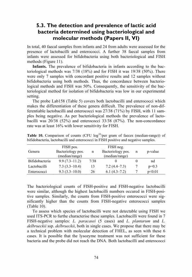
74
5.3. The detection and prevalence of lactic acid bacteria determined using bacteriological and
molecular methods (Papers II, VI)
In total, 40 faecal samples from infants and 24 from adults were assessed for the presence of lactobacilli and enterococci. A further 38 faecal samples from infants were assessed for bifidobacteria using both bacteriological and FISH methods (Figure 11).
Infants. The prevalence of bifidobacteria in infants according to the bac-teriological methods was 7/38 (18%) and for FISH it was 19/38 (50%). There were only 7 samples with concordant positive results and 12 samples without bifidobacteria using both methods. Thus, the concordance between bacterio-logical methods and FISH was 50%. Consequently, the sensitivity of the bac-teriological method for isolation of bifidobacteria was low in our experimental setting.
The probe Lab158 (Table 5) covers both lactobacilli and enterococci which makes the differentiation of these genera difficult. The prevalence of non-dif-ferentiable lactobacilli and enterococci was 27/38 (71%) by FISH, with 11 sam-ples being negative. As per bacteriological methods the prevalence of lacto-bacilli was 20/38 (52%) and enterococci 33/38 (87%). The non-concordance rate was at least 16% with lower sensitivity for FISH. Table 10. Comparison of counts (CFU log10/per gram of faeces (median-range)) of bifidobacteria, lactobacilli and enterococci in FISH positive and negative samples.
Genera FISH pos.
Bacteriology pos. (median/range)
n FISH neg.
Bacteriology pos. (median/range)
n p-value
Bifidobacteria 9.9 (7.3–11.2) 7/38 0 0 nd Lactobacilli 7.3 (3.3–10.4) 13 7.2 (4.4–7.3) 7 p=0.3 Enterococci 9.3 (3.3–10.0) 26 6.1 (4.3–7.2) 7 p<0.01
The bacteriological counts of FISH-positive and FISH-negative lactobacilli were similar, although the highest lactobacilli numbers occured in FISH-posi-tive samples. Similarly, the counts from FISH-positive enterococci were sig-nificantly higher than the counts from FISH-negative enterococci samples (Table 10).
To assess which species of lactobacilli were not detectable using FISH we used ITS-PCR to furthe characterise these samples. Lactobacilli were found in 7 FISH-negative samples: L. paracasei (5 cases) and L. plantarum and L. delbrueckii ssp. delbrueckii, both in single cases. We propose that there may be a technical problem with molecular detection of FHEL, as seen with these 6 cases. It is possible that the lysozyme treatment was not sufficient for these bacteria and the probe did not reach the DNA. Both lactobacilli and enterococci

75
are notably resistant to the lysozyme treatment as shown previously (Harmsen, 1999).
Adults. In 24 faecal samples from adults the prevalence of lactobacilli and enterococci using FISH was higher 24/24 (100%) than in the isolates from chil-dren (71%). Similarly, when using bacteriological methods the prevalence of lactobacilli 23/24 (96%) and enterococci 24/24 (100%) was higher than that of infants (52% and 87%, respectively). In adults the concordance of results for bacteriological methods and FISH was 96% for both lactobacilli and entero-cocci. In addition, the counts of LAB were higher using the FISH method for both children and adults (Table 11). Table 11. The counts of lactobacilli and enterococci in infants and adults using bac-teriology and FISH (CFU log10 per gram of faeces; median-range).
Age groups Bacteriology FISH Lactobacilli Enterococci Lactobacilli/Enterococci
Children 7.25 (3.3–10.4) 8.6 (4.3–10) 9.3 (8.0–9.6) Adults 5.9 (2.6–8.6) 5.9 (3.3–8.08) 8.4 (8.1–9.0)
Figure 11. Bifidobacteria (A) and lactobacilli-enterococci (B) in the faecal samples detected with FITC-labeled Bif164 and Lab158 probes.
5.4. Diversity of gut lactobacilli in healthy adults and the elderly (Paper IV)
The counts and species distribution of intestinal lactobacilli in 24 healthy adults and 37 elderly persons was assessed. Lactobacilli were cultured from specimens from 23/24 (96%) adults and 36/37 (97%) elderly people. In two individuals viable counts were below the detection limit. According to RT-qPCR results, lactobacilli were found in all faecal samples of adults and the elderly. Both methods showed that in the elderly persons the total counts of lactobacilli were significantly higher than in the adults: (range 3.6–10.8, median 6,4 vs. range 2.6–8.6, median 5,9; p=0.008) using both the bacteriological method (range

76
7.0–9.5, median 7,9; vs. range 5.3–8.8, median 7,2; p=0.0002) and using qRT-PCR (Figure 12).
According to the PCR method, the number of species in adults was lower than in the elderly, 4 versus 12 respectively [median 8, species vs. 5 to 11 (median 6) (p=0.042)]. There was no correlation between the number of species and the lactobacilli counts, either using bacteriological or RT-PCR methods. The most prevalent Lactobacillus species detected in both groups were L. casei from the FHEL group and L. ruminis from the OHOL group (≥70%; Figure 13). The prevalence of L. crispatus was the lowest (8%).
In the FHEL group, L. plantarum (22/37, 59% vs. 7/24, 29%; p=0.035) and L. paracasei (36/37, 97% vs. 14/24, 58%; p=0.0002) were more prevalent in the elderly people than adults. However, L. rhamnosus was detected in 7 (29%) of the adults (p<0.001), but not in any elderly person. The species difference was also found in the OHEL group where the prevalence of L. reuteri (19/37, 51% vs. 5/24, 21%; p=0.031) and L. buchneri (11/37, 29% vs. 2/24, 8%; p=0.059), was higher in elderly persons.
Figure 12. Total counts of Lactobacillus spp. in adults and the elderly persons using bacteriology (A) and real-time PCR (B).

77
Figure 13. Intestinal Lactobacillus species diversity in the adults and the elderly.
5.4.1. Association of Lactobacillus spp. with some anthropometrical and metabolic characteristics in healthy
adults and elderly (Paper IV)
Age-related, significant differences were found for 6 species of Lactobacillus. With regards to the OHOL metabolic pattern, the adults, in comparison to the elderly, were more often colonised with L. acidophilus (19/24, 79% vs. 18/37, 47%; p=0.031) and L. helveticus (16/24, 66% vs. 9/37, 24%; p=0.001). L. john-sonii was detected only in samples from the elderly (5/37, 13.5%) (Figure 13) and the age range for detection was older than 66, median 68 years old. The age range for detection of L. rhamnosus, found only in adults, was 48 with median of 31 years old.
A positive correlation between BMI and the age was found (r=0.301; p=0.0185), though the gender was a confounding factor (Table 12). Further-more, a positive correlation was found between BMI and fasting blood glucose content (r=0.463; p<0.0001) adjusted for the age and gender. A lower blood glucose level was predicted by the presence of intestinal L. paracasei (r2=0.281; Adj r2=0.213; p=0.031) with results adjusted for the age in the adult group. A negative correlation of borderline significance was found between the level of blood glucose and colonisation by L. fermentum (r=–0.321; p=0.052) in the elderly group. Multiple regression analysis revealed that the BMI was asso-ciated with the counts of cultivable lactobacilli adjusted for the age and gender (r2=0.187; Adj r2=0.144; p=0.008). The higher BMI in both groups of persons was directly predicted by the presence of OHOL and facultatively hetero-fermentative L. sakei species, both adjusted for the age and gender (Table 12).
20

78
Table 12. Spearman rank order correlation, linear, and multiple linear regression analy-sis between the counts and the species of lactobacilli, BMI, age, and gender.
Variable 1 Variable 2 Correlation coefficients r- and p-values
Linear multiple regression analysis (R2; Adj R2 ; p-values)
BMI (kg/m2) Age (years) r= 0.301; p=0.0185 NS BMI (kg/m2) Fasting plasma
glucose (mmol/L) r= 0.463; p=0.0001 Age, gender
R2=0.182; Adj R2=0.139; p=0.009
Fasting plasma glucose (mmol/L)
Presence of L. paracasei
r= –0.460; p=0.0238 Age (adult group) R2=0.281; Adj R2=0.213; p=0.031
Fasting plasma glucose (mmol/L)
Presence of L. fermentum
r=–0.321; p=0.052 NS
BMI (kg/m2) Counts of lactobacilli (bacteriology; log10
CFU/ g of faeces)
r=0.309; p=0.00279 Age, gender R2=0.187; Adj R2=0.144; p=0.008
BMI (kg/m2) Number of OHOL species
r=0.598; p=0.00213 NS
BMI (kg/m2) Presence of L. sakei r= 0.394; p=0.002 Age, gender R2=0.282; Adj R2=0.244; p<0.001
NS – no significant difference
5.5. Influence of some environmental factors on gut microbiota
5.5.1. Diversity of gut lactobacilli in 1–2-year-old Estonian and Swedish children (Paper III)
We found that Estonian children were more frequently colonised with lacto-bacilli than Swedish children (50/71 (70%) vs. 30/65 (46%); p<0.01). The distribution of different fermentation types (OHOL, FHEL and OHEL) of lacto-bacilli was similar in both Estonian and Swedish children with FHEL prevailing in both groups (72% and 80% of colonised children, respectively). The preva-lence of intestinal lactobacilli was compared for a set of Estonian (n=18) and Swedish (n=13) 1–2-year-old children. The Lactobacillus spp. identified in faecal samples of both groups of children included L. acidophilus, L. paracasei, L. coprophilus, L. delbrueckii, L. salivarius, L. plantarum and L. brevis. In contrast, L. fermentum and L. buchneri were detected only in faecal samples of

79
Estonian children. In addition, L. plantarum was identified as the prevailing species for six of the 18 Estonian children tested but none of the 13 Swedish children tested (p<0.05) (Figure 14).
Figure 14. Prevalence of Lactobacillus spp. in healthy 1–2-year-old Estonian and Swedish children (*–p<0.05).
5.6. The diversity of the bifidobacterial community in non-allergic and allergic children (Paper V)
The prevalence of intestinal bifidobacteria between allergic and non-allergic 5-year-old Estonian children was compared. Bifidobacteria were detected in all samples of allergic and non-allergic children. There were no differences in the prevalence of Bifidobcterium spp. between allergic and non-allergic children. Bifidobacterial profiles varied from 3 to 6 dominant bands on DGGE gels (Figure 15).
A total of 128 bifidobacterial clones (55 from allergic and 73 from non-allergic children) were subjected to sequence analysis. The sequence analysis of the 16S rDNA amplicons showed clear difference in composition of the faecal bifidobacterial species in allergic and non-allergic children. Comparison of the obtained sequences with those of the database revealed a significant degree of similarity of the fragments (97–100%) to the 16S rDNA sequences of known Bifidobacterium species. B. adolescentis was found in the faecal samples of 14 (70%) allergic children and in only 5 (25%) non-allergic children (Figure 16

80
(A); 14/6 vs. 5/15; OR = 5.8 (95% CI 1.7–19.7); p=0.01) while a significantly higher number of individual clones of B. adolescentis was present in allergic children (18/55 vs. 5/73; p<0,001). In contrast, in non-allergic children the B. catenulatum/pseudo-catenulatum group prevailed (14/6 vs. 5/15; p=0.01) showing a higher number of different clones as compared to allergic children (23/73 vs. 5/55; p=0.005) (Figure 16 (B)).
Figure 15. DGGE analysis of the Bifidobacterium spp. from fecal samples of allergic and non-allergic children. (M)-indicates the marker for DGGE, constructed from 16S rDNA amplicons: 1 – Staphylococcus aureus; 2 – Enterococcus faecalis; 3 – Lacto-bacillus acidophilus; 4 – Escherichia coli; 5 – Ruminococcus obeum; 6 – Bifido-bacterium longum.

81
Figure 16. Number of different species (A) and clones (B) of Bifidobacterium spp. in allergic and non-allergic children.
21

82
6. GENERAL DISCUSSION
Selection of identification and screening methods for functional properties of lactic acid bacteria
The intestinal microbiota is an important factor for human well-being and health. It provides an interface with the environment, particularly involving nutrition for the host and acting as a barrier against potentially harmful food components and pathogenic bacteria. The microbial ecology of the intestine is characteried with the balance between different groups of microbiota in the intestinal lumen and mucosa (Savage, 1978; Mikelsaar, 1993; Zoetendal et al., 2006; Sekirov et al., 2010; Cerf-Bensussan et al., 2010). Many different diseases and also their prevention can be linked to a dysfunctional intestinal microbiota. It is obvious that understanding the cross-talk that occurs between intestinal microbiota and its host promises to expand our views about the rela-tionship between intestinal microbiota and health. The recognition of LAB as a part of the human microbial ecosystem and the understanding of various inter-connected influences of that system is an important step for their application as healthy functional food (Tannock, 2004; Vaughan et al., 2005; Walter, 2008). There is still much to be learned about the normal LAB microbiota and how to achieve a desirable composition. Moreover, the precise identification of these bacteria at the species level is not an easy task. Prevalence, estimation and enumeration of LAB by molecular methods
In the present thesis, different methods for chracterisation of phenotypic and genetic properties of LAB intestinal isolates were compared. In particular, the human LAB microbiota from different age groups, geographical locations and health status were examined utilising methods fordifferentiation based on phe-notypic and molecular characterisitcs as laid out in the stated aims.
Previously, several methods have been used for quantitative and qualitative assessment of intestinal LAB, with a wide variation in results between the various authors. Our particular interest concerned both bifidobacteria and lacto-bacilli. Bifidobacteria have been described as strictly anaerobic bacteria. Identi-fication of anaerobic bacteria often requires very laborious, complex and time-consuming methods in addition to specifically developed anaerobic tehniques (Langendijk et al., 1995). Previous work in this field has been greatly hampered by the methodical inaccuracy that comes with handling these bacterial cultures in anaerobiosis. Many bacteria are fastidious in nature or are unculturable using current methods. Furthermore, media are frequently non-specific or are too selective for certain bacteria.
The main focus for quantitative and qualitative identification of microbes has moved from phenotypical to genotypical methods as the latter has generated more sensitive and accurate results (Lick et al., 2003). Molecular techniques for
83
quantitative microbial community analysis that do not require isolation of the microorganisms are very promising for providing an overview of the diversity of microorganisms present in particular samples or ecosystems. Without doubt, one of the most direct methods for the identification of single cells within a complex ecosystem is using FISH with specific 16S r-RNA-based oligonu-cletide probes (Amman et al., 1995; Langendijk et al., 1995; Harmsen et al., 1999). Approximately 90% of the total faecal bacteria can be detected using FISH with different group-specific probes. However, according to the literature the sensitivity of FISH based on microscopy is not high. The detection limit for bacteria is 107–108 cells per gram of faeces against a total population back-ground of 1011 cells or just 0.1% of the total population. The quantitative infor-mation derived from the FISH method and the qualitative information derived from other PCR-based methods may be complemented by more advanced quantitative PCR to obtain results which may reflect the real situation within this ecosystem.
In our study, the counts of intestinal lactobacilli from different age groups of people were some 10–100 times higher when molecular methods were used such as FISH and RT-PCR when compared to attempts to cultre these on media. This corresponds to the data of Eckburg et al. (2005) where molecular analysis showed that facultative anaerobic bacteria were only a minority within the colon (0.1%) and that strictly anaerobic microbes were by far the most dominant group of microorganisms. FISH has been relatively widely used for intestinal microbiota assessment and it has also been used to assess changes in levels of the predominant groups of intestinal bacteria as a result of the consumpsion of prebiotic substances or probiotic bacteria (Kirjavainen et al., 2002; Tuohy et al., 2003). Interestingly, using FISH it has also been shown that there are some dif-ferences in the gut microbiota between infants who later develop atopy and those who do not (Kalliomäki et al., 2001).
In our study we have used selective media for the simultaneous detection of bifidobacteria (CAN; BBL) and lactobacilli (MRS). The low corrsepondence between cultural and FISH methodologies, the latter using the Bif164 probe illustrated a methodical failure in the cultivation of bifidobacteria. Discrepancy between bacteriological and FISH methods may be related to the ability of bifi-dobacteria to grow on non-specific medium. The ability of bifidobacteria to grow on MRS medium is poor (Chapin et al., 1999). In contrast, Harmsen showed that 90% of colonies grown on media selective for lactobacilli, specifi-cally the MRS and Rogosa plates, were hybridised using the Bif164 probe and not with the Lab158 probe. They concluded that species such as Bifidobac-terium spp., which are dominant in the samples, grew also on the MRS selective media (Harmsen et al., 2000). However, in our study, all the isolates of the gram-positive rods from MRS media were identified as L. paracasei, L. plan-tarum, L. delbrueckii ssp. delbrueckii and no unidentified isolates (putatively bifidobacteria) were revealed. Our results were not affected by sample storage problems as in our study even more positive samples were detected using arti-

84
ficial culture media than molecular typing. This implies isolation problems exist with the strictly anaerobic bifidobacteria. This is in agreement with previous reports which showed that B. adolescentis could be better identified using PCR-DGGE, sequencing and FISH methods than with cultivation techniques (Matsuki et al., 1999; Welling et al., 1999). Moreover, a recent study using FISH combined with flow cytometry has confirmed that B. adolescentis-related species are likely to be dead or damaged by the time faecal samples are passed to the laboratory (Ben-Amor et al., 2005).
Simultaneously, issues have arisen using the Lab158 probe which detects enterococci-lactobacilli phyla. Using FISH faecal enterococci were frequently misidentified for lactobacilli, since the cells of both bacteria may show as plump rods. Furthermore, in some FISH-negative samples low counts of entero-cocci were detected on culture media which were seemingly missed using the molecular analysis. In general, probes containing a single labeled molecule give a strong signal only if cells are metabolically active and, hence, contain large numbers of ribosomes and target rRNA (Hahn et al., 1992; Manz et al., 1993). A limitation of FISH may include problems with cell wall permeability (Bin-denko et al., 1998) which gives rise to difference in the abilities of the probes to reach target sites (Fuchs et al., 1998). In the current study we used lysozyme to resolve the problems with cell wall permeability. Low fluorescence levels in positively hybridized cells can also significantly overlap with high background signals from negative controls (Langendijk et al., 1995). Coaggregation of bac-teria, broken cells, or contaminating substance can make cell identification and counting even more difficult. It has been reported that for estimation of Bifido-bacterium spp., molecular methods such as FISH and PCR-DGGE method combined with sequencing is a more appropriate approach. The use of DGGE/ TGGE profiles may represent 90–99% of the bacterial community, with the detection limit in faecal samples at 109 cells per gram of faeces (Zoetendal et al., 1998). This particular approach was successfully applied byour group in this present study to analyse the species composition of bifidobacteria in 5-year-old allergic children.
However, even the latest molecular approaches have several limitations, including the extraction of nucleic acids from samples, biases, and artefacts associated with enzymatic amplification of the nucleic acids, cloning of PCR products, as well as the previously described sensitivity and target site accessi-bility in whole-cell hybridization techniques (Head et al., 1998). Futhermore, as more rRNA sequences become available in sequence databases, it has been highlighted that may probes and primers do not respond to the taxonomic level desired. To date we are still far from having all the necessary specific primers and probes for LAB species and questions arise regarding the specificity and validity of existing ones which should be checked systematically with close genera, species and strains.
Historically of course, culture methods have been extensively used as the gold standard to quantitate LAB in faeces. However, it has been suggested that

85
the contrasting results in various studies seen may be due to the high variability of the intestinal microbiota between individuals in addition to the variability seen in the methods used, such as different culture media for the counting or isolation of strains or different methodologies used for identification of isolates. Conventional culture-dependent methods have limitations such as low sensi-tivity, long duration, as well as bias introduced due to the culture, and recovery of only the culturable species of the intestinal microbiota. Culture-based analy-sis may, however, give qualitative and quantitative fluctuations especially in the faecal microbiota compared to molecular methodology. This can lead to the overestimation of some species and underestimation of others.
However, culture-based methods have some advantages. We have found that using culture-based techniques when applied to the analysis of intestinal micro-biota, several new associations with human health indices can be detected. Thus, the bacteriological methods may provide a complementary picture of the LAB populations in humans. Identification methods of intestinal Lactobacillus isolates: phenotypic API 50CHL vs. molecular (ITS-PCR) methods
Though molecular methods have brought new aspects to the phylogenetic and quantitative relations of different bacteria in the gut, culture-based and molecu-lar-based profiling may be considered complementary with both giving a broadly similar overview of the intestinal ecosystem.
Frequently in the identification and species differnetiation of lactobacilli isolates methods have been based on their phenotypic properties, particularly the fermentation patterns of carbohydrates. The API 50CHL test was developed for the identification of human Lactobacillus strains in 1980s. However, its reliability is in question because the applied taxonomy of the genus Lacto-bacillus is unsatisfactory due to phenotypic heterogenecity (Andrighetto et al., 1998). The ITS-PCR method is a highly discriminatory molecular typing tech-nique for lactobacilli, which can give good differentiation at the strain level, thus allowing for a comprehensive assessment of the diversity of isolates re-covered on artificial media.
In this study, we identified the reference strains and faecal isolates of lacto-bacilli from different fermentation groups (OHOL, FHEL and OHEL) by using phenotyping (API 50CHL) and compared these results to molecular (ITS-PCR) methods.
According to the results using the API 50CHL test, in several cases, the exact identifications of closely related species was not reliable; some were doubtful or unacceptable and some strains were misidentified despite a stated “good” identification level. Differences in identification were found by both methods in all groups; especially for the L. acidophilus group. We found that the API system assigned to some species such as L. gasseri, L. johnsonii, L. jensenii, L. ruminis and L. helveticus for L. acidophilus. It may be concluded
22

86
that sometimes the low fermentation ability of carbohydrates is not sensitive enough for differentiation of species among the entire OHOL group. Also the manufacturer’s database may not be up to date in this dynamic area and that profiles for some prevalent Lactobacillus spp. of the OHOL group may be defi-cient. Moreover, 16S rRNA sequence analysis for Lactobacillus spp. has shown that some phenotypically generated taxa do not correspond with the suggested phylogenetic relations in particular in the case of L. acidophilus and L. para-casei groups (Holzapfel et al., 2001).
Such differences were also seen in the FHEL and OHEL groups. Within the FHEL group, we found that lactobacilli isolates identified by phenotyping such as L. rhamnosus, L. curvatus were reassigned using ITS-PCR as L. paracasei ssp. paracasei. Similarly to Tynkkynen et al. (1999) our study showed when using API 50CHL for identifying the strains of the L. casei group (L. rham-nosus, L. paracasei, L. casei and L. zeae) several reassignments were necessary based on molecular identification.
In our study, the species differentiation among OHEL group, including L. fermentum, L. cellobiosus, L. reuteri and L. buchneri was problematic using phenotypic typing methods. In addition, no genotypic differences were noted between L. fermentum and L. cellobiosus. It has been revealed by DNA/DNA hybridization that L. cellobiosus is identical to L. fermentum (Vescovo et al., 1979) and the first name is now considered as synonym to the latter (Hammes et al., 1992). In the study of Dellaglio et al. (2004) phylogenetic placement of L. cellobiosus was obtained based on 16S rDNA sequences, and genetic similarity was investigated by comparing partial recA gene sequences for the type strains of L. cellobiosus and L. fermentum. Based on the high identity values for 16S rDNA (99%) and recA gene (98%) sequences, the results of DNA–DNA hybridization assays and phenotypic traits available from the literature, it is proposed that L. cellobiosus be reclassified and, as a rule of priority, renamed as L. fermentum.
Thus, in conclusion, the present investigation highlighted a discrepancy between API 50CHL based phenotypic and genotypic identification techniques and suggested that API 50CHL analysis be used only as a preliminary identi-fication tool. This is in agreement with proposals by other researchers in this field (Nigatu, 2000; Paludan-Müller et al., 2002; Lei, 2004). However, it should be borne in mind that API 50CHL analysis provides important data regarding the carbohydrate fermentation patterns of the isolates, which are important from a functional and applied point of view (Sawadogo-Lingani et al., 2010). Lactoflora in children, adults and elderly
In our study faecal Lactobacillus populations were was studied in healthy chil-dren, adults and also in the elderly. We focussed on Lactobacillus species which although they comprise only a minor part of the bacterial community in human

87
faeces (Sghir et al., 2000) is more prevalent in the caecum and the physiologi-cally important area of the ileum (Marteau et al., 2001).
Previous culture-based studies have indicated higher viable counts of lacto-bacilli in healthy elderly people as compared to adults (Mikelsaar and Mändar, 1993). In our study, lactobacilli were detected in 90% of faecal samples using bacteriological methods and in all samples by RT-PCR. In the elderly, higher counts of intestinal lactobacilli were assessed by both methodical approaches. In addition, other data, based on viable counts, has shown that lactobacilli are pre-sent in the gastrointestinal tract of 70–73% of a population which consumes a Western-type diet whereas within those consuming a vegetarian diet a higher prevalence of lactobacilli has been assessed (Finegold et al., 1977,1983; Tannock, 1991). In Estonia, Western-type diet features have been attributed with the high use of fiber-rich and dairy products that could be the cause for differences.
The Lactobacillus species composition of the lower parts of the gastro-intestinal tract is still not well described. Moroever, the composition of intesti-nal lactobacilli also varies widely between individuals. Our results confirm that each person from the different age groups analysed had an individual pattern (from 4 to 12 species) of different lactobacilli species, which is in agreement with previous studies (Ahrne et. al., 1998; Delgado et al., 2007). Present knowledge indicates that 20 Lactobacillus species are putative inhabitants of the human gut, some of which are only recently detected using molecular methods with specific primers for LAB. From 20 lactobacilli species identified in this study, 18 were previously described as members of faecal microbiota of humans, although some have only been detected by molecular techniques using specific PCR primers. These include species such as L. gasseri and L. ruminis group. In the present study, L. acidophilus, L. salivarius, L. paracasei, L. casei, L. plantarum, L. brevis and L. fermentum were the most common species in all age groups, which is in agreement with previous studies. However, in our study we also found age-related differences in species profile. With regard to Lacto-bacillus species distribution between age groups, we found that L. paracasei and L. plantarum dominated in 1–2-year-old children and elderly persons. It has been previously reported that from 12 months, L. paracasei starts to be the most prevalent Lactobacillus species, and it becomes more prominent at 18 months, followed by L. delbrueckii, L. plantarum and L. acidophilus (Ahrne et al., 2005). L. gasseri and L. fermentum were also detected in faecal samples of chil-dren, which agree with the data of Vassos (2007).
Among the faecal lactobacilli of adults, L. acidophilus and L. helveticus were the most commonly detected habitants, being present in more than 60% of the individuals. Early studies based on bacteriological methods identified L. aci-dophilus as the predominant faecal Lactobacillus species in adults (Hentges et al., 1993; Moore et al., 1995). On the basis of the current taxonomy and using molecular methods, most of the L. acidophilus isolates were classified as L. gasseri, L. helveticus and L. crispatus in our study. Thus, isolates from

88
L. acidophilus group are difficult to distinguish, and it may lead to inaccurate results.
In our study, L. ruminis and L. casei were also detected as common species in the Lactobacillus biota of adults and the elderly as has been previously reported (Vaughan et al., 2005).
L. sakei and L. curvatus are food-associated lactobacilli and these could be detected by direct analysis of the 16S rRNA or 16–23S rRNA genes but not by bacteriological culture (Walter et al., 2001; Heilig et al., 2002).
A striking difference was noted concerning the L. rhamnosus species, which could be found in faecal samples of children and adults, but not in the elderly. L. rhamnosus has not been found to predominate studies of infants from the different countries such as the Netherlads, Japan and Scotland (Song et al., 1999; Harmsen et al., 2000; Satokari et al., 2002). However, it has been shown to be common in a study of Greek infants (Mitsou et al., 2008). Walter (2008) has suggested that L. rhamnosus is one of the dominant Lactobacillus spp., though only 26% of the healthy individuals and 6% of the elderly have been shown to harbour this species (Ahrne et al., 1998; Silvi et al., 2003). But why L. rhamnosus is completely absent in the elderly according to our data is not clear. Interestingly, Gefilus, one of the L. rhamnosus GG products in Estonia, is heavily marketed as a probiotic. We hypothesize that in the infancy of the cohort of elderly people studied (born between 1920–1940) when the resident Lactobacillus spp. biota of gastrointestinal tract were formed, the diet and envi-ronment did not favor the colonisation of gut with this species. Further, during the following years of life, even the intensive spread of this probiotic strain did not help to colonise this cohort which had a well-formed microbiota.
Reuter and coauthors (2001) have observed the predominance of L. reuteri among the indigenous intestinal lactobacilli in contrast to other authors who reported this species as only a minor component of the obligately hetero-fermentative group or completely absent in humans (Vaughan et al., 2005). In our investigations, altogether 39% of humans harboured L. reuteri.
A few studies have indicated that Lactobacillus populations in the human gut show temporal dynamics characterised by species and strain fluctuations (Mueller et al., 2006; Vassos, 2007). However, several authors (Mitsuoka et al., 1992; Reuter, 2001; Tannock et al., 2002) have identified the following as per-sistent strains in intestinal tract; L. gasseri, L. crispatus, L. reuteri, L. salivarius and L. ruminis and these were detected also in our study. However, seemingly in some persons the Lactobacillus spp. such as L. acidophilus, L. casei, L. para-casei, L. rhamnosus, L. delbrueckii, L. brevis, L. johnsonii, L. plantarum and L. fermentum still perform stable populations of the gut. The persistent coloni-sation of intestines with L. acidophilus, L. fermentum and L. casei has been shown in healthy persons over a 15 years period (Mikelsaar et al., 1998) in opposite to suggestion by (Walter, 2008).
89
Lactic acid bacteria metabolites and their association with some health indices of the human body While a first step in studying LAB in the GI tract is the identification of the microbes, a subsequent step is identifying the role of LAB metabolites in the health of its human host. Convincing evidence of the metabolic impact of lactic acid bacteria on their host remains deficient. It is not yet clear as to whether different fermentative groups and species of lactobacilli with different meta-bolic endproducts affect differentially the anthropometrical and biochemical indices of humans.
Phospholipid contents of bacteria vary across taxa, and fatty acid profiles of individual bacteria are known to vary quantitatively and qualitatively with changes in environmental factors (Rose, 1989). Cell fatty composition is espe-cially sensitive to medium composition, but also incubation temperature, cell age, and degree of anaerobiosis as occurs within the intestines (Moore, 1993). In our study we found that production of organic acids by LAB depends on environmental conditions.
There is considerable evidence that individual SCFA may have specific roles in connection with health implications. Furthermore, it is well known that SCFA and lactic acid stimulate gastric motility and gastric absorption, affect pancreatic secretion, promote trophics of intestinal mucosa and affect the pro-liferation of pathogenic bacteria in gastrointestinal tract. It has been previously described that lactic acid has a higher antimicrobial activity than acetic acid (Ouwehand, 1998). This activity may also be affected by the synergistic activity of different acids (Ouwehand, 1998). Thus, the test milieu, e.g. a micro- or anaerobic environment, seems to be very important for the expression of antimicrobial metabolites and subsequent selection of strains.
Microbes sometimes influence host physiology in a way what is not directly related to the GI tract function (Zoetendal et al., 2006). The principal substrate available for microbial fermentation in the large intestine includes a wide variety of dietary residues that have escaped digestion in the small intestine, the main ones being non-starch polysaccharides (NSP), various forms of starch resistant to digestion in the small intestine, sugar alcohols and proteins. Also, host-produced substrates such as glycoproteins of exfoliated epithelial cells and pancreatic secretions are other important sources. In particular, NSP are impor-tant energy substrates for large intestinal microbial fermentation, and the amount as well as the chemical and structural composition of the carbohydrate, are all important factors for the microbial activity within the gastrointestinal tract (Jensen and Jorgensen, 1994; Jensen, 2001). The energy available to the host after microbial fermentation is the energy found as SCFA, based on approximately 60% of that of the substrate being fermented (Jensen, 2001).
Whether these differences are also expressed in the different habitat of the gut (small and large intestine) with different oxygen tensions, has to be eluci-dated by GC testing of human samples from different parts of GI tract. The
23

90
intestinal lactobacilli can support intestinal epithelial cell proliferation through the production of SCFA which provides energy for the host (Shanahan, 2002). For example the main fermentation end product of OHOL group is lactic acid. In contrast, lactobacilli of the FHEL and OHEL groups, including L. paracasei and L. fermentum, play an important role in carbohydrate breakdown producing lactic acid, acetic acid and also succinate. It can be speculated that intestinal colonisation by some species of the latter groups decreases the content of both glucose (hexose) and fructose (pentose) due the effecient phosphoketolase pathway of carbohydrate fermentation (Axelsson, 1998). Thus, the complex metabolic activities between host and different groups of microbiota are capable of shaping the nutrient environment of the gut and subsequently the blood glu-cose level (Falk et al., 1998).
Previously, it has been shown that the elderly are more prone to metabolic syndrome, expressing a higher blood glucose level as part of this. To date, overweight children and adults also show an increased trend to develop a meta-bolic syndrome. A possible connection between the prevalence of Firmicutes and fat storage has been demonstrated (Bäckhed et al., 2004). Through the pro-duction of SCFA, the intestinal lactobacilli may positively influence intestinal epithelial cell proliferation and provide energy for the host.
We have shown that a reduction in oxidative stress indices (oxidized low density lipid content) was associated with higher numbers of lactobacilli in the elderly (Mikelsaar et al., 2010). Furthermore, we have raised the question as to whether the high counts of lactobacilli and prevalence of different species, par-ticularly those of different fermentation groups, could influence the health parameters such as blood glucose levels and BMI. According to our study, iso-lation of L. sakei was associated with a higher BMI. The ability of this species to metabolise complex carbohydrates to monosaccharides has been shown pre-viously and may also support their absorption and harvesting by host cells. Some studies indicate that L. sakei is one of the predominant food-associated Lactobacillus species that occurs in human faeces (Hammes et al., 1990, 1998). This may be explained by the frequent occurrence of this species in the food environment. L. sakei has been isolated from meat, sausages, and sauerkrauts and is one of the major spoilage organisms for vacuum-packaged meat products. In addition, it is an important component of starter cultures used for the produc-tion of fermented meat products.
Interestingly we found a reduced blood glucose level in adults colonised with L. paracasei from the FHEL group. This finding is supported by the data of Matzuzaki and coauthors (1998), which have shown that administration of the closely related L. casei to a diabetic mouse model, reduced blood glucose levels. Likewise, a diet enriched with another Lactobacillus species of the FHEL group (e.g. L. rhamnosus GG) has resulted in an improved glucose-tolerance test as well as in reduced blood glycated haemoglobin values in experimental rats and pregnant women (Yadav et al., 2007; Laitinen et al., 2009). Collectively, this data clearly hints at the potential for a positive impact

91
of probiotics using particular Lactobacillus species from different fermentative groups with different effects on the host metabolism.
In addition to SCFA, polyamines play an essential role in the maintenance and enhancement of the mucosal barrier. To illustrate, putrescine is produced straight from ornithine, arginine, is primarily converted into agmantine which is then converted into putrescine (Halaris et al., 2007). Diamine oxidase metabo-lises putrescine into gamma-butyric, which is an essential molecule in the metabolism of neurons.
In addition, arginine serves as the source of nitric oxide (NO) produced from nitric oxide synthase isofroms which provides a hyperemia-dependent protec-tion mechanism against stress-induced damage of the intestinal mucosa (Brzo-zowski et al., 2008). Putrescine is produced by several anaerobes, as well as Escherichia coli and lactobacilli with lactobacilli comprising a majority of the microflora of the proximal colon (Marteau et al., 2001). Lactobacilli produce polyamines through decarboxylation of amino acids, particularly at the high pH of the intestinal content (Lonvaud-Funel et al., 2001). On the other hand, strains of Lactobacillus acidophilus utilise putrescine and reduce the odour of faeces (WO 2008/019887, BASF AG).
Polyamines are produced endogenously or they are obtained actively from food (Stanton et al., 2006). In the case of the damage of epithelial cells, the pro-duction of polyamines by the intestinal microflora is considered one of the compensatory mechanisms for modification of immune response and apoptosis regulation.
It is especially important for elderly, whose polyamine-synthesizing ability is lowered, to consume food fortified with polyamines. Only 20% of consumed putrescine is absorbed from intestine into bloodstream, accompanied by a rise of acetylated putrescine while the majority is metabolised within hepatocytes and enterocytes (Milovic et al., 1997).
However, tha ability to perform some biochemical reaction in vitro will not always be reflected in host. Phenotypic methods for assessment of the LAB composition and metabolic as well as enzymatic activities of microbial LAB communities in the gut have shown certain inherent limitations but also useful features of the methods used. We have succeeded in showing that polyamine production in vitro, within cheese by a strain of L. plantarum Inducia was in good correlation with the increased amount of metabolised acetylated putrescine in host urine after consumption of the Inducia cheese.
On the other hand, the high amounts of polyamines could facilitate the absorption of glucose, increasing the number of glucose carriers in the mem-brane of enterocytes. In Finnish elderly people, it was found recently that there is a positive correlation between the counts of administered L. acidophilus NCFM (OHOL group) and the values of polyamine spermine. This may be the reason for an increased BMI tightly bound to the glucose level of blood in per-sons colonised predominantly with several species from the OHOL group in our study.

92
Changes of lactic acid bacteria composition by different environmental factors and diseases
Geographical differences
Our results demonstrated that certain regional-specific differences exist in the prevalence of colonisation with particular lactobacilli. Swedish children were less frequently colonised with lactobacilli than Estonian children. However, no differences were observed in the prevalence of different fermentation types among the prevailing Lactobacillus isolates in colonised children from either group, indicating a gross uniformity of intestinal lactobacilli profiles between the two populations. FHEL group was dominant in both countries and OHOL and OHEL were equally common among the Estonian and Swedish children. This is in agreement with geographical differences in microbiota species com-position which have been described previously (Mueller et al., 2006; Dicksved et al., 2007). In our study, L. plantarum strains were encountered only from Estonian children. One explanation for this difference may involve genetic and environmental differences in the human population between the two countries, as the implantation and the development of Lactobacillus in the neonate are greatly influenced by prematurity, method of birth, early feeding style (breast milk or formula fed), diet and environment (Fanaro et al., 2003). For example, in adults the prevalence of L. plantarum has been associated with a vegetarian as opposed to a typical Western type of diet (Stiles and Holzapfel, 1997). The Estonian diet to a large extent based on locally produced foods and many food-stuffs are fermented by lactic acid bacteria which are a prominent part of the diet, even in children. The individual specificity on the one hand and the tempo-ral modulation of intestinal microbiota by diet, lifestyle, and stress on the other hand complicate the understanding of the dynamic relationship between colonic microbiota signatures and host functions throughout the lifespan (Nielsen et al., 2003; Lay et al., 2005; Guarner, 2006). Bifidobacterium spp. and allergy
Differences in the Bifidobacterium population between allergic and non-allergic infants have been commonly found in previous studies (Bjorksten et al., 1999; 2001; Kalliomaki et al., 2001; Suzuki et al., 2007), in which lower numbers and lower prevalence of Bifidobacterium in allergic infants were observed. Also, inadequate bifidobacteria composition in GI tract microbiota has been suggested to be a risk factor for the development of allergy. Watanabe et al. in 2003 showed that the counts of Bifidobacterium spp. were significantly lower in patients with atopic dermatitis than in healthy control subjects. In particular, percentages of Bifidobacterium were significantly lower in patients with severe skin symptoms than in those with mild skin symptoms. In the previous studies performed in our laboratory using bacteriological methods we could not see any differences in prevalence of Bifidobacterium spp. between allergic and healthy
93
children at the age of 5 (Sepp et al., 2005) However, using specific Bifido-bacterium primers we observed significant differences in the distribution of Bifidobacterium species: allergic children were mainly colonised with B. ado-lescentis, whereas in non-allergic children the B. catenulatum/pseudocate-nulatum group predominated. These data correspond well to the results reported by Ouwehand and coworkers (2001) who showed using bacteriological methods that 50% of the total bifidobacterial isolates of allergic children were B. adolescentis. The culture-independent methods used have shown that the B. pseudocatenulam/catenulatum group was the most common species in the adult intestinal tract, followed by B. longum, B. adolescentis and B. bifidum, whereas B. breve, B. infantis and B. longum are the predominant species in infants (Matsuki et al., 1999, 2004). In contrast, Gore et al. (2008) showed that B. pseudocatenulatum is associated with atopic eczema in infants.
It is difficult to explain why at the age of five years the species composition of bifidobacteria in allergic children differs from healthy children of the same age and also from microbiota of adult persons. It may be that different lactic acid bacteria can modulate the immune response of the host by increasing the IgA synthesis and by inducing different pro-and anti-inflammatory cytokines. The species B. adolescentis prevailing in allergic children is more effective at triggering the pro-inflammatory cytokines TNF-, IL-6 and IL-12, but it is not able to induce the regulatory cytokine IL-10, which is an important anti-allergic immune response (Kramer et al., 1995; He et al., 2002). In contrast, some other species such as B. bifidum, B. longum and B. pseudocatenulatum/catenulatum induce the production of the regulatory cytokine IL-10 (Young et al., 2004).
Several studies have indicated that supplementation of the child’s diet with probiotic lactobacilli and bifidobacteria, such as Lactobacillus GG and Bifido-bacterium lactis Bb-12, can somewhat reduce the allergic disorders in atopic children and may have a strong impact on development, microbial cross-talk, evolution and modulation of a microbiota (Kirjavainen et al., 2002; Gueimonde et al., 2006). It is therefore tempting to suggest that in order to ameliorate aller-gic disease; the consumption of some probiotics including B. pseudo-catenulateum/catenulatum may be beneficial.
Characterisation of the probiotic strain, L. plantarum Inducia DSM 21379
To characterise our probiotic strain we looked at its molecular profile and its metabolic functional properties. The probiotic capacities are strain-dependent; therefore the methods for reliably identifying LAB at the strain level are of great importance. In the present study the 16S rDNA of the potential probiotic strain L. plantarum Inducia DSM 21379 has been sequenced. The sequencing method based on 16S or 23S rDNA is currently considered to be the most powerful and accurate method for determining the degree to which micro-organisms are phylogenetically related.
The application of PCR-DGGE and PFGE methods is also valuable, espe-cially for the quality control of approved Lactobacillus spp. strains, to avoid
24

94
health risks and misleading claims, as well as for the description of new strains. According to our PFGE results, the patented strain L. plantarum Inducia DSM 21379 showed clear difference from the other L. plantarum strains including the patentable L. plantarum Tensia DSM 21380 strain.
Among the functional properties of the Inducia strain the ability to produce putrescine in relatively high amounts was remarkable. Previously, the produc-tion of biogenic amines has been most extensively studied with respect to his-tamine and tyramine, probably the two most important BA of bacterial origin in food, due to their toxic effects. In our study we investigated the ability of some lactobacilli species for their production of bio- and polyamines such as putre-scine and cadaverine. Lactobacilli produce polyamines through decarboxlation of amino acids, particularly at the high pH of the intestinal content (Lonvaud-Funel, 2001). Bover-Cid et al. (1999) has shown that 40% of LAB strains were found to be tyramine formers. Only a few strains of lactobacilli produced putre-scine and small amount of cadaverine. Several BA-forming strains are of importance in food fermentation. Our study noted that minimal amounts of the biogenic amines were found during in vitro tests with L. plantarum, and that also minimal amounts were found in cheese comprising L. plantarum Inducia and urine samples of L. plantarum Inducia consumers. In contrast, the ability of L. plantarum strains to produce high levels of putrescine was refleted in urine samples from consumers of this strain. The results of BA production suggest that the capability to produce amines might be strain-dependent rather that being related to specific species. The composition of decarboxylase medium, the incubation time and the particular activity of strains within the cheese matrix and intestinal content play important roles in the production of polyamines by bacterial strains.

95
CONCLUSIONS
We applied phenotypic and genotypic methods to characterise the composition and the metabolic impact of intestinal lactic acid bacteria and to examine their relationship with different age groups, their environment and atopy. 1. The phenotypic methods used including culture on artifical media with sub-sequent biochemical profiling by API 50CHL and GC estimation of organic acids and ethanol are appropriate for estimation of the comparative Lacto-bacillus species composition providing well characterised Lactobacillus spp. strains for biotechnological applications.
The molecular methods such as ITS-PCR, controlled by 16S rRNA sequencing are clearly superior to API 50CHL in species identification, par-ticularly for the Lactobacillus acidophilus group. For quantitative estimation of Bifidobacterium spp. of infants the FISH method has remarkably higher sensi-tivity in comparison with bacteriological methods. However, for Lactobacillus spp. the concordance between bacteriology and FISH methods is high both in infants and adults. In comparison with bacteriological data, higher counts of lactobacilli can be detected in different age groups using FISH and RT-PCR. 2. The composition of intestinal indigenous lactobacilli is characterised with large individual variety both in counts, number and species composition depending on geographical location and age. A geographical comparison of microbiota from Estonian and Swedish 1–2-year-old children showed that the counts of cultivated lactobacilli were significantly higher in the nineties in fae-cal samples of the Estonians. Considering different age, the faecal microbiota of elderly people in comparison with adults express higher total counts of lacto-bacilli. In qualitative aspect, the Estonian children are characterised with higher prevalence of particular FHEL group species as L. plantarum whereas there is a predominance of L. paracasei among Swedish children apparent. In the elderly, the prevalence of the same FHEL species (L. plantarum and L. paracasei) and the higher species variety can be found whereas in adults the OHOL group (L. acidophilus and L. helveticus) is prevalent. 3. The higher counts of intestinal Lactobacillus spp. assessed using bacterio-logical methods can be associated with higher BMI and blood glucose content in adults and the elderly. A higher BMI in both groups of persons is predicted by the presence of OHOL species and L. sakei. Moreover, lower blood glucose levels are associated with L. paracasei in adults and L. fermentum in the elderly. Thus, the particular fermentation groups and species of intestinal lactobacilli are specifically associated with human anthropometrical data and biochemical indi-ces within the blood.

96
4. The previously assessed association between the shifts of prevalence of cul-turable bifidobacteria in infants and the development of allergy was confirmed with molecular methods in children at 5 years of age. Particularly, in allergic children, B. adolescentis is prevalent among intestinal Bifidobacterium spp., whereas in non-allergic children B. catenulatum/pseudocatenulatum is preva-lent. The impact of the different species of Bifidobacterium on host metabolism requires further elucidation. 5. Elaborated into probiotics and patented, the L. plantarum Inducia DSM 21379 strain was identified using phenotypic (API 50CHL) and genotypic (ITS-PCR, PFGE) methods followed by 16S rRNA sequencing. These methods serve as useful tools for strain identification and typing of lactobacilli from biological material and probiotic products. The concordance between the high proportions of putrescine amounts produced by strain L. plantarum Inducia both in decar-boxylation media and cheese with an increased metabolism of putrescine is identifiable in its consumer.

97
REFERENCES
Abd-El-Haleem D, Layton AC, Sayler GS. Long PCR-amplified rDNA for PCR-RFLP and rep-PCR-based approaches to recognize closely related microbial species. J Microbiol Methods 2002; 49: 315–319.
Adams MR, Moss MO. Food Microbiology, 2nd edn. Cambridge: Royal Society of Chemistry, 2000.
Adlerberth I, Lindberg E, Aberg N, Hesselmar B, Saalman R, Strannegard IL, Wold AE. Reduced enterobacterial and increased staphylococcal colonization of the infantile bowel: an effect of hygienic lifestyle? Pediatr Res 2006; 59: 96–101.
Adlerberth I, Strachan DP, Matricardi MP, Ahrne S, Orfei L, Aberg N, Perkin MR, Tripodi S, Hesselmar B, Saalman R, Coates AR, Bonanno CL, Panetta V, Wold AE. Gut microbiota and development of atopic eczema in 3 European birth cohorts. J Allergy Clin Immunol 2007; 120: 343–350.
Adlerberth I, Carlsson B, De Man P, Jalil F Khan, Larsson P, Mallander L, Svanborg C, Wold AE, Hanson LA. Intestinal colonization with Enterobacteriaceae in Pakistani and Swedish hospital-delivered infants. Acta Paediatr Scand 1991; 80: 602–610.
Ahlroos T, Tynkkynen S. Quantitative strain-specific detection of Lactobacillus rhamnosus GG in human fecal samples by real-time PCR. J Appl Microbiol 2009; 106: 506–514.
Ahnfelt NO, Hartvig P, Karlsson E. Derivatization of amines with chloroformate esters for gas chromatographic analysis. Chromatographia 1980; 16: 60–62.
Ahrne S, Lonnermark E, Wold AE, Aberg N, Hesselmar B, Saalman R, Strannegard IL, Molin G, Adlerberth I. Lactobacilli in the intestinal microbiota of Swedish infants. Microbes Infect 2005; 7: 1256–1262.
Ahrne S, Nobaek S, Jeppson B, Adlerberth I, Wold AE, Molin G. The normal Lactobacillus flora of healthy human rectal and oral mucosa. J Appl Microbiol 1998; 85: 88–94.
Alander M, De Smet I, Nollet L, Verstraete W, von Wright A, Mattila-Shandholm T. The effect of probiotic strains on the microbiota of the simulator of the human intestinal microbial ecosystem (SHIME). Int J Food Microbiol 1999; 46: 71–79.
Alm EW, Oerther DB, Larsen N, Stahl DA, Raskin L. The oligonucleotide probe database. Appl Environ Microbiol 1996; 62: 3557–3559.
Alm JS, Swartz J, Bjorksten B, Engstrand L, Engström J, Kühn I, Lilja G, Möllby R, Norin E, Pershagen G, Reinders C, Wreiber K, Scheynius A. An anthroposophic lifestyle and intestinal microflora in infancy. Pediatr Allergy Immunol 2002; 13: 402–411.
Amann RI, Ludwig W. Ribosomal RNA-targeted nucleic acid probes for studies in microbial ecology. FEMS Microbiol Rev 2000; 24: 555–565.
Amann RI, Krumholz L, Stahl DA. Fluorescent-oligonucleoide probing of whole cells for determinative, phylogenetic, and environmental studies in microbiology. J Bacteriol 1990; 172: 762–770.
Amman RI, Ludwig W, Schleifer KH. Phylogenetic identification and in situ detection of individual microbial cells without cultivation. Microbiol Rev 1995; 59: 143–169.
Andrighetto C, De Dea P, Lombardi A, Neviani E, Rossetti L, Giraffa G. Molecular identification and cluster analysis of homofermentative thermophilic lactobacilli isolated from dairy products. Res Microbiol 1998; 149: 631–643.
25

98
Annuk H. Selection of medical plants and intestinal lactobacilli as antimicrobial components for functional foods. Dissertation. Tartu, 2002.
Arena ME, Saguir FM, Manca de Nadra MC. Arginine dihydrolase pathway in Lactobacillus plantarum from orange. Int J Food Microbiol 1999; 47: 203–209.
Axelsson L. Lactic acid bacteria: classification and physiology. In: Salminen S., von Wright A, eds. Lactic acid bacteria. 1st edn. New York: Marcel Dekker, 1993; 1–64.
Axelsson L. Lactic acid bacteria: classification and physiology. In: Salminen S., von Wright A, eds. Lactic acid bacteria. Microbiology and functional aspects, 2nd edn. New York: Marcel Dekker, 1998; 1–72.
Aymerich T, Martín B, Garriga M, Hugas M. Microbial quality and direct PCR identification of lactic acid bacteria and nonpathogenic staphylococci from artisanal low-acid sausages. Appl Environ Microbiol 2003; 69: 4583–4594.
Bäckhed F, Ding H, Wang T, Hooper LV, Koh GY, Nagy A, Semenkovich CF, Gordon JI. The gut microbiota as an environmental factor that regulates fat storage. Proc Natl Acad Sci USA 2004; 101: 15718–15723.
Ballongue J. Bifidobacteria and probiotic action. In: Salminen S., von Wright A, Ouwehand A, eds. Lactic acid bacteria. Microbiological and functional aspects, 3rd edn. New York: Marcel Dekker, 2004; 67–123.
Bardóczs S, Grant G, Brown DS, Pusztai A. Putrescine as a source of instant energy in the small intestine of the rat. Gut 1998; 42: 24–28.
Bardócz S. Polyamines in food and their consequences for food quality and human health.Trends Food Sci Tech 1995; 6: 341–346.
Barry T, Colleran G, Glennon M, Dunican LK, Gannon F. The 16S/23S ribosomal spacer region as a target for DNA probes to identify eubacteria. PCR Methods Appl 1991; 1: 51–56.
Beck-Nielsen H. The metabolic syndrom in the daily clinic. Ugeskr Laeger 2010; 172: 1746–1751.
Ben Amor K. Microbial eco-physiology of the human intestinal tract. A flow cytometric approach. Dissertation. Wageningen, 2004.
Ben Amor K, Vaughan EE, Heilig H, Smidt H, Abee T, de Vos WM. Genetic diversity of viable, injured and dead fecal bacteria assessed by fluorescence activated cell sorting and 16S rRNA gene analysis. Appl Environ Microbiol 2005; 68: 114–123.
Bennet R, Eriksson M, Tafari N, Nord CE. Intestinal bacteria of newborn Ethiopian infants in relation to antibiotic treatment and colonisation by potentially pathogenic gram-negative bacteria. Scand J Infect Dis 1991; 23: 63–69.
Bergey’s Manual of Systematic Bacteriology. Krieg NR, Holt JG, eds. Williams and Wilkins, Baltimore, 1986.
Bezirtzoglou E. The intestinal microflora during the first weeks of life. Anaerobe 1997; 3: 173–177.
Biavati B, Mattarelli P. Bifidobacterium ruminantium sp. nov and Bifidobacterium merycicum sp. nov. from the rumen cattle. Int J Syst Bacteriol 1991; 41: 163–168.
Bidnenko E, Mercier C, Tremblay J, Tailler P, Kulakaukas S. Estimation of the state of the bacterial cell wall by fluorescent in situ hybridization. Appl Environ Microbiol 1998; 64: 3059–3062.
Björkstén B, Naaber P, Sepp E, Mikelsaar M. The intestinal microbiota during the first years of life and the development of allergy. J Allergy Clin Immunol 2001; 108: 516–520.
Björksten B, Naaber P, Sepp E, Mikelsaar M. The intestinal microflora in allergic Estonian and Swedish 2-year old children. Clin Exp Allergy 1999; 29: 342–346.

99
Björksten B, Sepp E, Julge K, Voor T, Mikelsaar M. Allergy development and the intestinal microflora during the first year of life. J Allergy Clin Immunol 2001; 108: 516–520.
Björksten B. Risk factors in early childhood for the development of atopic diseases. Allergy 1994; 49: 400–407.
Botaccini F, Medini D, Pavesi A, Turroni F, Foroni E, Riley D, Giubellini V, Tettelin H, van Sinderen D, Ventura M. Comparative genomics of the genus Bifidobacterium. Microbiology 2010; 156: 3243–3254.
Bottazzi V. Other fermented dairy products. Food and feed production with microorganisms. In: G. Reed, ed. Biotechnology. Weinheim: Verlag Chemie, 1983; 5: 315–364.
Bover-Cid S, Holzapfel WH. Improved screening procedure for biogenic amine production by lactic acid bacteria. Int J Food Microbiol 1999; 53: 33–41.
Brzozowski T, Konturek PC, Sliwowski Z, Drozdowicz D, Burnat G, Pajdo R, Pawlik M, Bielanski W, Kato I, Kuwaraha A, Konturek SJ, Pawlik WW. Gastroprotective action of orexin-A against stress-induced gastric damage is mediated by endogenous prostaglandins, sensory afferent neuropeptides and nitric oxide. Regular Pept 2008; 148: 6–20.
Buts JP, De Ketser N, Kolanovski J, Spkal E, van Hoof E. Maturation of villus and crypt cell functions in rat small intestine. Role of dietary polyamines. Dig Dis Sci 1993; 38: 1091–1098.
Drouet-Coassolo C, Aubert C, Coassolo P, Cano J. Capillary GC/MS method for the identification and qualification of some benzodiazepines and their unconjugated metabolites in plasma. J Chromatogr 1989; 487: 295–311.
Cagno R, Rizello CG, Garliardi F, Ricciuti P, Ndagijimana M, Francavilla R, M. Elisabetta Guerzoni ME, Crecchio C, Gobbetti M, De Angelis M. Imbalance of the faecal microbiota and volatile organic compounds between treated and untreated coeliac children. Appl Environ Microbiol 2009; 75: 3963–3971.
Cani PD, Delzenne MN. The role of the gut microbiota in energy metabolism and metabolic disease. Curr Pharm Des 2009; 15: 1546–1558.
Capano G, Bloch KJ, Schiffrin EJ, Dascoli JA, Israel EJ, Harmatz PR. Influence of the polyamines, spermidine, on intestinal maturation and dietary antigen uptake in the neonatal rats. J Pediatr Nutrit 1994; 19: 34–42.
Carlier JP, Sellier N. Identification by gas chromatography-mass spectrometry of short-chain hydroxy acids produced by Fusobacterium species and Clostridium innocuum. J Chromatogr B 1987; 420: 121–128.
Carlsson J; Grahntn H, Jonsson G. Lactobacilli and streptococci in the mouths of children, Caries Res 1975; 9: 333–339.
Cerf-Bensussan N, Gaboriau-Routhiau V. The immune system and the gut microbiota: friends or foes? Nature Rev Immunol 2010; 10: 735–744.
Chamaillard L, Quemener V, Havouis R, Moulinoux JP. Polyamine deprivation stimulates natural killer cell activity in cancerous mice. Anticancer Res 1993; 13: 1027–1033.
Chapin KC, Murray PR. Media. In: Murray PR, Baron EY, Pfaller MA,. Tenover FC, Yolken RH, eds. Manual of Clinical Microbiology, 7th edn. Washington DC: ASM Press. 1999: 1687–1707.
Christensen H, Moller PL, Vogensen FK, Olsen JE. 16S to 23S rRNA spacer fragment length polymorphism of Salmonella enterica at subspecies and serotype levels. J Appl Microbiol 2000; 89: 130–136.

100
Claesson JM, Cusack S, O’Sullivan O, Greene-Diniz R, de Weerd H, Flannery E, Marchesi JR, Falush D, Dinan T, Fitzgerald G, Stanton C, van Sinderen D, O’Connor M, Harnedy N, Kieran O’Connor K, Henry C, O’Mahony D, Fitzgerald AP, Shanahan F, Twomey C, Hilla C, Ross RP, O’Toole PW. Composition, variability, and temporal stability of the intestinal microbiota of the elderly. PNAS 2011; 108: 4586–4591.
Collins MD, Farrow AE, Philips BA, Ferusu S, Jones D. Classification of Lactobacillus divergens, Lactobacillus piscicola and some catalase-negative, asporogenous, rod-shaped bavteria from poultry in a new genus, Carnobacterium. Int J Syst Bacteriology 1987; 37: 310–316.
Collins MD, Rodrigues UM, Ash C, Aguirre M, Farrow JAE, Martinez-Murica A, Philips BA,Williams AM, Wallbanks S. Phylogenetic analysis of the genus Lacto-bacillus and related lactic acid bacteria as determined by reverse transcriptase sequencing of 16S rRNA. FEMS Microbiol Lett 1991; 77: 5–12.
Collins MD, Samelis J, Metaxopoulos J, Wallbanks S. Taxonomic studies on some leuconostoc-like organisms from fermented sausages: description of a new genus Weisella from Leuconostoc paramesenteroides group of species. J Appl Bacteriol 1993; 75: 595–603.
Condon C, Squires C, Squires CL. Control of rRNA transcription in Escherichia coli. Microbiol Rev 1995; 59: 623–645.
Corsetti A, Gobetti M, Rossi J, Damiani P. Antimould activity of sourdoughs lactic acid bacteria: identification of amixture of organic acids produced by Lactobacillus sanfrancisco CB1. Appl Microb Biotecnhol 1998; 50: 253–256.
Cox RP, Thomsen JK. Computer-aided identification of lactic acid bacteria using the API 50 CHL system. Lett Appl Microbiol 1990; 10: 257–259.
Cummings HJ, Pomare EW, Branch WJ, Naylor CPE, Macfarlane GT. Short chain fatty acids in human large intestine, portal, hepatic and venous blood. Gut 1987; 28: 1221–1227.
Dal Bello F, Clarke CI, Ryan LAM, Ulmer H, Ström K, Sjögren J, van Sinderen D, Schnürer, J, Arendt EK. Improvement of the quality and shelf life of wheat bread by using the antifungal strain Lactobacillus plantarum FST 1.7. J Cereal Sci 2006; 45: 309–318.
De Onis M, Habicht JP. Anthropometric reference data for international use: re-commendations from a WHO Expert Committee. Am J Clin Nutr 1996; 64: 650–658.
De Vries W, Stouthamer AH. Fermentation of glucose, lactose, galactose, mannitol and xylose by bifidobacteria. J Bacteriol 1968; 96: 472–478.
Decroos K, Vanhemmens S, Cattoir S, Boon N, Verstraete W. Isolation and characterization of an equol-producing mixed microbial culture from a human faecal sample and its activity under gastrointestinal conditions. Arch Microbiol 2005; 183: 45–55.
Delgado S, Suarez A, Mayo B. Dominant cultivable Lactobacillus species from the feces of healthy adults in northern Spain. Int Microbiol 2007; 10: 141–145.
Dellaglio T, Torriani S, Felis E. Reclassification of Lactobacillus cellobiosus Rogosa et al. 1953 a later synonym of Lactobacillus fermentum Beijerink 1901. Int J Sys Evol Microbiol 2004; 54: 809–812.
Deloyer P, Peulen O, Dandrifosse G. Dietary polyamines and non-neoplastic growth and disease. Eur J Gastroenterol Hepatol 2001; 13: 1027–1032.

101
Dicks LMT, Du Plessis EM, Dellaglio F, Lauer F. Reclassification of Lactobacillus casei subsp. casei ATCC 393 and Lactobacillus rhamnosus ATCC 15820 as Lacto-bacillus zeae nom. rev., designation of ATCC334 as the neotype of Lactobacillus casei, and rejection of name Lactobacillus paracasei. Int J Syst Bacteriol 1996; 46: 337–340.
Dicks LMT, Dellaglio F, Collins MD. Proposal to reclassify Leuconostoc oenos [corrig.] gen. nov., comb. nov. Int J Syst Bacteriol 1995; 45: 395–397.
Dicksved J, Flöistrup H, Bergström A, Rosenquist M, Pershagen G, Scheynius A, Roos S, Alm JS, Engstrand L, Braun-Fahrländer C, von Mutius E, Jannson JK. Molecular fingerprinting of the fecal microbiota of children raised according to different lifestyles. Appl Environ Microbiol 2007; 73: 2284–2289.
Donskey CJ, Hujer AM, Das SM, Pultz NJ, Bonomo RA, Rice LB. Use of denaturing gradient gel electrophoresis for analysis of the stool microbiota of hospitalized patients. J Microbiol Methods 2003; 1807: 1–8.
Douglas LC, Sanders ME. Probiotics and prebiotics in dietetics practice. J Am Diet Assoc 2008; 108: 510–521.
Drasar BS, Hill MJ. Human intestinal flora. New York: Academic Press, 1974. Dubez AM, Chaillous S, Hissler L, Stenz R, Champomier-Verges MC, Alpert CA,
Zagorec M. Physical and genetic map of the L. sakei 23K chromosome. Micro-biology 2002; 148: 421–431.
Dunder T, Kuikka L, Turtinen J, Räsänen L, Uhari M. Diet, serum fatty acids, and atopic diseases in childhood. Allergy 2001; 56: 425–428.
Eckburg PB, Bik MB, Bernstein CN, Purdom E, Dethlefsen L, Sargent M, Gill SR, Nelson KE, Relman DA. Diversity of the human intestinal microbial flora. Science 2005; 308: 1635–1638.
Edwards RA, Dainty RH, Hibbard CM. The relationship of bacterial numbers and types to diamine concentration in fresh and aerobically stored beef, pork and lamb. J Food Technology 1983; 18: 777–788.
Eerola S, Roig Sagués AX, Lilleberg L, Aalto H. Biogenic amines in dry sausages during shelf-life storage. Z Lebensm Unters Forsch A 1997; 205: 351–355.
Falckman RR, Washington JA. Streptococcus and catalase-negative gram-positive cocci. In: Balows A, Hausler WJ, Heemann KL, Isenberg HD, Shadomy HJ, eds. Manual of clinical microbiology Washington: American Society for Microbiology, 1991: 238–257.
Falk PG, Hooper LV, Midvedt T, Gordon JI. Creating and maintaining the gastrointestinal ecosystem: what we know and need to know from gnotobiology. Microbiol Mol Biol Rev 1998; 62: 1157–1170.
Fallani M, Young D, Scott E, Norin E, Amarri S, Adam R, Aquelere M, Khanna S, Gil A, Dore I. Intestinal microbiota of 6-week-old infants across Europe: geographic influence beyond delivery mode, breast-feeding, and antibiotics. J Pediatr Gastroenterol Nutr 2010; 51: 77–84.
Fanaro S, Chierici P, Guerrini P, Vigi V. Intestinal microflora in early: composition and development. Acta Paediatrica 2003; S441: 48–55.
FAO/WHO. Guidelines for the evaluation of probiotics in food. Food and Agriculture Organization of the United Nations and World Health Organization working group report. London, Ontario, Canada, 2002. Retrieved from: http://www.fao.org/es/ESN/Probio/probio.htm.
Felis GE, Dellaglio F. Taxonomy of lactobacilli and bifibobacteria. Curr Issues Intestinal Microbiol 2007; 8: 44–61.
26

102
Finegold SM, Attebery HR, Sutler, VL. Effect of diet on human fecal flora: comparison of Japanise and American diets. Am J Clin Nutr 1974; 27: 1456–1469.
Finegold SM, Sutter VL, Mathisen GE. Normal indigenous intestinal flora. In: Hentges DJ, ed. Human intestinal microflora in health and disease. New York: Academic Press, Inc. 1983: 3–31.
Finegold SM, Sutter VL, Sugihara PT. Fecal microbiol flora in seventh day Adventist populations and control subjects. Am J Clin Nutr 1977; 30: 1781–1792.
Finegold SM. Anaerobic bacteria in human disease. New York: Academic Press, 1977. Frank DN, Pace NR. Gastrointestinal microbiology enters the metagenomics era. Curr
Opin Gastroenterol 2008; 24: 4–10. Frank DN, St Amand AL, Feldman RA, Boedeker EC, Harpaz N, Pace NR. Molecular-
phylogenic characterization of microbial community imbalances in human inflammatory bowel diseases. PNAS 2007; 104: 13780–13785.
Franks AH, Harmsen HJM, Raangs GC, Jansen GJ, Schut E, Welling GW. Variation of bacterial populations in human feces meashured by fluorescent in situ hybridization with group-specific 16S rRNA-targeted oligonucleotide probes. Appl Environ Microbiol 1998; 64: 3336–3345.
Fuchs BM, Glockner FO, Wulf J, Amann R. Unlabeled helper oligonucleotides increase the in situ accessibility to 16S rRNA of fluorescently labeled oligonucleotide probes. Appl Environ Microbiol 2000; 66: 3603–3607.
Fuchs BM, Wallner G, Beisker W, Schwippl I, Ludwig W, Amann R. Flow cytometry analysis of the in situ accessibility of E. coli 16S rRNA for fluorescently labeled oligonucleotide probes. Appl Environ Microbiol 1998; 64: 4973–4982.
Fujisawa T, Kuno M, Kokubu T, Hirata R. Sasaki K, Fujisawa Y, Nakamura K, Mitsuoka T. Effects of apple and corn fiber supplemented with bifidobacteria and fructooligosaccharides preparation (A & C) on the fecal microflora and fecal properties in patients with dementia senilis. Bifidus 1992; 5: 173–176.
Furet JP, Quenee P, Tailliez P. Molecular quantification of lactic acid bacteria in fermented milk products using real-time quantitative PCR. Int J Food Microbiol 2004; 97: 197–207.
García-Martínez J, Martínez-Murcia AJ, Rodríguez-Valera F, Zorraquino A. Molecular evidence supporting the existence of two majors groups in uropathogenic Escherichia coli. FEMS Immunol Med Microbiol 1996; 14: 231–244.
Garcia-Martinez J, Acinas SG, Anton AI, Rodriguez-Valera F. Use of the 16S–23S ribosomal genes spacer region in studies of prokaryotic diversity. J Microbiol Methods 1999; 36: 55–64.
Garrity GM, Bell JA, Lilburn TG. Taxonomic outline of the procaryotes. Bergey’s manual of systematic bacteriology, 2nd edn, Release 5.0, New York: Springer-Verlag, 2004: 1–399.
Gill SR, Pop M, De Boy RT, Eckburg DB, Turnbaugh PJ, Samuel BS, Gordon JI, Relman DA, Fraser-Liggett CM, Nelson KE. Metagenomic analysis of the human distal gut microbiome. Science 2006; 312: 1355–1359.
Giovannoni SJ, DeLong EF, Olsen GJ, Pace NR. Phylogenetic group-specific oligo-deoxynucleotide probes for identification of single microbial cells. J Bacteriol 1988; 170: 720–726.
Gorbach SL, Nahas L, Lerner P, Weinstein L. Studies of intestinal microflora. I. Effects of diet, age and periodic sampling on numbers of fecal micro-organisms in man. Ibid 1967; 53: 845–855.

103
Gorbach SL, Plaut AG, Nahas L, Weinstein L, Spanknebel G, Levitan R. Studies of intestinal microflora. II. Microorganisms of the small intestine and their relations to oral and fecal flora. Gastroenterology 1967; 53: 856–867.
Gore C, Munro K, Lay C, Bibiloni R, Morris J, Woodcock A, Custovic A, Tannock GW. Bifidobacterium pseudocatenulatum is associated with atopic eczema: a nested case-control study investigating the fecal microbiota of infants. J Allergy Clin Immunol 2008; 12: 135–140.
Grönlund MM, Lehtonen OP, Eerola E, Kero P. Fecal microflora in healthy infants born by different methods of delivery: permanent changes in intestinal flora after cesarean delivery. J Pediatr Gastroenterol Nutr 1999; 28: 19–25.
Guarner F, Malagelada JR. Role of bacteria in experimental colitis. Best Pract Res Clin Gastroenterol 2003; 17: 793–804.
Guarner F. Enteric flora in health and disease. Digestion 2006; 73: 5–12. Guarner F. Prebiotics and mucosal barrier function. J Nutr 2006; 136: 2269. Gueimonde M, Sakata S, Kalliomaki M, Isolauri E, Benno Y, Salminen S. Effect of
maternal consumption of Lactobacillus GG on transfer and establishment of fecal bifidobacterial microbiota in neonates. J Pediatr Gastroenterol Nutr 2006; 42: 166–170.
Guerrini S, Mangani S, Granchi L, Vencenzini M. Biogenic amine production by Oenococcus oeni. Curr Microbiol 2002; 44: 374–378.
Gugliucci A, Menini T. The polyamines spermine and spermidine protect proteins from structural and functional damage by AGE precursors: a new role for old molecules? Life Sci 2003; 72: 2603–2616.
Gürtler V, Stanisich VA. New approaches to typing and identification of bacteria using the 16S–23S rDNA spacer. Microbiology 1996; 142: 3–16.
Haarman M, Knol J. Quantitative real-time PCR analysis of fecal Lactobacillus species in infants receiving a prebiotic infant formula. Appl Environ Microbiol 2006; 72: 2359–2365.
Hahn D, Amann RI, Ludwig W, Akkermans AD, Schleifer KH. Detection of micro-organisms in soil after in situ hybridization with rRNA-targeted, fluorescently labeled oligonucleotides. J Gen Microbiol 1992; 138: 879–887.
Halaris A, Plietz P. Agmatine: metabolic pathway and spectrum of activity in brain. CNS Drugs 2007; 21: 885–890.
Halász A, Barath A, Simon-Sarkadi L, Holzapfel W. Biogenic amines and their production by micro-organisms in food. Trends Food Sci Tech 1994; 5: 42–49.
Hammes WP, Hertel C. The Genera Lactobacillus and Carnobacterium. In: Dworkin M, Falkow S, Rosemberg E, Schleifer KH, Stackedbrandt E, eds. The Prokaryotes: An evolving slsctronic resource for the microbiological community. 3rd edn. Berlin: Springer, 2003: 302–404.
Hammes WP, Vogel R. The genus Lactobacillus. In: Wood BJB, Holzapfel W, eds. The Genera of lactic acid bacteria London: Chapman&Hall, 1995; 2: 19–54.
Hammes WP, Weiss N, Holzapfel W. The genera Lactobacillus and Carnobacterium. In: Balows A, Trüper HG, Dworkin M, Harder W, Schleifer KH eds. The Prokaryotes. New York: Springer-Verlag Inc., 1992: 1534–1594.
Harmsen HJ, Elfferich P, Schut F, Welling GW. A 16S rRNA-targeted probe for detection of lactobacilli and enterococci in faecal samples by fluorescent in situ hybridization. Microb Ecol Health Dis 1999; 11: 3–12.
Harmsen HJ, Gibson GR, Elfferich P, Raangs GC, Wildeboer.Veloo AC, Argaiz A, Roberfroid MB, Welling GW. Comparison of viable cell counts and fluorescence in

104
situ hybridization using specific rRNA-based probes for the quantification of human fecal bacteria. FEMS Microbiol Lett 1999; 183: 125–129.
Harmsen HJ, Wildeboer-Veloo AC, Raangs GC, Wagendorp AA, Klijn N, Bindels JG, Welling GW. Analysis of intestinal flora development in breast-fed and formula-fed infants by using molecular identification and detection methods. J Pediatr Gastroenterol Nutr 2000; 30: 61–67.
Harrow SA, Ravindran V, Butler RC, Marshall JW , Tannock GW. Real-time quanti-tative PCR measurement of ileal Lactobacillus salivarius populations from broiler chickens to determine the influence of farming practices. Appl Environ Microbiol 2007; 73: 7123–7127.
He F, Morita H, Hashimoto H, Hosoda M, Kunisaki J, Ouwehand AC. Intestinal bifidobacteria species induce varying cytokine production. J Clin Immunol 2002; 109: 1035–1036.
Head IM; Saunders JR, Pickup RW. Microbial evolution, diversity, and ecology: a decade of ribosomal RNA analysis of uncultivated microorganisms. Microbiol Ecol 1998; 35: 1–21.
Heavey PM, Rowland IR. The gut microbiology of the developing infant: microbiology and metabolism. Microbiol Ecol Health Dis 1999; 11: 75–83.
Hebuterne X. Gut changes attributed to ageing: effects on intestinal microflora. Gastroenterology 2003; 6: 49–54.
Heilig HG, Zoetendal EG, Vaughan EE, Marteau P, Akkermans ADL, de Vos WM. Molecular diversity of Lactobacillus spp. and other lactic acid bacteria in the human intestine as determined by specific amplification of 16S ribosomal DNA. Appl Environ Microbiol 2002; 68: 114–123.
Hentges DJ. The anaerobic microflora of the human body. Clin Infect Disease 1993; 4: S175–S180.
Holdeman LV, Cato EP, Moore WEC. Anaerobe Laboratory Manual. 4th edn. Virginia Polytechnic Institute and State Laboratory. Virginia: Blacksburg, 1977.
Holdeman LV, Moore WEC. Anaerobe laboratory manual. Blackburg, Virginia: Virginia Polytechnic Institute, 1975.
Holzapfel WH, Haberer P, Geisen R, Björkroth J, Schillinger U. Taxonomy and important features of probiotic microorganisms in food and nutrition. Am J Clin Nutrition 2001; 72: 365S–373S.
Holzapfel WH. Appropriate starter cultures technologies for small-scale fermentation in developing countries. Int J Food Microbiol 2001; 78: 119–131.
Hooper LV, Midtvedt T, Gordon JI. How host-microbial interactions shape the nutrient environment of the mammalian intestine. Annu Rev Nutr 2002; 22: 283–307.
Hopkins MJ, Sharp R, Macfarlane GT. Age and disease related changes inbintestinal bacterial populations assessed by cell culture, 16S rRNA abundance and community cellular fatty acid profiles. Gut 2001; 48, 198–205.
Hušek P. Chloroformates in gas chromatography as general purpose derivatizing agents. J Chromatogr B 1998; 717: 57–91.
Huys G, D’Haene K, Danielsen M, Matto J, Egervärn M, Vandamme P. Phenotypic and molecular assessment of antimicrobial resistance in Lactobacillus paracasei strains of food origin. J Food Protect 2008; 71: 339–344.
Jacobsen CN, Rosenfeld Nielsen V, Hayford AE, Moller PL, Michaelsen KF, Paerregard A, Sandström B, Tvede M, Jakobsen M. Screening of probiotic activities of forty-seven strains of Lactobacillus sp. by in vitro techniques and evaluation of

105
the colonization ability of five selected strains in humans. Appl Environ Microbiol 1999; 65: 4949–56.
Jansen GJ, Wildeboer-Veloo ACM, Tonk RHJ, Franks AH, Welling GW. Development and validation of an automated, microscopy-based method for enumeration of groups of intestinal bacteria. J Microb Methods 1999; 37: 215–221.
Jensen BB, Jorgensen H. Effect of dietary fiber on microbial activity and microbial gas production in various regions of the gastrointestinal tract. Appl Environ Microbiol 1994; 60: 1897–1904.
Jensen BB. Possible ways of modifying type and amount of products from microbial fermentation in the gut. In: Piva A, Bach Knudsen KE, Lindberg JE, eds. Gut Environment of Pigs Nottingham: Nottingham University Press, 2001: 181–200.
Jensen MA, Webster JA, Strauss N. Rapid identification of bacteria on the basis of polymerase chain reaction-amplified ribosomal DNA spacer polymorphisms. Appl Environ Microbiol 1993; 59: 943–952.
Julge K, Vasar M, Björkstén B. Development of allergy and IgE total serum during the first 5 years of life in Estonian children. Clin Exp Allergy 2001; 31: 1854–1861.
Kalaš P, Křížek M, Pelikanova T, Langova M, Veskrna O. Contents of polyamines in selected foods. Food Chemistry 2005; 90: 561–564.
Kalliomäki M, Collado MC, Salminen S, Isolauri E. Early differences in fecal micro-biota composition in children may prevent overweight. Am J Clin Nutr 2008; 87: 534–538.
Kalliomäki M, Kirjavainen P, Eerola E, Kero P, Salminen S, Isolauri E. Distinct patterns of neonatal gut microflora in infants in whom atopy was and was not developing. J Allergy Clin Immunol 2001; 107: 129–134.
Kandler O, Lauer E. Neuere Vorstellungen zur taxonomic der Bifidobacterien. Zentralbl. Bakteriol. Parasitenkd. Infektioskr. Hyg. Abt. 1, Orig. Reihe 1974; A228: 29–48.
Kandler O, Weis B. Regular, nonsporing gram-positive rod. In: Sneath PHA, Mair NS, Sharpe ME, Holt JG, eds. Bergeys Manual of Systematic Bacteriology. Baltimore: Williams and Wilkins, 1986; 2: 1208–1234.
Kandler O. Carbohydrate metabolism in lactic acid bacteria. Antonie van Leeuwehoek 1983; 49: 209–224.
Kao Y, Liu Y, Shyu Y. Identification of Lactobacillus spp. in probiotic products by real-time PCR and melting curve analysis. Food Res Int 2007; 40: 71–79.
Karki T. Quantitative composition of the human lactoflora and method for its examination. Dissertation. Tartu, 1996.
Karovicoka J, Kohajdova Z. Biogenic amines in food. Chem Pap 2005; 59: 70–79. Kataoka H. Derivatization reactions for the determination of amines by gas
chromatography and their application on inveronmental analysis. J Chromatogr A 1996; 733: 19–34.
Kaufmann P, Pfefferkorn A, Teuber M, Meile L. Identification and quantification of Bifidobacterium species isolated from food with genus-specific 16S rRNA-targeted probes by colony hybridization and PCR. Appl Environ Microbiol 1997; 63: 1268–1273.
Kimura K, McCartney AL, McConell MA, Tannock GW. Analysis of fecal populations of Bifidobacteria and Lactobacilli and investigation of the immunological responses of their humna hosts to the predominant strains. Appl Environ Microbiol 1997; 63: 3394–3398.
27

106
Kirjavainen PV, Arvola T, Salminen SJ, Isolauri E. Aberrant composition of gut microbiota of allergic infants: a target of bifidobacterial therapy at weaning? Gut 2002; 51: 51–55.
Kleessen B, Stoof G, Prool J, Smiedl D, Noack J, Blau M. Feeding resistant starch affects fecal and cecal microflora and short-chain fatty acids in rats. J Anim Sci 1997; 75: 2453–2462.
Kleessen B. Culture-based knowledge on biodiversity, development and stability of human gastrointestinal microflora. Microb Ecol Health Dis 2000; 12: 53–63.
Klein G, Pack A, Bonaparte C, Reuter G. Taxonomy and physiology of probiotic lactic acid bacteria. Int J Food Microbiol 1998; 41: 103–125.
Kok RD, de Waal A, Shut F, Welling GW, Weenk G, Hellingwerf KJ. Specific detection and analysis of a probiotic Bifidobacterium strain in infant feces. Appl Environ Microbiol 1997; 62: 3668–3672.
Kostman JR, Edling TD, Lipuma JJ, Stull TL. Molecular epidemiology of Pseudomonas cepacia determined by polymerase chain reaction ribotyping. J Clin Microbiol 1992; 30: 2084–2087.
Kramer D, Sutherland RN, Bao S, Husband A. Cytokine mediated effects in mucosal immunity. Immunol Cell Biol 1995; 73: 389–396.
Kullisaar T, Shepetova J, Zilmer K, Songisepp E, Rehema A, Mikelsaar M, Zilmer M. An antioxidant probiotic reduces postprandial lioemia and oxidative stress. Cent Eur J Biol 2010 (doi: 10.2478/s11535-010-0103-4).
Lambert MA, Moss CW. Gas-liquid chromatography of short-chain fatty acids on Dexsil 300 GC. J Chromatogr 1972; 74: 335–338.
Landendijk PS, Schut F, Jansen GI, Raangs GC, Kamphuis GR, Wikinson HF, Welling GW. Quantitative fluorescence in situ hybridization of Bifidobacterium ssp. with genus-specific 16S rRNA-targeted probes and its application in fecal samples. Appl Environ Microbiol 1995; 61: 3069–3075.
Lane DJ. 16S/23S rRNA sequencing.In: Stackebrandt ER, Goodfellow M, eds. Nucleic acid tehniques in bacterial systematics. UK, Chichester: John Wiley& Sons LTD, 1991: 115–175.
Larqué E, Sabater-Molina M, Zamora S. Biological significance of dietary polyamines. Nutrition 2007; 23: 87–95.
Larsen N, Vogensen FK, van den Berg FWJ, Nielsen DS, Andreasen AS, Pedersen BK, Abu Al-Soud W, Sorsen SJ, Hansen LH, Jacobsen M. Gut microbiota in human adults with type 2 diabetes differs from non-diabetic adults. PLOS ONE 2010; 5: 1–10.
Lauer E. Bifidobacterium gallicum sp. nov. isolated from human feces. Int J Syst Bacteriol 1990; 40: 100–102.
Lauer E, Kandler O. Lactobacillus gasseri sp. nov., a new species of the subgenus Thermobacterium. Zbl Bact, I Abt Orig 1980; 1: 75–78.
Lavizarri T, Breccia M, Bover-Cid S, Vidal-Carou MC, Veciana-Nogues MT. Histamine, cadaverine and putrescine produced in vitro by Enterobacteriaceae and Pseudomonadaceae isolated from spinach. J Food Protection 2010; 73: 385–389.
Lay C, Rigottier-Gois L, Holmstrom K, Rajilic M, Vaughan EE, de Vos WM, Collins MD, Thiel R, Namsolleck P. Colonic microbiota signatures across five northern European countries. Appl Environ Microbiol 2005; 71: 4153–4155.
Leblond-Bourget N, Philippe H, Mangin I, Decaris B. 16S rRNA and 16S–23S internal transcribed spacer sequence analyses reveal inter- and intraspecific Bifidobacterium phylogeny. Int J Syst Bacteriol 1996; 46: 102–111.

107
Lei V, Jakobsen M. Microbiological characterization and probiotic potential of koko and koko sour water, African spontaneously fermented millet porridge and drink. J Appl Microbiol 2004; 96: 384–397.
Ley RE, Bäckhed F, Turnbaugh P, Lozupone CA, Knight RD, Gordon JI. Obesity alters gut microbial ecology. Proc Natl Acad Sci USA 2005; 102: 11070–11075.
Ley RE, Peterson DA, Gordon JI. Ecological and evolutionary forces shaping microbial diversity in the human intestine. Cell 2006; 124: 837–848.
Lick S. Typing systems for lactobacilli. Milchwissenschaft 2003; 58: 256–260. Lindgren SE, Dobrogosz WJ. Antagonistic activities of lactic acid bacteria in food and
feed fermentations. FEMS Microbiol Rev 1990; 87: 149–164. Ljundberg B, Nillson-Ehle I, Edlund C, Nord CE. Influence of ciprofloxacin on the
colonic microflora in young and elderly voluteers: no impact of the altered drug absorption. Scan J Infect Dis 1990; 22: 205–208.
Lonvaud-Funel A. Biogenic amines in wine: role of lactic acid bacteria. FEMS Microbiol Letters 2001; 199: 9–13.
Löser C, Eisel A, Folsch UR. Dietary polyamines are essential luminal growth factors for small intestinal and colonic mucosal growth and development. Gut 1999; 44: 12–16.
Macfarlane S, Hopkins MJ, Macfarlane GT. Bacterial growth and methabolism on surfaces in the large intestine. Microb Ecol Health Dis 2000; S2: 64–72.
Macfarlane S, Macfarlane GT. Proteolysis and amino acids fermentations. In: Gibson GR, Macfarlane GT, eds. Human colonic bacteria: role in nutrition, physiology and pathology. CRC Press, Boca Raton, 1995.
Maidak BL, Cole JR, Lilburn TG, Parker CT, Saxman PR, Farris RJ, Garrity GM, Olsen GJ, Schmidt TM, Tiedje JM. The RDP-II (Ribosomal databese project). Nucleic Acids Res. 2001; 29: 173–174.
Malinen E, Kassinen A, Rinttila T, Palva A. Comparison of real-time PCR with SYBR Green I or 5’-nuclease assays and dot-blot hybridization with rDNA-targeted oligonucleotide probes in quantification of selected faecal bacteria. Microbiology 2003; 149: 269–277.
Mändar R. Vaginal microflora during pregnancy and its transmission to newborn. Dissertation. Tartu, 1996.
Manz W, Amann R, Ludwig W, Wagner M, Schleifer KH. Phylogenetic oli-deoxynucleotide probes for the major subclasses of proteobacteria: problems and solutions, Syst Appl Microbiol 1992; 15: 593–600.
Margin I, Suau A, Magne F, Garrido D, Gotteland M, Neut C, Pochart P. Charac-terization of human intestinal bifidobacteria using competitive PCR and PCR-TGGE. 2006; 1: 28–37.
Marteau P, Pochart P, Dore J, Bera-Maillet C, Bernalier A, Corthie G. Comparative study of bacterial groups withinthe human cecal and fecal microbiota. Appl Environ Microbiol 2001; 67: 4939–4942.
Martin B, Jofre A, Garriga M, Pla M, Aymerich T. Rapid quantitative detection of Lactobacillus sakei in meat and fermented sausages by real-time PCR. Appl Environ Microbiol 2006; 72: 6040–6048.
Maruo T, Sakamoto M, Toda T, Benno Y. Monitoring the cell number of Lactococcus lactis subsp. cremoris FC in human feces by real-time PCR with strain-specific primers designed using the RAPD technique. Int J Food Microbiol 2006; 110: 69–76.

108
Matricardi PM, Rosmini F, Riondin, S, Fortini M, Ferrigno L, Rapicetta M, Bonini S. Exposure to foodborne and orofecal microbes versus airborne viruses in relation to atopy and allergic asthma: epidemiological study. Brit Med J 2000; 320: 412–417.
Matsuda K, Tsuji H, Asahara T, Matsumoto K, Takada T, Nomoto K. Establishment of an analytical system for the human fecal microbiota, based on the reverse transcription-quantitative PCR targeting of multicopy rRNA molecules. Appl Environ Microbiol 2009; 75: 1961–1969.
Matsuki T, Watanabe K, Fujimoto J, Kado Y, Takada T, Matsumoto K, Tanaka R. Quantitative PCR with 16S rRNA-gene-targeted species-specific primers for analy-sis of human intestinal bifidobacteria. Appl Environ Microbiol 2004; 70: 167–173.
Matsuki T, Watanabe K, Fujimoto J, Takada T. Tanaka R. Use of 16S rRNA gene-targeted group-specific primers for real-time PCR analysis of predominant bacteria in human feces. Appl Environ Microbiol 2004; 70: 7220–7228.
Matsuki T, Watanabe K, Watanabe T, Oyaizu H. Phylogenetic analysis of genus Bifidobacterium and related genera based on 16S rDNA sequences. Microbiol Immunol 1998; 42: 661–667.
Matsuki T,Watanabe K, Tanaka R, Fukuda, Oyaizu H Distribution of bifidobacterial species in human intestinal microflora examined with 16S rRNA-gene-targeted species specific primers. Appl Environ Microbiol 1999; 65: 4506–4512.
Matsumoto M, Benno Y. The relationship between microbiota and polyamine in the human intestine: a pilot study. Microbiol Immunol 2007; 51: 25–35.
Matsuzaki T. Immunomodulation by treatment with Lactobacillus casei strain Shirota. Int J Food Microbiol 1998; 41: 133–140.
Maukonen J, Mättö J, Suihko M-L, Saarela M. Intra-individual diversity and similarity of salivary and faecal microbiota. J Med Microbiol 2008; 57: 1560–1568.
McCartney AL, Wang W, Tannock GW. Molecular analysis of the composition of the bifidobacterial and lactobacillus microflora of humans. Appl Environ Microbiol 1996; 62: 4608–4613.
McDonald P, Henderson AR, Heron SJE. The biochemistry of silage. 2nd edn. Marlow, UK: Chalcombe Publications, 1991.
Metchnikoff E. The Prolongation of Life: Optimistic Studies. New York: GP Putman’s Sons, 1908; 161–183.
Midtvedt AC, Midtvedt T. Production of short chain fatty acids by the intestinal microflora during the first 2 years of human life. J Pediatr Gastroenterol Nutr 1992; 15: 395–403.
Mikelsaar M, Mändar R, Sepp E, Annuk H. Human lactic acid flora and its role in the welfare of the host. In: Salminen S, von Wright A, Ouwehand A, eds. Lactic Acid Bacteria. Microbiological and functional aspects, 3rd edn. New York: Marcel Dekker Inc.; 2004: 453–506.
Mikelsaar M, Mändar R. Development of individual lactic acid microflora in the human microbial ecosystem. In: Salminen S, von Wright A, eds. Lactic acid bacteria. New York: Marcel Dekker, 1993; 237–293.
Mikelsaar M, Štšepetova J, Hütt P, Kolk H, Sepp E, Lõivukene K, Zilmer K, Zilmer M. Intestinal Lactobacillus sp. is associated with some cellular and metabolic cha-racteristics of blood in eldrly people. Anaerobe 2010; 16: 240–246.
Mikelsaar M, Zilmer M. Lactobacillus fermentum ME-3 – an antimicrobial and antioxidative probiotic. Microb Ecol Health Dis 2009; 21: 1–27.
Mikelsaar M. Evaluation of the gastrointestinal microbial ecosystem in health and disease. Dissertatsion. Tartu, 1992.

109
Milovic V, Odera G, Murphy GM, Dowling RH. Jejunal putrescine absoption and the pharmakokinetic/biotransformation of ingested putrescine in humans. Gut 1997; 41: A63.
Milovic V. Polyamines in the gut lumen: bioavailability and biodistribution. Europ J Gastroenterol Hepatol 2001; 13: 1021–1025.
Mitsou EK, Kirtzalidou E, Oikonomou I, Liosis G, Kyriacou A. Fecal microflora of Greek healthy neonates. Anaerobe 2008; 14: 94–101.
Mitsuoka T. The human gastrointestinal tract. In: Wood BFB, ed. The lactic acid bacteria in health and disease. The lactic acid bacteria. London, UK: Elsevier Science Publishers Ltd., 1992; 1: 69–114.
Mitsuoka T. Intestinal flora and aging. Nutr Rev 1992; 50: 438−446. Moinard C, Cynober L, de Brandt J-P. Polyamines: metabolism and implications in
human diseases. Clin Nutr 2004; 24: 184–197. Molin G, Jeppsson B, Johansson ML. Numerical taxonomy of Lactobacillus spp.
associated to healthy and diseased mucosa of the human intestines. J Appl Microbiol 1993; 74: 314–23.
Monstein HJ, Tiveljung A, Kraft CH, Borch K, Jonasson J. Profiling of bacterial flora in gastric biopsies from patients with Helicobacter pylori-associated gastritis and histologically normal control individuals by temperature gradient gel electrophoresis and 16S rDNA sequence analysis. J Med Microbiol 2000; 4: 817–822.
Moore WEC, Moore LH. Intestinal floras of populations that have a high risk of colon cancer. Appl Environ Microbiol 1995; 61: 3202–3207.
Moore WEC, Holdeman LV. Human fecal flora: the normal flora of 20 Japanese-Hawaiians. Appl Microbiol 1974; 27: 961–979.
Moore WEC. Effect of substrate composition on intestinal flora. Appl Environ Microbiol 1993; 59: 2763–2764.
Moreno-Arribas MV, Polo MC, Jorganes F, Muñoz R. Screening of biogenic amine production by lactic acid bacteria isolated from grape must and wine. Int J Food Microbiol 2003; 84: 117–123.
Moro E. Über die nach Gram-färbbaren Bacillen des Säuglingsstuhles. Wien Lin Wschr 1900a; 13: 114–115.
Moro E. Über B. acidophilus. Jb Kinderheilk 1900b; 52: 38–55. Mueller S, Saunier K, Hanisch C, Norin E, Alm L, Midtvedt T, Cresci A, Silvi S,
Orpianesi C, Verdenelli MC, Clavel T, Koebnick C, Zunft HJ, Dore J, Blaut M. Differences in fecal microbiota in different European study populations in relation to age, gender, and country: a cross-sectional study. Appl Environ Microbiol 2006; 72: 1027–1033.
Murray CS, Tannock GW, Simon MA, Harmsen HJM, Welling GW, Custovic A, Woodcock A. Fecal microbiota in sensitized and non-wheezy children: a nested case-control study. Clin Exp Allergy 2005; 35: 741–745.
Muyzer G, de Waal EC, Uitterlinden GA. Profiling of complex populations by denaturing gradient gel electroporesis analysis of polymerase chain reaction-amplified genes coding for 16S rRNA. Appl Environ Microb 1993; 59: 695–700.
Muyzer G, Smalla K. Application of gradient gel electrophoresis (DGGE) and temperature gradient gel electrohoresis (TGGE) in microbial ecology. Antonie van Leeuwenhoek 1998; 73: 127–141.
Naaber P. Clostridium difficile infection and intestinal microbial ecology. Dissertation. Tartu, 1997.
28

110
Nacowich L. Analysis of biogenic amines by GC/FID and GC/MS. Master’s thesis. 2009, Virginia.
Nadal I, Santacruz A, Marcos A, Warnberg J, Garagorri M, Moreno L. Shifts in clostridia, bacteroides and immunoglobulin-coating fecal bacteria associated with weight loss in obese adolescents. Int J Obesity 2009; 33: 758–767.
Nielsen DS, Möller PL, Rosenfeldt, V, Pærregaard A, Michaelsen KF, Jakobsen M. Case study of the distribution of mucosa-associated Bifidobacterium species, Lacto-bacillus species, and other lactic acid bacteria in the human colon. Appl Environ Microbiol 2003; 69: 7545–7548.
Nielsen JL, Christensen D, Kloppenborg M, Nielsen PH. Quantification of cell-specific substrate uptake by probe-defined bacteria under in situ conditions by micro-autoradiography and fluorescence in situ hybridization. Environ Microbiol 2003; 5: 202–211.
Nigatu A. Evaluation of numerical analyses of RAPD and API 50 CH patterns to differentiate Lactobacillus plantarum, Lact. fermentum, Lact. rhamnosus, Lact. sakei, Lact. parabuchneri, Lact. gallinarum, Lact. casei, Weissella minor and related taxa isolated from kocho and tef. J Appl Microbiol 2000; 89: 969–978.
Normand P, Ponsonnet C, Nesme X, Neyra M, Simonet P. ITS analysis of prokaryotes. In: Akkermans ADL, Van Elsas JD, De Bruijn FJ, eds. Molecular microbiology ecological manual. Dordrecht: Kluwer, 1996: 1–12.
Nubel U, Engelen B, Felske A. Sequence heterogeneities of genes encoding 16S rRNAs in Paenibacillus polymyxa detected by temperature gradient gel electrophoresis. J Bacteriol 1996; 178: 5636–5643.
Olsen R, Overbeek R, Pramanik S, Schmidt TM, Tiedje JM, Woese CR. A new version of the RDP (Ribosomal Database Project). Nucleic Acids Res 1986; 27: 171–173.
Orla-Jensen S. The lactic acid baceria. Copenhagen: Andr Host and Son, 1919: 1–196. O’Sullivan DJ. Methods for analysis of the intestinal microflora. In: Tannock GW, ed.
Probiotics. A Critical Review. Norfolk, UK: Horizon Scientific Press, 1999: 23–44. Ouwehand A, Vesterlund S. Health aspects of probiotics. IDrugs 2003; 6: 573–580. Ouwehand AC, Isolauri E, He F, Hashimoto H, Benno Y, Salminen S. Differences in
Bifidobacterium flora composition in allergic and healthy infants. J Allergy Clin Immunol 2001; 108: 144–145.
Ouwehand AC, Tiihonen K, Saarinen M, Putaala H, Rautonen N. Influence of a combination of Lactobacillus acidophilus NCFM and lactitol on healthy elderly: intestinal and immune parameters 2009; 101: 367–375.
Ouwehand AC. Antimicrobial components from lactic acid bacteria. In: Salminen S, von Wright A, eds. Lactic acid bacteria. microbiology and functional aspects, 2nd edn. Marcel Dekker, Inc., New York, 1998: 139–159.
Palleroni NJ. Prokaryotic diversity and the importance of culturing. Anton van Leeuwenhoek 1997; 72: 3–19.
Paludan-Müller C, Valyasevi R, Huss HH, Gram L. Genotypic and phenotypic characterization of garlic-fermenting lactic acid bacteria isolated from som-fak, a Thai low-salt fermented fish product. J Appl Microbiol 2002; 92: 307–314.
Penders J, Stobberingh EE, van den Brandt PA, van Ree R, Thijs C. Toxigenic and non-toxigenic Clostridium difficile: determinants of intestinal colonisation and role in childhood atopic manifestations. GUT 2008; 57: 1468–3288.
Penders J, Stobberingh EE, van den Brandt PA, Thijs C. The role of intestinal microbiota in the development of atopic disorders. Allergy 2007, 11: 1223–1236.

111
Penders J, Thijs C, Vink C, Stelma FF, Snijders B, Kummeling I, van den Brandt PA, Stobberingh EE. Factors influencing the composition of the intestinal microbiota in early infancy. Pediatrics 2006; 118: 511–521.
Penders J, Vink C, Driessen C, London N, Thijs C, Stobberingh E. Quantification of Bifidobacterium spp., Escherichia coli and Clostridium difficile in faecal samples of breast-fed and formulafed infants by real-time PCR. FEMS Microbiol Lett 2005; 243: 141–147.
Pot B, Ludwig W, Kersters K, Schleifer KH. Taxonomy of lactic acid bacteria. In: de Vuyst L, Vandamme EJ eds. Bacteriocins of lactic acid bacteria. Microbiology, genetics and applications. London: Blackie Academic and Professional, 1994: 13–90.
Poupard JA, Husain I, Norris RF. Biology of the bifidobacteria. Bacteriol Rev 1973; 37: 136–165.
Radler F. The metabolism of organic acids by lactic acid bacteria. In Carr JG, Cutting CV, Whiting GC, eds. Lactic acid bacteria in beverages and food. London: Academic Press Inc., 1975: 17–27.
Rainey FA, Ward N, Sly LI, Stackebrandt E. Dependence on the taxon composition of clone libraries for PCR amplified naturally occuring 16S rDNA, on the primer pair and the cloning system used. Experientia 1994; 50: 796–797.
Raskin L, Capman WC, Sharp R, Poulson LK, Stahl DA. Molecular ecology of gastrointestinal system. In: Macckie RI, White BA, Isaacson R, eds. Gastrointestinal Microbiology. New York: Chapman and Hall, 1997; 2: 243–298.
Reddy BS, Pleasants JR, Wostmann BS. Metabolic enzymes in liver and kidney of the germfree rat. Biochim Biophys Acta 1973; 320: 1–8.
Reid G, Burton J. Use of Lactobacillus to prevent infection by pathogenic bacteria. Microbes Infect 2002; 4: 319–324.
Requena T, Burton J, Matsuki T, Munro K, Simon MA, Tanaka R, Watanabe K, Tannock GW. Identification, detection and enumeration of Bifidobacterium species by PCR targeting the transaldolase gene. Appl Environ Microbiol 2002; 68: 2420–2427.
Reuter G. The Lactobacillus and Bifidobacterium microflora of the human intestine: composition and succession. Curr Issues Intest Microbiol 2001; 2: 42–53.
Reuter G. Vergleichende Untersuchunge über die Bifidus-Flora im Sauglings-und Erwahsenenstuhl. Zentralbl Bakteriol [Origin A] 1963; 1: 286–507.
Riikjärv MA, Julge K, Vasar M, Brabäck L, Knutsson A, Björkstén B. The prevalence of atopic sensitization and respiratory symptoms among Estonian schoolchildren. Clin Experiment Allergy 1995; 25: 1198–1204.
Rinttilä T, Kassinen A, Malinen E, Krogius L, Palva A. Development of an extensive set of 16S rDNA-targeted primers for quantification of pathogenic and indigenous bacteria in faecal samples by real-time PCR. J Appl Microbiol 2004; 97: 1166–1177.
Robinson RK. The microbiology of milk and milk products. In: Robinson RK, ed. Dairy Microbiology Handbook, 3rd edn. New York: John Wiley and Sons, 2002: 431–466.
Rose, AH. Influence of the environment on microbial lipid composition. In: Ratledge C, Wilkinson SG, eds. Microbial lipids. London, UK: Academic Press, 1989; 2: 255–278.
Rotimi VO, Duerden BI. The development of the bacterial flora in normal neonates. J Med Microbiol 1981; 14: 51–61.

112
Roussel Y, Colmin C, Simonet JM, Decaris B. Strain characterization, genome size and plasmid content in the Lactobacillus acidophilus group (Hansen and Mocquot). J Appl Bacteriol 1993; 74: 549–556.
Sakata S, Kitahara M, Sakamoto M, Hayashi H, Fukuyama M, Benno Y. Unification of Bifidobacterium infantis and Bifidobacterium suis as Bifidobacterium longum. Int J Syst Evol Microbiol 2002; 52: 1945–1951.
Salminen S, Isolauri E. Onnela T. Gut flora in normal and disordered states. Chemotherapy 1995; 41: 5–15.
Salminen S, von Wright A, Ouwehand A, eds. (2004) Lactic acid bacteria: microbiological and functional aspects. Marcel Dekker: New York.
Salminen SA, von Wright A, Morelli L, Marteau P, Brassart D, de Vos WM, Fonden R, Saxelin M, Collins K, Mogensen G, Birkeland SE, Mattila-Sandholm T. Demonstration of safety of probiotics – a review. Int J Food Microbiol 1998; 44: 93–106.
Sambrook J, Russell, D.W. Molecular cloning, a laboratory manual. New York: Cold Spring Harbor Laboratory Press, 2001.
Santacruz A, Marcos A, Warnberg J, Matri A, Martin-Matillas, M. Interplay between weight loss and gut microbiota composition in overweight adolescents. Obesity 2009; 17: 1906–1915.
Satokari RM, Vaughan EE, Akkermans ADL, Saarela M, de Vos WM. Bifidobacterial diversity in human feces detected by genus-specific PCR and – denaturing gradient gel electrophoresis, Appl Environ Microbil 2001; 67: 504–513.
Satokari RM, Vaughan EE, Favier CF, Dore J, Edwards C, de Vos WM. Diversity of Bifidobacterium and Lactobacillus spp. in breast-fed and formula-fed infants as assessed by 16S rDNA sequence differences. Microbiol Ecol Health Dis 2002; 14: 97–105.
Savage DC. Gastrointestinal microecology: one opinion. In: Lautit MW, Miles JA, eds. Microbial ecology. Berlin: Springer-Verlag, 1978: 234–249.
Sawadogo-Lingani H, Diawara B, Traore AS, Jakobsen M. Technological properties of Lactobacillus fermentum involved in the processing of dolo and pito, West African sorghum beers, for the selection of starter cultures. J Appl Microbiol 2010; 104: 873–882.
Scardovi V, Crociani F. Bifidobacterium catenulatum, Bifidobacterium dentium, and Bifidobacterium angulatum: three new species and their deoxyribonucleinic acid homology and relationships. Int J Sys Bacteriol 1974; 24: 21–28.
Scardovi V, Trovatelli LD, Biavati B, Zani G. Bifidobacterium cuniculi, Bifido-bacterium choerinum, Bifidobacterium boum, and Bifidobacterium pseudocatenu-latum: four new species and their deoxyribonucleic acid homology relationships. Int J Syst Bacteriol 1979; 29: 291–311.
Scardovi V, Trovatelli LD. New species of bifidobacteria from Apis mellifica L. and Apis indica F. A contribution to the taxonomy and biochemistry of the genus Bifidobacterium. Zentrabl Bakteriolb [Orig A], 1965; 12: 64–88.
Scardovi V. Bifidobacterium. In: Sneath PH, Mair NS, Sharpe ME, Holt GE, eds. Bergey’s Manual of Determinative Bacteriology, 9th edn. Baltimore: Williams and Wilkins, 1986, 1418–1434.
Schleifer KH, Ludwig W. Phylogeny of the genus Lactobacillus and related genera. Systemic and Applied Microbiology 1995; 18: 461–467.
Schmalenberger A, Schwieger F, Tebbe CC. Effect of primers hybridising to different evolutionary conserved regions of the small-subunit rRNA gene in PCR based

113
microbial community analyses and genetic profiling. Appl Environ Microbiol 2001; 67: 3557–3563.
Schroff KE, Meslin K, Cebra JJ. Commensal enteric bacteria engender a self limiting humoral mucosal immune response while permanently colonizing the gut. Infect Immun 1995; 63: 3904–3913.
Sebald M, Gasser F, Werner H. DNA based composition oand classification. Appli-cation to group of bifidobacteria and to related genera. Ann Inst. Pasteur 1965, 109: 251–269.
Sekirov I, Russel SL, Antunes LC, Finlay BB. Gut microbiota in health and disease. Physiol Rev 2010; 90: 859–904.
Sepp E, Julge K, Vasar M , Naaber P, Björksten B, Mikelsaar M. Intestinal microflora of Estonian and Swedish infants. Acta Paediatria 1997; 86: 956–961.
Sepp E, Naaber P, Voor T, Mikelsaar M, Björksten B. Development of intestinal microflora during the first month of life in Estonian and Swedish infants. Microb Ecol Health Dis 2000; 12: 22–26.
Sepp E, Voor T, Julge K, Lõivukene K, Björksten B, Mikelsaar M. In intestinal micro-biota bound up with changing lifestyle. In: Mendez-Vilas A, ed. Modern multi-disciplinary applied microbiology: exploiting microbes and their interactions. Germany: Wiley, 2006: 708–712.
Sepp E. Formation of intestinal microbial ecosystem in children. Dissertation. Tartu, 1998.
Sepp T, Julge K, Mikelsaar M, Björksten B. Intestinal microbiota and immunoglobulin E responses in 5-year-old Estonian children. Clin Exp Allergy 2005; 35: 1141–1146.
Servin AL. Antagonistic activities of lactobacilli and bifidobacteria against microbial pathogens. FEMS Microbial Rev 2004; 28: 405–440.
Sghir A, Gramet G, Suau A, Rochet V, Pochart P, Dore J. Quantification of bacterial groups within human fecal flora by oligonucleotide probe hybridization. Appl Environ Microbiol 2000; 66: 2263–2266.
Shanahan F. The host-microbe interface within the the gut. Best Pract Res Clin Gastroenterol 2002; 16: 915–931.
Sharpe ME, Dellaglio F. Desoxyribonucleic acid homology in anaerobic lactobacilli and in possibly related species. Intern J System Bact 1977; 27: 19–21.
Sharpe ME. The genus Lactobacillus. In: Starr M, Stolp H, Trüper HG, Balows A, Schelegel HG, eds.The Prokaryotes Berlin, Germany: Springer-Verlag, 1981; 2: 1653–1679.
Shaver YJ, Nagpal ML, Rudner R, Nakamura LK, Fox KF, Fox A. Restriction fragment length polymorphism of rRNA operons for discrimination and intergenic spacer sequences for cataloging Bacillus subtilis sub-groups. J Microbiol Methods 2002; 50: 215–223.
Shinki T, Tanaka H, Takito J, Yamaguchi A, Nakamura Y, Yoshiki S, Suda T. Putrescine is involved in the Vitamin D action in chick intestine. Gastroenterology 1991; 100: 113–122.
Silvi S, Verdanelli MC, Orpianesi C, Cresci A. EU Project Crownalife: functional foods, gut microflora and healthy aging. Isolation and identification of Lactobacillus and Bifidobacterium strains from fecal samples of elderly subjects for a possible probiotic use in functional foods J Food Engin 2003; 56: 195–200.
Simon GL, Gorbach SL. Intestinal flora in health and disease. Gastroenterology 1984; 86: 174–193.
29

114
Simpson PJ, Stanton C, Fitzgerald GF, Ross RP. Genomic diversity and relatedness of bifidobacteria isolated from a porcine cecum. J Bacteriol 2003; 185: 2571–2581.
Sjogren YM, Jenmalm MC, Bottcher MF, Björksten B, Sverremark-Ekstrom E. Altered early infant gut microbiotain chidren developing allergy up to 5 years of age. Clin J Exp Allergy 2009; 39: 518–526.
Socolowsky S, Hohne C, Sandow D. The direct detection of volatile fatty acids by gas chromatography in microbiological diagnosis. Zeitschrift Med Lab Diagn 1990; 31: 445–452.
Song YL, Kato N, Liu CX, Matsumiya Y, Kato H, Watanabe K. Rapid identification of 11 human intestinal Lactobacillus species by multiplex PCR assays using group- and species-specific primers derived from the 16S–23S rRNA intergenic spacer region and its flanking 23S rRNA. FEMS Microbiol Lett 2000; 187: 167–173.
Songisepp E. Evaluation of technological and functional properties of the new probiotic Lactobacillus fermentum ME-3. Dissertation. Tartu, 2005.
Southwick PL, Ernst LA, Tauriello EW, Parker SR, Mujumdar RB, Mujumdar SR, Clever HA, Waggoner AS. Cyanine dye labeling reagents-carboxymethy-lindocyanine succinimudyl esters. Cytometry 1990; 11: 418–430.
Stackebrandt E, Goebel BM. A place for DNA-DNA reassociation and 16S rRNA sequence analysis in the present species definition in bacteriology. Int J Syst Bacteriol 1994; 44: 846–849
Stackebrandt E, Goebel BM. Taxonomic note: a place DNA-DNA reassociation and 16S rRNA sequence analysis in the present species definition in bacteriology. Int J Sys Bacteriol 1994; 44: 846–849.
Stanton C, Ross PR, Fitzgerald GF, van Sinderen D. Fermented functional foods based on probiotics and their biogenic metabolites. Current Opin Biotechnol 2005; 16: 198–203.
Stevenson DM, Muck RE, Shinners KJ, Weimer PJ. Use of real time PCR to determine population profiles of individual species of lactic acid bacteria in alfalfa silage and stored corn stover. Appl Microbiol Biotechnol 2005; 71: 329–338.
Strachan DP. Hay fever, hygiene and household size. BMJ 1989; 299: 1259–1260. Stratton JE, Hutkins RW, Taylor SL. Biogenic amines in foods, a review. J Food
Protection 1991; 54: 460–470. Suau A, Bonnet R, Sutren M, Godon JJ, Gibson GR, Collins MD, Dorè J. Direct
analysis of genes encoding 16S rRNA from complex communities reveals many novel molecular species within the human gut. Appl Environ Microbiol 1999; 65: 4799–4807.
Sutter VL, Citron DM, Edelstein MAC, Finegold SM. Wadsworth Anaerobic Bacteriology Manual, 4th edn. Belmont, CA: Star Publishing Company, 1985.
Suzuki S, Shimojo N, Tajiri Y, Kumemura M, Kohno Y. Differences in the composition of intestinal Bifidobacterium species and the development of allergic diseases in infants in rural Japan. Clin Exp Microbiol 2007; 37: 506–511.
Tannock GW. A special fondness for lactobacilli. Appl Environ Microbiol 2004; 70: 3189–3194.
Tannock GW, Munro K, Harmsen HJM, Welling GW, Smart J, Gopal PA. Analysis of the fecal microflora of human subjects consuming a probiotic containing Lactobacillus rhamnosus DR20. Appl Environ Microbiol 2000; 66: 2578–2588.
Tannock GW. Normal microflora. An introduction to microbes inhabiting the human body. London: Chapman and Hall, 1995.

115
Tannock GW. Introduction. In: Tannock GW, edn. Probiotics: a critical review. Norfolk, UK: Horizon Scientific Press, 1999: 1–4.
Tannock GW. Probiotics and prebiotics: where are we going? In: Tannock GW, edn. Probiotics and prebiotics: where are we going? Wymondham, UK: Caister Academic Press, 2002: 1–39.
Tannock GW. Analysis of bifidobacterial populations in bowel ecology studies. In: Mayo B, van Sideren D, eds. Bifidobacteria: genomics and molecular aspects. Norfolk, UK: Caister Academic Press, 2010: 1–15.
Tannock GW. Analysis of fecal populations of bifidobacteria and lactobacilli and investigation of the immunological responses of their human hosts to predominant strains. Appl Environ Microbiol 1997; 63: 3394–3398.
Tannock GW. Analysis of intestinal microflora, a renassance. Antonie von Leeuwenhoek 1999; 76: 265–278.
Tannock GW. Identification of lactobacilli and bifidobacteria. Current Issues Molec Biol 1999; 1: 53–64.
Tannock GW. The microecology of lactobacilli inhabiting the gastrointestinal tract. Adv Microb Ecol 1991; 11: 147–171.
Burtis CA, Ashwood ER, eds. Tietz text book of clinical chemistry. 3rd edn. Philadelphia, PA: Saunders WB, 1998: 1241–1242.
Tiihonen K, Ouwehand AC, Rautonen N. Human intestinal microbiota and healthy ageing. Ageing Res Rev 2010; 9: 107–116.
Tiihonen K, Tynkkynen S, Ouwehand A, Ahlroos T, Rautonen N. The effect of ageing with and without non-steroidal antiinflamatory drugs on gastrointestinal microbiology and immunology. Br J Nutr 2008; 100: 130–137.
Tilsala-Timisjarvi A, Alatossava T. Development of oligonucleotide primers from the 16S–23S rRNA intergenic sequences for identifying different dairy and probiotic lactic acid bacteria by PCR. Int J Food Microbiol 1997; 35: 49–56.
Tissier H. Recherchers sur la flora intestinale normale et pathologique du nourisson. In: Medicine Paris, Universitè de Paris, 1900.
Toth IK, Avroka AO, Hyman LJ. Rapid identification and differentiation of the soft rot erwinias by 16S–23S intergenic transcribed spacer-PCR and restriction fragment length polymorphism analysis. Appl Environ Microbiol 2001; 67: 4070–4076.
Tuohy KM, Probert HM, Smejkal CW, Gidson GR. Using probiotics and prebiotics to improve gut health. DDT 2003; 8: 692–700.
Tynkkynen S, Satokari R, Saarela M, Mattila-Sandholm T, Saxelin M. Comparison of ribotyping, randomly amplified polymorphic DNA analysis, and pulsed-field gel electrophoresis in typing of Lactobacillus rhamnosus and Lactobacillus casei. Appl Environ Microbiol 1999; 65: 3908–3914.
Ugland HG, Krogh M, Rasmussen KE. Aqueous alkylchloroformate derivatisation and solid-phase microextraction: determination of amphetamines in urine by capillary gas chromatography. J Chromatogr 1997, 701: 29–38.
Van der Waaij L, Harmsen H, Madjipour F, Kroese F, Zwiers M, van Dullemen H, de Boer N, Welling G, Jansen P. Bacterial population analysis of human colon and terminal ileum biopsies with 16S rRNA-based fluorescent probes: commensal bacteria live in suspension and have no direct contact with epithelial cells. Inflamm Bowel Dis 2005; 11: 865–871.
Vankerckhoven V, Huys G, Vancanneyt CV, Vael C, Klare I, Romond MB, Entenza JM, Moreillon P, Wind RD, Knol J, Wiertz E, Pot B, Vaughan EE, Kahlmeter G, Goossens H. Biosafety assessment of probiotics used for human consumption:

116
recommendations from the EU – PROSAFE project. Trends Food Sci & Technol 2008; 19: 102–144.
Vassos D, Maipa V, Voidarous C, Alexopoulos A, Bezirtzoglou E. Development of human lactic acid gastrointestinal microbiotain a Greek rural population. Cent Eur J Biol 2007; 3: 55–60.
Vaughan EE, de Vries MC, Zoetendal EG, Ben-Amor K, Akkermans AD, de Vos WM. The intestinal LABs. Antonie van Leeuwenhoek, 2002; 82: 341–352.
Vaughan EE, Heilig HG, Ben-Amor K, de Vos WM. Diversity, vitality and activities of intestinal lactic acid bacteria and bifidobacteria assessed by molecular approaches. FEMS Microbiol Rev 2005; 29: 477–490.
Vaughan EE, Schut F, Heilig HG, Zoetendal EG, de Vos WM, Akkermans AD. A molecular view of the intestinal ecosystem. Curr Issues Intest Microbiol 2000; 1: 1–12
Vaugien L, Prevots F, Roques C. Bifidobacteria identification based on 16S rRNA and pyruvate kinase partial gene sequence analysis. Anaerobe 2002; 8: 341–344.
Ventura M, Turroni F, Canchaya C, Vaughan EE, O’Toole PW, van Sinderen D. Microbial diversity in the human intestine and novel insights from metagenomics. Front BioSci, 2009; 14: 3214–3221.
Ventura M, van Sinderen D, Fitzgerald GF, Zink R. Insights into the taxinomy, genetics and physiology of bifidobacteria. Antonie van Leeuwenhoek 2004; 86: 205–223.
Ventura M, Zink R. Comparative sequence analysis of the tuf and recA genes and restriction fragment length polymorphism of the internal transcribed spacer region sequences supply additional tools for discrimnating Bifidobacterium lactis from Bifidobacterium animalis. Appl Environ Microbiol 2003; 69: 7517–7522.
Vescovo M, Dellaglio F, Bottazzi V, Sarra PG. Deoxyribonucleic acid homology among Lactobacillus species of the subgenus Betabacterium Orla–Jensen. Micro-biology 1979; 2: 317–330.
Vitali B, Candela M, Matteuzzi D, Brigidi P. Quantitative detection of probiotic Bifidobacterium strains in bacterial mixtures by using real-time PCR. Syst Appl Microbiol 2003; 26: 269–276.
De Vos WM, Castenmiller JJM, Hamer RJ, Brummer RJM. Nutridynamics – studying the dynamics of food components in products and in the consumer. Current Opin Biotechnol 2006; 17: 217–225.
von Wintzingerode F, Göbel UB, Stackebrandt E. Determination of microbial diversity in environmental samples: pitfalls of PCR-based rRNA analysis. FEMS Microbiol Rev 1997; 21: 213–229
Vrieze A, Holleman F, Zoetendal EG, de Vos WM, Hoekstra JBL, Nieuwdrorp M. The environment within: how gut microbiota may influence metabolismand body composition. Diabetologia 2010; 53: 606–613.
Wallace HM, Fraser AV. Polyamine analogues as anticancer drugs. Biochem Soc Trans 2003; 31: 393–396.
Walter J, Hertel C, Tannock GW, Lis CM, Munro K, Hammes WP. Detection of Lactobacillus, Pediococcus, Leuconostoc and Weisella species in human feces by using group-specific PCR primers and denaturing gradient gel electrophoresis. Appl Environ Microbiol 2001; 67: 2578–2685.
Walter J. Discovering the molecular foundations of Lactobacillus autochthony in the gastrointestinal tract. Japanese J Lactic Acid Bacteria 2008; 1: 9–20.
117
Walter J. Ecological role of lactobacilli in the gastrointestinal tract: implication for fundamental and biomedical research. Appl Environ Microbiol 2008: 74: 4985–4996.
Walter JG, Tannock W, Tilsala-Timisjarvi A, Rodtong S, Loach DM, Alatossava T. Detection and identification of gastrointestinal Lactobacillus species by using denaturing gradient gel electrophoresis and species-specific PCR primers. Appl Environ Microbiol 2000; 66: 297–303.
Wang IJ, Guo YL, Weng HS, Hsieh WS, Chuang YL, Lin SJ, Chen PC. Environmental risk factors for early infantile atopic dermatitis. Pediatric Allergy Immunol 2007; 18: 441–447.
Wang JY, Mccormack S, Viar MJ, Johnson L. Stimulation of proximal small intestinal mucosal growth by luminal polyamines. Am J Physiol 1991; 261: G504–G511.
Watanabe K. Microorganisms relevant to bioremediation. Curr Opin Biotechnol 2001; 12: 237–241.
Watanabe K. Primers for Lactobacillus. Japan patent application 1998-260041; publi-cation 1999-151097.
Watanabe S, Narisawa Y, Arase S, Okamatsu H, Ikenaga T, Kumemura M. Differences in fecal microbiota between patients with atopic and healthy control subjects. J Allergy Clin Immunol 2003; 111: 587–591.
Welling GW, Wideboer-Veloo ACM, Raangs GS, Franks AH, Haarmsen HJM, Tonk RHJ, Degener JE. Enumeration of bacteria in fecal samples using fluorescent in situ hybridization (FISH). Microb Ecol Health Dis 1999; 28: 115–118.
Wenzel R. Health care – associated infections. Major issues in early years of 21st century. Clin Inf Dis 2007; 45: 85–88.
Wessels S, Axelsson L, Hansen EB, De Vuyst L, Laulund S, Lähteenmäki L, Lindgren S, Mollel B, Salminen S, von Wright A. The lactic acid bacteria, the food chain, and their regulation. Trends Food Sci Technol 2004; 15: 498–505.
Whiting GC. Some biochemical and flavour aspects of lactic acid bacteria in ciders and other alcoholic beverages. In: Carr JG, Cutting CV, Whiting GC, eds. Lactic acid bacteria in beverages and food. London: Academic Press Inc., 1975: 69–85.
Wiggins RJ, Wilks M, Tabaqchali S. Analysis by gas liquid chromatography of production of volatile fatty acids by anaerobic bacteria grown on solid medium. J Clin Pathol 1985; 38: 933–936.
Wittwer CT, Herrmann MG, Gundry CN, Elenitoba-Johnson KSJ. Real-time multiplex PCR assay. Methods 2001; 25: 430–442.
Woese CR. Bacterial evolution. Microbiol Rev 1987; 51: 221–271. Wold AE, Adlerberth I, Herías V. Bacterial regulation of immunity. In: Heidt PJ, Cebra
JC, Rusch VD, van der Waaij D, Walker RI, eds. Immunomodulation of the gastrointestinal mucosa. Herborn-Dill: Herborn Litterae, 1998; 11: 14–27.
Wollowski I, Rechkemmer G, Pool-Zobel BL. Protective role of probiotics and prebiotics in colon cancer. Am J Clin Nutr 2001; 73: 451S–455S.
Woodmansey EJ, McMurdo ME, Macfarlane GT, Macfarlane S. Comparison of compositions and metabolic activities of fecal microbiota in young adults and in antibiotic-treated and non-antibiotic-treated elderly subjects. Appl Environ Micro-biol 2004; 70: 6113–6122.
Woodmansey EJ. Intestinal bacteria and aging. J Appl Microbiol 2007; 102: 1178–1186.
Yanagihara K, Kitagawa Y, Tomonaga M, Tsukasaki K, Kohno S, Seki M, Sugimoto H, Shimazu T, Tasaki O, Matsushima A, Ikeda Y, Okamoto S, Aikawa N, Hori S,
30

118
Obara H, Ishizaka A, Hasegawa N, Takeda J, Kamihira S, Sugahara K, Asari S, Murata M, Kobayashi Y, Ginba H, Sumiyama Y, Kitajima M. Evaluation of pathogen detection from clinical samples by real-time polymerase chain reaction using a sepsis pathogen DNA detection kit. Critical Care 2010; 14: R159.
Yin X, Chambers JR, Barlow K, Park AS, Wheatcroft R. The gene encoding xylose-5-phosphate/fructose-6-phosphate phosphoketolase (xfp) is conserved among Bifidobacterium species within a more variable region of the genome and both are useful for strain identification. FEMS Microbiol Lett 2005; 246: 251–257.
Young SL, Simon AS, Baird MA. Bifidobacterial species differentially affect expression of cell surface markers and cytokines of dendritic cells harvested from cord blood. Clin Diagn Lab Immunol 2004; 11: 686–690.
Yuan Q, Ray RM, Viar MJ, Johnson LR. Polyamine regulation of ornithine decarboxylase and its antienzyme in intestinal epithelial cells. Am J Physiol Gastrointest Liver Physiol 2001; 280: 130–138.
Zalan Z, Hudaček J, Štetina J, Chumchalova J, Halasz A. Production of organic acids by Lactobacillus strains in three different media. Eur Food Res Technol 2010; 230: 395–404.
Zhong W, Millsap K, Bialkowska-Hobrzanska H, Reid G. Differentiation of Lactobacillus species by molecular typing. Appl Eniron Microbiol 1998; 64: 2418-2423.
Zoetendal EG, Akkermans ADL, Akkermans-van Vliet WM, de Visser JAGM, de Vos WM. The host genotype affects the bacterial community in the human gastrointestinal tract. Microb Ecol Health Dis 2001; 13: 129–134.
Zoetendal EG, Akkermans ADL, de Vos WM. Molecular characterization of microbial communitiesbased on 16S rRNA sequence diversity. In: Dijkhoorn L, Towner KJ, Struelens M, eds. New approaches for generation and analysis of microbial typing data. Amsterdam, The Netherlands: Elsevier Science, 2001: 267–298.
Zoetendal EG, Akkermans ADL, de Vos WM. Temperature gradient gel electrophoresis analysis of 16S rRNA from human fecal samples reveals stable and host-specific communities of active bacteria. Appl Environ Microbiol 1998; 64: 3854–3859.
Zoetendal EG, Collier CT, Koike S, Mackie RI, Gaskins HR. Molecular ecological analysis of the gastrointestinal microbiota: A Review. J Nutr 2004; 134: 465–472.
Zoetendal EG, Vaughan EE, de Vos WM. A microbial world within us. Mol Microbiol 2006; 59: 1639–1650.

119
SUMMARY IN ESTONIAN
Soole piimhappebakterite iseloomustus bakterioloogiliste, biokeemiliste ja molekulaarsete meetoditega
Piimhappebakterid (lactic acid bacteria, LAB), sealhulgas Lactobacillus spp. ja Bifidobacterium spp., kuuluvad inimese normaalse mikrobioota hulka. Seede-kulgla kasulike mikroobidena omavad nad olulist tähtsust mao- ja sooltrakti mikroobikoosluste tasakaalu säilitamisel. Samal ajal kutsuvad nad oma rakkude ja ainevahetusproduktidega organismis esile üldefekte, mis võivad mõjustada inimese tervist.
Seedekulgla piimhappebakterite liigilist jaotumist ja metaboliite pole erine-vatesse eagruppidesse kuuluvatel isikutel, erinevates geograafilistes piirkonda-des elavatel lasetel ning allergilistel ja mitteallergilistel lastel uuritud. Bakterite metaboolset aktiivsust hinnatakse tavaliselt bakterioloogiliste ja biokeemiliste metoodikate abil. Samas täiendavad molekulaarsed uurimismeetodid arusaamist mikroobide tegelikust mõjust organsüsteemidele. Käesolevas töös iseloomus-tatakse erineva vanuse, elukoha ja tervisliku seisundiga isikute seedekulglast isoleeritud piimhappebaktereid vastavalt nende feno- ja genotüübilistele oma-dustele. Pikemaajalise sihina pakuvad saadud tulemused võimalusi uute pro-biootiliste toodete väljatöötamiseks. Uurimistöö eesmärgid ja ülesanded
Uurimistöö peamiseks ülesandeks oli iseloomustada bakterioloogiliste, bio-keemiliste ja molekulaarsete meetodite abil soolestiku piimhappebakterite liigi-list koostist ja metaboolset toimet erineva vanuse, elukoha ja tervisliku seisun-diga isikutel. Uurimistöös püstitati järgmised ülesanded: 1. Võrrelda soole piimahappebakterite kvalitatiivset ja kvantitatiivset
koostist bakterioloogiliste, biokeemiliste ja molekulaarsete meeto-ditega ning nende ainevahetust.
2. Eesti ja Rootsi laste, Eesti tervete täiskasvanute ja eakate isikute seedekulgla Lactobacillus spp. liigilise koostise ja hulkade selgitamine.
3. Täiskasvanud ja eakate inimeste seedekulgla Lactobacillus spp. hulkade, fermentatsiooni gruppide ja liikide seostamine inimese kehamassi indeksi ja vere glükoosisisaldusega.
4. Seedekulgla Bifidobacterium spp. liigilise koostise võrdlus tervetel ja allergi-listel 5-aastatel lastel rakendades bifidobakterite genotüpeerimist.
5. Probiootilise tüve Lactobacillus plantarum Inducia DSM 21379 genotüübi-lise samastamismustri, orgaaniliste rasvhapete (SCFA), polüamiinide ja biogeensete amiinide profiili iseloomustamine.

120
Uuritav materjal ja meetodid
Uurimisalusteks olid 71 Eesti ja 73 Rootsi 1–2 aastast last, 40 5-aastast last (20 allergilist ja 20 mitteallergilist), 24 tervet täiskasvanut ja 37 eakat inimest.
Kokku uuriti 133 laktobatsilli tüve: 35 juhuslikult valitud seedetrakti tüve kultuurikollektsioonist, 76 Eesti ja Rootsi laste seedetraktist isoleeritud tüve, 21 referentstüve ja üht L. plantarum Inducia DSM 21379 tüve.
Isoleeritud laktobatsillide tüved samastati liigi tasemel feno- ja genotüübi-liste meetoditega (API 50CHL ja ITS-PCR). Laktobatsillide metaboliite määrati gaaskromatograafiaga: orgaanilisi rasvhappeid (äädik-, piim-, ja merevaikhape ja etanool) erinevates keskkonnatingimustes ning polü-ja biogeenseid amiine erinevate aminohapete keskkonnas (arginiin, glutamiin, lüsiin, ornitiin, histi-diin), inimese uriinis ja piimatoodetes. Inimeste rooja analüüsides määrati lakto-batsillide olemasolu PCR meetodiga ja hulk FISH ning real-time PCR meeto-ditega.
Allergiliste ja mitteallergiliste laste bifidobakterite liikide määramiseks kasu-tati liigi-spetsiifilist PCR-DGGE meetodit, järgnevalt kloneerides ja sekve-neerides PCR üksikuid bände.
L. plantarum Inducia DSM 21379 samastati liigi tasemel fenotüübilise laktobatsillide API 50CHL samastamiskitiga ning molekulaarselt ITS-PCR-ga, võrdluses tüüptüvedega ja uuritavate tüvede 16S rRNA sekveneerimisega. Laktobatsilli Inducia tüve molekulaarsed näpujäljed määrati PFGE meetodi abil.
Täiskasvanute ja eakate kehamassi indeks arvutati kehakaalu ja pikkuse ruudu suhte alusel. Vereplasma glükoosisisaldus (mmol/L) määrati vastavalt standarditele Tartu Ülikooli Kliinikumi ühendlaboris. Uurimistöö kokkuvõtte
Erineva vanuse, geograafilise päritolu ja allergilise staatusega isikute seede-kulgla piimhappebakterite liigilise koostise ja metaboolse aktiivsuse määra-miseks kasutati bakterioloogilisi, biokeemilisi ja molekulaarseid meetodeid. 1. Erinevates biotehnoloogilistes rakendustes kasutatavate laktobatsillide oma-duste kirjeldamiseks sobivad fenotüübilised meetodid, mis põhinevad bakterite ainevahetusel, nagu biokeemilise profiili kindlakstegemine API 50CHL-ga ja metaboliitide määramine gaaskromatograafiaga.
Laktobatsillide liikide, eelkõige Lactobacillus acidophilus gruppi kuuluvate bakterite määramiseks on molekulaarsed meetodid nagu ITS-PCR ja 16S rRNA sekveneerimine võrreldes API 50CHLga selgelt eelistatavamad. Bifidobacte-rium spp. hulga määramiseks imikutel on FISH meetod märkimisväärselt kõr-gema tundlikkusega kui bakterioloogiline meetod. Kuid Lactobacillus spp. hulga määramisel nii lastel kui täiskasvanutel ühtib bakterioloogilise ja FISH meetodi kõrge tundlikkus. Erinevates vanuserühmades on laktobatsillide hulgad

121
FISH ja real-time PCR meetoditega määramisel kõrgemad võrreldes bakterio-loogiliste meetoditega. 2. Erinevate inimeste soole indigeensete laktobatsillide hulkade ja liikide mitmekesisus on seotud erinevate geograafiliste piirkondadega ja inimese vanu-sega. Eesti ja Rootsi 1–2-aastaste laste mikrofloora võrdlusel oli 1990-ndate aastate alguses Eesti laste roojaanalüüsidest isoleeritud laktobatsillide hulk märgatavalt kõrgem kui rootslastel. Eesti eakatel isikutel oli laktobatsillide üldhulk kõrgem kui täiskasvanutel. Leiti erinevusi fakultatiivsete heterofermen-tatiivsete laktobatsillide (FHEL) levimuses, näiteks sagedasem L. plantarum (Eesti lastel) või L. paracasei (Rootsi lastel) leid. Eakatel inimestel esines võrreldes täiskasvanutega rohkem ja suuremates hulkades erinevaid laktobat-sillide liike, sagedamini leiti samuti FHEL grupi L. plantarum ja L. paracasei liike. Samas domineeris täiskasvanutel OHOL grupp (L. acidophilus, L. helveticus). 3. Bakterioloogiliseltl määratud Lactobacillus spp. kõrgemad hulgad seede-kulglas seostuvad täiskasvanutel ja eakatel isikutel kõrgema kehamassi indeksi ja vere glükoosi sisaldusega. Mõlema vanusegrupi kõrgem kehamassiindeks on prognoositav OHOL grupi laktobatsillide ja L. sakei esinemise põhjal; mada-lamat veresuhkru taset ennustab täiskasvanutel L. paracasei ja eakatel L. fer-mentum esinemine. Seega võib järeldada, et teatud kindlatesse fermentatsiooni-gruppidesse ja liikidesse kuuluvate laktobatsillide esinemine seedekulglas seos-tub inimese antropomeetriliste andmete ja vere biokeemiliste näitajatega. 4. Molekulaarsete metoodikatega kinnitusid eelnevate bakterioloogiliste uurin-gute tulemused allergilistel ja mitteallergilistel 5 aastastel lastel. Allergilistel lastel prevaleerib seedekulgla bifidobakterite perekonnas B. adolescentis, samas kui mitte-allergilistel lastel on sagedasem B. catenulatum/pseudocatenulatum. Bifidobacterium spp. erinevate liikide mõju peremeesorganismi ainevahetusele vajab edasist selgitamist. 5. Probiootikumina patenteeritud tüvi Lactobacillus plantarum Inducia DSM 21379 samastati fenotüpeerimis- (API 50CHL) ja genotüpeerimismeetoditega (ITS-PCR, PFGE), millele lisandus 16S rRNA sekveneerimine. Need uurimis-meetodid on heaks võimaluseks laktobatsillide samastamisel ja tüpiseerimisel bioloogilisest materjalist ja probiootilistest toodetest. Putrestsiini märkimis-väärne hulk söötmes ja juustus on vastavuses laktobatsilli tüve Inducia poolse putrestsiini metabolismiga organismis. Seejuures kinnitab atsetüleeritud putrest-siini hulga tõus uriinis putrestsiini suurenenud ringlust probiootilise juustu tarbijail.
31

122
ACKNOWLEDGMENTS
This study was carried out at the Department of Microbiology of University of Tartu, Estonia and the Laboratory of Microbiology, Wageningen University.
The study was supported by the grants from the Estonian Science Foun-dation (base fundings No. 0418, 6782, DARMB0893), the Ministry of Higher Education and Research (grant No. SF018255), Marie Curie Fellowship (QLK1-1999-51298) and the EU Structural Funds (Project EU 22868, 27789, 28662, 30002).
I wish to express my sincere gratitude to all my colleagues and friends who have helped me and contributed to the completion of this thesis, and especially to:
Professor Marika Mikelsaar, my excellent supervisor, for giving me the opportunity to work in the field of microbiology, for her advice, inspiring scientific discussion and also her great support throughout my studies.
Dr. Epp Sepp, my co-supervisor, for sharing her scientific experience and knowledge with me. Her advice was the invaluable help for my research.
Professor Irja Lutsar, for her constructive criticism and proposals during the review process connected with this work.
Professor Willem de Vos, for introducing me to the fascinating field of molecular biology and for the great collaboration.
Dr. Elaine Vaughan, for her wise advice and excellent scientific knowledge in molecular biology, as well as for creative discussions.
My colleagues and co-workers, Imbi Smidt, Heidi Annuk, Pirje Hütt, Merle Rätsep, Kai Truusalu, from the Department of Microbiology for practical help and assisting these studies through discussions.
Ene Tammsaar and Epp Songisepp for lively discussions about probiotics and excellent collaboration.
My colleagues from the Wageningen Laboratory of Microbiology, Muriel Derrien and Hans Heilig, for sharing their extensive knowledge of molecular and microbiology, for their excellent technical assistance and friendly atmos-phere.
I thank co-authors of my publications for their valuable help and also all my colleagues who have contributed to the completion of this thesis.
My colleagues Reet Mändar, Tatjana Brilene, Siiri Kõljalg, Epp Songisepp for your valuable comments in revising of the thesis.
Professor Pärt Peterson and senior researcher Meeme Utt, for the competent revision of the thesis.
Ms. Sean Hynes and Mrs. Heidi Annuk for their careful revision of the lan-guage of this work.
Mrs. Irja Roots for the technical revision of the manuscript. My beloved parents, Rima and Vladimir Štšepetov, and my dear brother
Maksim Štšepetov, for your unconditional love and support and for always being there when I needed you. Спасибо Вам большое!

PUBLICATIONS

CURRICULUM VITAE
Jelena Štšepetova
Citizenship: Estonia Born: April 15, 1975 in Tartu, Estonia Address: University of Tartu, Department of Microbiology
Ravila 19, 50411, Tartu, Estonia Phone: +372 737 4174 Fax: +372 737 4172 E-mail: [email protected]
Education
1982–1992 Tartu Secondary shcool No 9. 1992–1997 University of Tartu, Faculty of Chemistry, Institute of Organic
Chemistry, B.Sc 1997–2000 University of Tartu, Faculty of Chemistry, Institute of Organic
Chemistry, M.Sc
Professional employment
1999–2001 University of Tartu, Medical Faculty, Institute of Microbiology, senior technician
2001–2007 University of Tartu, Medical Faculty, Institute of Microbiology, extraordinary research fellow
2007– University of Tartu, Medical Faculty, Institute of Microbiology, research fellow
Special courses
2003–2004 1 year in the Netherlands, Wageningen University 2003 2 weeks in France, EU training workshop “Molecular characteri-
zation of the human intestinal microbiota”. 2007 1 week in Italy, ESCMID postgraduate technical workshop “Gene
expression during infection”
Professional organisations
Society for Microbial Ecology and Therapy
Scientific work
The main subject of the research deals with the intestinal microbial qualitative and quantitative studies of healthy and diseased people in various age groups using molecular methods. Determination of lactic acid bacteria and their metabolic products in various biological materials, and dairy products in vivo and in vitro experiments. The obtained results have been used for characte-
219

rization of lactoflora composition in the human gastrointestinal tract and linking them with health indicators. 14 scientific publications and 32 conference presentations.
Publications Štšepetova J, Sepp E, Kolk H, Lõivukene K, Songisepp E; Mikelsaar M. Diversity and
metabolic impact of intestinal Lactobacillus sp. in healthy adults and the elderly. British Journal of Nutrition 2011; 105: 1235–1244. (Medline)
Štšepetova J, Sepp E, Julge K, Hütt P, Vaughan E, Mikelsaar M, de Vos WM. Molecularly assessed shifts of Bifidobacterium ssp. and less diverse microbial communities are characteristic of 5–year-old allergic children. FEMS Immunology and Medical Microbiology 2007; 51: 260–269. (Medline)
Hütt P, Kõll P, Štšepetova J, Alvarez B, Mändar R, Krogh-Andersen K, Marcotte H, Hammarström L, Mikelsaar M. Safety and persistence of orally administered human Lactobacillus sp. strains in healthy adults. Beneficial Microbes 2011; 2(1): 79–90.
Mikelsaar M, Štšepetova J, Hütt P, Kolk H, Sepp E, Lõivukene K, Zilmer K, Zilmer M. Intestinal Lactobacillus sp. is associated with zome cellular and metabolic characteristics of blood in eldrly people. Anaerobe 2010; 16: 240–246. (Medline)
Kullisaar T, Shepetova J; Zilmer K, Songisepp E, Rehema A, Mikelsaar M, Zilmer M. An antioxidant probiotic reduces postprandial lipemia and oxidative stress. Central European Journal of Biology 2010 (DOI: 10.2478/s11535-010-0103-4).
Kõll P, Mändar R, Smidt I, Hütt P, Truusalu K, Mikelsaar R-H, Shchepetova J, Krogh-Andersen K, Marcotte H, Hammarström L, Mikelsaar M. Screening and evaluation of human intestinal lactobacilli for the development of novel gastrointestinal probiotics. Current Microbiology 2010; 61: 560–656. (Medline)
Kõljalg S, Truusalu K, Vainumäe I, Štšepetova J, Sepp E, Mikelsaar M. Persistence of Escherichia coli clones and phenotypic and genotypic antibiotic resistance in recurrent tract infections in childhood. Journal of Clinical Microbiology 2009; 47 (1): 99–105. (Medline)
Sepp E, Štšepetova J, Lõivukene K, Truusalu K, Kõljalg S, Naaber P, Mikelsaar M. The occurrence of antimicrobial resistance and 1 class integrons among commensal Escherichia coli isolates from infants and elderly persons. Annals of Clinical Microbiology and Antimicrobials 2009; 8 (34): 1–6. (Medline)
Hütt P, Shchepetova J, Lõivukene K, Kullisaar T, Mikelsaar M. Antagonistic activity of probiotic lactobacilli and bifidobacteria against entero- and uropathogens. Journal of Applied Microbiology 2006; 100(6): 1324–1332. (Medline)
Derrien M, Adawi D, Ahrne S, Jeppsson B, Molin G, Osman N, Štšepetova J, Vaughan E, de Vos WM, Ouwehand A. The intestinal mucosa as a habitant of the gut microbiota and rational target for probiotic and safety. Microbial Ecology in Health and DiseaseCC 2004; 16: 137–144. (InformaWorld, Taylor&Francis)
Naaber P, Smidt I, Štšepetova J, Brilene T, Annuk H, Mikelsaar M. Inhibition of Clostridium difficile strains by intestinal Lactobacillus species. Journal of Medical Microbiology 2004; 53(6): 551–554. (Medline)
Mikelsaar M, Annuk H, Shchepetova J, Mändar R, Sepp E, Björksten B. Intestinal lactobacilli of Estonian and Swedish children. Microbial Ecology in Health and DiseaseCC 2002; 14: 75 – 80. (InformaWorld, Taylor&Francis)
220

Štšepetova J, Sepp E, Hütt P, Mikelsaar M. Estimation of lactobacilli, enterococci and bifidobacteria in fecal samples by quantitative bacteriology and FISH. Microecology and Therapy 2002; 29: 51–59.
Annuk H, Shchepetova J, Kullisaar T, Songisepp E, Zilmer M, Mikelsaar M. Characterization of intestinal lactobacilli as putative probiotic candidates. Journal of Applied Microbiology 2003; 94(3): 403 – 412. (Medline)
Patentne leiutis: Mikelsaar M, Songisepp E, Smidt I; Štšepetova J; Zilmer M; Hütt P; Truusalu K; Kilk K. Isoleeritud mikroorganismi tüvi L. plantarum Inducia DSM 21379 kui organismi loomulikku kaitsevõimet tõstev probiootik, seda sisaldav toiduaine ja kompositsioon ning mikroorganismi kasutamine rakulist immuunsust tõstva ravimi valmistamiseks. (2008) Omanik: Tervisliku Piima Biotechnoloogiate Arenduskeskus. (Prioriteedinumber: P200800027)
221 56

ELULOOKIRJELDUS
Jelena Štšepetova
Kodakondsus: Eesti Sündinud: 15. aprillil 1975 Tartus Aadress: Tartu Ülikool, Mikrobioloogia instituut
Ravila 19, 50411, Tartu, eesti Tel.: +372 737 4174 Fax: +372 737 4172 E-mail: [email protected]
Haridus
1982–1992 Tartu 9. Keskkool 1992–1997 Tartu Ülikool, Füüsika-Keemia teaduskond, orgaanilise keemia
instituut, B.Sc 1997–2000 Tartu Ülikool, Füüsika-Keemia teaduskond, orgaanilise keemia
instituut, M.Sc
Teenistuskäik
1999–2001 Tartu Ülikool, Arstiteaduskond, mikrobioloogia instituut, va-nemlaborant
2001–2007 Tartu Ülikool, Arstiteaduskond, mikrobioloogia instituut, era-korraline teadur
2007– Tartu Ülikool, Arstiteaduskond, mikrobioloogia instituut, teadur
Erialane täiendus
2003–2004 1 aasta Hollandis, Wageningeni Ülikool 2003 2 nädalat Prantsusmaal, EU seminar (training workshop)
“Molecular characterization of the human intestinal micro-biota”.
2007 1 nädal Itaalias, ESCMID seminar (postgraduate technical workshop) “Gene expression during infection”.
Kutseorganisatsioonid
Society for Microbial Ecology and Therapy
Teadustöö
Sooletrakti mikroobiökoloogia kvalitatiivsed ja kvantitatiivsed uuringud mit-metes tervete ja haigete inimeste eagruppides, kasutades molekulaarseid metoodikaid. Piimhappebakterite ning nende ainevahetusproduktide määramine erinevates bioloogilistes materjalides ja piimatoodetes in vivo ja in vitro
222

katsetes. Saadud tulemusi on kasutatud inimese seedetrakti laktofloora koostise iseloomustamisel ja seostamisel tervise näitajatega. Tulemuste alusel on publit-seeritud 14 teadusartiklit ja 32 konverentsiteesi.
Olulisemad publikatsioonid Štšepetova J, Sepp E, Kolk H, Lõivukene K, Songisepp E; Mikelsaar M. Diversity and
metabolic impact of intestinal Lactobacillus sp. in healthy adults and the elderly. British Journal of Nutrition 2011; 105: 1235–1244. (Medline)
Štšepetova J, Sepp E, Julge K, Hütt P, Vaughan E, Mikelsaar M, de Vos WM. Molecularly assessed shifts of Bifidobacterium ssp. and less diverse microbial communities are characteristic of 5–year-old allergic children. FEMS Immunology and Medical Microbiology 2007; 51: 260–269. (Medline)
Hütt P, Kõll P, Štšepetova J, Alvarez B, Mändar R, Krogh-Andersen K, Marcotte H, Hammarström L, Mikelsaar M. Safety and persistence of orally administered human Lactobacillus sp. strains in healthy adults. Beneficial Microbes 2011; 2(1): 79–90.
Mikelsaar M, Štšepetova J, Hütt P, Kolk H, Sepp E, Lõivukene K, Zilmer K, Zilmer M. Intestinal Lactobacillus sp. is associated with zome cellular and metabolic characteristics of blood in eldrly people. Anaerobe 2010; 16: 240–246. (Medline)
Kullisaar T, Shepetova J; Zilmer K, Songisepp E, Rehema A, Mikelsaar M, Zilmer M. An antioxidant probiotic reduces postprandial lipemia and oxidative stress. Central European Journal of Biology 2010 (DOI: 10.2478/s11535-010-0103-4). (Medline)
Kõll P, Mändar R, Smidt I, Hütt P, Truusalu K, Mikelsaar R-H, Shchepetova J, Krogh-Andersen K, Marcotte H, Hammarström L, Mikelsaar M. Screening and evaluation of human intestinal lactobacilli for the development of novel gastrointestinal probiotics. Current Microbiology 2010; 61: 560–656. (Medline)
Kõljalg S, Truusalu K, Vainumäe I, Štšepetova J, Sepp E, Mikelsaar M. Persistence of Escherichia coli clones and phenotypic and genotypic antibiotic resistance in recurrent tract infections in childhood. Journal of Clinical Microbiology 2009; 47 (1): 99–105. (Medline)
Sepp E, Štšepetova J, Lõivukene K, Truusalu K, Kõljalg S, Naaber P, Mikelsaar M. The occurrence of antimicrobial resistance and 1 class integrons among commensal Escherichia coli isolates from infants and elderly persons. Annals of Clinical Microbiology and Antimicrobials 2009; 8 (34): 1–6. (Medline)
Hütt P, Shchepetova J, Lõivukene K, Kullisaar T, Mikelsaar M. Antagonistic activity of probiotic lactobacilli and bifidobacteria against entero- and uropathogens. Journal of Applied Microbiology 2006; 100(6): 1324–1332. (Medline)
Derrien M, Adawi D, Ahrne S, Jeppsson B, Molin G, Osman N, Štšepetova J, Vaughan E, de Vos WM, Ouwehand A. The intestinal mucosa as a habitant of the gut microbiota and rational target for probiotic and safety. Microbial Ecology in Health and DiseaseCC 2004; 16: 137–144. (InformaWorld, Taylor&Francis)
Naaber P, Smidt I, Štšepetova J, Brilene T, Annuk H, Mikelsaar M. Inhibition of Clostridium difficile strains by intestinal Lactobacillus species. Journal of Medical Microbiology 2004; 53(6): 551–554. (Medline)
Mikelsaar M, Annuk H, Shchepetova J, Mändar R, Sepp E, Björksten B. Intestinal lactobacilli of Estonian and Swedish children. Microbial Ecology in Health and DiseaseCC 2002; 14: 75 – 80. (InformaWorld, Taylor&Francis)
223

Štšepetova J, Sepp E, Hütt P, Mikelsaar M. Estimation of lactobacilli, enterococci and bifidobacteria in fecal samples by quantitative bacteriology and FISH. Microecology and Therapy 2002; 29: 51–59.
Annuk H, Shchepetova J, Kullisaar T, Songisepp E, Zilmer M, Mikelsaar M. Characterization of intestinal lactobacilli as putative probiotic candidates. Journal of Applied Microbiology 2003; 94(3): 403 – 412. (Medline)
Patentne leiutis: Mikelsaar M, Songisepp E, Smidt I; Štšepetova J; Zilmer M; Hütt P; Truusalu K; Kilk K. Isoleeritud mikroorganismi tüvi L. plantarum Inducia DSM 21379 kui organismi loomulikku kaitsevõimet tõstev probiootik, seda sisaldav toiduaine ja kompositsioon ning mikroorganismi kasutamine rakulist immuunsust tõstva ravimi valmistamiseks. (2008) Omanik: Tervisliku Piima Biotechnoloogiate Arenduskeskus. (Prioriteedinumber: P200800027)
224
DISSERTATIONES MEDICINAE UNIVERSITATIS TARTUENSIS
1. Heidi-Ingrid Maaroos. The natural course of gastric ulcer in connection with chronic gastritis and Helicobacter pylori. Tartu, 1991.
2. Mihkel Zilmer. Na-pump in normal and tumorous brain tissues: Structu-ral, functional and tumorigenesis aspects. Tartu, 1991.
3. Eero Vasar. Role of cholecystokinin receptors in the regulation of beha-viour and in the action of haloperidol and diazepam. Tartu, 1992.
4. Tiina Talvik. Hypoxic-ischaemic brain damage in neonates (clinical, biochemical and brain computed tomographical investigation). Tartu, 1992.
5. Ants Peetsalu. Vagotomy in duodenal ulcer disease: A study of gastric acidity, serum pepsinogen I, gastric mucosal histology and Helicobacter pylori. Tartu, 1992.
6. Marika Mikelsaar. Evaluation of the gastrointestinal microbial ecosystem in health and disease. Tartu, 1992.
7. Hele Everaus. Immuno-hormonal interactions in chronic lymphocytic leu-kaemia and multiple myeloma. Tartu, 1993.
8. Ruth Mikelsaar. Etiological factors of diseases in genetically consulted children and newborn screening: dissertation for the commencement of the degree of doctor of medical sciences. Tartu, 1993.
9. Agu Tamm. On metabolic action of intestinal microflora: clinical aspects. Tartu, 1993.
10. Katrin Gross. Multiple sclerosis in South-Estonia (epidemiological and computed tomographical investigations). Tartu, 1993.
11. Oivi Uibo. Childhood coeliac disease in Estonia: occurrence, screening, diagnosis and clinical characterization. Tartu, 1994.
12. Viiu Tuulik. The functional disorders of central nervous system of che-mistry workers. Tartu, 1994.
13. Margus Viigimaa. Primary haemostasis, antiaggregative and anticoagulant treatment of acute myocardial infarction. Tartu, 1994.
14. Rein Kolk. Atrial versus ventricular pacing in patients with sick sinus syndrome. Tartu, 1994.
15. Toomas Podar. Incidence of childhood onset type 1 diabetes mellitus in Estonia. Tartu, 1994.
16. Kiira Subi. The laboratory surveillance of the acute respiratory viral infections in Estonia. Tartu, 1995.
17. Irja Lutsar. Infections of the central nervous system in children (epidemi-ologic, diagnostic and therapeutic aspects, long term outcome). Tartu, 1995.
18. Aavo Lang. The role of dopamine, 5-hydroxytryptamine, sigma and NMDA receptors in the action of antipsychotic drugs. Tartu, 1995.
19. Andrus Arak. Factors influencing the survival of patients after radical surgery for gastric cancer. Tartu, 1996.
20. Tõnis Karki. Quantitative composition of the human lactoflora and method for its examination. Tartu, 1996.
225 57

21. Reet Mändar. Vaginal microflora during pregnancy and its transmission to newborn. Tartu, 1996.
22. Triin Remmel. Primary biliary cirrhosis in Estonia: epidemiology, clinical characterization and prognostication of the course of the disease. Tartu, 1996.
23. Toomas Kivastik. Mechanisms of drug addiction: focus on positive rein-forcing properties of morphine. Tartu, 1996.
24. Paavo Pokk. Stress due to sleep deprivation: focus on GABAA receptor-chloride ionophore complex. Tartu, 1996.
25. Kristina Allikmets. Renin system activity in essential hypertension. Associations with atherothrombogenic cardiovascular risk factors and with the efficacy of calcium antagonist treatment. Tartu, 1996.
26. Triin Parik. Oxidative stress in essential hypertension: Associations with metabolic disturbances and the effects of calcium antagonist treatment. Tartu, 1996.
27. Svetlana Päi. Factors promoting heterogeneity of the course of rheumatoid arthritis. Tartu, 1997.
28. Maarike Sallo. Studies on habitual physical activity and aerobic fitness in 4 to 10 years old children. Tartu, 1997.
29. Paul Naaber. Clostridium difficile infection and intestinal microbial ecology. Tartu, 1997.
30. Rein Pähkla. Studies in pinoline pharmacology. Tartu, 1997. 31. Andrus Juhan Voitk. Outpatient laparoscopic cholecystectomy. Tartu, 1997. 32. Joel Starkopf. Oxidative stress and ischaemia-reperfusion of the heart.
Tartu, 1997. 33. Janika Kõrv. Incidence, case-fatality and outcome of stroke. Tartu, 1998. 34. Ülla Linnamägi. Changes in local cerebral blood flow and lipid peroxida-
tion following lead exposure in experiment. Tartu, 1998. 35. Ave Minajeva. Sarcoplasmic reticulum function: comparison of atrial and
ventricular myocardium. Tartu, 1998. 36. Oleg Milenin. Reconstruction of cervical part of esophagus by revascular-
ised ileal autografts in dogs. A new complex multistage method. Tartu, 1998.
37. Sergei Pakriev. Prevalence of depression, harmful use of alcohol and alcohol dependence among rural population in Udmurtia. Tartu, 1998.
38. Allen Kaasik. Thyroid hormone control over -adrenergic signalling system in rat atria. Tartu, 1998.
39. Vallo Matto. Pharmacological studies on anxiogenic and antiaggressive properties of antidepressants. Tartu, 1998.
40. Maire Vasar. Allergic diseases and bronchial hyperreactivity in Estonian children in relation to environmental influences. Tartu, 1998.
41. Kaja Julge. Humoral immune responses to allergens in early childhood. Tartu, 1998.
42. Heli Grünberg. The cardiovascular risk of Estonian schoolchildren. A cross-sectional study of 9-, 12- and 15-year-old children. Tartu, 1998.
226

43. Epp Sepp. Formation of intestinal microbial ecosystem in children. Tartu, 1998.
44. Mai Ots. Characteristics of the progression of human and experimental glomerulopathies. Tartu, 1998.
45. Tiina Ristimäe. Heart rate variability in patients with coronary artery disease. Tartu, 1998.
46. Leho Kõiv. Reaction of the sympatho-adrenal and hypothalamo-pituitary-adrenocortical system in the acute stage of head injury. Tartu, 1998.
47. Bela Adojaan. Immune and genetic factors of childhood onset IDDM in Estonia. An epidemiological study. Tartu, 1999.
48. Jakov Shlik. Psychophysiological effects of cholecystokinin in humans. Tartu, 1999.
49. Kai Kisand. Autoantibodies against dehydrogenases of -ketoacids. Tartu, 1999.
50. Toomas Marandi. Drug treatment of depression in Estonia. Tartu, 1999. 51. Ants Kask. Behavioural studies on neuropeptide Y. Tartu, 1999. 52. Ello-Rahel Karelson. Modulation of adenylate cyclase activity in the rat
hippocampus by neuropeptide galanin and its chimeric analogs. Tartu, 1999. 53. Tanel Laisaar. Treatment of pleural empyema — special reference to
intrapleural therapy with streptokinase and surgical treatment modalities. Tartu, 1999.
54. Eve Pihl. Cardiovascular risk factors in middle-aged former athletes. Tartu, 1999.
55. Katrin Õunap. Phenylketonuria in Estonia: incidence, newborn screening, diagnosis, clinical characterization and genotype/phenotype correlation. Tartu, 1999.
56. Siiri Kõljalg. Acinetobacter – an important nosocomial pathogen. Tartu, 1999.
57. Helle Karro. Reproductive health and pregnancy outcome in Estonia: association with different factors. Tartu, 1999.
58. Heili Varendi. Behavioral effects observed in human newborns during exposure to naturally occurring odors. Tartu, 1999.
59. Anneli Beilmann. Epidemiology of epilepsy in children and adolescents in Estonia. Prevalence, incidence, and clinical characteristics. Tartu, 1999.
60. Vallo Volke. Pharmacological and biochemical studies on nitric oxide in the regulation of behaviour. Tartu, 1999.
61. Pilvi Ilves. Hypoxic-ischaemic encephalopathy in asphyxiated term infants. A prospective clinical, biochemical, ultrasonographical study. Tartu, 1999.
62. Anti Kalda. Oxygen-glucose deprivation-induced neuronal death and its pharmacological prevention in cerebellar granule cells. Tartu, 1999.
63. Eve-Irene Lepist. Oral peptide prodrugs – studies on stability and absorption. Tartu, 2000.
64. Jana Kivastik. Lung function in Estonian schoolchildren: relationship with anthropometric indices and respiratory symptomas, reference values for dynamic spirometry. Tartu, 2000.
227

65. Karin Kull. Inflammatory bowel disease: an immunogenetic study. Tartu, 2000.
66. Kaire Innos. Epidemiological resources in Estonia: data sources, their quality and feasibility of cohort studies. Tartu, 2000.
67. Tamara Vorobjova. Immune response to Helicobacter pylori and its association with dynamics of chronic gastritis and epithelial cell turnover in antrum and corpus. Tartu, 2001.
68. Ruth Kalda. Structure and outcome of family practice quality in the changing health care system of Estonia. Tartu, 2001.
69. Annika Krüüner. Mycobacterium tuberculosis – spread and drug resistance in Estonia. Tartu, 2001.
70. Marlit Veldi. Obstructive Sleep Apnoea: Computerized Endopharyngeal Myotonometry of the Soft Palate and Lingual Musculature. Tartu, 2001.
71. Anneli Uusküla. Epidemiology of sexually transmitted diseases in Estonia in 1990–2000. Tartu, 2001.
72. Ade Kallas. Characterization of antibodies to coagulation factor VIII. Tartu, 2002.
73. Heidi Annuk. Selection of medicinal plants and intestinal lactobacilli as antimicrobil components for functional foods. Tartu, 2002.
74. Aet Lukmann. Early rehabilitation of patients with ischaemic heart disease after surgical revascularization of the myocardium: assessment of health-related quality of life, cardiopulmonary reserve and oxidative stress. A clinical study. Tartu, 2002.
75. Maigi Eisen. Pathogenesis of Contact Dermatitis: participation of Oxida-tive Stress. A clinical – biochemical study. Tartu, 2002.
76. Piret Hussar. Histology of the post-traumatic bone repair in rats. Elabora-tion and use of a new standardized experimental model – bicortical perfora-tion of tibia compared to internal fracture and resection osteotomy. Tartu, 2002.
77. Tõnu Rätsep. Aneurysmal subarachnoid haemorrhage: Noninvasive moni-toring of cerebral haemodynamics. Tartu, 2002.
78. Marju Herodes. Quality of life of people with epilepsy in Estonia. Tartu, 2003.
79. Katre Maasalu. Changes in bone quality due to age and genetic disorders and their clinical expressions in Estonia. Tartu, 2003.
80. Toomas Sillakivi. Perforated peptic ulcer in Estonia: epidemiology, risk factors and relations with Helicobacter pylori. Tartu, 2003.
81. Leena Puksa. Late responses in motor nerve conduction studies. F and A waves in normal subjects and patients with neuropathies. Tartu, 2003.
82. Krista Lõivukene. Helicobacter pylori in gastric microbial ecology and its antimicrobial susceptibility pattern. Tartu, 2003.
83. Helgi Kolk. Dyspepsia and Helicobacter pylori infection: the diagnostic value of symptoms, treatment and follow-up of patients referred for upper gastrointestinal endoscopy by family physicians. Tartu, 2003.
228

84. Helena Soomer. Validation of identification and age estimation methods in forensic odontology. Tartu, 2003.
85. Kersti Oselin. Studies on the human MDR1, MRP1, and MRP2 ABC transporters: functional relevance of the genetic polymorphisms in the MDR1 and MRP1 gene. Tartu, 2003.
86. Jaan Soplepmann. Peptic ulcer haemorrhage in Estonia: epidemiology, prognostic factors, treatment and outcome. Tartu, 2003.
87. Margot Peetsalu. Long-term follow-up after vagotomy in duodenal ulcer disease: recurrent ulcer, changes in the function, morphology and Helico-bacter pylori colonisation of the gastric mucosa. Tartu, 2003.
88. Kersti Klaamas. Humoral immune response to Helicobacter pylori a study of host-dependent and microbial factors. Tartu, 2003.
89. Pille Taba. Epidemiology of Parkinson’s disease in Tartu, Estonia. Pre-valence, incidence, clinical characteristics, and pharmacoepidemiology. Tartu, 2003.
90. Alar Veraksitš. Characterization of behavioural and biochemical pheno-type of cholecystokinin-2 receptor deficient mice: changes in the function of the dopamine and endopioidergic system. Tartu, 2003.
91. Ingrid Kalev. CC-chemokine receptor 5 (CCR5) gene polymorphism in Estonians and in patients with Type I and Type II diabetes mellitus. Tartu, 2003.
92. Lumme Kadaja. Molecular approach to the regulation of mitochondrial function in oxidative muscle cells. Tartu, 2003.
93. Aive Liigant. Epidemiology of primary central nervous system tumours in Estonia from 1986 to 1996. Clinical characteristics, incidence, survival and prognostic factors. Tartu, 2004.
94. Andres, Kulla. Molecular characteristics of mesenchymal stroma in human astrocytic gliomas. Tartu, 2004.
95. Mari Järvelaid. Health damaging risk behaviours in adolescence. Tartu, 2004.
96. Ülle Pechter. Progression prevention strategies in chronic renal failure and hypertension. An experimental and clinical study. Tartu, 2004.
97. Gunnar Tasa. Polymorphic glutathione S-transferases – biology and role in modifying genetic susceptibility to senile cataract and primary open angle glaucoma. Tartu, 2004.
98. Tuuli Käämbre. Intracellular energetic unit: structural and functional aspects. Tartu, 2004.
99. Vitali Vassiljev. Influence of nitric oxide syntase inhibitors on the effects of ethanol after acute and chronic ethanol administration and withdrawal. Tartu, 2004.
100. Aune Rehema. Assessment of nonhaem ferrous iron and glutathione redox ratio as markers of pathogeneticity of oxidative stress in different clinical groups. Tartu, 2004.
101. Evelin Seppet. Interaction of mitochondria and ATPases in oxidative muscle cells in normal and pathological conditions. Tartu, 2004.
229 58
102. Eduard Maron. Serotonin function in panic disorder: from clinical expe-riments to brain imaging and genetics. Tartu, 2004.
103. Marje Oona. Helicobacter pylori infection in children: epidemiological and therapeutic aspects. Tartu, 2004.
104. Kersti Kokk. Regulation of active and passive molecular transport in the testis. Tartu, 2005.
105. Vladimir Järv. Cross-sectional imaging for pretreatment evaluation and follow-up of pelvic malignant tumours. Tartu, 2005.
106. Andre Õun. Epidemiology of adult epilepsy in Tartu, Estonia. Incidence, prevalence and medical treatment. Tartu, 2005.
107. Piibe Muda. Homocysteine and hypertension: associations between homocysteine and essential hypertension in treated and untreated hypertensive patients with and without coronary artery disease. Tartu, 2005.
108. Külli Kingo. The interleukin-10 family cytokines gene polymorphisms in plaque psoriasis. Tartu, 2005.
109. Mati Merila. Anatomy and clinical relevance of the glenohumeral joint capsule and ligaments. Tartu, 2005.
110. Epp Songisepp. Evaluation of technological and functional properties of the new probiotic Lactobacillus fermentum ME-3. Tartu, 2005.
111. Tiia Ainla. Acute myocardial infarction in Estonia: clinical characteristics, management and outcome. Tartu, 2005.
112. Andres Sell. Determining the minimum local anaesthetic requirements for hip replacement surgery under spinal anaesthesia – a study employing a spinal catheter. Tartu, 2005.
113. Tiia Tamme. Epidemiology of odontogenic tumours in Estonia. Patho-genesis and clinical behaviour of ameloblastoma. Tartu, 2005.
114. Triine Annus. Allergy in Estonian schoolchildren: time trends and characteristics. Tartu, 2005.
115. Tiia Voor. Microorganisms in infancy and development of allergy: com-parison of Estonian and Swedish children. Tartu, 2005.
116. Priit Kasenõmm. Indicators for tonsillectomy in adults with recurrent tonsillitis – clinical, microbiological and pathomorphological investi-gations. Tartu, 2005.
117. Eva Zusinaite. Hepatitis C virus: genotype identification and interactions between viral proteases. Tartu, 2005.
118. Piret Kõll. Oral lactoflora in chronic periodontitis and periodontal health. Tartu, 2006.
119. Tiina Stelmach. Epidemiology of cerebral palsy and unfavourable neuro-developmental outcome in child population of Tartu city and county, Estonia Prevalence, clinical features and risk factors. Tartu, 2006.
120. Katrin Pudersell. Tropane alkaloid production and riboflavine excretion in the field and tissue cultures of henbane (Hyoscyamus niger L.). Tartu, 2006.
230

121. Külli Jaako. Studies on the role of neurogenesis in brain plasticity. Tartu, 2006.
122. Aare Märtson. Lower limb lengthening: experimental studies of bone regeneration and long-term clinical results. Tartu, 2006.
123. Heli Tähepõld. Patient consultation in family medicine. Tartu, 2006. 124. Stanislav Liskmann. Peri-implant disease: pathogenesis, diagnosis and
treatment in view of both inflammation and oxidative stress profiling. Tartu, 2006.
125. Ruth Rudissaar. Neuropharmacology of atypical antipsychotics and an animal model of psychosis. Tartu, 2006.
126. Helena Andreson. Diversity of Helicobacter pylori genotypes in Estonian patients with chronic inflammatory gastric diseases. Tartu, 2006.
127. Katrin Pruus. Mechanism of action of antidepressants: aspects of sero-toninergic system and its interaction with glutamate. Tartu, 2006.
128. Priit Põder. Clinical and experimental investigation: relationship of ischaemia/reperfusion injury with oxidative stress in abdominal aortic aneurysm repair and in extracranial brain artery endarterectomy and possi-bilities of protection against ischaemia using a glutathione analogue in a rat model of global brain ischaemia. Tartu, 2006.
129. Marika Tammaru. Patient-reported outcome measurement in rheumatoid arthritis. Tartu, 2006.
130. Tiia Reimand. Down syndrome in Estonia. Tartu, 2006. 131. Diva Eensoo. Risk-taking in traffic and Markers of Risk-Taking
Behaviour in Schoolchildren and Car Drivers. Tartu, 2007. 132. Riina Vibo. The third stroke registry in Tartu, Estonia from 2001 to 2003:
incidence, case-fatality, risk factors and long-term outcome. Tartu, 2007. 133. Chris Pruunsild. Juvenile idiopathic arthritis in children in Estonia.
Tartu, 2007. 134. Eve Õiglane-Šlik. Angelman and Prader-Willi syndromes in Estonia.
Tartu, 2007. 135. Kadri Haller. Antibodies to follicle stimulating hormone. Significance in
female infertility. Tartu, 2007. 136. Pille Ööpik. Management of depression in family medicine. Tartu, 2007. 137. Jaak Kals. Endothelial function and arterial stiffness in patients with
atherosclerosis and in healthy subjects. Tartu, 2007. 138. Priit Kampus. Impact of inflammation, oxidative stress and age on
arterial stiffness and carotid artery intima-media thickness. Tartu, 2007. 139. Margus Punab. Male fertility and its risk factors in Estonia. Tartu, 2007. 140. Alar Toom. Heterotopic ossification after total hip arthroplasty: clinical
and pathogenetic investigation. Tartu, 2007. 141. Lea Pehme. Epidemiology of tuberculosis in Estonia 1991–2003 with
special regard to extrapulmonary tuberculosis and delay in diagnosis of pulmonary tuberculosis. Tartu, 2007.
142. Juri Karjagin. The pharmacokinetics of metronidazole and meropenem in septic shock. Tartu, 2007.
231

143. Inga Talvik. Inflicted traumatic brain injury shaken baby syndrome in Estonia – epidemiology and outcome. Tartu, 2007.
144. Tarvo Rajasalu. Autoimmune diabetes: an immunological study of type 1 diabetes in humans and in a model of experimental diabetes (in RIP-B7.1 mice). Tartu, 2007.
145. Inga Karu. Ischaemia-reperfusion injury of the heart during coronary surgery: a clinical study investigating the effect of hyperoxia. Tartu, 2007.
146. Peeter Padrik. Renal cell carcinoma: Changes in natural history and treat-ment of metastatic disease. Tartu, 2007.
147. Neve Vendt. Iron deficiency and iron deficiency anaemia in infants aged 9 to 12 months in Estonia. Tartu, 2008.
148. Lenne-Triin Heidmets. The effects of neurotoxins on brain plasticity: focus on neural Cell Adhesion Molecule. Tartu, 2008.
149. Paul Korrovits. Asymptomatic inflammatory prostatitis: prevalence, etio-logical factors, diagnostic tools. Tartu, 2008.
150. Annika Reintam. Gastrointestinal failure in intensive care patients. Tartu, 2008.
151. Kristiina Roots. Cationic regulation of Na-pump in the normal, Alzhei-mer’s and CCK2 receptor-deficient brain. Tartu, 2008.
152. Helen Puusepp. The genetic causes of mental retardation in Estonia: fragile X syndrome and creatine transporter defect. Tartu, 2009.
153. Kristiina Rull. Human chorionic gonadotropin beta genes and recurrent miscarriage: expression and variation study. Tartu, 2009.
154. Margus Eimre. Organization of energy transfer and feedback regulation in oxidative muscle cells. Tartu, 2009.
155. Maire Link. Transcription factors FoxP3 and AIRE: autoantibody associations. Tartu, 2009.
156. Kai Haldre. Sexual health and behaviour of young women in Estonia. Tartu, 2009.
157. Kaur Liivak. Classical form of congenital adrenal hyperplasia due to 21-hydroxylase deficiency in Estonia: incidence, genotype and phenotype with special attention to short-term growth and 24-hour blood pressure. Tartu, 2009.
158. Kersti Ehrlich. Antioxidative glutathione analogues (UPF peptides) – molecular design, structure-activity relationships and testing the protective properties. Tartu, 2009.
159. Anneli Rätsep. Type 2 diabetes care in family medicine. Tartu, 2009. 160. Silver Türk. Etiopathogenetic aspects of chronic prostatitis: role of myco-
plasmas, coryneform bacteria and oxidative stress. Tartu, 2009. 161. Kaire Heilman. Risk markers for cardiovascular disease and low bone
mineral density in children with type 1 diabetes. Tartu, 2009. 162. Kristi Rüütel. HIV-epidemic in Estonia: injecting drug use and quality of
life of people living with HIV. Tartu, 2009. 163. Triin Eller. Immune markers in major depression and in antidepressive
treatment. Tartu, 2009.
232

164. Siim Suutre. The role of TGF-β isoforms and osteoprogenitor cells in the pathogenesis of heterotopic ossification. An experimental and clinical study of hip arthroplasty. Tartu, 2010.
165. Kai Kliiman. Highly drug-resistant tuberculosis in Estonia: Risk factors and predictors of poor treatment outcome. Tartu, 2010.
166. Inga Villa. Cardiovascular health-related nutrition, physical activity and fitness in Estonia. Tartu, 2010.
167. Tõnis Org. Molecular function of the first PHD finger domain of Auto-immune Regulator protein. Tartu, 2010.
168. Tuuli Metsvaht. Optimal antibacterial therapy of neonates at risk of early onset sepsis. Tartu, 2010.
169. Jaanus Kahu. Kidney transplantation: Studies on donor risk factors and mycophenolate mofetil. Tartu, 2010.
170. Koit Reimand. Autoimmunity in reproductive failure: A study on as-sociated autoantibodies and autoantigens. Tartu, 2010.
171. Mart Kull. Impact of vitamin D and hypolactasia on bone mineral density: a population based study in Estonia. Tartu, 2010.
172. Rael Laugesaar. Stroke in children – epidemiology and risk factors. Tartu, 2010.
173. Mark Braschinsky. Epidemiology and quality of life issues of hereditary spastic paraplegia in Estonia and implemention of genetic analysis in everyday neurologic practice. Tartu, 2010.
174. Kadri Suija. Major depression in family medicine: associated factors, recurrence and possible intervention. Tartu, 2010.
175. Jarno Habicht. Health care utilisation in Estonia: socioeconomic determinants and financial burden of out-of-pocket payments. Tartu, 2010.
176. Kristi Abram. The prevalence and risk factors of rosacea. Subjective disease perception of rosacea patients. Tartu, 2010.
177. Malle Kuum. Mitochondrial and endoplasmic reticulum cation fluxes: Novel roles in cellular physiology. Tartu, 2010.
178. Rita Teek. The genetic causes of early onset hearing loss in Estonian children. Tartu, 2010.
179. Daisy Volmer. The development of community pharmacy services in Estonia – public and professional perceptions 1993–2006. Tartu, 2010.
180. Jelena Lissitsina. Cytogenetic causes in male infertility. Tartu, 2011. 181. Delia Lepik. Comparison of gunshot injuries caused from Tokarev,
Makarov and Glock 19 pistols at different firing distances. Tartu, 2011. 182. Ene-Renate Pähkla. Factors related to the efficiency of treatment of
advanced periodontitis. Tartu, 2011. 183. Maarja Krass. L-Arginine pathways and antidepressant action. Tartu,
2011. 184. Taavi Lai. Population health measures to support evidence-based
health policy in Estonia. Tartu, 2011.
23359

185. Tiit Salum. Similarity and difference of temperature-dependence of the brain sodium pump in normal, different neuropathological, and aberrant conditions and its possible reasons. Tartu, 2011.
186. Tõnu Vooder. Molecular differences and similarities between histo-logical subtypes of non-small cell lung cancer. Tartu, 2011.
Copyright © 2022 FDOKUMEN