Clinical and laboratory features of intestinal tuberculosis
7
© 2018 Patel and Yagnik. This work is published and licensed by Dove Medical Press Limited. The full terms of this license are available at https://www.dovepress.com/terms. php and incorporate the Creative Commons Attribution – Non Commercial (unported, v3.0) License (http://creativecommons.org/licenses/by-nc/3.0/). By accessing the work you hereby accept the Terms. Non-commercial uses of the work are permitted without any further permission from Dove Medical Press Limited, provided the work is properly attributed. For permission for commercial use of this work, please see paragraphs 4.2 and 5 of our Terms (https://www.dovepress.com/terms.php). Clinical and Experimental Gastroenterology 2018:11 97–103 Clinical and Experimental Gastroenterology Dovepress submit your manuscript | www.dovepress.com Dovepress 97 ORIGINAL RESEARCH open access to scientific and medical research Open Access Full Text Article http://dx.doi.org/10.2147/CEG.S154235 Clinical and laboratory features of intestinal tuberculosis Bhumit Patel 1 Vipul D Yagnik 2 1 Department of Medical Gastroenterology, Akshar Bhoomi Liver and Gastro Care, Ahmedabad, Gujarat, India; 2 Department of Surgical Gastroenterology, Ronak Endo- laparoscopy and General Surgical Hospital, Patan, Gujarat, India Background/aims: As increasing numbers of Crohn’s disease (CD) cases are being recognized in India, so the differential diagnosis of CD and gastrointestinal tuberculosis (GITB) is becom- ing increasingly important. If patients are misdiagnosed with GITB, toxicity may result from unnecessary anti-TB therapy and treatment of the primary disease (ie, CD) gets delayed. We therefore aimed to assess the accuracy of various parameters that can be used to predict GITB diagnosis at index evaluation. Materials and methods: This was a prospective, unicentric, observational study carried out in the gastroenterology department of a tertiary care hospital between August 2011 and January 2013. Patients who presented to our hospital and were suspected of having GITB were included in our study. Patients were then followed up over a 6-month period. Statistical analysis: Chi-square test was used to analyze the data. Results: Of the 69 patients with GITB, 49 (71.01%) had thickening of the involved part of the colon and 33 (47.83%) had abdominal lymphadenopathy. The ileocecal valve was involved in 58 patients (84.05%) Histological detection of granulomas had 78.95% specificity, 36.23% sensitivity, and 51.40% accuracy. Tuberculosis polymerase chain reaction was found to have 78.95% specificity, 71.01% sensitivity, and 73.83% accuracy. BACTEC-MGIT culture was found to have 100% specificity, 20.29% sensitivity, and 48.60% accuracy. Conclusion: Although histology is helpful in ruling out other conditions, TB-specific findings such as caseating granuloma and acid-fast bacilli are rarely seen. Instead, tuberculosis polymerase chain reaction has the highest diagnostic accuracy followed by BACTEC culture. Keywords: gastrointestinal tuberculosis, colonoscopy, TB polymerase chain reaction, BACTEC culture, antituberculous treatment, biopsy Introduction The incidence of abdominal tuberculosis (TB) has been steadily increasing world- wide over the past 20 years, 1–4 including in India where gastrointestinal tuberculosis (GITB) is very common. However, incidences of Crohn’s disease (CD) are also being increasingly reported throughout the country. 5–8 Both GITB and CD are granulomatous diseases of the intestine and have a close resemblance in terms clinical, radiological, endoscopic, surgical, and histological features. Thus, differential diagnosis of these 2 conditions remains a major challenge to clinicians. 3,9–16 As increasing numbers of CD cases are being recognized in India, differential diagnosis of CD and GITB is becoming increasingly important. 10,12 If patients are mis- diagnosed with GITB, toxicity may result from unnecessary anti-TB therapy (ATT) and treatment of the primary disease (ie, CD) gets delayed. Conversely, the administration Correspondence: Vipul D Yagnik 77 Siddhraj Nagar, Rajmahal Road, Patan 384265, Gujarat, India Email [email protected] Video abstract Point your SmartPhone at the code above. If you have a QR code reader the video abstract will appear. Or use: http://youtu.be/FbsRQcmj4OI
-
Upload
khangminh22 -
Category
Documents
-
view
1 -
download
0
Transcript of Clinical and laboratory features of intestinal tuberculosis
© 2018 Patel and Yagnik. This work is published and licensed by Dove Medical Press Limited. The full terms of this license are available at https://www.dovepress.com/terms. php and incorporate the Creative Commons Attribution – Non Commercial (unported, v3.0) License (http://creativecommons.org/licenses/by-nc/3.0/). By accessing the work
you hereby accept the Terms. Non-commercial uses of the work are permitted without any further permission from Dove Medical Press Limited, provided the work is properly attributed. For permission for commercial use of this work, please see paragraphs 4.2 and 5 of our Terms (https://www.dovepress.com/terms.php).
Clinical and Experimental Gastroenterology 2018:11 97–103
Clinical and Experimental Gastroenterology Dovepress
submit your manuscript | www.dovepress.com
Dovepress 97
O R I G I N A L R E S E A R C H
open access to scientific and medical research
Open Access Full Text Article
http://dx.doi.org/10.2147/CEG.S154235
Clinical and laboratory features of intestinal tuberculosis
Bhumit Patel1
Vipul D Yagnik2
1Department of Medical Gastroenterology, Akshar Bhoomi Liver and Gastro Care, Ahmedabad, Gujarat, India; 2Department of Surgical Gastroenterology, Ronak Endo-laparoscopy and General Surgical Hospital, Patan, Gujarat, India
Background/aims: As increasing numbers of Crohn’s disease (CD) cases are being recognized
in India, so the differential diagnosis of CD and gastrointestinal tuberculosis (GITB) is becom-
ing increasingly important. If patients are misdiagnosed with GITB, toxicity may result from
unnecessary anti-TB therapy and treatment of the primary disease (ie, CD) gets delayed. We
therefore aimed to assess the accuracy of various parameters that can be used to predict GITB
diagnosis at index evaluation.
Materials and methods: This was a prospective, unicentric, observational study carried out
in the gastroenterology department of a tertiary care hospital between August 2011 and January
2013. Patients who presented to our hospital and were suspected of having GITB were included
in our study. Patients were then followed up over a 6-month period.
Statistical analysis: Chi-square test was used to analyze the data.
Results: Of the 69 patients with GITB, 49 (71.01%) had thickening of the involved part of
the colon and 33 (47.83%) had abdominal lymphadenopathy. The ileocecal valve was involved
in 58 patients (84.05%) Histological detection of granulomas had 78.95% specificity, 36.23%
sensitivity, and 51.40% accuracy. Tuberculosis polymerase chain reaction was found to have
78.95% specificity, 71.01% sensitivity, and 73.83% accuracy. BACTEC-MGIT culture was
found to have 100% specificity, 20.29% sensitivity, and 48.60% accuracy.
Conclusion: Although histology is helpful in ruling out other conditions, TB-specific findings
such as caseating granuloma and acid-fast bacilli are rarely seen. Instead, tuberculosis polymerase
chain reaction has the highest diagnostic accuracy followed by BACTEC culture.
Keywords: gastrointestinal tuberculosis, colonoscopy, TB polymerase chain reaction, BACTEC
culture, antituberculous treatment, biopsy
IntroductionThe incidence of abdominal tuberculosis (TB) has been steadily increasing world-
wide over the past 20 years,1–4 including in India where gastrointestinal tuberculosis
(GITB) is very common. However, incidences of Crohn’s disease (CD) are also being
increasingly reported throughout the country.5–8 Both GITB and CD are granulomatous
diseases of the intestine and have a close resemblance in terms clinical, radiological,
endoscopic, surgical, and histological features. Thus, differential diagnosis of these 2
conditions remains a major challenge to clinicians.3,9–16
As increasing numbers of CD cases are being recognized in India, differential
diagnosis of CD and GITB is becoming increasingly important.10,12 If patients are mis-
diagnosed with GITB, toxicity may result from unnecessary anti-TB therapy (ATT) and
treatment of the primary disease (ie, CD) gets delayed. Conversely, the administration
Correspondence: Vipul D Yagnik77 Siddhraj Nagar, Rajmahal Road, Patan 384265, Gujarat, IndiaEmail [email protected]
Journal name: Clinical and Experimental GastroenterologyArticle Designation: ORIGINAL RESEARCHYear: 2018Volume: 11Running head verso: Patel and YagnikRunning head recto: Clinical and laboratory features of intestinal tuberculosisDOI: http://dx.doi.org/10.2147/CEG.S154235
Video abstract
Point your SmartPhone at the code above. If you have a QR code reader the video abstract will appear. Or use:
http://youtu.be/FbsRQcmj4OI
Clinical and Experimental Gastroenterology 2018:11submit your manuscript | www.dovepress.com
Dovepress
Dovepress
98
Patel and Yagnik
of steroids alone for CD treatment in the event where GITB
is overlooked can be highly detrimental to patients. Cur-
rently, patient response to empirical ATT administration is
considered an important factor in whether a final diagnosis
of TB is made. This poses a further problem as it encourages
the emergence of multidrug-resistant TB strains, decreasing
drug efficacy in patients who do have TB. These situations
highlight the need to definitively diagnose either GITB or CD
before any form of empirical treatment is started.
We therefore aimed in our study to assess the accuracy of
various parameters that can be used to predict GITB diagnosis
at index evaluation.
Materials and methodsStudy designThis was a prospective, unicentric, observational study car-
ried out in the gastroenterology department of a tertiary care
hospital between August 2011 and January 2013. The study
was commenced after receiving approval and clearance from
the hospital ethics committee of the Lokmanya Tilak Munici-
pal General Hospital and Medical College, Sion, Mumbai.
Written informed consent was obtained from all patients.
Patient evaluationPatients who presented to our hospital and were suspected of
having GITB were included in our study. Totally, 69 patients
were included in the study. The following parameters were
prospectively evaluated: patient characteristics and clinical
symptoms, gastrointestinal morphology as assessed by colo-
noscopy, radiological findings on contrast-enhanced computed
tomography (CECT) scans of the abdomen, histological
findings, and microbiology of involved intestinal segments.
Patients were then followed up over a 6-month period, during
which they were evaluated at regular intervals to assess the
clinical and laboratory parameters. Patients were diagnosed
with GITB if they responded favorably to TB treatment.
Patient characteristics and clinical featuresPatient characteristics that were recorded included age and
sex. Clinical features that were noted included symptom
duration, abdominal pain, weight loss, fever, chronic diarrhea,
features of intestinal obstruction, ascites, bloody stools, and
extraintestinal disease manifestations.
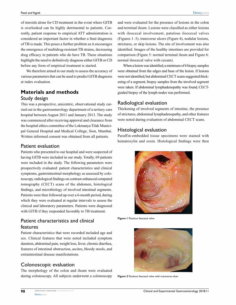
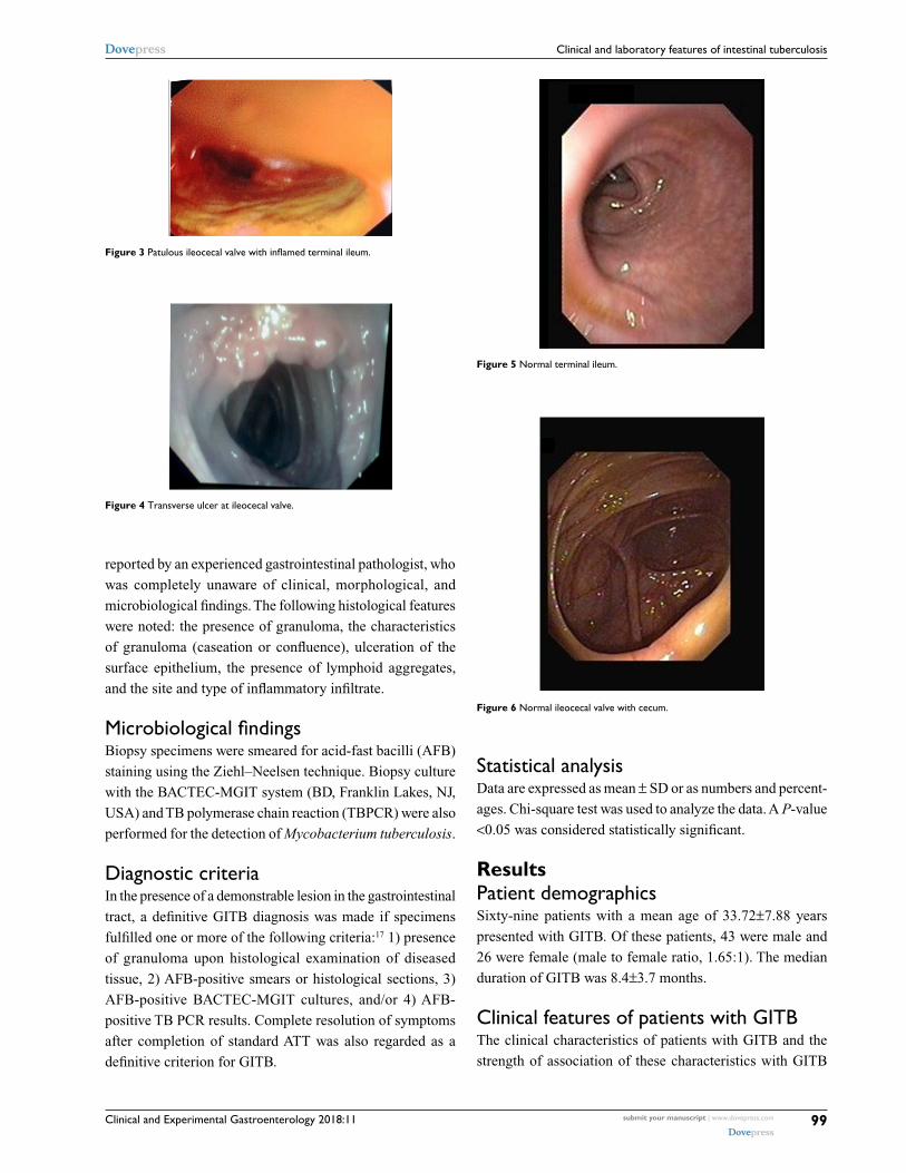
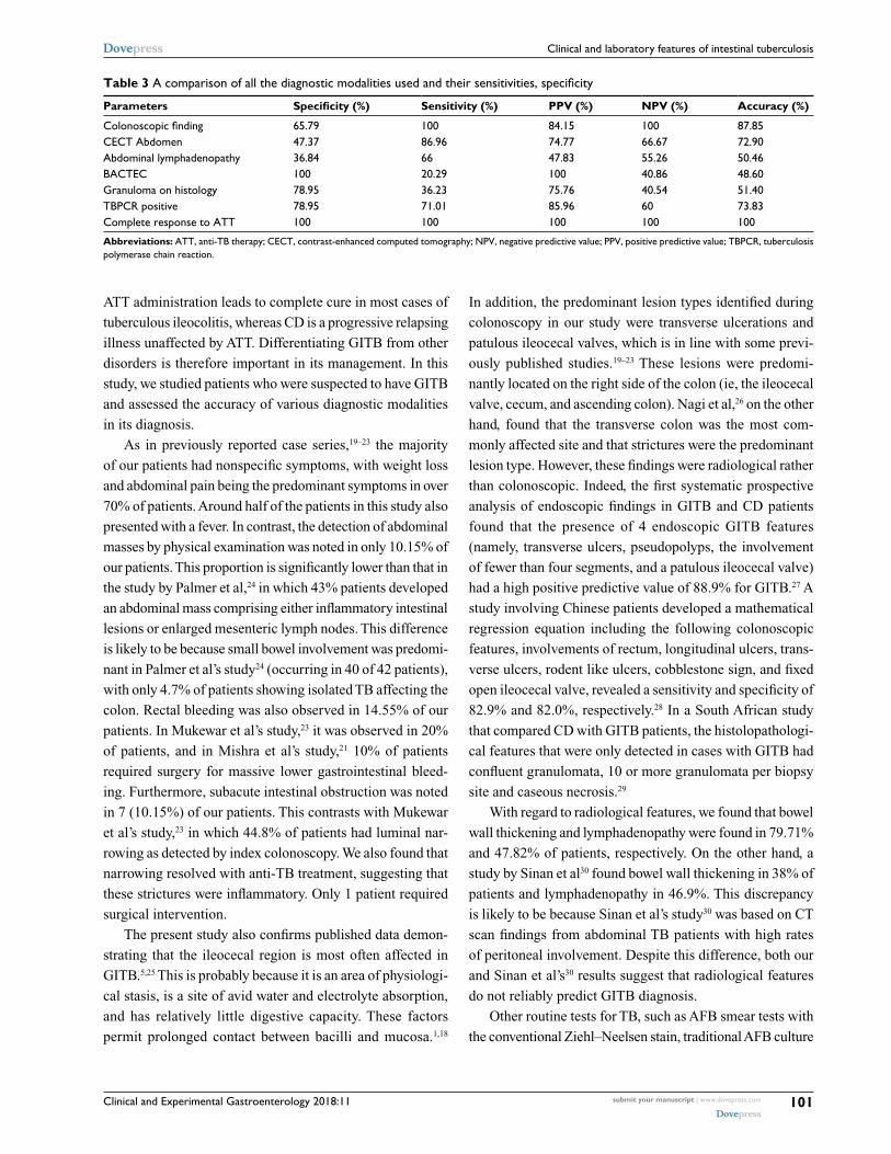
Colonoscopic evaluationThe morphology of the colon and ileum were evaluated
during colonoscopy. All subjects underwent a colonoscopy
and were evaluated for the presence of lesions in the colon
and terminal ileum. Lesions were classified as either lesions
with ileocecal involvement, patulous ileocecal valves
( Figures 1–3), transverse ulcers (Figure 4), nodular lesions,
strictures, or skip lesions. The site of involvement was also
identified. Images of the healthy intestines are provided for
comparison (Figure 5: normal terminal ileum and Figure 6:
normal ileocecal valve with cecum).
When a lesion was identified, a minimum of 6 biopsy samples
were obtained from the edges and base of the lesion. If lesions
were not identified, but abdominal CECT scans suggested thick-
ening of a segment, biopsy samples from the involved segment
were taken. If abdominal lymphadenopathy was found, CECT-
guided biopsy of the lymph nodes was performed.
Radiological evaluationThickening of involved segments of intestine, the presence
of strictures, abdominal lymphadenopathy, and other features
were noted during evaluation of abdominal CECT scans.
Histological evaluationParaffin-embedded tissue specimens were stained with
hematoxylin and eosin. Histological findings were then
Figure 1 Patulous ileocecal valve.
Figure 2 Patulous ileocecal valve with transverse ulcer.
Clinical and Experimental Gastroenterology 2018:11 submit your manuscript | www.dovepress.com
Dovepress
Dovepress
99
Clinical and laboratory features of intestinal tuberculosis
reported by an experienced gastrointestinal pathologist, who
was completely unaware of clinical, morphological, and
microbiological findings. The following histological features
were noted: the presence of granuloma, the characteristics
of granuloma (caseation or confluence), ulceration of the
surface epithelium, the presence of lymphoid aggregates,
and the site and type of inflammatory infiltrate.
Microbiological findingsBiopsy specimens were smeared for acid-fast bacilli (AFB)
staining using the Ziehl–Neelsen technique. Biopsy culture
with the BACTEC-MGIT system (BD, Franklin Lakes, NJ,
USA) and TB polymerase chain reaction (TBPCR) were also
performed for the detection of Mycobacterium tuberculosis.
Diagnostic criteriaIn the presence of a demonstrable lesion in the gastrointestinal
tract, a definitive GITB diagnosis was made if specimens
fulfilled one or more of the following criteria:17 1) presence
of granuloma upon histological examination of diseased
tissue, 2) AFB-positive smears or histological sections, 3)
AFB-positive BACTEC-MGIT cultures, and/or 4) AFB-
positive TB PCR results. Complete resolution of symptoms
after completion of standard ATT was also regarded as a
definitive criterion for GITB.
Statistical analysisData are expressed as mean ± SD or as numbers and percent-
ages. Chi-square test was used to analyze the data. A P-value
<0.05 was considered statistically significant.
ResultsPatient demographicsSixty-nine patients with a mean age of 33.72±7.88 years
presented with GITB. Of these patients, 43 were male and
26 were female (male to female ratio, 1.65:1). The median
duration of GITB was 8.4±3.7 months.
Clinical features of patients with GITBThe clinical characteristics of patients with GITB and the
strength of association of these characteristics with GITB
Figure 3 Patulous ileocecal valve with inflamed terminal ileum.
Figure 4 Transverse ulcer at ileocecal valve.
Figure 5 Normal terminal ileum.
Figure 6 Normal ileocecal valve with cecum.

Clinical and Experimental Gastroenterology 2018:11submit your manuscript | www.dovepress.com
Dovepress
Dovepress
100
Patel and Yagnik
are shown in Table 1. The clinical features most strongly
associated with a diagnosis of GITB were the presence of
ascites and presentation with subacute intestinal obstruction.
Endoscopic features of patients with GITBSites within the gastrointestinal tract that were commonly
found to be involved in our 69 GITB patients were (in
descending order of prevalence) the ileocecal valve in 58
patients (84.05%), the ileum in 25 patients (36.23%), the
ascending colon in 22 patients (31.88%), the transverse
colon in 4 patients (5.80%), and the descending colon in 2
patients (2.90%). No patients were found to have GITB with
involvement of the sigmoid colon, rectum, or the anal and
perianal region.
Lesions detected during endoscopy were also character-
ized and noted, with their features summarized in Table 2. Of
these lesions, transverse ulcers and patulous ileocecal valves
were found to be significantly associated with a diagnosis
of GITB.
Radiological features of patients with GITBOf the 69 patients with GITB, 49 (71.01%) had thicken-
ing of the involved part of the colon and 33 (47.83%) had
abdominal lymphadenopathy. Thickening of an involved
portion of the colon was found to have 47.37% specificity,
86.96% sensitivity, a positive predictive value of 74.77%, a
negative predictive value of 66.67%, and 72.90% accuracy.
For abdominal lymphadenopathy, these values were 36.84%,
66%, 47.83%, 55.26%, and 50.46%, respectively.
Histological features of patients with GITBOf the 69 patients diagnosed with GITB, granulomas were
found in the stained biopsy sections of 49 patients (71.01%).
Histological detection of granulomas had 78.95% specificity,
36.23% sensitivity, a positive predictive value of 75.76%, a
negative predictive value of 40.54%, and 51.40% accuracy.
TBPCR results in patients with GITBOf the 69 patients with GITB, TB PCR was positive for
M. tuberculosis in 49 (71.01%). TBPCR was found to have
78.95% specificity, 71.01% sensitivity, a positive predictive
value of 85.96%, a negative predictive value of 60%, and
73.83% accuracy.
BACTEC-MGIT culture results in patients with GITBBACTEC-MGIT cultures established from colonoscopic
biopsy specimens were positive for M. tuberculosis in 14 of
our 69 (20.29%) GITB patients. BACTEC-MGIT culture was
found to have 100% specificity, 20.29% sensitivity, a posi-
tive predictive value of 100%, a negative predictive value of
40.86%, and 48.60% accuracy.
A comparison of all the diagnostic modalities used and
their sensitivities, specificity, etc. is shown in Table 3.
DiscussionTB can affect any part of the gastrointestinal tract from the
mouth to the anus, as well as the pancreatobiliary system. Its
symptoms can also mimic those of various other intestinal
disorders, both common and rare. As intestinal infections
occur at a high rate in the Asia-Pacific region, the area was
previously thought to have a low incidence rate of inflam-
matory bowel disorder (IBD), though this could also be due
to a lack of population-based studies and low diagnostic
awareness of IBD. However, current literature and recent
information suggest that a true increase of IBD is occurring
throughout this region.5 At the same time, the incidence of
TB is also rising elsewhere, including in both the US and
the UK.3,18
Two disorders that present similarly in terms of both
clinical features and colonoscopy findings are CD and
tuberculous colitis. Despite their similar features, however,
their pathogenesis and therefore treatment differ. Appropriate
Table 1 Clinical features
Characteristics GITB, n=69 (%) P-value
Abdominal pain 53 (76) >0.05Weight loss 42 (60.87) >0.05Fever 50 (72.46) >0.05Chronic diarrhea 20 (28.99) >0.05Constipation 5 (7.25) >0.05Abdominal mass 7 (10.15) <0.05Blood in stool 10 (14.50) >0.05Ascites 7 (10.15) <0.05Subacute intestinal obstruction 7 (10.15) <0.05Extraintestinal manifestation 10 (14.50) >0.05
Abbreviation: GITB, gastrointestinal tuberculosis.
Table 2 Types of lesions
Endoscopic findings n=69 (%) P-value
Transverse ulcer 40 (57.97) <0.05Patulous ileocecal valve 20 (28.99) <0.05Nodularity 15 (21.74) >0.05Stricture 6 (8.70) >0.05Skip lesions 5 (7.25) >0.05
Clinical and Experimental Gastroenterology 2018:11 submit your manuscript | www.dovepress.com
Dovepress
Dovepress
101
Clinical and laboratory features of intestinal tuberculosis
ATT administration leads to complete cure in most cases of
tuberculous ileocolitis, whereas CD is a progressive relapsing
illness unaffected by ATT. Differentiating GITB from other
disorders is therefore important in its management. In this
study, we studied patients who were suspected to have GITB
and assessed the accuracy of various diagnostic modalities
in its diagnosis.
As in previously reported case series,19–23 the majority
of our patients had nonspecific symptoms, with weight loss
and abdominal pain being the predominant symptoms in over
70% of patients. Around half of the patients in this study also
presented with a fever. In contrast, the detection of abdominal
masses by physical examination was noted in only 10.15% of
our patients. This proportion is significantly lower than that in
the study by Palmer et al,24 in which 43% patients developed
an abdominal mass comprising either inflammatory intestinal
lesions or enlarged mesenteric lymph nodes. This difference
is likely to be because small bowel involvement was predomi-
nant in Palmer et al’s study24 (occurring in 40 of 42 patients),
with only 4.7% of patients showing isolated TB affecting the
colon. Rectal bleeding was also observed in 14.55% of our
patients. In Mukewar et al’s study,23 it was observed in 20%
of patients, and in Mishra et al’s study,21 10% of patients
required surgery for massive lower gastrointestinal bleed-
ing. Furthermore, subacute intestinal obstruction was noted
in 7 (10.15%) of our patients. This contrasts with Mukewar
et al’s study,23 in which 44.8% of patients had luminal nar-
rowing as detected by index colonoscopy. We also found that
narrowing resolved with anti-TB treatment, suggesting that
these strictures were inflammatory. Only 1 patient required
surgical intervention.
The present study also confirms published data demon-
strating that the ileocecal region is most often affected in
GITB.5,25 This is probably because it is an area of physiologi-
cal stasis, is a site of avid water and electrolyte absorption,
and has relatively little digestive capacity. These factors
permit prolonged contact between bacilli and mucosa.1,18
In addition, the predominant lesion types identified during
colonoscopy in our study were transverse ulcerations and
patulous ileocecal valves, which is in line with some previ-
ously published studies.19–23 These lesions were predomi-
nantly located on the right side of the colon (ie, the ileocecal
valve, cecum, and ascending colon). Nagi et al,26 on the other
hand, found that the transverse colon was the most com-
monly affected site and that strictures were the predominant
lesion type. However, these findings were radiological rather
than colonoscopic. Indeed, the first systematic prospective
analysis of endoscopic findings in GITB and CD patients
found that the presence of 4 endoscopic GITB features
(namely, transverse ulcers, pseudopolyps, the involvement
of fewer than four segments, and a patulous ileocecal valve)
had a high positive predictive value of 88.9% for GITB.27 A
study involving Chinese patients developed a mathematical
regression equation including the following colonoscopic
features, involvements of rectum, longitudinal ulcers, trans-
verse ulcers, rodent like ulcers, cobblestone sign, and fixed
open ileocecal valve, revealed a sensitivity and specificity of
82.9% and 82.0%, respectively.28 In a South African study
that compared CD with GITB patients, the histolopathologi-
cal features that were only detected in cases with GITB had
confluent granulomata, 10 or more granulomata per biopsy
site and caseous necrosis.29
With regard to radiological features, we found that bowel
wall thickening and lymphadenopathy were found in 79.71%
and 47.82% of patients, respectively. On the other hand, a
study by Sinan et al30 found bowel wall thickening in 38% of
patients and lymphadenopathy in 46.9%. This discrepancy
is likely to be because Sinan et al’s study30 was based on CT
scan findings from abdominal TB patients with high rates
of peritoneal involvement. Despite this difference, both our
and Sinan et al’s30 results suggest that radiological features
do not reliably predict GITB diagnosis.
Other routine tests for TB, such as AFB smear tests with
the conventional Ziehl–Neelsen stain, traditional AFB culture
Table 3 A comparison of all the diagnostic modalities used and their sensitivities, specificity
Parameters Specificity (%) Sensitivity (%) PPV (%) NPV (%) Accuracy (%)
Colonoscopic finding 65.79 100 84.15 100 87.85CECT Abdomen 47.37 86.96 74.77 66.67 72.90Abdominal lymphadenopathy 36.84 66 47.83 55.26 50.46BACTEC 100 20.29 100 40.86 48.60Granuloma on histology 78.95 36.23 75.76 40.54 51.40TBPCR positive 78.95 71.01 85.96 60 73.83Complete response to ATT 100 100 100 100 100
Abbreviations: ATT, anti-TB therapy; CECT, contrast-enhanced computed tomography; NPV, negative predictive value; PPV, positive predictive value; TBPCR, tuberculosis polymerase chain reaction.

Clinical and Experimental Gastroenterology 2018:11submit your manuscript | www.dovepress.com
Dovepress
Dovepress
102
Patel and Yagnik
using egg-based or agar-based media, and guinea pig inocula-
tion lack sensitivity and are time-consuming.31 In contrast,
smear tests using fluorescence staining and culture with the
BACTEC technique are more rapid and sensitive.31 As GITB
is a paucibacillary disease, however, the sensitivity of these
methods for detecting M. tuberculosis in clinical specimens
remains poor. Furthermore, although many serological tests
for TB are commercially available, they are not satisfactory
when a differential diagnosis needs to be made.31
Recently, Pulimood et al32 reported that in addition to
AFB detection, biopsy features suggestive of TB include
confluent granulomas, a lymphoid cuff around granulomas,
granulomas larger than 400 μm in diameter, 5 or more
granulomas in biopsies from 1 segment, granulomas located
in the submucosa or in granulation tissue (often as palisaded
epithelioid histiocytes), and disproportionate submucosal
inflammation. In our study, we collected mucosal biopsies
during colonoscopy. However, the usefulness of mucosal
biopsies is limited because granulomas, the primary feature
that differentiates TB from CD, are found in only 50%–80%
of intestinal mucosal biopsies from patients with clinically
confirmed TB.33–35 Furthermore, caseation of granulomas and
AFB, the diagnostic features of TB, are, respectively, found
in only 18%–33%19,35 and as low as 5% of cases.19 Indeed, we
found that mucosal biopsies showing caseation of granulomas
could predict TB diagnosis with 51.40% accuracy, which is
not sufficient for differentially diagnosing GITB and CD.
Du et al36 published a meta-analysis including 10 ran-
domized trials in PLoS One showing that the sensitivity
and specificity of confluent granulomas for differentiation
of GITB from CD were 38% and 99%, respectively, while
those of epithelioid histiocytes in ulcer bases were 41% and
94%, respectively.36
For culture-based TB diagnosis, the BACTEC 460TB
system works on the principle of early specific detection of
mycobacterial growth.37 This metabolism-based diagnostic
system is advantageous as its speed of diagnosis is double that
of conventional Lowenstein–Jensen medium; while a culture
observation period of 6 weeks is required when Lowenstein–
Jensen medium is used, it is possible to provide a negative
report in 3 weeks when the BACTEC 460TB system is used. In
our study, a positive BACTEC culture was observed in 20.29%
of TB patients and had 48.60% accuracy. The rate of culture
positivity was higher in Shah et al’s study,38 where 76% of TB
patients showed positive BACTEC 460TB cultures. This dif-
ference may be because while all cultures were established in
our study using colonoscopic biopsies, Shah et al’s38 samples
were drawn from a range of procedures including colono-
scopic, laparoscopic, and open surgical biopsies.
Finally, amplification of mycobacterial DNA sequences
by PCR, which is a rapid and accurate diagnostic method, has
been shown to be more promising for mycobacterial detec-
tion in clinical specimens. Indeed, we found that TBPCR
had an accuracy of 73.83% in the diagnosis of GITB. This
is within the range of previous studies, including a study by
Amarapurkar et al,39 where TBPCR had a diagnostic accuracy
of over 80%, and a study by Gan et al,40 where 64.1% of TB
patients had a positive TBPCR result.
Limitations of the study1) This study is not a comparative study between GITB and
CD which, given the minimal incidence of CD, would be
difficult. 2) We mentioned CD as the background motivation
for study, not as a component of research question.
ConclusionPatients with GITB tend to present with nonspecif ic
symptoms. Hence, diagnosis in countries where TB is
nonendemic involves a high index of suspicion (migrant
population, immunosuppressed individuals). While abdom-
inal pain and weight loss are the predominant symptoms,
ulceration, patulous ileocecal valves, and luminal narrow-
ing are the prominent endoscopic findings and appear to
mostly affect the right colon. Although histology is helpful
in ruling out other conditions, TB-specific findings such
as caseating granuloma and AFB are rarely seen. Instead,
TBPCR has the highest diagnostic accuracy followed by
BACTEC culture. Furthermore, the majority of colonic
lesions, including strictures, resolve with anti-TB treat-
ment, suggesting that strictures are inflammatory rather
than fibrotic. Follow-up colonoscopy is not required in
those who have symptomatic improvement after anti-TB
treatment.
AcknowledgmentWe would like to acknowledge the help provided by Dr Sushil
Dawka (Professor of Surgery, Mauritius) for his kind help in
revising the manuscript.
Author contributionsThis work was carried out in collaboration between both
authors. Author Bhumit Patel designed the study, per-
formed the statistical analysis, wrote the protocol, and
wrote the first draft of the manuscript. Author Vipul Yagnik
managed the analyses of the study, and critically revised the
manuscript. Both authors contributed toward data analysis,
drafting and critically revising the paper and agree to be
accountable for all aspects of the work.

Clinical and Experimental Gastroenterology 2018:11 submit your manuscript | www.dovepress.com
Dovepress
Dovepress
Clinical and Experimental Gastroenterology
Publish your work in this journal
Submit your manuscript here: https://www.dovepress.com/clinical-and-experimental-gastroenterology-journal
Clinical and Experimental Gastroenterology is an international, peer-reviewed, open access, online journal publishing original research, reports, editorials, reviews and commentaries on all aspects of gastroenterology in the clinic and laboratory. This journal is included on PubMed. The manuscript management system is completely online
and includes a very quick and fair peer-review system, which is all easy to use. Visit http://www.dovepress.com/testimonials.php to read real quotes from published authors.
Dovepress
103
Clinical and laboratory features of intestinal tuberculosis
DisclosureThe authors report no conflicts of interest in this work.
References1. Guth AA, Kim U. The reappearance of abdominal tuberculosis. Surg
Gynecol Obstet. 1991;172(6):432–436.2. Watson JM, Gill ON. HIV infection and tuberculosis. BMJ.
1990;300(6717):63–65.3. Marshall JB. Tuberculosis of the gastrointestinal tract and peritoneum.
Am J Gastroenterol. 1993;88(7):989–999.4. Snider DJ, Roper WL. The new tuberculosis. N Engl J Med. 1992;
326(10):703–705.5. Ouyang Q, Tandon R, Goh KL, Ooi CJ, Ogata H, Fiocchi C. The emer-
gence of inflammatory bowel disease in the Asian Pacific region. Curr Opin Gastroenterol. 2005;21(4):408–413.
6. Jayanthi V, Robinson RJ, Malathi S, et al. Does Chron’s disease need differentiation from tuberculosis? J Gastroenterol Hepatol. 1996;11(2):183–186.
7. Bhansali SK. Abdominal tuberculosis. Experiences with 300 cases. Am J Gastroenterol. 1977;67(4):324–337.
8. Chung KM, Kim HS, Park SY, et al. The changes in incidence of Chron’s disease and intestinal tuberculosis in Korea. Korean J Gastroenterol. 2008;52(6):351–358.
9. Amarapurkar D, Patel N. Crohn’s disease in India. Gastroenterol Today. 2002;6:73–75.
10. Pulimood AB, Ramakrishna BS, Kurian G, et al. Endoscopic mucosal biopsies are useful in distinguishing granulomatous colitis due to Crohn’s disease from tuberculosis. Gut. 1999;45(4):537–541.
11. Pai CG, Khandige GK. Is Crohn’s disease rare in India? Indian J Gas-troentero. 2000;19(1):17–20.
12. Patel N, Amarapurkar DN, Agal S, et al. Gastrointestinal luminal tuberculosis: establishing the diagnosis. Gastroenterol Hepatol. 2004;19(11):1240–1246.
13. Ouyang Q. Inflammatory bowel disease in China. J Gastroenterol Hepatol. 2000;15:S25.
14. Das P, Shukla HS. Clinical diagnosis of abdominal tuberculosis. Br J Surg. 1976;63:941–946.
15. Anand BS. Distinguishing Crohn’s disease from intestinal tuberculosis. Nat Med J Ind. 1989;2:172–175.
16. Chuttani HK, Sarin SK. Intestinal tuberculosis. Ind J Tuber. 1985;32: 117–125.
17. Amarapurkar DN, Patel ND, Rane PS. Diagnosis of Crohn’s disease in India where tuberculosis is widely prevalent. World J Gastroenterol. 2008;14(5):741–746.
18. Hayward AC, Watson JM. Tuberculosis in England and Wales 1982–1993: notifications exceeded predictions. Commun Dis Rep CDR Rev. 1995;5(3):R29–R33.
19. Alvares JF, Devarbhavi H, Makhija P, Rao S, Kottoor R. Clinical, colo-noscopic, and histological profile of colonic tuberculosis in a tertiary hospital. Endoscopy. 2005;37(4):351–356.
20. Villanueva SE, Martinez HMP, Fernando ATFJ, Valdés OM. Colonic tuberculosis. Dig Dis Sci. 2002;47(9):2045–2048.
21. Misra SP, Misra V, Dwivedi M, Arora JS, Kunwar BK. Tuberculous colonic strictures: impact of dilation on diagnosis. Endoscopy. 2004;36(12):1099–1103.
22. Chong VH, Lim KS. Gastrointestinal tuberculosis. Singapore Med J. 2009;50(6):638–645; quiz 646.
23. Mukewar S, Mukewar S, Ravi R, Prasad A, Dua KS. Colon tuberculosis:endoscopic feature and prospective endoscopic follow up after anti tuberculous treatment. Clin Transl Gastroenterol. 2012; 3(10):e24.
24. Palmer KR, Patil DH, Basran GS, Riordan JF, Silk DB. Abdominal tuberculosis in urban Britain – a common disease. Gut. 1985;26: 1296–1305.
25. Misra SP, Misra V, Dwivedi M, Gupta SC. Colonic tuberculosis: clini-cal features, endoscopic appearance and management. J Gastroenterol Hepatol. 1999;14(7):723–729.
26. Nagi B, Kochhar R, Bhasin DK, Singh K. Colorectal tuberculosis. Eur Radiol. 2003;13(8):1907–1912.
27. Lee YJ, Yang SK, Byeon JS, et al. Analysis of colonoscopic findings in the differential diagnosis between intestinal tuberculosis and Crohn’s disease. Endoscopy. 2006;38(6):592–597.
28. Li X, Liu X, Zou Y, et al. Predictors of clinical and endoscopic findings in differentiating Crohn’s disease from intestinal tuberculosis. Dig Dis Sci. 2011;56:188–196.
29. Kirsch R, Pentecost M, Hall PM, et al. Role of colonoscopic biopsy in distinguishing between Crohn’s disease and intestinal tuberculosis. J Clin Pathol. 2006;59:840–844.
30. Sinan T, Sheikh M, Ramadan S, Sahwney S, Behbehani A. Ct features in abdominal tuberculosis: 20 years experience. BMC Med Imaging. 2002;2(1):3.
31. Woods GL. The mycobacteriology laboratory and new diagnostic tech-niques. Infect Dis Clin North Am. 2002;16(1):127–144.
32. Pulimood AB, Amarapurkar DN, Ghoshal U, et al. Differentiation of Crohn’s disease from intestinal tuberculosis in India in 2010. World J Gastroenterol. 2011;17(4):433–443.
33. Makharia GK, Srivastava S, Das P, et al. Clinical, endoscopic, and histological differentiations between Crohn’s disease and intestinal tuberculosis. Am J Gastroenterol. 2010;105(3):642–651.
34. Pettengell KE, Larsen C, Garb M, Mayet FG, Simjee AE, Pirie D. Gastrointestinal tuberculosis in patients with pulmonary tuberculosis. Q J Med. 1990;74(275):303–308.
35. Shah S, Thomas V, Mathan M, et al. Colonoscopic study of 50 patients with colonic tuberculosis. Gut. 1992;33(3):347–351.
36. Du J, Ma YY, Xiang H, Li YM. Confluent granulomas andulcers lined by epithelioid histiocytes: new ideal method for differentiation of ITB and CD? A meta analysis. PLoS One. 2014;9:e103303.
37. Katoch VM. Newer diagnostic techniques for tuberculosis. Indian J Med Res. 2004;120:418–428.
38. Shah SR, Shenai S, Desai DC, Joshi A, Abraham P, Rodrigues C. Comparison of Mycobacterium tuberculosis culture using liquid culture medium and Lowenstein Jensen medium in abdominal tuberculosis. Indian J Gastroenterol. 2010;29(6):237–239.
39. Amarapurkar DN, Patel ND, Amarapurkar AD, Agal S, Baigal R, Gupte P. Tissue polymerase chain reaction in diagnosis of intestinal tuberculosis and Crohn’s disease in India. J Assoc Physicians India. 2004;52:863–867.
40. Gan HT, Chen YQ, Ouyang Q, Bu H, Yang XY. Differentiation between intestinal tuberculosis and Crohn’s disease in endoscopic biopsy specimens by polymerase chain reaction. Am J Gastroenterol. 2002;97(6):1446–1451.