Resolving multiple epigenetic pathways to adolescent depression

Epigenetic Inactivation of RASSF1A in Lung and BreastCancers and Malignant Phenotype Suppression
David G. Burbee, Eva Forgacs, Sabine Zochbauer-Muller, Latha Shivakumar,Kwun Fong, Boning Gao, Dwight Randle, Masashi Kondo, Arvind Virmani, ScottBader, Yoshitaka Sekido, Farida Latif, Sara Milchgrub, Shinichi Toyooka, Adi F.Gazdar, Michael I. Lerman, Eugene Zabarovsky, Michael White, John D. Minna
Background: The recently identified RASSF1 locus is locatedwithin a 120-kilobase region of chromosome 3p21.3 that fre-quently undergoes allele loss in lung and breast cancers. Weexplored the hypothesis that RASSF1 encodes a tumor sup-pressor gene for lung and breast cancers. Methods: We as-sessed expression of two RASSF1 gene products, RASSF1Aand RASSF1C, and the methylation status of their respectivepromoters in 27 non-small-cell lung cancer (NSCLC) celllines, in 107 resected NSCLCs, in 47 small-cell lung cancer(SCLC) cell lines, in 22 breast cancer cell lines, in 39 resectedbreast cancers, in 104 nonmalignant lung samples, and inthree breast and lung epithelial cultures. We also transfecteda lung cancer cell line that lacks RASSF1A expression withvectors containing RASSF1A complementary DNA to deter-mine whether exogenous expression of RASSF1A wouldaffect in vitro growth and in vivo tumorigenicity of this cellline. All statistical tests were two-sided. Results: RASSF1Amessenger RNA was expressed in nonmalignant epithelialcultures but not in 100% of the SCLC, in 65% of theNSCLC, or in 60% of the breast cancer lines. By contrast,RASSF1C was expressed in all nonmalignant cell culturesand in nearly all cancer cell lines. RASSF1A promoter hy-permethylation was detected in 100% of SCLC, in 63% ofNSCLC, in 64% of breast cancer lines, in 30% of primaryNSCLCs, and in 49% of primary breast tumors but in noneof the nonmalignant lung tissues. RASSF1A promoter hy-permethylation in resected NSCLCs was associated with im-paired patient survival (P = .046). Exogenous expression ofRASSF1A in a cell line lacking expression decreased in vitrocolony formation and in vivo tumorigenicity. Conclusion:RASSF1A is a potential tumor suppressor gene that under-goes epigenetic inactivation in lung and breast cancersthrough hypermethylation of its promoter region. [J NatlCancer Inst 2001;93:691–9]
Allelic loss of human chromosome 3p is an early and frequentevent in the development of several cancers, including lung andbreast cancers (1–5). Identification of a series of nested 3p21.3homozygous deletions in small-cell lung cancers (SCLCs) di-rected an intensive effort to positionally clone tumor suppressorgenes from a 630-kilobase (kb) region, which was recently nar-rowed to a 120-kb subregion by identification of a breast cancerhomozygous deletion (6–8). Sequencing the entire 630-kb re-gion identified at least 25 genes, several of which may encodetumor suppressor genes for lung cancer (7). Nine genes arelocated in or on the border of the breast cancer-defined subre-gion. One of these genes, which spans 7.6 kb of genomic DNA,has a predicted Ras-association domain and homology to the
Ras-effector Nore1 (Fig. 1); it has, therefore, been termed“RASSF1” (9,10).
The RASSF1 gene encodes two major transcripts, RASSF1Aand RASSF1C, which are produced by alternative promoter se-lection and alternative messenger RNA (mRNA) splicing.RASSF1A is encoded by RASSF1 exons 1A, 1C, and 2–5.RASSF1C is encoded by RASSF1 exons 1–5 (Fig. 1). The startsites for RASSF1A and RASSF1C are approximately 2 kb apartand have two independent CpG island-containing putative pro-moter regions. RASSF1A is predicted to encode a 39-kd peptidethat contains an N-terminal diacylglycerol (DAG)-bindingdomain and a Ras-association domain (Fig. 1). RASSF1C ispredicted to encode a 32-kd peptide that lacks a DAG-bindingdomain but contains a Ras-association domain (7,11). Immedi-ately adjacent to the DAG-binding domain of RASSF1A isa sequence PxxP, which is the minimal sequence required for ansrc homology 3-binding domain. RASSF1A has a central linkerthat contains a number of prolines, as well as acidic and hy-droxyl-bearing residues. These regions, called PEST sequences,are found in proteins that are rapidly turned over by ubiquitina-tion-dependent pathways (12). For RASSF1C, the amino-terminal region unique to this isoform is enriched for thesePEST sequences. Within the PEST sequences common to bothRASSF1A and RASSF1C is a serine residue that is phosphor-ylated in vitro by DNA-dependent ataxia-telangiectasia-mutated(ATM) and ataxia-telangiectasia-related kinases (13). The Ras-association domain is more than 50% identical and more than70% similar to the carboxyl terminal 225 residues of mouseNore1 (10). The Ras-association domain, consisting of a core of90 amino acids, is flanked on the amino terminal side by a regionhomologous with a region found in Nore1 and Caenorhabditiselegans orthologue T24F1.3 protein.
In this article, we characterized RASSF1A and RASSF1C aspotential tumor suppressor genes in lung and breast cancers.
Affiliations of authors: D. G. Burbee, E. Forgacs, S. Zochbauer-Muller,B. Gao, D. Randle, M. Kondo, A. Virmani, S. Bader, Y. Sekido, S. Toyooka,A. F. Gazdar, J. D. Minna (Hamon Center for Therapeutic Oncology Research),L. Shivakumar, M. White (Department of Cell Biology), S. Milchgrub (Depart-ment of Pathology), The University of Texas Southwestern Medical Center atDallas; K. Fong, Department of Thoracic Medicine, The Prince Charles Hospital,Queensland, Australia; F. Latif, Department of Reproductive and Child Health,University of Birmingham, U.K.; M. Lerman, Laboratory of Immunobiology,National Cancer Institute-Frederick Cancer Research and Development Center,Frederick, MD; E. Zabarovsky, Karolinska Institute, Stockholm, Sweden.
Correspondence to: John D. Minna, M.D., Hamon Center for TherapeuticOncology Research, The University of Texas Southwestern Medical Center atDallas, 6000 Harry Hines Blvd., Dallas, TX 75390-8593 (e-mail: [email protected]).
See “Notes” following “References.”
© Oxford University Press
Journal of the National Cancer Institute, Vol. 93, No. 9, May 2, 2001 ARTICLES 691

Because loss of gene expression can be caused by tumor-acquired aberrant methylation, we assessed the methylation sta-tus of the RASSF1A promoter region in these tumors (14). Inaddition, we tested the ability of RASSF1A to suppress themalignant phenotype. Previously, Dammann et al. (15) showedthat the RASSF1A promoter is hypermethylated in lung cancercells and that exogenous expression of RASSF1A suppressestumorigenesis in nude mice. We have confirmed and extendedthose findings by analyzing the expression and methylation sta-tus of both the RASSF1A and RASSF1C genes in lung andbreast cancers.
METHODS
Patient Population
Resected lung tumor samples and clinical data were collected from patientsafter obtaining appropriate institutional review board approval and patients’written informed consent. Primary tumor samples and corresponding nonin-volved lung tissues were obtained from 107 patients with non-small-cell lungcarcinoma (NSCLC) who had received curative resection surgery at the PrinceCharles Hospital, Brisbane, Australia, from June 1990 through March 1993, andfor whom clinical and survival data of 5 or more years were available (16,17).Among the 107 patients, there were 76 males and 31 females (range, 28–81years; mean age, 61 years at diagnosis). Among the patients, 61 had stage Icancers, 21 had stage II, 24 had stage IIIA, and one had stage IIIB (18). Histo-logically, there were 45 adenocarcinomas, 43 squamous cell carcinomas,11 adenosquamous carcinomas, four large-cell carcinomas, three atypical carci-noids, and one typical carcinoid. Ninety-eight patients were smokers, with amean exposure of 31 pack-years, and nine were never smokers or nonsmokers.
We also obtained 39 primary breast tumors from patients aged 31–84 yearsundergoing breast cancer treatment in The University of Texas SouthwesternHospital system. Among the patients, three had stage I cancers, 15 had stage IIA,two had stage IIB, eight had stage IIIA, five had stage IIB, and six had stage IV.Histologically, there were 30 infiltrating ductal carcinomas, four invasive lobularcarcinomas, one lobular carcinoma in situ, two ductal carcinomas in situ, and
two breast adenocarcinomas at metastatic sites. Clinical information was ob-tained by retrospective review of clinical records.
Cell Lines and Cell Cultures
Lung and breast tumor cell lines generated by us have been described previ-ously (19–21). Complementary DNAs (cDNAs) and genomic DNAs were ob-tained from cell lines, most of which have been deposited in the American TypeCulture Collection (ATTC) (Manassas, VA), that represented the spectrum oflung cancer histologies. These cell lines include the following: (all Hxxxx lineshave the prefix National Cancer Institute [NCI]-) SCLCs (i.e., H69, H82, H128,H146, H182, H187, H196, H209, H249, H289, H290, H345, H378, H524, H526,H592, H735, H738, H740, H748, H774, H841, H847, H862, H889, H1092,H1105, H1184, H1304, H1339, H1450, H1607, H1618, H1672, H1688, H1963,H2028, H2029, H2081, H2108, H2171, H2195, H2227, and HCC970) andNSCLCs (i.e., H23, H28, H125, H157, H226, H358, H720, H727, H838, H920,H1155, H1299, H1437, H1466, H1573, H1648, H1770, H1792, H1819, H1838,H1993, H2009, H2052, H2077, H2087, H2347, H2452, H2882, H2887, HCC44,HCC78, HCC95, HCC193, HCC515, HCC827, and HCC1171). Breast cancercell lines used in these studies were the following: HTB19, HTB20, HTB22,HTB23, HTB24, HTB25, HTB26, HTB27, HTB121, HTB130, HTB131,HTB132, HTB133 (all HTB lines are available from the ATCC), HCC38,HCC70, HCC202, HCC712, HCC1007, HCC1143, HCC1187, HCC1395,HCC1419, HCC1428, HCC1500, HCC1569, HCC1739, HCC1806, HCC1937,HCC1599, HCC1954, HCC2157, HCC2185, HCC2218, HCC2688, andHCC2713. Normal human bronchial epithelial (NHBE) and small-airway epi-thelial (SAE) cell cultures were obtained from Clonetics (San Diego, CA) andwere grown and harvested as directed by the vendor.
Expression Analysis of RASSF1 Isoforms
The identification of the RASSF1 gene (initially called 123F2) and its majorisoforms RASSF1C and RASSF1A was reported as part of the overall charac-terization of the genes in the larger 630-kb 3p21.3 homozygous-deletion region(7) (Fig. 1, A–C).
Sequence information from exons 1A and 3 was used to design the forwardprimer PKCDF (5�-GGCGTCGTGCGCAAAGGCC-3�) and the reverse primerR182 (5�-GGGTGGCTTCTTGCTGGAGGG-3�) (Fig. 1, C). This primer pair
Fig. 1. Map of the RASSF1 locus, transcripts, and proteindomains. A) The exon–intron structure of the RASSF1 locuswith the location of the CpG islands in the predicted promoterregions (the locations of which are shown by double-headedarrows) of RASSF1A and RASSF1C. RASSF1A transcrip-tion is predicted to come from the most centromeric promoterregion located within a CpG island and begins with exon 1A.RASSF1F also commences at this promoter but is missingexon 1C. Transcription of RASSF1C is predicted to begin inthe most telomeric promoter region, which is approximately 2kilobases from that of RASSF1A and begins with exon 1.Blocks represent exons; lines represent introns. B) Schematicof the RASSF1A transcript and predicted protein-sequencedomains. The location of the various primers (PKCDF, NF,R182, and R292) used for isoform-specific reverse transcrip-tion (RT)–polymerase chain reaction (PCR) analyses are in-dicated. Tick marks identify the exon boundaries. The po-tential src homology 3 (SH3)-binding region, putativediacylglycerol (DAG)-binding domain, PEST sequence, Ras-association domain, and ataxia-telangiectasia-mutated (ATM)phosphorylation site are labeled. C) Schematic of theRASSF1C transcript and predicted protein-sequence domains.The locations of the various primers (NOX3, R182, and R292)used for isoform-specific RT–PCR analyses are indicated. D)Schematic of the RASSF1F transcript and predicted protein-sequence domains.
692 ARTICLES Journal of the National Cancer Institute, Vol. 93, No. 9, May 2, 2001

was used in reverse transcription (RT)–polymerase chain reaction (PCR) screensof lung, heart, and pancreatic tissue-specific cDNA libraries (Clontech Labora-tories, Inc., Palo Alto, CA). The conditions used TaqGold (The Perkin-ElmerCorp., Norwalk, CT) with 1× TaqGold buffer adjusted to 2 mM MgCl2. Allreactions used a 70 °C–60 °C touchdown, with 5% dimethyl sulfoxide for35 rounds (denaturation for 30 seconds, annealling for 30 seconds, and extensionfor 60 seconds) of PCR. The RASSF1A cDNA sequence is identical to that ofthe RASSF1C cDNA from the second exon to the carboxyl terminus, but the twocDNAs have different 5� exons (RASSF1A, GenBank Accession #AF102770:exons 1A and 1C; RASSF1C, GenBank Accession #AF040703: exon 1 [Fig. 1]).We also isolated tissue-specific isoforms from the heart (RASSF1D, GenBankAccession #AF102771) and the pancreas (RASSF1E, GenBank Assession#AF102772) cDNA libraries.
Primers derived from exon–intron junctions (sequences available online at theJournal website) were used for genomic DNA single-strand conformation poly-morphism mutation analysis of the coding regions of RASSF1A on a panel ofNSCLC, SCLC, and breast cancer cell line DNAs. DNA was prepared fromtumors and cell lines by standard methods (22), and aberrantly migrating frag-ments were sequenced as described previously (16,23).
RNA Analysis
Isoform-specific RT–PCR assays were used for analysis of RASSF1A andRASSF1C expression. Primers for RASSF1C were Nox3 (5�-CTGCAGC-CAAGAGGACTCGG-3�) and R182 and for RASSF1A were either PKCDFor NF (5�-TGCAAGTTCACCTGCCAC-3�) and R182 (Fig. 1, C). Total RNAwas isolated from previously described lung and breast cancer cell lines grownin RPMI-1640 medium supplemented with 5% fetal bovine serum (completemedium) (19–21) by Trizol extraction (Life Technologies, Inc. [GIBCO BRL],Rockville, MD). Four micrograms of total RNA was reverse transcribed by useof GIBCO-BRL Superscript First Strand cDNA Kit. All cDNA preparationswere tested for the ability to amplify a nontranscribed genomic sequence im-mediately upstream of the first exon of the RASSF1A transcript. Any cDNAsthat produced a product from this sequence were discarded because they werecontaminated with genomic DNA.
We also assessed the expression of RASSF1A after exposure to 5-aza-2�-deoxycytidine, a drug that inhibits DNA methylation. We exposed subconfluentcultures of the RASSF1A-nonexpressing NSCLC line NCI-H157 to 0.5 �M5-aza-2�-deoxycytidine for 48 hours, after which we isolated total RNA andperformed RT–PCR for RASSF1A, RASSF1C, and glyceraldehyde-3-phosphatedehydrogenase (GAPDH). RT–PCR of GAPDH transcripts was performed withthe use of forward primer GAPDH-C (5�-CATGACAACTTTGGTATCGTG-3�)and reverse primer GAPDH-D (5�-GTGTCGCTGTTGAAGTCAGA-3�). RT–PCR products were separated by agarose gel electrophoresis and visualized afterstaining with ethidium bromide.
Methylation Analysis
The methylation status of the presumed RASSF1A and RASSF1C promoterregions was determined by methylation-specific PCR. Genomic DNAs fromlung cancer cell lines not expressing RASSF1A (NCI lines H1299, H1184,H1304, H841, H2108, and H128) or expressing RASSF1A (H1792 and H2009)were modified by sodium bisulfite treatment as described previously (24,25).Bisulfite treatment converts cytosine bases to uracil bases but has no effect onmethylcytosine bases. PCR amplification followed by sequencing of the PCRfragments identifies specific CpG dinucleotides in the promoter region that aremodified by methylation (24,26,27). PCR primers (sequences available online atthe Journal website) were designed to amplify genomic sequences in the pre-sumed promoter regions of RASSF1A (cosmid Luca12; GenBank Accession#AC002481 nucleotides 17 730–18 370) and RASSF1C (GenBank Accession#AC002481 nucleotides 21 022–21 152 and 21 194–21 332). The resulting PCRfragments were sequenced by automated fluorescence-based DNA sequencing todetermine the methylation status.
The data on CpG methylation in RASSF1A-nonexpressing lung cancer celllines (data available online at the Journal website) were used to design methyl-ation-specific PCR (24) primers for the RASSF1A 5� promoter region: Theprimers to detect the methylated form were 5�-GGGTTTTGCGAGAGCGCG-3�
(forward) and 5�-GCTAACAAACGCGAACCG-3� (reverse), and the primers todetect the unmethylated form were 5�-GGTTTTGTGAGAGTGTGTTTAG-3�
(forward) and 5�-CACTAACAAACACAAACCAAAC-3� (reverse). Eachprimer set generated a 169-base-pair (bp) product. Methylation-specific PCRcycling conditions consisted of one incubation of 15 minutes at 95 °C, followed
by 40 cycles of a 30-second denaturation at 94 °C, 50 seconds at an annealingtemperature (64 °C for methylation-specific and 59 °C for unmethylated-specificprimers), a 30-second extension at 72 °C, and a final extension at 72 °C for10 minutes. PCR products were separated in 2% agarose gels. LymphocyteDNA, methylated in vitro by CpG (SssI) methylase (New England Biolabs, Inc.,Beverly, MA) following the manufacturer’s directions, was used as a positivecontrol. A water blank was used as a negative control.
Generation of Transfectants
RASSF1A cDNA was cloned into pcDNA3.1+ (Invitrogen Corp., Carlsbad,CA), resequenced to confirm that the cDNAs were in the correct orientationand reading frame, transcribed, and translated in vitro with commercial kits(Clonetech Laboratories, Inc.). The expression vector containing RASSF1A pro-duced a 42-kd protein, and the vector containing RASSF1C produced a 32-kdprotein on sodium dodecyl sulfate–polyacrylamide gels, close to their respectivepredicted molecular masses of 39 and 32 kd (data not shown). Expression vec-tors in pcDNA3.1 for mutant and wild-type p53 and their transfection andactivity have been described previously (28).
The RASSF1A expression vector was transfected into NSCLC NCI-H1299cells expressing RASSF1C, but not RASSF1A, by use of Lipofectamine plus(Life Technologies, Inc.) according to the manufacturer’s recommendations. Fortransient transfection studies, approximately 5 × 10⁵ NSCLC NCI-H1299 cells,harvested from 80%–90% confluent cultures in complete medium, were trans-fected with 1 �g of purified plasmid DNA. Samples were plated in a minimumof triplicate, and cells were collected 48 hours after transfection. Because thepcDNA3.1+ expression vector contains a neomycin resistance gene, clones ex-pressing RASSF1A were selected in complete medium supplemented with G418(800 �g/mL). Stable clones were maintained in complete medium supplementedwith G418 (600 �g/mL). We confirmed that the clones were expressing thetransfected RASSF1A gene by isolating total RNA from individual clones andperforming RT–PCR as described above. We also transfected NCI-H1299 cellswith the vector containing no inserts and isolated stable clones.
The RASSF1A and RASSF1C cDNAs were also cloned in the retroviralvector pBABEpuro and were resequenced to confirm that the genes were in thecorrect sequence and orientation (29). Virus was prepared in the 293 cell-basedPhoenix packaging cell line as described previously (29) from cells infectedeither with the vector alone or with constructs containing the RASSF1A orRASSF1C cDNA. Culture supernatants were collected by centrifugation at 500gat 37 °C for 10 minutes and used to infect NSCLC NCI-H1299 cells as describedpreviously (29). Because the viral vector contains a puromycin resistance gene,infected cells were selected with 1 �g/mL of puromycin for 7 days. Cellssurviving the selection and containing the transgene were pooled, and total cellextracts were made. Western blot analysis was performed as described previ-ously (30) to verify protein expression of the transfected genes. The proteinbands were visualized with the Pierce SuperSignal Kit (Pierce Chemical Co.,Rockford, IL).
Tumorigenicity Testing
The in vitro growth characteristics of NSCLC NCI-H1299 clones that expressRASSF1A were tested for anchorage-dependent and anchorage-independent(soft agar) growth. After 48 hours of growth in nonselective medium, transientlytransfected NSCLC NCI-H1299 cells were detached with trypsin and diluted,usually 10- to 25-fold, in complete medium containing 800 �g/mL of G418 andplated into fresh 100-mm dishes. The medium was changed twice weekly. After14 days, the medium was removed, the plates were washed with phosphate-buffered saline (PBS), and the colonies were stained with 1% methylene blue in50% (vol/vol) ethanol. For the anchorage-independent, soft agar-growth assays,1000 RASSF1A-expressing cells were suspended and plated in 0.33% Nobleagar (Sigma Chemical Co., St. Louis, MO) in complete medium supplementedwith 600 �g/mL G418 and layered over a 0.50% agar base in complete medium.After 21 days, colonies greater than 0.2 mm in diameter were counted.
For retrovirally infected cells, anchorage-independent growth assays wereperformed as follows: 10 000 viable selected cells from each infection were platedin 0.33% soft agar over a 0.50% agar base in Dulbecco’s modified Eagle medium(Life Technologies, Inc.) with 10% heat-inactivated fetal bovine serum. After21 days, colonies greater than 0.2 mm in diameter were counted.
We also tested the ability of RASSF1A-infected cells to grow in vivo in nudemice. Male BALB/c nude (nu/nu) 3- to 6-week-old mice (Charles River Labo-ratories, Wilmington, DE) were irradiated on day 0 of the experiment in groupsof five animals by a 5-minute exposure to 350 cGy from a cesium source. The
Journal of the National Cancer Institute, Vol. 93, No. 9, May 2, 2001 ARTICLES 693
next day, each mouse was given an injection subcutaneously on its flank with 0.2mL of sterile PBS containing 107 viable parental, vector control, or RASSF1Aretroviral-infected NSCLC NCI-H1299 tumor cells. Mice were monitored every2–3 days for tumor size; once tumors reached greater than 1500 mm3, the micewere killed. All animal care was in accord with institutional guidelines.
Antibody Preparation
The entire RASSF1C open reading frame was used to make a glutathioneS-transferase (GST) fusion protein, which was expressed in Escherichia coli byuse of an established procedure (31), and was used to make rabbit polyclonalantibodies to be described in detail elsewhere. For the western blot analysis, theantiserum was used at a 1 : 1000 dilution in 5% nonfat milk in PBS. Specificitywas determined by western blotting of H1299 cells transfected with vector(negative control) and the various RASSF1-expression constructs (e.g., see Fig. 6).
Statistical Analysis
Statistical analysis was performed by use of �2 and Fisher’s exact tests fordifferences between groups. Overall survival curves were calculated by use ofthe Kaplan–Meier method, and survival curves were compared with the log-rankstatistic (32). All analyses, including univariate, multivariate, and Cox analyses,were performed by use of SPSS Windows version 9.0.1 (SPSS Inc., Chicago,IL). All statistical tests were two-sided.
RESULTS
Characterization of the RASSF1 Gene
To determine if the RASSF1A gene was mutated in lung andbreast cancers, we performed extensive mutational analysis ofthe RASSF1A isoform with the use of single-strand conforma-tion polymorphism assays on genomic DNA. We had previouslyfound no RASSF1C mutations in 77 lung cancer cell linesamples (7). By use of the RASSF1A sequence as a reference,we found several polymorphisms, including the following:codon 21 (AAG to CAG), Lys to Gln; codon 28 (CGT to CGA),no amino acid change; codon 49 (GGC to GGT), no amino acidchange; codon 53 (CGC to TGC), Arg to Cys; codon 129 (GACto GAG), Asp to Glu; codon 133 (GCT to TCT), Ala to Ser; andcodon 325 (TAT to TGT), Tyr to Cys.
Expression of RASSF1A and RASSF1C in Lung andBreast Cancer Cell Lines
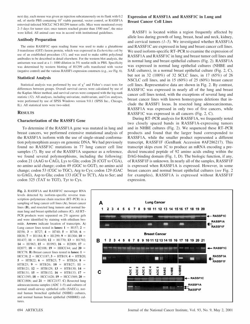
RASSF1 is located within a region frequently affected byallele loss during growth of lung, breast, head and neck, kidney,and cervical tumors (1–5). We investigated whether RASSF1Aand RASSF1C are expressed in lung and breast cancer cell lines.We used isoform-specific RT–PCR to examine the expression ofRASSF1A and RASSF1C in lung and breast tumor cell lines andin normal lung and breast epithelial cultures (Fig. 2). RASSF1Awas expressed in normal lung epithelial cultures (NHBE andSAE cultures), in a normal breast epithelial culture (Fig. 2, C),but not in 32 (100%) of 32 SCLC lines, in 17 (65%) of 26NSCLC cell lines, and in 15 (60%) of 25 (60%) breast cancercell lines. Representative data are shown in Fig. 2. By contrast,RASSF1C was expressed in nearly all of the lung and breastcancer cell lines tested, with the exceptions of several lung andbreast cancer lines with known homozygous deletions that in-clude the RASSF1 locus. In resected lung adenocarcinomas,RASSF1A was expressed in only two of five cancers, whileRASSF1C was expressed in all cancers (Fig. 2, C).
During RT–PCR analysis for RASSF1A, we frequently notedtwo closely spaced bands in RASSF1A-expressing tumorsand in NHBE cultures (Fig. 2). We sequenced these RT–PCRproducts and found that the larger band corresponded toRASSF1A, while the smaller product represented a differenttranscript, RASSF1F (GenBank Accession #AF286217). Thistranscript skips exon 1C to produce an mRNA encoding a pre-dicted truncated peptide of 92 amino acids ending within theDAG-binding domain (Fig. 1, D). The biologic function, if any,of RASSF1F is unknown. In nearly all of the samples, RASSF1Fis expressed when RASSF1A is expressed. However, in somebreast cancers and normal breast epithelial cultures (see Fig. 2for examples), RASSF1A is expressed without RASSF1Fexpression.
Fig. 2. RASSF1A and RASSF1C messenger RNAlevels detected by isoform-specific reverse tran-scription–polymerase chain reaction (RT–PCR) in asampling of lung cancer cell lines (A), breast cancerlines (B), and resected lung tumors and normal hu-man lung and breast epithelial cultures (C). All RT–PCR products were separated on 2% agarose gelsand were identified by staining with ethidium bro-mide. Arrows indicate location of transcripts. A)Lung cancer lines tested in lanes: 1 � H157; 2 �
H358; 3 � H727; 4 � H740; 5 � H748; 6 �
H838; 7 � H1184; 8 � H1299; 9 � H1304; 10 �
H1437; 11 � H1450; 12 � H1770; 13 � H1792;14 � H1963; 15 � H1993; 16 � H2009; 17 �
H2077; 18 � H2108; 19 � HHCC44; and 20 �
HCC78. B) Breast cancer lines tested in lanes: 1 �
HCC38; 2 � HCC1187; 3 � HTB19; 4 � HTB20;5 � HTB22; 6 � HTB23; 7 � HTB24; 8 �
HTB25; 9 � HTB26; 10 � HTB27; 11 �
HTB121; 12 � HTB129; 13 � HTB130; 14 �
HTB131; 15 � HTB132; 16 � HTB133; 17 �
HCC1395; 18 � HCC1428; 19 � HCC1569; 20 �
HCC1806; and 21 � HCC2157. C) Resected lungadenocarcinoma samples (ADC 1–5) and cultures ofnormal small-airway epithelial cells (SAECs), nor-mal human bronchial epithelial (NHBE) cultures,and normal human breast epithelial (NHBRE) cul-tures.
694 ARTICLES Journal of the National Cancer Institute, Vol. 93, No. 9, May 2, 2001
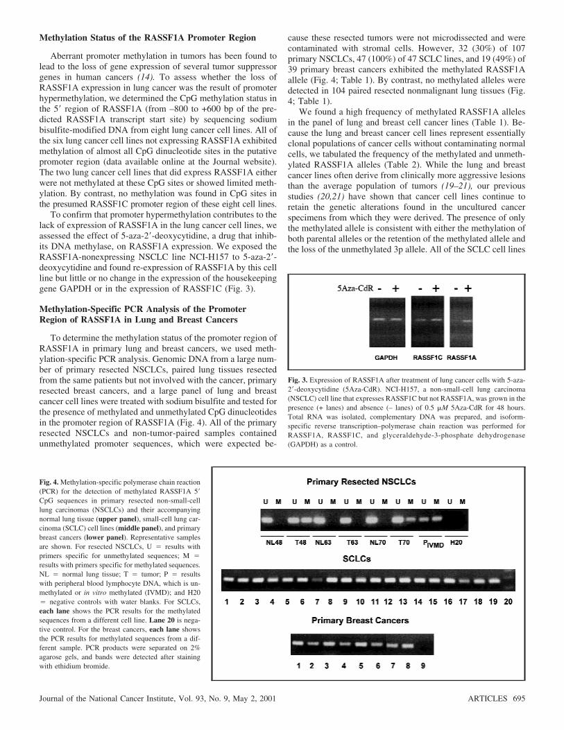
Methylation Status of the RASSF1A Promoter Region
Aberrant promoter methylation in tumors has been found tolead to the loss of gene expression of several tumor suppressorgenes in human cancers (14). To assess whether the loss ofRASSF1A expression in lung cancer was the result of promoterhypermethylation, we determined the CpG methylation status inthe 5� region of RASSF1A (from –800 to +600 bp of the pre-dicted RASSF1A transcript start site) by sequencing sodiumbisulfite-modified DNA from eight lung cancer cell lines. All ofthe six lung cancer cell lines not expressing RASSF1A exhibitedmethylation of almost all CpG dinucleotide sites in the putativepromoter region (data available online at the Journal website).The two lung cancer cell lines that did express RASSF1A eitherwere not methylated at these CpG sites or showed limited meth-ylation. By contrast, no methylation was found in CpG sites inthe presumed RASSF1C promoter region of these eight cell lines.
To confirm that promoter hypermethylation contributes to thelack of expression of RASSF1A in the lung cancer cell lines, weassessed the effect of 5-aza-2�-deoxycytidine, a drug that inhib-its DNA methylase, on RASSF1A expression. We exposed theRASSF1A-nonexpressing NSCLC line NCI-H157 to 5-aza-2�-deoxycytidine and found re-expression of RASSF1A by this cellline but little or no change in the expression of the housekeepinggene GAPDH or in the expression of RASSF1C (Fig. 3).
Methylation-Specific PCR Analysis of the PromoterRegion of RASSF1A in Lung and Breast Cancers
To determine the methylation status of the promoter region ofRASSF1A in primary lung and breast cancers, we used meth-ylation-specific PCR analysis. Genomic DNA from a large num-ber of primary resected NSCLCs, paired lung tissues resectedfrom the same patients but not involved with the cancer, primaryresected breast cancers, and a large panel of lung and breastcancer cell lines were treated with sodium bisulfite and tested forthe presence of methylated and unmethylated CpG dinucleotidesin the promoter region of RASSF1A (Fig. 4). All of the primaryresected NSCLCs and non-tumor-paired samples containedunmethylated promoter sequences, which were expected be-
cause these resected tumors were not microdissected and werecontaminated with stromal cells. However, 32 (30%) of 107primary NSCLCs, 47 (100%) of 47 SCLC lines, and 19 (49%) of39 primary breast cancers exhibited the methylated RASSF1Aallele (Fig. 4; Table 1). By contrast, no methylated alleles weredetected in 104 paired resected nonmalignant lung tissues (Fig.4; Table 1).
We found a high frequency of methylated RASSF1A allelesin the panel of lung and breast cell cancer lines (Table 1). Be-cause the lung and breast cancer cell lines represent essentiallyclonal populations of cancer cells without contaminating normalcells, we tabulated the frequency of the methylated and unmeth-ylated RASSF1A alleles (Table 2). While the lung and breastcancer lines often derive from clinically more aggressive lesionsthan the average population of tumors (19–21), our previousstudies (20,21) have shown that cancer cell lines continue toretain the genetic alterations found in the uncultured cancerspecimens from which they were derived. The presence of onlythe methylated allele is consistent with either the methylation ofboth parental alleles or the retention of the methylated allele andthe loss of the unmethylated 3p allele. All of the SCLC cell lines
Fig. 3. Expression of RASSF1A after treatment of lung cancer cells with 5-aza-2�-deoxycytidine (5Aza-CdR). NCI-H157, a non-small-cell lung carcinoma(NSCLC) cell line that expresses RASSF1C but not RASSF1A, was grown in thepresence (+ lanes) and absence (– lanes) of 0.5 �M 5Aza-CdR for 48 hours.Total RNA was isolated, complementary DNA was prepared, and isoform-specific reverse transcription–polymerase chain reaction was performed forRASSF1A, RASSF1C, and glyceraldehyde-3-phosphate dehydrogenase(GAPDH) as a control.
Fig. 4. Methylation-specific polymerase chain reaction(PCR) for the detection of methylated RASSF1A 5�
CpG sequences in primary resected non-small-celllung carcinomas (NSCLCs) and their accompanyingnormal lung tissue (upper panel), small-cell lung car-cinoma (SCLC) cell lines (middle panel), and primarybreast cancers (lower panel). Representative samplesare shown. For resected NSCLCs, U � results withprimers specific for unmethylated sequences; M �
results with primers specific for methylated sequences.NL � normal lung tissue; T � tumor; P � resultswith peripheral blood lymphocyte DNA, which is un-methylated or in vitro methylated (IVMD); and H20� negative controls with water blanks. For SCLCs,each lane shows the PCR results for the methylatedsequences from a different cell line. Lane 20 is nega-tive control. For the breast cancers, each lane showsthe PCR results for methylated sequences from a dif-ferent sample. PCR products were separated on 2%agarose gels, and bands were detected after stainingwith ethidium bromide.
Journal of the National Cancer Institute, Vol. 93, No. 9, May 2, 2001 ARTICLES 695

showed only the methylated allele or lacked RASSF1A entirelybecause of a homozygous deletion, consistent with the nearlyuniversal 3p21.3 allele loss in SCLC (1,20,33). Of the NSCLCcell lines, 13 (48%) of 27 (Table 2) had only the methylatedRASSF1A allele, and 10 (37%) of 27 had only the unmethylatedallele, consistent with a lower rate of 3p21.3 allele loss in thistumor type (1). Likewise, 10 (45%) of 22 samples (Table 2) ofbreast cancer cell lines had only the methylated allele, and seven(32%) of 22 had only the unmethylated allele, again consistentwith the rate of 3p21.3 allele loss found in breast cancer (21). Asexpected, two tumor lines shown previously to have homozy-gous deletions involving the 3p21.3 region were negative forboth the methylated and the unmethylated allele (Table 2) (7,8).
For a subset of 61 lung and breast cancer cell lines, weperformed both expression and methylation analysis and found astatistically significant association (P<.001, Fisher’s exact test)between the presence of methylated RASSF1A alleles and theloss of RASSF1A expression. In 12 samples, RASSF1A wasexpressed in the absence of a methylated allele; in 44 samples,RASSF1A was not expressed in the presence of a methylatedallele; in four samples, RASSF1A was not expressed in theabsence of methylated allele (presumably because of some otherinactivating mechanism); and in one sample (a breast cancer cellline), RASSF1A was expressed in the presence of both a meth-ylated and an unmethylated allele. These data show the criticalassociation of RASSF1A methylation with loss of RASSF1Aexpression.
We next assessed whether there was any association betweenRASSF1A promoter methylation and clinical findings in thepatients with primary NSCLC. We found no statistically signifi-
cant association between RASSF1A methylation and age, sex,tumor–node–metastasis (TNM) pathologic stage (18), or tumorhistology in 107 resected NSCLCs (data not shown). In addition,we found no statistically significant association betweenRASSF1A methylation and age, TNM pathologic stage, tumorhistology, estrogen or progesterone receptor status, or HER2/Neuexpression in 39 primary resected breast cancers (data not shown).
Survival among lung cancer patients differed by the methyl-ation status of RASSF1A (P � .046) (Fig. 5). Also, by univari-ate analysis, in this group of 107 patients with NSCLC treatedwith an attempt at curative surgical resection, tumor (T1, T2, andT3), lymph node stage (N1 and N2), and reported weight losswere statistically significant predictors of adverse survival. Nei-ther smoking history (yes/no or pack-years with 40 pack-yearcutoff) nor treatment differences (all patients had surgical resec-tion of lobectomy or pneumonectomy, and only five had priorradiotherapy or chemotherapy) accounted for the adverse sur-vival. Because a multivariate analysis is of limited use with asmall sample size, we performed a Cox proportional hazardsregression analysis by use of RASSF1A methylation and themain univariate factors (tumor, lymph node stage, and weightloss). RASSF1A methylation was not found to be an indepen-dent prognostic factor of survival. However, this result could bedue to small numbers because even lymph node stage (a knownprognostic factor) was also no longer an independent factor inthe analysis. Currently, we are studying a much larger cohort ofNSCLC patients to determine whether RASSF1A methylation isan independent prognostic factor of survival.
Effect of Exogenous Expression of RASSF1A on TumorCell Phenotype
We examined the effect of RASSF1A on the tumor cell phe-notype by three methods. We used anchorage-dependent colony
Table 1. Frequency of methylation-specific polymerase chain reaction assayfor detection of RASSF1A CpG island-methylated alleles in lung and
breast cancers
DNA sample source* No. testedNo. of methylation alleles
(positive) (%)
Primary resected NSCLCs 107 32 (30%)Corresponding nonmalignant lung 104 0 (0%)NSCLC lines 27 17 (63%)SCLC lines 47 47 (100%)Primary resected breast cancers 39 19 (49%)Breast cancer lines 22 14 (64%)
*NSCLC � non-small-cell lung carcinoma; SCLC � small-cell lung carci-noma.
Table 2. Presence of methylated and unmethylated RASSF1A alleles in97 lung and breast cancer cell lines*
RASSF1A CpG genotype
Methylated allele Unmethylated allele SCLC NSCLC BCCL Total
+ + 0 4 4 8+ − 47 13 10 70− + 0 10 7 17− − 1 0 1 2†
Total 48 27 22 97
*SCLC � small-cell lung cancer; NSCLC � non-small-cell lung cancer;BCCL � breast cancer cell lines.
†The two tumor cell lines with methylation-specific polymerase chain reactiongenotypes lacking both methylated and unmethylated alleles (SCLC line NCI-H740 and breast cancer line HCC1500) were known to have homozygous de-letions including the RASSF1 locus in chromosome region 3p21.3.
Fig. 5. Kaplan–Meier survival curve for 107 patients with resected non-small-cell lung carcinomas based on RASSF1A methylation status (32 methylated and75 not methylated). For the patients with unmethylated RASSF1A alleles, thenumber of cases � 75, censored � 39, and events � 36, with a mean overallsurvival of 52 months (95% confidence interval [CI] � 44 to 59) and a medianoverall survival of 49 months (95% CI � 44 to 59); for the patients withmethylated RASSF1A alleles, the number of cases � 32, censored � nine, andevents � 23, with a mean overall survival of 37 months (95% CI � 27 to 46)and a median overall survival of 28 months (95% CI � 9 to 47). The log-ranktest statistic for equality of survival distributions for RASSF1A methylation was3.97, with df 1, P � .0463. The patients at risk for each group were: RASSF1Aunmethylated—12 months (n � 63), 36 months (n � 34), and 60 months (n �
16); RASSF1A methylated—12 months (n � 24), 36 months (n � 13), and 60months (n � 5).
696 ARTICLES Journal of the National Cancer Institute, Vol. 93, No. 9, May 2, 2001

formation as a measure of proliferation and anchorage-independent colony formation as a measure of malignant poten-tial. We also directly assessed in vivo tumor formation.
We first cloned RASSF1A cDNA into pcDNA3.1+, an ex-pression vector that contains a selectable marker, and transfectedNCI-H1299 cells, which lack endogenous RASSF1A expres-sion. After selection for 14–21 days, we determined colony for-mation of NCI-H1299 cells in both anchorage-dependent andanchorage-independent assays. Expression of RASSF1A inNCI-H1299 cells resulted in a 40%–60% decrease in anchorage-dependent colony formation and in an approximate 90% de-crease in anchorage-independent colony formation comparedwith cells transfected with the pcDNA3.1 vector alone (Fig. 6,
A). Because NCI-H1299 cells have an intragenic p53 homozy-gous deletion (34), transient expression of wild-type p53 canserve as a positive control for growth inhibition. Indeed, expres-sion of wild-type p53 in NCI-H1299 cells resulted in a 80% and95% reduction in colony formation in anchorage-dependent andanchorage-independent assays, respectively (Fig. 6, A). Severalclones of NCI-H1299 cells transfected with RASSF1A wereisolated in selective medium and were found to expressRASSF1A by northern blot analysis (Fig. 6, B). Although theclones grew well in vitro, each had reduced anchorage-independent colony formation by approximately 90% comparedwith the vector-transfected control clones (Fig. 6, C).
To eliminate the possibility that the pcDNA3.1+ vector me-
Fig. 6. Effect of RASSF1A on the in vitro and in vivogrowth of the non-small-cell lung carcinoma(NSCLC) cell line NCI-H1299. A) Anchorage-dependent and anchorage-independent colony forma-tion after transfection of NCI-H1299 cells with theempty vector (pcDNA3.1+) or pcDNA3.1+ expres-sion vectors containing wild-type p53 or RASSF1A.For analysis of anchorage-dependent growth, after 2days in nonselective growth medium, transfectedNCI-H1299 cells were diluted into 100-mm2 disheswith selective medium. Transfected cells were platedin liquid medium (for anchorage-dependent assays) orsoft agar (for anchorage-independent assays) contain-ing 800 �g/mL of G418. Colonies were stained withmethylene blue in anchorage-dependent experimentsafter 14 days. Results represent the average of eight to12 experiments in liquid medium and three soft-agarexperiments. Standard deviations are shown or areless than 2%. Solid bars � anchorage-dependentgrowth (95% confidence interval [CI] � 0 to 36 forwt-p53 (wild-type) and 52 to 60 for RASSF1A); openbars � anchorage-independent growth (95% CI � 0to 6 for wild-type (wt)-p53 and 0 to 39 forRASSF1A). B) Northern blot analysis of theRASSF1A expression in stable clones of NCI-H1299cells transfected with the pcDNA3.1+ vector orpcDNA3.1+ containing RASSF1A complementaryDNA (cDNA). The vector control (vector) and fourseparate clones with various RASSF1A messengerRNA levels are shown. Several of these clones wereused in the anchorage-independent growth assayshown in D. Ethidium bromide staining of the ribo-somal RNA is shown as a loading control. The cloneswere also verified to express the RASSF1A isoformby reverse transcription–polymerase chain reactionwith the use of isoform-specific primers (data notshown). C) Soft-agar (anchorage-independent) colonyformation in stable clones of NCI-H1299 cells trans-fected with the pcDNA3.1+ vector or pcDNA3.1+containing RASSF1A cDNA. The means and standarddeviations are shown. For each of the RASSF1A-expressing clones, the 95% CI � 0 to 4 for F1A.4, 2to 16 for F1A.5, and 3 to 14 for F1A.19. D) NCI-H1299 cells were infected with the pBABEpuro retrovirus expression vectorscontaining either the vector control or the RASSF1A or RASSF1C cDNAs.Infected cells (10 000 per plate) were suspended in 0.33% agar, and the suspen-sion was layered over a 0.5% agar base. Colonies greater than 0.2 mm indiameter were counted after 21 days. The lower right panel shows a represen-tative western blot, developed with a rabbit antibody to the RASSF1-glutathioneS-transferase fusion protein, to verify the expression of the RASSF1 proteins. C� positive control generated by transient transfection of NCI-H1299 cells withpcDNA3.1+ containing RASSF1A cDNA; V � infection of NCI-H1299 cellswith the retroviral vector control (note runover from positive control); 1A �
infection of NCI-H1299 cells with the retroviral vector containing RASSF1A;and 1C � infection of NCI-H1299 cells with the retroviral vector containingRASSF1C. E) Effect of RASSF1A on the in vivo growth of NCI-H1299 cells.Approximately 107 viable NCI-H1299 cells expressing RASSF1A were injectedinto the flanks of each of five previously irradiated BALB/c (nu/nu) nude mice.Tumor size was monitored over time, and size is shown in cubic millimeters. Theaverage volume of tumors grown in more than 20 mice that were given aninjection of vector-transfected NCI-H1299 cells is shown (H1299 parent). Micethat were given an injection of RASSF1A-infected NCI-H1299 cells grew nomeasurable tumors.
Journal of the National Cancer Institute, Vol. 93, No. 9, May 2, 2001 ARTICLES 697
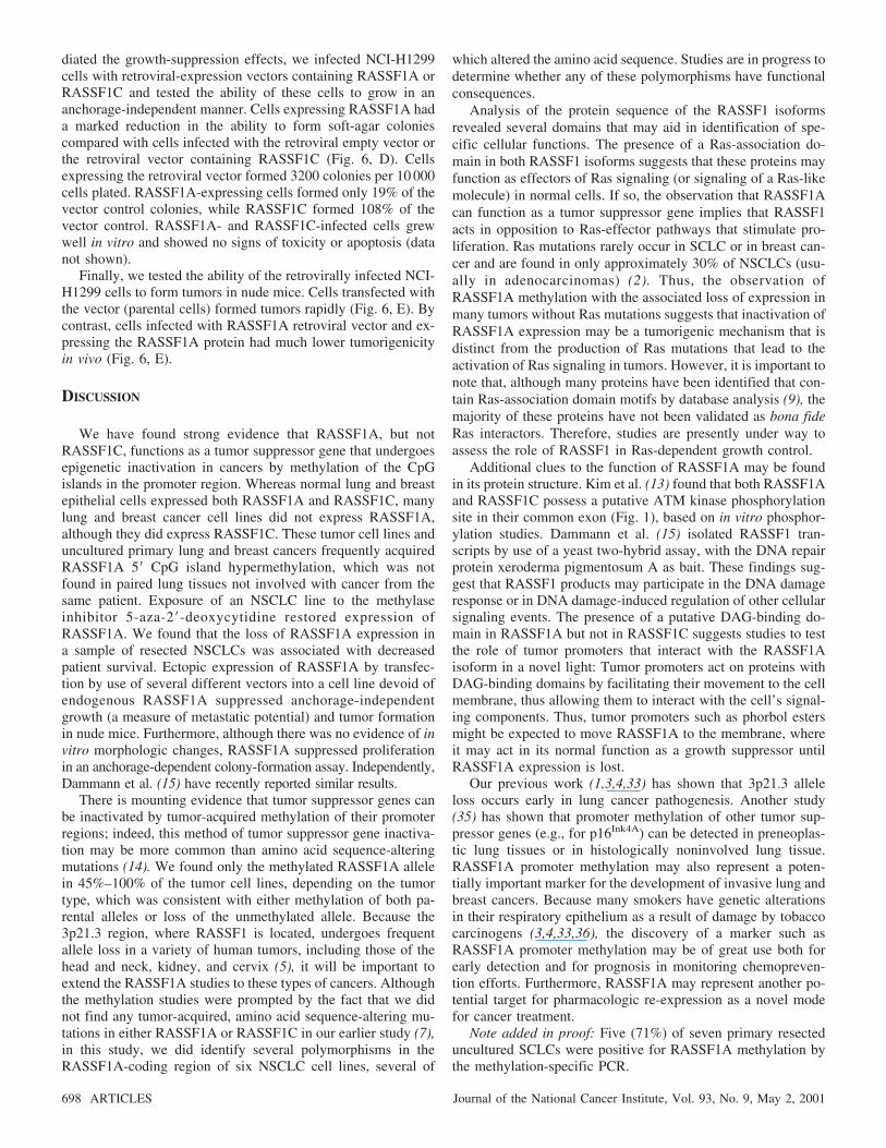
diated the growth-suppression effects, we infected NCI-H1299cells with retroviral-expression vectors containing RASSF1A orRASSF1C and tested the ability of these cells to grow in ananchorage-independent manner. Cells expressing RASSF1A hada marked reduction in the ability to form soft-agar coloniescompared with cells infected with the retroviral empty vector orthe retroviral vector containing RASSF1C (Fig. 6, D). Cellsexpressing the retroviral vector formed 3200 colonies per 10 000cells plated. RASSF1A-expressing cells formed only 19% of thevector control colonies, while RASSF1C formed 108% of thevector control. RASSF1A- and RASSF1C-infected cells grewwell in vitro and showed no signs of toxicity or apoptosis (datanot shown).
Finally, we tested the ability of the retrovirally infected NCI-H1299 cells to form tumors in nude mice. Cells transfected withthe vector (parental cells) formed tumors rapidly (Fig. 6, E). Bycontrast, cells infected with RASSF1A retroviral vector and ex-pressing the RASSF1A protein had much lower tumorigenicityin vivo (Fig. 6, E).
DISCUSSION
We have found strong evidence that RASSF1A, but notRASSF1C, functions as a tumor suppressor gene that undergoesepigenetic inactivation in cancers by methylation of the CpGislands in the promoter region. Whereas normal lung and breastepithelial cells expressed both RASSF1A and RASSF1C, manylung and breast cancer cell lines did not express RASSF1A,although they did express RASSF1C. These tumor cell lines anduncultured primary lung and breast cancers frequently acquiredRASSF1A 5� CpG island hypermethylation, which was notfound in paired lung tissues not involved with cancer from thesame patient. Exposure of an NSCLC line to the methylaseinhibitor 5-aza-2�-deoxycytidine restored expression ofRASSF1A. We found that the loss of RASSF1A expression ina sample of resected NSCLCs was associated with decreasedpatient survival. Ectopic expression of RASSF1A by transfec-tion by use of several different vectors into a cell line devoid ofendogenous RASSF1A suppressed anchorage-independentgrowth (a measure of metastatic potential) and tumor formationin nude mice. Furthermore, although there was no evidence of invitro morphologic changes, RASSF1A suppressed proliferationin an anchorage-dependent colony-formation assay. Independently,Dammann et al. (15) have recently reported similar results.
There is mounting evidence that tumor suppressor genes canbe inactivated by tumor-acquired methylation of their promoterregions; indeed, this method of tumor suppressor gene inactiva-tion may be more common than amino acid sequence-alteringmutations (14). We found only the methylated RASSF1A allelein 45%–100% of the tumor cell lines, depending on the tumortype, which was consistent with either methylation of both pa-rental alleles or loss of the unmethylated allele. Because the3p21.3 region, where RASSF1 is located, undergoes frequentallele loss in a variety of human tumors, including those of thehead and neck, kidney, and cervix (5), it will be important toextend the RASSF1A studies to these types of cancers. Althoughthe methylation studies were prompted by the fact that we didnot find any tumor-acquired, amino acid sequence-altering mu-tations in either RASSF1A or RASSF1C in our earlier study (7),in this study, we did identify several polymorphisms in theRASSF1A-coding region of six NSCLC cell lines, several of
which altered the amino acid sequence. Studies are in progress todetermine whether any of these polymorphisms have functionalconsequences.
Analysis of the protein sequence of the RASSF1 isoformsrevealed several domains that may aid in identification of spe-cific cellular functions. The presence of a Ras-association do-main in both RASSF1 isoforms suggests that these proteins mayfunction as effectors of Ras signaling (or signaling of a Ras-likemolecule) in normal cells. If so, the observation that RASSF1Acan function as a tumor suppressor gene implies that RASSF1acts in opposition to Ras-effector pathways that stimulate pro-liferation. Ras mutations rarely occur in SCLC or in breast can-cer and are found in only approximately 30% of NSCLCs (usu-ally in adenocarcinomas) (2). Thus, the observation ofRASSF1A methylation with the associated loss of expression inmany tumors without Ras mutations suggests that inactivation ofRASSF1A expression may be a tumorigenic mechanism that isdistinct from the production of Ras mutations that lead to theactivation of Ras signaling in tumors. However, it is important tonote that, although many proteins have been identified that con-tain Ras-association domain motifs by database analysis (9), themajority of these proteins have not been validated as bona fideRas interactors. Therefore, studies are presently under way toassess the role of RASSF1 in Ras-dependent growth control.
Additional clues to the function of RASSF1A may be foundin its protein structure. Kim et al. (13) found that both RASSF1Aand RASSF1C possess a putative ATM kinase phosphorylationsite in their common exon (Fig. 1), based on in vitro phosphor-ylation studies. Dammann et al. (15) isolated RASSF1 tran-scripts by use of a yeast two-hybrid assay, with the DNA repairprotein xeroderma pigmentosum A as bait. These findings sug-gest that RASSF1 products may participate in the DNA damageresponse or in DNA damage-induced regulation of other cellularsignaling events. The presence of a putative DAG-binding do-main in RASSF1A but not in RASSF1C suggests studies to testthe role of tumor promoters that interact with the RASSF1Aisoform in a novel light: Tumor promoters act on proteins withDAG-binding domains by facilitating their movement to the cellmembrane, thus allowing them to interact with the cell’s signal-ing components. Thus, tumor promoters such as phorbol estersmight be expected to move RASSF1A to the membrane, whereit may act in its normal function as a growth suppressor untilRASSF1A expression is lost.
Our previous work (1,3,4,33) has shown that 3p21.3 alleleloss occurs early in lung cancer pathogenesis. Another study(35) has shown that promoter methylation of other tumor sup-pressor genes (e.g., for p16Ink4A) can be detected in preneoplas-tic lung tissues or in histologically noninvolved lung tissue.RASSF1A promoter methylation may also represent a poten-tially important marker for the development of invasive lung andbreast cancers. Because many smokers have genetic alterationsin their respiratory epithelium as a result of damage by tobaccocarcinogens (3,4,33,36), the discovery of a marker such asRASSF1A promoter methylation may be of great use both forearly detection and for prognosis in monitoring chemopreven-tion efforts. Furthermore, RASSF1A may represent another po-tential target for pharmacologic re-expression as a novel modefor cancer treatment.
Note added in proof: Five (71%) of seven primary resecteduncultured SCLCs were positive for RASSF1A methylation bythe methylation-specific PCR.
698 ARTICLES Journal of the National Cancer Institute, Vol. 93, No. 9, May 2, 2001

REFERENCES
(1) Wistuba I, Behrens C, Virmani A, Mele G, Milchgrub S, Girard L, et al.High resolution chromosome 3p allelotyping of human lung cancer andpreneoplastic/preinvasive bronchial epithelium reveals multiple, discon-tinuous sites of 3p allele loss and three regions of frequent breakpoints.Cancer Res 2000;60:1949–60.
(2) Sekido Y, Fong KM, Minna JD. Progress in understanding the molecularpathogenesis of human lung cancer. Biochim Biophys Acta 1998;1378:F21–59.
(3) Wistuba II, Lam S, Behrens C, Virmani AK, Fong KM, LeRiche J, et al.Molecular damage in the bronchial epithelium of current and former smok-ers. J Natl Cancer Inst 1997;89:1366–73.
(4) Wistuba II, Behrens C, Milchgrub S, Bryant D, Hung J, Minna JD, et al.Sequential molecular abnormalities are involved in the multistage devel-opment of squamous cell lung carcinoma. Oncogene 1999;18:643–50.
(5) Kok K, Naylor SL, Buys CH. Deletions of the short arm of chromosome3 in solid tumors and the search for suppressor genes. Adv Cancer Res1997;71:27–92.
(6) Wei MH, Latif F, Bader S, Kashuba V, Chen JY, Duh FM, et al. Con-struction of a 600-kilobase cosmid clone contig and generation of a tran-scriptional map surrounding the lung cancer tumor suppressor gene (TSG)locus on human chromosome 3p21.3: progress toward the isolation of alung cancer TSG. Cancer Res 1996;56:1487–92.
(7) Lerman MI, Minna JD. The 630-kb lung cancer homozygous deletionregion on human chromosome 3p21.3: identification and evaluation of theresident candidate tumor suppressor genes. The International Lung CancerChromosome 3p21.3 Tumor Suppressor Gene Consortium. Cancer Res2000;60:6116–33.
(8) Sekido Y, Ahmadian M, Wistuba II, Latif F, Bader S, Wei MH, et al.Cloning of a breast cancer homozygous deletion junction narrows the re-gion of search for a 3p21.3 tumor suppressor gene. Oncogene 1998;16:3151–7.
(9) Schultz J, Milpetz F, Bork P, Ponting CP. SMART, a simple modulararchitecture research tool: identification of signaling domains. Proc NatlAcad Sci U S A 1998;95:5857–64.
(10) Vavvas D, Li X, Avruch J, Zhang XF. Identification of Nore1 as a potentialRas effector. J Biol Chem 1998;273:5439–42.
(11) Hurley JH, Newton AC, Parker PJ, Blumberg PM, Nishizuka Y. Taxonomyand function of C1 protein kinase C homology domains. Protein Sci 1997;6:477–80.
(12) Rogers S, Wells R, Rechsteiner M. Amino acid sequences common torapidly degraded proteins: the PEST hypothesis. Science 1986;234:364–8.
(13) Kim ST, Lim DS, Canman CE, Kastan MB. Substrate specificities andidentification of putative substrates of ATM kinase family members. J BiolChem 1999;274:37538–43.
(14) Baylin SB, Herman JG, Graff JR, Vertino PM, Issa JP. Alterations in DNAmethylation: a fundamental aspect of neoplasia. Adv Cancer Res 1998;72:141–96.
(15) Dammann R, Li C, Yoon JH, Chin PL, Bates S, Pfeifer GP. Epigeneticinactivation of a RAS association domain family protein from the lungtumour suppressor locus 3p21.3. Nat Genet 2000;25:315–9.
(16) Fong K, Biesterveld EJ, Virmani A, Wistuba I, Sekido Y, Bader SA, et al.FHIT and FRA3B 3p14.2 allele loss are common in lung cancer and pre-neoplastic bronchial lesions and are associated with cancer-related FHITcDNA splicing aberrations. Cancer Res 1997;57:2256–67.
(17) Geradts J, Fong KM, Zimmerman PV, Maynard R, Minna JD. Correlationof abnormal RB, p16ink4a, and p53 expression with 3p loss of heterozy-gosity, other genetic abnormalities, and clinical features in 103 primarynon-small cell lung cancers. Clin Cancer Res 1999;5:791–800.
(18) Mountain CF. Revisions in the International System for Staging LungCancer. Chest 1997;111:1710–7.
(19) Phelps RM, Johnson BE, Ihde DC, Gazdar AF, Carbone DP, McClintockPR, et al. NCI-Navy Medical Oncology Branch cell line data base. J CellBiochem Suppl 1996;24:32–91.
(20) Wistuba II, Bryant D, Behrens C, Milchgrub S, Virmani AK, Ashfaq R, etal. Comparison of features of human lung cancer cell lines and their cor-responding tumors. Clin Cancer Res 1999;5:991–1000.
(21) Gazdar AF, Kurvari V, Virmani A, Gollahon L, Sakaguchi M, WesterfieldM, et al. Characterization of paired tumor and non-tumor cell lines estab-lished from patients with breast cancer. Int J Cancer 1998;78:766–74.
(22) Sambrook J, Fritsch E, Maniatis T, editors. Molelcular cloning: a labora-tory manual. Cold Spring Harbor (NY): Cold Spring Harbor Laboratory; 1989.
(23) Orita M, Iwahana H, Kanazawa H, Hayashi K, Sekita T. Detection ofpolymorphisms of human DNA by gel electrophoresis as single-strandconformation polymorphisms. Proc Natl Acad Sci U S A 1989;86:2766–70.
(24) Herman JG, Graff JR, Myohanen S, Nelkin BD, Baylin SB. Methylation-specific PCR: a novel PCR assay for methylation status of CpG islands.Proc Natl Acad Sci U S A 1996;93:9821–6.
(25) Zochbauer-Muller S, Fong KM, Virmani AK, Geradts J, Gazdar AF, MinnaJD. Aberrant promoter methylation of multiple genes in non-small cell lungcancers. Cancer Res 2001;61:249–55.
(26) Clark SJ, Harrison J, Paul CL, Frommer M. High sensitivity mapping ofmethylated cytosines. Nucleic Acids Res 1994;22:2990–7.
(27) Tanaka H, Shimada Y, Harada H, Shinoda M, Hatooka S, Imamura M, etal. Methylation of the 5� CpG island of the FHIT gene is closely associatedwith transcriptional inactivation in esophageal squamous cell carcinomas.Cancer Res 1998;58:3429–34.
(28) Chen JY, Funk WD, Wright WE, Shay JW, Minna JD. Heterogeneity oftranscriptional activity of mutant p53 proteins and p53 DNA target se-quences. Oncogene 1993;8:2159–66.
(29) Claudio PP, Howard CM, Pacilio C, Cinti C, Romano G, Minimo C, et al.Mutations in the retinoblastoma-related gene RB2/p130 in lung tumors andsuppression of tumor growth in vivo by retrovirus-mediated gene transfer.Cancer Res 2000;60:372–82.
(30) Gao B, Sekido Y, Maximov A, Saad M, Forgacs E, Latif F, et al. Func-tional properties of a new voltage-dependent calcium channel alpha(2)deltaauxiliary subunit gene (CACNA2D2). J Biol Chem 2000;275:12237–42.
(31) Smith DB, Johnson KS. Single-step purification of polypeptides expressedin Escherichia coli as fusions with glutathione S-transferase. Gene 1988;67:31–40.
(32) Kaplan E, Meier P. Nonparametric estimation from incomplete observa-tions. J Am Stat Assoc 1958;53:457–81.
(33) Wistuba II, Barry J, Behrens C, Maitra A, Shivapurkar N, Milchgrub S, etal. Molecular changes in the bronchial epithelium of patients with small celllung cancer. Clin Cancer Res 2000;6:2604–10.
(34) Unger T, Nau MM, Segal S, Minna JD. p53: a transdominant regulator oftranscription whose function is ablated by mutations occurring in humancancer. EMBO J 1992;11:1383–90.
(35) Belinsky SA, Nikula KJ, Palmisano WA, Michels R, Saccomanno G, Gab-rielson E, et al. Aberrant methylation of p16(INK4a) is an early event inlung cancer and a potential biomarker for early diagnosis. Proc Natl AcadSci U S A 1998;95:11891–6.
(36) Park IW, Wistuba II, Maitra A, Milchgrub S, Virmani AK, Minna JD, et al.Multiple clonal abnormalities in the bronchial epithelium of patients withlung cancer. J Natl Cancer Inst 1999;91:1863–8.
NOTES
Supported by Public Health Service grants CA71618, CA71443, andP50CA70907 and contract N01CO56000 (to M. I. Lerman and F. Latif) from theNational Cancer Institute, National Institutes of Health, Department of Healthand Human Services; by the Early Detection Research Network for the breastcancer portion; by the G. Harold and Leila Y. Mathers Charitable Foundation;by grants J1658-MED and J1860-MED from the Austrian Science Foundation(to S. Zochbauer-Muller); by the Association For International Cancer Research,Cancer Research Campaign, Fundacao para a Cientia e a Technologia (to F.Latif); and by grants from the Swedish Cancer Society, Karolinska Institute,Stockholm, and by the Royal Swedish Academy of Science (to E. Zabarovsky).
The content of the publication does not necessarily reflect the views or poli-cies of the Department of Health and Human Services nor does mention of tradenames, commercial products, or organizations imply endorsement by the U.S.Government.
Manuscript received August 21, 2000; revised February 5, 2001; acceptedFebruary 26, 2001.
Journal of the National Cancer Institute, Vol. 93, No. 9, May 2, 2001 ARTICLES 699
Copyright © 2022 FDOKUMEN