Metabolomic, Transcriptional, Hormonal, and Signaling Cross-Talk in Superroot2
Upload
independentCategory
view
0download
0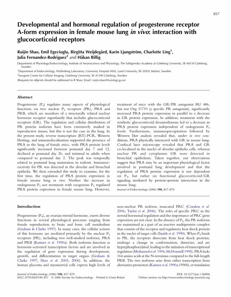
857
Developmental and hormonal regu
lation of progesterone receptorA-form expression in female mouse lung in vivo: interaction withglucocorticoid receptorsRuijin Shao, Emil Egecioglu, Birgitta Weijdegard, Karin Ljungstrom, Charlotte Ling1,
Julia Fernandez-Rodriguez2 and Hakan Billig
Department of Physiology/Endocrinology, Institute of Neuroscience and Physiology, The Sahlgrenska Academy at Goteborg University, SE-40530 Goteborg,Sweden
1Department of Endocrinology, Wallenberg Laboratory, University Hospital MAS, Lund University, SE-20502 Malmo, Sweden2Swegene Centre for Cellular Imaging, Goteborg University, SE-41390 Goteborg, Sweden
(Requests for offprints should be addressed to R Shao; Email: [email protected])
Abstract
Progesterone (P4) regulates many aspects of physiological
functions via two nuclear P4 receptors (PR), PRA and
PRB, which are members of a structurally related nuclear
hormone receptor superfamily that includes glucocorticoid
receptors (GR). The regulation and cellular distribution of
PR protein isoforms have been extensively studied in
reproductive tissues, but this is not the case in the lung. In
the present study, reverse transcriptase (RT)-PCR, Western
blotting, and immunolocalization supported the presence of
PRA in the lung of female mice, with PRA protein levels
significantly increased between postnatal day 7 and 12,
declined at postnatal day 26, and minimal in adults when
compared to postnatal day 2. The peak was temporally
related to postnatal lung maturation in rodents. Immunor-
eactivity for PR was detected in the alveolar and bronchial
epithelia. We then extended this study to examine, for the
first time, the regulation of PRA protein expression in
female mouse lung in vivo. Neither the increase in
endogenous P4 nor treatment with exogenous P4 regulated
PRA protein expression in female mouse lung. However,
Journal of Endocrinology (2006) 190, 857–8700022–0795/06/0190–857 q 2006 Society for Endocrinology Printed in Great
treatment of mice with the GR/PR antagonist RU 486,
but not Org 31710 (a specific PR antagonist), significantly
increased PRA protein expression in parallel to a decrease
in GR protein expression. In addition, treatment with the
synthetic glucocorticoid dexamethasone led to a decrease in
PRA protein expression independent of endogenous P4levels. Furthermore, immunoprecipitation followed by
Western blot analysis revealed that, under in vivo con-
ditions, PRA physically interacted with GR in mouse lung.
Confocal laser microscopy revealed that PRA and GR
co-localized in the nuclei of alveolar epithelia cells, whereas
nuclear PR and cytoplasmic GR were detected in
bronchial epithelium. Taken together, our observations
suggest that PRA may be an important physiological factor
involved in postnatal lung development and that the
regulation of PRA protein expression is not dependent
on P4, but rather on functional glucocorticoid/GR
signaling mediated by protein–protein interaction in the
mouse lung.
Journal of Endocrinology (2006) 190, 857–870
Introduction
Progesterone (P4), an ovarian steroid hormone, exerts diverse
functions in several physiological processes ranging from
female reproduction to brain and bone cell metabolism
(Graham & Clarke 1997). In many cases, the cellular actions
of this hormone are mediated primarily by the nuclear P4receptors (PR), including two well-studied isoforms, PRA
and PRB (Kastner et al. 1990a). Both isoforms function as
hormone-activated transcription factors and are involved in
the regulation of gene expression during development,
growth, and differentiation in target organs (Graham &
Clarke 1997, Shao et al. 2003, 2006). In addition, the
human placenta and myometrial cells express high levels of
non-nuclear PR isoform, truncated PRC (Condon et al.
2006, Taylor et al. 2006). The roles of specific PRC in the
steroid hormonal regulation and the importance of PRC gene
expression are not clear. In the absence of P4, the PR isoforms
are maintained as a part of an inactive multiprotein complex
that consists of the receptor and regulatory heat shock protein
in the nuclei of target cells (Smith et al. 1990). When P4 binds
to PR, the receptors dissociate from heat shock proteins,
undergo a change in conformation, dimerize, and are
hyperphosphorylated, leading to the initiation of transcriptional
regulation (Mohamed et al. 1994,McDonnell 1995). PRA lacks
164 amino acids at the N-terminus compared to the full-length
PRB. The two isoforms arise from either transcription from
alternative promoters (Kastner et al. 1990a,1990b), or translation
DOI: 10.1677/joe.1.06896Britain Online version via http://www.endocrinology-journals.org
R SHAO and others $ PRA interacts with GR in female mouse lung858
from alternative sites in the samemRNA (Conneely et al. 1987,
Giangrande &McDonnell 1999). The relative expression of the
twoPRisoforms is conserved in rodents andhumans (Schneider
et al. 1991, Kraus et al. 1993, Giangrande &McDonnell 1999),
but varies dramatically between different tissues, cell types,
physiological, and disease states, and is under developmental
control (Ilenchuk & Walters 1987, Schneider et al. 1991,
Graham & Clarke 1997, Bethea & Widmann 1998). Although
both isoforms have similar hormone- and DNA-binding
affinities, and form homo- and/or hetero-dimers (Lessey et al.
1983, Kastner et al. 1990a), several in vitro studies have shown
that PRA and PRB display different transcriptional effects on
progestin-responsive promoters in a cell- and promoter context-
dependent manner (Vegeto et al. 1993, Wen et al. 1994,
Giangrande & McDonnell 1999). For instance, PRA is often
transcriptionally inactive and acts as a potent dominant repressor
of PRB-mediated transcription (Tung et al. 1993). Further-
more, PRA has been found to diminish the activities of other
nuclear receptors such as the glucocorticoid receptor (GR)
under some conditions (Vegeto et al. 1993), whereas PRB is
shown to activate transcription in several target tissues
(McDonnell & Goldman 1994, McDonnell et al. 1994, Wen
et al. 1994). Recently, studies in PR knockout mice confirmed
that the two isoforms are not functionally equivalent and even
imply tissue-selective isoform activity in vivo (Curtis & Korach
2000, Mulac-Jericevic et al. 2000). Thus, it is conceivable that
the differential expression of PR protein isoforms in certain
target tissuesmay influence the action of P4. Several studies have
demonstrated the potential for differential steroid receptor
interactions at the level of transcription in numerous cell lines
and tissue types in vitro (Uht et al. 1997, Kinyamu & Archer
2003). However, much less work has been done on the
interaction between PR and other steroid receptors in vivo.
Although previous data suggest that P4 might be important
in lung physiology and diseases (e.g. asthma and cancer)
(Milne 1979, Haggerty et al. 2003, Ishibashi et al. 2005), the
regulation and cellular distribution of PR protein isoforms in
lung remain largely unknown. Previous studies have shown
that PR mRNAs and proteins are found in rabbit
(Giannopoulos et al. 1982, Bullock et al. 1987, Nielsen et al.
1987, Camacho-Arroyo et al. 1994, 1998), rodent (Moser &
Daxenbichler 1982, Beyer et al. 2003), and chicken (Pasanen
et al. 1997) lungs. In the present study, we sought to initially
establish the presence of PR isoforms in mouse lung using
reverse transcriptase (RT)-PCR and Western blotting
analyses and on finding that PRA mRNA and protein were
specifically expressed in mouse lung, we examined the
possibility of regulation of PRA protein expression in mice
during postnatal growth and development. It is generally
accepted that P4’s actions on target tissues are mediated
primarily by binding to its cognate nuclear receptors.
Therefore, we examined the steroid hormone regulation of
PRA protein expression in female mouse lung by different
experimental approaches, including treatment with gonado-
tropins, P4, dexamethasone, and PR and/or GR antagonists.
Furthermore, we investigated whether under in vivo
Journal of Endocrinology (2006) 190, 857–870
conditions, PRA physically interacted with GR in mouse
lung by using immunoprecipitation followed by Western blot
and double-immunofluorescent assays.
Materials and Methods
Hormones and chemicals
Equine gonadotropin (eCG), dexamethasone, monoclonal
anti-b-actin, and alkaline phosphatase-conjugated goat-anti-
mouse immunoglobulin were obtained from Sigma. Human
chorionic gonadotropin (hCG) and the specific PR-antago-
nist Org 31710 (Kloosterboer et al. 1994) were provided by N
V Organon (Oss, Holland). The PR/GR antagonist RU486
(Cadepond et al. 1997) was provided by Exelgyn (Paris,
France). Rabbit polyclonal anti-PR (c-19 and c-20) and their
respective blocking peptides (c-19p and c-20p), as well as
mouse monoclonal anti-GR were obtained from Santa Cruz
Biotechnologies, Inc. (Santa Cruz, CA, USA). Mouse
monoclonal anti-PRB ( Joensuu et al. 1990) was obtained
from Affinity BioReagents, Inc. (ABR, Golden, CO, USA).
Mouse monoclonal anti-proliferating cell nuclear antigen
(PCNA) was obtained from Novocastra Laboratories Ltd
(Newcastle upon Tyne, UK). Normal rabbit serum and
normal goat serum were obtained from DAKO Corp.
(Carpinteria, CA, USA). Alkaline phosphatase conjugated
goat-anti-rabbit immunoglobulin was obtained from Tropix
(Bedford, MA, USA). Biotin-SP-conjugated donkey anti-
rabbit IgG, fluorescein (DTAF)-conjugated streptavidin, and
Cy 3-conjugated donkey anti-mouse IgG were purchased
from Jackson ImmunoResearch Laboratories, Inc. (West
Grove, PA, USA). All other chemicals were purchased from
Merck, unless otherwise specified.
Animals and experimental design
Timed pregnant femalemice (C57BL/6) were purchased from
Taconic M&B, Copenhagen, Denmark. They were mon-
itored daily between 0900 and 0930 h for delivery of pups and
the day of birth was defined as postnatal day (PND) 0. Pups
were sexed according to ano-genital distance and male pups
were removed. All animals were housed and acclimatized to
controlled temperature (21G2 8C) and with a ratio of 12 h
light:12 h darkness cycles, with the standard rodent chow and
tap water ad libitum. The animal studies were reviewed and
approved by the ethics committee at Goteborg University,
Sweden. At least five animals in each groupwere used for every
experimental approach unless otherwise specified.
Experiment 1 This experiment was designed to determine
the presence of the two PR isoforms (PRA and PRB) in
mouse lung. Mice were killed following deep anesthetization
with pentobarbitone sodium and selected whole tissues (lung,
liver, fallopian tube, and uterus) were carefully removed and
www.endocrinology-journals.org
PRA interacts with GR in female mouse lung $ R SHAO and others 859
lung tissue was frozen in liquid nitrogen for subsequent
Western blot or RT-PCR analysis.
Experiment 2 This experiment was designed to examine
the regulation and cellular localization of PRA protein
expression in female mouse lung during postnatal growth and
development. Female mice were deeply anesthetized with
pentobarbitone sodium at various time intervals (PND 2, 7,
12, 26, and 90) between 0800 and 1000 h, whole lung tissue
isolated from three mice at each time point were quickly
dissected and either frozen in liquid nitrogen or stored at
K135 8C, or fixed in 4% formaldehyde in neutral buffer
(Sigma) for 24 h at 4 8C and embedded in paraffin wax for
either Western blot or immunohistochemical analysis.
Experiment 3 This experiment was designed to examine the
effects of endogenous or exogenous P4 on regulation of PRA
protein expression in female mouse lung. In our experiments,
sequential administration of eCG and hCG is substituted for
effects of follicle-stimulating hormone and luteinizing hor-
mones in vivo. This in vivomodel has been used for induction of
ovarian P4 production and further elevates serum P4 levels in
female mice in vivo (Shao et al. 2003, 2006). Female mice (PND
26) were given a single injection of eCG (5 IU, i.p.) followed by
treatment with hCG (5 IU i.p.) after 48 h. Six hours after hCG
treatment, mice were killed by approved methods and tissues
were dissected. To determine the specific effect of P4 on
regulation of PR protein expression, female mice (PND 26)
were randomly selected and received a single i.p. injection of P4(4 mg/kg per body weight in 100 ml sesame oil). A single
pharmacological dose of P4 was chosen on the basis of previous
studies (Russo et al. 2001, Shao et al. 2006). Lung tissue was
obtained at different time intervals (6, 24, 36, 48, and 72 h) for
Western blot analysis.
Experiment 4 This experiment was designed to examine
the effects of PR/GR antagonist RU 486 and PR antagonist
Org 31710 on the regulation of PRA and GR protein
expression in female mouse lung. Thirty female mice (PND
26), chosen at random, received eCG (5 IU, i.p.). The animals
were then randomly divided into six groups of five mice for
each group. Mice in the first two groups were given either
2 mg of RU 486 or 1 mg Org 31710 in 100 ml sesame oil by
i.p. injection after treatment with eCG for 44 h. Mice in the
remaining groups were injected with either 100 ml sesame oil
(two groups, as controls), 2 mg RU 486 or 1 mg Org 31710
dissolved in 100 ml sesame oil after treatment with eCG for
48 h. All mice in the three groups received hCG (5 IU, i.p.) at
48 h after eCG treatment. Six hours after hCG treatment, the
mice were killed by approved methods. Lung tissue was frozen
in liquid nitrogen for subsequent Western blot analysis.
Experiment 5 This experiment was designed to study
the effect of glucocorticoids on the regulation of PRA and
GR expression in female mouse lung. Female mice (PND 26)
were treated with gonadotropins and/or dexamethasone,
www.endocrinology-journals.org
as described in Experiments 3 and 4. Animals were given a
single i.p. injection of dexamethasone (600 mg/kg per body
weight) dissolved in saline, chosen on the basis of a previous
study (Russo et al. 2001). Mice were killed by approved
methods, lung tissues removed and frozen in liquid nitrogen
for Western blot analysis.
RT-PCR
Total RNA was isolated from individual tissues using Trizol
Reagent (Life Technologies) according to the manufacturer’s
instructions.Thequantityof extractedRNAwas determined by
spectrophotometry. Any contaminating genomic DNA was
digested by RNase-free DNase. cDNA was reversely tran-
scribed from 5 mg total RNA using 0$25 mg random hexamer
(LifeTechnologies) in 20 ml of a 1!AMV-RTbuffer containing
9 U AMV-RT (Invitrogen), 39 U ribonuclease inhibitor
(RNasin) (Promega), and 4 ml 5 mM deoxyNTP (dNTP)
mixtures (Amersham). Reactions were incubated for 1 h at
42 8C, heated to 95 8C for 5 min and then chilled on ice. An
aliquot (5 ml) ofRTproductwas used forPCRamplification in a
total volume of 50 ml PCR-buffer presented with 2$5 U Taq-
DNA polymerase (Promega), 2 ml 5 mM dNTP mixtures and
the appropriate primer pairs as mentioned below. The PCR
consisted of a denaturing step at 94 8C for 5 min. The thermal
cycle profile used in this study was (1) denaturing at 94 8C for
30 s, (2) annealing the primers at 55 8C for 30 s, (3) extending
the primers at 72 8C for 2 min. After the final amplification, the
reactionwas elongated for an additional 7 min at 72 8C. PRA/B
and PRB were amplified after 30 or 39 cycles respectively. The
amount of cDNA and the number of PCR cycles needed to
detect PR were determined in a preliminary experiment. A
portion (10 ml) of the PCR mixture was electrophoresed in
1$2% agarose gel along with a 100 bp DNA ladder (Life
Technologies) and visualized by ethidium bromide staining.
Specificity of amplified products was confirmed by including
RT control reactions. Ribosomal protein L19was selected as an
internal control to confirm that cDNA had been synthesized
and to check for genomic DNA contamination (Chan et al.
1987). PCR was performed using primers specific for mouse
PRA/B (sense 5 0-GGCAAATCCCACAGGAGTTTG-3 0,
antisense 50-AGACATCATTTCCGGAAATTC-30; expected
PCR-amplified product size, 327 bp) (Beyer et al. 2002);mouse
PRB (sense 5 0-ATGACTGAGCTGCAGGCAAAG-30, anti-
sense 5 0-CTTCTACCCCAGAGAAAGCGC-3 0; expected
PCR-amplified product size, 277 bp) (Beyer et al. 2002);
ribosomal protein L19 (sense 5 0-GAAATCGCCAATGC-
CAACTC-30, antisense 50-ACCTTCAGGTACAGGCTG-
TG-30, 290 bp) (Robker et al. 2000).
Western blot and co-immunoprecipitation analyses
Protein extraction and Western blotting were performed
according to standard methods as described previously in
Shao et al. (2003). Equal amounts of protein in the cell
extracts were pretreated with 4! SDS before loading and
Journal of Endocrinology (2006) 190, 857–870
R SHAO and others $ PRA interacts with GR in female mouse lung860
resolved by 4–12% SDS-polyacrylamide gels (Novex, San
Diego, CA, USA) under reducing conditions. The separated
samples were electrotransferred to polyvinyldifluoride mem-
branes (Amersham) and treated with blocking buffer (0$2%I-Block, 0$2% BSA, 5 mM MgCl2, 3 mM NaN3, and 0$3%Tween 20 in PBS, pH 7$4) for 2 h. The antibodies used to
detect PR in this study were raised against human PRs and
recognized ligand-binding domain (PR c-19) or DNA-
binding domain (PR c-20) of PR in rodent tissues byWestern
blotting and immunohistochemical analyses. The membranes
were incubated with primary antibody for either anti-PRA/B
(c-19Cc-20, 1:250), anti-PRB (1:50), anti-PCNA (1:1000),
anti-b-actin (1:1000), or anti-GR (1:500) in blocking buffer
overnight at 4 8C. After washing in blocking buffer for 2 h,
the membranes were incubated with alkaline phosphatase-
linked secondary antibody (polyclonal secondary antibody at
1:40 000 or monoclonal secondary antibody at 1:80 000
dilutions respectively) in blocking buffer for 2 h with gentle
shaking and detected using CDP-Star as substrate (Tropix,
Bedford, MA, USA). Immunoblotted signals were exposed,
developed on ECL-film (Amersham) and subsequently
scanned into a computer. Individual bands were quantified
directly from membranes by densitometry using ImageQuant
(version 5$0) software program (Molecular Dynamics, Inc.,
Sunnyvale, CA, USA). Equal protein loading was determined
by the expression of b-actin, as well as confirmed by staining
the gels with Coomassie blue after the transfer. As a negative
control, the two PR antibodies were pre-absorbed with
tenfold excess of neutralizing synthetic PR peptides for 4 h at
room temperature before use to demonstrate antigen
specificity.
For co-immunoprecipitation experiments, tissues were
extracted with ice-cold lysis buffer (25 mM Tris–HCl,
pH 8$0, 150 mM NaCl, 0$5% Nonidet P-40, 1% SDS,
200 mM sodium deoxycholate, 1 mM dithiothreitol (DTT),
5 mM EDTA, 0$5 mM phenylmethylsulphonyl fluoride
(PMSF), 10 mM N-ethylmaleimide, 10 mM iodoacetamide,
and a cocktail of protease inhibitors (Roche). Either anti-PR
or anti-GR antibodies were added to 500 mg protein extracts
and incubated for 4 h at room temperature. Immune
complexes were obtained by the addition of 50 ml Pansorbincells (Calbiochem, San Diego, CA, USA). The resulting
immobilized immune complexes were washed in RIRA
buffer (50 mM Tris–HCl, pH 7$8, 150 mM NaCl, 15 mM
MgCl2, 0$5% Nonidet P-40, 0$3% Triton X-100, 0$5%sodium deoxycholate, 5 mM EDTA, 1 mM DTT, and a
cocktail of protease inhibitors). The bound protein was eluted
by boiling in 30 ml SDS sample reducing loading buffer
(Novex) for 5 min. Immunoprecipitated complexes for either
GR or PRA/B were loaded in the same gels, which were run
at the same time within the same electrophoresis unit and
examined by immunoblotting as described above.
To re-probe the blot with another antibody, the blot was
re-hydrated in methanol, rinsed and incubated with stripping
buffer (62$5 mM Tris–HCl, 2% SDS and 100 mM
b-mercaptoethanol, pH 6$8) at 50 8C for 30 min.
Journal of Endocrinology (2006) 190, 857–870
Immunohistochemistry and microscopy
PR immunolabeling was undertaken essentially as described
previously (Shao et al. 2003, 2006). Paraffin-embedded lung
sections were deparaffinized in xylene and rehydrated through
graded series of ethanol and water. Antigens were retrieved by
10 mM sodium citrate buffer. After three washes with Tris-
buffered saline (TBS 50 mM Tris, 0$9% NaCl, pH 7$5),endogenous peroxidase activity was abolished by 3%
hydrogen peroxide in TBS and non-specific binding was
blocked by incubating sections in 10% normal goat serum.
Sections were incubated with primary antibodies (anti-
PRA/B (c-19Cc-20) at 1:100, and anti-PRB at 1:50
dilutions respectively) in TBS containing 1% BSA overnight
at 4 8C in a humidified chamber. After a series of washing
with TBS, sections were then stained using the avidin-
biotinylated-peroxidase complex detection system (ABC kit,
Vector Laboratories, Inc., Burlingame, CA, USA) according
to the manufacturer’s instructions. Immunostaining was then
visualized using 3, 3-diaminobenzidine tetrahydrochloride
(0$5 mg/ml in PBS and 0$01% H2O2, pH 7$6) for 10 min.
Slides were viewed on Nikon E-1000 microscope (Bergstrom
Instruments AB, Stockholm, Sweden) under brightfield
optics, and photomicrographed using Easy Image 1 (Berg-
strom Instrument AB, Goteborg, Sweden). To determine the
specificity for detection of PR, the primary antibodies (c-
19Cc-20, 1:250 each) were pre-incubated with tenfold
excess of neutralizing synthetic PR peptides (c-19 pCc-20 p)
for 4 h at room temperature. These pre-absorbed antibodies
were substituted for primary antibodies, while adjacent
sections were incubated with PR primary antibodies alone.
Serial sections of lung were examined blind under light
microscopy.
Double-immunofluorescent staining of PR and GR was
performed according to a previously described procedure
(Shao et al. 2004). Sections were incubated with a mixture of
antibodies against PR and GR diluted to 1:100 and 1:150
respectively, in PBS containing 1% BSA and 5% fat-free milk.
Each primary antibody was serially diluted to optimize the
concentration used to achieve maximum sensitivity and
specificity. Immunodetection was accomplished using the
species-specific secondary antibodies, either a biotin-con-
jugated anti-rabbit antibody together with streptavidin
conjugated with DTAF or Cy 3-conjugated anti-mouse
antibody. Sections were washed and mounted with Fluor-
escent Mounting Media (DAKO). Slides were viewed on an
Axiovert 200 microscope (Zeiss, Jena, Germany) equipped
with a laser-scanning confocal imaging LSM 510 META
system (Carl Zeiss, Jena, Germany) and photomicrographed.
The resulting stain was evaluated blind by three observers. The
immunohistochemical figures illustrated are representative of
those observed in numerous sections from multiple animals.
Serum P4 assay
Serum P4 concentration was measured by RIA according to a
protocol provided by the manufacturer (Perkin–Elmer Life
www.endocrinology-journals.org
PRA interacts with GR in female mouse lung $ R SHAO and others 861
and Analytical Sciences, Wallac Oy, Turku, Finland) (Shao
et al. 2006). Trunk blood was obtained from cardiac puncture
in immature mice treated with or without gonadotropins, PR
antagonists, and dexamethasone. All samples were tested as
duplicates together in one run. The sensitivity of the assay was
typically better than 0$8 nmol/l, and the intra-assay
coefficient of variation was 3$3–7$3%.
Statistical analysis
Results are presented as the meanGS.E.M. Statistical analysis
was performed by one-way ANOVA using Analyse-It
program (Analyse-It Software, Ltd, Leeds, UK). Significant
differences between the treatment groups were determined by
the post hoc Tukey’s test. Effects were considered to be
significant if P value was less than 0$05.
Results
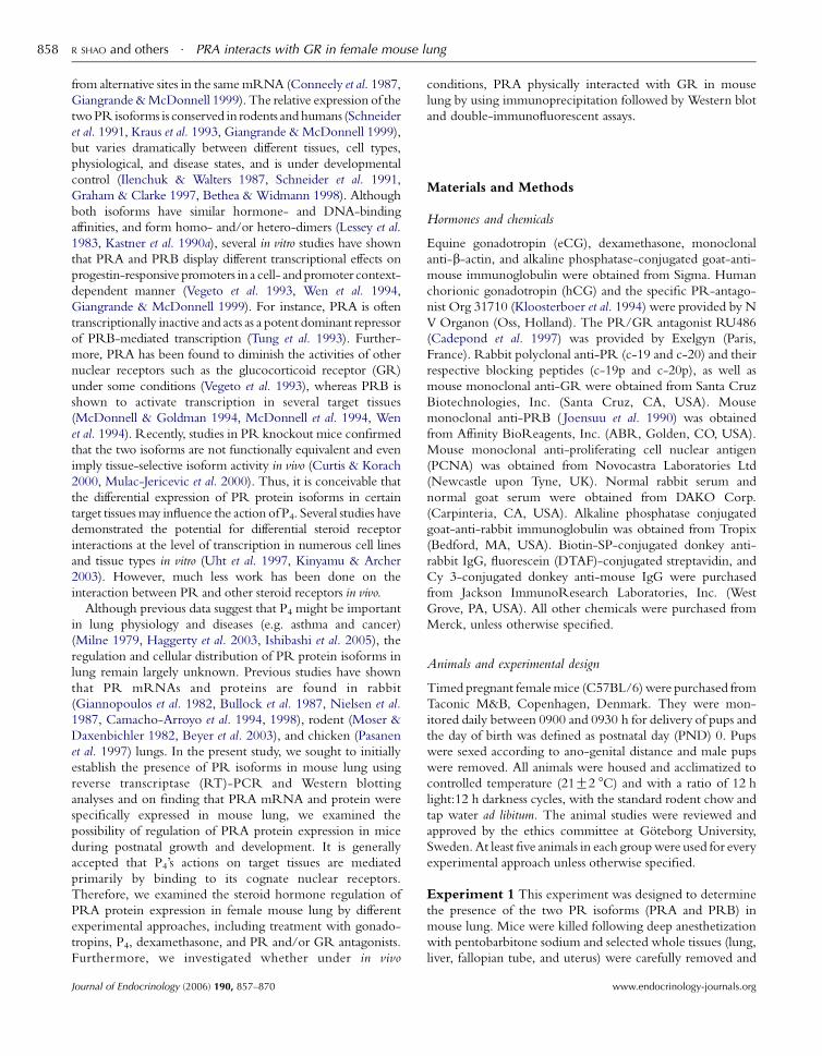
PRA is the predominant PR in mouse lung
PRA/BmRNAwas detected in female mouse lung at PND 7
and 26, whereas PRB transcripts in the same lung tissues
PND
7 lu
ng (
1)
PND
7 lu
ng (
2)
PND
26
lung
(1)
PND
26
lung
(2)
191
97
MW (kDa)
PRA/B (327bp)
PND
7 lu
ng
PND
26
lung
Ute
rus
Liv
er
L-19 (290bp)
PRB (277bp)
MW (1
A B
C
Figure 1 Expression of PR isoforms in mousefemale mouse lungs was reversely transcribed afor mouse PRA and PRB as well as L-19 (as descproducts were resolved on 1$5% agarose gelsstaining. A mouse uterus-positive control and aincluded. Similar analysis with two different Rtissues (nZ3 each). This figure is representativeresults were obtained. (B) and (C) Protein sampselected tissues (nZ3 each). The equal proteinanalysis with either anti-PRA/B (B) or anti-PRBMethods. The immunoblots are representativeexperiments from both female and male mice.indicated on the left. X denotes protein band thPR peptide-PR antibodies. PND, postnatal day
www.endocrinology-journals.org
were absent (Fig. 1A). In parallel with PCR analysis, we used
the same selected tissues for Western blot analysis to
demonstrate PR protein isoform expression, with only
PRA protein expression found in mouse lung, at PND 7
and 26 (Fig. 1B). Male mice at PND 26 also displayed similar
levels of PRA protein expression in lung (data not shown).
Furthermore, we used a specific PRB antibody to confirm
this result under PRA/B antibodies, and no PRB immuno-
reactive band was detected in all lung tissues examined, while
expression of PRB was observed in mouse fallopian tube and
uterus (Fig. 1C) (Shao et al. 2006).
Developmental regulation of PRA protein expression in femalemouse lung
To determine whether PRA protein levels are regulated
during postnatal development in female mouse lung, we
examined PRA protein expression by Western blot analysis.
PRA immunoreactive protein was detected in all lung tissue
extracts isolated from female mice of different ages. Levels of
PRA protein increased with age, peaking at PND 7, although
significantly increased levels of PRA protein were also
Liver
Fallo
pian
tube
Ute
rus
PRB(115 kDa)
kDa)91
97
64
PND
7 lu
ng
PND
26
lung
Liv
er
Ute
rus
PRB (115 kDa)
PRA (83 kDa)
X
lung. (A) Total RNA from the indicatednd amplified by PCR with primers specificribed in Materials and Methods). Amplifiedand visualized by ethidium bromide
mouse liver-negative control were alsoNA preparations from different animalof two separate experiments in which sameles were isolated from mouse lungs and(50 mg/lane) underwent Western blotantibody as described in Materials andof duplicate blots of two independentThe molecular mass markers (kDa) are
at cross-reacted with neutralizing synthetics.
Journal of Endocrinology (2006) 190, 857–870
R SHAO and others $ PRA interacts with GR in female mouse lung862
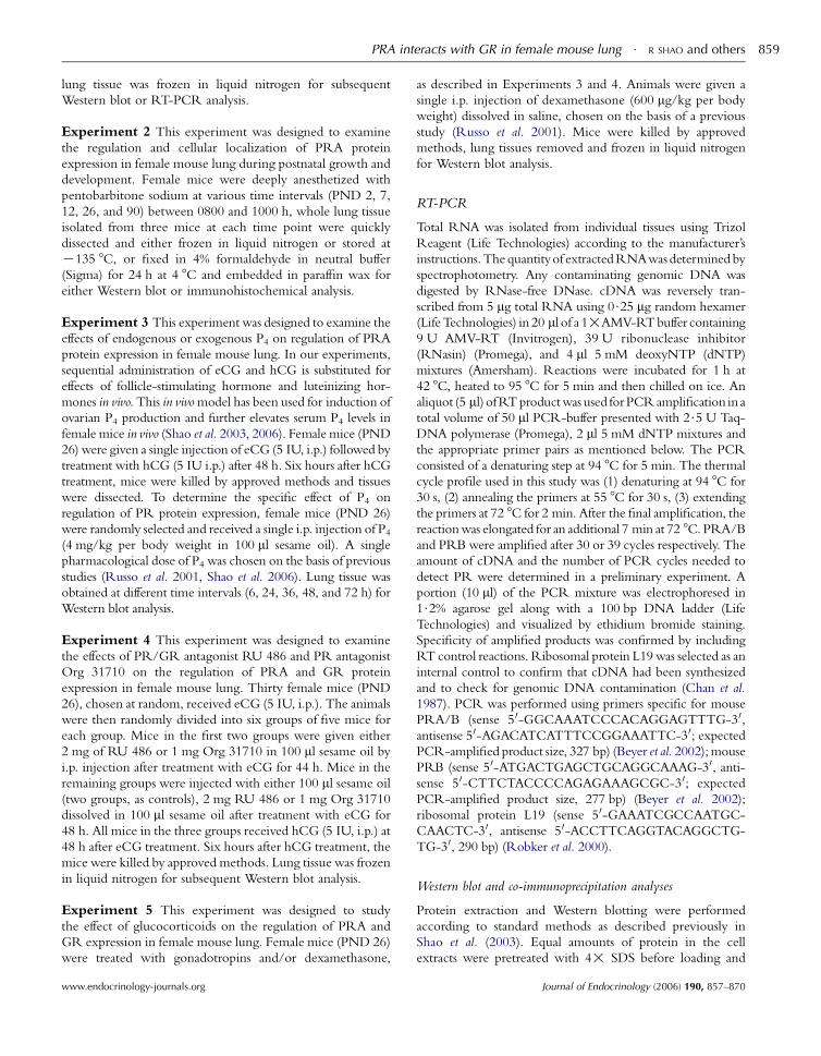
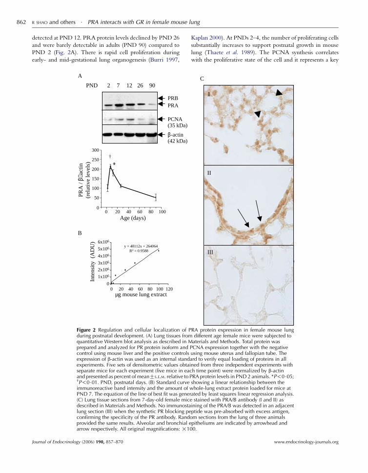
detected at PND 12. PRA protein levels declined by PND 26
and were barely detectable in adults (PND 90) compared to
PND 2 (Fig. 2A). There is rapid cell proliferation during
early- and mid-gestational lung organogenesis (Burri 1997,
A
B
y = 48112x + 264064 R2 = 0.9588
0
1x10⁶
2x10⁶
3x10⁶
4x10⁶
5x10⁶
6x10⁶
µg mouse lung extract
Inte
nsity
(A
DU
)
PRA
/ β−
actin
(rel
ativ
e le
vels
)
0 20 40 60 80 100
0 20 40 60 80 100 120
0
50
100
150
200
250
300
Age (days)
*
PRBPRA
PCNA(35 kDa)
β-actin(42 kDa)
PND 2 7 12 26 90
Figure 2 Regulation and cellular localization of Pduring postnatal development. (A) Lung tissues fromquantitative Western blot analysis as described in Mprepared and analyzed for PR protein isoform andcontrol using mouse liver and the positive controlsexpression of b-actin was used as an internal standexperiments. Five sets of densitometric values obtaiseparate mice for each experiment (five mice in eacand presented as percent of meanGS.E.M. relative to P†P!0$01. PND, postnatal days. (B) Standard curveimmunoreactive band intensity and the amount of wPND 7. The equation of the line of best fit was gener(C) Lung tissue sections from 7-day-old female micdescribed in Materials and Methods. No immunostalung section (III) when the synthetic PR blocking peconfirming the specificity of the PR antibody. Randprovided the same results. Alveolar and bronchial earrow respectively. All original magnifications: !1
Journal of Endocrinology (2006) 190, 857–870
Kaplan 2000). At PNDs 2–4, the number of proliferating cells
substantially increases to support postnatal growth in mouse
lung (Thaete et al. 1989). The PCNA synthesis correlates
with the proliferative state of the cell and it represents a key
C
I
II
III
RA protein expression in female mouse lungdifferent age female mice were subjected toaterials and Methods. Total protein was
PCNA expression together with the negativeusing mouse uterus and fallopian tube. Theard to verify equal loading of proteins in allned from three independent experiments withh time point) were normalized by b-actinRA protein levels in PND 2 animals. *P!0$05;showing a linear relationship between thehole-lung extract protein loaded for mice at
ated by least squares linear regression analysis.e stained with PRA/B antibody (I and II) asining of the PRA/B was detected in an adjacentptide was pre-absorbed with excess antigen,
om sections from the lung of three animalspitheliums are indicated by arrowhead and
00.
www.endocrinology-journals.org
1500
2000
2500
este
rone
(nM
ol/L
)
n=3 per group
PRA
β-actin
P4 (h) 0 6 24 36 48 72
A
B
C
eCG (h) - - 48 48 54 54
hCG (h) - - - - 6 6
PRA
β-actin
PRA interacts with GR in female mouse lung $ R SHAO and others 863
gene necessary for the transition of cells from quiescence to S
phase (Tsurimoto 1999). PCNA was used previously as
a reliable marker protein for cell proliferation in mouse lung
(Thaete et al. 1989). We demonstrated that the expression
pattern of PCNA protein was similar to that of PRA protein
in female mice in a parallel experiment. Next, we determined
the cellular localization of PR in a 7-day-old mouse lung by
immunohistochemical analysis using a mixture of two
different polyclonal PRA/B antibodies (c-19Cc-20). Positive
immunostaining for PR was observed in the alveolar and
bronchial epitheliums (Fig. 2C I–II). Comparison of
immunostaining between adjacent sections was used to
confirm immunoreactive specificity. In adjacent sections of
lung stained with either PRA/B antibody and blocking
peptides (Fig. 2C III), or PRB antibody (data not shown), no
positive signals were detected. Sections from human breast
carcinoma as positive controls (data not shown), were run in
parallel to reduce discrepancies related to inter-assay
variability in immunostaining intensity.
0
500
1000
Seru
m p
rog
P4 (h) 0 6 24 36 48 72
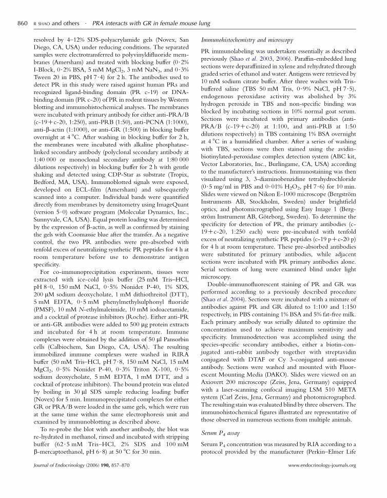
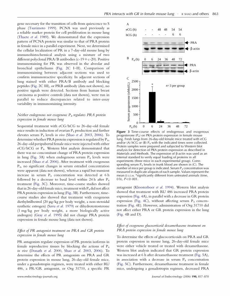
Figure 3 Time-course effects of endogenous and exogenousprogesterone (P4) on PRA protein expression in female mouselung. Fresh lungs from 26-day-old female mice treated with eCGand/or (A) hCG or (B) P4 with the indicated times were collected.Protein samples were prepared and subjected to Western blotanalysis for detection of PRA protein expression as described inMaterials and Methods. The expression of b-actin was used as aninternal standard to verify equal loading of proteins in allexperiments (three mice in each experimental group). Corre-sponding serum P4 levels in trunk blood are shown in (C). Thenumber of mice per group is indicated. Serum P4 concentration wasmeasured in duplicate aliquots of each sample. Values represent themeanGS.E.M. *significantly different from untreated animals (time,0 h), P!0$001.
Neither endogenous nor exogenous P4 regulates PRA proteinexpression in female mouse lung
Sequential treatment with eCG/hCG to 26-day-old female
mice results in induction of ovarian P4 production and further
elevates serum P4 levels in vivo (Shao et al. 2003, 2006). To
determinewhether PRA protein expression is regulated by P4,
26-day-old peripubertal female micewere injectedwith either
eCG/hCG or P4. Western blot analysis demonstrated that
there was no concomitant change of PRA protein expression
in lung (Fig. 3A) when endogenous serum P4 levels were
increased (Shao et al. 2006). After treatment with exogenous
P4, no significant changes in serum estradiol concentration
were apparent (data not shown), whereas a rapid but transient
increase in serum P4 concentration was detected at 6 h
followed by a decrease to basal level within 24 h after P4treatment (Fig. 3C). Moreover, time-course studies showed
that in 26-day-old femalemice, treatmentwithP4 did not affect
PRA protein expression in lung (Fig. 3B). Furthermore, time-
course studies also showed that treatment with exogenous
diethylstilbestrol (20 mg/kg per body weight, a non-steroidal
synthetic estrogen) (Savu et al. 1979) or dihydrotestosterone
(1 mg/kg per body weight, a more biologically active
androgen) (Gray et al. 1995) did not change PRA protein
expression in female mouse lung (data not shown).
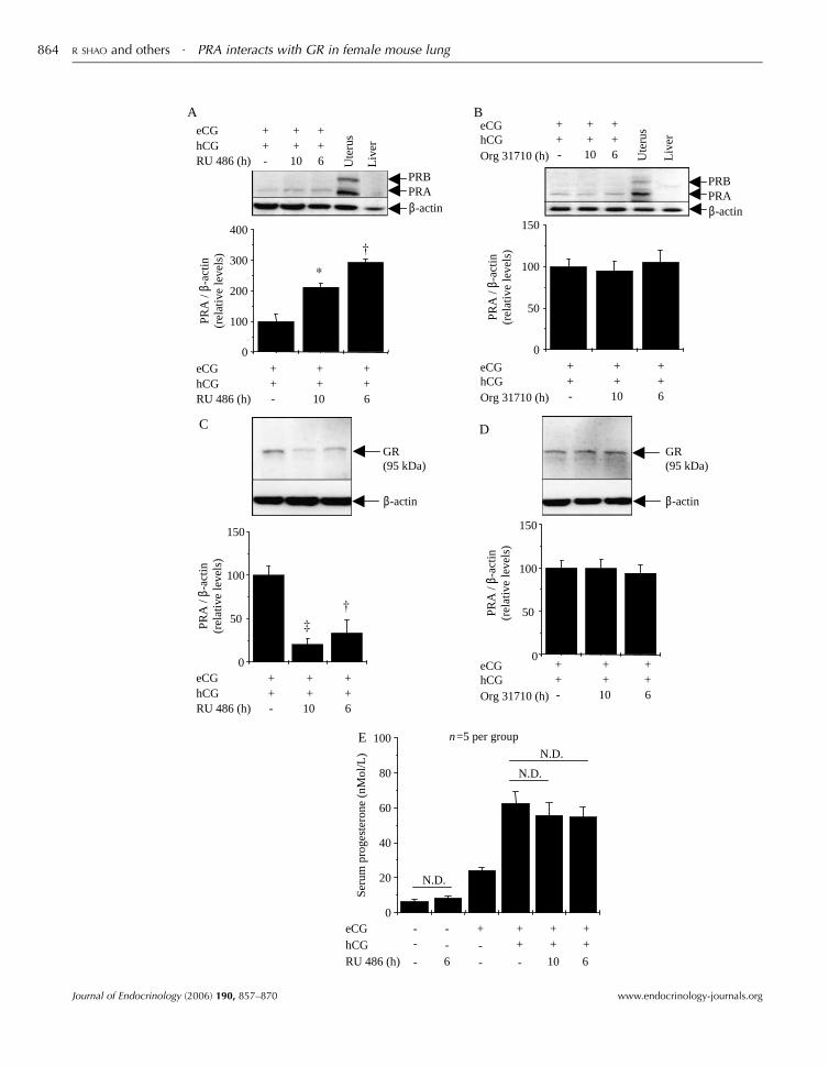
Effect of PR antagonist treatment on PRA and GR proteinexpression in female mouse lung
PR antagonists regulate expression of PR protein isoforms in
female reproductive tissues by blocking the actions of P4in vivo (Donath et al. 2000, Shao et al. 2003, 2006). To
determine the effects of PR antagonists on PRA and GR
protein expression in mouse lung, 26-day-old female mice,
under a gonadotropin regimen, were treated with either RU
486, a PR/GR antagonist, or Org 31710, a specific PR
www.endocrinology-journals.org
antagonist (Kloosterboer et al. 1994). Western blot analysis
showed that treatment with RU 486 increased PRA protein
expression (Fig. 4A), in parallel with a decrease in GR protein
expression (Fig. 4C), without affecting serum P4 concen-
tration (Fig. 4E). However, administration of Org 31710 did
not affect either PRA or GR protein expression in the lung
(Fig. 4B and D).
Effect of exogenous glucocorticoid dexamethasone treatment onPRA protein expression in female mouse lung
To determine the effects of glucocorticoids on PRA and GR
protein expression in mouse lung, 26-day-old female mice
were either vehicle treated or treated with dexamethasone.
Western blot analysis indicated that GR protein expression
was increased at 6 h after dexamethasone treatment (Fig. 5A),
in association with a decrease in serum P4 concentration
(Fig. 5C). Furthermore, dexamethasone treatment in female
mice, undergoing a gonadotropin regimen, decreased PRA
Journal of Endocrinology (2006) 190, 857–870
0
50
100
150
eCGhCGOrg 31710 (h)
0
50
100
150
0
50
100
150
GR(95 kDa)
0
100
200
300
400
PRA
/ β-
actin
(rel
ativ
e le
vels
)PR
A /
β-ac
tin(r
elat
ive
leve
ls)
PRA
/ β-
actin
(rel
ativ
e le
vels
)PR
A /
β-ac
tin(r
elat
ive
leve
ls)
*
eCG +
A B
C D
E
hCG +++
++
RU 486 (h) - 10 6
eCG +hCG +
++
++
RU 486 (h) - 10 6
eCG +hCG +
++
++
RU 486 (h) - 10 6
Ute
rus
Liv
er
++
++
++
- 10 6
eCGhCGOrg 31710 (h)
++
++
++
- 10 6
eCGhCGOrg 31710 (h)
++
++
++
- 10 6
Ute
rus
Liv
er
PRBPRAβ-actin
β-actin
GR(95 kDa)
β-actin
PRBPRAβ-actin
0
20
40
60
80
100
Seru
m p
roge
ster
one
(nM
ol/L
)
N.D.
N.D.
N.D.
eCG - --
-
+ ++
++
++hCG - -
- -RU 486 (h) 6 10 6
n=5 per group
R SHAO and others $ PRA interacts with GR in female mouse lung864
Journal of Endocrinology (2006) 190, 857–870 www.endocrinology-journals.org
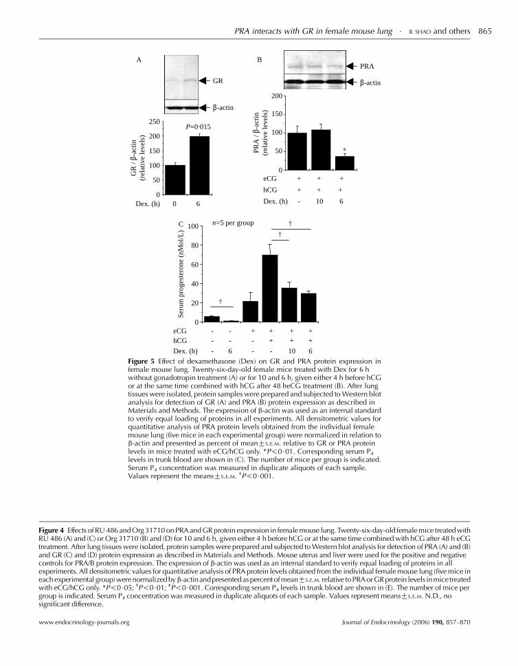
Figure 4 Effects of RU 486 and Org 31710 on PRA and GR protein expression in female mouse lung. Twenty-six-day-old female mice treated withRU 486 (A) and (C) or Org 31710 (B) and (D) for 10 and 6 h, given either 4 h before hCG or at the same time combined with hCG after 48 h eCGtreatment. After lung tissues were isolated, protein samples were prepared and subjected to Western blot analysis for detection of PRA (A) and (B)and GR (C) and (D) protein expression as described in Materials and Methods. Mouse uterus and liver were used for the positive and negativecontrols for PRA/B protein expression. The expression of b-actin was used as an internal standard to verify equal loading of proteins in allexperiments. All densitometric values for quantitative analysis of PRA protein levels obtained from the individual female mouse lung (five mice ineach experimental group) were normalized byb-actinandpresented as percent ofmeanGS.E.M. relative toPRA orGRprotein levels inmice treatedwith eCG/hCG only. *P!0$05; †P!0$01; ‡P!0$001. Corresponding serum P4 levels in trunk blood are shown in (E). The number of mice pergroup is indicated. Serum P4 concentration was measured in duplicate aliquots of each sample. Values represent meansGS.E.M. N.D., nosignificant difference.
eCG + +
hCG +
+
+ +
Dex. (h) - 10 6
0
50
100
150
200
PRA
/ β-
actin
(rel
ativ
e le
vels
)
PRA
β-actinGR
A
C
B
β-actin
0
50
100
150
200
250
GR
/ β-
actin
(rel
ativ
e le
vels
)
P=0·015
Dex. (h) 0 6
0
20
40
60
80
100
Seru
m p
roge
ster
one
(nM
ol/L
)
eCG --
--
-
-
- -
+ ++
++
++hCG
Dex. (h) 6 10 6
n=5 per group
Figure 5 Effect of dexamethasone (Dex) on GR and PRA protein expression infemale mouse lung. Twenty-six-day-old female mice treated with Dex for 6 hwithout gonadotropin treatment (A) or for 10 and 6 h, given either 4 h before hCGor at the same time combined with hCG after 48 heCG treatment (B). After lungtissues were isolated, protein samples were prepared and subjected to Western blotanalysis for detection of GR (A) and PRA (B) protein expression as described inMaterials and Methods. The expression of b-actin was used as an internal standardto verify equal loading of proteins in all experiments. All densitometric values forquantitative analysis of PRA protein levels obtained from the individual femalemouse lung (five mice in each experimental group) were normalized in relation tob-actin and presented as percent of meanGS.E.M. relative to GR or PRA proteinlevels in mice treated with eCG/hCG only. *P!0$01. Corresponding serum P4
levels in trunk blood are shown in (C). The number of mice per group is indicated.Serum P4 concentration was measured in duplicate aliquots of each sample.Values represent the meansGS.E.M. †P!0$001.
PRA interacts with GR in female mouse lung $ R SHAO and others 865
www.endocrinology-journals.org Journal of Endocrinology (2006) 190, 857–870
R SHAO and others $ PRA interacts with GR in female mouse lung866
protein expression (Fig. 5B), depending on injection time.
However, dexamethasone treatment decreased serum P4concentration regardless of injection time (6 and 10 h)
(Fig. 5C). Our findings demonstrate that dexamethasone-
induced suppression of PRA protein expression is indepen-
dent of circulating P4 concentration.
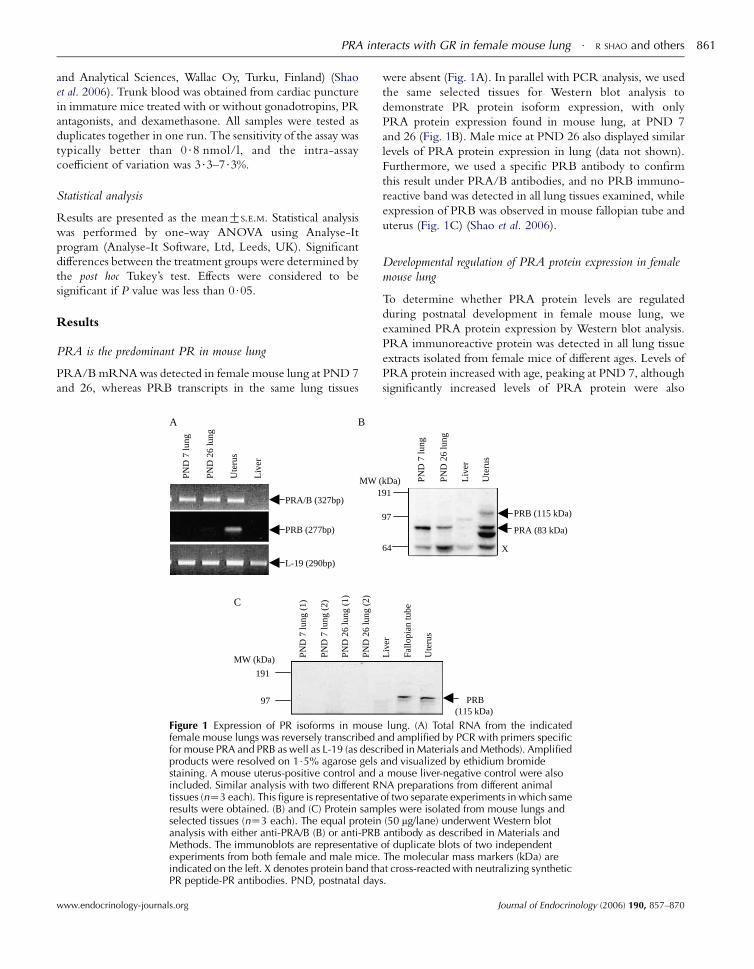
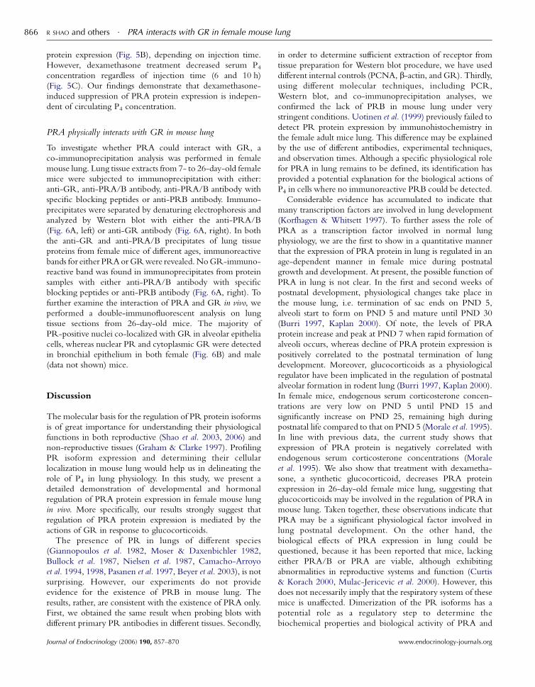
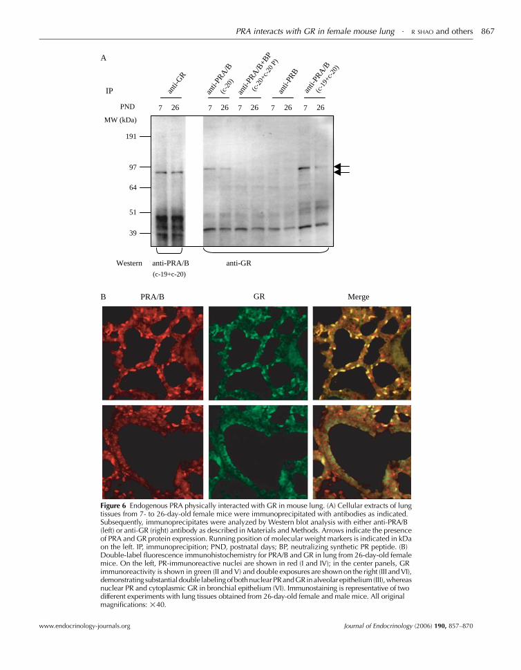
PRA physically interacts with GR in mouse lung
To investigate whether PRA could interact with GR, a
co-immunoprecipitation analysis was performed in female
mouse lung. Lung tissue extracts from 7- to 26-day-old female
mice were subjected to immunoprecipitation with either:
anti-GR, anti-PRA/B antibody, anti-PRA/B antibody with
specific blocking peptides or anti-PRB antibody. Immuno-
precipitates were separated by denaturing electrophoresis and
analyzed by Western blot with either the anti-PRA/B
(Fig. 6A, left) or anti-GR antibody (Fig. 6A, right). In both
the anti-GR and anti-PRA/B precipitates of lung tissue
proteins from female mice of different ages, immunoreactive
bands for either PRAorGRwere revealed.NoGR-immuno-
reactive band was found in immunoprecipitates from protein
samples with either anti-PRA/B antibody with specific
blocking peptides or anti-PRB antibody (Fig. 6A, right). To
further examine the interaction of PRA and GR in vivo, we
performed a double-immunofluorescent analysis on lung
tissue sections from 26-day-old mice. The majority of
PR-positive nuclei co-localized with GR in alveolar epithelia
cells, whereas nuclear PR and cytoplasmic GR were detected
in bronchial epithelium in both female (Fig. 6B) and male
(data not shown) mice.
Discussion
The molecular basis for the regulation of PR protein isoforms
is of great importance for understanding their physiological
functions in both reproductive (Shao et al. 2003, 2006) and
non-reproductive tissues (Graham & Clarke 1997). Profiling
PR isoform expression and determining their cellular
localization in mouse lung would help us in delineating the
role of P4 in lung physiology. In this study, we present a
detailed demonstration of developmental and hormonal
regulation of PRA protein expression in female mouse lung
in vivo. More specifically, our results strongly suggest that
regulation of PRA protein expression is mediated by the
actions of GR in response to glucocorticoids.
The presence of PR in lungs of different species
(Giannopoulos et al. 1982, Moser & Daxenbichler 1982,
Bullock et al. 1987, Nielsen et al. 1987, Camacho-Arroyo
et al. 1994, 1998, Pasanen et al. 1997, Beyer et al. 2003), is not
surprising. However, our experiments do not provide
evidence for the existence of PRB in mouse lung. The
results, rather, are consistent with the existence of PRA only.
First, we obtained the same result when probing blots with
different primary PR antibodies in different tissues. Secondly,
Journal of Endocrinology (2006) 190, 857–870
in order to determine sufficient extraction of receptor from
tissue preparation for Western blot procedure, we have used
different internal controls (PCNA, b-actin, and GR). Thirdly,
using different molecular techniques, including PCR,
Western blot, and co-immunoprecipitation analyses, we
confirmed the lack of PRB in mouse lung under very
stringent conditions. Uotinen et al. (1999) previously failed to
detect PR protein expression by immunohistochemistry in
the female adult mice lung. This difference may be explained
by the use of different antibodies, experimental techniques,
and observation times. Although a specific physiological role
for PRA in lung remains to be defined, its identification has
provided a potential explanation for the biological actions of
P4 in cells where no immunoreactive PRB could be detected.
Considerable evidence has accumulated to indicate that
many transcription factors are involved in lung development
(Korfhagen & Whitsett 1997). To further assess the role of
PRA as a transcription factor involved in normal lung
physiology, we are the first to show in a quantitative manner
that the expression of PRA protein in lung is regulated in an
age-dependent manner in female mice during postnatal
growth and development. At present, the possible function of
PRA in lung is not clear. In the first and second weeks of
postnatal development, physiological changes take place in
the mouse lung, i.e. termination of sac ends on PND 5,
alveoli start to form on PND 5 and mature until PND 30
(Burri 1997, Kaplan 2000). Of note, the levels of PRA
protein increase and peak at PND 7 when rapid formation of
alveoli occurs, whereas decline of PRA protein expression is
positively correlated to the postnatal termination of lung
development. Moreover, glucocorticoids as a physiological
regulator have been implicated in the regulation of postnatal
alveolar formation in rodent lung (Burri 1997, Kaplan 2000).
In female mice, endogenous serum corticosterone concen-
trations are very low on PND 5 until PND 15 and
significantly increase on PND 25, remaining high during
postnatal life compared to that on PND 5 (Morale et al. 1995).
In line with previous data, the current study shows that
expression of PRA protein is negatively correlated with
endogenous serum corticosterone concentrations (Morale
et al. 1995). We also show that treatment with dexametha-
sone, a synthetic glucocorticoid, decreases PRA protein
expression in 26-day-old female mice lung, suggesting that
glucocorticoids may be involved in the regulation of PRA in
mouse lung. Taken together, these observations indicate that
PRA may be a significant physiological factor involved in
lung postnatal development. On the other hand, the
biological effects of PRA expression in lung could be
questioned, because it has been reported that mice, lacking
either PRA/B or PRA are viable, although exhibiting
abnormalities in reproductive systems and function (Curtis
& Korach 2000, Mulac-Jericevic et al. 2000). However, this
does not necessarily imply that the respiratory system of these
mice is unaffected. Dimerization of the PR isoforms has a
potential role as a regulatory step to determine the
biochemical properties and biological activity of PRA and
www.endocrinology-journals.org
PRA/B GR Merge
A
B
anti-
PRA
/B+B
P
(c-2
0+c-
20 P
)an
ti-PR
B
anti-
PRA
/B(c
-19+
c-20
)
191
MW (kDa)
97
64
51
39
Western anti-PRA/B anti-GR(c-19+c-20)
PND 7 26 7 26 7 26 7 26 7 26
IP anti-
PRA
/B(c
-20)
anti-
GR
Figure 6 Endogenous PRA physically interacted with GR in mouse lung. (A) Cellular extracts of lungtissues from 7- to 26-day-old female mice were immunoprecipitated with antibodies as indicated.Subsequently, immunoprecipitates were analyzed by Western blot analysis with either anti-PRA/B(left) or anti-GR (right) antibody as described in Materials and Methods. Arrows indicate the presenceof PRA and GR protein expression. Running position of molecular weight markers is indicated in kDaon the left. IP, immunoprecipition; PND, postnatal days; BP, neutralizing synthetic PR peptide. (B)Double-label fluorescence immunohistochemistry for PRA/B and GR in lung from 26-day-old femalemice. On the left, PR-immunoreactive nuclei are shown in red (I and IV); in the center panels, GRimmunoreactivity is shown in green (II and V) and double exposures are shown on the right (III and VI),demonstrating substantial double labelingofbothnuclearPRandGRinalveolar epithelium (III),whereasnuclear PR and cytoplasmic GR in bronchial epithelium (VI). Immunostaining is representative of twodifferent experiments with lung tissues obtained from 26-day-old female and male mice. All originalmagnifications: !40.
PRA interacts with GR in female mouse lung $ R SHAO and others 867
www.endocrinology-journals.org Journal of Endocrinology (2006) 190, 857–870
R SHAO and others $ PRA interacts with GR in female mouse lung868
PRB (Mohamed et al. 1994, McDonnell 1995). Since PRA
cannot only dimerize to PRB, but also homodimerize to
modulate target cell responsiveness to hormones (Leonhardt
et al. 1998) and RU 486 (Mohamed et al. 1994), it is possible
that PRA plays a transient role in mouse lung development
during the early postnatal period. Future studies will be
needed to address the effects of PRA on cell differentiation
and function in lung during postnatal development.
Previously, we have shown that PR protein isoform
expression in female reproductive tissues including ovary,
fallopian tube, and uterus is regulated by gonadotropin
treatment that increase endogenous P4 concentration, as well
as acute treatment with P4 in female mice (Shao et al. 2003,
2006). The current study demonstrates that neither endogen-
ous nor exogenous P4-regulate PRA protein expression in
lung using the same in vivo experimental model. In addition, a
time-course study of both RU 486 and dexamethasone
treatment supports the idea that changes in PRA protein
expression are independent of circulating P4 concentration in
female mouse lung. To our knowledge, this is the first report
that demonstrates a functional dissimilarity of P4 and
glucocorticoids in the regulation of PRA protein expression
in female mouse lung in vivo. Although previous studies have
reported that the pure antagonist effect of RU 486 is mediated
by the PRA signaling pathway within target cells in vitro
(McDonnell & Goldman 1994, Hurd et al. 1999), RU 486
can interact with both PR and GR (Guettari et al. 1990,
McDonnell & Goldman 1994, Cadepond et al. 1997). We
therefore used Org 31710, a specific PR antagonist
(Kloosterboer et al. 1994), to distinguish the effects of RU
486 on PRA from the effects on GR. There is no change in
PRA protein expression after Org 31710 treatment,
suggesting that RU 486 binding to GR regulates PRA
protein expression in mouse lung. Furthermore, GR appears
to overlap PR cellular distribution in lung (Camacho-Arroyo
et al. 1994, Adcock et al. 1996), interacts with estrogen
receptors (ER) (Kinyamu & Archer 2003), and PR has been
shown to interact with ER (Uht et al. 1997), suggesting a
possibility of interaction between PRA and GR in vivo. The
amount of PRA/GR complex detected in PRA and GR
immunoprecipitates in this study suggests that PRA and GR
are able to crosstalk with each other in response to
glucocorticoids through protein–protein interaction in vivo.
Interestingly, cellular localization patterns of PR and GR are
diverse. Unliganded PR is predominantly nuclear (Smith et al.
1990) whereas unliganded GR resides entirely in the
cytoplasm (Hache et al. 1999). Lung is composed of multiple
cell types (Amy et al. 1977, Burri 1997) and our
immunohistochemistry results show that in the presence of
low levels of endogenous corticosterone (Ferrandez & De la
Fuente 1999), PRA is localized predominantly in the nucleus
of both alveolar and bronchial epithelia, whereas GR is
distributed in either the nucleus of alveolar epithelium or
cytoplasm of bronchial epithelium, suggesting that interaction
between PRA and GR may differ in a cell type-specific
manner. Additional studies will determine whether the
Journal of Endocrinology (2006) 190, 857–870
interactions of PRA and GR are ligand-dependent using a
cell fractionation procedure. Evaluating the significance of the
interactions described here requires identification of targets of
GR signal activated by glucocorticoids in mouse lung.
Clinically, corticosteroids are the most potent and effective
therapy for treatment of chronic inflammatory diseases such as
asthma, and they are generally believed to exert their
therapeutic benefits by specifically binding to GR, which
on activation translocates to the nucleus and either increases
or decreases of expression of responsive genes and also
suppresses inflammation (Adcock et al. 1996, Umland et al.
2002). It is known that receptor regulation is an important
mechanism for modulating target cell responsiveness to
hormones. As previously mentioned, several in vitro studies
have shown that PRA functions as a strong inhibitor of other
steroid receptors including GR (Vegeto et al. 1993,
McDonnell et al. 1994). Since the cellular sensitivity to
glucocorticoid is directly proportional to GR concentration
and the anti-inflammatory activity of GR is mediated by
protein–protein interaction (Reichardt et al. 2000), our results
suggest that suppression of PRA protein expression by
glucocorticoids may affect the regulation and/or modulation
of GR levels or activity to enhance lung responsiveness to
circulating glucocorticoids, i.e. activation of GR signaling.
In summary, the results of this study document the presence
of PRA mRNA and protein in the female mouse lung and
demonstrate that PRA protein is expressed and regulated in
female mice during postnatal lung development. The
relationship between PRA, PCNA protein expression, and
alveoli maturation suggests that PRA may play a role in
postnatal lung development. However, there was no direct
relationship between circulating P4 concentrations and PRA
protein expression, suggesting that it is unlikely that P4 is
responsible for the regulation of PRA expression in female
mouse lung. Moreover, we have shown that exposure to
dexamethasone and RU 486, but not Org 31710, in vivo
significantly changes PRA protein expression independently
of circulating P4 levels. Furthermore, the interaction and
co-localization of PRA and GR at physiologically relevant
levels depends on the lung cell type, suggesting that further
study of such interactions may be relevant to our under-
standing of the mechanism of action and interaction between
PRA and GR signaling pathway in the lung.
Acknowledgements
We would like to thank Prof. Ola Nilsson, Lundberg
Laboratory for Cancer Research, Department of Pathology,
Sahlgrenska University Hospital, Goteborg University, for
help with lung morphological studies, Emilia Rung for
critically reading the manuscript and the Swegene Centre
for Cellular Imaging at Goteborg University for the use of
imaging equipment. The authors declare that there is no
conflict of interest that would prejudice the impartiality of
this scientific work.
www.endocrinology-journals.org
PRA interacts with GR in female mouse lung $ R SHAO and others 869
Funding
This work was supported by Grants 10380 and 13550 from
the Swedish Medical Research Council to HB, and the Assar
Gabrielssons Forsknings Foundation, the Scientific Foun-
dation of Eva och Oscar Ahrens, Wilhelm och Martina
Lundgrens,Rudolf ochEllenMariaMornes, aswell asHjalmar
Svensson and Adlerbertska Research Foundation to RS.
References
Adcock IM, Gilbey T, Gelder CM, Chung KF & Barnes PJ 1996
Glucocorticoid receptor localization in normal and asthmatic lung.
American Journal of Respiratory and Critical Care Medicine 154 771–782.
Amy RW, Bowes D, Burri PH, Haines J & Thurlbeck WM 1977 Postnatal
growth of the mouse lung. Journal of Anatomy 124 131–151.
Bethea CL &Widmann AA 1998 Differential expression of progestin receptor
isoforms in the hypothalamus, pituitary, and endometrium of rhesus
macaques. Endocrinology 139 677–687.
Beyer C, Damm N, Brito V & Kuppers E 2002 Developmental expression of
progesterone receptor isoforms in the mouse midbrain. Neuroreport 13
877–880.
Beyer C, Kuppers E, Karolczak M & Trotter A 2003 Ontogenetic expression
of estrogen and progesterone receptors in the mouse lung. Biology of the
Neonate 84 59–63.
Bullock DW, Lamb DJ, Rider VC & Kima PE 1987 The rabbit progesterone
receptor and uteroglobin gene expression. Advances in Experimental Medicine
and Biology 230 79–97.
Burri PH 1997 Postnatal development and growth. In The Lung: Scientific
Foundations, pp 1013–1026. Eds RG Crystal, JB West, ERWeibel &
PJ Barnes. Philadelphia, PA, USA: Lippincott-Raven.
Cadepond F, Ulmann A & Baulieu EE 1997 RU486 (mifepristone):
mechanisms of action and clinical uses. Annual Review of Medicine 48
129–156.
Camacho-Arroyo I, Ruiz A, Gamboa-Dominguez A, Perez-Palacios G &
Cerbon MA 1994 Immunohistochemical localization of intracellular
progesterone and glucocorticoid receptors in the rabbit lung. Journal of
Endocrinology 142 311–316.
Camacho-Arroyo I, Mendez-Cruz ST, Guerra-Araiza C & Cerbon MA 1998
Changes in progesterone receptor mRNA content in the rabbit lung during
early pregnancy and after sex steroid hormone treatment. Journal of
Endocrinology 157 71–74.
Chan YL, Lin A, McNally J, Peleg D, Meyuhas O & Wool IG 1987 The
primary structure of rat ribosomal protein L19. A determination from the
sequence of nucleotides in a cDNA and from the sequence of amino acids
in the protein. Journal of Biological Chemistry 262 1111–1115.
Condon JC, Hardy DB, Kovaric K & Mendelson CR 2006 Up-regulation of
the progesterone receptor (PR)-C isoform in laboring myometrium by
activation of nuclear factor-kappaB may contribute to the onset of labor
through inhibition of PR function. Molecular Endocrinology 20 764–775.
Conneely OM, Maxwell BL, Toft DO, Schrader WT & O’Malley BW 1987
The A and B forms of the chicken progesterone receptor arise by alternate
initiation of translation of a unique mRNA. Biochemical and Biophysical
Research Communications 149 493–501.
Curtis SH & Korach KS 2000 Steroid receptor knockout models: phenotypes
and responses illustrate interactions between receptor signaling pathways
in vivo. Advances in Pharmacology 47 357–380.
Donath J, Nishino Y, Schulz T &Michna H 2000 The antiovulatory potential
of progesterone antagonists correlates with a down-regulation of
progesterone receptors in the hypothalamus, pituitary and ovaries.
Anatomischer Anzeiger 182 143–150.
Ferrandez MD&De la Fuente M 1999 Effects of age, sex and physical exercise
on the phagocytic process of murine peritoneal macrophages. Acta
Physiologica Scandinavica 166 47–53.
www.endocrinology-journals.org
Giangrande PH & McDonnell DP 1999 The A and B isoforms of the human
progesterone receptor: two functionally different transcription factors
encoded by a single gene. Recent Progress in Hormone Research 54 291–313
(discussion 313–314).
Giannopoulos G, Phelps DS & Munowitz P 1982 Heterogeneity and
ontogenesis of progestin receptors in rabbit lung. Journal of Steroid
Biochemistry 17 503–510.
Graham JD & Clarke CL 1997 Physiological action of progesterone in target
tissues. Endocrine Reviews 18 502–519.
GrayK, Eitzman B,RaszmannK, SteedT,Geboff A,McLachlan J&BidwellM
1995 Coordinate regulation by diethylstilbestrol of the platelet-derived
growth factor-A (PDGF-A) and -B chains and the PDGF receptor alpha- and
beta-subunits in themouse uterus and vagina: potential mediators of estrogen
action. Endocrinology 136 2325–2340.
Guettari N, Dufour ME &Marin L 1990 Effects of the antiglucocorticoid RU
486 on the initiation of ultrastructural type-II cell differentiation in fetal rat
lung. Biology of the Neonate 58 173–180.
Hache RJ, Tse R, Reich T, Savory JG & Lefebvre YA 1999 Nucleocyto-
plasmic trafficking of steroid-free glucocorticoid receptor. Journal of
Biological Chemistry 274 1432–1439.
Haggerty CL, Ness RB, Kelsey S &Waterer GW 2003 The impact of estrogen
and progesterone on asthma. Annals of Allergy, Asthma and Immunology 90
284–291 (quiz 291–293, 347).
Hurd C, Nag K, Khattree N, Alban P, Dinda S & Moudgil VK 1999 Agonist-
and antagonist-induced qualitative and quantitative alterations of progesterone
receptor frombreast cancer cells.Molecular andCellular Biochemistry 199 49–56.
Ilenchuk TT &Walters MR 1987 Rat uterine progesterone receptor analyzed
by [3H]R5020 photoaffinity labeling: evidence that the A and B subunits
are not equimolar. Endocrinology 120 1449–1456.
IshibashiH, Suzuki T, Suzuki S,NiikawaH, Lu L,Miki Y,MoriyaT,Hayashi S,
Handa M, Kondo Tet al. 2005 Progesterone receptor in non-small cell lung
cancer – a potent prognostic factor and possible target for endocrine therapy.
Cancer Research 65 6450–6458.
Joensuu TK, Ylikomi TJ, Toft DO, KeinanenRA,KulomaaMS&Tuohimaa PJ
1990 Progesterone-induced avidin as a marker of cytodifferentiation in the
oviduct: comparison to ovalbumin. Endocrinology 126 1143–1155.
Kaplan F 2000 Molecular determinants of fetal lung organogenesis. Molecular
Genetics and Metabolism 71 321–341.
Kastner P, Krust A, Turcotte B, Stropp U, Tora L, Gronemeyer H & Chambon
P 1990a Two distinct estrogen-regulated promoters generate transcripts
encoding the two functionally different human progesterone receptor forms
A and B. EMBO Journal 9 1603–1614.
Kastner P, Bocquel MT, Turcotte B, Garnier JM, Horwitz KB, Chambon P &
Gronemeyer H 1990b Transient expression of human and chicken
progesterone receptors does not support alternative translational initiation
from a single mRNA as the mechanism generating two receptor isoforms.
Journal of Biological Chemistry 265 12163–12167.
Kinyamu HK & Archer TK 2003 Estrogen receptor-dependent proteasomal
degradation of the glucocorticoid receptor is coupled to an increase in
mdm2 protein expression. Molecular and Cellular Biology 23 5867–5881.
Kloosterboer HJ, Deckers GH & Schoonen WG 1994 Pharmacology of two
new very selective antiprogestagens: Org 31710 and Org 31806. Human
Reproduction 9 (Suppl 1) 47–52.
Korfhagen TR & Whitsett JA 1997 Transcriptional control in the developing
lung. The Parker B. Francis lectureship. Chest 111 83S–88S.
Kraus WL, Montano MM & Katzenellenbogen BS 1993 Cloning of the rat
progesterone receptor gene 5 0-region and identification of two functionally
distinct promoters. Molecular Endocrinology 7 1603–1616.
Leonhardt SA, Altmann M & Edwards DP 1998 Agonist and antagonists
induce homodimerization and mixed ligand heterodimerization of human
progesterone receptors in vivo by a mammalian two-hybrid assay. Molecular
Endocrinology 12 1914–1930.
Lessey BA, Alexander PS & Horwitz KB 1983 The subunit structure of
human breast cancer progesterone receptors: characterization by chroma-
tography and photoaffinity labeling. Endocrinology 112 1267–1274.
McDonnell DP 1995 Unraveling the human progesterone receptor signal
transduction pathway. Trends in Endocrinology and Metabolism 6 133–138.
Journal of Endocrinology (2006) 190, 857–870
R SHAO and others $ PRA interacts with GR in female mouse lung870
McDonnell DP & Goldman ME 1994 RU486 exerts antiestrogenic activities
through a novel progesterone receptor A form-mediated mechanism.
Journal of Biological Chemistry 269 11945–11949.
McDonnell DP, Shahbaz MM, Vegeto E & Goldman ME 1994 The human
progesterone receptor A-form functions as a transcriptional modulator of
mineralocorticoid receptor transcriptional activity. Journal of Steroid
Biochemistry and Molecular Biology 48 425–432.
Milne JA 1979 The respiratory response to pregnancy. Postgraduate Medical
Journal 55 318–324.
Mohamed MK, Tung L, Takimoto GS & Horwitz KB 1994 The leucine
zippers of c-fos and c-jun for progesterone receptor dimerization:
A-dominance in the A/B heterodimer. Journal of Steroid Biochemistry and
Molecular Biology 51 241–250.
Morale MC, Batticane N, Gallo F, Barden N & Marchetti B 1995 Disruption
of hypothalamic-pituitary-adrenocortical system in transgenic mice
expressing type II glucocorticoid receptor antisense ribonucleic acid
permanently impairs T cell function: effects on T cell trafficking and T cell
responsiveness during postnatal development. Endocrinology 136
3949–3960.
Moser EH & Daxenbichler G 1982 Detection of a heat- and acid-stable
‘progesterone’-binding protein in the rat lung. FEBS Letters 150 347–353.
Mulac-Jericevic B, Mullinax RA, DeMayo FJ, Lydon JP & Conneely OM
2000 Subgroup of reproductive functions of progesterone mediated by
progesterone receptor-B isoform. Science 289 1751–1754.
Nielsen ST, Conaty JM & DiPasquale G 1987 Progesterone receptor of adult
rabbit lung. Pharmacology 35 217–226.
Pasanen S, Ylikomi T, Syvala H & Tuohimaa P 1997 Distribution of
progesterone receptor in chicken: novel target organs for progesterone and
estrogen action. Molecular and Cellular Endocrinology 135 79–91.
Reichardt HM, Tronche F, Bauer A & Schutz G 2000 Molecular genetic
analysis of glucocorticoid signaling using the Cre/loxP system. Biological
Chemistry 381 961–964.
Robker RL, Russell DL, Espey LL, Lydon JP, O’Malley BW & Richards JS
2000 Progesterone-regulated genes in the ovulation process: ADAMTS-1
and cathepsin L proteases. PNAS 97 4689–4694.
Russo LA, Calabro SP, Filler TA, Carey DJ & Gardner RM 2001 In vivo
regulation of syndecan-3 expression in the rat uterus by 17 beta-estradiol.
Journal of Biological Chemistry 276 686–692.
Savu L, Benassayag C, Vallette G & Nunez EA 1979 Ligand properties of
diethylstilbestrol: studies with purified native and fatty acid-free rat alpha
1-fetoprotein and albumin. Steroids 34 737–748.
Schneider W, Ramachandran C, Satyaswaroop PG & Shyamala G 1991
Murine progesterone receptor exists predominantly as the 83-kilodalton ‘A’
form. Journal of Steroid Biochemistry and Molecular Biology 38 285–291.
Shao R, Markstrom E, Friberg PA, Johansson M & Billig H 2003 Expression
of progesterone receptor (PR) A and B isoforms in mouse granulosa cells:
stage-dependent PR-mediated regulation of apoptosis and cell prolifer-
ation. Biology of Reproduction 68 914–921.
Journal of Endocrinology (2006) 190, 857–870
Shao R, Zhang FP, Tian F, Anders Friberg P, Wang X, Sjoland H & Billig H
2004 Increase of SUMO-1 expression in response to hypoxia: direct
interaction with HIF-1alpha in adult mouse brain and heart in vivo. FEBS
Letters 569 293–300.
Shao R, Weijdegrd B, Ljungstrom K, Friberg A, Zhu C, Wang X, Zhu Y,
Fernandez-Rodriguez J, Egecioglu E, Rung E, et al. 2006 Nuclear
progesterone receptor (PR) A and B isoforms in mouse fallopian tube and
uterus: implications for expression, regulation and cellular function.
American Journal of Physiology. Endocrinology and Metabolism 291 E59–E72.
Smith DF, Faber LE & Toft DO 1990 Purification of unactivated progesterone
receptor and identification of novel receptor-associated proteins. Journal of
Biological Chemistry 265 3996–4003.
Taylor AH, McParland PC, Taylor DJ & Bell SC 2006 The progesterone
receptor in human term amniochorion and placenta is isoform C.
Endocrinology 147 687–693.
Thaete LG, Ahnen DJ & Malkinson AM 1989 Proliferating cell nuclear
antigen (PCNA/cyclin) immunocytochemistry as a labeling index in mouse
lung tissues. Cell and Tissue Research 256 167–173.
Tsurimoto T 1999 PCNA binding proteins. Frontiers in Bioscience 4
D849–D858.
Tung L, Mohamed MK, Hoeffler JP, Takimoto GS & Horwitz KB 1993
Antagonist-occupied human progesterone B-receptors activate transcrip-
tion without binding to progesterone response elements and are dominantly
inhibited by A-receptors. Molecular Endocrinology 7 1256–1265.
Uht RM, Anderson CM, Webb P & Kushner PJ 1997 Transcriptional
activities of estrogen and glucocorticoid receptors are functionally
integrated at the AP-1 response element. Endocrinology 138 2900–2908.
Umland SP, Schleimer RP & Johnston SL 2002 Review of the molecular and
cellular mechanisms of action of glucocorticoids for use in asthma.
Pulmonary Pharmacology and Therapeutics 15 35–50.
Uotinen N, Puustinen R, Pasanen S, Manninen T, Kivineva M, Syvala H,
Tuohimaa P & Ylikomi T 1999 Distribution of progesterone receptor in
female mouse tissues. General and Comparative Endocrinology 115 429–441.
Vegeto E, Shahbaz MM, Wen DX, Goldman ME, O’Malley BW &
McDonnell DP 1993 Human progesterone receptor A form is a cell- and
promoter-specific repressor of human progesterone receptor B function.
Molecular Endocrinology 7 1244–1255.
Wen DX, Xu YF, Mais DE, Goldman ME &McDonnell DP 1994 The A and
B isoforms of the human progesterone receptor operate through distinct
signaling pathways within target cells. Molecular and Cellular Biology 14
8356–8364.
Received 30 March 2006Received in final form 17 May 2006Accepted 18 May 2006Made available online as an Accepted Preprint9 June 2006
www.endocrinology-journals.org
Copyright © 2022 FDOKUMEN