Susceptibility of northern British Columbia forests to spruce budworm defoliation
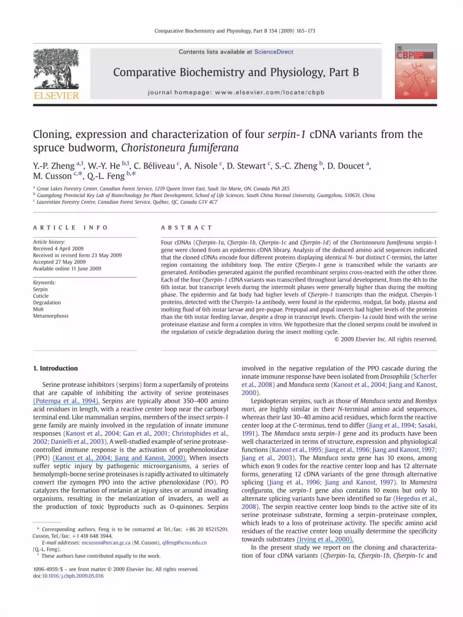
Comparative Biochemistry and Physiology, Part B 154 (2009) 165–173
Contents lists available at ScienceDirect
Comparative Biochemistry and Physiology, Part B
j ourna l homepage: www.e lsev ie r.com/ locate /cbpb
Cloning, expression and characterization of four serpin-1 cDNA variants from thespruce budworm, Choristoneura fumiferana
Y.-P. Zheng a,1, W.-Y. He b,1, C. Béliveau c, A. Nisole c, D. Stewart c, S.-C. Zheng b, D. Doucet a,M. Cusson c,⁎, Q.-L. Feng b,⁎a Great Lakes Forestry Center, Canadian Forest Service, 1219 Queen Street East, Sault Ste Marie, ON, Canada P6A 2E5b Guangdong Provincial Key Lab of Biotechnology for Plant Development, School of Life Sciences, South China Normal University, Guangzhou, 510631, Chinac Laurentian Forestry Centre, Canadian Forest Service, Québec, QC, Canada G1V 4C7
⁎ Corresponding authors. Feng is to be contacted aCusson, Tel./fax: +1 418 648 3944.
E-mail addresses: [email protected] (M. Cusson)(Q.-L. Feng).
1 These authors have contributed equally to the work
1096-4959/$ – see front matter © 2009 Elsevier Inc. Aldoi:10.1016/j.cbpb.2009.05.016
a b s t r a c t
a r t i c l e i n f oArticle history:Received 4 April 2009Received in revised form 23 May 2009Accepted 27 May 2009Available online 11 June 2009
Keywords:SerpinCuticleDegradationMoltMetamorphosis
Four cDNAs (Cfserpin-1a, Cfserpin-1b, Cfserpin-1c and Cfserpin-1d) of the Choristoneura fumiferana serpin-1gene were cloned from an epidermis cDNA library. Analysis of the deduced amino acid sequences indicatedthat the cloned cDNAs encode four different proteins displaying identical N- but distinct C-termini, the latterregion containing the inhibitory loop. The entire CfSerpin-1 gene is transcribed while the variants aregenerated. Antibodies generated against the purified recombinant serpins cross-reacted with the other three.Each of the four Cfserpin-1 cDNA variants was transcribed throughout larval development, from the 4th to the6th instar, but transcript levels during the intermolt phases were generally higher than during the moltingphase. The epidermis and fat body had higher levels of Cfserpin-1 transcripts than the midgut. Cfserpin-1proteins, detected with the Cfserpin-1a antibody, were found in the epidermis, midgut, fat body, plasma andmolting fluid of 6th instar larvae and pre-pupae. Prepupal and pupal insects had higher levels of the proteinsthan the 6th instar feeding larvae, despite a drop in transcript levels. Cfserpin-1a could bind with the serineproteinase elastase and form a complex in vitro. We hypothesize that the cloned serpins could be involved inthe regulation of cuticle degradation during the insect molting cycle.
© 2009 Elsevier Inc. All rights reserved.
1. Introduction
Serine protease inhibitors (serpins) form a superfamily of proteinsthat are capable of inhibiting the activity of serine proteinases(Potempa et al., 1994). Serpins are typically about 350–400 aminoacid residues in length, with a reactive center loop near the carboxylterminal end. Likemammalian serpins, members of the insect serpin-1gene family are mainly involved in the regulation of innate immuneresponses (Kanost et al., 2004; Gan et al., 2001; Christophides et al.,2002; Danielli et al., 2003). Awell-studied example of serine protease-controlled immune response is the activation of prophenoloxidase(PPO) (Kanost et al., 2004; Jiang and Kanost, 2000). When insectssuffer septic injury by pathogenic microorganisms, a series ofhemolymph-borne serine proteinases is rapidly activated to ultimatelyconvert the zymogen PPO into the active phenoloxidase (PO). POcatalyzes the formation of melanin at injury sites or around invadingorganisms, resulting in the melanization of invaders, as well asthe production of toxic byproducts such as O-quinones. Serpins
t Tel./fax: +86 20 85215291.
.
l rights reserved.
involved in the negative regulation of the PPO cascade during theinnate immune response have been isolated fromDrosophila (Scherferet al., 2008) andManduca sexta (Kanost et al., 2004; Jiang and Kanost,2000).
Lepidopteran serpins, such as those of Manduca sexta and Bombyxmori, are highly similar in their N-terminal amino acid sequences,whereas their last 30–40 amino acid residues, which form the reactivecenter loop at the C-terminus, tend to differ (Jiang et al., 1994; Sasaki,1991). The Manduca sexta serpin-1 gene and its products have beenwell characterized in terms of structure, expression and physiologicalfunctions (Kanost et al., 1995; Jiang et al., 1996; Jiang and Kanost,1997;Jiang et al., 2003). The Manduca sexta gene has 10 exons, amongwhich exon 9 codes for the reactive center loop and has 12 alternateforms, generating 12 cDNA variants of the gene through alternativesplicing (Jiang et al., 1996; Jiang and Kanost, 1997). In Mamestraconfigurata, the serpin-1 gene also contains 10 exons but only 10alternate splicing variants have been identified so far (Hegedus et al.,2008). The serpin reactive center loop binds to the active site of itsserine proteinase substrate, forming a serpin–proteinase complex,which leads to a loss of proteinase activity. The specific amino acidresidues of the reactive center loop usually determine the specificitytowards substrates (Irving et al., 2000).
In the present study we report on the cloning and characteriza-tion of four cDNA variants (Cfserpin-1a, Cfserpin-1b, Cfserpin-1c and
166 Y.-P. Zheng et al. / Comparative Biochemistry and Physiology, Part B 154 (2009) 165–173
Cfserpin-1d) of the serpin-1 gene from the spruce budworm, Choris-toneura fumiferana. We compared their sequences and expressionpatterns and assessed their possible functions. Our data supportearlier speculation by Hegedus et al. (2008) and Chamankhah et al.(2003) that insect serpin-1 isoforms may be involved in processesdistinct from plasma coagulation and melanization during the innateimmune response, namely the regulation of the activity of proteases incuticle degradation during the molting cycle.
2. Materials and methods
2.1. Experimental animals
Spruce budworm (Choristoneura fumiferana Clem., Lepidoptera:Tortricidae) were reared at the Great Lake Forestry Centre (Sault Ste.Marie, Ontario, Canada). Third instar larvae were transferred onto asynthetic diet (McMorran, 1965) and reared at 22 °C, 70% relativehumidity and under a photoperiodic cycle of 12-h light:12-h dark,until they reached the adult stage. To ensure accurate staging of laterinstars, 4th instar larvaewere selected after the insects had undergonehead capsule slippage for sampling.
To collect molting fluid samples, a small hole was made with aneedle at the bottom of a 0.5 ml PCR tube, which was then insertedinto a 1.5 ml microfuge tube containing 1.5–2.0 μl cold PBS (0.1 M, pH8.0). Old integument that had just separated from prepupal larvaewascollected and put in the 0.5 ml PCR tube, which was then centrifuged(12,000g) for 5s at 4 °C. The resultant buffered molting fluid wascollected in the 1.5 ml tube and kept at −20°C until used for SDS-PAGE, western blotting analyses and antibody production.
2.2. Cloning and sequence analysis of Cfserpin-1 cDNAs
An expression cDNA library was constructed in the Uni-ZAP XRvector (Stratagene, La Jolla, CA, USA) using mRNA isolated from theepidermis of larvae molting from the 5th to the 6th instar. This librarywas screened with antibody produced against total proteins of themolting fluid as described below. The positive clones were isolatedand the cDNA inserts were sequenced. Those cDNA clones whosepartial sequence revealed high similarity to insect serpins werecompletely sequenced in both directions.
Annotation of sequences was performed using the National Centerfor Biotechnology Information BLAST search services (http://www.ncbi.nlm.nih.gov; Altschul et al., 1990). Multiple sequence alignmentand phylogenetic tree analysis were performed using the Clustal WAlignment Program of DNASTAR (DNASTAR, Inc., Madison, WI) inMegAlign 5.01 version at a gap penalty of 10, a gap length penalty of0.2, Gonnet series matrix of 250 and a bootstrap value at 10,000 times.
2.3. Production of recombinant Cfserpin-1 proteins
To produce protein in a bacterial expression system, the openreading frames (ORF) of the four Cfserpin-1 cDNAs were amplified byPCR and inserted between the BamH I and Xho I restriction sites of the
Table 1Designations, oligonucleotide sequences, length, Tm and amplicon sizes for primers used in
Specificity Name Sequence
Cfserpin-1a Ser-1a F2 5′-CATCGCAGACCGTCAGTTTGTTTACCA-3′Ser-1a R4 5′-GCCTCAGCTTCAACAACTAATTTCGG-3′
Cfserpin-1b Ser-1b F3 5′-GACCACCGCCGCCTGTG-3′Ser-1b R3 5′-ACCCAATTACATTACTTGGTATGGACGTGTTGA
Cfserpin-1c Ser-1c F4 5′-GCAGCATTCGAGGAAACGCCA-3′Ser-1c R3 5′-CAAGCGAGTCGTCACATAAACTTTGCTTTATTG
Cfserpin-1d Ser-1d F2 5′-GGACCGAAGCCGCCCAAA-3′Ser-1d R2 5′-GCAAAGACACCGCTGAATAACTTGTTGTCG-3′
“F” and “R” in the primer names designate forward and reverse primers.
pPROEXTM HT expression vector so as to place a hexahistidine tag onthe C-terminal ends (Invitrogen, USA). The recombinant pPROEXTMHT-Cfserpin-1 plasmids were used to transform DH10BTM Escherichiacoli cells for protein expression in vitro. Protein expression wasinduced by adding IPTG at a final concentration of 1 mM. The cellscontaining the expressed recombinant proteins were homogenized bysonication and the protein inclusion bodies were isolated bycentrifugation at 10,000×g for 20 min. The purified occlusion bodieswere denatured with 8 M urea at 4 °C overnight, followed by refoldingin a series of dialysis solutions of 4 M urea and 0.15 MNaCl for 2 h; 2 Murea and 0.15 M NaCl for 2 h; 0.15 M NaCl for 6 h using a D-TubeDialyzer (Novagen, Canada).
2.4. Antibody production
Total proteins of the molting fluid from 6th instar larvae were usedto produce a polyclonal antiserum by injecting a New Zealand rabbit(Cocalico Biologicals, Inc., Reamstown, PA, USA). The antiserum wascollected after three booster injections, each with 0.3 mg of totalmolting fluid proteins.
To produce antibody against individual recombinant serpinproteins, the recombinant proteins were separated by SDS-PAGE andthe target protein bands were excised from the gel. The protein wasextracted and injected into New Zealand rabbits. The antiserum wascollected after three booster injections, each containing 0.1 mg of therecombinant protein.
2.5. RNA isolation and northern blot analysis
Total RNA was isolated from larval tissues using the guanidiniumisothiocyanate method (Chomyczynski and Sacchi, 1987). Total RNA(10 μg per lane) was separated on a 1.2% formaldehyde agarose geland transferred onto nylon membrane. Variant-specific regions(nucleotides 1060–1411 for Cfserpin-1a; 1058–1394 for Cfserpin-1b;1039–1334 for Cfserpin-1c and 1063–1496 for Cfserpin-1d) of Cfserpin-1cDNA were labeled with 32P-dCTP using the RadPrime DNA LabelingSystem (Life Technologies, Burlington, Canada) and used as probes fornorthern blotting analysis. Pre-hybridization, hybridization and post-hybridization washes were carried out as described (Beliveau et al.,2000). The membranes were exposed to X-ray films at−75 °C.
2.6. Quantitative real-time PCR
Post-diapause 2nd instar larvae of the spruce budworm werereared on artificial diet and sampled at 26 different times duringdevelopment, starting with 3rd instars at the time of head capsuleslippage (L3HCS) and ending with day-7 pupae (PD7). Total RNA wasextracted from a pool of 3–5 whole insects in three biologicalreplicates, using Trizol reagent (Life Technologies, USA) according tothe manufacturer's instructions. The RNA was treated for 15 min with1U DNAse I (Invitrogen, USA) per µg of RNA. Reverse transcriptionwascarried out for 50 min at 42 °C, in 20 µL 1X-RT buffer containing 0.5 µgof an oligo(dT)12-18 primer (Invitrogen, USA), 50U SuperScript II
q-PCR quantification of four individual Choristoneura fumiferana serpin-1 transcripts.
Length (nt) Tm (°C) Amplicon size (bp)
27 70.2 9226 68.017 72.5 189
TGAA-3′ 37 70.221 70.5 150
AATACT-3′ 39 69.518 71.2 9530 70.8
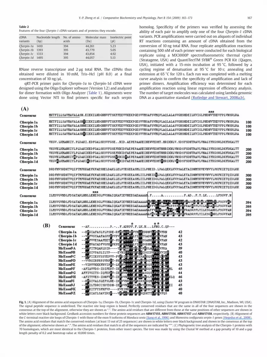
Table 2Features of the four Cfserpin-1 cDNA variants and of proteins they encode.
cDNAvariants
Nucleotide length(bp)
No. of aminoacids
Molecular mass(Da)
Isoelectric point(pI)
Cfserpin-1a 1410 394 44,261 5.23Cfserpin-1b 1393 395 43,779 5.05Cfserpin-1c 1333 394 43,854 4.92Cfserpin-1d 1495 395 44,057 5.13
167Y.-P. Zheng et al. / Comparative Biochemistry and Physiology, Part B 154 (2009) 165–173
RNase reverse transcriptase and 2 µg total RNA. The cDNAs thusobtained were diluted in 10 mM, Tris–Hcl (pH 8.0) at a finalconcentration of 10 ng/µL.
qRT-PCR primer pairs for Cfserpin-1a to Cfserpin-1d cDNA weredesigned using the Oligo Explorer software (Version 1.2) and analyzedfor dimer formation with Oligo Analyzer (Table 1). Alignments weredone using Vector NTI to find primers specific for each serpin
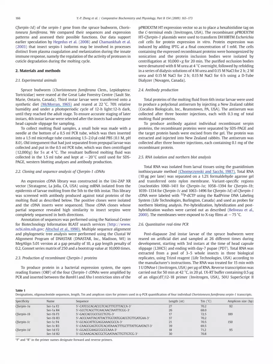
Fig. 1. (A) Alignment of the amino acid sequences of Cfserpin-1a, Cfserpin-1b, Cfserpin-1c andThe signal peptide sequence is underlined. The reactive site loop region is boxed. Perfectlconsensus at the top of the alignment, otherwise they are shown as “.”. The amino acid residwhite letters over black background. GenBank accession numbers for these protein sequencethe C-terminal reactive site loops of Cfserpin-1 with those of the exon 9 isoforms ofManducaThe amino acid residues that match the conserved residues (at least 13 out of 25 sequences)of the alignment, otherwise shown as “.”. The amino acid residues that match in all of the sequ70 homologues, which are most identical to the Cfserpin-1 proteins, from other insect speclength penalty of 0.2 and bootstrap value at 10,000 times.
homolog. Specificity of the primers was verified by assessing theability of each pair to amplify only one of the four Cfserpin-1 cDNAvariants. PCR amplifications were carried out on aliquots of individualRT reactions containing an amount of cDNA obtained from theconversion of 10 ng total RNA. Four replicate amplification reactionscontaining 500 nM of each primer were conducted for each biologicalreplicate using a MX3000P spectrofluorometric thermal cycler(Stratagene, USA) and QuantiTectTM SYBR® Green PCR Kit (Qiagen,USA), initiated with a 15-min incubation at 95 °C, followed by acycling regime of denatuation at 95 °C for 10 s; annealing andextension at 65 °C for 120 s. Each run was completed with a meltingcurve analysis to confirm the specificity of amplification and lack ofprimer dimers. Amplification efficiency was determined for eachamplification reaction using linear regression of efficiency analysis.The number of target molecules was calculated using lambda genomicDNA as a quantitative standard (Rutledge and Stewart, 2008a,b).
Cfserpin-1d, using ClusterW program in DNASTAR (DNASTAR, Inc., Madison, WI, USA).y conserved residues that are the same in all of the four sequences are shown in theues that are different from those at the same positions of other sequences are shown ins are ABW17155, ABW17156, ABW17157 and ABW17158, respectively. (B) Alignment ofsexta (Jiang et al., 1996) andMamestra configurata serpin-1 genes (Hegedus et al., 2008).are shown inwhite letters over black background and shown in the consensus at the topences are indicated by “*”. (C) Phylogenetic tree analysis of the Cfserpin-1 proteins withies. The tree was made by using the Clustal W method at a gap penalty of 10 and a gap
Fig. 1 (continued).
168 Y.-P. Zheng et al. / Comparative Biochemistry and Physiology, Part B 154 (2009) 165–173
2.7. SDS-PAGE and western blot analysis
Protein samples were denatured at 100 °C for 3 min in an equalvolume of 2× protein loading buffer (0.1 M Tris buffer, pH 6.8, 4% SDS,0.2% β-mercaptoethanol, 40% glycerol, and 0.002% bromophenolblue). SDS-PAGE was performed in 7.5% polyacrylamide gels, whichwere stained with Coomassie Brilliant Blue R-250 after electrophor-esis. For western blotting analysis, proteins were transferred from thepolyacrylamide gel to a nitrocellulose membrane, which was thenblocked with 3% bovine serum albumin (BSA) in 1× phosphatebuffered saline (PBS) for 30 min at room temperature, and incubatedwith the anti-Cfserpin-1 antibodies (1:2000) at room temperature for1 h. Goat anti-rabbit IgG conjugated with alkaline phosphatase wasused as the secondary antibody at a dilution of 1:2000. Nitrobluetetrazolium and 5-bromo-4-chloro-3-indolyl phosphate were used assubstrates for color development.
2.8. Binding assay
Binding of the recombinant Cfserpin-1a protein, which waspurified by a hexahistidine-tag affinity column, to elastase wasassayed by incubating 2 μg of the Cfserpin-1a recombinant proteinwith 8 μg of porcine pancreatic elastase (Sigma, Canada) in TBS buffer(25 mM Tris, 50 mM NaCl, pH 8.0) at 22 °C for 5 min. The proteinmixtures were then separated in 7.5% polyacrylamide native gel todetermine whether protein complexes had formed.
3. Results
3.1. Cloning of Cfserpin-1 cDNAs and properties of the Cfserpin-1 proteins
An expression cDNA library constructed with mRNA isolated fromthe epidermis of larvae molting from the 5th to the 6th stadium was
169Y.-P. Zheng et al. / Comparative Biochemistry and Physiology, Part B 154 (2009) 165–173
screened using the antiserum raised against molting fluid proteins.Out of 9×10⁴ colonies screened, 278 positive clones were identified.The inserts of these positive clones were partially sequenced. Thirteenclones (4.5% of the positive clones) were found to encode proteinsdisplaying similarity to insect serpins. These clones were thencompletely sequenced. Four full-length cDNA variants closely relatedto theManduca sexta serpin-1 gene (Jiang et al., 1996), while the othernine clones encoded other serpins (data not shown). On the basis oftheir high levels of identity with the Manduca sexta serpin-1 cDNAs,these four serpin-1 cDNAs were named Cfserpin-1a, Cfserpin-1b, Cfserpin-1c and Cfserpin-1d (GenBank accession numbers: EU040207, EU040208,EU040209 and EU040210, respectively),. Table 2 summarizes the lengthof the nucleotide and amino acid sequences of the ORFs, as well asmolecularmass and isoelectric points (pI) of these fourCfserpin-1 cDNAs.A 16 amino acid signal peptide was predicted for all four deducedproteins using the SignalP 3.0 program (http://www.cbs.dtu.dk/services/SignalP/; Bendtsen et al., 2004) (Fig. 1). Amino acid sequencealignment of the four Cfserpin-1 variants revealed that they had almostidentical N-termini and central regions, whereas they displayeddifferent C-terminal ends, starting at residue 353 (Fig. 1A). The highlysimilar regions correspond to exons 1–8 of the Manduca sexta (Jianget al., 1996) and Mamestra configurata (Hegedus et al., 2008) serpin-1genes. The C-terminal regions (i.e., the reactive loop region) of Cfserpin-1a and Cfserpin-1b proteins were most similar to that encoded by exon9Z of the Manduca sexta serpin-1 gene (ca 40% identity at the aminoacid level; Fig. 1B). The C-terminal region of Cfserpin-1c protein wasmost similar to that encoded by exon 9G of the Mamestra configurataserpin-1 gene (46% identity) and the C-terminal region of Cfserpin-1d was most similar to that encoded by exon 9D of the Mamestraconfigurata serpin-1 gene (37% identity). Only three amino acid residues(Phe, Phe andGly)within the reactive center loopwere identical amongall Choristoneura fumiferana, Manduca sexta and Mamestra configurataserpin-1 proteins, although a few others were common to a majority ofserpins (Fig. 1B).
Phylogenetic tree analysis of the Cfserpin-1 proteins with 70homologues from other insect species revealed three groups: theLepidoptera group, including the proteins from Choristoneura fumi-ferana (this study), Manduca sexta (Jiang et al., 1996), Mamestra con-
Fig. 2. SDS-PAGE analysis of Cfserpin-1a, Cfserpin-1b, Cfserpin-1c and Cfserpin-1d proteinseparation after centrifugation to pellet the protein occlusion bodies. Lane 1: total proteinsfraction. (C) Purified and refolded proteins, whichwere used for the protein binding activity afor anti-Cfserpin-1a, anti-Cfserpin-1b, anti-Cfserpin-1c and anti-Cfserpin-1d antibodies, restransformed with the pPROEXTM HTempty vector alone. 1a–1d: the proteins from the bacter
figurata (Hegedus et al., 2008), B. mori (Sasaki, 1991), Plutellaxylostella (Song et al., 2008), Lonomia oblique (Veiga et al., 2005),and Spodoptera exigua, was separated from the Diptera group, whichincluded Drosophila, Anopheles gambiae, Culex quinquefasciatus, Cte-nocephalides felis and Lutzomyia longipalpis, and the Coleoptera group,which included Tribolium castaneum and Sphenophorus levis (Fig. 1C).It appears that the different isoforms of lepidopteran serpin-1 proteinsare paralogues diverged from gene duplication or alternative splicingwithin a species after speciation, because these isoforms weregrouped into the separate species clusters. The selected serpins ofDiptera and Coleoptera for phylogenetic tree analysis were the mosthomologous to the Cfserpin-1 proteins, but it is not clear yet if theycan be classified into the serpin-1 gene family, although they do havethe similar conserved domains to the serpin-1 proteins and shareabout 30–40% identities to the lepidopteran serpin-1 proteins.Additionally, they appeared not to have as many as paralogues asthe lepidopteran serpin-1 genes.
3.2. In vitro production of recombinant Cfserpin-1
All four Cfserpin-1 variant proteins were produced as hexahisti-dine-tagged fusion proteins in E. coli expression system (Fig. 2A). Therecombinant proteins were produced as protein inclusion bodies(Fig. 2B). The apparent molecular mass of the recombinant proteins,as assessed by SDS-PAGE, was approximately 45 kDa, close to thecalculated size based on the deduced amino acid sequences. Theantibody generated against individual recombinant Cfserpin-1 pro-teins cross-reacted with each of the other three recombinant serpins(Fig. 2D). In addition, the anti-Cfserpin-1b antibody showed cross-reactivity with an unknown high molecular-mass protein of bacterialorigin (Fig. 2Db).
3.3. Assessment of Cfserpin-1 transcript and protein abundance in vivo
Quantitative PCR assessment of transcript abundance was con-ducted for the four Cfserpin-1 variants as a function of developmentalstage (Fig. 3A). Absolute amounts of transcripts varied widely amongthe four Cfserpin-1 cDNA variants, with Cfserpin-1a being the most
s produced in vitro. (A) Total proteins from the transformed E. coli cells; (B) proteinfrom the cell lysate; Lane 2: supernatant fraction of the cell lysate; Lane 3: the pelletssays. (D)Western blot analysis of the specificity of anti-Cfserpin-1 antibodies. (a–d) arepectively. M: protein mass marker; CK: control, the proteins isolated from the bacteriaia transformed with Cfserpin-1a, Cfserpin-1b, Cfserpin-1c and Cfserpin-1d, respectively.
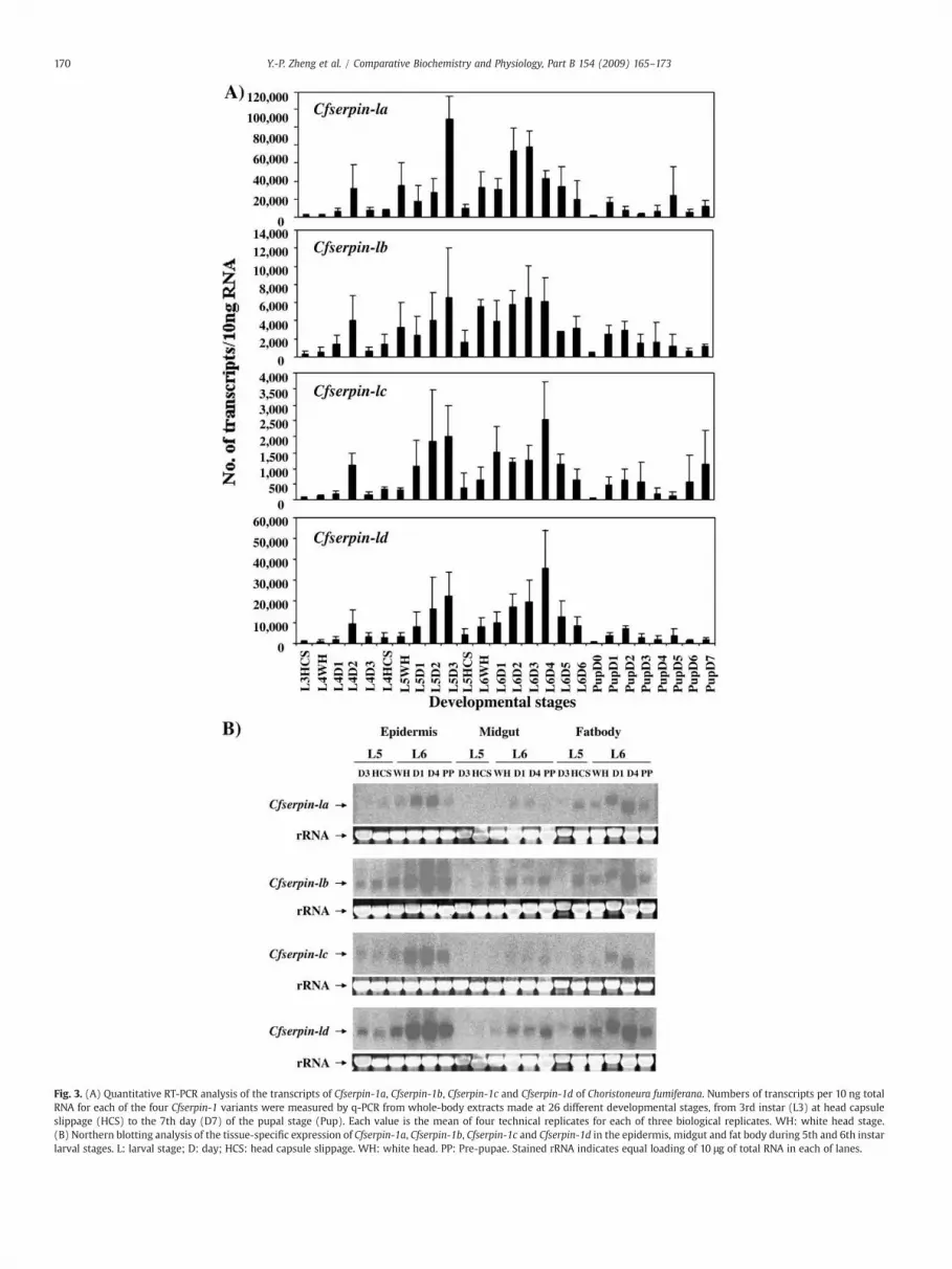
Fig. 3. (A) Quantitative RT-PCR analysis of the transcripts of Cfserpin-1a, Cfserpin-1b, Cfserpin-1c and Cfserpin-1d of Choristoneura fumiferana. Numbers of transcripts per 10 ng totalRNA for each of the four Cfserpin-1 variants were measured by q-PCR from whole-body extracts made at 26 different developmental stages, from 3rd instar (L3) at head capsuleslippage (HCS) to the 7th day (D7) of the pupal stage (Pup). Each value is the mean of four technical replicates for each of three biological replicates. WH: white head stage.(B) Northern blotting analysis of the tissue-specific expression of Cfserpin-1a, Cfserpin-1b, Cfserpin-1c and Cfserpin-1d in the epidermis, midgut and fat body during 5th and 6th instarlarval stages. L: larval stage; D: day; HCS: head capsule slippage. WH: white head. PP: Pre-pupae. Stained rRNA indicates equal loading of 10 μg of total RNA in each of lanes.
170 Y.-P. Zheng et al. / Comparative Biochemistry and Physiology, Part B 154 (2009) 165–173
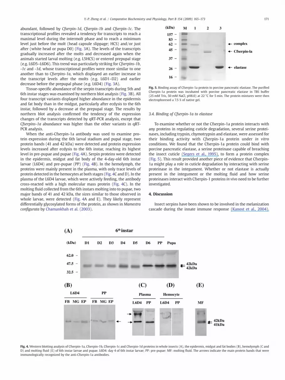
Fig. 5. Binding assay of Cfserpin-1a protein to porcine pancreatic elastase. The purifiedCfserpin-1a protein was incubated with porcine pancreatic elastase in TBS buffer(25 mM Tris, 50 mM NaCl, pH8.0) at 22 °C for 5 min. The protein mixtures were thenelectrophoresed a 7.5 % of native gel.
171Y.-P. Zheng et al. / Comparative Biochemistry and Physiology, Part B 154 (2009) 165–173
abundant, followed by Cfserpin-1d, Cfserpin-1b and Cfserpin-1c. Thetranscriptional profiles revealed a tendency for transcripts to reach amaximal level during the intermolt phase and to reach a minimumlevel just before the molt (head capsule slippage; HCS) and/or justafter (white head or pupa D0) (Fig. 3A). The levels of the transcriptsgradually increased after the molts and decreased again when theanimals started larval molting (e.g. L5HCS) or entered prepupal stage(e.g. L6D5–L6D6). This trend was particularly striking for Cfserpins-1b,-1c and -1d, whose transcriptional profiles were more similar to oneanother than to Cfserpins-1a, which displayed an earlier increase inthe transcript levels after the molts (e.g. L6D1–D2) and earlierdecrease before the prepupal phase (e.g. L6D4) (Fig. 3A).
Tissue-specific abundance of the serpin transcripts during 5th and6th instar stages was examined by northern blot analysis (Fig. 3B). Allfour transcript variants displayed higher abundance in the epidermisand fat body than in the midgut, particularly after ecdysis to the 6thinstar, followed by a decrease at the prepupal stage. The results bynorthern blot analysis confirmed the tendency of the expressionchanges of the transcripts detected by qRT-PCR analysis, except thatCfserpins-1a abundance was higher than the other variants in qRT-PCR analysis.
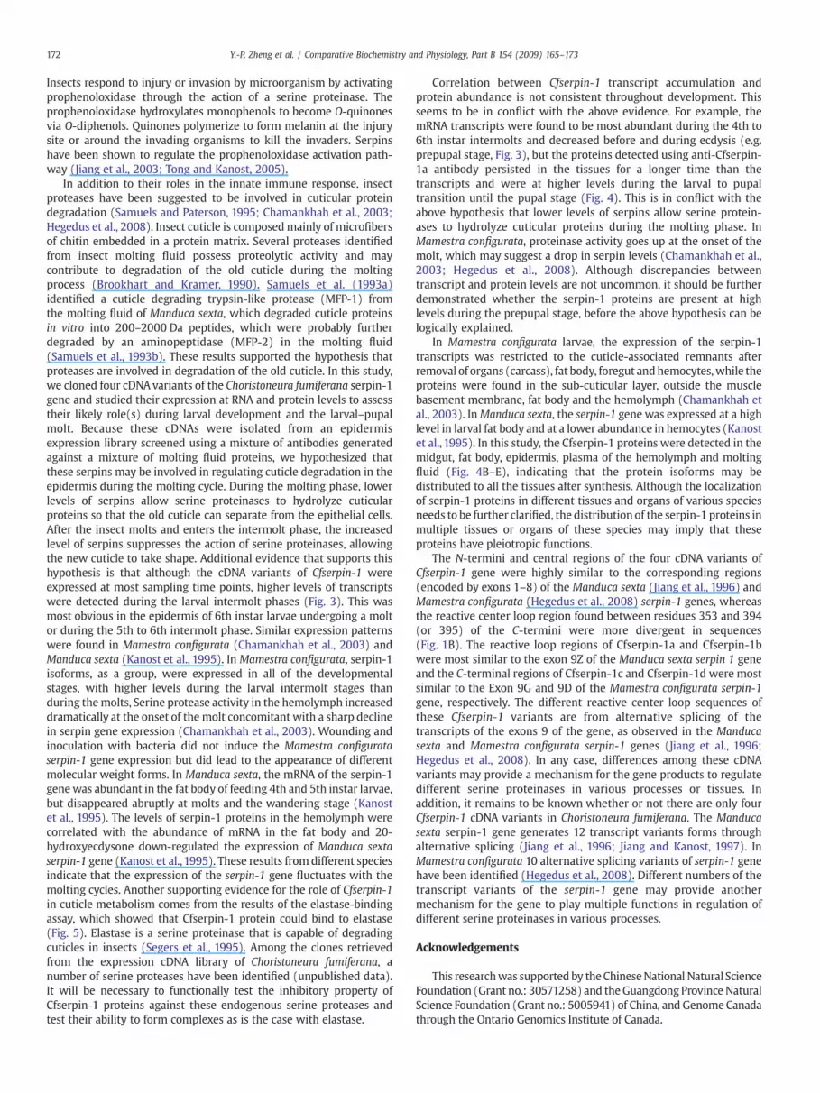
When the anti-Cfserpin-1a antibody was used to examine pro-tein expression during the 6th larval stadium and pupal stage, twoprotein bands (41 and 42 kDa) were detected and protein expressionlevels increased after ecdysis to the 6th instar, reaching its highestlevel in pre-pupae and pupae (Fig. 4A). Serpin proteins were detectedin the epidermis, midgut and fat body of the 4-day-old 6th instarlarvae (L6D4) and pre-pupae (PP) (Fig. 4B). In the hemolymph, theproteins were mainly present in the plasma, with only trace levels ofprotein detected in the hemocytes at both stages (Fig. 4C andD). In theplasma of the L6D4 larvae, which were actively feeding, the antibodycross-reacted with a high molecular mass protein (Fig. 4C). In themolting fluid collected from the 6th instars molting into to pupae, twomajor bands of 41 and 42 kDa, the sizes similar to those observed inwhole larvae, were detected (Fig. 4A and E). They likely representdifferentially glycosylated forms of the protein, as shown in Mamestraconfigurata by Chamankhah et al. (2003).
Fig. 4.Western blotting analysis of Cfserpin-1a, Cfserpin-1b, Cfserpin-1c and Cfserpin-1d proD) and molting fluid (E) of 6th instar larvae and pupae. L6D4: day 4 of 6th instar larvae; PPimmunologically recognized by the anti-Cfserpin-1a antibodies.
3.4. Binding of Cfserpin-1a to elastase
To examine whether or not the Cfserpin-1a protein interacts withany proteins in regulating cuticle degradation, several serine protei-nases, including trypsin, chymotrypsin and elastase, were assessed fortheir binding activity with Cfserpin-1a protein under in vitroconditions. We found that the Cfserpin-1a protein could bind withporcine pancreatic elastase, a serine proteinase capable of breachingthe insect cuticle (Segers et al., 1995), to form a protein complex(Fig. 5). This result provided another piece of evidence that Cfserpin-1a might play a role in cuticle degradation by interacting with serineproteinase in the integument. Whether or not elastase is actuallypresent in the integument or the molting fluid and how serineproteinases interact with Cfserpin-1 proteins in vivo need to be furtherinvestigated.
4. Discussion
Insect serpins have been shown to be involved in the melanizationcascade during the innate immune response (Kanost et al., 2004).
teins in whole insects (A), the epidermis, midgut and fat bodies (B), hemolymph (C and: pre-pupae: MF: molting fluid. The arrows indicate the main protein bands that were
172 Y.-P. Zheng et al. / Comparative Biochemistry and Physiology, Part B 154 (2009) 165–173
Insects respond to injury or invasion by microorganism by activatingprophenoloxidase through the action of a serine proteinase. Theprophenoloxidase hydroxylates monophenols to become O-quinonesvia O-diphenols. Quinones polymerize to form melanin at the injurysite or around the invading organisms to kill the invaders. Serpinshave been shown to regulate the prophenoloxidase activation path-way (Jiang et al., 2003; Tong and Kanost, 2005).
In addition to their roles in the innate immune response, insectproteases have been suggested to be involved in cuticular proteindegradation (Samuels and Paterson, 1995; Chamankhah et al., 2003;Hegedus et al., 2008). Insect cuticle is composedmainly of microfibersof chitin embedded in a protein matrix. Several proteases identifiedfrom insect molting fluid possess proteolytic activity and maycontribute to degradation of the old cuticle during the moltingprocess (Brookhart and Kramer, 1990). Samuels et al. (1993a)identified a cuticle degrading trypsin-like protease (MFP-1) fromthe molting fluid of Manduca sexta, which degraded cuticle proteinsin vitro into 200–2000 Da peptides, which were probably furtherdegraded by an aminopeptidase (MFP-2) in the molting fluid(Samuels et al., 1993b). These results supported the hypothesis thatproteases are involved in degradation of the old cuticle. In this study,we cloned four cDNAvariants of the Choristoneura fumiferana serpin-1gene and studied their expression at RNA and protein levels to assesstheir likely role(s) during larval development and the larval–pupalmolt. Because these cDNAs were isolated from an epidermisexpression library screened using a mixture of antibodies generatedagainst a mixture of molting fluid proteins, we hypothesized thatthese serpins may be involved in regulating cuticle degradation in theepidermis during the molting cycle. During the molting phase, lowerlevels of serpins allow serine proteinases to hydrolyze cuticularproteins so that the old cuticle can separate from the epithelial cells.After the insect molts and enters the intermolt phase, the increasedlevel of serpins suppresses the action of serine proteinases, allowingthe new cuticle to take shape. Additional evidence that supports thishypothesis is that although the cDNA variants of Cfserpin-1 wereexpressed at most sampling time points, higher levels of transcriptswere detected during the larval intermolt phases (Fig. 3). This wasmost obvious in the epidermis of 6th instar larvae undergoing a moltor during the 5th to 6th intermolt phase. Similar expression patternswere found in Mamestra configurata (Chamankhah et al., 2003) andManduca sexta (Kanost et al., 1995). In Mamestra configurata, serpin-1isoforms, as a group, were expressed in all of the developmentalstages, with higher levels during the larval intermolt stages thanduring themolts, Serine protease activity in the hemolymph increaseddramatically at the onset of themolt concomitant with a sharp declinein serpin gene expression (Chamankhah et al., 2003). Wounding andinoculation with bacteria did not induce the Mamestra configurataserpin-1 gene expression but did lead to the appearance of differentmolecular weight forms. In Manduca sexta, the mRNA of the serpin-1genewas abundant in the fat body of feeding 4th and 5th instar larvae,but disappeared abruptly at molts and the wandering stage (Kanostet al., 1995). The levels of serpin-1 proteins in the hemolymph werecorrelated with the abundance of mRNA in the fat body and 20-hydroxyecdysone down-regulated the expression of Manduca sextaserpin-1 gene (Kanost et al., 1995). These results from different speciesindicate that the expression of the serpin-1 gene fluctuates with themolting cycles. Another supporting evidence for the role of Cfserpin-1in cuticle metabolism comes from the results of the elastase-bindingassay, which showed that Cfserpin-1 protein could bind to elastase(Fig. 5). Elastase is a serine proteinase that is capable of degradingcuticles in insects (Segers et al., 1995). Among the clones retrievedfrom the expression cDNA library of Choristoneura fumiferana, anumber of serine proteases have been identified (unpublished data).It will be necessary to functionally test the inhibitory property ofCfserpin-1 proteins against these endogenous serine proteases andtest their ability to form complexes as is the case with elastase.
Correlation between Cfserpin-1 transcript accumulation andprotein abundance is not consistent throughout development. Thisseems to be in conflict with the above evidence. For example, themRNA transcripts were found to be most abundant during the 4th to6th instar intermolts and decreased before and during ecdysis (e.g.prepupal stage, Fig. 3), but the proteins detected using anti-Cfserpin-1a antibody persisted in the tissues for a longer time than thetranscripts and were at higher levels during the larval to pupaltransition until the pupal stage (Fig. 4). This is in conflict with theabove hypothesis that lower levels of serpins allow serine protein-ases to hydrolyze cuticular proteins during the molting phase. InMamestra configurata, proteinase activity goes up at the onset of themolt, which may suggest a drop in serpin levels (Chamankhah et al.,2003; Hegedus et al., 2008). Although discrepancies betweentranscript and protein levels are not uncommon, it should be furtherdemonstrated whether the serpin-1 proteins are present at highlevels during the prepupal stage, before the above hypothesis can belogically explained.
In Mamestra configurata larvae, the expression of the serpin-1transcripts was restricted to the cuticle-associated remnants afterremoval of organs (carcass), fat body, foregut andhemocytes,while theproteins were found in the sub-cuticular layer, outside the musclebasement membrane, fat body and the hemolymph (Chamankhah etal., 2003). InManduca sexta, the serpin-1 gene was expressed at a highlevel in larval fat body and at a lower abundance in hemocytes (Kanostet al., 1995). In this study, the Cfserpin-1 proteins were detected in themidgut, fat body, epidermis, plasma of the hemolymph and moltingfluid (Fig. 4B–E), indicating that the protein isoforms may bedistributed to all the tissues after synthesis. Although the localizationof serpin-1 proteins in different tissues and organs of various speciesneeds to be further clarified, the distribution of the serpin-1 proteins inmultiple tissues or organs of these species may imply that theseproteins have pleiotropic functions.
The N-termini and central regions of the four cDNA variants ofCfserpin-1 gene were highly similar to the corresponding regions(encoded by exons 1–8) of the Manduca sexta (Jiang et al., 1996) andMamestra configurata (Hegedus et al., 2008) serpin-1 genes, whereasthe reactive center loop region found between residues 353 and 394(or 395) of the C-termini were more divergent in sequences(Fig. 1B). The reactive loop regions of Cfserpin-1a and Cfserpin-1bwere most similar to the exon 9Z of the Manduca sexta serpin 1 geneand the C-terminal regions of Cfserpin-1c and Cfserpin-1d were mostsimilar to the Exon 9G and 9D of the Mamestra configurata serpin-1gene, respectively. The different reactive center loop sequences ofthese Cfserpin-1 variants are from alternative splicing of thetranscripts of the exons 9 of the gene, as observed in the Manducasexta and Mamestra configurata serpin-1 genes (Jiang et al., 1996;Hegedus et al., 2008). In any case, differences among these cDNAvariants may provide a mechanism for the gene products to regulatedifferent serine proteinases in various processes or tissues. Inaddition, it remains to be known whether or not there are only fourCfserpin-1 cDNA variants in Choristoneura fumiferana. The Manducasexta serpin-1 gene generates 12 transcript variants forms throughalternative splicing (Jiang et al., 1996; Jiang and Kanost, 1997). InMamestra configurata 10 alternative splicing variants of serpin-1 genehave been identified (Hegedus et al., 2008). Different numbers of thetranscript variants of the serpin-1 gene may provide anothermechanism for the gene to play multiple functions in regulation ofdifferent serine proteinases in various processes.
Acknowledgements
This researchwas supported by the ChineseNational Natural ScienceFoundation (Grant no.: 30571258) and theGuangdong Province NaturalScience Foundation (Grant no.: 5005941) of China, and Genome Canadathrough the Ontario Genomics Institute of Canada.
173Y.-P. Zheng et al. / Comparative Biochemistry and Physiology, Part B 154 (2009) 165–173
References
Altschul, S.F., Gish, W., Miller, W., Myers, E.W., Lipman, D.J., 1990. Basic local alignmentsearch tool. J. Mol. Biol. 215, 403–410.
Beliveau, C., Laforge, M., Cusson, M., Bellemare, G., 2000. Expression of a Tranosemarostrale polydnavirus gene in the spruce budworm, Choristoneura fumiferana.J. Gen. Virol. 81, 1871–1880.
Bendtsen, J.D., Nielsen, H., von Heijne, G., Brunak, S., 2004. Improved prediction of signalpeptides: SignalP 3.0. J. Mol. Biol. 340, 783–795.
Brookhart, G.L., Kramer, K.J., 1990. Proteinases inmolting fluid of the tobacco hornworm,Manduca sexta. Insect Biochem. 20, 467–477.
Chamankhah, M., Braun, L., Visal-Shah, S., O'Grady, M., Baldwin, D., Shi, X., Hemmingsen,S.M., Alting-Mees, M., Hegedus, D.D., 2003. Mamestra configurata serpin-1 homo-logues: implications for a regulatory role for serpins in molting. Insect Biochem. Mol.Biol. 33, 355–369.
Chomyczynski, P., Sacchi, N., 1987. Methods of RNA isolation by acid guanidiniumthiocyanate-phenol-chloroform extraction. Anal. Biochem. 162, 156–159.
Christophides, G.K., Zdobnov, E., Barillas-Mury, C., Birney, E., Blandin, S., Blass, C., Brey,P.T., Collins, F.H., Danielli, A., Dimopoulos, G., Hetru, C., Hoa, N.T., Hoffmann, J.A.,Kanzok, S.M., Letunic, I., Levashina, E.A., Loukeris, T.G., Lycett, G., Meister, S.,Michel, K., Moita, L.F., Muller, H.M., Osta, M.A., Paskewitz, S.M., Reichhart, J.M.,Rzhetsky, A., Troxler, L., Vernick, K.D., Vlachou, D., Volz, J., von Mering, C., Xu, J.,Zheng, L., Bork, P., Kafatos, F.C., 2002. Immunity-related genes and gene families inAnopheles gambiae. Science 298, 159–165.
Danielli, A., Kafatos, F.C., Loukeris, T.G., 2003. Cloning and characterization of fourAnopheles gambiae serpin isoforms, differentially induced in the midgut by Plas-modium berghei invasion. J. Biol. Chem. 278, 4184–4193.
Gan, H., Wang, Y., Jiang, H., Mita, K., Kanost, M.R., 2001. A bacteria-induced, intracellularserpin in granular hemocytes of Manduca sexta. Insect Biochem. Mol. Biol. 31,887–898.
Hegedus, D.D., Erlandson, M., Baldwin, D., Hou, X., Chamankhah, M., 2008. Differentialexpansion and evolution of the exon family encoding the serpin-1 reactive centreloop has resulted in divergent serpin repertoires among the Lepidoptera. Gene 418,15–21.
Irving, J.A., Pike, R.N., Lesk, A.M., Whisstock, J.C., 2000. Phylogeny of the serpinsuperfamily: implications of patterns of amino acid conservation for structure andfunction. Genome Res. 10, 1845–1864.
Jiang, H.B., Kanost, M.R., 1997. Characterization and functional analysis of 12 naturallyoccurring reactive site variants of serpin-1 from Manduca sexta. J. Biol. Chem. 272,1082–1087.
Jiang, H., Kanost, M.R., 2000. The clip-domain family of serine proteinases in arthropods.Insect Biochem. Mol. Biol. 30, 95–105.
Jiang, H., Wang, Y., Kanost, M.R., 1994. Mutually exclusive exon use and reactive centerdiversity in insect serpins. J. Biol. Chem. 269, 55–58.
Jiang, H., Wang, Y., Huang, Y., Mulnix, A.B., Kadel, J., Cole, K., Kanost, M.R., 1996. Organi-zation of serpin gene-1 fromManduca sexta. Evolution of a family of alternate exonsencoding the reactive site loop. J. Biol. Chem. 271, 28017–28023.
Jiang, H., Wang, Y., Yu, X.Q., Zhu, Y., Kanost, M., 2003. Prophenoloxidase-activatingproteinase-3 (PAP-3) from Manduca sexta hemolymph: a clip-domain serineproteinase regulated by serpin-1J and serine proteinase homologs. Insect Biochem.Mol. Biol. 33, 1049–1060.
Kanost, M.R., Prasad, S.V., Huang, Y.L., Willott, E., 1995. Regulation of serpin gene-1 inManduca sexta. Insect. Biochem. Mol. Biol. 25, 285–291.
Kanost, M.R., Jiang, H., Yu, X.Q., 2004. Innate immune responses of a lepidopteran insect,Manduca sexta. Immunol. Rev. 198, 97–105.
McMorran, A., 1965. A synthetic diet for the spruce budworm, Choristoneura fumiferana(Lepidoptera: Tortricidae). Can. Entomol. 97, 58–62.
Potempa, J., Korzus, E., Travis, J., 1994. The serpin superfamily of proteinase inhibitors:structure, function, and regulation. J. Biol. Chem. 269, 15957–15960.
Rutledge, R.G., Stewart, D., 2008a. A kinetic-based sigmoidal model for the polymerasechain reaction and its application to high-capacity absolute quantitative real-timePCR. BMC Biotechnol. 8, 47.
Rutledge, R.G., Stewart, D., 2008b. Critical evaluation of methods used to determineamplification efficiency refutes the exponential character of real-time PCR. BMCMol. Biol. 9, 96.
Samuels, R.I., Paterson, I.C., 1995. Cuticle degrading proteases from insect moulting fluidand culture filtrates of entomopathogenic fungi. Comp. Biochem. Physiol. 110B,661–669.
Samuels, R.I., Charnley, A.K., Reynolds, S.E., 1993a. A cuticle degrading proteinase fromthe moulting fluid of the tobacco hornworm, Manduca sexta. Insect Biochem. Mol.Biol. 23, 607–614.
Samuels, R.I., Charnley, A.K., Reynolds, S.E.,1993b. An aminopeptidase from themoultingfluid of the tobacco hornworm, Manduca sexta. Insect Biochem. Mol. Biol. 23,615–620.
Sasaki, T., 1991. Patchwork-structure serpins from silkworm (Bombyx mori) larvalhemolymph. Eur. J. Biochem. 202, 255–261.
Scherfer, C., Tang, H., Kambris, Z., Lhocine, N., Hashimoto, C., Lemaitre, B., 2008.Drosophila Serpin-28D regulates hemolymph phenoloxidase activity and adultpigmentation. Dev. Biol. 323, 189–196.
Segers, R., Butt, T.M., Keen, J.N., Kerry, B.R., Peberdy, J.F., 1995. The subtilisins of theinvertebrate mycopathogens Verticillium chlamydosporium and Metarhizium aniso-pliae are serologically and functionally related. FEMS Microbiol. Lett. 126, 227–231.
Song, K., Jung, M., Eum, J., Hwang, I., Han, S., 2008. Proteomic analysis of parasitizedPlutella xylostella larvae plasma. J. Insect Physiol. 54, 1271–1280.
Tong, Y., Kanost, M.R., 2005.Manduca sexta serpin-4 and serpin-5 inhibit the prophenoloxidase activation pathway: cDNA cloning, protein expression, and characteriza-tion. J. Biol. Chem. 280, 14923–14931.
Veiga, A.B., Ribeiro, J.M., Guimaraes, J.A., Francischetti, I.M., 2005. A catalog for thetranscripts from the venomous structures of the caterpillar Lonomia obliqua:identification of the proteins potentially involved in the coagulation disorder andhemorrhagic syndrome. Gene 355, 11–27.
Copyright © 2022 FDOKUMEN