Employer Duties and Defenses to OSHA Violations-Janice Gail Murphy
Upload
independentCategory
view
0download
0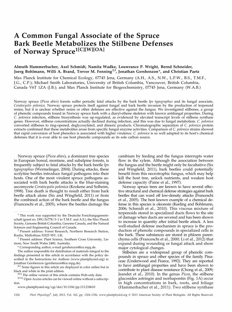
A Common Fungal Associate of the SpruceBark Beetle Metabolizes the Stilbene Defensesof Norway Spruce1[C][W][OA]
Almuth Hammerbacher, Axel Schmidt, Namita Wadke, Louwrance P. Wright, Bernd Schneider,Joerg Bohlmann, Willi A. Brand, Trevor M. Fenning2,3, Jonathan Gershenzon*, and Christian Paetz
Max Planck Institute for Chemical Ecology, 07745 Jena, Germany (A.H., A.S., N.W., L.P.W., B.S., T.M.F.,J.G., C.P.); Michael Smith Laboratories, University of British Columbia, Vancouver, British Columbia,Canada V6T 1ZA (J.B.); and Max Planck Institute for Biogeochemistry, 07745 Jena, Germany (W.A.B.)
Norway spruce (Picea abies) forests suffer periodic fatal attacks by the bark beetle Ips typographus and its fungal associate,Ceratocystis polonica. Norway spruce protects itself against fungal and bark beetle invasion by the production of terpenoidresins, but it is unclear whether resins or other defenses are effective against the fungus. We investigated stilbenes, a groupof phenolic compounds found in Norway spruce bark with a diaryl-ethene skeleton with known antifungal properties. DuringC. polonica infection, stilbene biosynthesis was up-regulated, as evidenced by elevated transcript levels of stilbene synthasegenes. However, stilbene concentrations actually declined during infection, and this was due to fungal metabolism. C. polonicaconverted stilbenes to ring-opened, deglycosylated, and dimeric products. Chromatographic separation of C. polonica proteinextracts confirmed that these metabolites arose from specific fungal enzyme activities. Comparison of C. polonica strains showedthat rapid conversion of host phenolics is associated with higher virulence. C. polonica is so well adapted to its host’s chemicaldefenses that it is even able to use host phenolic compounds as its sole carbon source.
Norway spruce (Picea abies), a dominant tree speciesin European boreal, montane, and subalpine forests, isfrequently subject to fatal attacks by the bark beetle Ipstypographus (Wermelinger, 2004). During attacks, thesescolytine beetles introduce fungal pathogens into theirhosts. One of the most virulent spruce pathogens as-sociated with bark beetle attacks is the blue-stainingascomycete Ceratocystis polonica (Krokene and Solheim,1998). Tree death is thought to result either from barkbeetle attack alone (Six and Wingfield, 2011) or fromthe combined action of the bark beetle and the fungus(Franceschi et al., 2005), where the beetles damage the
cambium by feeding and the fungus interrupts waterflow in the xylem. Although the association betweenthe fungus and the beetle might only be facultative (Sixand Wingfield, 2011), bark beetles could potentiallybenefit from this necrotrophic fungus, which may helpkill the host tree, unlock nutrients, and weaken hostdefense capacity (Paine et al., 1997).
Norway spruce trees are known to have several effec-tive structural and chemical defense strategies against barkbeetles that can ward off low-density attacks (Franceschiet al., 2005). The best known example of a chemical de-fense in this species is oleoresin (Keeling and Bohlmann,2006; Schmidt et al., 2010). This viscous mixture ofterpenoids stored in specialized ducts flows to the siteof damage when ducts are severed and has been shownto increase in quantity after initial beetle attack. A lesswell-studied defense mechanism in spruce is the pro-duction of phenolic compounds in specialized cells inthe bark. These substances are stored in phloem paren-chyma cells (Franceschi et al., 2000; Li et al., 2012) thatexpand during wounding or fungal attack and showmajor cytological changes.
Stilbenes are a widespread group of phenolic com-pounds in spruce and other species of the family Pina-ceae (Underwood and Pearce, 1992). They are reportedto have antifungal properties and have been shown tocontribute to plant disease resistance (Chong et al., 2009;Jeandet et al., 2010). In the genus Picea, the stilbeneglucosides astringin and isorhapontin (Fig. 1A) occurin high concentrations in bark, roots, and foliage(Hammerbacher et al., 2011). Two stilbene synthase
1 This work was supported by the Deutsche Forschungsgemein-schaft (grant no. DFG Fe778/3–1 to T.M.F. and A.S.), the Max PlanckSociety, Genome British Columbia, Genome Canada, and the NaturalSciences and Engineering Council of Canada.
2 Present address: Forest Research, Northern Research Station,Roslin, Midlothian EH25 9SY, UK.
3 Present address: Plant Science, Southern Cross University, Lis-more, New South Wales 2480, Australia.
* Corresponding author; e-mail [email protected] author responsible for distribution of materials integral to the
findings presented in this article in accordance with the policy de-scribed in the Instructions for Authors (www.plantphysiol.org) is:Jonathan Gershenzon ([email protected]).
[C] Some figures in this article are displayed in color online but inblack and white in the print edition.
[W] The online version of this article contains Web-only data.[OA] Open Access articles can be viewed online without a subscrip-
tion.www.plantphysiol.org/cgi/doi/10.1104/pp.113.218610
1324 Plant Physiology�, July 2013, Vol. 162, pp. 1324–1336, www.plantphysiol.org � 2013 American Society of Plant Biologists. All Rights Reserved.
(STS) enzymes that contribute to the biosynthesis of thesecompounds have been described in Norway spruce(Hammerbacher et al., 2011). These enzymes seem toplay a role in tree defense, since fungal infection inducedelevated amounts of STS transcript (Hammerbacheret al., 2011) and increased enzyme levels (Brignolaset al., 1995). However, there are contradictory reports onstilbene glucoside accumulation in spruce bark duringfungal infection. Significant increases in astringin wereobserved after inoculation of Picea glauca saplings withavirulent C. polonica (Hammerbacher et al., 2011).However, stilbene glycoside concentrations in matureNorway spruce were stable or even declined duringthe course of C. polonica infection (Brignolas et al., 1995;Viiri et al., 2001; Li et al., 2012).After host infection, fungi follow different strategies
to gain access to host nutrients. Biotrophic fungi ac-quire nutrients directly from living cells by penetratingthem and causing little visible damage (Voegele andMendgen, 2003). Necrotrophic fungi such as C. polonica
lyse cells in order to release nutrients that are thenassimilated by the fungi (Oliver and Solomon, 2010).During plant cell lysis, potential antifungal defensecompounds may come into contact with necrotrophicfungi. However, it is not yet known how effective Nor-way spruce defense compounds are against C. polonicaand whether this specialized, bark beetle-vectored path-ogen has developed any resistance to them. This missinginformation may be critical in understanding howC. polonica contributes to the success of bark beetle attacksand why so many mutualistic relationships between barkbeetles and blue-staining fungi have evolved.
In this work, we demonstrate that C. polonica cancircumvent the antifungal activity of Norway sprucestilbenes during infection. Although high levels of STStranscripts accumulate in fungus-infected bark, a netloss of stilbene glucosides was detected at the site ofinfection. This reduction in stilbenes was explained byfungal biotransfomation processes, including the for-mation of stilbene dimers, aglycones, and ring-opened
Figure 1. Stilbene biosynthesis and stilbene concentrations in Norway spruce bark in response to fungal infection. A, Route ofstilbene biosynthesis. STS transcription (B) and concentrations of the stilbene glucosides astringin (C) and isorhapontin (D) weremeasured in Norway spruce saplings after inoculation and infection by two C. polonica isolates and after control woundingtreatment (inoculation with sterile agar). Error bars represent SE (n = 5). Significant differences were detected between 14 and28 d after treatment for both fungus-infected and wounded control saplings (P , 0.005). [See online article for color version ofthis figure.]
Plant Physiol. Vol. 162, 2013 1325
Fungal Biotransformation of Spruce Stilbenes

lactones, which may represent the first step of theb-ketoadipate pathway for microbial utilization ofaromatic compounds as a carbon source. We could alsoshow that different C. polonica isolates follow differentstilbene biotransformation strategies and that rapid bio-transformation and formation of ring-opened lactonesis associated with greater levels of fungal virulence inNorway spruce bark.
RESULTS
Fungal Inoculation of Norway Spruce Bark IncreasesStilbene Biosynthetic Gene Transcripts But ReducesStilbene Accumulation
When Norway spruce saplings were inoculated withthe blue-stain fungus C. polonica, the transcript levels ofSTS genes PaSTS1 and PaSTS2 (Hammerbacher et al.,2011) increased significantly compared with the sterileagar-inoculated control (P = 0.014). The same pattern wasobserved for two different C. polonica isolates (Fig. 1B).However, the levels of the stilbene glucosides, astringinand isorhapontin, declined over the 28-d time course afterinoculation of the two fungal isolates (P = 0.037), whilethey increased in the sterile agar-inoculated control (Fig.1, C and D).
To determine if C. polonica was the agent responsiblefor the reduced stilbene levels seen in infected bark, iso-lates 1 and 2 were grown in nutrient broth amended with2 mg mL21 astringin. A significant decrease in astringinwas observed in the fungal culture medium comparedwith a sterile control medium amended with astringin(P, 0.0001) over a time course of 28 h (Table I). The rateof astringin decrease in the medium was significantlyhigher for isolate 2 compared with isolate 1 (P , 0.0001).
The Stilbene Astringin Is Metabolized by the Fungus toRing-Opened, Deglucosylated, and Dimeric Products
When astringin was added to C. polonica cultures, sev-eral products of fungal biotransformation were detectedin culture filtrates (Fig. 2), including the ring-opened
lactones 1 and 4, the deglucosylated piceatannol 2, as wellas several dimeric products (3, 5, and 6). These com-pounds were identified after solid-phase extraction on RP-18 material followed by HPLC coupled to solid-phaseextraction (SPE)-NMR spectroscopy.
To our knowledge, compounds 1 and 4 are reportedhere for the first time, and their structural elucidationis described in Supplemental Materials and MethodsS1. The structures of 2a and 2b were identified asthe deglucosylated derivatives of astringin, E- andZ-piceatannol in a ratio of 2:3, based on comparison ofthe mass spectrometry and 1H-NMR data with reporteddata (Li et al., 2007). Two inseparable pairs of astringindimers, 3a and 3b, were also isolated. Each pair com-prises two diastereomers in a 1:1 ratio with R,R or S,Sconfigurations at the indicated centers. Mass spectro-metric and NMR spectroscopic analysis identified themas piceasides A and B and piceasides G and H (Li et al.,2008; Supplemental Figs. S2 and S3). Further pairs ofdimers lacking one (6a and 6b) or both (5a and 5b) Glcmoieties were also isolated and identified by massspectrometry and 1H-NMR spectroscopy. Again, eachpair represents a mixture of diastereomers as describedabove.
Separate Strains of C. polonica Metabolize Astringin atDifferent Rates Favoring Different Pathways
Although all 10 biotransformation products, some ofwhich occurred as stereoisomers, could be detected inthe cultures of both fungal isolates, the major pathwaysof stilbene breakdown differed between the two isolates.For example, the ring-opened lactone 1 (Fig. 2; Table I)was produced at significantly higher levels by isolate 2than isolate 1 (P , 0.0001). Lactone 4, formed at lowerconcentrations than 1, was also detected at higher levelsin cultures from isolate 2 than isolate 1 (P , 0.0001).
In contrast, E- and Z-piceatannol (2a and 2b) wereproduced at higher levels by isolate 1 than by isolate 2(P , 0.0001). Moreover, a linear increase in piceatannolconcentration could be observed in isolate 1 between 4and 28 h of incubation, whereas a decrease in piceatannolcontent was noted in cultures colonized by isolate 2.Changes in piceatannol content in culture medium of
Table I. Biotransformation of the Norway spruce stilbene astringin to various metabolites by C. polonica
Listed are the percentages 6 SD of substrate recovered in individual biotransformation products 4 and 28 h after adding astringin to C. polonicaisolate 1 and isolate 2 (n = 4 replicates per isolate per time point). Control indicates medium without any inoculated fungus. Cultures were amendedwith 2 mg mL21 astringin. Metabolite numbers and structures are listed in Figure 2.
Astringin MetaboliteIsolate 1 Isolate 2 Control
4 h 28 h 4 h 28 h 4 h 28 h
Astringin lactone (1) 0.05 6 0.02 0.13 6 0.04 6.54 6 1.47 9.01 6 1.28 0 0Piceatannol (2a and 2b) 10.3 6 1.14 20.7 6 4.94 3.93 6 0.74 0.32 6 0.27 0 0Astringin dimers (3a and 3b) 3.92 6 0.50 13.9 6 2.92 22.3 6 3.21 10.1 6 4.36 0.69 6 0.11 2.14 6 0.42Piceatannol lactone (4) 0.05 6 0.01 0.08 6 0.01 0.15 6 0.04 0.54 6 0.20 0 0Piceatannol dimers (5a and 5b) 0.14 6 0.02 1.12 6 0.31 0.16 6 0.01 0.15 6 0.43 0 0Astringin-piceatannol dimers (6a and 6b) 1.34 6 0.87 15.0 6 3.95 4.56 6 0.87 3.83 6 3.77 0 0Total identified stilbene metabolites 15.8 6 2.53 50.9 6 12.2 37.6 6 6.63 24.0 6 10.3 0.69 6 0.11 2.14 6 0.42Remaining astringin 77.5 6 4.46 46.6 6 11.6 53.7 6 8.67 0.94 6 0.27 96.86 6 0.44 95.64 6 0.27
1326 Plant Physiol. Vol. 162, 2013
Hammerbacher et al.

both fungi were statistically significant between 4 and28 h (P , 0.0001).Two pairs of diastereomeric astringin dimers (3a
and 3b) were observed in the culture medium of bothfungal isolates as well as in noninoculated controlmedium, but the patterns of change differed. In cul-tures containing isolate 1 as well as in the sterile control,
a gradual increase in astringin dimer concentrationswas observed, with levels significantly higher in thefungal cultures than in the control (P = 0.0006). How-ever, in cultures containing isolate 2, the highest con-centration of astringin dimers was observed 4 h afterthe onset of the experiment, followed by a gradual de-crease. Further biotransformation of astringin dimers
Figure 2. Biotransformation of the Norway spruce stilbene astringin by C. polonica isolate 1 and isolate 2. The lengths of thearrows denote the relative velocity of each reaction when comparing the two isolates in the first 4 h after astringin was added tothe growth medium.
Plant Physiol. Vol. 162, 2013 1327
Fungal Biotransformation of Spruce Stilbenes
(diglycosides, 3a and 3b) to astringin-piceatannol di-mers (monoglucosides, 6a and 6b) and piceatannol di-mers (aglycones, 5a and 5b) followed the same relativekinetics observed for conversion to the astringin dimers,with statistically significant differences between fungalisolates 1 and 2 (P , 0.0001).
Consistent with the greater production of astringinmetabolites 1, 3a, 3b, and 4 in isolate 2 versus isolate 1,the overall rate of astringin degradation was faster inisolate 2 than isolate 1. After 28 h, less than 1% of theadded astringin remained in isolate 2 cultures, whilenearly 50% remained in isolate 1 cultures, and more than95% of the astringin remained in the noninoculatedmedium (Table I).
C. polonica Protein Extracts Metabolize Astringin in inVitro Assays
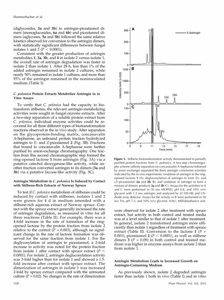
To verify that C. polonica had the capacity to bio-transform stilbenes, the relevant astringin-metabolizingactivities were sought in fungal enzyme extracts. Aftera two-step separation of a soluble protein extract fromC. polonica, individual enzyme activities could be re-covered for all three different types of biotransformationreactions observed in the in vivo study. After separationon the glycoprotein-binding matrix, concanavalinA-Sepharose, an unbound protein fraction hydrolyzedastringin to E- and Z-piceatannol 2 (Fig. 3B). Fractionsthat bound to concanavalin A-Sepharose were furtherpurified by anion-exchange chromatography. One frac-tion from this second chromatographic step formed thering-opened lactone 1 from astringin (Fig. 3A) via aputative catechol dioxygenase-like activity, while an-other fraction converted astringin to its dimers (3a and3b) via a putative laccase-like activity (Fig. 3C).
Astringin Metabolism in C. polonica Is Induced by Contactwith Stilbene-Rich Extracts of Norway Spruce
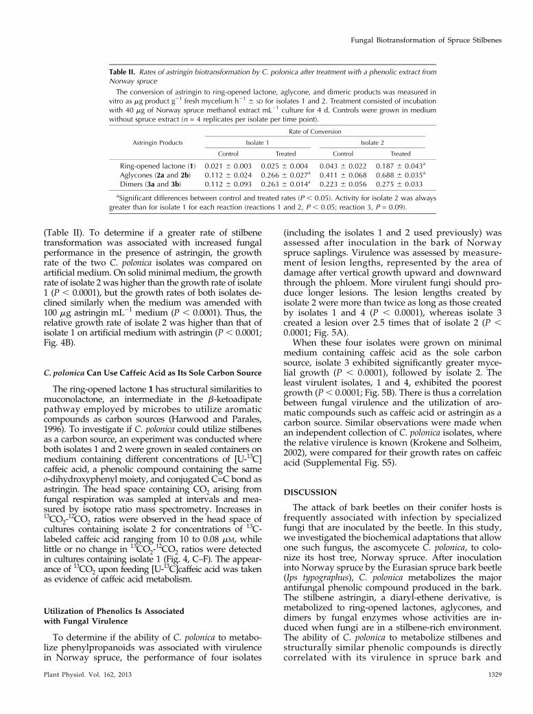
To test if C. polonica metabolism of stilbenes could beinduced by contact with stilbenes, isolates 1 and 2were grown for 4 d in medium amended with astilbene-rich aqueous extract of Norway spruce. Con-tact with the spruce extract generally increased the rateof astringin degradation, as measured in vitro for allthree reactions (Table II). For example, there was a4-fold increase in the rate of formation of the ring-opened lactone by the protein fraction from isolate 2relative to the control (P = 0.003), although no signif-icant change in the rate of lactone formation was ob-served for the same fraction from isolate 1. For thedeglycosylation of astringin to piceatannol, a 2-foldincrease in activity was noted for the protein fractionfrom isolate 1 after contact with spruce extract (P ,0.0001). For isolate 2, astringin deglucosylation activitywas 3-fold higher than for isolate 1 and showed a 1.5-fold increase after contact with spruce extract. Thedimerization of astringin in isolate 1 was increased2-fold by spruce extract compared with the untreatedculture (P = 0.02). No changes in the rate of dimerization
were observed for isolate 2 after treatment with spruceextract, but activity in both control and treated mediawas at a level similar to that of isolate 1 after treatment.In general, isolate 2 transformed astringin more effi-ciently than isolate 1 regardless of treatment with spruceextract (Table II). Conversion to the lactone 1 (P =0.001), piceatannol 2 (P , 0.0001), as well as stilbenedimers 3 (P = 0.09) in both control and treated me-dium was higher in enzyme assays from isolate 2 thanfrom isolate 1.
Astringin Metabolism Leads to Increased Growth onAstringin-Containing Medium
As previously shown, isolate 2 degraded astringinfaster than isolate 1 both in vivo (Table I) and in vitro
Figure 3. Stilbene biotransformation activity demonstrated in partiallypurified protein fractions from C. polonica. A two-step chromatogra-phy scheme (affinity separation on concanavalin A-Sepharose followedby anion exchange) separated the three astringin conversion activitiesindicated by the in vivo experiments: oxidation of astringin to the ring-opened lactone 1 (A); deglucosylation of astringin to form (E )- and(Z)-piceatannol (2a and 2b; B); and oxidation of astringin to form amixture of dimeric products 3a and 3b (C). Assays for the activities in Aand C were performed in 50 mM MOPSO, pH 6.8, and 10% (v/v)glycerol with 1.2 mM astringin and analyzed by LC-ESI-MS and UV-diode array detector. Assays for the activity in B were performed in 50mM Tris, pH 7.5, and 10% (v/v) glycerol. mAU, Milliabsorbance unit.
1328 Plant Physiol. Vol. 162, 2013
Hammerbacher et al.
(Table II). To determine if a greater rate of stilbenetransformation was associated with increased fungalperformance in the presence of astringin, the growthrate of the two C. polonica isolates was compared onartificial medium. On solid minimal medium, the growthrate of isolate 2 was higher than the growth rate of isolate1 (P , 0.0001), but the growth rates of both isolates de-clined similarly when the medium was amended with100 mg astringin mL21 medium (P , 0.0001). Thus, therelative growth rate of isolate 2 was higher than that ofisolate 1 on artificial medium with astringin (P , 0.0001;Fig. 4B).
C. polonica Can Use Caffeic Acid as Its Sole Carbon Source
The ring-opened lactone 1 has structural similarities tomuconolactone, an intermediate in the b-ketoadipatepathway employed by microbes to utilize aromaticcompounds as carbon sources (Harwood and Parales,1996). To investigate if C. polonica could utilize stilbenesas a carbon source, an experiment was conducted whereboth isolates 1 and 2 were grown in sealed containers onmedium containing different concentrations of [U-13C]caffeic acid, a phenolic compound containing the sameo-dihydroxyphenyl moiety, and conjugated C=C bond asastringin. The head space containing CO2 arising fromfungal respiration was sampled at intervals and mea-sured by isotope ratio mass spectrometry. Increases in13CO2-
12CO2 ratios were observed in the head space ofcultures containing isolate 2 for concentrations of 13C-labeled caffeic acid ranging from 10 to 0.08 mM, whilelittle or no change in 13CO2-
12CO2 ratios were detectedin cultures containing isolate 1 (Fig. 4, C–F). The appear-ance of 13CO2 upon feeding [U-13C]caffeic acid was takenas evidence of caffeic acid metabolism.
Utilization of Phenolics Is Associatedwith Fungal Virulence
To determine if the ability of C. polonica to metabo-lize phenylpropanoids was associated with virulencein Norway spruce, the performance of four isolates
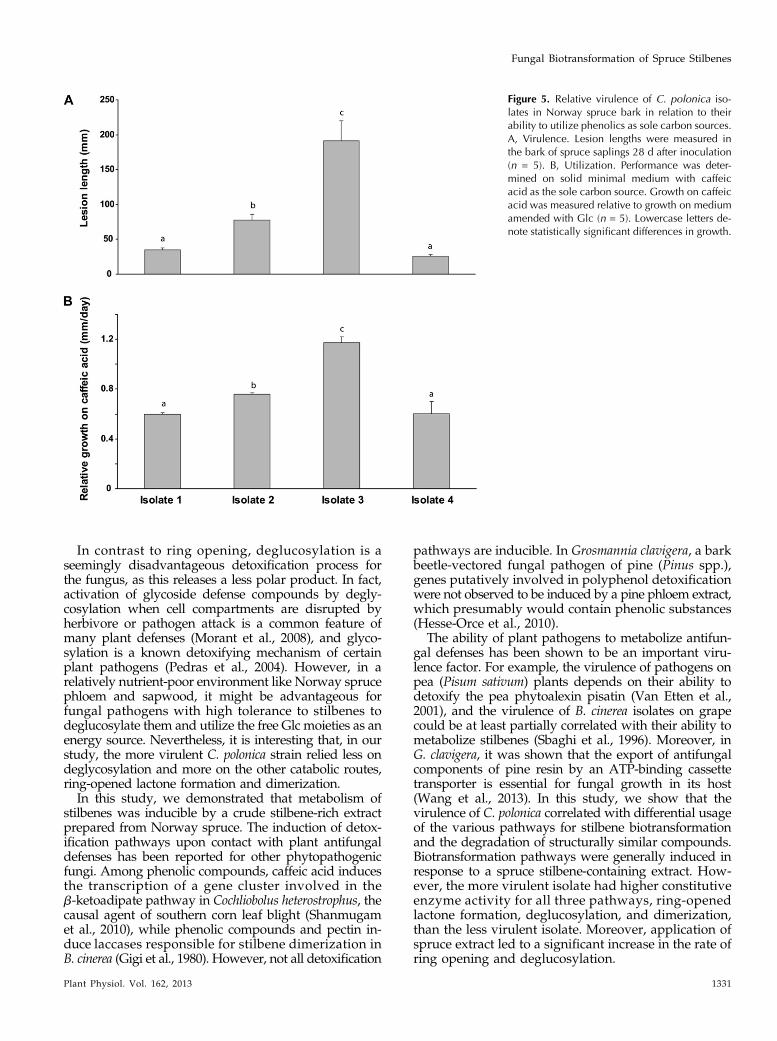
(including the isolates 1 and 2 used previously) wasassessed after inoculation in the bark of Norwayspruce saplings. Virulence was assessed by measure-ment of lesion lengths, represented by the area ofdamage after vertical growth upward and downwardthrough the phloem. More virulent fungi should pro-duce longer lesions. The lesion lengths created byisolate 2 were more than twice as long as those createdby isolates 1 and 4 (P , 0.0001), whereas isolate 3created a lesion over 2.5 times that of isolate 2 (P ,0.0001; Fig. 5A).
When these four isolates were grown on minimalmedium containing caffeic acid as the sole carbonsource, isolate 3 exhibited significantly greater myce-lial growth (P , 0.0001), followed by isolate 2. Theleast virulent isolates, 1 and 4, exhibited the poorestgrowth (P, 0.0001; Fig. 5B). There is thus a correlationbetween fungal virulence and the utilization of aro-matic compounds such as caffeic acid or astringin as acarbon source. Similar observations were made whenan independent collection of C. polonica isolates, wherethe relative virulence is known (Krokene and Solheim,2002), were compared for their growth rates on caffeicacid (Supplemental Fig. S5).
DISCUSSION
The attack of bark beetles on their conifer hosts isfrequently associated with infection by specializedfungi that are inoculated by the beetle. In this study,we investigated the biochemical adaptations that allowone such fungus, the ascomycete C. polonica, to colo-nize its host tree, Norway spruce. After inoculationinto Norway spruce by the Eurasian spruce bark beetle(Ips typographus), C. polonica metabolizes the majorantifungal phenolic compound produced in the bark.The stilbene astringin, a diaryl-ethene derivative, ismetabolized to ring-opened lactones, aglycones, anddimers by fungal enzymes whose activities are in-duced when fungi are in a stilbene-rich environment.The ability of C. polonica to metabolize stilbenes andstructurally similar phenolic compounds is directlycorrelated with its virulence in spruce bark and
Table II. Rates of astringin biotransformation by C. polonica after treatment with a phenolic extract fromNorway spruce
The conversion of astringin to ring-opened lactone, aglycone, and dimeric products was measured invitro as mg product g21 fresh mycelium h21 6 SD for isolates 1 and 2. Treatment consisted of incubationwith 40 mg of Norway spruce methanol extract mL21 culture for 4 d. Controls were grown in mediumwithout spruce extract (n = 4 replicates per isolate per time point).
Astringin Products
Rate of Conversion
Isolate 1 Isolate 2
Control Treated Control Treated
Ring-opened lactone (1) 0.021 6 0.003 0.025 6 0.004 0.043 6 0.022 0.187 6 0.043a
Aglycones (2a and 2b) 0.112 6 0.024 0.266 6 0.027a 0.411 6 0.068 0.688 6 0.035a
Dimers (3a and 3b) 0.112 6 0.093 0.263 6 0.014a 0.223 6 0.056 0.275 6 0.033
aSignificant differences between control and treated rates (P , 0.05). Activity for isolate 2 was alwaysgreater than for isolate 1 for each reaction (reactions 1 and 2, P , 0.05; reaction 3, P = 0.09).
Plant Physiol. Vol. 162, 2013 1329
Fungal Biotransformation of Spruce Stilbenes

appears to allow growth on stilbenes as a sole carbonsource.
A number of previous studies had reported that thestilbene content of spruce bark declined during fungalattack (Brignolas et al., 1995; Viiri et al., 2001; Li et al.,2012). These declines were considered very puzzlingbecause they occurred despite increases in the enzymeactivity of STS (Brignolas et al., 1995), which catalyzethe formation of the stilbene skeleton. Furthermore,increases in piceatannol concentrations (Viiri et al.,2001) and stilbene dimers (Li et al., 2012) were observedin spruce bark after C. polonica infection. Our resultsnow clearly show that these metabolites originate fromthe fungal metabolism of host defense compounds andthat C. polonica metabolism of stilbenes can overrideeven increases in host stilbene biosynthesis.
Stilbenes have long been known as antifungal de-fenses in plants that inhibit fungal growth by inter-fering with microtubule assembly (Woods et al., 1995;Adrian et al., 1997), disrupting plasma membranes anduncoupling electron transport in fungal spores andgerm tubes (Pont and Pezet, 1990; Adrian and Jeandet,2012). It is not surprising that fungi specialized to livein stilbene-rich plant material, such as C. polonica, havedeveloped mechanisms to circumvent the deleteriouseffects of these compounds. Here, we demonstrated thatC. polonica not only metabolizes stilbenes but employsseveral different degradation routes. Using the tetra-hydroxystilbene glucoside, astringin, which is producedin high amounts by Norway spruce, as a model, weidentified ring-opened lactone, aglycone, and dimericmetabolites in C. polonica cultures. Some of these reac-tion types have been reported for other stilbenes (Breuilet al., 1998, 1999; Rodriguez-Bonilla et al., 2011). Forexample, stilbene dimer formation has been previouslyreported in the grape (Vitis vinifera) pathogen Botrytiscinerea. The dimerization of resveratrol and ptero-stilbenes has been shown to be an oxidative processinvolving the 49-hydroxyl group of the stilbene skeletonand catalyzed by laccases and peroxidases (Breuil et al.,1998; Rodriguez-Bonilla et al., 2011).
Ring opening and lactonization has not previouslybeen reported for stilbenes but is a logical early step ofcatabolism, since it results in a more polar product. Stil-benes exert their toxicity on fungi by diffusing throughmembranes into vegetative and reproductive structures(Adrian et al., 1998). A ring-opened and lactonized pro-duct with increased polarity should diffuse more slowlyinto fungal cells and, therefore, show decreased toxicity.Converting plant defense compounds to more polarproducts has been shown to be a successful detoxifica-tion strategy for other plant pathogens (Weltring andBarz, 1980; Esaki et al., 1998). A more recent study(Sobolev et al., 2006) reported the appearance of a novelstilbene phytoalexin in peanut (Arachis hypogaea) kernelsinfected by Aspergillus species. This compound has aprenylated but-2-enolide skeleton that bears a strikingresemblance to the astringin lactone produced byC. polonica and so might also represent a fungal biotrans-formation product derived from a peanut phytoalexin.
Figure 4. Relative growth of C. polonica isolates 1 and 2 in artificialmedium amended with astringin in relation to the rates of phenolicbiotransformation and the ability to utilize phenolics as sole carbonsources. A, Biotransformation. Depletion of astringin added to artificialmedium over a period of 72 h was compared with changes in astringinconcentration in sterile control medium (n = 4). B, Growth. In vitrogrowth on solid minimal medium was determined in the presence ofastringin relative to growth without astringin (n = 10). Error bars in Aand B represent SE. C to F. Utilization. Changes in the ratio of 13CO2 to12CO2 were determined in fungal head space arising from respirationof [U-13C]caffeic acid. Samples were collected over 18 d of fungalgrowth on agar containing 1 mM caffeic acid amended with 10 mM (C),2 mM (D), 0.4 mM (E), and 0.08 mM (F) [U-13C]caffeic acid (n = 1).VPDB, Vienna Pee Dee Belemnite. [See online article for color versionof this figure.]
1330 Plant Physiol. Vol. 162, 2013
Hammerbacher et al.
In contrast to ring opening, deglucosylation is aseemingly disadvantageous detoxification process forthe fungus, as this releases a less polar product. In fact,activation of glycoside defense compounds by degly-cosylation when cell compartments are disrupted byherbivore or pathogen attack is a common feature ofmany plant defenses (Morant et al., 2008), and glyco-sylation is a known detoxifying mechanism of certainplant pathogens (Pedras et al., 2004). However, in arelatively nutrient-poor environment like Norway sprucephloem and sapwood, it might be advantageous forfungal pathogens with high tolerance to stilbenes todeglucosylate them and utilize the free Glc moieties as anenergy source. Nevertheless, it is interesting that, in ourstudy, the more virulent C. polonica strain relied less ondeglycosylation and more on the other catabolic routes,ring-opened lactone formation and dimerization.In this study, we demonstrated that metabolism of
stilbenes was inducible by a crude stilbene-rich extractprepared from Norway spruce. The induction of detox-ification pathways upon contact with plant antifungaldefenses has been reported for other phytopathogenicfungi. Among phenolic compounds, caffeic acid inducesthe transcription of a gene cluster involved in theb-ketoadipate pathway in Cochliobolus heterostrophus, thecausal agent of southern corn leaf blight (Shanmugamet al., 2010), while phenolic compounds and pectin in-duce laccases responsible for stilbene dimerization inB. cinerea (Gigi et al., 1980). However, not all detoxification
pathways are inducible. In Grosmannia clavigera, a barkbeetle-vectored fungal pathogen of pine (Pinus spp.),genes putatively involved in polyphenol detoxificationwere not observed to be induced by a pine phloem extract,which presumably would contain phenolic substances(Hesse-Orce et al., 2010).
The ability of plant pathogens to metabolize antifun-gal defenses has been shown to be an important viru-lence factor. For example, the virulence of pathogens onpea (Pisum sativum) plants depends on their ability todetoxify the pea phytoalexin pisatin (Van Etten et al.,2001), and the virulence of B. cinerea isolates on grapecould be at least partially correlated with their ability tometabolize stilbenes (Sbaghi et al., 1996). Moreover, inG. clavigera, it was shown that the export of antifungalcomponents of pine resin by an ATP-binding cassettetransporter is essential for fungal growth in its host(Wang et al., 2013). In this study, we show that thevirulence of C. polonica correlated with differential usageof the various pathways for stilbene biotransformationand the degradation of structurally similar compounds.Biotransformation pathways were generally induced inresponse to a spruce stilbene-containing extract. How-ever, the more virulent isolate had higher constitutiveenzyme activity for all three pathways, ring-openedlactone formation, deglucosylation, and dimerization,than the less virulent isolate. Moreover, application ofspruce extract led to a significant increase in the rate ofring opening and deglucosylation.
Figure 5. Relative virulence of C. polonica iso-lates in Norway spruce bark in relation to theirability to utilize phenolics as sole carbon sources.A, Virulence. Lesion lengths were measured inthe bark of spruce saplings 28 d after inoculation(n = 5). B, Utilization. Performance was deter-mined on solid minimal medium with caffeicacid as the sole carbon source. Growth on caffeicacid was measured relative to growth on mediumamended with Glc (n = 5). Lowercase letters de-note statistically significant differences in growth.
Plant Physiol. Vol. 162, 2013 1331
Fungal Biotransformation of Spruce Stilbenes
The metabolism of Norway spruce stilbenes byC. polonicamay serve not only to detoxify them but alsoto provide the fungus with nutrition. Other aromaticcompounds, such as lignin breakdown products, havebeen shown to be metabolized by the b-ketoadipatepathway in soil bacteria and fungi, yielding energy,reducing equivalents, and releasing CO2 (Harwood andParales, 1996). The structural similarities between thering-opened lactone formed from astringin and an in-termediate in the b-ketoadipate pathway hinted thatstilbenes might also be further catabolized by C. polonicavia this process. Using caffeic acid, whose structureclosely matches that of astringin in the region of themolecule giving rise to the ring-opened lactone, weestablished that virulent C. polonica isolates did employthis aromatic compound as an energy source based onrelease of 13CO2 from [U-13C]caffeic acid and growth oncaffeic acid as a sole carbon source. Thus, C. polonicacould conceivably use the stilbenes of its host tree asnutrients, resulting in an increased growth rate andmore successful colonization of the tree. The use of hostdefense chemicals as a carbon source to support thegrowth of a bark beetle-associated fungus has alsopreviously been reported for the mountain pine beetleassociate G. clavigera, which can grow on monoterpenecompounds of pine resin as a sole carbon source(DiGuistini et al., 2011).
The fungus C. polonica and the bark beetle are fre-quent associates that are usually viewed as killingtheir Norway spruce host by their combined efforts(Franceschi et al., 2005), but this opinion is not universallyaccepted (Six and Wingfield, 2011). In any case, the exactbasis for their mutualism is not fully understood (Paineet al., 1997). It has been proposed that the fungus benefitsby being dispersed to new hosts and gaining entry intothe tree. The beetle, on the other hand, may benefit if thefungus helps weaken or kill the tree, since tree death isessential for beetle reproduction (Franceschi et al., 2005).Our study demonstrates that fungal growth in thestilbene-rich bark of Norway spruce depends on suc-cessful metabolism of these host defense compounds,which should also benefit the bark beetle by neutral-izing tree defenses.
An alternative explanation for the cooccurrence ofbark beetles with C. polonica might be the nutritionalbenefits associated with polyphenol biotransformationby the fungus. Degradation of stilbenes may benefitbark beetles by improving the quality of substrateavailable for feeding their larvae, since the stilbenesremaining in the bark could be toxic. For example, thecatechol groups present in the astringin core structuremay be spontaneously or enzymatically rearranged toform reactive quinones (Haruta et al., 2001; Lin et al.,2010) under the semialkaline conditions prevailing inbeetle guts (Balogun, 1969). These quinones can thenalkylate reactive nucleophiles such as sulfhydryl andamino groups in proteins or amino acids (Felton et al.,1992; Son et al., 2010). Protein alkylation may reducethe nutritional quality of ingested food or destabilizethe peritrophic membrane in the bark beetle gut
(Barbehenn et al., 2008). Lactonization or dimerizationof the catechol groups, as we showed for C. polonica,should prohibit quinone formation and thus protectbark beetles from the harmful effects of stilbene de-fense compounds.
We have shown in this study that C. polonica cantransform phenolic defense compounds from Norwayspruce and that it is even able to use these compoundsas its sole carbon source. Further research should re-veal more about how the biochemical capabilities ofthis fungus can contribute to its relationship with barkbeetles and how its metabolism of spruce defensesmodifies host tree resistance to both the fungus and thebeetle.
MATERIALS AND METHODS
Inoculation of Norway Spruce Saplingswith Ceratocystis polonica
Eight-year-oldNorway spruce (Picea abies) saplings originating from the 3369-Schongau clone (Samenklenge and Pflanzengarten) were grown in an outdoorplot for 4 years prior to the experiment. Two C. polonica isolates (CMW 7749 =isolate 1 and CMW 7135 = isolate 2) were provided by the culture collection ofthe Forestry and Agricultural Biotechnology Institute (University of Pretoria).These were characterized as the least and most virulent, respectively, out of 12isolates by measurement of lesion lengths created after inoculation of maturespruce trees (Marin, 2003). Both were grown on 2% (w/v) malt extract agar for14 d at 25°C. Inoculations of saplings with C. polonica isolates 1 and 2 wereperformed 3 weeks after buds had broken during the flush of spring growth(June 10, 2008). A bark plug, 8 mm in diameter, was removed midway betweenthe second and third branch whorl from the upper part of the sapling (2-year-oldsegment) with a cork borer. An 8-mm plug from one of the two C. polonicacultures was placed into the wound with the mycelium oriented toward thewood surface and sealed with Parafilm. For the control treatment, plugs of sterilemalt extract agar were inserted into the wound. All treatments were appliedduring the same growth phase.
Bark tissue samples from five inoculated and five wounded saplings wereharvested 2, 7, 14, and 28 d after the onset of the experiment. Five nonwoundedcontrol saplings were harvested at 2 d after the onset of the experiment. Fungallesions were measured with a caliper. Bark material was flash frozen imme-diately after harvest in liquid nitrogen and stored at 280°C.
Inoculations of isolates 2, 3, and 4 were performed as described above usinga 5-mm cork borer to wound 5-year-old clonal Picea glauca saplings. Lesionlength data from this experiment were related to data from the first experi-ment by comparison of lesions of isolate 2 under the different conditions.
Quantitative Real-Time PCR of STS from Norway Spruce
Total RNA from inoculated treatment andwounded control barkwas isolatedwith the Invitrap Spin Plant RNAMini Kit (Invitek) following the protocols of themanufacturer except that an additional DNA digestion stepwas included (RNaseFree DNase set; Qiagen). RNA was quantified by spectrophotometry. Reversetranscription of 1 mg of RNA into complementary DNA (cDNA) was achievedby using SuperScript II reverse transcriptase (Invitrogen) and 50 pmol of PolyT
(12-18) primer (Invitrogen) in a reaction volume of 20 mL. cDNA was diluted to10% (v/v) with deionized water. One microliter of diluted cDNA was used astemplate for quantitative real-time PCR in a reaction mixture containing BrilliantSYBR Green QPCR Master Mix (Stratagene), 10 pmol of forward primer, and 10pmol of reverse primer. PaSTS transcripts were amplified using the forwardprimer 59-GTGGCGAGCAGAACACAGACTTC-39 and the reverse primer59-CAGCGATGGTACCTCCATGAACG-39. This primer pair was designed toamplify 140 bp of both STS1 (GenBank accession no. JN400048) and STS2(GenBank accession no. JN400047) simultaneously. PCR was performed using aStratagene MX3000P thermocycler using the following cycling parameters: 5 minat 95°C, followed by 40 cycles of 30 s at 95°C, 30 s at 55°C, and 30 s at 72°C, anda melting curve analysis from 55°C to 95°C. Reaction controls included non-template controls as well as non-reverse-transcribed RNA. STS gene abundancewas normalized to the abundance of the Norway spruce ubiquitin gene
1332 Plant Physiol. Vol. 162, 2013
Hammerbacher et al.

(Schmidt et al., 2010; GenBank accession no. EF681766) amplified with the for-ward primer 59-GTTGATTTTTGCTGGCAAGC-39 and the reverse primer59-CACCTCTCAGACGAAGTAC-39. Relative transcript abundance was cal-culated from three technical replicates of five biological replicates and cali-brated against the transcript abundance of five nonwounded control saplings(relative transcript abundance = 1).
Phenolic Extraction from Spruce to Investigate Changesafter C. polonica Infection
For the extraction of phenolic compounds, spruce tissue was ground to afine powder under liquid nitrogen and lyophilized. Approximately 40 mg ofdried tissue was extracted with 2 mL of HPLC-grade methanol for 4 h at 4°C.Insoluble material was pelleted by centrifugation, and the supernatant wasrecovered. Insoluble material was reextracted with 1.5 mL of methanol for16 h. Extracts were combined and evaporated to dryness under a stream ofnitrogen. Dried samples were redissolved in 1 mL of methanol containing100 mg mL21 of the internal standard apigenin glucoside. For liquid chromatog-raphy (LC)-electrospray ionization (ESI)-mass spectrometry (MS), samples werediluted to 20% (v/v) with water.
HPLC-ESI-MS
Phenolic metabolites from spruce bark and astringin biotransformationproducts were separated on a Nucleodur Sphinx RP18ec column with di-mensions of 250 3 4.6 mm and a particle size of 5 mm (Macherey-Nagel) usingan 1100 series HPLC apparatus (Agilent Technologies). The total mobile phaseflow rate for chromatographic separation was 1.0 mL min21. The columntemperature was maintained at 25°C. Phenolic compounds from spruce andfungal biotransformation were separated using 0.2% (v/v) formic acid andacetonitrile as mobile phases A and B, respectively, with the following elutionprofile: 0 to 1 min, 100% A; 1 to 25 min, 0% to 65% B in A; 25 to 28 min, 100%B; and 28.1 to 32 min, 100% A. Products from enzyme assays were separatedwith the following elution profile: 0 to 1 min, 100% A; 1 to 18 min, 0% to 100%B in A; 18 to 19 min, 100% B; and 19.1 to 22 min, 100% A.
Compound detection and quantification were accomplished with an Es-quire 6000 ESI ion-trap mass spectrometer (Brucker Daltronics). Flow comingfrom the column was diverted in a ratio of 4:1 before entering the massspectrometer electrospray chamber. The MS device was operated in negativemode scanning mass-to-charge ratio between 50 and 1,600 with an optimaltarget mass of mass-to-charge ratio 405. The mass spectrometer was operatedusing the following specifications: skimmer voltage, 60 V; capillary voltage,4,200 V; nebulizer pressure, 35 p.s.i.; drying gas, 11 L min21; gas temperature,330°C. Capillary exit potential was kept at 2121 V.
Compounds in chromatograms were identified based on retention time,their apparent molecular masses, and fragmentation spectra (SupplementalTable S3; Supplemental Fig. S4) and ultimately by NMR (see below). BruckerDaltronics Quant Analysis version 3.4 software was used for data processingand compound quantification using a standard smoothing width of 3 andPeak Detection Algorithm version 2. Linearity in ionization efficiencies wasverified by analyzing serial dilutions of randomly selected samples. Ion sup-pression was controlled for by calculating analyte-to-internal standard ratiosin serially diluted samples in which the internal standard was maintained atthe same concentration. An external calibration curve for astringin was createdby linear regression. Variability in the processing of individual samples wascorrected by adjustment relative to the internal standard, the flavonoid api-genin glycoside.
In Vivo Biotransformation of Astringin by C. polonicain Culture
C. polonica isolate 1 and isolate 2 were grown on 2% (w/v) malt extract agarfor 12 d at 25°C in darkness. Agar plugs (diameter = 4 mm) from stationarycultures were placed in 15-mL test tubes for biotransformation assays. Then,2 mL of sterile 2% (v/v) malt extract amended with astringin to a final con-centration of 2 mg mL21 was added to the test tubes and incubated at 28°Cwith shaking at 220 rpm. Negative control treatments contained astringin-amended medium without fungus and fungus grown in medium withoutastringin. Cultures were harvested 4, 8, 24, and 72 h after the onset of theexperiment. Biotransformation processes were stopped by adding 50 mL ofHCl (2 N) to the test tubes. The internal standard apigenin-3-O-glucoside was
added to a final concentration of 0.1 mg mL21 prior to analysis by LC-ESI-MS.The concentrations of biotransformation products were calculated relative tothe internal standard.
Preparation of Astringin Biotransformation Products forNMR Analysis
Culture medium samples from biotransformation experiments were firstsubjected to SPE with RP-18 as the stationary phase. After loading, the col-umns were washed with water, dried with nitrogen gas, and finally elutedwith methanol. The methanol extracts were separated by means of HPLC, andpeaks of interest were collected online by postcolumn SPE. The HPLC-SPEsystem consisted of an Agilent 1100 chromatography system (Agilent Tech-nologies) and a J&M photodiode array detector (J&M Analytik) connected to aSpark Prospekt 2 SPE device (Spark Holland) equipped with HySphere resingeneral phase cartridges (10 3 2 mm, 10 mm). Separations were performedwith linear gradient elution, with water (A) and methanol (B) as solvents, bothcontaining 0.1% formic acid: 0 to 1 min, 100% A; 1 to 20 min, 0% to 100% B inA; 20 to 25 min, 100% B; 25 to 27 min, 100% to 0% B in A; 27 to 34 min, 100% A.The column was a Macherey-Nagel Isis RP-18e column (250 3 4 mm). Themake-up flow for postcolumn SPE trapping was set to 2.5 mL min21 usingHPLC-grade water. The SPE cartridges were subsequently dried using pres-surized nitrogen. HPLC-grade acetonitrile was used to extract the trappedanalytes from the general phase cartridges into HPLC glass vials. Afterevaporation to dryness using nitrogen gas, the samples were reconstitutedwith 80 mL of MeOH-d4 and DMSO-d6, transferred into 2-mm (i.d.) capillaryNMR tubes, and subjected to NMR measurements. Hystar 3.2 software wasused to coordinate the LC-SPE experiments, and Topspin 2.1 software wasused to control the NMR spectrometer and to perform data processing.
NMR and High-Resolution MS Analysis of AstringinBiotransformation Products
1H-NMR, 13C-NMR, 1H-1H-correlation spectroscopy, total correlated spec-troscopy, heteronuclear multiple bond correlation, and heteronuclear singlequantum coherence spectra were measured on a Bruker Avance 500 NMRspectrometer (Bruker Biospin) operating at 500.13 MHz for 1H and 125.75MHz for 13C. An inverse triple channel cryoprobe (5 mm) was used to measurespectra at a probe temperature of 300 K. Spectra are referenced to the re-sidual solvent signals: for methanol-d4 at d 3.31/49.05 ppm and for DMSO-d6at d 2.49/39.51 ppm. Capillary tubes (2 mm i.d.) were used for all NMRmeasurements.
Attempts to determine the structure of compound 1 in methanol-d4 resultedin degradation during time-consuming two-dimensional heteronuclear ex-periments. Thus, DMSO-d6 was used.
High-resolution MS was recorded on a ultra-performance liquidchromatography-MS/MS system consisting of an Ultimate 3000 series RapidSeparation LC (Dionex) system and an Orbitrap mass spectrometer (ThermoFisher Scientific). Ultra-performance liquid chromatography was performedusing a Dionex Acclaim C18 column (150 3 2.1 mm 3 2.2 mm) at a constantflow rate of 300 mL min21. A binary solvent system of water (A) and aceto-nitrile (B), both containing 0.1% formic acid, was used as follows: 0 min, 20% Bin A; 0 to 6 min, 20% to 95% B in A; 6 to 10 min, 95% B; 10 to 14 min, 20% B inA. Full-scan mass spectra were generated using 30,000 resolving power: themass accuracy was better than 3 ppm.
Partial Purification of Fungal Protein Fractions ShowingStilbene Biotransformation Activity
C. polonica isolates 1 and 2 were cultured in 10% (v/v) carrot (Daucuscarota) juice liquid culture for 6 d, harvested by centrifugation, and lyophi-lized. Dried material was finely ground using a vibrating ball mill. Powderedmycelium (0.5 g) was extracted with 10 mL of extraction buffer (50 mM Tris,pH 7.5, 5 mM ascorbic acid, 5 mM dithiothreitol, 10 mM MgCl2, 10 mM CaCl2,10 mM MnCl2, 0.5 M NaCl, 10% glycerol, 1% polyvinylpyrrolidone [molecularweight of 360,000], 4% polyvinylpolypyrrolidone, and 0.1% Tween 20) at 4°Cfor 30 min with shaking. Crude fungal protein extract in extraction buffer wasloaded onto an open column of concanavalin A-Sepharose (GE Healthcare)with a bed volume of 5 mL. The protein extract was incubated with theconcanavalin A matrix for 30 min at 4°C before washing unbound proteins offwith 5 bed volumes of washing buffer (50 mM Tris, pH 7.5, 0.5 M NaCl, and
Plant Physiol. Vol. 162, 2013 1333
Fungal Biotransformation of Spruce Stilbenes
10% glycerol). Elution of bound proteins was achieved using 2 column vol-umes of washing buffer amended with 500 mM a-D-methylglucopyranoside.Proteins from the column eluate were desalted at 4°C into 50 mM 3-(N-morpholino)-2-hydroxypropanesulfonic acid (MOPSO; pH 6.8) containing10% glycerol using a HiPrep 26/10 (GE Healthcare) desalting column on anÄKTA 900 chromatography system (GE Healthcare). The desalted proteinfraction was loaded onto a 5-mL DEAE-Sepharose column and washed with2 column volumes of DEAE washing buffer (50 mM MOPSO, pH 6.8, and 10%glycerol) at a flow rate of 5 mL min21. Proteins were eluted from the columnwith DEAE wash buffer adjusted with NaCl using a step-wise gradient(100 mM, 200 mM, 300 mM, 500 mM, and 1 M NaCl). Elution steps and fractionvolumes were 10 mL. Stilbene biotransformation activity was determined forcrude extracts as well as for proteins that were eluted from the concanavalinA- and DEAE-Sepharose columns and for samples from the flow through ofboth columns. Enzyme activities were assayed in 300-mL reaction volumescontaining 200 mL of enzyme from the purification steps described above and100 mg of astringin in DEAE washing buffer. Reaction mixtures were incubatedat 30°C for 4 h before stopping the reaction with 10 mL of 0.1 N HCl. After re-moving the protein by centrifugation, 20 mL of the reaction mixture was ana-lyzed by LC-ESI-MS.
In Vitro Assay of Astringin Biotransformation Activity
Mycelium was cultured as above for 3 d. Each culture (10 mL) was sub-cultured in 50 mL of 10% (v/v) carrot medium. To determine if the biosynthesisof enzymes for stilbene biotransformation could be induced, the medium offour biological replicates was amended with 4 mg of crude spruce extract(methanol-soluble bark extract with solvent evaporated) in 2 mL of sterilewater. As controls, four other replicates were subcultured after adding sterilewater instead of spruce extract. Subcultured mycelium was harvested after4 d by centrifugation and ground to a fine powder using a mortar and pestle.Then, 100 mg of groundmyceliumwas extracted with 5 mL of extraction bufferas above. Insoluble material was removed from extracts by centrifugation, andthe resulting supernatant was filteredwith a syringe filter with an exclusion sizeof 0.2 mm.
Astringin biotransformation activity was assayed at 30°C in 2-mL reactionvolumes containing fungal protein in extraction buffer and 100 mg mL21
astringin. A 200-mL subsample was taken from each assay reaction 45 min and2, 4, and 8 h after the assays were initiated. The biotransformation reactionwas stopped and analyzed as above.
Assessment of Fungal Growth in the Presence of Astringin
Petri dishes with synthetic nutrient agar (Nirenberg and O’Donnell, 1998)amended with astringin were prepared by cooling the autoclaved medium to55°C and adding astringin in ethanol to a final concentration of 100 mg mL21
medium. Medium for negative control treatments was amended with ethanolonly. Medium (25 mL) was dispensed in petri dishes (diameter = 10 cm). Afterthe medium set, agar plugs (diameter = 4 mm) from 14-d-old C. polonica sta-tionary cultures (isolate 1 or 2) were placed in the middle of each petri dish.Cultures were sealed with Parafilm and incubated at 26°C. Diameters of theexpanding fungal cultures were measured every 24 h for 5 d. Growth rateswere calculated using the slope of linear growth curves. Relative growth onastringin was calibrated against the mean growth rate of each isolate oncontrol medium (100%) by calculating the fraction of each isolate’s growth rateon astringin.
Phenolic Extraction from Spruce to Induce FungalBiotransformation Pathways
Finely ground lyophilized spruce bark (100 g) was extracted overnight with400 mL of methanol. The mixture was filtered and the solvent was removedunder reduced pressure in a rotary evaporator (Buechi Rotavapor R-114). Thedried extract was weighed and redissolved in sterile water to a concentration of2 mg mL21.
Measurement of Fungal Utilization of Caffeic Acidas a Carbon Source
Synthetic nutrient agar (Nirenberg and O’Donnell, 1998) was prepared.Modified synthetic nutrient agar was prepared by replacing Glc and Suc with
the equivalent amount of caffeic acid. Agar plugs (5-mm diameter) from fungalcultures (isolates 1–4 and 1994-169/113, 1980-53/7/A, 1993-208/115, 1980-53/7/B, 1980-53/7/C, and 1980-53/7/A9; Krokene and Solheim, 2002) were platedon both synthetic nutrient agar and modified synthetic nutrient agar and grownat 25°C in the dark for 14 d. Radial mycelial growth was measured every 2 to5 d, and growth rates were calculated as above. Relative growth rates weredetermined by normalization with growth on synthetic nutrient agar.
Water agar (2.5%, w/v) amended with 1 mM caffeic acid was prepared.Filter-sterilized caffeic acid with a natural 13C-12C carbon isotope ratio (Sigma)was mixed with [U-13C]caffeic acid (Campro Scientific) at ratios of 100:1,100:0.2, 100:0.04, and 100:0.008 in different batches of medium. The growthmedium (2 mL) was dispensed into heat-sterilized 10-mL vials (Exetainer;Labco). Mycelial plugs (length approximately 2 mm) from isolate 1 or isolate2 stationary cultures, grown on modified synthetic nutrient agar containing2 mM caffeic acid with a natural 13C-12C carbon isotope ratio, were placed ontothe medium inside the vials and sealed with a septum under open-air con-ditions. Using an autosampler (CTC Combi-PAL; CTC Zwingen), 200-mL airaliquots were taken from each vial 5, 8, 11, 15, and 18 d after initiating theexperiment. The aliquots were injected into a homemade, room temperature,constant pressure gas chromatograph modified from a commercial GC/general phase system (Thermo Fisher Scientific). Between the injector and a30-m Poraplot Q capillary column, a Nafion (a sulfonated tetrafluoroethylene-based fluoropolymer; Perma Pure Products) online water-removal unit wasplaced. A second such unit was mounted postcolumn, followed by a thirdwater-removal step realized by immersing the transfer line into a dry ice bath.The transfer line led to an active open splitter that was connected to an isotoperatio mass spectrometer (Delta+ XL; Thermo Fisher Scientific). The opensplitter reduces the effluent such that only the eluting CO2 peak enters themass spectrometer; other air constituents (mainly nitrogen, oxygen, and ar-gon) are diverted to the vent and do not interfere with the isotopic analysis.Before each sample was injected, air from a continuously bleeding referenceair tank, whose mixing ratio and isotopic composition were independentlycalibrated at the Max Planck Institute for Biogeochemistry, was analyzed. Inaddition, control samples were included for each 13C-12C caffeic acid ratiofrom medium incubated without fungus.
The actual 13CO2-12CO2 ratio values in air samples from control and sample
vials were calculated from the measured d13C values using the InternationalUnion of Pure and Applied Chemistry reference value of 0.0111802 for13CO2:
12CO2 ratio in the international Vienna Pee Dee Belemnite reference(Zhang et al., 1990; Werner and Brand, 2001). Then, isotopic enrichment offungal samples was obtained by subtracting the measured d13C values of theblank samples without fungus.
Statistical Analysis
The results are presented graphically as means 6 SE. Tabulated results arepresented as means6 SD. The normality of data was tested for with the Shapiro-Wilk test. The statistical significance of differences in PaSTS transcript accu-mulation and stilbene concentrations in living trees was determined using linearmodels with tree responses as the dependent variable and time as the explanatoryvariable on log-transformed data. Differences between fungal biotransformationsof astringin as well as differences of fungal growth on astringin-amended me-dium were analyzed with the nonparametric Kruskal-Wallis test, as data couldnot be normalized. Differences in fungal growth in spruce bark and on caffeic acidwere analyzed using one-way ANOVA. Differences in reaction rates of astringinbiotransformation activity by the fungus measured in vitro were analyzed usingtwo-way ANOVA of untransformed or reciprocally (1/3) transformed data.Following ANOVA, differences in means were calculated using Tukey’s post-hoc pairwise comparisons test at a 95% confidence level. Analyses were conductedusing the open-source software R (version 2.81) and the LAERCIO package forTukey’s pairwise comparisons.
Supplemental Data
The following materials are available in the online version of this article.
Supplemental Figure S1. Ring-opened lactone products derived fromC. polonica transformation of astringin.
Supplemental Figure S2. NMR spectrum of one set of dimeric products(3a) derived from C. polonica biotransformation of astringin in this study.
Supplemental Figure S3. NMR spectrum of another set of dimeric products(3b) derived from C. polonica biotransformation of astringin in this study.
1334 Plant Physiol. Vol. 162, 2013
Hammerbacher et al.

Supplemental Figure S4. Diagnostic mass spectral fragments of astringinand its biotransformation products.
Supplemental Figure S5. Relative virulence of six C. polonica isolates firstdescribed in Krokene and Solheim (2002) is correlated with their abilityto grow on caffeic acid as the sole carbon source.
Supplemental Table S1. 1H NMR (500 MHz) and 13C NMR data (125MHz) of compounds 1 in DMSO-d6 and 4 in MeOH-d4.
Supplemental Table S2. Mass spectrometry data for astringin and identi-fied astringin metabolites.
Supplemental Materials and Methods S1. Structural elucidation of com-pounds 1 and 4.
ACKNOWLEDGMENTS
We thank Bettina Raguschke, Petra Linke, and Michael Reichelt fortechnical assistance and Paal Krokene and Mike Wingfield for providing C.polonica isolates.
Received March 26, 2013; accepted May 28, 2013; published June 1, 2013.
LITERATURE CITED
Adrian M, Jeandet P (2012) Effects of resveratrol on the ultrastructure ofBotrytis cinerea conidia and biological significance in plant/pathogeninteractions. Fitoterapia 83: 1345–1350
Adrian M, Jeandet P, Veneau J, Weston LA, Bessis R (1997) Biological activityof resveratrol, a stilbenic compound from grapevines, against Botrytis cinerea,the causal agent for grey mould. J Chem Ecol 23: 1689–1702
Adrian M, Rajaei H, Jeandet P, Veneau J, Bessis R (1998) Resveratroloxidation in Botrytis cinerea conidia. Phytopathology 88: 472–476
Balogun RA (1969) Digestive enzymes of alimentary canal of larch barkbeetle Ips cembrae (Heer). Comp Biochem Physiol 29: 1267–1270
Barbehenn RV, Maben RE, Knoester JJ (2008) Linking phenolic oxidationin the midgut lumen with oxidative stress in the midgut tissues of a tree-feeding caterpillar Malacosoma disstria (Lepidoptera: Lasiocampidae).Environ Entomol 37: 1113–1118
Breuil AC, Adrian M, Pirio N, Meunier P, Bessis R, Jeandet P (1998)Metabolism of stilbene phytoalexins by Botrytis cinerea. 1. Characteri-zation of a resveratrol dehydrodimer. Tetrahedron Lett 39: 537–540
Breuil AC, Jeandet P, Adrian M, Chopin F, Pirio N, Meunier P, Bessis R(1999) Characterization of a pterostilbene dehydrodimer produced bylaccase of Botrytis cinerea. Phytopathology 89: 298–302
Brignolas F, Lacroix B, Lieutier F, Sauvard D, Drouet A, Claudot AC, YartA, Berryman AA, Christiansen E (1995) Induced responses in phenolicmetabolism in two Norway spruce clones after wounding and inocula-tions with Ophiostoma polonicum, a bark beetle-associated fungus. PlantPhysiol 109: 821–827
Chong JL, Poutaraud A, Hugueney P (2009) Metabolism and roles of stil-benes in plants. Plant Sci 177: 143–155
DiGuistini S, Wang Y, Liao NY, Taylor G, Tanguay P, Feau N, HenrissatB, Chan SK, Hesse-Orce U, Alamouti SM, et al (2011) Genome andtranscriptome analyses of the mountain pine beetle-fungal symbiontGrosmannia clavigera, a lodgepole pine pathogen. Proc Natl Acad SciUSA 108: 2504–2509
Esaki H, Onozaki H, Morimitsu Y, Kawakishi S, Osawa T (1998) Potentantioxidative isoflavones isolated from soybeans fermented with As-pergillus saitoi. Biosci Biotechnol Biochem 62: 740–746
Felton GW, Donato KK, Broadway RM, Duffey SS (1992) Impact of oxi-dized plant phenolics on the nutritional quality of dietary protein to anoctuid herbivore, Spodoptera exigua. J Insect Physiol 38: 277–285
Franceschi VR, Krokene P, Christiansen E, Krekling T (2005) Anatomicaland chemical defenses of conifer bark against bark beetles and otherpests. New Phytol 167: 353–375
Franceschi VR, Krokene P, Krekling T, Christiansen E (2000) Phloemparenchyma cells are involved in local and distant defense responses tofungal inoculation or bark-beetle attack in Norway spruce (Pinaceae).Am J Bot 87: 314–326
Gigi O, Marbach I, Mayer AM (1980) Induction of laccase formation inBotrytis. Phytochemistry 19: 2273–2275
Hammerbacher A, Ralph SG, Bohlmann J, Fenning TM, Gershenzon J,Schmidt A (2011) Biosynthesis of the major tetrahydroxystilbenes inspruce, astringin and isorhapontin, proceeds via resveratrol and is en-hanced by fungal infection. Plant Physiol 157: 876–890
Haruta M, Pedersen JA, Constabel CP (2001) Polyphenol oxidase andherbivore defense in trembling aspen (Populus tremuloides): cDNAcloning, expression, and potential substrates. Physiol Plant 112: 552–558
Harwood CS, Parales RE (1996) The beta-ketoadipate pathway and thebiology of self-identity. Annu Rev Microbiol 50: 553–590
Hesse-Orce U, DiGuistini S, Keeling CI, Wang Y, Li M, Henderson H,Docking TR, Liao NY, Robertson G, Holt RA, et al (2010) Gene dis-covery for the bark beetle-vectored fungal tree pathogen Grosmanniaclavigera. BMC Genomics 11: 536
Jeandet P, Delaunois B, Conreux A, Donnez D, Nuzzo V, Cordelier S,Clément C, Courot E (2010) Biosynthesis, metabolism, molecular engi-neering, and biological functions of stilbene phytoalexins in plants. Bio-factors 36: 331–341
Keeling CI, Bohlmann J (2006) Genes, enzymes and chemicals of terpenoiddiversity in the constitutive and induced defence of conifers againstinsects and pathogens. New Phytol 170: 657–675
Krokene P, Solheim H (1998) Pathogenicity of four blue-stain fungi asso-ciated with aggressive and nonaggressive bark beetles. Phytopathology88: 39–44
Krokene P, Solheim H (2002) Loss of pathogenicity in the blue-stain fungusCeratocystis polonica. For Pathol 50: 497–502
Li SH, Nagy NE, Hammerbacher A, Krokene P, Niu XM, Gershenzon J,Schneider B (2012) Localization of phenolics in phloem parenchymacells of Norway spruce (Picea abies). ChemBioChem 13: 2707–2713
Li SH, Niu XM, Zahn S, Gershenzon J, Weston J, Schneider B (2008)Diastereomeric stilbene glucoside dimers from the bark of Norwayspruce (Picea abies). Phytochemistry 69: 772–782
Li SH, Schneider B, Gershenzon J (2007) Microchemical analysis of laser-microdissected stone cells of Norway spruce by cryogenic nuclearmagnetic resonance spectroscopy. Planta 225: 771–779
Lin LM, Wu HY, Li WS, Chen WL, Lee YJ, Wu DC, Li P, Yeh A (2010)Kinetic studies of the oxidation of quercetin, rutin and taxifolin in thebasic medium by (ethylenediaminetetraacetato) cobalt(III) complex. In-org Chem Commun 13: 633–635
Marin M (2003) Molecular taxonomy of Ceratocystis polonica sensu lato. PhDdissertation. University of Pretoria, Pretoria, South Africa
Morant AV, Jørgensen K, Jørgensen C, Paquette SM, Sánchez-Pérez R,Møller BL, Bak S (2008) b-Glucosidases as detonators of plant chemicaldefense. Phytochemistry 69: 1795–1813
Nirenberg HI, O’Donnell K (1998) New Fusarium species and combinationswithin the Gibberella fujikuroi species complex. Mycologia 90: 434–458
Oliver RP, Solomon PS (2010) New developments in pathogenicity andvirulence of necrotrophs. Curr Opin Plant Biol 13: 415–419
Paine TD, Raffa KF, Harrington TC (1997) Interactions among scolytidbark beetles, their associated fungi, and live host conifers. Annu RevEntomol 42: 179–206
Pedras MSC, Ahiahonu PWK, Hossain M (2004) Detoxification of thecruciferous phytoalexin brassinin in Sclerotinia sclerotiorum requires aninducible glucosyltransferase. Phytochemistry 65: 2685–2694
Pont V, Pezet R (1990) Relation between the chemical structure and the biologicalactivity of hydroxystilbenes against Botrytis cinerea. Phytopathol Z 130: 1–8
Rodríguez-Bonilla P, Méndez-Cazorla L, López-Nicolás JM, García-Carmona F (2011) Kinetic mechanism and product characterization ofthe enzymatic peroxidation of pterostilbene as model of the detoxifica-tion process of stilbene-type phytoalexins. Phytochemistry 72: 100–108
Sbaghi M, Jeandet P, Bessis R, Leroux P (1996) Degradation of stilbene-type phytoalexins in relation to the pathogenicity of Botrytis cinerea tograpevines. Plant Pathol 45: 139–144
Schmidt A, Wächtler B, Temp U, Krekling T, Séguin A, Gershenzon J(2010) A bifunctional geranyl and geranylgeranyl diphosphate synthaseis involved in terpene oleoresin formation in Picea abies. Plant Physiol152: 639–655
Shanmugam V, Ronen M, Shalaby S, Larkov O, Rachamim Y, Hadar R, RoseMS, Carmeli S, Horwitz BA, Lev S (2010) The fungal pathogen Cochliobolusheterostrophus responds to maize phenolics: novel small molecule signals in aplant-fungal interaction. Cell Microbiol 12: 1421–1434
Six DL, Wingfield MJ (2011) The role of phytopathogenicity in bark beetle-fungus symbioses: a challenge to the classic paradigm. Annu Rev En-tomol 56: 255–272
Plant Physiol. Vol. 162, 2013 1335
Fungal Biotransformation of Spruce Stilbenes
Sobolev VS, Deyrup ST, Gloer JB (2006) New peanut (Arachis hypogaea)phytoalexin with prenylated benzenoid and but-2-enolide moieties. JAgric Food Chem 54: 2111–2115
Son PS, Park SA, Na HK, Jue DM, Kim S, Surh YJ (2010) Piceatannol, acatechol-type polyphenol, inhibits phorbol ester-induced NF-kappaBactivation and cyclooxygenase-2 expression in human breast epithelialcells: cysteine 179 of IKKbeta as a potential target. Carcinogenesis 31:1442–1449
Underwood CDT, Pearce RB (1992) Stilbene glucoside levels and the re-sistance of sitka spruce (Picea sitchensis) tissues to colonization by root-rotting and butt-rotting fungi. Plant Pathol 41: 722–729
Van Etten H, Temporini E, Wasmann C (2001) Phytoalexin (and phy-toanticipin) tolerance as a virulence trait: why is it not required by allpathogens? Physiol Mol Plant Pathol 59: 83–93
Viiri H, Annila E, Kitunen V, Niemela P (2001) Induced responses instilbenes and terpenes in fertilized Norway spruce after inoculation withblue-stain fungus, Ceratocystis polonica. Trees Struct Funct 15: 112–122
Voegele RT, Mendgen K (2003) Rust haustoria: nutrient uptake and be-yond. New Phytol 159: 93–100
Wang Y, Lim L, DiGuistini S, Robertson G, Bohlmann J, Breuil C (2013) Aspecialized ABC efflux transporter GcABC-G1 confers monoterpene re-sistance to Grosmannia clavigera, a bark beetle-associated fungal patho-gen of pine trees. New Phytol 197: 886–898
Weltring KM, Barz W (1980) Degradation of 3,9-dimethoxypterocarpanand medicarpin by Fusarium proliferatum. Z Naturforsch 35: 399–405
Wermelinger B (2004) Ecology and management of a spruce bark beetle: areview of recent research. For Ecol Manage 202: 67–82
Werner RA, Brand WA (2001) Referencing strategies and techniques instable isotope ratio analysis. Rapid Commun Mass Spectrom 15: 501–519
Woods JA, Hadfield JA, Pettit GR, Fox BW, McGown AT (1995) The in-teraction with tubulin of a series of stilbenes based on combretastatinA-4. Br J Cancer 71: 705–711
Zhang QL, Chang TL, Li WJ (1990) A calibrated measurement of the atomicweight of carbon. Chin Sci Bull 35: 290–296
1336 Plant Physiol. Vol. 162, 2013
Hammerbacher et al.
Copyright © 2022 FDOKUMEN