Grassroots Organizations: Recurrent Themes and Research Approaches
ORIGINAL RESEARCH ARTICLEpublished: 29 May 2013
doi: 10.3389/fncom.2013.00070
Biological oscillations for learning walking coordination:dynamic recurrent neural network functionally modelsphysiological central pattern generatorThomas Hoellinger 1*, Mathieu Petieau 1, Matthieu Duvinage 2, Thierry Castermans2,Karthik Seetharaman 1, Ana-Maria Cebolla1, Ana Bengoetxea 1, Yuri Ivanenko 3, Bernard Dan 1 and
Guy Cheron1,4
1 Laboratory of Neurophysiology and Movement Biomechanics, CP601, ULB Neuroscience Institute, Université Libre de Bruxelles, Brussels, Belgium2 TCTS Lab, Faculty of Engineering, Université de Mons, Mons, Belgium3 Laboratory of Neuromotor Physiology, Fondazione Santa Lucia, Rome, Italy4 Laboratory of Electrophysiology, Université de Mons, Mons, Belgium
Edited by:
Tamar Flash, Weizmann Institute,Israel
Reviewed by:
Abdelmalik Moujahid, University ofthe Basque Country UPV/EHU,SpainThierry Pozzo, INSERM, FranceJacques Duysens, KU-Leuven,Belgium
*Correspondence:
Thomas Hoellinger, Laboratory ofNeurophysiology and MovementBiomechanics, CP601, ULBNeuroscience Institute, UniversitéLibre de Bruxelles, 808 route deLennik, 1070 Brussels, Belgiume-mail: [email protected]
The existence of dedicated neuronal modules such as those organized in the cerebralcortex, thalamus, basal ganglia, cerebellum, or spinal cord raises the question of howthese functional modules are coordinated for appropriate motor behavior. Study of humanlocomotion offers an interesting field for addressing this central question. The coordinationof the elevation of the 3 leg segments under a planar covariation rule (Borghese et al.,1996) was recently modeled (Barliya et al., 2009) by phase-adjusted simple oscillatorsshedding new light on the understanding of the central pattern generator (CPG) processingrelevant oscillation signals. We describe the use of a dynamic recurrent neural network(DRNN) mimicking the natural oscillatory behavior of human locomotion for reproducingthe planar covariation rule in both legs at different walking speeds. Neural networklearning was based on sinusoid signals integrating frequency and amplitude featuresof the first three harmonics of the sagittal elevation angles of the thigh, shank, andfoot of each lower limb. We verified the biological plausibility of the neural networks.Best results were obtained with oscillations extracted from the first three harmonics incomparison to oscillations outside the harmonic frequency peaks. Physiological replicationsteadily increased with the number of neuronal units from 1 to 80, where similarity indexreached 0.99. Analysis of synaptic weighting showed that the proportion of inhibitoryconnections consistently increased with the number of neuronal units in the DRNN.This emerging property in the artificial neural networks resonates with recent advancesin neurophysiology of inhibitory neurons that are involved in central nervous systemoscillatory activities. The main message of this study is that this type of DRNN mayoffer a useful model of physiological central pattern generator for gaining insights in basicresearch and developing clinical applications.
Keywords: central pattern generator (CPG), human locomotion, biological oscillations, dynamical recurrent neural
network (DRNN), kinematics, neurophysiology of walking
INTRODUCTIONNeuronal modules profoundly influence many aspects of motorbehavior. However, little is currently known about the con-trol mechanisms that allow the coordination of these modularentities. In this theoretical context, human locomotion is a chal-lenging movement because of the numerous neuroanatomicalmodules implicated in the different aspects of whole body move-ment, ranging from the cyclic propulsion of the limb to balancecontrol. In spite of these intricate movement components andneuronal modules involved, it has been proposed that the over-all control can be realized by reducing the number of degreesof freedom of the system into global variables (Bernstein, 1967;Lacquaniti et al., 1999, 2002; Flash and Hochner, 2005; Latash,2008). The fact that the elevation angles of the three main lowerlimb segments are coordinated during gait within a covariation
plane (Borghese et al., 1996), forming an elliptic loop corrobo-rates the idea that control of locomotion is also submitted to thegeneral law of reducing variables (Barliya et al., 2009). This gen-eral law is also applicable for different walking speeds (Bianchiet al., 1998), for forward and backward directions (Grasso et al.,1998), rectilinear or curvilinear walking paths (Courtine andSchieppati, 2004), walking with stilts (Dominici et al., 2009; Leurset al., 2011), or with a transfemoral prosthesis walk (Leurs et al.,2012), with different levels of body weight unloading (Ivanenkoet al., 2002) and for running (Ivanenko et al., 2007). Notably,this inter-segmental coordination is not present in newly walk-ing toddlers (Cheron et al., 2001a,b; Ivanenko et al., 2005), butcovariation planarity rapidly emerges over the first few days ofindependent walking experience, while the shape of the loopand plane orientation “mature” gradually over several years.
Frontiers in Computational Neuroscience www.frontiersin.org May 2013 | Volume 7 | Article 70 | 1
COMPUTATIONAL NEUROSCIENCE
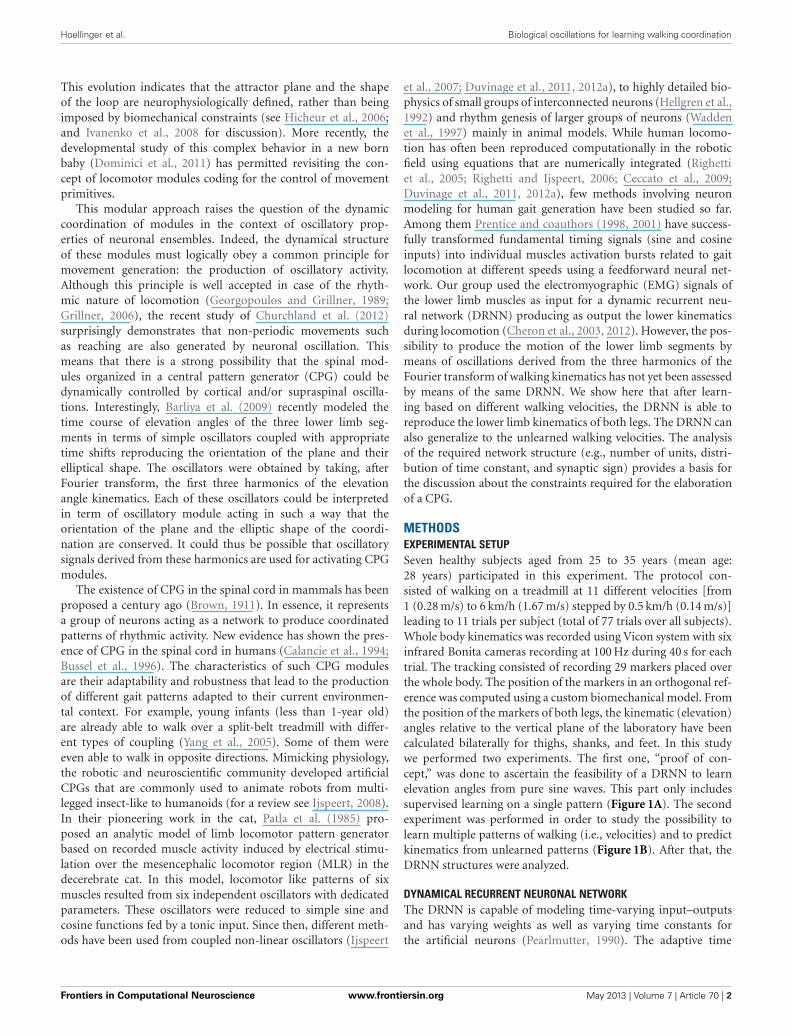
Hoellinger et al. Biological oscillations for learning walking coordination
This evolution indicates that the attractor plane and the shapeof the loop are neurophysiologically defined, rather than beingimposed by biomechanical constraints (see Hicheur et al., 2006;and Ivanenko et al., 2008 for discussion). More recently, thedevelopmental study of this complex behavior in a new bornbaby (Dominici et al., 2011) has permitted revisiting the con-cept of locomotor modules coding for the control of movementprimitives.
This modular approach raises the question of the dynamiccoordination of modules in the context of oscillatory prop-erties of neuronal ensembles. Indeed, the dynamical structureof these modules must logically obey a common principle formovement generation: the production of oscillatory activity.Although this principle is well accepted in case of the rhyth-mic nature of locomotion (Georgopoulos and Grillner, 1989;Grillner, 2006), the recent study of Churchland et al. (2012)surprisingly demonstrates that non-periodic movements suchas reaching are also generated by neuronal oscillation. Thismeans that there is a strong possibility that the spinal mod-ules organized in a central pattern generator (CPG) could bedynamically controlled by cortical and/or supraspinal oscilla-tions. Interestingly, Barliya et al. (2009) recently modeled thetime course of elevation angles of the three lower limb seg-ments in terms of simple oscillators coupled with appropriatetime shifts reproducing the orientation of the plane and theirelliptical shape. The oscillators were obtained by taking, afterFourier transform, the first three harmonics of the elevationangle kinematics. Each of these oscillators could be interpretedin term of oscillatory module acting in such a way that theorientation of the plane and the elliptic shape of the coordi-nation are conserved. It could thus be possible that oscillatorysignals derived from these harmonics are used for activating CPGmodules.
The existence of CPG in the spinal cord in mammals has beenproposed a century ago (Brown, 1911). In essence, it representsa group of neurons acting as a network to produce coordinatedpatterns of rhythmic activity. New evidence has shown the pres-ence of CPG in the spinal cord in humans (Calancie et al., 1994;Bussel et al., 1996). The characteristics of such CPG modulesare their adaptability and robustness that lead to the productionof different gait patterns adapted to their current environmen-tal context. For example, young infants (less than 1-year old)are already able to walk over a split-belt treadmill with differ-ent types of coupling (Yang et al., 2005). Some of them wereeven able to walk in opposite directions. Mimicking physiology,the robotic and neuroscientific community developed artificialCPGs that are commonly used to animate robots from multi-legged insect-like to humanoids (for a review see Ijspeert, 2008).In their pioneering work in the cat, Patla et al. (1985) pro-posed an analytic model of limb locomotor pattern generatorbased on recorded muscle activity induced by electrical stimu-lation over the mesencephalic locomotor region (MLR) in thedecerebrate cat. In this model, locomotor like patterns of sixmuscles resulted from six independent oscillators with dedicatedparameters. These oscillators were reduced to simple sine andcosine functions fed by a tonic input. Since then, different meth-ods have been used from coupled non-linear oscillators (Ijspeert
et al., 2007; Duvinage et al., 2011, 2012a), to highly detailed bio-physics of small groups of interconnected neurons (Hellgren et al.,1992) and rhythm genesis of larger groups of neurons (Waddenet al., 1997) mainly in animal models. While human locomo-tion has often been reproduced computationally in the roboticfield using equations that are numerically integrated (Righettiet al., 2005; Righetti and Ijspeert, 2006; Ceccato et al., 2009;Duvinage et al., 2011, 2012a), few methods involving neuronmodeling for human gait generation have been studied so far.Among them Prentice and coauthors (1998, 2001) have success-fully transformed fundamental timing signals (sine and cosineinputs) into individual muscles activation bursts related to gaitlocomotion at different speeds using a feedforward neural net-work. Our group used the electromyographic (EMG) signals ofthe lower limb muscles as input for a dynamic recurrent neu-ral network (DRNN) producing as output the lower kinematicsduring locomotion (Cheron et al., 2003, 2012). However, the pos-sibility to produce the motion of the lower limb segments bymeans of oscillations derived from the three harmonics of theFourier transform of walking kinematics has not yet been assessedby means of the same DRNN. We show here that after learn-ing based on different walking velocities, the DRNN is able toreproduce the lower limb kinematics of both legs. The DRNN canalso generalize to the unlearned walking velocities. The analysisof the required network structure (e.g., number of units, distri-bution of time constant, and synaptic sign) provides a basis forthe discussion about the constraints required for the elaborationof a CPG.
METHODSEXPERIMENTAL SETUPSeven healthy subjects aged from 25 to 35 years (mean age:28 years) participated in this experiment. The protocol con-sisted of walking on a treadmill at 11 different velocities [from1 (0.28 m/s) to 6 km/h (1.67 m/s) stepped by 0.5 km/h (0.14 m/s)]leading to 11 trials per subject (total of 77 trials over all subjects).Whole body kinematics was recorded using Vicon system with sixinfrared Bonita cameras recording at 100 Hz during 40 s for eachtrial. The tracking consisted of recording 29 markers placed overthe whole body. The position of the markers in an orthogonal ref-erence was computed using a custom biomechanical model. Fromthe position of the markers of both legs, the kinematic (elevation)angles relative to the vertical plane of the laboratory have beencalculated bilaterally for thighs, shanks, and feet. In this studywe performed two experiments. The first one, “proof of con-cept,” was done to ascertain the feasibility of a DRNN to learnelevation angles from pure sine waves. This part only includessupervised learning on a single pattern (Figure 1A). The secondexperiment was performed in order to study the possibility tolearn multiple patterns of walking (i.e., velocities) and to predictkinematics from unlearned patterns (Figure 1B). After that, theDRNN structures were analyzed.
DYNAMICAL RECURRENT NEURONAL NETWORKThe DRNN is capable of modeling time-varying input–outputsand has varying weights as well as varying time constants forthe artificial neurons (Pearlmutter, 1990). The adaptive time
Frontiers in Computational Neuroscience www.frontiersin.org May 2013 | Volume 7 | Article 70 | 2
Hoellinger et al. Biological oscillations for learning walking coordination
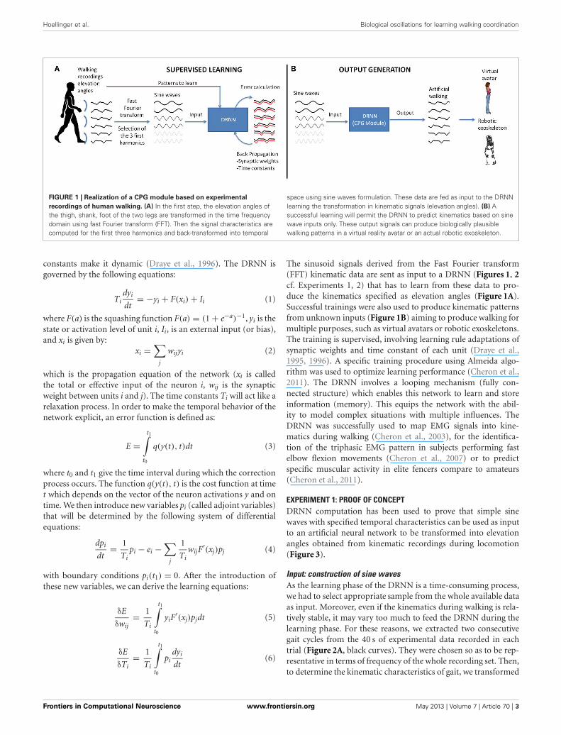
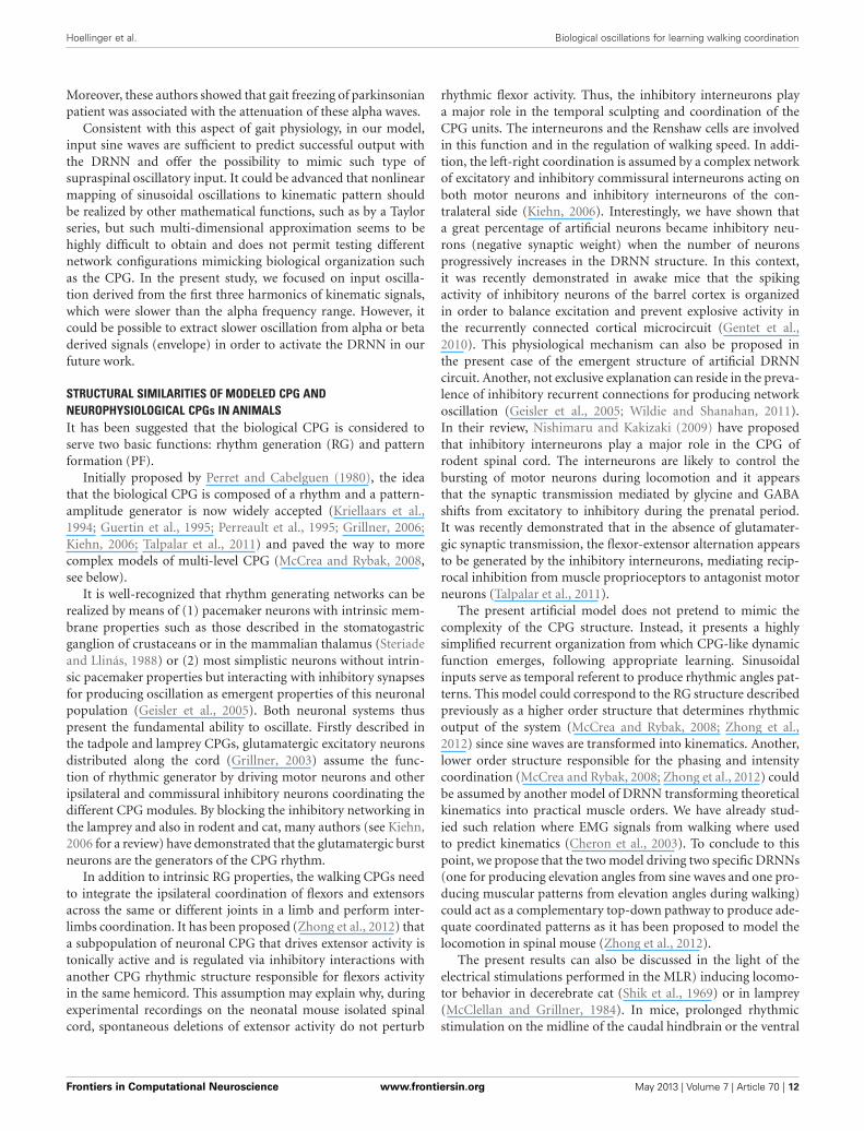
FIGURE 1 | Realization of a CPG module based on experimental
recordings of human walking. (A) In the first step, the elevation angles ofthe thigh, shank, foot of the two legs are transformed in the time frequencydomain using fast Fourier transform (FFT). Then the signal characteristics arecomputed for the first three harmonics and back-transformed into temporal
space using sine waves formulation. These data are fed as input to the DRNNlearning the transformation in kinematic signals (elevation angles). (B) Asuccessful learning will permit the DRNN to predict kinematics based on sinewave inputs only. These output signals can produce biologically plausiblewalking patterns in a virtual reality avatar or an actual robotic exoskeleton.
constants make it dynamic (Draye et al., 1996). The DRNN isgoverned by the following equations:
Tidyi
dt= −yi + F(xi) + Ii (1)
where F(a) is the squashing function F(a) = (1 + e−a)−1, yi is thestate or activation level of unit i, Ii, is an external input (or bias),and xi is given by:
xi =∑
j
wijyi (2)
which is the propagation equation of the network (xi is calledthe total or effective input of the neuron i, wij is the synapticweight between units i and j). The time constants Ti will act like arelaxation process. In order to make the temporal behavior of thenetwork explicit, an error function is defined as:
E =t1∫
t0
q(y(t), t)dt (3)
where t0 and t1 give the time interval during which the correctionprocess occurs. The function q(y(t), t) is the cost function at timet which depends on the vector of the neuron activations y and ontime. We then introduce new variables pi (called adjoint variables)that will be determined by the following system of differentialequations:
dpi
dt= 1
Tipi − ei −
∑j
1
TiwijF
′(xj)pj (4)
with boundary conditions pi(t1) = 0. After the introduction ofthese new variables, we can derive the learning equations:
δE
δwij= 1
Ti
t1∫t0
yiF′(xj)pjdt (5)
δE
δTi= 1
Ti
t1∫t0
pidyi
dt(6)
The sinusoid signals derived from the Fast Fourier transform(FFT) kinematic data are sent as input to a DRNN (Figures 1, 2cf. Experiments 1, 2) that has to learn from these data to pro-duce the kinematics specified as elevation angles (Figure 1A).Successful trainings were also used to produce kinematic patternsfrom unknown inputs (Figure 1B) aiming to produce walking formultiple purposes, such as virtual avatars or robotic exoskeletons.The training is supervised, involving learning rule adaptations ofsynaptic weights and time constant of each unit (Draye et al.,1995, 1996). A specific training procedure using Almeida algo-rithm was used to optimize learning performance (Cheron et al.,2011). The DRNN involves a looping mechanism (fully con-nected structure) which enables this network to learn and storeinformation (memory). This equips the network with the abil-ity to model complex situations with multiple influences. TheDRNN was successfully used to map EMG signals into kine-matics during walking (Cheron et al., 2003), for the identifica-tion of the triphasic EMG pattern in subjects performing fastelbow flexion movements (Cheron et al., 2007) or to predictspecific muscular activity in elite fencers compare to amateurs(Cheron et al., 2011).
EXPERIMENT 1: PROOF OF CONCEPTDRNN computation has been used to prove that simple sinewaves with specified temporal characteristics can be used as inputto an artificial neural network to be transformed into elevationangles obtained from kinematic recordings during locomotion(Figure 3).
Input: construction of sine wavesAs the learning phase of the DRNN is a time-consuming process,we had to select appropriate sample from the whole available dataas input. Moreover, even if the kinematics during walking is rela-tively stable, it may vary too much to feed the DRNN during thelearning phase. For these reasons, we extracted two consecutivegait cycles from the 40 s of experimental data recorded in eachtrial (Figure 2A, black curves). They were chosen so as to be rep-resentative in terms of frequency of the whole recording set. Then,to determine the kinematic characteristics of gait, we transformed
Frontiers in Computational Neuroscience www.frontiersin.org May 2013 | Volume 7 | Article 70 | 3

Hoellinger et al. Biological oscillations for learning walking coordination
FIGURE 2 | (A) Elevation angles (in degree) of the three segments (thigh,shank, and foot) for each leg as a function of time for a subject walkingat 3 km/h (0.83 m/s). The whole pattern is presented for duration of 5 s.The black lines represent a pattern for two gait cycles used to determinethe FFT characteristics. (B) The mean FFT transformation for six joints
for 40 s (in gray) and the two gait cycles (in black). Note that the twogait cycle patterns are selected so as to preserve the FFT characteristicsin terms of amplitude and frequency (stars) for the overall pattern of40 s. These characteristics are used as parameters to generate sinewaves as input of the DRNN.
FIGURE 3 | DRNN learning method for Experiment 1. For each subject,we trained 100 DRNNs to learn a pattern of kinematic [corresponding toa velocity of 3 km/h (3.83 m/s)] from sine waves (Equations 7 and 8).Three sets of learning were defined as the input differed (SEA, SEB,SEC). The structures of DRNNs were modeled with 30 hidden units for
each set of training. Each network iterated 10,000 times to change itssynaptic weights and time constants. For each subject and structure weselected the network with the highest SI value. For each condition, thedesign of the network is then processed with 6 inputs, 30 hidden units,and 6 outputs.
the data of the lower limb segments elevation angles into the timefrequency domain using the Matlab fft function to get the FFTpower amplitude and their related frequency values of the firstthree harmonic peaks (Figure 2B). It has been shown previouslythat the first two Fourier harmonics accounted for approximately98% of the experimental variance of the thigh, shank, and foot
angles (Bianchi et al., 1998). We decided to create sine wavesbased on the characteristics of the first three harmonics to ensurethat the signal proposed as learning input to the DRNN containsenough information to match the desired output precisely.
We extracted the values of the three frequencies (f 1, f 2, f 3)corresponding to the maximal amplitudes (a1, a2, a3). It was
Frontiers in Computational Neuroscience www.frontiersin.org May 2013 | Volume 7 | Article 70 | 4
Hoellinger et al. Biological oscillations for learning walking coordination
verified that f 2 = 2f 1 and f 3 = 3f 1. We then artificially pro-duced six sinusoidal signals using frequency values as parameters(Equations 7 and 8).
yfi,1 = sin(2π × fi × t) (7)
yfi,2 = sin(−(2π × fi × t)) (8)
For i = 1, 2, 3
These six sine waves thus correspond to the frequency charac-teristics of the kinematic patterns that will be further used aspattern to be learned. For the sake of clarity we called the set oforiginal inputs set SEA (for set of Equations A). Additionally, weproduced six new sine waves using the preceding computations(Equations 7 and 8) where f 1′ = f 1 + 0.25 Hz and six other sinewaves where f 1′′ = f 1 − 0.25 Hz, respectively called SEB (for setof Equations B) and SEC (for set of Equations C). Please notethat in the latter two cases the original relations f 2 = 2f 1 andf 3 = 3f 1 were respected and hence f 2′ = 2f 1 + 0.50 Hz; f 3′ =3f 1 + 0.75 Hz in the set SEB and f 2′′ = 2f 1 − 0.50 Hz; f 3′′ =3f 1 − 0.75 Hz in the set SEC. Three different input sets (SEA,SEB, SEC) were thus defined for learning.
Pattern to learn: kinematic dataThe pattern to learned corresponds to the elevation angles of theright and left thigh, shank, foot segments in the two gait cyclesof a 3 km/h (0.83 m/s) walk, normalized between −1 and 1. Theoutputs were the same for SEA, SEB, and SEC whereas inputsdiffered.
DRNN learning phaseIt was expected that the DRNN would learn how to transform theinput to output thanks to its dynamical and recurrent structureof 30 hidden neurons. For each of the seven subjects, the networkiterated 10,000 times. This process was repeated 100 times, lead-ing to 100 different networks. At the end of the learning phase,we selected for each subject the network for which the differ-ence between predicted and real kinematics was minimal. Thiscomputation was performed by calculating a similarity index (SI)(Bengoetxea et al., 2010) defined by the following equation:
SI =
∫ Tc2
− Tc2
p1(t)p2(t)dt
[∫ Tc2
− Tc2
p1(t)2dt
∫ Tc2
− Tc2
p2(t)2dt
] 12
(9)
where Tc is the period of the limit cycle, p1 and p2 being syn-chronized, i.e., the matching between both patterns is based onthe maximum of each pattern. Note that if both functions areidentical, SI = 1. SI was calculated with the recorded pattern ofelevation angles and the output of the DRNN.
EXPERIMENT 2: MULTIPLE PATTERN LEARNING AND PREDICTIONIn this experiment sine waves were modulated in frequencyand amplitude and transformed into kinematic data using
multi-pattern training. The prediction of kinematic pattern fromunknown sine wave pattern was also tested.
InputAs for the proof of concept methods, we extracted twogait cycles of each trial for multiple velocities (in km/h)(v = {1.5, 2.5, 3.5, 4.5, 5.5}). We transformed the kinematicdata into the time-frequency domain to get their frequency(f 1, f 2, f 3) and amplitude (a1, a2, a3) (Figure 2B) parametersusing the following set of Equations (10 and 11).
yv, fiv, aiv,1 = aiv × (sin(2π × fiv × t)) (10)
yv, fiv, aiv,2 = aiv × (sin(−(2π × fiv × t))) (11)
For i = 1, 2, 3
Patterns to learn: kinematic dataThe patterns to be learned consisted of elevation angles of theright and left thigh, shank, and foot segments corresponding tothe two gait cycles, normalized between –1 and 1. These patternswere obtained from recordings at multiple velocities (in km/h)(v = {1.5, 2.5, 3.5, 4.5, 5.5}).
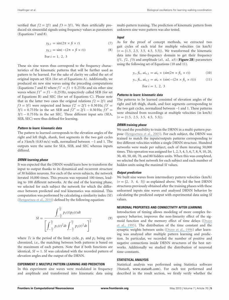
DRNN training phaseWe used the possibility to train the DRNN in a multi-pattern pur-pose (Bengoetxea et al., 2005). For each subject, the DRNN wastrained to match the inputs/outputs patterns corresponding tofive different velocities within a single DRNN structure. Hundrednetworks were made per subject, each of them iterating 50,000times. This operation was assigned for 1, 2, 3, 4, 5, 6, 7, 8, 9, 10, 20,30, 40, 50, 60, 70, and 80 hidden units. When this was completed,we selected the best network for each subject and each number ofhidden units using the maximal SI values.
Output predictionWe built sine waves from intermediary pattern velocities (km/h)(v = {2, 3, 4, 5}) as explained above. We fed the best DRNNstructures previously obtained after the training phases with theseunlearned inputs sine waves and analyzed DRNN behavior bycalculating the predicted output with experimental data using SIvalues.
NEURONAL PROPERTIES AND CONNECTIVITY AFTER LEARNINGIntroduction of timing allows modeling of more complex fre-quency behavior, improves the non-linearity effect of the sig-moid function and the memory effect of time delays (Drayeet al., 1995). The distribution of the time constant and thesynaptic weights between units (Draye et al., 1996) after learn-ing was analyzed after multiple pattern learning and predic-tion. In particular, we recorded the number of positive andnegative connections inside DRNN structures of the best net-works. Additionally we studied the distribution of neuronaltime-constants.
STATISTICAL ANALYSISStatistical analysis was performed using Statistica software(Statsoft, www.statsoft.com). For each test performed anddescribed in the result section, we firstly verify whether the
Frontiers in Computational Neuroscience www.frontiersin.org May 2013 | Volume 7 | Article 70 | 5
Hoellinger et al. Biological oscillations for learning walking coordination
data were distributed normally using Kolmogorov–Smirnov test.When the data were normally distributed we use ANOVA withrepeated measures and post-hoc Fisher analyses. When it was notpossible to use ANOVA we used non-parametric tests such asFriedman ANOVA or sign tests.
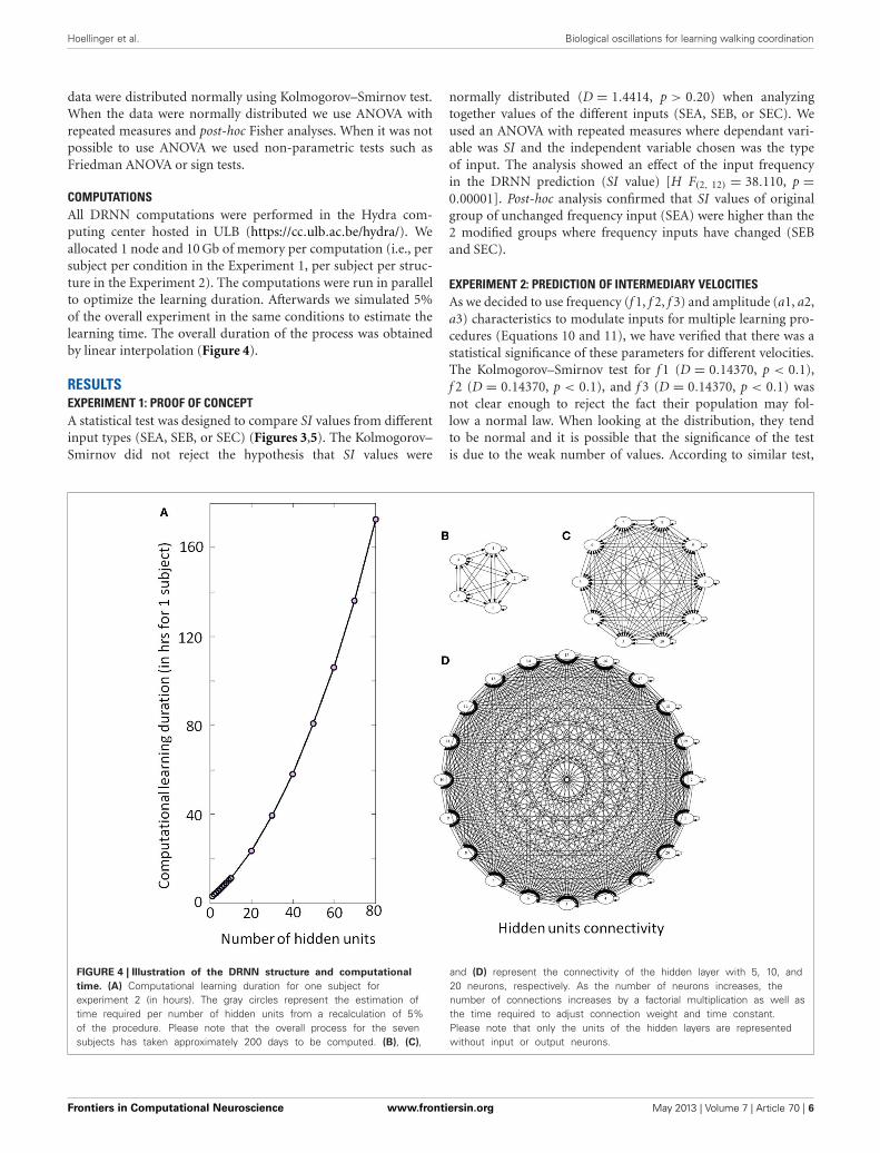
COMPUTATIONSAll DRNN computations were performed in the Hydra com-puting center hosted in ULB (https://cc.ulb.ac.be/hydra/). Weallocated 1 node and 10 Gb of memory per computation (i.e., persubject per condition in the Experiment 1, per subject per struc-ture in the Experiment 2). The computations were run in parallelto optimize the learning duration. Afterwards we simulated 5%of the overall experiment in the same conditions to estimate thelearning time. The overall duration of the process was obtainedby linear interpolation (Figure 4).
RESULTSEXPERIMENT 1: PROOF OF CONCEPTA statistical test was designed to compare SI values from differentinput types (SEA, SEB, or SEC) (Figures 3,5). The Kolmogorov–Smirnov did not reject the hypothesis that SI values were
normally distributed (D = 1.4414, p > 0.20) when analyzingtogether values of the different inputs (SEA, SEB, or SEC). Weused an ANOVA with repeated measures where dependant vari-able was SI and the independent variable chosen was the typeof input. The analysis showed an effect of the input frequencyin the DRNN prediction (SI value) [H F(2, 12) = 38.110, p =0.00001]. Post-hoc analysis confirmed that SI values of originalgroup of unchanged frequency input (SEA) were higher than the2 modified groups where frequency inputs have changed (SEBand SEC).
EXPERIMENT 2: PREDICTION OF INTERMEDIARY VELOCITIESAs we decided to use frequency (f 1, f 2, f 3) and amplitude (a1, a2,a3) characteristics to modulate inputs for multiple learning pro-cedures (Equations 10 and 11), we have verified that there was astatistical significance of these parameters for different velocities.The Kolmogorov–Smirnov test for f 1 (D = 0.14370, p < 0.1),f 2 (D = 0.14370, p < 0.1), and f 3 (D = 0.14370, p < 0.1) wasnot clear enough to reject the fact their population may fol-low a normal law. When looking at the distribution, they tendto be normal and it is possible that the significance of the testis due to the weak number of values. According to similar test,
FIGURE 4 | Illustration of the DRNN structure and computational
time. (A) Computational learning duration for one subject forexperiment 2 (in hours). The gray circles represent the estimation oftime required per number of hidden units from a recalculation of 5%of the procedure. Please note that the overall process for the sevensubjects has taken approximately 200 days to be computed. (B), (C),
and (D) represent the connectivity of the hidden layer with 5, 10, and20 neurons, respectively. As the number of neurons increases, thenumber of connections increases by a factorial multiplication as well asthe time required to adjust connection weight and time constant.Please note that only the units of the hidden layers are representedwithout input or output neurons.
Frontiers in Computational Neuroscience www.frontiersin.org May 2013 | Volume 7 | Article 70 | 6
Hoellinger et al. Biological oscillations for learning walking coordination
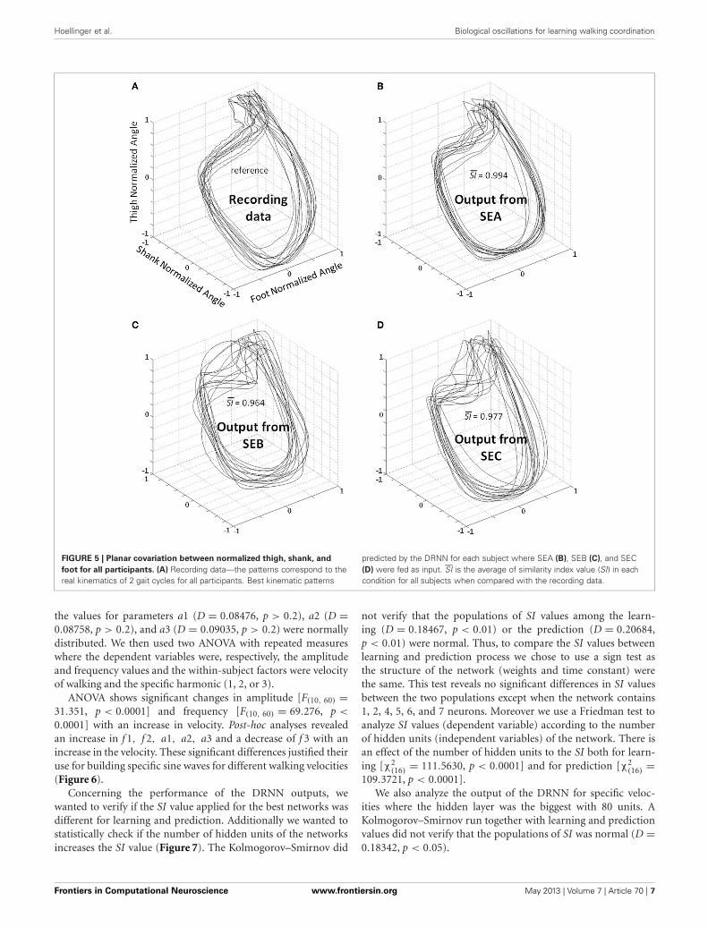
FIGURE 5 | Planar covariation between normalized thigh, shank, and
foot for all participants. (A) Recording data—the patterns correspond to thereal kinematics of 2 gait cycles for all participants. Best kinematic patterns
predicted by the DRNN for each subject where SEA (B), SEB (C), and SEC(D) were fed as input. SI is the average of similarity index value (SI) in eachcondition for all subjects when compared with the recording data.
the values for parameters a1 (D = 0.08476, p > 0.2), a2 (D =0.08758, p > 0.2), and a3 (D = 0.09035, p > 0.2) were normallydistributed. We then used two ANOVA with repeated measureswhere the dependent variables were, respectively, the amplitudeand frequency values and the within-subject factors were velocityof walking and the specific harmonic (1, 2, or 3).
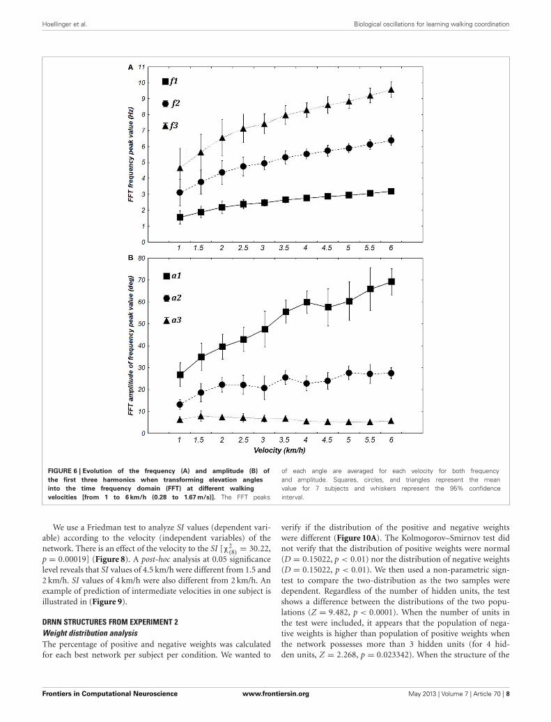
ANOVA shows significant changes in amplitude [F(10, 60) =31.351, p < 0.0001] and frequency [F(10, 60) = 69.276, p <
0.0001] with an increase in velocity. Post-hoc analyses revealedan increase in f 1, f 2, a1, a2, a3 and a decrease of f 3 with anincrease in the velocity. These significant differences justified theiruse for building specific sine waves for different walking velocities(Figure 6).
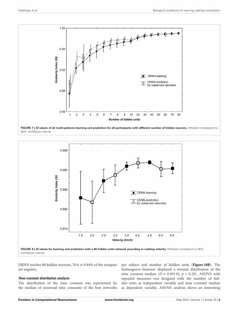
Concerning the performance of the DRNN outputs, wewanted to verify if the SI value applied for the best networks wasdifferent for learning and prediction. Additionally we wanted tostatistically check if the number of hidden units of the networksincreases the SI value (Figure 7). The Kolmogorov–Smirnov did
not verify that the populations of SI values among the learn-ing (D = 0.18467, p < 0.01) or the prediction (D = 0.20684,p < 0.01) were normal. Thus, to compare the SI values betweenlearning and prediction process we chose to use a sign test asthe structure of the network (weights and time constant) werethe same. This test reveals no significant differences in SI valuesbetween the two populations except when the network contains1, 2, 4, 5, 6, and 7 neurons. Moreover we use a Friedman test toanalyze SI values (dependent variable) according to the numberof hidden units (independent variables) of the network. There isan effect of the number of hidden units to the SI both for learn-ing [χ2
(16)= 111.5630, p < 0.0001] and for prediction [χ2
(16)=
109.3721, p < 0.0001].We also analyze the output of the DRNN for specific veloc-
ities where the hidden layer was the biggest with 80 units. AKolmogorov–Smirnov run together with learning and predictionvalues did not verify that the populations of SI was normal (D =0.18342, p < 0.05).
Frontiers in Computational Neuroscience www.frontiersin.org May 2013 | Volume 7 | Article 70 | 7
Hoellinger et al. Biological oscillations for learning walking coordination
FIGURE 6 | Evolution of the frequency (A) and amplitude (B) of
the first three harmonics when transforming elevation angles
into the time frequency domain (FFT) at different walking
velocities [from 1 to 6 km/h (0.28 to 1.67 m/s)]. The FFT peaks
of each angle are averaged for each velocity for both frequencyand amplitude. Squares, circles, and triangles represent the meanvalue for 7 subjects and whiskers represent the 95% confidenceinterval.
We use a Friedman test to analyze SI values (dependent vari-able) according to the velocity (independent variables) of thenetwork. There is an effect of the velocity to the SI [χ2
(8)= 30.22,
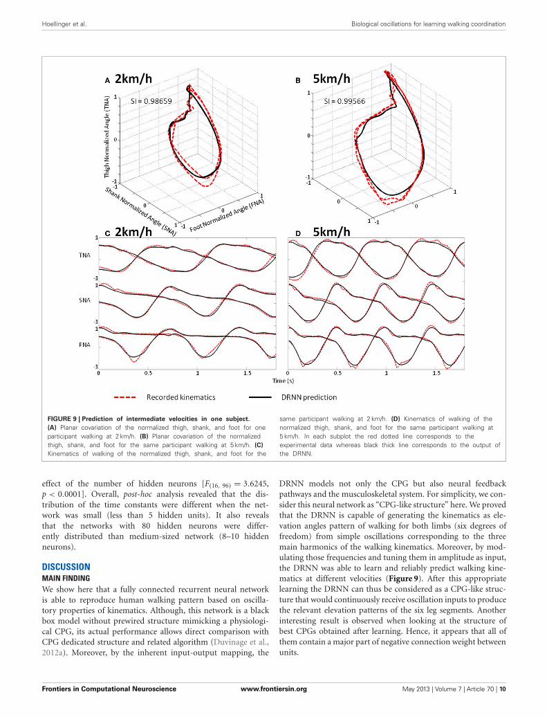
p = 0.00019] (Figure 8). A post-hoc analysis at 0.05 significancelevel reveals that SI values of 4.5 km/h were different from 1.5 and2 km/h. SI values of 4 km/h were also different from 2 km/h. Anexample of prediction of intermediate velocities in one subject isillustrated in (Figure 9).
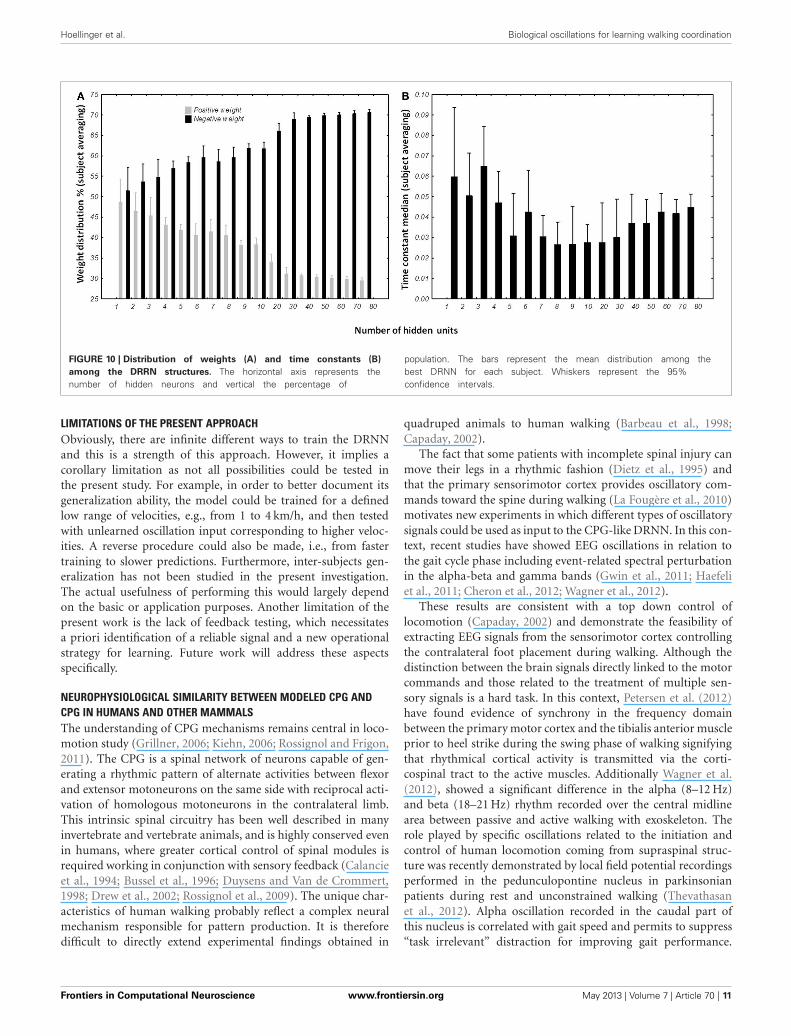
DRNN STRUCTURES FROM EXPERIMENT 2Weight distribution analysisThe percentage of positive and negative weights was calculatedfor each best network per subject per condition. We wanted to
verify if the distribution of the positive and negative weightswere different (Figure 10A). The Kolmogorov–Smirnov test didnot verify that the distribution of positive weights were normal(D = 0.15022, p < 0.01) nor the distribution of negative weights(D = 0.15022, p < 0.01). We then used a non-parametric sign-test to compare the two-distribution as the two samples weredependent. Regardless of the number of hidden units, the testshows a difference between the distributions of the two popu-lations (Z = 9.482, p < 0.0001). When the number of units inthe test were included, it appears that the population of nega-tive weights is higher than population of positive weights whenthe network possesses more than 3 hidden units (for 4 hid-den units, Z = 2.268, p = 0.023342). When the structure of the
Frontiers in Computational Neuroscience www.frontiersin.org May 2013 | Volume 7 | Article 70 | 8
Hoellinger et al. Biological oscillations for learning walking coordination
FIGURE 7 | SI values of all multi-patterns learning and prediction for all participants with different number of hidden neurons. Whiskers correspond to95% confidence interval.
FIGURE 8 | SI values for learning and prediction with a 80 hidden units network according to walking velocity. Whiskers correspond to 95%confidence interval.
DRNN reaches 80 hidden neurons, 70.6 ± 0.84% of the synapsesare negative.
Time constant distribution analysisThe distribution of the time constant was represented bythe median of neuronal time constants of the best networks
per subject and number of hidden units (Figure 10B). TheKolmogorov–Smirnov displayed a normal distribution of thetime constant median (D = 0.09110, p > 0.20). ANOVA withrepeated measures was designed with the number of hid-den units as independent variable and time constant medianas dependent variable. ANOVA analysis shows an interesting
Frontiers in Computational Neuroscience www.frontiersin.org May 2013 | Volume 7 | Article 70 | 9
Hoellinger et al. Biological oscillations for learning walking coordination
FIGURE 9 | Prediction of intermediate velocities in one subject.
(A) Planar covariation of the normalized thigh, shank, and foot for oneparticipant walking at 2 km/h. (B) Planar covariation of the normalizedthigh, shank, and foot for the same participant walking at 5 km/h. (C)
Kinematics of walking of the normalized thigh, shank, and foot for the
same participant walking at 2 km/h. (D) Kinematics of walking of thenormalized thigh, shank, and foot for the same participant walking at5 km/h. In each subplot the red dotted line corresponds to theexperimental data whereas black thick line corresponds to the output ofthe DRNN.
effect of the number of hidden neurons [F(16, 96) = 3.6245,p < 0.0001]. Overall, post-hoc analysis revealed that the dis-tribution of the time constants were different when the net-work was small (less than 5 hidden units). It also revealsthat the networks with 80 hidden neurons were differ-ently distributed than medium-sized network (8–10 hiddenneurons).
DISCUSSIONMAIN FINDINGWe show here that a fully connected recurrent neural networkis able to reproduce human walking pattern based on oscilla-tory properties of kinematics. Although, this network is a blackbox model without prewired structure mimicking a physiologi-cal CPG, its actual performance allows direct comparison withCPG dedicated structure and related algorithm (Duvinage et al.,2012a). Moreover, by the inherent input-output mapping, the
DRNN models not only the CPG but also neural feedbackpathways and the musculoskeletal system. For simplicity, we con-sider this neural network as “CPG-like structure” here. We provedthat the DRNN is capable of generating the kinematics as ele-vation angles pattern of walking for both limbs (six degrees offreedom) from simple oscillations corresponding to the threemain harmonics of the walking kinematics. Moreover, by mod-ulating those frequencies and tuning them in amplitude as input,the DRNN was able to learn and reliably predict walking kine-matics at different velocities (Figure 9). After this appropriatelearning the DRNN can thus be considered as a CPG-like struc-ture that would continuously receive oscillation inputs to producethe relevant elevation patterns of the six leg segments. Anotherinteresting result is observed when looking at the structure ofbest CPGs obtained after learning. Hence, it appears that all ofthem contain a major part of negative connection weight betweenunits.
Frontiers in Computational Neuroscience www.frontiersin.org May 2013 | Volume 7 | Article 70 | 10
Hoellinger et al. Biological oscillations for learning walking coordination
FIGURE 10 | Distribution of weights (A) and time constants (B)
among the DRRN structures. The horizontal axis represents thenumber of hidden neurons and vertical the percentage of
population. The bars represent the mean distribution among thebest DRNN for each subject. Whiskers represent the 95%confidence intervals.
LIMITATIONS OF THE PRESENT APPROACHObviously, there are infinite different ways to train the DRNNand this is a strength of this approach. However, it implies acorollary limitation as not all possibilities could be tested inthe present study. For example, in order to better document itsgeneralization ability, the model could be trained for a definedlow range of velocities, e.g., from 1 to 4 km/h, and then testedwith unlearned oscillation input corresponding to higher veloc-ities. A reverse procedure could also be made, i.e., from fastertraining to slower predictions. Furthermore, inter-subjects gen-eralization has not been studied in the present investigation.The actual usefulness of performing this would largely dependon the basic or application purposes. Another limitation of thepresent work is the lack of feedback testing, which necessitatesa priori identification of a reliable signal and a new operationalstrategy for learning. Future work will address these aspectsspecifically.
NEUROPHYSIOLOGICAL SIMILARITY BETWEEN MODELED CPG ANDCPG IN HUMANS AND OTHER MAMMALSThe understanding of CPG mechanisms remains central in loco-motion study (Grillner, 2006; Kiehn, 2006; Rossignol and Frigon,2011). The CPG is a spinal network of neurons capable of gen-erating a rhythmic pattern of alternate activities between flexorand extensor motoneurons on the same side with reciprocal acti-vation of homologous motoneurons in the contralateral limb.This intrinsic spinal circuitry has been well described in manyinvertebrate and vertebrate animals, and is highly conserved evenin humans, where greater cortical control of spinal modules isrequired working in conjunction with sensory feedback (Calancieet al., 1994; Bussel et al., 1996; Duysens and Van de Crommert,1998; Drew et al., 2002; Rossignol et al., 2009). The unique char-acteristics of human walking probably reflect a complex neuralmechanism responsible for pattern production. It is thereforedifficult to directly extend experimental findings obtained in
quadruped animals to human walking (Barbeau et al., 1998;Capaday, 2002).
The fact that some patients with incomplete spinal injury canmove their legs in a rhythmic fashion (Dietz et al., 1995) andthat the primary sensorimotor cortex provides oscillatory com-mands toward the spine during walking (La Fougère et al., 2010)motivates new experiments in which different types of oscillatorysignals could be used as input to the CPG-like DRNN. In this con-text, recent studies have showed EEG oscillations in relation tothe gait cycle phase including event-related spectral perturbationin the alpha-beta and gamma bands (Gwin et al., 2011; Haefeliet al., 2011; Cheron et al., 2012; Wagner et al., 2012).
These results are consistent with a top down control oflocomotion (Capaday, 2002) and demonstrate the feasibility ofextracting EEG signals from the sensorimotor cortex controllingthe contralateral foot placement during walking. Although thedistinction between the brain signals directly linked to the motorcommands and those related to the treatment of multiple sen-sory signals is a hard task. In this context, Petersen et al. (2012)have found evidence of synchrony in the frequency domainbetween the primary motor cortex and the tibialis anterior muscleprior to heel strike during the swing phase of walking signifyingthat rhythmical cortical activity is transmitted via the corti-cospinal tract to the active muscles. Additionally Wagner et al.(2012), showed a significant difference in the alpha (8–12 Hz)and beta (18–21 Hz) rhythm recorded over the central midlinearea between passive and active walking with exoskeleton. Therole played by specific oscillations related to the initiation andcontrol of human locomotion coming from supraspinal struc-ture was recently demonstrated by local field potential recordingsperformed in the pedunculopontine nucleus in parkinsonianpatients during rest and unconstrained walking (Thevathasanet al., 2012). Alpha oscillation recorded in the caudal part ofthis nucleus is correlated with gait speed and permits to suppress“task irrelevant” distraction for improving gait performance.
Frontiers in Computational Neuroscience www.frontiersin.org May 2013 | Volume 7 | Article 70 | 11
Hoellinger et al. Biological oscillations for learning walking coordination
Moreover, these authors showed that gait freezing of parkinsonianpatient was associated with the attenuation of these alpha waves.
Consistent with this aspect of gait physiology, in our model,input sine waves are sufficient to predict successful output withthe DRNN and offer the possibility to mimic such type ofsupraspinal oscillatory input. It could be advanced that nonlinearmapping of sinusoidal oscillations to kinematic pattern shouldbe realized by other mathematical functions, such as by a Taylorseries, but such multi-dimensional approximation seems to behighly difficult to obtain and does not permit testing differentnetwork configurations mimicking biological organization suchas the CPG. In the present study, we focused on input oscilla-tion derived from the first three harmonics of kinematic signals,which were slower than the alpha frequency range. However, itcould be possible to extract slower oscillation from alpha or betaderived signals (envelope) in order to activate the DRNN in ourfuture work.
STRUCTURAL SIMILARITIES OF MODELED CPG ANDNEUROPHYSIOLOGICAL CPGs IN ANIMALSIt has been suggested that the biological CPG is considered toserve two basic functions: rhythm generation (RG) and patternformation (PF).
Initially proposed by Perret and Cabelguen (1980), the ideathat the biological CPG is composed of a rhythm and a pattern-amplitude generator is now widely accepted (Kriellaars et al.,1994; Guertin et al., 1995; Perreault et al., 1995; Grillner, 2006;Kiehn, 2006; Talpalar et al., 2011) and paved the way to morecomplex models of multi-level CPG (McCrea and Rybak, 2008,see below).
It is well-recognized that rhythm generating networks can berealized by means of (1) pacemaker neurons with intrinsic mem-brane properties such as those described in the stomatogastricganglion of crustaceans or in the mammalian thalamus (Steriadeand Llinás, 1988) or (2) most simplistic neurons without intrin-sic pacemaker properties but interacting with inhibitory synapsesfor producing oscillation as emergent properties of this neuronalpopulation (Geisler et al., 2005). Both neuronal systems thuspresent the fundamental ability to oscillate. Firstly described inthe tadpole and lamprey CPGs, glutamatergic excitatory neuronsdistributed along the cord (Grillner, 2003) assume the func-tion of rhythmic generator by driving motor neurons and otheripsilateral and commissural inhibitory neurons coordinating thedifferent CPG modules. By blocking the inhibitory networking inthe lamprey and also in rodent and cat, many authors (see Kiehn,2006 for a review) have demonstrated that the glutamatergic burstneurons are the generators of the CPG rhythm.
In addition to intrinsic RG properties, the walking CPGs needto integrate the ipsilateral coordination of flexors and extensorsacross the same or different joints in a limb and perform inter-limbs coordination. It has been proposed (Zhong et al., 2012) thata subpopulation of neuronal CPG that drives extensor activity istonically active and is regulated via inhibitory interactions withanother CPG rhythmic structure responsible for flexors activityin the same hemicord. This assumption may explain why, duringexperimental recordings on the neonatal mouse isolated spinalcord, spontaneous deletions of extensor activity do not perturb
rhythmic flexor activity. Thus, the inhibitory interneurons playa major role in the temporal sculpting and coordination of theCPG units. The interneurons and the Renshaw cells are involvedin this function and in the regulation of walking speed. In addi-tion, the left-right coordination is assumed by a complex networkof excitatory and inhibitory commissural interneurons acting onboth motor neurons and inhibitory interneurons of the con-tralateral side (Kiehn, 2006). Interestingly, we have shown thata great percentage of artificial neurons became inhibitory neu-rons (negative synaptic weight) when the number of neuronsprogressively increases in the DRNN structure. In this context,it was recently demonstrated in awake mice that the spikingactivity of inhibitory neurons of the barrel cortex is organizedin order to balance excitation and prevent explosive activity inthe recurrently connected cortical microcircuit (Gentet et al.,2010). This physiological mechanism can also be proposed inthe present case of the emergent structure of artificial DRNNcircuit. Another, not exclusive explanation can reside in the preva-lence of inhibitory recurrent connections for producing networkoscillation (Geisler et al., 2005; Wildie and Shanahan, 2011).In their review, Nishimaru and Kakizaki (2009) have proposedthat inhibitory interneurons play a major role in the CPG ofrodent spinal cord. The interneurons are likely to control thebursting of motor neurons during locomotion and it appearsthat the synaptic transmission mediated by glycine and GABAshifts from excitatory to inhibitory during the prenatal period.It was recently demonstrated that in the absence of glutamater-gic synaptic transmission, the flexor-extensor alternation appearsto be generated by the inhibitory interneurons, mediating recip-rocal inhibition from muscle proprioceptors to antagonist motorneurons (Talpalar et al., 2011).
The present artificial model does not pretend to mimic thecomplexity of the CPG structure. Instead, it presents a highlysimplified recurrent organization from which CPG-like dynamicfunction emerges, following appropriate learning. Sinusoidalinputs serve as temporal referent to produce rhythmic angles pat-terns. This model could correspond to the RG structure describedpreviously as a higher order structure that determines rhythmicoutput of the system (McCrea and Rybak, 2008; Zhong et al.,2012) since sine waves are transformed into kinematics. Another,lower order structure responsible for the phasing and intensitycoordination (McCrea and Rybak, 2008; Zhong et al., 2012) couldbe assumed by another model of DRNN transforming theoreticalkinematics into practical muscle orders. We have already stud-ied such relation where EMG signals from walking where usedto predict kinematics (Cheron et al., 2003). To conclude to thispoint, we propose that the two model driving two specific DRNNs(one for producing elevation angles from sine waves and one pro-ducing muscular patterns from elevation angles during walking)could act as a complementary top-down pathway to produce ade-quate coordinated patterns as it has been proposed to model thelocomotion in spinal mouse (Zhong et al., 2012).
The present results can also be discussed in the light of theelectrical stimulations performed in the MLR) inducing locomo-tor behavior in decerebrate cat (Shik et al., 1969) or in lamprey(McClellan and Grillner, 1984). In mice, prolonged rhythmicstimulation on the midline of the caudal hindbrain or the ventral
Frontiers in Computational Neuroscience www.frontiersin.org May 2013 | Volume 7 | Article 70 | 12
Hoellinger et al. Biological oscillations for learning walking coordination
spinal cord (C1–C4) induces a stable locomotor activity (Talpalaret al., 2011). Typically, low-frequency stimulation leads to slow-frequency movements and inversely fast-frequency stimulationleads to fast-frequency movements. Our model is in accordancewith this physiological behavior, when amplitude of the artificialsine wave inputs increases, the amplitude of stepping increasesas well, leading to a change in walking velocities (Figure 6B). Interms of neurological development, there is some evidence for theexistence of CPG very early in CNS maturation (Yang et al., 1998;Dominici et al., 2011). Neonatal, so-called “infant” stepping hasbeen ascribed to similar EMG patterns activity in different direc-tions inducing stereotyped yet non-functional walking patterns(Lamb and Yang, 2000). This leads the authors to conclude thatthe same CPG controls different stepping in human infants incontrast with some studies in adults (Thorstensson, 1986; Grassoet al., 1998). Interestingly, we found that DRNN with only fourhidden artificial neurons can generate walking pattern, whereasat least 50 hidden neurons are the prerequisite to generate accu-rate movements (Figure 7). Obviously, recruitment and trainingof such high numbers of neurons requires long computationaltime. For example, with a 4 hidden units DRNN structure, thelearning process lasts about 5 min based on our computer per-formance while 160 min are necessary for a DRNN containing80 units (Figure 4).
PERSPECTIVESSuch a tool can be used to produce gait kinematics in numer-ous and various applications. For rehabilitation it can be usedto train people for recovering a walking pattern correspondingto their physical characteristics by training with an appropri-ate feedback. Specifically dedicated DRNN based on the properdynamics of participants could be used for medical applicationssuch as in prosthesis and exoskeleton control (Cheron et al.,2012). It can also be integrated in BCI applications in whichhigher order commands can be used, e.g., from steady state visual
or somatosensory evoked potentials (Cheron et al., 2012) or P300(Castermans et al., 2011; Duvinage et al., 2012b). This neuronalavenue might lead to the decoding of higher neuronal commandsthat govern CPG mechanisms. Since these CPG can be trainedusing specific sinusoidal frequency signals, it might be possibleto extract this type of signals from specific EEG rhythms sincethe brain itself is an effective machine for producing oscilla-tions (Buzsáki and Draguhn, 2004). One of the strengths of thisapproach is that it is not necessary to determine in advance thetopology and the timing sequences between the artificial neu-rons. This contrasts with other CPGs, such as a recently developedones (Duvinage et al., 2011) based on coupled oscillators (Righettiand Ijspeert, 2006), where adjustment of intrinsic parameters byoptimization techniques was necessary.
In future studies, by introducing an informational delay(Draye et al., 1997) or an artificial distance based on a Gaussianfactor modulating the weights between the different neurons(Draye et al., 2002), it will be possible to analyze the self-tailoredorganization of the links between neurons and the possible emer-gence of specific topologies. In this case it will also be possible toselect different modular architectures of the DRNN.
ACKNOWLEDGMENTSThis work was funded by the Belgian Federal Science PolicyOffice, the European Space Agency, (AO-2004, 118), the BelgianNational Fund for Scientific Research (FNRS), the researchfunds of the Université Libre de Bruxelles and of the Universitéde Mons (Belgium), the FEDER support (BIOFACT), and theMINDWALKER project (FP7 – 2007–2013) supported by theEuropean Commission. M. Duvinage is a FNRS Research Fellow.This paper presents research results of the Belgian NetworkDYSCO (Dynamical Systems, Control, and Optimization),funded by the Interuniversity Attraction Poles Program, initi-ated by the Belgian State, Science Policy Office. The scientificresponsibility rests with its author(s).
REFERENCESBarbeau, H., Pépin, A., Norman, K.
E., Ladouceur, M., and Leroux,A. (1998). Review?: walking afterspinal cord injury: control andrecovery. Neuroscientist 4, 14–24.
Barliya, A., Omlor, L., Giese, M. A.,and Flash, T. (2009). An analyticalformulation of the law of interseg-mental coordination during humanlocomotion. Exp. Brain Res. 193,371–385.
Bengoetxea, A., Dan, B., Leurs, F.,Cebolla, A. M., De Saedeleer,C., Gillis, P., et al. (2010).Rhythmic muscular activationpattern for fast figure-eight move-ment. Clin. Neurophysiol. 121,754–765.
Bengoetxea, A., Leurs, F., Cebolla,A., Wellens, S., Draye, J. P., andCheron, G. (2005). A dynamicrecurrent neural network for draw-ing multi-directional trajectories.
Comput. Methods Biomech. Biomed.Engin. 8, 29–30.
Bernstein, N. A. (1967). The Co-Ordination and Regulation ofMovements. Oxford; New York:Pergamon Press.
Bianchi, L., Angelini, D., Orani, G.P., and Lacquaniti, F. (1998).Kinematic coordination in humangait: relation to mechanicalenergy cost. J. Neurophysiol. 79,2155–2170.
Borghese, N. A., Bianchi, L., andLacquaniti, F. (1996). Kinematicdeterminants of human locomo-tion. J. Physiol. 494, 863–879.
Brown, T. G. (1911). The intrinsic fac-tors in the act of progression in themammal. R. Soc. Lond. Proc. Ser. B84, 308–319.
Bussel, B., Roby-Brami, A., Néris,O. R., and Yakovleff, A. (1996).Evidence for a spinal stepping gen-erator in man. Electrophysiological
study. Acta Neurobiol. Exp. (Wars)56, 465–468.
Buzsáki, G., and Draguhn, A. (2004).Neuronal oscillations in corticalnetworks. Science 304, 1926–1929.
Calancie, B., Needham-Shropshire, B.,Jacobs, P., Willer, K., Zych, G., andGreen, B. A. (1994). Involuntarystepping after chronic spinal cordinjury evidence for a central rhythmgenerator for locomotion in man.Brain 117, 1143–1159.
Capaday, C. (2002). The special natureof human walking and its neu-ral control. Trends Neurosci. 25,370–376.
Castermans, T., Duvinage, M., Petieau,M., Hoellinger, T., Saedeleer, C.D., Seetharaman, K., et al. (2011).Optimizing the performances ofa P300-based Brain-ComputerInterface in ambulatory conditions.IEEE J. Emerg. Sel. Topics CircuitsSyst. 1, 566–577.
Ceccato, J.-C., Azevedo-Coste, C., andCazalets, J.-R. (2009). Real timecontrol of a CPG-based modelof the human trunk in differ-ent walking conditions. Conf. Proc.IEEE Eng. Med. Biol. Soc. 2009,1388–1391.
Cheron, G., Bengoetxea, A., Bouillot,E., Lacquaniti, F., and Dan, B.(2001a). Early emergence of tem-poral co-ordination of lower limbsegments elevation angles in humanlocomotion. Neurosci. Lett. 308,123–127.
Cheron, G., Bouillot, E., Dan, B.,Bengoetxea, A., Draye, J. P.,and Lacquaniti, F. (2001b).Development of a kinematiccoordination pattern in toddlerlocomotion: planar covariation.Exp. Brain Res. 137, 455–466.
Cheron, G., Cebolla, A., Bengoetxea,A., Leurs, F., and Dan, B. (2007).Recognition of the physiological
Frontiers in Computational Neuroscience www.frontiersin.org May 2013 | Volume 7 | Article 70 | 13
Hoellinger et al. Biological oscillations for learning walking coordination
actions of the triphasic EMG pat-tern by a dynamic recurrent neu-ral network. Neurosci. Lett. 414,192–196.
Cheron, G., Duvinage, M., Castermans,T., Leurs, F., Cebolla, A.,Bengoetxea, A., et al. (2011).“Toward an integrative dynamicrecurrent neural network for sen-sorimotor coordination dynamics,”in Recurrent Neural Networksfor Temporal Data Processing,Chapter 5, ed H. Cardot (INTECH),67–80.
Cheron, G., Duvinage, M., DeSaedeleer, C., Castermans, T.,Bengoetxea, A., Petieau, M., et al.(2012). From spinal central patterngenerators to cortical network:integrated BCI for walking rehabil-itation. Neural Plast. 2012:375148.doi: 10.1155/2012/375148
Cheron, G., Leurs, F., Bengoetxea, A.,Draye, J. P., Destrée, M., and Dan,B. (2003). A dynamic recurrent neu-ral network for multiple muscleselectromyographic mapping to ele-vation angles of the lower limbin human locomotion. J. Neurosci.Methods 129, 95–104.
Churchland, M. M., Cunningham, J.P., Kaufman, M. T., Foster, J. D.,Nuyujukian, P., Ryu, S. I., et al.(2012). Neural population dynam-ics during reaching. Nature 487,51–56.
Courtine, G., and Schieppati, M.(2004). Tuning of a basic coor-dination pattern constructsstraight-ahead and curved walkingin humans. J. Neurophysiol. 91,1524–1535.
Dietz, V., Colombo, G., Jensen, L., andBaumgartner, L. (1995). Locomotorcapacity of spinal cord in paraplegicpatients. Ann. Neurol. 37, 574–582.
Dominici, N., Daprati, E., Nico, D.,Cappellini, G., Ivanenko, Y. P., andLacquaniti, F. (2009). Changes inthe limb kinematics and walking-distance estimation after shankelongation: evidence for a locomo-tor body schema? J. Neurophysiol.101, 1419–1429.
Dominici, N., Ivanenko, Y. P.,Cappellini, G., D’ Avella, A., Mondì,V., Cicchese, M., et al. (2011).Locomotor primitives in newbornbabies and their development.Science 334, 997–999.
Draye, J., Pavisic, D. A., Cheron, G. A.,and Libert, G. A. (1996). Dynamicrecurrent neural networks: adynamical analysis. IEEE Trans.Syst. Man Cybern. B Cybern. 26,692–706.
Draye, J. P., Cheron, G., Libert, G.,and Godaux, E. (1997). Emergenceof clusters in the hidden layer of a
dynamic recurrent neural network.Biol. Cybern. 76, 365–374.
Draye, J.-P., Pavisic, D., Cheron, G.,and Libert, G. (1995). Adaptativetime constants improve the predic-tion capability of recurrent neuralnetworks. Neural Process. Lett. 2,12–16.
Draye, J.-P., Winters, J. M., and Cheron,G. (2002). Self-selected modularrecurrent neural networks withpostural and inertial subnetworksapplied to complex movements.Biol. Cybern. 87, 27–39.
Drew, T., Jiang, W., and Widajewicz,W. (2002). Contributions of themotor cortex to the control of thehindlimbs during locomotion inthe cat. Brain Res. Rev. 40, 178–191.
Duvinage, M., Castermans, T.,Hoellinger, T., and Reumaux, J.(2012a). Human walk modeledby PCPG to control a lower limbneuroprosthesis by high-level com-mands. J. Syst. Cybern. Inform. 10,70–80.
Duvinage, M., Castermans, T.,Jimenez-Fabian, R., Petieau,M., Seetharaman, K., Hoellinger, T.,et al. (2012b). “A five-state P300-based foot lifter orthosis: proof ofconcept,” in The 3rd IEEE Biosignalsand Biorobotics conference (ISSNIP)(Manaus), 1–6.
Duvinage, M., Castermans, T.,Hoellinger, T., Cheron, G., andDutoit, T. (2011). “Modelinghuman walk by PCPG for lowerlimb neuroprosthesis control,”in 5th International IEEE/EMBSConference on Neural Engineering(NER) (Cancun), 317–321.
Duysens, J., and Van de Crommert, H.(1998). Neural control of locomo-tion; the central pattern generatorfrom cats to humans. Gait Posture 7,131–141.
Flash, T., and Hochner, B. (2005).Motor primitives in vertebrates andinvertebrates. Curr. Opin. Neurobiol.15, 660–666.
Geisler, C., Brunel, N., and Wang,X.-J. (2005). Contributions ofintrinsic membrane dynamicsto fast network oscillations withirregular neuronal discharges.J. Neurophysiol. 94, 4344–4361.
Gentet, L. J., Avermann, M., Matyas,F., Staiger, J. F., and Petersen, C.C. H. (2010). Membrane potentialdynamics of GABAergic neurons inthe barrel cortex of behaving mice.Neuron 65, 422–435.
Georgopoulos, A. P., and Grillner, S.(1989). Visuomotor coordination inreaching and locomotion. Science245, 1209–1210.
Grasso, R., Bianchi, L., and Lacquaniti,F. (1998). Motor patterns for human
gait: backward versus forwardlocomotion. J. Neurophysiol. 80,1868–1885.
Grillner, S. (2003). The motor infras-tructure: from ion channels to neu-ronal networks. Nat. Rev. Neurosci.4, 573–586.
Grillner, S. (2006). Biological patterngeneration: the cellular and com-putational logic of networks inmotion. Neuron 52, 751–766.
Guertin, P., Angel, M. J., Perreault, M.C., and McCrea, D. A. (1995). Ankleextensor group I afferents exciteextensors throughout the hindlimbduring fictive locomotion in thecat. J. Physiol. (Lond.) 487(Pt 1),197–209.
Gwin, J. T., Gramann, K., Makeig,S., and Ferris, D. P. (2011).Electrocortical activity is cou-pled to gait cycle phase duringtreadmill walking. Neuroimage 54,1289–1296.
Haefeli, J., Vögeli, S., Michel, J., andDietz, V. (2011). Preparation andperformance of obstacle steps: inter-action between brain and spinalneuronal activity. Eur. J. Neurosci.33, 338–348.
Hellgren, J., Grillner, S., and Lansner,A. (1992). Computer simulationof the segmental neural networkgenerating locomotion in lampreyby using populations of networkinterneurons. Biol. Cybern. 68,1–13.
Hicheur, H., Terekhov, A. V., andBerthoz, A. (2006). Intersegmentalcoordination during human loco-motion: does planar covariationof elevation angles reflect cen-tral constraints? J. Neurophysiol. 96,1406–1419.
Ijspeert, A. J. (2008). Central patterngenerators for locomotion controlin animals and robots: a review.Neural Netw. 21, 642–653.
Ijspeert, A. J., Crespi, A., Ryczko, D.,and Cabelguen, J.-M. (2007). Fromswimming to walking with a sala-mander robot driven by a spinalcord model. Science 315, 1416–1420.
Ivanenko, Y. P., Cappellini, G.,Dominici, N., Poppele, R. E.,and Lacquaniti, F. (2007). Modularcontrol of limb movements duringhuman locomotion. J. Neurosci. 27,11149–11161.
Ivanenko, Y. P., D’Avella, A., Poppele,R. E., and Lacquaniti, F. (2008).On the origin of planar covari-ation of elevation angles duringhuman locomotion. J. Neurophysiol.99, 1890–1898.
Ivanenko, Y. P., Dominici, N.,Cappellini, G., and Lacquaniti, F.(2005). Kinematics in newly walk-ing toddlers does not depend upon
postural stability. J. Neurophysiol.94, 754–763.
Ivanenko, Y. P., Grasso, R., Macellari, V.,and Lacquaniti, F. (2002). Controlof foot trajectory in human loco-motion: role of ground contactforces in simulated reduced gravity.J. Neurophysiol. 87, 3070–3089.
Kiehn, O. (2006). Locomotor circuits inthe mammalian spinal cord. Annu.Rev. Neurosci. 29, 279–306.
Kriellaars, D. J., Brownstone, R. M.,Noga, B. R., and Jordan, L. M.(1994). Mechanical entrainmentof fictive locomotion in the decer-ebrate cat. J. Neurophysiol. 71,2074–2086.
Lacquaniti, F., Grasso, R., and Zago, M.(1999). Motor patterns in walking.Physiology 14, 168–174.
Lacquaniti, F., Ivanenko, Y. P., andZago, M. (2002). Kinematic con-trol of walking. Arch. Ital. Biol. 140,263–272.
La Fougère, C., Zwergal, A., Rominger,A., Förster, S., Fesl, G., Dieterich,M., et al. (2010). Real versus imag-ined locomotion: a [18F]-FDGPET-fMRI comparison. Neuroimage50, 1589–1598.
Lamb, T., and Yang, J. F. (2000). Coulddifferent directions of infant step-ping be controlled by the samelocomotor central pattern genera-tor? J. Neurophysiol. 83, 2814–2824.
Latash, M. L., (2008). Synergy, Edn 1.Oxford: Oxford University Press.
Leurs, F., Bengoetxea, A., Cebolla, A.M., De Saedeleer, C., Dan, B., andCheron, G. (2012). Planar covaria-tion of elevation angles in prostheticgait. Gait Posture 35, 647–652.
Leurs, F., Ivanenko, Y. P., Bengoetxea,A., Cebolla, A.-M., Dan, B.,Lacquaniti, F., et al. (2011). Optimalwalking speed following changesin limb geometry. J. Exp. Biol. 214,2276–2282.
McClellan, A. D., and Grillner, S.(1984). Activation of “fictive swim-ming” by electrical microstimula-tion of brainstem locomotor regionsin an in vitro preparation of the lam-prey central nervous system. BrainRes. 300, 357–361.
McCrea, D. A., and Rybak, I. A. (2008).Organization of mammalian loco-motor rhythm and pattern genera-tion. Brain Res. Rev. 57, 134–146.
Nishimaru, H., and Kakizaki, M.(2009). The role of inhibitoryneurotransmission in locomotorcircuits of the developing mam-malian spinal cord. Acta Physiol.(Oxf.) 197, 83–97.
Patla, A. E., Calvert, T. W., and Stein, R.B. (1985). Model of a pattern gen-erator for locomotion in mammals.Am. J. Physiol. 248, R484–R494.
Frontiers in Computational Neuroscience www.frontiersin.org May 2013 | Volume 7 | Article 70 | 14
Hoellinger et al. Biological oscillations for learning walking coordination
Pearlmutter, B. (1990). “Dynamicrecurrent neural networks,”in Carnegie Mellon UniversityRepository. Available onlineat: http://repository.cmu.edu/about.html
Perreault, M. C., Angel, M. J., Guertin,P., and McCrea, D. A. (1995). Effectsof stimulation of hindlimb flexorgroup II afferents during fictivelocomotion in the cat. J. Physiol.(Lond.) 487(Pt 1), 211–220.
Perret, C., and Cabelguen, J. M. (1980).Main characteristics of the hindlimblocomotor cycle in the decorticatecat with special reference to bifunc-tional muscles. Brain Res. 187,333–352.
Petersen, T. H., Willerslev-Olsen, M.,Conway, B. A., and Nielsen, J. B.(2012). The motor cortex drives themuscles during walking in humansubjects. J. Physiol. (Lond.) 590,2443–2452.
Prentice, S. D., Patla, A. E., and Stacey,D. A. (1998). Simple artificial neuralnetwork models can generate basicmuscle activity patterns for humanlocomotion at different speeds. Exp.Brain Res. 123, 474–480.
Prentice, S. D., Patla, A. E., and Stacey,D. A. (2001). Artificial neural net-work model for the generationof muscle activation patterns forhuman locomotion. J. Electromyogr.Kinesiol. 11, 19–30.
Righetti, L., Buchli, J., and Ijspeert,A. J. (2005). “From dynamic heb-bian learning for oscillators toadaptive central pattern generators,”in Proceedings of 3rd InternationalSymposium on Adaptive Motion in
Animals and Machines – AMAM2005 (Ilmenau: Verlag ISLE), 45.
Righetti, L., and Ijspeert, A. J.(2006). “Programmable centralpattern generators: an appli-cation to biped locomotioncontrol,” in Proceedings of the2006 IEEE International Conferenceon Robotics and Automation,BIOROB-CONF-2006-011 (Pisa),1585–1590.
Rossignol, S., Barrière, G., Alluin,O., and Frigon, A. (2009). Re-expression of locomotor functionafter partial spinal cord injury.Physiology (Bethesda) 24, 127–139.
Rossignol, S., and Frigon, A. (2011).Recovery of locomotion after spinalcord injury: some facts and mech-anisms. Annu. Rev. Neurosci. 34,413–440.
Shik, M. L., Severin, F. V., and Orlovsky,G. N. (1969). Control of walkingand running by means of elec-trical stimulation of the mesen-cephalon. Electroencephalogr. Clin.Neurophysiol. 26, 549.
Steriade, M., and Llinás, R. R. (1988).The functional states of the tha-lamus and the associated neuronalinterplay. Physiol. Rev. 68, 649–742.
Talpalar, A. E., Endo, T., Löw, P.,Borgius, L., Hägglund, M.,Dougherty, K. J., et al. (2011).Identification of minimal neu-ronal networks involved inflexor-extensor alternation inthe mammalian spinal cord. Neuron71, 1071–1084.
Thevathasan, W., Pogosyan, A., Hyam,J. A., Jenkinson, N., Foltynie, T.,Limousin, P., et al. (2012). Alpha
oscillations in the pedunculopon-tine nucleus correlate with gait per-formance in parkinsonism. Brain135, 148–160.
Thorstensson, A. (1986). How isthe normal locomotor programmodified to produce backwardwalking? Exp. Brain Res. 61,664–668.
Wadden, T., Hellgren, J., Lansner,A., and Grillner, S. (1997).Intersegmental coordination inthe lamprey: simulations using anetwork model without segmentalboundaries. Biol. Cybern. 76, 1–9.
Wagner, J., Solis-Escalante, T.,Grieshofer, P., Neuper, C.,Müller-Putz, G., and Scherer,R. (2012). Level of participationin robotic-assisted treadmillwalking modulates midlinesensorimotor EEG rhythms inable-bodied subjects. Neuroimage63, 1203–1211.
Wildie, M., and Shanahan, M. (2011).Establishing communicationbetween neuronal populationsthrough competitive entrainment.Front. Comput. Neurosci. 5:62. doi:10.3389/fncom.2011.00062
Yang, J. F., Lamont, E. V., and Pang,M. Y. C. (2005). Split-belt tread-mill stepping in infants suggestsautonomous pattern generators forthe left and right leg in humans.J. Neurosci. 25, 6869–6876.
Yang, J. F., Stephens, M. J., andVishram, R. (1998). Infant step-ping: a method to study thesensory control of human walk-ing. J. Physiol. (Lond.) 507(Pt 3),927–937.
Zhong, G., Shevtsova, N. A., Rybak,I. A., and Harris-Warrick, R. M.(2012). Neuronal activity in theisolated mouse spinal cord duringspontaneous deletions in fictivelocomotion: insights into loco-motor central pattern generatororganization. J. Physiol. (Lond.) 590,4735–4759.
Conflict of Interest Statement: Theauthors declare that the researchwas conducted in the absence of anycommercial or financial relationshipsthat could be construed as a potentialconflict of interest.
Received: 20 December 2012; accepted:09 May 2013; published online: 29 May2013.Citation: Hoellinger T, PetieauM, Duvinage M, Castermans T,Seetharaman K, Cebolla A-M,Bengoetxea A, Ivanenko Y, Dan Band Cheron G (2013) Biological oscilla-tions for learning walking coordination:dynamic recurrent neural networkfunctionally models physiological centralpattern generator. Front. Comput.Neurosci. 7:70. doi: 10.3389/fncom.2013.00070Copyright © 2013 Hoellinger, Petieau,Duvinage, Castermans, Seetharaman,Cebolla, Bengoetxea, Ivanenko, Dan andCheron. This is an open-access arti-cle distributed under the terms of theCreative Commons Attribution License,which permits use, distribution andreproduction in other forums, providedthe original authors and source are cred-ited and subject to any copyright noticesconcerning any third-party graphics etc.
Frontiers in Computational Neuroscience www.frontiersin.org May 2013 | Volume 7 | Article 70 | 15
Copyright © 2022 FDOKUMEN