Antimicrobial activity of hemocompatible silver doped hydroxyapatite nanoparticles synthesized by...
9
ORIGINAL ARTICLE Antimicrobial activity of hemocompatible silver doped hydroxyapatite nanoparticles synthesized by modified sol–gel technique Sushma Jadalannagari • Ketaki Deshmukh • Sutapa Roy Ramanan • Meenal Kowshik Received: 17 October 2012 / Accepted: 22 October 2012 Ó The Author(s) 2013. This article is published with open access at Springerlink.com Abstract Silver doped hydroxyapatite (Ag x Ca 100-x (PO 4 ) 6 (OH) 2 ) nanorods were synthesized using a modified sol gel method at a low temperature of 100 °C. Silver concentration was varied as x = 1, 3 and 5. X-ray diffraction studies showed that the synthesized silver doped hydroxyapatite (Ag-HAp) was fully crystalline with hexagonal structure and an average crystallite size of 25 nm. At all the doping con- centrations, the nanoparticles were rod shaped with an aver- age length of 110–180 nm and diameter of 20–25 nm as determined from transmission electron microscopy (TEM) studies. These compounds were tested for their antimicrobial activities against E. coli (MTCC 2345) and S. aureus (MTCC 737). Antimicrobial activity was observed for all the three silver doping concentrations with the highest activity for x = 3, in terms of the zone of inhibition and the percentage reduction in the number of colonies. Hemolysis ratios for x = 1 and 3 Ag-HAp samples were below 2 %, indicating that they are highly hemocompatible and can be a promising biomaterial for tissue engineering applications in orthopedics. Keywords Hydroxyapatite Silver doping Antimicrobial activity Hemocompatibility Introduction Hydroxyapatite (HAp) is a calcium phosphate ceramic material which has similarity to human bone composition and the ability to form a strong bond to human hard tissue (Ramanan and Venkatesh 2004). Due to its high osteo- conductive properties, porosity, and longer degradation times, it finds application as an attractive implant material; as filler in dental prosthesis, for reconstructing bone defects, as thin coatings on metals like titanium or CoCrMo alloys for hip and knee prosthesis (Diaz et al. 2009). Its high biocompatibility, allows tissue to infiltrate and chemically bond with it. The success of these orthopedic and dental implants is dependent on implant osteointegra- tion, but the long term survival is dependent on its resis- tance to bacterial infections (Chen et al. 2006). A disadvantage of HAp is that, proteins, amino acids, and other organic substances are easily adsorbed, which could create an environment conducive for the growth of bacteria on its surface leading to infection at the implant site, accompanied by decreased penetration of antibiotics and reduced blood supply (Rameshbabu and Prasad Rao 2007). When microorganisms adhere onto implant surfaces, they can form biofilms wherein microorganisms tend to be more resistant to antimicrobial agents (Stanic et al. 2011). In addition, the presence of implant materials inside the body interferes with the host defense mechanism and influences the clinical dose of antibiotics that is needed to protect against infections. Moreover, antibiotics loaded in the implant material tend to be quickly washed out by body fluids and cannot prevent long term post surgical infections The authors, S. Jadalannagari and K. Deshmukh contributed equally. S. Jadalannagari K. Deshmukh M. Kowshik (&) Biological Sciences Department, Birla Institute of Technology and Science Pilani, K K Birla Goa Campus, Zuarinagar 403726, Goa, India e-mail: [email protected] S. Jadalannagari e-mail: [email protected] K. Deshmukh e-mail: [email protected] S. R. Ramanan (&) Chemical Engineering Department, Birla Institute of Technology and Science Pilani, K K Birla Goa Campus, Zuarinagar 403726, Goa, India e-mail: [email protected] 123 Appl Nanosci DOI 10.1007/s13204-013-0197-x
-
Upload
independent -
Category
Documents
-
view
1 -
download
0
Transcript of Antimicrobial activity of hemocompatible silver doped hydroxyapatite nanoparticles synthesized by...
ORIGINAL ARTICLE
Antimicrobial activity of hemocompatible silver dopedhydroxyapatite nanoparticles synthesized by modified sol–geltechnique
Sushma Jadalannagari • Ketaki Deshmukh •
Sutapa Roy Ramanan • Meenal Kowshik
Received: 17 October 2012 / Accepted: 22 October 2012
� The Author(s) 2013. This article is published with open access at Springerlink.com
Abstract Silver doped hydroxyapatite (AgxCa100-x (PO4)6
(OH)2) nanorods were synthesized using a modified sol gel
method at a low temperature of 100 �C. Silver concentration
was varied as x = 1, 3 and 5. X-ray diffraction studies
showed that the synthesized silver doped hydroxyapatite
(Ag-HAp) was fully crystalline with hexagonal structure and
an average crystallite size of 25 nm. At all the doping con-
centrations, the nanoparticles were rod shaped with an aver-
age length of 110–180 nm and diameter of 20–25 nm as
determined from transmission electron microscopy (TEM)
studies. These compounds were tested for their antimicrobial
activities against E. coli (MTCC 2345) and S. aureus (MTCC
737). Antimicrobial activity was observed for all the three
silver doping concentrations with the highest activity for
x = 3, in terms of the zone of inhibition and the percentage
reduction in the number of colonies. Hemolysis ratios for
x = 1 and 3 Ag-HAp samples were below 2 %, indicating
that they are highly hemocompatible and can be a promising
biomaterial for tissue engineering applications in orthopedics.
Keywords Hydroxyapatite � Silver doping �Antimicrobial activity � Hemocompatibility
Introduction
Hydroxyapatite (HAp) is a calcium phosphate ceramic
material which has similarity to human bone composition
and the ability to form a strong bond to human hard tissue
(Ramanan and Venkatesh 2004). Due to its high osteo-
conductive properties, porosity, and longer degradation
times, it finds application as an attractive implant material;
as filler in dental prosthesis, for reconstructing bone
defects, as thin coatings on metals like titanium or CoCrMo
alloys for hip and knee prosthesis (Diaz et al. 2009). Its
high biocompatibility, allows tissue to infiltrate and
chemically bond with it. The success of these orthopedic
and dental implants is dependent on implant osteointegra-
tion, but the long term survival is dependent on its resis-
tance to bacterial infections (Chen et al. 2006). A
disadvantage of HAp is that, proteins, amino acids, and
other organic substances are easily adsorbed, which could
create an environment conducive for the growth of bacteria
on its surface leading to infection at the implant site,
accompanied by decreased penetration of antibiotics and
reduced blood supply (Rameshbabu and Prasad Rao 2007).
When microorganisms adhere onto implant surfaces, they
can form biofilms wherein microorganisms tend to be more
resistant to antimicrobial agents (Stanic et al. 2011). In
addition, the presence of implant materials inside the body
interferes with the host defense mechanism and influences
the clinical dose of antibiotics that is needed to protect
against infections. Moreover, antibiotics loaded in the
implant material tend to be quickly washed out by body
fluids and cannot prevent long term post surgical infections
The authors, S. Jadalannagari and K. Deshmukh contributed equally.
S. Jadalannagari � K. Deshmukh � M. Kowshik (&)
Biological Sciences Department, Birla Institute of Technology
and Science Pilani, K K Birla Goa Campus,
Zuarinagar 403726, Goa, India
e-mail: [email protected]
S. Jadalannagari
e-mail: [email protected]
K. Deshmukh
e-mail: [email protected]
S. R. Ramanan (&)
Chemical Engineering Department, Birla Institute of Technology
and Science Pilani, K K Birla Goa Campus,
Zuarinagar 403726, Goa, India
e-mail: [email protected]
123
Appl Nanosci
DOI 10.1007/s13204-013-0197-x
(Stanic et al. 2011; Bahadir et al. 2009). Besides, excessive
use of antibiotics could lead to the development of resistant
microorganisms. In view of this, use of inorganic anti-
bacterial agents has attracted interest for control of
microbial infections. Doping HAp with antimicrobial metal
ions has been exploited to address these issues (Rameshbabu
and Prasad Rao 2007; Stanic et al. 2011; Xiao et al. 2010;
Kim et al. 2011).
Transition metal ions like silver, zinc and copper have
stronger antibacterial properties than other metallic ions
(Gristina and Costerton 1985). Silver ions in particular
exhibit oligodynamic activity with a broad spectrum of
antibacterial susceptibility which is particularly effective
against the polymicrobial colonization associated with bio-
material infection (Gristina and Costerton 1985; Kramer
et al. 1981) Silver containing antimicrobial biomaterials
have silver in the form of elemental silver or Ag ions
incorporated into organic (polymers) or inorganic (bio-
glasses and HAp) matrices (Diaz et al. 2009). Ag ions
released from matrices are absorbed into the surface of
negatively charged bacterial cell walls due to electrostatic
attraction leading to the disruptions of the cell wall and cell
membrane (Woo et al. 2008). It also forms stable bonds with
thiol group (–SH) containing proteins in the cell membrane
including enzymes involved in trans-membrane energy
generation and ion transport (Bragg and Rainnie 1974; Furr
et al. 1994). Thus, silver catalyzed formation of disulfide
bonds can change protein structure and deactivate important
enzymes such as those involved in cellular respiration
(Schreus and Rosenberg 1982). In addition it interacts with
phosphate groups of DNA, resulting in bacterial degenera-
tion and loss of ability to replicate (Feng et al. 2000). It is also
known to promote the formation of reactive oxygen species
(ROS) inside bacterial cells which can cause significant
damage to cells ultimately leading to cell death. (Woo et al.
2008; Furr et al. 1994; Schreus and Rosenberg 1982; Feng
et al. 2000) Thus, it has recently become one of the preferred
ions to confer microbial resiliency on biomaterials and
medical devices (Gristina and Costerton 1985). Several
in vitro studies have reported that silver ions in HAp play an
important role in preventing or minimizing initial bacterial
adhesions (Mo et al. 2008; Chen et al. 2008).
Silver hydroxyapatite (Ag-HAp) has been synthesized
using various starting materials and calcination tempera-
tures in the range of 100 to 900 �C by different methods
such as powder processing, sol gel, microwave processing
etc. (Diaz et al. 2009; Rameshbabu and Prasad Rao 2007;
Stanic et al. 2011; Zheng et al. 2009). In this study crys-
talline Ag-HAp nanorods have been synthesized at 100 �C
using a modified sol–gel method followed by dialysis
(Jadalannagari et al. 2011). This material exhibits good
antimicrobial activity against E. coli and S. aureus, along
with good hemocompatibility, and therefore could be a
potential candidate for bone tissue engineering applications
(Chen et al. 2006).
Materials and methods
Preparation of the materials
Ag-HAp was prepared using CaCl2�2H2O (Hi-Media),
H3PO4 (Merck), triethylamine (Merck), AgNO3 (Hi-
Media) and de-ionized water as starting materials (Jadal-
annagari et al. 2011). The concentration of Ag was varied
as x = 1, 3 and 5, in (AgxCa100-x (PO4)6 (OH)2). Required
amount of silver nitrate was added to 2 M calcium chloride
solution in de-ionized water under continuous stirring in
dark. 1 M phosphoric acid in triethylamine was added
drop-wise to the above solution. (Ca ? Ag): P atomic ratio
was maintained at 1.67. The pH was adjusted to 10.0 using
liquid ammonia and stirring was continued till the com-
pletion of gel formation. The gel obtained was dialyzed
(using dialysis bag, Bangalore Genie 110) against de-ion-
ized water for 12 h with frequent change of water for active
removal of adsorbed ions (Jadalannagari et al. 2011). The
dialyzed samples were dried in a hot air oven (Quality
instruments) at 100 �C for 48 h and the samples were
powdered using an agate mortar and pestle.
Characterization of the materials
X-ray diffraction (XRD) studies of the powdered samples
were carried out for phase identification using X-Ray
Diffractometer (Miniflex II Rigaku) with monochromatic
CuKa radiation (k = 1.5405 A) and a scan range of
2h = 20� to 80�. The crystallite sizes were determined
using Scherrer’s equation, D ¼ kkb cos h; where D is the
crystallite size, k the shape constant (0.9), k is the X-ray
(CuKa) wavelength, h the diffraction angle in degrees and
b (in radians) is the half width measured for the [211] peak.
The crystal lattice parameters, a and c, were calculated
using the formula.
1
d2hkl
¼ 4
3h2 þ k2 þ hk� �
þ l2 a
c
� �2� �
1
a2
where, d is obtained from the formula, d ¼ nk2�sin h
The degree of crystallinity (Xc), corresponding to [002]
peak was determined using the relation,
Xc ¼ kb002
� �3
, where k is a constant, with a value of 0.24
for HAp (Landi et al. 2000).
The particle shape and size of Ag-HAp were studied
using transmission electron microscope (TEM) (Phillips
CM 200). The functional groups present in the ‘as syn-
thesized’ compounds were ascertained by Fourier
Appl Nanosci
123
Transform Infrared Spectroscopy (FTIR 8201 PC Shima-
dzu), over the region 450–4,000 cm-1. The pellets for
analysis were obtained by mixing 1 mg of the powdered
samples with spectroscopic grade KBr (Merck).
Studies on antimicrobial activity
Antimicrobial activities of the Ag-HAp powders were
investigated against E. coli (MTCC 2345) and S. aureus
(MTCC 737) by agar diffusion assay and Resazurin
Microtitre Assay (REMA) (Sarker and Nahar 2007). The
initial concentration of the microorganisms used was
1 9 108 cells/ml (test culture).
The agar diffusion assay was performed on Mueller–
Hinter (MH) agar. 1 % test culture was seeded into the
medium before pouring it into sterile petri plates to form a
layer of 4 mm thickness. 200 lg/ml of 1, 3 and 5 % Ag-
HAp was loaded on sterile discs (Himedia) which were
placed on the MH agar plates and incubated for 24 h at
37 �C. The results were recorded by observing the zone of
inhibition.
Quantitative tests for measuring the minimum inhibitory
concentration (MIC) and minimum bactericidal concen-
tration (MBC) were performed in 96 well microtitre plates
using the resazurin dye. The Ag-HAp powders were added
to 100 ll of 29 MH media, 2 ll of test culture (1 %),
10 ll resazurin dye and the final volume was made up to
200 ll using sterile distilled water. The plates were incu-
bated in dark on a shaking incubator for 24 h at 37 �C. Un-
doped HAp was used as a control. After 24 h, the culture
from the wells was plated onto nutrient agar plates and the
concentration at which no growth was obtained was
recorded as the MBC value. The test was performed in
triplicates for each of the 1, 3 and 5 % Ag doping. Same set
of experiments were performed using M9 minimal media
(Sambrook and Russell 2001).
Kill curve analysis was performed to determine the time
taken for the total reduction of cell number at MBC con-
centration. The study was performed using 5 ml MH broth
to which required amount of Ag-HAp and 1 % of the test
culture was added. The solution was kept under stirring;
samples were withdrawn at regular intervals, and spread
plated on nutrient agar plates after appropriate dilution.
The number of colony forming units on the plate was
counted after 24 h of incubation at 37 �C.
Hemolysis test
The hemolytic activity of the samples was investigated
according to the method described by Parnham, Yinghui
et al. (Landi et al. 2000; Sarker and Nahar 2007). Sterile
saline solution (1.25 ml) was added to 1 mg of Ag-HAp;
4 ml blood was collected from a healthy human and diluted
with 5 ml of sterile saline solution; 20 ll of the diluted
blood was then added to the tubes containing 1 mg/ml of
Ag-HAp in sterile saline solution and incubated at 37 �C
for 30 min. Subsequently, the tubes were incubated for
60 min in water bath shaker at 37 �C and centrifuged at
7009 g for 10 min sterile saline solution and distilled
water were used as the negative and positive controls,
respectively. The amount of free hemoglobin was deter-
mined by measuring the absorbance of the supernatant at
540 nm (UV Spectrophotometer Shimadzu UV 2450). The
hemolysis rate (HR) was calculated using (Zheng et al.
2009):
HR ¼ Dt � Dnc
Dpc� Dncx100 %;
where Dt, Dnc and Dpc are the absorbance of the sample,
negative control and positive control, respectively. All the
experiments were run in triplicate.
Silver ion release studies
The Ag-HAp samples were suspended in simulated body
fluid (SBF) at the concentration of 1 mg/ml in brown bottles.
SBF was prepared according to the Cuneyt Tas method (Tas
2000). The samples were sonicated (MicrosonTM Sonicator)
for 30 min at 3 RPS (Rotation per second), followed by
centrifugation (Eppendorf 5415R) at 12,000 rpm for 30 min
and the supernatant was used to analyze the silver ion using
ICP-MS (LAM–MC–ICP–MS).
Results and discussion
The synthesized Ag-HAp nanoparticles were elongated and
rod like in morphology. Most methods for HAp synthesis
are elaborate and require long processing hours as well as
presence of various organic chelating agents (Stanic et al.
2011; Bahadir et al. 2009). In the present study dialysis was
used for the active removal of the adsorbed ions and thus,
pure Ag-HAp nanorods were obtained by heating at 100 �C
in hot air oven. It has been reported that high temperature
processing of silver and silver-doped polymer films led to
increase in crystallinity and migration of Ag? ions into the
bulk, resulting in reduction in silver release kinetics
(Babapour et al. 2006; Hyung et al. 2003). For practical
applications controlled silver release can increase stability
of the medical implant due to sustained antimicrobial
activity. Hence, low temperature synthesis of silver doped
materials, can be a solution to this problem.
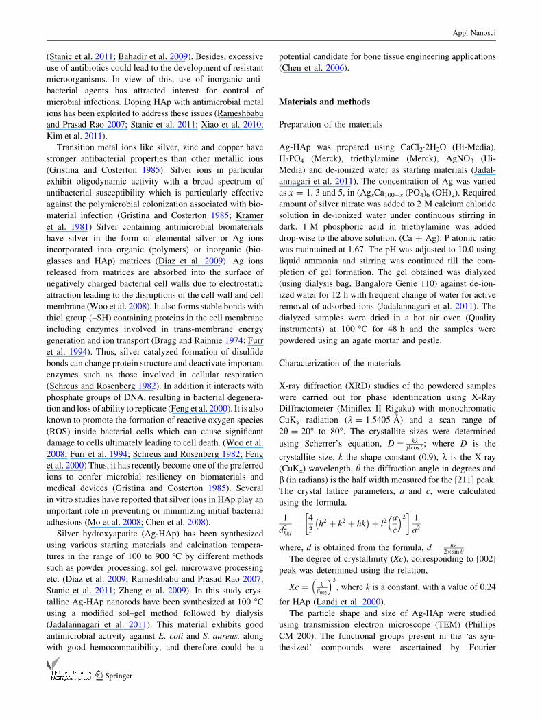
The XRD spectra of the Ag-HAp (1, 3 and 5 %) and HAp
are shown in Fig. 1. The samples with silver content x = 3
and 5 exhibited a shoulder at 2h = 388, attributable to
metallic silver. Trace amount of b-TCP phase (JCPDS 9-16)
Appl Nanosci
123
was observed in all the samples. The degree of crystallinity,
the lattice parameters and crystallite sizes are presented in
Table 1. The lattice parameters a and c increased with
increasing Ag concentration. Changes in cell parameters
may imply that silver substituted calcium in the HAp lattice
and since Ag? (0.128 nm) is larger than Ca2? (0.099 nm) it
resulted in increase in lattice parameters (Rameshbabu and
Prasad Rao 2007; Stanic et al. 2011).
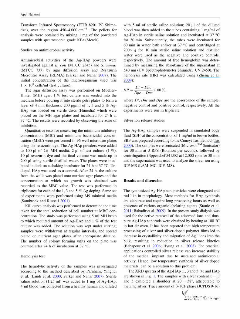
Figure 2, shows the FTIR spectra of HAp and Ag-HAp
samples. The characteristic signature HAp double bands
near 600 cm-1 are attributed to the bending modes of P–O
bonds in phosphate groups with contribution from the –OH
of the apatite group at about 630 cm-1 (Ramanan and
Venkatesh 2004). The bands in the region 3300 cm-1 are
attributed to –OH bonds and those observed near
3,571 cm-1 are associated with OH-1 stretching vibration
of HAp (Tas 2000). The broad band extending from 2,500
to about 3,700 cm-1 and the band at about 1,635 cm-1
corresponds to absorbed water (Stanic et al. 2011). The
bands noted around 1,100–1,000 cm-1 are assigned to the
stretching modes of the PO43- bonds in HAp (Smith 1999).
The strongest band between 1,020 to 1,100 cm-1, which
appeared as a doublet or a band with a shoulder, in all
samples was due to the P–O stretching vibration of the
phosphate group (Stanic et al. 2011).
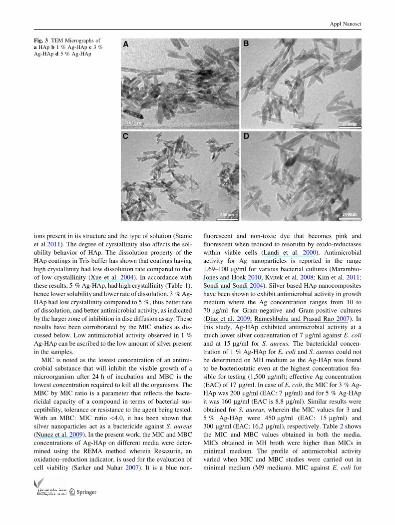
The TEM micrographs (Fig. 3) show particles of uniform
size distribution with elongated rod-like morphology inde-
pendent of the dopant concentration. The length of the
nanorods varied from 110–180 nm and the diameter was
between 20 and 25 nm. Silver aggregates on HAp particles
have been reported during synthesis of Ag-HAp
nanocomposites, using a colloidal chemical route and sub-
sequent reduction in H2/Ar atmosphere at 350 �C. This has
been attributed to partial destabilization of HAp in aqueous
suspension favoring the growth of Ag2O nuclei (Diaz et al.
2009). Chung et al. (2006) also reported the presence of
Ag2O crystallite phase in addition to the HAp and the nitrate
phases, depending upon the degree of substitution by silver.
In this procedure no such formation of Ag2O nuclei was
detected.
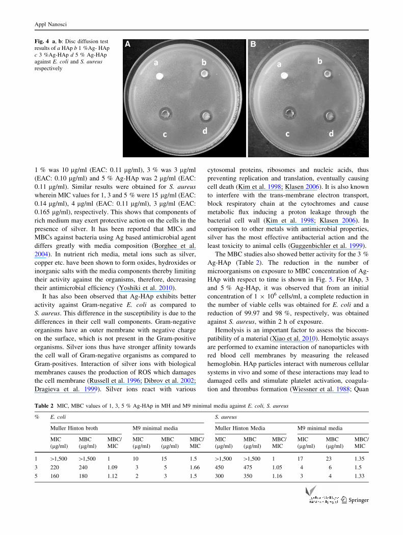
Antimicrobial studies were carried out against two rep-
resentative pathogenic organisms, Gram-negative E. coli
and Gram-positive S. aureus. It was observed that all the Ag-
HAp samples (1, 3 and 5 %) exhibited antimicrobial activity;
however, no inhibition was obtained for the pure HAp. The
zones of inhibition obtained are shown in Fig. 4. It was
observed that 3 % Ag-HAp exhibited maximum activity as
compared to 1 and 5 % Ag-HAp for both the test organisms.
Antimicrobial activity of Ag-HAp based compounds such as
plasma sprayed silver containing HAp coatings on Ti plates
(Chen et al. 2008), magnetron co-sputtered silver containing
HAp coatings (Chen et al. 2006), Ag-nHAp/TiO2/PA66
scaffolds (Xia et al. 2010), silver-hydroxyapatite/titania film
on titanium plates (Mo et al. 2008) and HAp self assembled
calcium phosphate glasses (Simon et al. 2008) has been
attributed to the Ag ions released into the medium. It is well
known that silver ions and silver based compounds can
destroy cell membranes and cell walls of bacteria and affect
their growth (Woo et al. 2008; Furr et al. 1994). In order to
estimate the concentration of Ag ions that are released when
Ag-HAp is suspended in aqueous medium, ICP-MS studies
were carried out. It was observed that the amount of silver
Fig. 1 XRD Micrograph of
HAp, 1 % Ag-HAp, 3 %
Ag-HAp and 5 % Ag-HAp
Appl Nanosci
123
leached out from all the three samples was almost the same
and in the range of 406–499 ng/ml after 1 h. No further
increase in the release of Ag ion was noted even after 24 h.
Thus, negligible amount of silver ions (1–4 %) were released
from the silver present in the Ag-HAp samples. However, the
antimicrobial activity in case of 3 % Ag-HAp was higher
than that of 1 and 5 % Ag-HAp. Hence, the antimicrobial
activity cannot be solely attributed to the leaching of Ag ions
into the media. Dissolution of the HAp matrices can increase
the availability of Ag ions for antimicrobial action. The
solubility of HAp at the solid liquid interface depends on its
physicochemical properties, types and amounts of foreign
Table 1 Crystallite size, degree
of crystallinity and unit cell
parameters of HAp and Ag-HAp
nanorods
Crystallite size Xs (nm) Degree of crystallinity Xc Unit cell parameters
a-axis (A) c-axis (A)
HAp 10 0.216 9.3550 6.1490
1 % Ag-HAp 31 0.027 9.5144 6.7108
3 % Ag-HAp 24.89 0.064 9.5753 6.7140
5 % Ag-HAp 38.7 0.110 9.3630 6.7996
Fig. 2 FTIR graphs of a HAp b 1 % Ag-HAp c 3 % Ag-HAp d 5 % Ag-HAp
Appl Nanosci
123
ions present in its structure and the type of solution (Stanic
et al.2011). The degree of cyrstallinity also affects the sol-
ubility behavior of HAp. The dissolution property of the
HAp coatings in Tris buffer has shown that coatings having
high crystallinity had low dissolution rate compared to that
of low crystallinity (Xue et al. 2004). In accordance with
these results, 5 % Ag-HAp, had high crystallinity (Table 1),
hence lower solubility and lower rate of dissolution. 3 % Ag-
HAp had low crystallinity compared to 5 %, thus better rate
of dissolution, and better antimicrobial activity, as indicated
by the larger zone of inhibition in disc diffusion assay. These
results have been corroborated by the MIC studies as dis-
cussed below. Low antimicrobial activity observed in 1 %
Ag-HAp can be ascribed to the low amount of silver present
in the samples.
MIC is noted as the lowest concentration of an antimi-
crobial substance that will inhibit the visible growth of a
microorganism after 24 h of incubation and MBC is the
lowest concentration required to kill all the organisms. The
MBC by MIC ratio is a parameter that reflects the bacte-
ricidal capacity of a compound in terms of bacterial sus-
ceptibility, tolerance or resistance to the agent being tested.
With an MBC: MIC ratio \4.0, it has been shown that
silver nanoparticles act as a bactericide against S. aureus
(Nunez et al. 2009). In the present work, the MIC and MBC
concentrations of Ag-HAp on different media were deter-
mined using the REMA method wherein Resazurin, an
oxidation–reduction indicator, is used for the evaluation of
cell viability (Sarker and Nahar 2007). It is a blue non-
fluorescent and non-toxic dye that becomes pink and
fluorescent when reduced to resorufin by oxido-reductases
within viable cells (Landi et al. 2000). Antimicrobial
activity for Ag nanoparticles is reported in the range
1.69–100 lg/ml for various bacterial cultures (Marambio-
Jones and Hoek 2010; Kvitek et al. 2008; Kim et al. 2011;
Sondi and Sondi 2004). Silver based HAp nanocomposites
have been shown to exhibit antimicrobial activity in growth
medium where the Ag concentration ranges from 10 to
70 lg/ml for Gram-negative and Gram-positive cultures
(Diaz et al. 2009; Rameshbabu and Prasad Rao 2007). In
this study, Ag-HAp exhibited antimicrobial activity at a
much lower silver concentration of 7 lg/ml against E. coli
and at 15 lg/ml for S. aureus. The bactericidal concen-
tration of 1 % Ag-HAp for E. coli and S. aureus could not
be determined on MH medium as the Ag-HAp was found
to be bacteriostatic even at the highest concentration fea-
sible for testing (1,500 lg/ml); effective Ag concentration
(EAC) of 17 lg/ml. In case of E. coli, the MIC for 3 % Ag-
HAp was 200 lg/ml (EAC: 7 lg/ml) and for 5 % Ag-HAp
it was 160 lg/ml (EAC is 8.8 lg/ml). Similar results were
obtained for S. aureus, wherein the MIC values for 3 and
5 % Ag-HAp were 450 lg/ml (EAC: 15 lg/ml) and
300 lg/ml (EAC: 16.2 lg/ml), respectively. Table 2 shows
the MIC and MBC values obtained in both the media.
MICs obtained in MH broth were higher than MICs in
minimal medium. The profile of antimicrobial activity
varied when MIC and MBC studies were carried out in
minimal medium (M9 medium). MIC against E. coli for
Fig. 3 TEM Micrographs of
a HAp b 1 % Ag-HAp c 3 %
Ag-HAp d 5 % Ag-HAp
Appl Nanosci
123
1 % was 10 lg/ml (EAC: 0.11 lg/ml), 3 % was 3 lg/ml
(EAC: 0.10 lg/ml) and 5 % Ag-HAp was 2 lg/ml (EAC:
0.11 lg/ml). Similar results were obtained for S. aureus
wherein MIC values for 1, 3 and 5 % were 15 lg/ml (EAC:
0.14 lg/ml), 4 lg/ml (EAC: 0.11 lg/ml), 3 lg/ml (EAC:
0.165 lg/ml), respectively. This shows that components of
rich medium may exert protective action on the cells in the
presence of silver. It has been reported that MICs and
MBCs against bacteria using Ag based antimicrobial agent
differs greatly with media composition (Borghee et al.
2004). In nutrient rich media, metal ions such as silver,
copper etc. have been shown to form oxides, hydroxides or
inorganic salts with the media components thereby limiting
their activity against the organisms, therefore, decreasing
their antimicrobial efficiency (Yoshiki et al. 2010).
It has also been observed that Ag-HAp exhibits better
activity against Gram-negative E. coli as compared to
S. aureus. This difference in the susceptibility is due to the
differences in their cell wall components. Gram-negative
organisms have an outer membrane with negative charge
on the surface, which is not present in the Gram-positive
organisms. Silver ions thus have stronger affinity towards
the cell wall of Gram-negative organisms as compared to
Gram-positives. Interaction of silver ions with biological
membranes causes the production of ROS which damages
the cell membrane (Russell et al. 1996; Dibrov et al. 2002;
Dragieva et al. 1999). Silver ions react with various
cytosomal proteins, ribosomes and nucleic acids, thus
preventing replication and translation, eventually causing
cell death (Kim et al. 1998; Klasen 2006). It is also known
to interfere with the trans-membrane electron transport,
block respiratory chain at the cytochromes and cause
metabolic flux inducing a proton leakage through the
bacterial cell wall (Kim et al. 1998; Klasen 2006). In
comparison to other metals with antimicrobial properties,
silver has the most effective antibacterial action and the
least toxicity to animal cells (Guggenbichler et al. 1999).
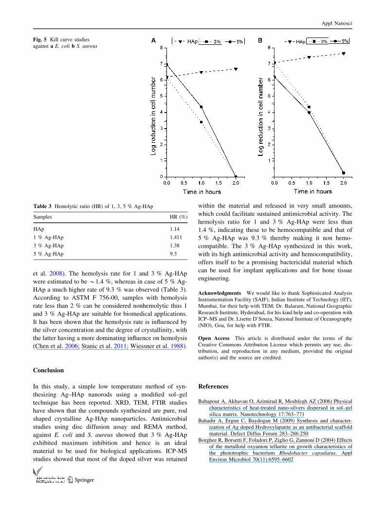
The MBC studies also showed better activity for the 3 %
Ag-HAp (Table 2). The reduction in the number of
microorganisms on exposure to MBC concentration of Ag-
HAp with respect to time is shown in Fig. 5. For HAp, 3
and 5 % Ag-HAp, it was observed that from an initial
concentration of 1 9 106 cells/ml, a complete reduction in
the number of viable cells was obtained for E. coli and a
reduction of 99.97 and 98 %, respectively, was obtained
against S. aureus, within 2 h of exposure.
Hemolysis is an important factor to assess the biocom-
patibility of a material (Xiao et al. 2010). Hemolytic assays
are performed to examine interaction of nanoparticles with
red blood cell membranes by measuring the released
hemoglobin. HAp particles interact with numerous cellular
systems in vivo and some of these interactions may lead to
damaged cells and stimulate platelet activation, coagula-
tion and thrombus formation (Wiessner et al. 1988; Quan
Fig. 4 a, b: Disc diffusion test
results of a HAp b 1 %Ag- HAp
c 3 %Ag-HAp d 5 % Ag-HAp
against E. coli and S. aureusrespectively
Table 2 MIC, MBC values of 1, 3, 5 % Ag-HAp in MH and M9 minimal media against E. coli, S. aureus
% E. coli S. aureus
Muller Hinton broth M9 minimal media Muller Hinton Media M9 minimal media
MIC
(lg/ml)
MBC
(lg/ml)
MBC/
MIC
MIC
(lg/ml)
MBC
(lg/ml)
MBC/
MIC
MIC
(lg/ml)
MBC
(lg/ml)
MBC/
MIC
MIC
(lg/ml)
MBC
(lg/ml)
MBC/
MIC
1 [1,500 [1,500 1 10 15 1.5 [1,500 [1,500 1 17 23 1.35
3 220 240 1.09 3 5 1.66 450 475 1.05 4 6 1.5
5 160 180 1.12 2 3 1.5 300 350 1.16 3 4 1.33
Appl Nanosci
123
et al. 2008). The hemolysis rate for 1 and 3 % Ag-HAp
were estimated to be *1.4 %, whereas in case of 5 % Ag-
HAp a much higher rate of 9.3 % was observed (Table 3).
According to ASTM F 756-00, samples with hemolysis
rate less than 2 % can be considered nonhemolytic thus 1
and 3 % Ag-HAp are suitable for biomedical applications.
It has been shown that the hemolysis rate is influenced by
the silver concentration and the degree of crystallinity, with
the latter having a more dominating influence on hemolysis
(Chen et al. 2006; Stanic et al. 2011; Wiessner et al. 1988).
Conclusion
In this study, a simple low temperature method of syn-
thesizing Ag–HAp nanorods using a modified sol–gel
technique has been reported. XRD, TEM, FTIR studies
have shown that the compounds synthesized are pure, rod
shaped crystalline Ag-HAp nanoparticles. Antimicrobial
studies using disc diffusion assay and REMA method,
against E. coli and S. aureus showed that 3 % Ag-HAp
exhibited maximum inhibition and hence is an ideal
material to be used for biological applications. ICP-MS
studies showed that most of the doped silver was retained
within the material and released in very small amounts,
which could facilitate sustained antimicrobial activity. The
hemolysis ratio for 1 and 3 % Ag-HAp were less than
1.4 %, indicating these to be hemocompatible and that of
5 % Ag-HAp was 9.3 % thereby making it non hemo-
compatible. The 3 % Ag-HAp synthesized in this work,
with its high antimicrobial activity and hemocompatibility,
offers itself to be a promising bactericidal material which
can be used for implant applications and for bone tissue
engineering.
Acknowledgments We would like to thank Sophisticated Analysis
Instrumentation Facility (SAIF), Indian Institute of Technology (IIT),
Mumbai, for their help with TEM; Dr. Balaram, National Geographic
Research Institute, Hyderabad, for his kind help and co-operation with
ICP–MS and Dr. Lisette D’Souza, National Institute of Oceanography
(NIO), Goa, for help with FTIR.
Open Access This article is distributed under the terms of the
Creative Commons Attribution License which permits any use, dis-
tribution, and reproduction in any medium, provided the original
author(s) and the source are credited.
References
Babapour A, Akhavan O, Azimirad R, Moshfegh AZ (2006) Physical
characteristics of heat-treated nano-silvers dispersed in sol–gel
silica matrix. Nanotechnology 17:763–771
Bahadir A, Ergun C, Baydogan M (2009) Synthesis and character-
ization of Ag doped Hydroxylapatite as an antibacterial scaffold
material. Defect Diffus Forum 283–286:250
Borghee R, Borsetti F, Foladori P, Ziglio G, Zannoni D (2004) Effects
of the metalloid oxyanion tellurite on growth characteristics of
the phototrophic bacterium Rhodobacter capsulatus. Appl
Environ Microbiol 70(11):6595–6602
Fig. 5 Kill curve studies
against a E. coli b S. aureus
Table 3 Hemolytic ratio (HR) of 1, 3, 5 % Ag-HAp
Samples HR (%)
HAp 1.14
1 % Ag-HAp 1.411
3 % Ag-HAp 1.38
5 % Ag-HAp 9.3
Appl Nanosci
123
Bragg PD, Rainnie DJ (1974) The effect of silver ions on the
respiratory chain of Escherichia coli. J Microbiol 20(6):883–889
Chen W, Liu Y, Curtney HS, Bettenga M (2006) In vitro anti-bacterial
and biological properties of magnetron co-sputtered silver—
containing hydroxyapatite coating. Biomaterials 27:5512–5517
Chen Y, Zheng X, Xie Y, Ding C, Ruan H, Fan C (2008) Anti-
bacterial and cytotoxic properties of plasma sprayed silver—
containing HA coating. J Mater Sci Mater Med 19:3603–3609
Chung RJ, Ming FH, Huang CW, Perng LH, Wen HW, Chin TS
(2006) Antimicrobial effects and human gingival biocompati-
bility of hydroxyapatite sol-gel coatings. J Biomed Mater Res B
Appl Biomater 76:169–178
Diaz M, Barba F, Miranda M, Guitian F, Torrecillas R, Moya SJ
(2009) Synthesis and antimicrobial activity of a silver hydroxy-
apatite nanocomposite. J Nanomater. doi:10.1155/2009/498505
Dibrov P, Dzioba J, Gosink KK, Hase CC (2002) Chemiosmotic
mechanism of antimicrobial activity of Ag in vibrio cholera.
Antimicrob Agents Chemother 46:2668–2670
Dragieva I, Stoeva S, Stoimenov P, Pavlikianov E, Klabunde K
(1999) Complex formation in solutions for chemical synthesis of
nanoscaled particles prepared by borohydride reduction process.
Nanostruct Mater 12:267–270
Feng QL, Wu J, Chen GQ, Cui FZ, Kim TN, Kim JO (2000) A
Mechanistic study of the antibacterial effect of silver ions on
Escherichia coli and Staphylococcus aureus, J Biomed Mater
Res 52:662–668
Furr JR, Russell AD, Turner TD, Andrews A (1994) Antibacterial
activity of Actisorb Plus, Actisorb and silver nitrate. J Hosp
Infect 27(3):201–208
Gristina AG, Costerton JW (1985) Bacterical adherence to biomate-
rials and tissue: the significance of its role in clinical species.
J Bone Joint Surg Am 67:264–273
Guggenbichler JP, Boswald S, Lugauer Krall T (1999) A new
technology of microdispersed silver in polyurethane induces
antimicrobial activity in central venous catheters. Infection
27:S16–S23
Hyung JJ, Sung CY, Seong GO (2003) Preparation and antibacterial
effects of Ag–SiO2 thin films by sol–gel method. Biomaterials
24:4921–4928
Jadalannagari S, More S, Kowshik M, Ramanan SR (2011) Low
temperature synthesis of hydroxyapatite nanorods using a
modified sol-gel technique. Mater Sci Engg C 31:1534–1538
Kim TN, Feng QL, Kim JO, Wu J, Wang H, Chen GC, Cui FZ (1998)
Antimicrobial effects of metal ions (Ag?, Cu2?, Zn2?) in
hydroxyapatite. J Mater Sci Mater Med 9:129–134
Kim SH, Lee HS, Ryu DS, Choi SJ, Lee DS (2011) Antibacterial
activity of silver-nanoparticles against Staphylococcus aureusand Escherichia coli, Korean. J Microbiol Biotechnol 39:77–85
Klasen JH (2006) A historical review of the use of silver in treatment
of burns. II. Renewed interest for silver. Burns 26:131–138
Kramer SJ, Spadaro JA, Webster DA (1981) Antibacterial and
osteoinductive properties of demineralized bone matrix treated
with silver. Clin Orthop Relat Res 16:1154–1162
Kvıtek L, Panacek A, Soukupova J, Kolar M, Vecerova R, Prucek R,
Holecova M, Zboril R (2008) Effect of surfactants and polymers
on stability and antibacterial activity of silver nanoparticles
(NPs). J Phys Chem C 112:5825
Landi E, Tampieri A, Celotti G, Spiro S (2000) Densification
behavior and mechanisms of synthetic hydroxyapatite. J Eur
Ceram Soc 20:2377–2387
Marambio-Jones C, Hoek EMV (2010) A review of the antibacterial
effects of silver nanomaterials and potential implications for human
health and the environment. J Nanopart Res 12(5):1531–1551
Mo A, Liao J, Xu W, Xian S, Li Y, Bai S (2008) Preparation and
antibacterial effect of silver-hydroxyapatite/titania nano com-
posite thin film on titanium. Appl Surf Sci 255:435–438
Nunez NN, Villegas H, Turrent L, Padilla C (2009) Silver nanopar-
ticles toxicity and bactericidal effect against methicillin resistant
staphylococcus aureus nanoscale does matter. Nanobiotechnol
5:2–9
Quan R, Yang D, Wu X, Wang H, Miao X, Li W (2008) In vitro
biocompatibility of graded hydroxyapatite-zirconia composite
bioceramic. J Mater Sci Mater Med 19:183–187
Ramanan SR, Venkatesh R (2004) A study of hydroxyapatite fibers
prepared via sol-gel route. Mat Lett 58:3320–3323
Rameshbabu N, Sampath Kumar TS, Prabhakar TG, Sastry VS,
Murty KVGK, Prasad Rao K (2007) Antibacterial nanosized
silver substituted hydroxylapatite: Synthesis and characteriza-
tion. J Biomed Mater Res A 80:581–591
Russell SW, Luptak KA, Suchicital CTA, Alford TL, Pizziconi VB
(1996) Chemical and structural evolution of sol-gel derived
hydroxyapatite thin films under rapid thermal processing. J Am
Ceram 79:837–842
Sambrook J, Russell D (2001) Molecular cloning: a laboratory
manual. Cold Spring Harbor: Cold Spring Harbor Laboratory
Sarker SD, Nahar L, Kumarasamy Y (2007) Microtitre plate based
antibacterial assay incorporating resazuring as an indicator of
cell growth, and its application in the in vitro antibacterial
screening of phytochemicals. Methods 42(4):321–324
Schreus WJA, Rosenberg H (1982) Effect of silver ions on transport
and retention of phosphate by Escherichia coli. J Bacteriol
152:7–13
Simon V, Albon C, Simon S (2008) Silver release from hydroxyap-
atite self assembling calcium phosphate glasses. J Non-cry Sol
354:1751–1755
Smith B (1999) Infrared spectral interpretation: a systematic
approach.CRC press, USA
Sondi I, Sondi BS (2004) Silver nanoparticles as antimicrobial agent:
a case study on E. coli as a model for Gram-negative bacteria.
J Colloid Interface Sci 275:117–182
Stanic V, Janackovic D, Dimitrijevic S, Tanaskovic BS, Mitric M,
Pavlovic SM, Krstic A, Jovanovic D, Raicevic S (2011)
Synthesis of antimicrobial monophase silver—doped hydroxy-
apatite nanopowders for bone tissue engineering. Appl Surf Sci
257:4510–4518
Tas AC (2000) Synthesis of biomimetic Ca-Hydroxyapatite powders
at 37 �C in synthetic body fluids. Biomaterials 21:1429–1438
Wiessner J, Mandel G, Halverson P, Mandel N (1988) The effect of
hydroxyapatite crystallinity on hemolysis. Calcif Tissue Int
42:210–219
Woo K. J, Hye CK, Ki WK, Sook S, So H, K, Yong HP (2008)
Antibacterial activity and mechanism of action of the silver ion
in Staphylococcus aureus and Escherichia coli, Appl Enviorn
Microb 74:2171–2178
Xia W, Li J, Wang L, Huang D, Zuo Y, Yubao L (2010) The release
properties of silver ions from Ag-nHA/TiO2/PA66 antimicrobial
composite scaffolds. Biomed Mater 5:044105
Xiao B, Karren M, Christopher MR, Rabiei A (2010) Functionally
graded hydroxyapatite coatings doped with antibacterial com-
ponents. Acta biomaterialia 6:2264–2273
Xue W, Tao S, Liu X, Zheng X, Ding C (2004) In vivo evaluation of
plasma sprayed hydroxyapatite coatings having different crys-
tallinity. Biomaterials 25:415–421
Yoshiki A, Hiroshi M, Iwao N, Fumiaki M, Takafumi S, Yutaka Y,
Masaki M, Takao H (2010) Effect of bacterial media on the
evaluation of the antibacterial activity of biomaterial containing
inorganic antibacterial reagents or antibiotics. Biocontr Sci
15(1):15–19
Zheng X, Chen Y, Xie Y, Ji Heng, Huang L, Ding C (2009)
Antibacterial property and biocompatibility of plasma sprayed
hydroxyapatite/silver composite coatings. J Therm Spray Tech
16:463
Appl Nanosci
123


![High capacity Li[Ni0.8Co0.1Mn0.1]O2 synthesized by sol–gel and co-precipitation methods as cathode materials for lithium-ion batteries](https://static.fdokumen.com/doc/165x107/6336e10720d9c9602f0b0e64/high-capacity-lini08co01mn01o2-synthesized-by-solgel-and-co-precipitation.jpg)