Magnetic and optical properties of manganese doped ZnO nanoparticles synthesized by sol–gel...
9
Magnetic and optical properties of manganese doped ZnO nanoparticles synthesized by sol–gel technique K. Omri a , J. El Ghoul a , O.M. Lemine b , M. Bououdina c , B. Zhang d , L. El Mir a,b,⇑ a Laboratory of Physics of Materials and Nanomaterials Applied at Environment (LaPhyMNE), Faculty of Sciences in Gabes, Gabes, Tunisia b Al Imam Mohammad Ibn Saud Islamic University (IMSIU), College of Sciences, Department of Physics, Riyadh 11623, Saudi Arabia c Department of Physics, College of Science, University of Bahrain, Bahrain d Advanced Nanofabrication, Imaging and Characterization Core Lab, KAUST, Thuwal, Saudi Arabia article info Article history: Received 27 April 2013 Accepted 29 April 2013 Available online 9 May 2013 Keywords: Nanostructures Zinc oxide Optical materials Magnetic properties Sol–gel technique abstract Mn doped ZnO nanoparticles with different doping concentration (1, 2, 3, 4, 5 at.%) were prepared by sol–gel method using supercrit- ical drying conditions of ethyl alcohol. The structural, morpholog- ical, optical and magnetic properties of the as-prepared nanoparticles were investigated by X-ray diffraction (XRD), trans- mission electron microscopy (TEM), UV measurements and super- conducting quantum interference device (SQUID). The structural properties showed that the undoped and Mn doped ZnO nanopar- ticles exhibit hexagonal wurtzite structure. From the optical stud- ies, the transmittance in UV region was decreased with the increase of Mn concentration. For Mn doped ZnO nanoparticles the optical band gap varies between 3.34 eV and 3.22 eV. It was found that the doping Mn 2+ ions have a significant influence on the optical properties. The magnetic characterization of the sam- ples with 1% and 5% Mn concentrations reveal diamagnetic behav- ior for the first one and the presence of both paramagnetic and ferromagnetic behavior for the second. The room ferromagnetic component is due to the presence of the secondary phase ZnOMn 3 which is confirmed by XRD study. Ó 2013 Elsevier Ltd. All rights reserved. 0749-6036/$ - see front matter Ó 2013 Elsevier Ltd. All rights reserved. http://dx.doi.org/10.1016/j.spmi.2013.04.029 ⇑ Corresponding author at: Al Imam Mohammad Ibn Saud Islamic University (IMSIU), College of Sciences, Department of Physics, Riyadh 11623, Saudi Arabia. Tel.: +966 531448340; fax: +966 12581584. E-mail address: [email protected] (L. El Mir). Superlattices and Microstructures 60 (2013) 139–147 Contents lists available at SciVerse ScienceDirect Superlattices and Microstructures journal homepage: www.elsevier.com/locate/superlattices
Transcript of Magnetic and optical properties of manganese doped ZnO nanoparticles synthesized by sol–gel...
Superlattices and Microstructures 60 (2013) 139–147
Contents lists available at SciVerse ScienceDirect
Superlattices and Microstructures
j o u r n a l h o m e p a g e : w w w . e l s e v i e r . c o m / l o ca t e / s u p e r l a t t i c es
Magnetic and optical properties of manganesedoped ZnO nanoparticles synthesized by sol–geltechnique
0749-6036/$ - see front matter � 2013 Elsevier Ltd. All rights reserved.http://dx.doi.org/10.1016/j.spmi.2013.04.029
⇑ Corresponding author at: Al Imam Mohammad Ibn Saud Islamic University (IMSIU), College of Sciences, DeparPhysics, Riyadh 11623, Saudi Arabia. Tel.: +966 531448340; fax: +966 12581584.
E-mail address: [email protected] (L. El Mir).
K. Omri a, J. El Ghoul a, O.M. Lemine b, M. Bououdina c, B. Zhang d, L. El Mir a,b,⇑a Laboratory of Physics of Materials and Nanomaterials Applied at Environment (LaPhyMNE), Faculty of Sciences in Gabes,Gabes, Tunisiab Al Imam Mohammad Ibn Saud Islamic University (IMSIU), College of Sciences, Department of Physics, Riyadh 11623, Saudi Arabiac Department of Physics, College of Science, University of Bahrain, Bahraind Advanced Nanofabrication, Imaging and Characterization Core Lab, KAUST, Thuwal, Saudi Arabia
a r t i c l e i n f o
Article history:Received 27 April 2013Accepted 29 April 2013Available online 9 May 2013
Keywords:NanostructuresZinc oxideOptical materialsMagnetic propertiesSol–gel technique
a b s t r a c t
Mn doped ZnO nanoparticles with different doping concentration(1, 2, 3, 4, 5 at.%) were prepared by sol–gel method using supercrit-ical drying conditions of ethyl alcohol. The structural, morpholog-ical, optical and magnetic properties of the as-preparednanoparticles were investigated by X-ray diffraction (XRD), trans-mission electron microscopy (TEM), UV measurements and super-conducting quantum interference device (SQUID). The structuralproperties showed that the undoped and Mn doped ZnO nanopar-ticles exhibit hexagonal wurtzite structure. From the optical stud-ies, the transmittance in UV region was decreased with theincrease of Mn concentration. For Mn doped ZnO nanoparticlesthe optical band gap varies between 3.34 eV and 3.22 eV. It wasfound that the doping Mn2+ ions have a significant influence onthe optical properties. The magnetic characterization of the sam-ples with 1% and 5% Mn concentrations reveal diamagnetic behav-ior for the first one and the presence of both paramagnetic andferromagnetic behavior for the second. The room ferromagneticcomponent is due to the presence of the secondary phase ZnOMn3
which is confirmed by XRD study.� 2013 Elsevier Ltd. All rights reserved.
tment of
140 K. Omri et al. / Superlattices and Microstructures 60 (2013) 139–147
1. Introduction
Doping semiconductor nanostructures in order to control their physical properties is an active fieldof research related to the development of nanotechnology applications. Due to the variety of ZnOapplications, nanowires and other nanostructures of this semiconducting oxide have been doped withdifferent metal ions, including transition metal elements (Ni, Fe, Mn, Co, V, etc.) [1–8]. However, con-siderable effort has been devoted to developing various semiconductor nanostructures. Zinc oxide, asa wide direct band-gap (3.37 eV) semiconductor with a large exciton binding energy of 60 meV atroom temperature, has recently attracted a beige attention due to its potential uses in ultraviolet(UV) luminescence devices, light emitting diodes (LEDs), gas sensors, solar cells and transparent thinfilm transistors [3,8,9]. ZnO is also a material that is biocompatible and biosafe for applications asimplantable biosensors. Diluted magnetic semiconductors (DMSs) have attracted considerable atten-tion in the past few years due to their promising applications in spintronic devices [10]. Among theDMS, ZnO doped Mn attracted more attention in the last two decades due to its specific technologicalapplications [11–24]. Most of the ZnO:Mn nanostructure materials exhibit strong ferromagnetism atroom temperature, strong absorption in the UV and visible range and strong UV emission [11]. Theseproperties may be due to the difficulty in controlling the interaction between Mn dopant and intrinsicdefects such as oxygen vacancies (VO) during the synthesis process [12]. Hence, it is necessary tosearch another approach to realize efficient optical properties from ZnO:Mn for the applications inmagneto-optic devices. However, it has been previously observed by other groups [13,14] that thedoping concentration of Mn has a significant influence on the optical properties. For low doping con-centrations (<3 at.% of Mn), the band gap of ZnO:Mn is reduced but for higher concentrations (>3 at.%)it increases due to the presence of secondary phases [15]. To date, a number of publications have dealtwith the fabrication of ZnO and metal-doped ZnO nanoparticles by sol–gel method. Ma and Wang [15]synthesized Mn-doped ZnO nanoparticles; they reported that the absorbance of UV light increasedwith the decreasing of particle size. They showed also that the particle size increased with calcinationtemperature [2,16]. In addition, Senthilkumaar et al. [17] reported the decrease of optical transmit-tance, optical band gap and the particle size of ZnO nanopowder by increasing the Mn concentration[18]. Furthermore, systematic studies on the effect of the Mn concentration on the structural and theoptical properties of ZnO nanoparticles play a very important role in the enhancing of device efficiency[19].
Regarding the magnetic properties, several groups reported various magnetic behavior; such asspin-glass behavior [20], paramagnetism [21] and ferromagnetism [22] in Mn-doped ZnO. Thesemagnetic properties are strongly depending on the sample preparation conditions. Mn-doped ZnOnanoparticles were grown by spin coating [23], sol–gel [5,24], spray [25], metal organic chemical va-por deposition [26], radio frequency magnetron sputtering [27] and pulsed laser deposition [21,28,29].However, there are few reports on the structural, optical and magnetic properties of Mn-doped ZnOsynthesis by sol–gel method [24].
In this study, we report a simple protocol using a slightly modified version of the conventional sol–gel method used to synthesize zinc oxide aerogels. The effects of Mn concentration on structural, mor-phological, optical and magnetic properties of the obtained nanopowders were investigated.
2. Experimental procedure
2.1. Synthesis of ZnO:Mn nanoparticles
For the synthesis of the samples, we dissolved 2 g of zinc acetate dehydrate in 14 ml of methanol,under magnetic stirring for 2 h and then we added 0.083 g of manganese (II) chloride-4-hydrate withstirring until complete dissolution of the precursors. Then, the resulting solution underwent rapid dry-ing to obtain aerogel powder in an autoclave under supercritical conditions of ethanol (EtOH), with aheating rate of 45 �C/h.
K. Omri et al. / Superlattices and Microstructures 60 (2013) 139–147 141
2.2. Characterization
The crystalline phases of the obtained nanopowders were identified by X-ray diffraction (XRD)using a Bruker D5005 powder X-ray diffractometer using a Co Ka source (1.78901 Å radiation). Crys-tallite sizes (G, in Å) were estimated from the Scherrer’s equation [30]:
G ¼ 0:9kB cos hB
ð1Þ
where k is the X-ray wavelength (1.78901 Å), hB is the maximum of the Bragg diffraction peak (in radi-ans) and B is the linewidth at half maximum.
Scanning electron microscope (SEM, JEOL JSM-6300) and transmission electron microscopy (TEM,JEM-200CX) were used to study the morphology and particle size of the phosphor powders. The spec-imens for TEM were prepared by putting the as-grown products in EtOH and immersing them in anultrasonic bath for 15 min, then dropping a few drops of the resulting suspension containing the syn-thesized materials onto TEM grid. The magnetic properties of the samples were characterized by acommercial Quantum Design SQUID–VSM magnetometer. All the powder samples enwrapped byquite low magnetic background parafilms were mounted inside the standard brass sample holderfor magnetic measurements. Field dependent magnetizations up to 6 Tesla have been measured atroom temperature.
3. Results and discussions
3.1. Structural properties
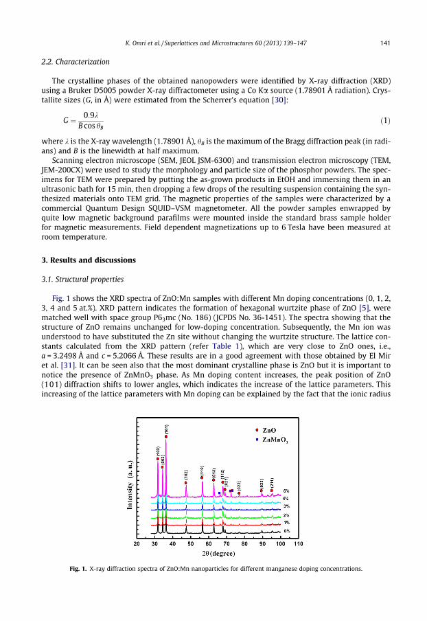
Fig. 1 shows the XRD spectra of ZnO:Mn samples with different Mn doping concentrations (0, 1, 2,3, 4 and 5 at.%). XRD pattern indicates the formation of hexagonal wurtzite phase of ZnO [5], werematched well with space group P63mc (No. 186) (JCPDS No. 36-1451). The spectra showing that thestructure of ZnO remains unchanged for low-doping concentration. Subsequently, the Mn ion wasunderstood to have substituted the Zn site without changing the wurtzite structure. The lattice con-stants calculated from the XRD pattern (refer Table 1), which are very close to ZnO ones, i.e.,a = 3.2498 Å and c = 5.2066 Å. These results are in a good agreement with those obtained by El Miret al. [31]. It can be seen also that the most dominant crystalline phase is ZnO but it is important tonotice the presence of ZnMnO3 phase. As Mn doping content increases, the peak position of ZnO(101) diffraction shifts to lower angles, which indicates the increase of the lattice parameters. Thisincreasing of the lattice parameters with Mn doping can be explained by the fact that the ionic radius
Fig. 1. X-ray diffraction spectra of ZnO:Mn nanoparticles for different manganese doping concentrations.
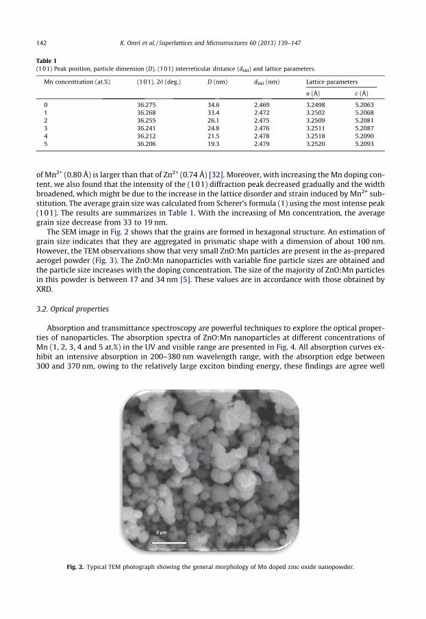
Table 1(101) Peak position, particle dimension (D), (101) interreticular distance (dhkl) and lattice parameters.
Mn concentration (at.%) (101), 2h (deg.) D (nm) dhkl (nm) Lattice parameters
a (Å) c (Å)
0 36.275 34.6 2.469 3.2498 5.20631 36.268 33.4 2.472 3.2502 5.20682 36.255 26.1 2.475 3.2509 5.20813 36.241 24.8 2.476 3.2511 5.20874 36.212 21.5 2.478 3.2518 5.20905 36.206 19.3 2.479 3.2520 5.2093
142 K. Omri et al. / Superlattices and Microstructures 60 (2013) 139–147
of Mn2+ (0.80 Å) is larger than that of Zn2+ (0.74 Å) [32]. Moreover, with increasing the Mn doping con-tent, we also found that the intensity of the (101) diffraction peak decreased gradually and the widthbroadened, which might be due to the increase in the lattice disorder and strain induced by Mn2+ sub-stitution. The average grain size was calculated from Scherer’s formula (1) using the most intense peak(101). The results are summarizes in Table 1. With the increasing of Mn concentration, the averagegrain size decrease from 33 to 19 nm.
The SEM image in Fig. 2 shows that the grains are formed in hexagonal structure. An estimation ofgrain size indicates that they are aggregated in prismatic shape with a dimension of about 100 nm.However, the TEM observations show that very small ZnO:Mn particles are present in the as-preparedaerogel powder (Fig. 3). The ZnO:Mn nanoparticles with variable fine particle sizes are obtained andthe particle size increases with the doping concentration. The size of the majority of ZnO:Mn particlesin this powder is between 17 and 34 nm [5]. These values are in accordance with those obtained byXRD.
3.2. Optical properties
Absorption and transmittance spectroscopy are powerful techniques to explore the optical proper-ties of nanoparticles. The absorption spectra of ZnO:Mn nanoparticles at different concentrations ofMn (1, 2, 3, 4 and 5 at.%) in the UV and visible range are presented in Fig. 4. All absorption curves ex-hibit an intensive absorption in 200–380 nm wavelength range, with the absorption edge between300 and 370 nm, owing to the relatively large exciton binding energy, these findings are agree well
Fig. 2. Typical TEM photograph showing the general morphology of Mn doped zinc oxide nanopowder.
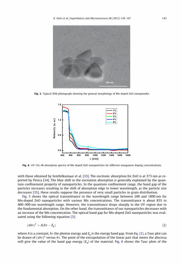
Fig. 3. Typical SEM photograph showing the general morphology of Mn doped ZnO nanopowder.
Fig. 4. UV–Vis–IR absorption spectra of Mn doped ZnO nanoparticles for different manganese doping concentrations.
K. Omri et al. / Superlattices and Microstructures 60 (2013) 139–147 143
with those obtained by Senthilkumaar et al. [33]. The excitonic absorption for ZnO is at 373 nm as re-ported by Pesica [34]. The blue shift in the excitation absorption is generally explained by the quan-tum confinement property of nanoparticles. In the quantum confinement range, the band gap of theparticles increases resulting in the shift of absorption edge to lower wavelength, as the particle sizedecreases [35], these results suppose the presence of very small particles in grain distribution.
Fig. 5 shows the optical transmittance in the wavelength range between 200 and 1800 nm forMn-doped ZnO nanoparticles with various Mn concentrations. The transmittance is about 85% in400–900 nm wavelength range. However, the transmittance drops sharply in the UV region due tothe fundamental absorption. On the other hand, the transmittance of our nanoparticles decreases withan increase of the Mn concentration. The optical band gap for Mn-doped ZnO nanoparticles was eval-uated using the following equation [3]:
ðahmÞ2 ¼ Aðhm� EgÞ ð2Þ
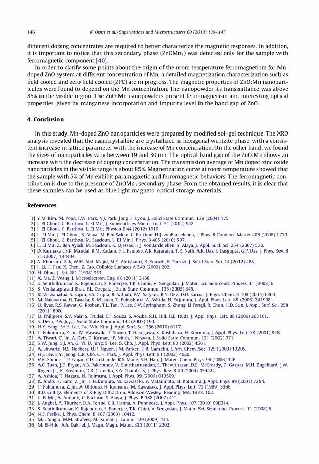
where A is a constant, hm the photon energy and Eg is the energy band gap. From Eq. (2), a Tauc plot canbe drawn of (ahm)2 versus hm. The point of the extrapolation of the linear part that meets the abscissawill give the value of the band gap energy (Eg) of the material. Fig. 6 shows the Tauc plots of the
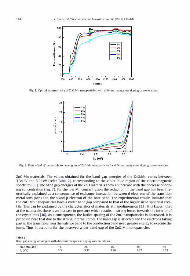
Fig. 5. Optical transmittance of ZnO:Mn nanoparticles with different manganese doping concentrations.
Fig. 6. Plots of (ahm)2 versus photon energy hm of ZnO:Mn nanoparticles for different manganese doping concentrations.
144 K. Omri et al. / Superlattices and Microstructures 60 (2013) 139–147
ZnO:Mn materials. The values obtained for the band gap energies of the ZnO:Mn varies between3.34 eV and 3.22 eV (refer Table 2), corresponding to the violet–blue region of the electromagneticspectrum [33]. The band gap energies of the ZnO materials show an increase with the decrease of dop-ing concentration (Fig. 7). For the low Mn concentration the reduction in the band gap has been the-oretically explained as a consequence of exchange interaction between d electrons of the transitionmetal ions (Mn) and the s and p electron of the host band. The experimental results indicate thatthe ZnO:Mn nanoparticles have a wider band gap compared to that of the bigger sized spherical crys-tals. This can be explained by the characteristics of materials at nanodimension [33]. It is known thatat the nanoscale, there is an increase in pressure which results in strong forces towards the interior ofthe crystallites [36]. As a consequence, the lattice spacing of the ZnO nanoparticles is decreased. It isproposed here that due to the strong internal forces, the band gap is affected and the electrons takingpart in the transition from the valence band to the conduction band need greater energy to execute thejump. Thus, it accounts for the observed wider band gap of the ZnO:Mn nanoparticles.
Table 2Band gap energy of samples with different manganese doping concentrations.
ZnO:Mn (at.%) 1% 2% 3% 4% 5%Eg (eV) 3.34 3.32 3.30 3.27 3.22
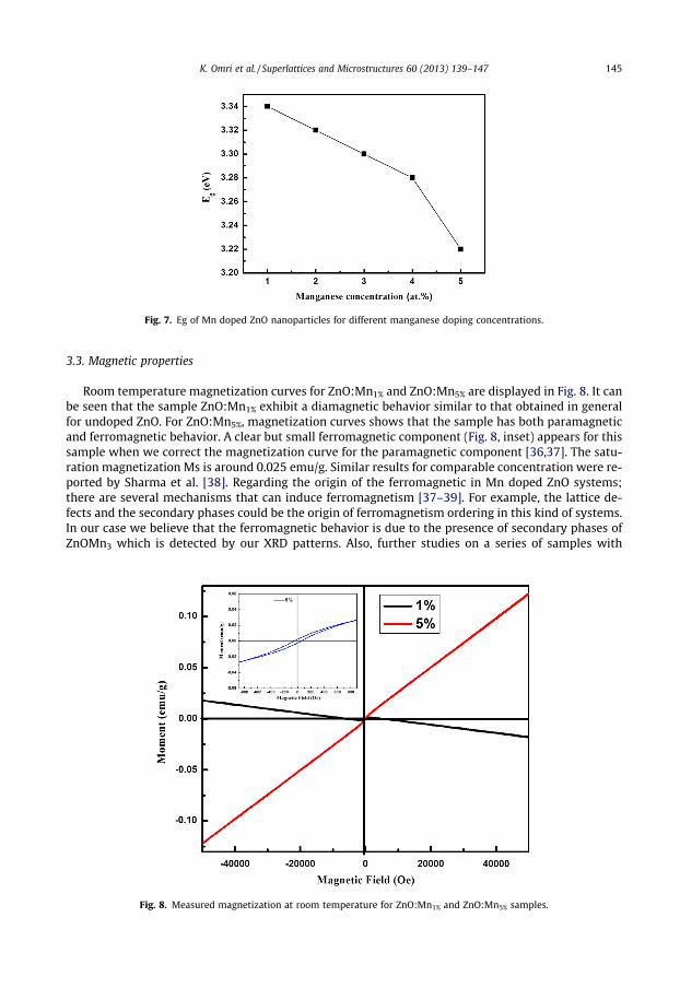
Fig. 7. Eg of Mn doped ZnO nanoparticles for different manganese doping concentrations.
K. Omri et al. / Superlattices and Microstructures 60 (2013) 139–147 145
3.3. Magnetic properties
Room temperature magnetization curves for ZnO:Mn1% and ZnO:Mn5% are displayed in Fig. 8. It canbe seen that the sample ZnO:Mn1% exhibit a diamagnetic behavior similar to that obtained in generalfor undoped ZnO. For ZnO:Mn5%, magnetization curves shows that the sample has both paramagneticand ferromagnetic behavior. A clear but small ferromagnetic component (Fig. 8, inset) appears for thissample when we correct the magnetization curve for the paramagnetic component [36,37]. The satu-ration magnetization Ms is around 0.025 emu/g. Similar results for comparable concentration were re-ported by Sharma et al. [38]. Regarding the origin of the ferromagnetic in Mn doped ZnO systems;there are several mechanisms that can induce ferromagnetism [37–39]. For example, the lattice de-fects and the secondary phases could be the origin of ferromagnetism ordering in this kind of systems.In our case we believe that the ferromagnetic behavior is due to the presence of secondary phases ofZnOMn3 which is detected by our XRD patterns. Also, further studies on a series of samples with
Fig. 8. Measured magnetization at room temperature for ZnO:Mn1% and ZnO:Mn5% samples.
146 K. Omri et al. / Superlattices and Microstructures 60 (2013) 139–147
different doping concentrates are required to better characterize the magnetic responses. In addition,it is important to notice that this secondary phase (ZnOMn3) was detected only for the sample withferromagnetic component [40].
In order to clarify some points about the origin of the room temperature ferromagnetism for Mn-doped ZnO system at different concentration of Mn, a detailed magnetization characterization such asfield cooled and zero field cooled (ZFC) are in progress. The magnetic properties of ZnO:Mn nanopart-icules were found to depend on the Mn concentration. The nanopowder its transmittance was above85% in the visible region. The ZnO:Mn nanopowders present ferromagnetism and interesting opticalproperties, given by manganese incorporation and impurity level in the band gap of ZnO.
4. Conclusion
In this study, Mn-doped ZnO nanoparticles were prepared by modified sol–gel technique. The XRDanalysis revealed that the nanocrystalline are crystallized in hexagonal wurtzite phase, with a consis-tent increase in lattice parameter with the increase of Mn concentration. On the other hand, we foundthe sizes of nanoparticles vary between 19 and 30 nm. The optical band gap of the ZnO:Mn shows anincrease with the decrease of doping concentration. The transmission average of Mn doped zinc oxidenanoparticles in the visible range is about 85%. Magnetization curve at room temperature showed thatthe sample with 5% of Mn exhibit paramagnetic and ferromagnetic behaviors. The ferromagnetic con-tribution is due to the presence of ZnOMn3 secondary phase. From the obtained results, it is clear thatthese samples can be used as blue light magneto-optical storage materials.
References
[1] Y.M. Kim, M. Yoon, I.W. Park, Y.J. Park, Jong H. Lyou, J. Solid State Commun. 129 (2004) 175.[2] J. El Ghoul, C. Barthou, L. El Mir, J. Superlattices Microstruct. 51 (2012) 942.[3] J. El Ghoul, C. Barthou, L. El Mir, Physica E 44 (2012) 1910.[4] L. El Mir, J. El Ghoul, S. Alaya, M. Ben Salem, C. Barthou, H.J. vonBardeleben, J. Phys. B Condens. Matter 403 (2008) 1770.[5] J. El Ghoul, C. Barthou, M. Saadoun, L. El Mir, J. Phys. B 405 (2010) 597.[6] L. El Mir, Z. Ben Ayadi, M. Saadoun, K. Djessas, H.J. vonBardeleben, S. Alaya, J. Appl. Surf. Sci. 254 (2007) 570.[7] D. Karmakar, S.K. Mandal, R.M. Kadam, P.L. Paulose, A.K. Rajarajan, T.K. Nath, A.K. Das, I. Dasgupta, G.P. Das, J. Phys. Rev. B
75 (2007) 144404.[8] A. Khorsand Zak, W.H. Abd. Majid, M.E. Abrishami, R. Yousefi, R. Parvizi, J. Solid State Sci. 14 (2012) 488.[9] J. Li, H. Fan, X. Chen, Z. Cao, Colloids Surfaces A 349 (2009) 202.
[10] H. Ohno, J. Sci. 281 (1998) 951.[11] X. Ma, Z. Wang, J. Microelectron. Eng. 88 (2011) 3168.[12] S. Senthilkumaar, K. Rajendran, S. Banerjee, T.K. Chinic, V. Sengodan, J. Mater. Sci. Semicond. Process. 11 (2008) 6.[13] S. Venkataprasad Bhat, F.L. Deepak, J. Solid State Commun. 135 (2005) 345.[14] R. Viswanatha, S. Sapra, S.S. Gupta, B. Satpati, P.V. Satyam, B.N. Dev, D.D. Sarma, J. Phys. Chem. B 108 (2004) 6303.[15] M. Nakayama, H. Tanaka, K. Masuko, T. Fukushima, A. Ashida, N. Fujimura, J. Appl. Phys. Lett. 88 (2006) 241908.[16] U. Ilyas, R.S. Rawat, G. Roshan, T.L. Tan, P. Lee, S.V. Springham, S. Zhang, Li Fengji, R. Chen, H.D. Sun, J. Appl. Surf. Sci. 258
(2011) 890.[17] U. Philipose, S.V. Nair, S. Trudel, C.F. Souza, S. Aouba, R.H. Hill, H.E. Ruda, J. Appl. Phys. Lett. 88 (2006) 263101.[18] S. Deka, P.A. Joy, J. Solid State Commun. 142 (2007) 190.[19] H.Y. Yang, Se H. Lee, Tae Wh. Kim, J. Appl. Surf. Sci. 256 (2010) 6117.[20] T. Fukumura, Z. Jin, M. Kawasaki, T. Shono, T. Hasegawa, S. Koshihara, H. Koinuma, J. Appl. Phys. Lett. 78 (2001) 958.[21] A. Tiwari, C. Jin, A. Kvit, D. Kumar, J.F. Muth, J. Nrayan, J. Solid State Commun. 121 (2002) 371.[22] S.W. Jung, S.J. An, G. Yi, U. Jung, S. Lee, S. Cho, J. Appl. Phys. Lett. 80 (2002) 4561.[23] A. Shwartz, N.S. Norberg, O.P. Nguen, J.M. Parker, D.R. Gamelin, J. Am. Chem. Soc. 125 (2003) 13205.[24] H.J. Lee, S.Y. Jeong, C.R. Cho, C.H. Park, J. Appl. Phys. Lett. 81 (2002) 4020.[25] V.R. Shinde, T.P. Gujar, C.D. Lokhande, R.S. Mane, S.H. Han, J. Mater. Chem. Phys. 96 (2006) 326.[26] A.C. Tuan, J.D. Bryan, A.B. Pakhomov, V. Shutthannandan, S. Thevuthasan, D.E. McCready, D. Gaspar, M.H. Engelhard, J.W.
Rogers Jr., K. Krishnan, D.R. Gamelin, S.A. Chambers, J. Phys. Rev. B 70 (2004) 054424.[27] A. Ashida, T. Nagata, N. Fujimura, J. Appl. Phys. 99 (2006) 013509.[28] K. Ando, H. Saito, Z. Jin, T. Fukumura, M. Kawasaki, Y. Matsumoto, H. Koinuma, J. Appl. Phys. 89 (2001) 7284.[29] T. Fukumura, Z. Jin, A. Ohtomo, H. Koinuma, M. Kawasaki, J. Appl. Phys. Lett. 75 (1999) 3366.[30] B.D. Cullity, Elements of X-Ray Diffraction, Addison-Wesley, Reading, MA, 1978. 102.[31] L. El Mir, A. Amlouk, C. Barthou, S. Alaya, J. Phys. B 388 (2007) 412.[32] J. Anghel, A. Thurber, D.A. Tenne, C.B. Hanna, A. Punnoose, J. Appl. Phys. 107 (2010) 09E314.[33] S. Senthilkumaar, K. Rajendran, S. Banerjee, T.K. Chini, V. Sengodan, J. Mater. Sci. Semicond. Process. 11 (2008) 6.[34] N.S. Pesika, J. Phys. Chem. B 107 (2003) 10412.[35] M.L. Singla, M.M. Shafeeq, M. Kumar, J. Lumin. 129 (2009) 434.[36] M. El-Hilo, A.A. Dakhel, J. Magn. Magn. Mater. 323 (2011) 2202.

K. Omri et al. / Superlattices and Microstructures 60 (2013) 139–147 147
[37] M.K. Sharma, R.N. Gayen, A.K. Pal, D. Kanjilal, R. Chatterjee, J. Alloys Compd. 509 (2011) 7259.[38] P. Sharma, A. Gupta, K.V. Rao, F.J. Owens, R. Sharma, R. Ahuja, J.M. Osorio Guillen, B. Johansson, G.A. Gehring, J. Nat. Mater. 2
(2003) 673.[39] J.H. Li, D.J. Shen, J.Y. Zhang, D.X. Zhao, B.S. Li, Y.M. Lu, Y.C. Liu, X.W. Fan, J. Magn. Magn. Mater. 302 (2006) 118.[40] D. Rubi, J. Fontcuberta, J. Phys. Rev. B 75 (2007) 155322.