NR LATEX COATED NANO ZnO DISKS FOR POTENTIAL DIELECTRIC APPLICATIONS
Send Orders for Reprints to [email protected]
Current Organic Chemistry, 2014, 18, 000-000 1
1385-2728/14 $58.00+.00 © 2014 Bentham Science Publishers
ZnO Applications and Challenges
Ovidiu Oprea1, Ecaterina Andronescu1, Denisa Ficai1, Anton Ficai1, Faik N. Oktar2-4* and Mehmet Yetmez5
1Politehnica University of Bucharest, Faculty of Applied Chemistry and Material Science; 1-7 Polizu St., 011061 Bucharest,
Romania; 2Bioengineering Dept., Faculty of Engineering, Marmara University, Istanbul, Turkey;
3Medical Imaging Techniques
Dept., School of Health Related Professions, Marmara University, Istanbul, Turkey; 4Nanotechnology and Biomaterials Research and
Application Center, Marmara University, Istanbul, Turkey; 5Mechanical Engineering Dept., Faculty of Engineering, Bulent Ecevit
University, Zonguldak, Turkey
Abstract: Metal oxide nanoparticles represent a new class of important materials that are increasingly being developed for use in re-
search and health-related applications. Although the in vitro antibacterial activity of zinc oxides and some other zinc compounds has been
known for quite some time, only in the last few years nanoparticles of ZnO have been investigated for their antibacterial activity, the
knowledge about it remaining deficient. The antimicrobial activity of ZnO nanoparticles is strongly influenced by some factors such as
size and the presence of light. The potential applications include, but are not limited to, topic drugs, cosmetics or component for agents
that control the spread of bacterial strains (antibacterial paint in hospitals, antibacterial coatings for fabrics, antibacterial packaging for
food, etc). The current review aims to present the level of knowledge accumulated on the antibacterial and antifungal activity of ZnO.
Keywords: ZnO nanoparticles, antibacterial activity, antifungal activity.
INTRODUCTION
In recent years, nanoparticles have received increasing attention due to their unique physical and chemical properties. Currently, metal oxide nanomaterials are among the most highly produced nanomaterials; their available applications include catalysis, sen-sors, environmental remediation, medicine, varistors, solar cells, rubber, concrete, foods, cosmetics and personal care products [1-11].
One promising approach is the use of metal oxides nanoparti-cles in health-related applications (hyperthermia, drug delivery, coated nanoparticles) [12-31]. The metal oxides such as titanium oxide, magnesium oxide, zinc oxide and copper oxide are better than nanosilver because of their low cost.
ZnO nanoparticles have been prepared by various methods such as thermal decomposition [32, 33], spray pyrolysis [34, 35], sol-vothermal reaction [36, 37], forced hydrolysis [13], reactive elec-tron beam evaporation technique [38], sol-gel method [39, 40], chemical vapor deposition [41], and hydrothermal method [42, 43]. They can also be produced into a variety of morphologies [44] such as nanowires [45, 46], nanorods [47-49], nanocombs [50, 51], tetra-legs [52], nanoflowers and nanosheet [53, 54].
ZnO has found its way in many applications in daily life such as in drug delivery, cosmetics, medical devices [55-62], dentistry and orthopedics [63-66]. Zinc is an essential trace element, which is found in the human body and has a stimulatory effect on bone for-mation. Human bone contains 0.0120 – 0.0250 wt% Zn [67]. The Zn content of various human tissues is shown in (Table 1) [68]. It can also be used in new bioglass formulations. Rui Lin Du et al. (2006) had shown that a certain concentration of the ionic products from Zn-containing bioactive glass could stimulate osteoblast pro-liferation [67] (Table 2).
*Address correspondence to this author at the Bioengineering Dept., Faculty of Engi-neering, Marmara University, Istanbul, Turkey; Tel: +90 216 348 0292/732; Fax: +90 216 348 0293; E-mail: [email protected]
The use of ZnO in cosmetics is not limited to the sunscreens [69] where (together with TiO2) the nanoparticles formulations have an improved performance as they scatter better the ultraviolet light than their respective bulk formulations [70] (Table 2).
In dentistry, ZnO alone or together with eugenol is used as a fill material due to its ability to block microbial leakage [71-74] (Table 2). For this reason, ZnO usually is included in the temporary ce-mentation material for cementation of temporary crowns and bridges. Most of those cements are based with eugenol and they are called as zinc oxide-eugenol (ZOE) cements. Beside temporary cementation, they are also very suitable for permanent cementation and as a cavity liner. They are also commonly used in the tempo-rary filling of teeth, and as a cavity basement in deep cavity prepa-rations. These ZnO containing products are non-toxic to oral tissues when they come into direct contact with freshly cut dentine tissue and the oral mucosa [65]. According to a recent study on ZnO, when it was used in nano-hydroxyapatite (HA)/chitosan compos-ites, it was proven to shorten the setting time, enhance the mechani-cal strength and improve the bioactivity and antibacterial activity of those composites [75]. Otsuka et al. (2004) have used Zn-tri cal-cium phosphate (TCP), as carrier, for controlled zinc release. The results showed that long-term sustained Zn release from Zn-TCP may improve bone mineral density in Zn-deficient osteoporotic rats [76]. Other researchers have discovered that HA-based system holds wide potential for application in clinic as an antibacterial biomaterial, as well as for dental filing and repair [77]. The struc-tural behavior of Zn in synthetic HA provides valuable information for future studies of Zn storage and transport involving calcified hard tissues and therefore the regulation of Zn metabolism in the body [78].
Zn holds also an inhibitory effect on osteoclastic bone resorp-tion. Several researchers have attempted to doper materials with Zn+2 at low concentrations for increasing the bioactivity of bone cells and decreasing and regulating the inflammatory reaction [79].
Besides being an antiseptic material, ZnO is also used as rein-forcing oxide for orthopedic various calcium phosphate based bio-
2 Current Organic Chemistry, 2014, Vol. 18, No. 2 Oprea et al.
ceramics (i.e. hydroxyapatite - HA). But there are limited studies in the literature on the reinforcement of HA structures with Zn or ZnO. Usually HA materials have poor mechanical properties. They cannot be used as heavy-loaded implants [80]. For improving me-chanical reliability of HA metallic materials, ceramic oxides, whiskers, or fibers are used [81]. ZnO seems be a good reinforcing material for HA [63, 64, 82, 83].
D. Norhidayu et al. (2008), have also proved that zinc plays a very important role in proliferative effects on osteoblastic cells apart from inhibiting osteoclastic resorption. Because of this zinc-substituted calcium phosphate has received a considerable attention [83].
Zn substitution in HA structures has been focused to the par-ticular interest in human body. Zn is present in all biological tissues and plays diverse roles in many different biological functions (i.e. enzyme activity, nucleic acid metabolism, maintenance of mem-brane structure and function, hormonal activity, biomineralization, potential pathological calcification etc.). The uptake and release of Zn material in the body are strongly mediated by the bone reservoir, even though it only contains a small portion of the total body Zn. For example, teeth contain a significant amount of Zn, (144.5 mi-crograms Zn/ g tooth substance). Zn concentration has been used also as an indicator of environmental exposure [84]. Kawamura et
al. (2000) found that the optimum Zn content in calcium phosphate ceramics is 0.316 wt % for promoting bone formation [85].
ZnO effect on epithelialization of wounds as well as ZnO bacte-riostatic property promote it as a topical wound dressing. It can be used in treatment of various dermatitis, diaper rashes, diaper wipes, blisters, and open skin sores [59, 86-88] (Table 2).
ZnO has for decades been used in medicine as a mild topical as-tringent (and possible antibacterial agent) in eczema and slight ex-coriations, in wounds, and for hemorrhoids [89]. Although the in
vitro antibacterial activity and efficacy of zinc oxides and some other zinc compounds have been investigated, only in the last few years nanoparticles of ZnO have been investigated for their antibac-terial activity [90-92]. Studies have indicated that ZnO nanoparti-cles at a concentration of between 3 and 10 mM caused 100% inhi-
bition of bacterial growth as a result of the intracellular accumula-tion of nanoparticles [93].
The antibacterial activity of ZnO may be dependent on the size and the presence of UV or visible light. The data suggest that ZnO nanoparticles have a potential application as a bacteriostatic agent in visible light and may have future applications in the development of derivative agents to control the spread and infection of a variety of bacterial strains.
ANTIBACTERIAL AND ANTIFUNGAL ACTIVITY OF ZnO
ZnO is one of five zinc compounds that are currently listed as generally recognized as safe (GRAS) by the U.S. Food and Drug Administration (21CFR182.8991) [102, 103].
Many studies have focused on the in vitro antibacterial activity on various bacterial strains (Staphyloccocus aureus, Staphylococcus epidermidis 1487, Streptococcus pyogenes, E. coli, E. coli O157:H7, Listeria monocytogenes, Bacillus subtilis, Bacillus atro-phaeus, Pseudomonas fluorescens, Pseudomonas aeruginosa, Sal-monella enteritidis, Salmonella typhimurium, Enterococcus fae-calis, Enterobacter cloacae, Lactobacillus helveticus) [103-108] and indicate that ZnO nanoparticles are more efficient as antibacterial agent than bulk powder and generally Gram-positive bacteria are more sensitive to ZnO than Gram negatives. This could be simply explained as smaller particles normally have a larger surface to volume ratio which provides a more efficient mean for antibacterial activity [109, 110]. Alternatively, small particles exhibit stronger cellular internalization, higher surface defect concentration, gener-ating larger amount of hydroxyl radicals [110]. ZnO nanoparticles also have antibacterial activity against spores that are high-temperature and high-pressure resistant [111-114].
Jones et al. (2008) demonstrated the influence of the particle size, by choosing three different values: 1 m, 50-70nm and 8nm. While ZnO particles ~1 m reduced growth rates (c. 50%) of Staphylococcus aureus, ZnO nanoparticles, with smaller particle size, were able to reduce c. 99% of growth in colloidal suspension with concentration of 2mM. While 8nm diameter ZnO nanoparti-
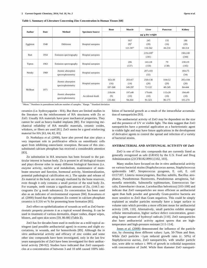
Table 1. Summary of Literature Concerning Zinc Concentration in Human Tissues [68]
Bone Muscle Liver Pancreas Kidney Author Date Method Specimen Source
g/ g dry weight
Eggleton 1940 Dithizone Hospital autopsies
163a
(9)b
121-307c
245
(20)
116-562
135
(16)
44-216
186
(20)
68-314
Butt 1954 Emission spectrography Hospital autopsies 219±109d
(241)
190±100
(244)
Tipton 1963 Emission spectrography Hospital autopsies 206
(137)
141±18
(150)
79
(138)
230±19
(145)
Netsky 1969 Atomic absorption
spectrophotometry Hospital autopsies
207±103
(32)
184±75
(34)
Atomic absorption
spectrophotometry Hospital autopsies
163±30
(16)
187-368
203±67
(14)
149-287
258±138
(20)
72-532
104±52
(20)
48-249
205±104
(20)
84-444 McBean 1971
Atomic absorption
spectrophotometry Accidental death
218±94
(9)
135-461
197±48
(10)
94-264
179±66
(10)
91-323
115±28
(10)
86-175
194±49
(10)
101-274
a Mean. b Numbers in parentheses indicate number of samples. c Range. d Standard deviation

ZnO Applications and Challenges Current Organic Chemistry, 2014, Vol. 18, No. 2 3
cles showed more than 95% growth inhibition at 1mM concentra-tion (0.008%), ZnO nanoparticles with relatively larger particle sizes (e.g. 50-70 nm) showed only 40-50% growth inhibition at 5mM [106]. The minimum inhibitory concentration (MIC) for ZnO nanoparticles with smaller size was calculated to be 1mM (0.08 mg mL-1) for Staphylococcus aureus, whereas that for larger particles of ZnO was calculated to be 15mM (1.2 mg mL-1). Increasing the contact time from 10h to 24h leads to no significant variation of the bacterial growth inhibition.
Another approach is represented by the studies which measure the antibacterial activity of ZnO nanoparticles in the presence of light [106].
The study made by Jones et al. (2008) clearly suggests that re-gardless of ZnO nanoparticles size the antibacterial activity of ZnO nanoparticles in the dark is less than that in ambient laboratory
conditions. In the case of the nanoparticles with 8nm diameter, only 30-50% of the antibacterial activity is retained at 5mM concentra-tion up to 10h. By photoactivation of ZnO nanoparticles suspen-sions, performed by exposure with UV light at 254nm for 30 min, the original antibacterial activity can be obtained [106].
By using the light factor in measuring the antibacterial activity, researchers take advantage of the well known photocatalytic activ-ity of ZnO nanoparticles. This photocatalytic activity can act syner-gic with the intrinsic antibacterial effect of ZnO, and promotes it as a good candidate for future topic drugs, cosmetics or simply as a component for agents that control the spread of bacterial strains (antibacterial paint in hospitals [115-119], antibacterial coatings for fabrics [120-125], etc). The overall growth analyses suggest that the ambient laboratory conditions are sufficient for the optimal biocidal activity of the ZnO nanoparticles.
Table 2. Some Applications of ZnO-based Materials and their Characteristics
Applications Material Characteristics Ref.
Biotinylated
silica-coated ZnO-
quantum dots
New biotinylated silica-coated ZnO quantum dots (QDs) were developed for improved bioimaging special due to the
low cytotoxicity, high luminescence, high stability in aqueous solutions and high sensitivity. These materials could
be used as advanced fluorescence probes for biological cell-labeling applications.
[94]
ZnO/Eugenol ZnO/eugenol based sealers are extensively used in dentistry because of its antiseptic role. [71-74]
Co-doped ZnO
particles in silk-fibroin
(SF) peptides
Co-doped ZnO NPs with the diameter of 50 nm can be obtained via a biomineralization technique in presence of silk
as templates. SF-coated Co-ZnO NPs remarkably stimulate the growth of cells when NP concentrations are less than
0.25mg/mL stimulating and the adhesion and proliferation of cell due to the SF peptide coating, indicating that the
prepared Co-doped ZnO particles have a good cytocompatibility and a potential in several biomedical applications.
[95]
ZnO nanosheets
Highly crystalline ZnO hexagonal nanosheets, with an average edge length of 46.6±8.5nm can be obtained under
mild reaction conditions. The obtained ZnO nanosheets own the visible yellow-orange emission, allowing the
bio-imaging applications and showing the potential prospects in the biomedical fields.
[96]
Biomedical
applications
ZnxCa5-x(PO4)3OH
Zinc doped hydroxyapatite is used especially for bone grafts materials and dental applications, zinc being co-factor
of many enzymes including alkaline phosphates (directly involved in bone metabolism). Zinc doped hydroxyapatite
is also used as drug delivery system for different drugs, such as ciprofloxacin. Zinc based calcium phosphates are
also interesting because they allow a slow release of ionic zinc.
[78, 85,
97]
Photocatalytic
applications
ZnO-TiO2 electrospun
materials
ZnO-TiO2 based hierarchical flower shaped mesostructures have been successfully synthesized by electrospinning
their precursors followed by calcinations.
It was demonstrated ZnO-TiO2 flower that, the photocatalytic activity exhibited by the ZnO-TiO2 flowers was supe-
rior to that of pure ZnO nanoparticles
[98]
Solid state gas
sensor
Flower-like ZnO
microstructures
The flower-like ZnO microstructures materials were tested for solid state gas sensor. Gas sensing device was pre-
pared by thick film technology and tested for NO2, CO, ethanol and acetaldehyde gases at different testing tempera-
tures as well as concentrations, and increased with gas concentration as well as temperature (except 400 C). The
response for NO2 was higher than the CO, ethanol and acetaldehyde at low operating temperatures.
[99]
Oriented hierarchical
ZnO flower-rod
architectures
Oriented hierarchical ZnO flower-rod arrays (FRs) were prepared on indium doped tin oxide (ITO) glass using a
facile solution-based method assisted with ZnO seed layer. The as prepared ZnO FRs/ITO materials can be used as a
photocatalytic device. The ZnO FRs are pure wurtzite phase. The length of flower petal and rod is 1.2-1.5 and 0.9-
1.1 m, respectively. The ZnO FRs are single crystal grown along the c axis and the flowers are cone-like building
blocks. The photoluminescence (PL) spectra illustrate that there are more oxygen vacancies on the surface of ZnO
FRs compared with ZnO nanoparticles (NPs).
For ZnO FRs, the higher photo-induced currents under UV irradiation and current density prove that the recombina-
tion of electron-hole pairs is restrained with oxygen vacancies, and the lower charge transfer resistance suggests that
the charges could move quickly through ZnO oriented structures. The photocatalytic activity of ZnO FRs is better as
compared to the activity of other ZnO NPs, against Rhodamine B (RhB), degradation efficiency of ZnO FRs being
nearly 100% on UV irradiation for 1.5 h.
[100]
Photovoltaic
application
Aligned CdSe@ZnO
flower-rod core-shell
nanocable
CdSe@ZnO flower-rod core-shell nanocable arrays (CSZFRs) were prepared by a two steps procedure meaning
ZFRs growth on ITO surface followed by CdSe deposition onto the ZFRs by ion exchange. The core-shell configura-
tion, the type II band alignment, and the favorable absorption properties of CdSe contribute to high photovoltaic
performance (higher open-circuit photovoltage -0.93 VAg/AgCl and short-circuit photocurrent 1.00 mA/cm2).
[101]
4 Current Organic Chemistry, 2014, Vol. 18, No. 2 Oprea et al.
Thati et al. (2010) reported that the antibacterial activity of various classes of antibiotics (penicillin, amoxyclav, amikacin, methicillin, oxacillin, amphicilin, amoxicillin, genthamycin, clox-icillin, cephaxine, cefotoxime, ceftazidime, vancomycin, strepto-mycin, erythromycin, clindamycin, tetracycline, ciprofloxacin, rifampicin, sulphozidime, cotrimaxizole, chloramphinical, norflax-acin and claritromycin) is greatly enhanced in the presence of sub inhibitory concentration of zinc nano particles against Staphylocco-
cus aureus strain, [126]. Also Voicu et al. (2013) reported that the ZnO-gentamicin hybrid material has a synergic activity on Bacillus
Cereus, Escherichia coli, Pseudomonas aeruginosa, Staphylococ-
cus aureus and Listeria monocytogenes, the increasing of inhibition diameter ranging between 15% and 51%, very good results being obtained for Gram-positive bacteria [127].
Overall, the actual knowledge suggests that ZnO nanoparticles can be used externally to control the spreading of bacterial infec-tions. It would also be interesting to determine which (if any) de-rivatives of ZnO nanoparticles with various chemical groups or bioagents are more effective at eliminating different microorgan-isms.
Nanotechnology is being envisioned as a dynamically develop-ing field, it has potential to revolutionize food systems and improve the conditions of the food quality [128-130], but it also influences domains as paints industry, textile industry or medical applications.
As different bacterial strains become increasingly resistant to antibiotics, the search for new strategies to deal with the diseases causing bacteria intensifies. Researchers need to identify and de-velop the next generation of drugs or agents to control bacterial infections [131].
Another major concern is the growth of fungal pathogens which presents the main cause of economic loss during the post harvest handling of crops. It is difficult to control fungal growth because fungi have developed resistance to many conventional fungicides such as benzimidazoles and dicarboximides. To overcome this re-sistance, it is important to explore novel antifungal agents, which may replace current control strategies.
The most important pathogenic agent in food industry, Salmo-
nella, is the causal agent of a significant range of foodborne ill-nesses [132, 133].
Staphylococcus is also a major group of the foodborne patho-gens which is mostly associated with community acquired and nosocomial infections, and may be life threatening in immunodefi-cient conditions. The most important representative is Staphylococ-
cus aureus, which is a natural inhabitant of human and animal skin, but can sometimes cause infections that affect many organs [134].
Botrytis cinerea and Penicillium expansum can cause severe postharvest fruit diseases including grey and blue mould even when the most advanced postharvest technologies are applied [135]. Botrytis cinerea is considered as one of the most important diseases of table grapes [136], while Penicillium expansum primarily causes the rot of stored apples and pears [137]. Furthermore, Penicillium
expansum is regarded as the major producer of a mycotoxin, patu-lin, which is commonly found in rotting apples. U.S. Food and Drug Administration (FDA) limits patulin to 50 mg/l in apple juices [138].
There is a need to develop new antimicrobials to ensure food safety and extend shelf life. The use of antimicrobial agents directly added to foods or through antimicrobial packaging is one effective approach. ZnO has high biocompatibility and fast electron transfer kinetics, such features promote the use of this material as a biomimic membrane to immobilize and modify the biomolecules [139].
According to [104, 140], there are two classes of antibacterial agents used in the food industry: organic and inorganic agents. Among these, the inorganic antibacterial agents are more stable at high temperatures and pressures compared with the organic materi-als, and the metallic oxide powders could be suggested as powerful antimicrobial agents in this field.
Since ZnO is classified as a safe material for human sand ani-mal shaving superior durability, greater selectivity and heat resis-tance compared to it sorganic/ inorganic analogues and owing to its stability under harsh processing conditions [105, 141-143], it be-comes a promising antibacterial agent in food industry.
Moreover, there are some studies [93, 144, 145] that have indi-cated that ZnO nanoparticles exhibit minimal effect on human cells while having selective toxicity to bacteria, which recommend their prospective uses in agricultural and food industries.
The antibacterial and antifungal activities of bulk ZnO powders have been demonstrated already [146, 147]. In agriculture, zinc compounds are mainly used as fungicides [148].
Among metal oxides nanoparticles (zinc, aluminium, silicon and titanium) Jiang et al. (2009) [149] reported that ZnO nanoparti-cles were the most toxic antibacterial agents against Bacillus sub-
tilis, E. coli and Pseudomonas fluorescens. Also Liu et al. (2009), [150], indicated that ZnO nanoparticles can be used as an effective antibacterial agent against foodborne pathogens (e.g. E. coli
O157:H7) thus being a promise candidate for agricultural and food safety.
Tayel A. et al. (2006), [104], tested ZnO nanoparticles and bulk ZnO against 9 strains (Staphyloccocus aureus, E. coli, E. coli
O157:H7, Bacillus cereus, Pseudomonas fluorescens, Pseudo-
monas aeruginosa, Salmonella enteritidis, Salmonella typhimurium,
Enterobacter cloacae). Their study indicates that for all strains tested (2 Gram-positive and 7 Gram-negative) at dimensions of ~50nm ZnO nanoparticles are more efficient than bulk ZnO (~5 m), and the best activity was recorded against Gram-positive strains. The influence of concentration and contact time were also accounted for. The diameter of growth inhibition zone increased with the decrement of required MIC from ZnO for each strain, but with no clear linear correlation between the used assays. The most sensitive strain was Bacillus cereus while the most resistant one was Pseudomonas spp. Regarding the contact time influence for MIC (minimum inhibitory concentration) to be effective the study indicates that Salmonella typhimurium required almost double val-ues vs. Staphyloccocus aureus (8h vs 4 h), after one hour of expos-ing the death rate being 3 times greater for the latest.
Jin et al. (2009) [103] recommended several approaches (pow-der, Polyvinylpyrolidone (PVP) capped, film and coating) for incorporation of ZnO into food systems. They concluded that ZnO nanoparticles exhibit antimicrobial effects against Listeria monocy-
togenes and Salmonella enteritidis in the white, liquid part of egg and in culture media. ZnO sedimentation in a liquid system should be reduced or avoided. The dispersion of ZnO nanoparticles in liq-uid media is very important. As ZnO sedimentation occurs, in the low concentrated solution the ZnO nanoparticles dispersed well and exhibited better antibacterial activity compared to its high concen-trated analogue [103]. This is to confirm that intimate contact be-tween ZnO nanoparticles and cell membrane is required for antibac-terial activity expression.
Similar results were obtained for ZnO-PVP coating system. If ZnO-PVP gel is applied as coating on the wall of the recipient, poor results will be obtained. The direct addition of ZnO-PVP nanoparti-cles (3.2 mg ZnO/mL) in the liquid egg white resulted in 5.3 lo-

ZnO Applications and Challenges Current Organic Chemistry, 2014, Vol. 18, No. 2 5
greduction of Listeriamonocytogenes and 6.0 logreduction of E.
coli O157:H7 after 48 h incubation, as compared to the controls [103].
To eliminate the sedimentation process some authors investi-gated the antibacterial behavior of ZnO nanofluids [144, 151, 152]. The results show that the use of two types of dispersants (Polyeth-ylene Glycol - PEG 400 and PVP) does not affect much the antibac-terial activity of ZnO nanofluids, in the dark, but enhances the sta-bility of the suspensions.
The same conclusion was formulated by Tam et al. (2008). The presence of surfactant (3-aminopropyltrimethoxysilane) or Au nanoparticles on the ZnO surface did not affect the antibacterial properties of ZnO nanoparticles [108].
The SEM images of the bacteria treated with ZnO nanofluids show considerable damage to some E. coli and that the damage has caused the breakdown of the membrane of the bacteria [144].
The storage time and the influence of light conditions over anti-bacterial activity were also investigated for ZnO nanofluids [151]. Best activity was obtained for nanofluids stored for 120 days, under light, most probably due to increased quantities of H2O2 produced in this time.
When ZnO was bound to a polystyrene film (ZnO-PS), no anti-bacterial activity was noticed, for concentrations up to 1% ZnO, as the nanoparticles were not released from the film into the growth broth or agar. The SEM image reflected the smooth surface of the ZnO-PS film, suggesting that the ZnO molecules were tightly bound within the film, which prevented ZnO release and expression of the antimicrobial action [103].
Other studies have shown that ZnO powder coated PVC (poly-vinyl chloride) films exhibit antibacterial activity against food borne pathogens like Escherichia coli and Staphylococcus aureus, but no antifungal activity [153]. The inhibition diameter was greater for Staphylococcus aureus (18.5 mm) than for Escherichia coli
(17.5 mm) at a concentration of 93.75 g cm-2, which is consistent with the fact that Gram-positive bacteria are more sensible to ZnO action than Gram-negative ones.
Antimicrobial active packaging is a new generation of nano food packaging based on metal oxide nanocomposites which are made by incorporating the oxide nanoparticles into polymer films [102]. Because of the thermal stability metal oxide nanoparticles can be incorporated in an LDPE film as a contacting layer in the package. Emamifar et al. (2010) reported an extended shelf life for orange juice up to 28 days when used package was embedded with ZnO and Ag nanoparticles (vs. a normal 14 days shelf life) [102]. Emamifar et al. (2011) continued their studies using nanocomposite packaging containing Ag and ZnO on inactivation of Lactobacillus
plantarum in orange juice up to 128 days [154]. Nanosilver was found to have a higher antimicrobial activity on Lactobacillus plan-
tarum, yeast and moulds compared with ZnO nanoparticles, espe-cially for longer storage times. These results can help the industry to combine pasteurization with antimicrobial nanocomposite pack-ages, developing new cost-effective pasteurization method to con-trol microorganisms and to preserve the desirable food qualities [155].
The antifungal activity of ZnO nanoparticles has not received same attention, as there are a limited number of studies carried out. There are reports of antifungal activity against Aspergillus niger [128, 147], Botrytis cinerea and Penicillium expansum [105], Sac-
charomyces cerevisiae, Candida albicans and Rhizopus stolonifer [147], Fusarium sp. [156], and Aspergillus brasiliensis [157].
Li X et al. (2009) reported that antifungal activity of the ZnO-PVC coated films against Aspergillus flavus and Penicillium ci-
trinum was not observed. They concluded that it is likely due to the complexity of the fungal cell wall and the ZnO nanoparticles with-out irradiation or the insufficient amount of nanoparticles at the interface [153].
Sawai and Yoshikawa (2004) reported that the minimum inhibi-tory concentration of bulk ZnO powder against Saccharomyces
cerevisiae, Candida albicans, Aspergillus niger, and Rhizopus
stolonifer was over 100 mg ml-1 by an indirect conductimetric assay [147].
On the other hand, Kasemets et al. (2009) found that nano and bulk ZnO were of comparable toxicity against Saccharomyces cer-
evisiae [158].
Solutions that contain 12mmol l-1 exhibit a reduction rate of fungal growth of 80% for Botrytis cinerea and 91% for Penicillium
expansum. For Aspergillus niger, the growth inhibition was total for 0.4 mg l-1 [128].
Sharma D. et al. (2010) reported a good antifungal activity of ZnO nanoparticles against Fusarium sp. At concentration of 0.1M ZnO, they reported a better antifungal activity than that by the stan-dard CuSO4 and proposed that ZnO nanoparticles lead to the rup-ture of the fungal cell membrane resulting the possible decrease of fungal enzymatic activity [156].
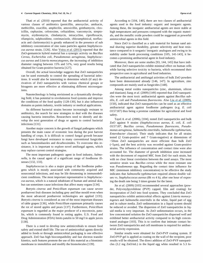
Vlad et al. (2012) reported antifungal activity of a membrane ZnO-PU (polyurethane) at concentration of 5% and 10% ZnO up to 60 days (Fig. 1). Such modified ZnO membranes have important antifungal properties and can be successfully used in food or bio-medical applications, blocking the formation of biofilms [157].
Textile industry is one domain that can largely benefit from the antibacterial and antifungal activity of ZnO nanoparticles. One objective is to impact the additional value on natural textiles by adding to them ultraviolet (UV) absorption and antimicrobial pro-tection properties with ZnO nano-level coatings It was found that the treatment with ZnO formulation caused a net reduction in bacte-rial cells of 78% and 62% in the case of treated cotton and cot-ton/polyester fabrics while the net reduction in fungi was calculated to be 80.7% and 32%, respectively [159-161].
ANTIBACTERIAL AND ANTIFUNGAL ACTION MECHA-
NISMS OF ZnO NANOPARTICLES
Tam et al. (2008) revealed that ZnO nanorods in the liquid phase (without UV light) lead to bacterial cell death which is due to the cell membrane damage [108]. TEM images present cells with obviously damaged membrane in addition to partially leaked out contents. Also, some of the cells exhibit shrinkage of cytoplasmatic material inside the cell wall. Internalization of the ZnO rod or ZnO nanoparticles can be observed in some cases, but this is not a gen-eral occurrence. Therefore the mechanical damage of the cell mem-brane cannot be considered as the main mechanism of antibacterial activity of ZnO. In the case of Bacillus atrophaeus, different ZnO morphologies cause similar types of cell damage. Cells with par-tially leaked out contents can be observed, and there exist very few intact cells. A TEM image of a single cell was published in [108], to more clearly observe the cell damage in E. coli and Bacillus at-
rophaeus cells exposed to ZnO nanoparticles. For Bacillus atro-
phaeus cells, can be observed multiple breaches of the cell walls, while this is not the case for E. coli. Tam’s study has demonstrated that the ZnO is more effective for killing Gram-positive than Gram-negative bacteria because they have simpler cell membrane struc-ture.
6 Current Organic Chemistry, 2014, Vol. 18, No. 2 Oprea et al.
Repeated incubation tests result in eventual loss of antibacterial activity of ZnO nanoparticles. This is likely due to the fact that it is necessary for nanoparticles to be in contact with the cells to present antibacterial activity. Thus no antibacterial activity will be observed once contamination by dead bacteria residue etc. has covered the surface of nanoparticles [108].
The interactions between the nanomaterial and the individual cell components can elucidate the impact of nanomaterials on bac-teria. The first interaction that can occur between a nanoparticle and a cell is at the membrane interface. Some studies, [162], suggested that nanoparticles were able even to embed themselves in the cell membrane.
Similar results were reported by Liu et al. (2009). ZnO nanopar-ticles may distort and damage bacterial cell membrane, resulting in a leakage of intracellular contents and eventually the death of bacte-
rial cells. The inhibitory effects against E. coli O157:H7 increase as the concentration of ZnO nanoparticles increases [150].
A study conducted by Tayel A. et al. (2011), [104], based on scanning electron micrographs of the treated bacterial cells (Salmo-
nella typhimurium and Staphyloccocus aureus) revealed that ZnO nanoparticles attached to the cellular wall, softening and broadening it, having as result that the cells were lysed and their internal com-ponents began to release.
As Gram-positive bacteria were more sensitive than Gram-negative bacteria to ZnO nanoparticles, this can be explained by ZnO interaction with specific cell wall compounds; these com-pounds may be found or increased in Gram-positive rather than in Gram-negative bacteria. The cell wall structure for Gram negative bacteria is also relatively more complex than that of Gram positive cells [163].
Fig. (1). SEM images: fungal growing profile of the samples PUDMPA-0 (0% nano-ZnO), PUDMPA-2 (5% nano-ZnO) and PUDMPA-4 (10% nano-ZnO) [157].

ZnO Applications and Challenges Current Organic Chemistry, 2014, Vol. 18, No. 2 7
Jeng et al. (2006), [164], proposed that nanomaterials that can physically attach to a cell can be bactericidal if they come into con-tact with this cell. If the membrane of a cell is compromised, the cell may repair itself or, if the damage is severe, the cell component may release and eventually the cell will die [165].
To explain the adhesion of ZnO nanoparticles to the cell wall, Tayel A. et al. (2006) in [104] indicated as potential candidates the outer thick peptidoglycan layer and its amino acid constituent, sur-face proteins and teichoic acids plus lipoids (forming lipoteichoic acids), which act as chelating agents. Among the compounds that can block the adhesion of ZnO (compounds found only in Gram-negative bacteria) the possible nominees include the extra outer membranes and the pathogen-associated molecular patterns which include lipopolysaccharide (consisting of lipid A, core polysaccha-ride and O antigen), porins and particular fragments of peptidoglycan.
Leone et al. (2007) proposed that carboxyl, amide, phosphate, hydroxyl groups and carbohydrate-related moieties in the bacterial cell wall may provide sites for the molecular-scale interactions with the oxide nanoparticles [166].
The exact nature of the interaction of the nanoparticles with bacterial membrane and possible permeation of the nanoparticles into the bacterial cells is still under investigation as exact informa-tion and consensus are lacking [149].
The antibacterial activity can be explained also by a mechanism proposed by Yamamoto et al. (2001), [146], which relies on the generation of hydrogen peroxide (H2O2). H2O2 that generates from the surface of ZnO is considered as an effective mean for the inhibi-tion of bacterial growth. It can be assumed that the concentration of H2O2 generated from the surface increases with decreasing particle size. As the size of particles decreases the surface increases, so more H2O2 will be generated. Li et al., (2009) reported that genera-tion of H2O2 from ZnO leads to the penetration of particles into the cell membrane of bacteria, that leads to the formation of injuries and finally the death of bacterium [153]. Same mechanism was reported for antibacterial activity against E. coli and Pseudomonas
aeruginosa [128].
The size of ZnO nanoparticles also directly influences the anti-bacterial activity. On one hand while the nanoparticles size de-creases, the surface/volume ratio increases. On the other hand, smaller nanoparticles tend to have a larger surface defect density, which leads to better photocatalytic activity by generating increased levels of reactive oxygen species (ROS) [167].
Electron resonance measurements show that aqueous suspen-sion of ZnO nanoparticles generates increased level of ROS [110]. Theoretically, ROS could be produced as a consequence of ZnO contact with some bacterial electron carriers. Applerot et al., (2009) found that an increased oxidative stress is detected after the antibac-terial treatment, beyond the level that is yielded by the ZnO itself. Moreover, the quantity of ROS detected was different for various bacterial strains. When ZnO slurry was added to E. coli, an increase of 124% in the level of ROS was detected, but when ZnO was added with Staphyloccocus aureus, an immediate decrease of ROS level with 31% was noticed [110]. These findings were explained by authors in the light of a relatively new hypothesis “the bacterial suicide response” [168]. The central idea is that when bacteria ex-perience continuous stress conditions, cells may eventually suffer growth arrest while maintaining their metabolism. An excessive burst of free-radical production then results from this uncoupling of growth from metabolism, and it is this free-radical burst that is lethal to the cells, rather than the stress per se. This mode of trigger-ing bacterial cell death can differ for various kinds of bacteria, ex-
plicating the difference between E. coli and Staphyloccocus aureus. As Staphyloccocus aureus is a more antioxidant protected bacte-rium, any burst of free-radical production can be neutralized more easily, ending up, in the short term, with smaller net number of hydroxyl radicals [110].
Also at it was demonstrated by Yamamoto et al. (2001) that for ZnO powders, the influence of particle size on Staphyloccocus
aureus was less than that on E. coli. The structures and chemical compositions of the cell surface of the bacteria used in the study are quite different. Thin layers of lipid A, lipopolysaccharide and pep-tidoglycan are present on the cell surface of E. coli, whereas there is only a peptidoglycan layer for Staphyloccocus aureus. However, the differences in antibacterial action towards Staphyloccocus
aureus and E. coli are assumed to be due to the different sensitivi-ties towards H2O2 [146].
According to Tam et al. (2008), ZnO nanoparticles were con-siderably more effective against Bacillus atrophaeus than against E.
coli (at 5 mM vs. 15 mM concentration, respectively, showing zero viable cell count). For both bacterial strains, damage of the cell membranes was found, and the effect was more pronounced for Bacillus atrophaeus. At the same time, significant differences were observed in the effect of H2O2 and ZnO nanostructures on Bacillus
atrophaeus. TEM investigation of the effects of H2O2 (30 mg/ mL) on the E. coli and Bacillus atrophaeus reveals that no damage of Bacillus atrophaeus cells is observed at concentrations causing damage to E. coli. This is different from the behavior observed with all ZnO morphologies, indicating existence of a different mecha-nism of antibacterial activity. While the release of H2O2 from ZnO may contribute to the cell damage of E. coli, cell damage of Bacil-
lus atrophaeus is caused by a different mechanism since no effect of H2O2 is observed in this case [108]. They also reported that an-nealing the ZnO nanorods array used for antibacterial test, changed the luminescence spectra, altering the surface defects density re-sponsible for green emission, but with no effect on the antibacterial activity. This suggested that there are other mechanisms, beside those mediated by H2O2, contributing to the antibacterial activity of ZnO nanoparticles [108].
Another possible mechanism for ZnO antibacterial activity is the release of Zn2+ ions [169]. The study of Doménech and Prieto
(1986), [170], indicates that the Zn2+ ion concentration is increased when H2O2 is produced. The dissolution of ZnO nanoparticles into Zn ions was found to be size dependent. Thus engineered ZnO nanostructures may change their toxicity by influencing their disso-lution rate [167, 171-177].
Applerot et al., (2009), [110] also suggested that ROS, at a very low concentration, are able to modulate biological processes in the bacterial cells. It has already been shown that low ROS fluxes (in contrast to high fluxes which have detrimental effects on cells) may act as messengers which activate cell processes such as transcrip-tion factor release, gene expression, and cell growth [178].
Green and Howman (2005), [179], indicated the probable pro-duction of free radicals under dark conditions as a mechanism for antibacterial activity. Tayel A. et al. (2006), [104], confirmed that the observed antibacterial activity reported by them was in the ab-sence of light.
On the other hand Applerot et al., (2009), [110], compared the generation of hydroxyl radicals in ZnO suspensions in the dark and before and after illumination with visible light. The results revealed that with dark prepared suspensions the antibacterial activity was to some extent reduced, but was still present. In contrast, upon brief illumination of the suspensions with visible light emitted from a

8 Current Organic Chemistry, 2014, Vol. 18, No. 2 Oprea et al.
lamp, the ROS level increased up to nearly 400% compared to sus-pensions without illumination. Such results may be advantageous for developing medical and water purification devices based on ZnO nanoparticles.
The mechanism of toxicity is still only partially understood [180]. However, the toxicity of ZnO nanoparticles seems not to be directly related to their entering into the cell, but rather to their intimate contact with the cell, contact that causes changes in the microenvironment in the vicinity of the organism-particle contact area to either increase metal solubilization or to generate ROS, that may ultimately damage cell membrane [172].
Moreover, the toxicity of ZnO nanoparticles is not only affected by the light via ROS production, but may also be affected in the dark although its mechanism is not yet defined [181].
There are also many studies that suggest that the mechanism of ZnO toxicity is related to its photosensitivity (or can be enhanced by it) and production of ROS under specific wavelength light. These ROS can impair the cell membrane architecture through lipid peroxidation. By increasing the irradiation time, the quantity of ROS formed on ZnO surface increases, yielding a better antibacte-rial activity. Brayner R. et al. (2006) reported damage and disor-ganization in the cell wall in the bacteria exposed to ZnO nanopar-ticles [93].
The generation of highly ROS such as OH , H2O2 and O22 is
explained as follows. Since ZnO with defects can be activated by both UV and visible light, electron-hole pairs (e-h+) can be created. The holes split H2O molecules (from the suspension of ZnO) into OH- and H+. Dissolved oxygen molecules are transformed to super-oxide radical anions (·O2
-), which in turn react with H+ to generate (HO2
·) radicals, which upon subsequent collision with electrons
produce hydrogen peroxide anions (HO2-). They then react with
hydrogen ions to produce molecules of H2O2. The generated H2O2 can penetrate the cell membrane and kill the bacteria. Since, the hydroxyl radicals and super oxides are negatively charged particles, they cannot penetrate into the cell membrane and must remain in direct contact with the outer surface of the bacteria; however, H2O2 can penetrate into the cell [107, 143].
Liu H.L. et al. (2003) reported that under UV irradiation with 365nm ZnO nanoparticles exhibit better antibacterial activity than TiO2 nanoparticles. Almost all the initial E. coli cells (108 CFU/ml) were inactivated in 40 min in the presence of 2 g/l ZnO. Photocata-lytic inactivation of bacteria was found to follow first order kinetics with the highest rate constants being 4.5x10⁻¹ and 2.2x10⁻¹/min for E. coli and Lactobacillus Helveticus, respectively, treated with 2 g/l ZnO. As Gram-negative bacteria was more sensitive to ZnO than Gram-positive one, authors hypothesized that no significant cell wall destruction by photocatalytic oxidation could be observed [107], but most probably cells may adapt different photocatalytic inactivation mechanism [110, 146].
The use of air as the purging gas provides a stronger oxidizing environment and gives a higher catalytic effect on bacteria inactiva-tion than that of nitrogen. Such methods are suitable not only for food industry but also for water treatment (simultaneous treatment of photolytic sterilization of microbial cells and photocatalytic de-composition of organic compounds) [107, 152, 182, 183].
The literature data suggest a different mechanism for the inhibi-tory effect of ZnO nanoparticles on fungi compared to those re-ported for bacteria. For the antifungal activity of ZnO nanoparticles, the mechanisms proposed are related to the fungus nature.
The fine structure of Botrytis cinerea mycelia was preserved af-ter treatment with ZnO nanoparticles, but the surface of fungal hy-
phae was deformed. The mechanism of cell functions of Botrytis
cinerea was affected by ZnO nanoparticles due to increased produc-tion of nucleic acids and carbohydrates through the stress response in fungal hyphae, that leads to cells death [105]. The increase of carbohydrates may be due to the self-protecting mechanism against the ZnO nanoparticles [184].
ZnO nanoparticles treatment inhibited the conidial development and distorted the conidiophores of Penicillium expansum. Treated cells of Penicillium expansum release proteins, carbohydrates and lipids through the damaged cell membrane, that leads to cells death [105].
Several studies reported that integration of ZnO nanoparticles into bacterial cells may induce continuous release of membrane lipids and proteins, which changes the membrane permeability of bacterial cells [93, 185].
The antifungal and antibacterial activities reported by Emamifar et al. (2010) were recorded for samples kept in the dark at 4°C, which exclude the photocatalytic activity as a mechanism [102].
The literature data suggest that ZnO nanoparticles have a poten-tial application as a bacteriostatic agent, especially in visible light and may have future applications in the development of derivative agents to control the spread and infection in a variety of domains.
In the prevention and control of bacterial spreading and infec-tions, the main target is the cell wall structure. The cell wall of most pathogenic bacteria is composed of surface proteins for adhesion and colonization, and components such as polysaccharides and teichoic acid that protect against host defenses and environmental conditions [186]. These components are charged macromolecules; therefore, specific interactions to disrupt their main function and location may be triggered by introducing specific groups on the surface of the nanoparticles. It has been reported that certain long-chain polycations coated onto surfaces can efficiently kill on con-tact both Gram-positive and Gram-negative bacteria [187, 188]. These studies have indicated that families of unrelated hydrophobic groups are equally efficient at killing bacteria. Therefore, in the future, ZnO nanoparticles containing formulations may be utilized for external uses as antibacterial agents in ointments, lotions, mouthwashes, and surface coatings on various substrates to prevent microorganisms from attaching, colonizing, spreading, and forming biofilms in indwelling medical devices [106].
CONCLUSION
The main conclusions of these studies can be summarized as follows:
ZnO particles are effective for inhibiting both Gram-positive and Gram-negative bacteria. Usually Gram-positive bacteria are more sensible but there are few studies, especially on E. coli and S.
aureus, where Gram-negative bacteria are killed faster than Gram-positive ones. In such cases, authors proposed alternative mecha-nisms to try elucidate this behavior.
The antibacterial activity of ZnO nanoparticles appeared in close vicinity of the surface, or on the surface.
Smaller ZnO particles have a better antibacterial activity. High concentration and large surfaces lead to better antibacterial activity. Shape of the nanoparticles and their crystalline structure do not have any influence on the antibacterial activity. The antibacterial activity depends only on the surface area and concentration.
The mechanisms of the antibacterial activity of ZnO particles are not well understood although two major hypotheses try to eluci-date it:
ZnO Applications and Challenges Current Organic Chemistry, 2014, Vol. 18, No. 2 9
a) That the generation of hydrogen peroxide is the main factor of the antibacterial activity.
b) That the binding of the particles on the bacteria surface due to the electrostatic forces is driving force behind antibacterial ac-tivity.
Antibacterial activity was noticed, with some difference in in-tensity, in the dark and under UV/visible light. The antibacterial activity is in direct correlation with photocatalytic activity of ZnO.
High-temperature treatment of ZnO particles has a significant effect on their antibacterial activity. Treatment at a higher tempera-ture leads to a lower activity. Antibacterial activity has no direct connection with the luminescence spectra (the green emission is usually attributed to surface defects, so in theory a higher defect density should lead to better luminescence and better antibacterial activity). These sustain the idea that ZnO nanoparticles act through more than one simple mechanism against bacteria.
CONFLICT OF INTEREST
The authors confirm that this article content has no conflict of interest.
ACKNOWLEDGEMENTS
Declared none.
REFERENCES
[1] Kumar, C. Nanomaterials: Toxicity, health, and environmental issues. Nanotechnologies for the Life Sciences, 2006, Wiley-VHC: Weinheim, Ger-many.
[2] Klingshirn, C. ZnO: Material, physics and applications. Chemphys., 2007, 8(6), 782-803.
[3] Safaei, H.R.; Safaei, M.R.; Rahmanian, V. Film formation and anticorrosive behavior of Zn-ZSM-5 nano-sized zeolite composite coatings. Open Electro-
chem. J., 2012, 4, 1-8. [4] Baraton, M.I. Nano-TiO2 for solar cells and photocatalytic water splitting:
Scientific and technological challenges for commercialization. Open Nano-
sci. J., 2011, 5, 64-77. [5] Denotti, G.; Piga, R.; Montaldo, C.; Erriu, M.; Pilia, F.; Piras, A.; DeLuca,
M.; Orru, G. In vitro evaluation of enterococcus faecalis adhesion on various endodontic medicaments. Open Dent. J., 2009, 3, 120-124.
[6] Al-Mossawi, M.A.; Al-Khursan, A.H.; Al-Ansari, R.A. ZnO-MgZnO quan-tum-dot semiconductor optical amplifiers. Recent Patents on Elect. Eng., 2009, 2, 226-238.
[7] Singh, S.; Kaur, H.; Pathak, D.; Bedi, R.K. Zinc oxide nanostructures as transparent window layer for photovoltaic application. Digest J. Nanomater.
Biostruc., 2011, 6(2), 689-698. [8] Ooi, M.D.J.; Aziz, A.A.; Abdullah, M.J.; Al-Hardan, N.H. The complex
impedance behavior of ZnO particles synthesized via fast crystallization pre-cipitation techniques. Digest J. Nanomater. Biostruc., 2012, 7(3), 1179-1187.
[9] Goto, T.; Yin, S.; Sato, T.; Tanaka, T. Morphological control of zinc oxide and application to cosmetics. Int. J. Nanotech., 2013, 10(1-2), 48-56.
[10] Gingasu, D.; Mindru, I.; Patron, L.; Culita, D.C.; Calderon-Morero, J.M.; Diamandescu, L.; Feder, M.; Oprea, O. Precursor method- A nonconven-tional route for the synthesis of ZnCr2O4 spinel. J. Phys. Chem. Solids, 2013, 74(9), 1295-1302.
[11] Lacatusu, I.; Mitrae, E.; Badea, N.; Stan, R.; Oprea, O.; Meghea, A. Lipid nanoparticles based on omega-3 fatty acids as effective carriers for lutein de-livery. Preparation and in vitro characterization studies. J. Funct. Foods, 2013, 5(3), 1260-1269.
[12] Florea, M.G.; Ficai, A.; Oprea, O.; Guran, C.; Ficai, D.; Pall, L.; An-dronescu, E. Drug delivery systems based on silica with prolonged delivery of folic acid. Romanian J. Mat., 2012, 42(3), 313-316.
[13] Oprea, O.; Andronescu, E.; Vasile, B.S.; Voicu, G.; Covaliu, C. Synthesis and characterization of ZnO nanopowder by non-basic route. Digest J.
Nanomater. Biostruc., 2011, 6(3), 1393-1401. [14] Ficai, D.; Ficai, A.; Vasile, B.S.; Ficai, M.; Guran, C.; Andronescu, E. Syn-
thesis of rod-like magnetite by using low magnetic field. Digest J. Nanoma-
ter. Biostruc., 2011, 6(3), 943-951. [15] Ianculescu, A.; Gheorghiu, F.P.; Postolache, P.; Oprea, O.; Mitoseriu, L. The
role of doping on the structural and functional properties of BiFe1-xMnxO3 magnetoelectric ceramics. J. Alloys Compounds, 2010, 504(2), 420-426.
[16] Zaid, H.; Rayan, A.; Said, O.; Saad, B. Cancer Treatment by Greco-Arab and Islamic Herbal Medicine. Open Nutraceutic. J., 2010, 3, 203-212.
[17] Brayner, R. Current approaches in natural biopolymer-nanoparticle hybrid functional materials: From drug delivery to bio-detection applications, in
frontiers in drug design & discovery. 2006, 241-258, Bentham Science Pub-lishers.
[18] Andronescu, E.; Grumezescu, AM.; Ficai, A.; Gheorghe, I.; Chifiriuc, M.; Mihaiescu, D.E.; Lazar, V. In vitro efficacy of antibiotic magnetic dextran microspheres complexes against Staphylococcus aureus and Pseudomonas aeruginosa strains. Biointerface Res. Appl. Chem., 2012, 2(3), 332-338.
[19] Beck-Broichsitter, M.; Gauss, J.; Schweiger, C.; Roesler, S.; Schmehl, T.; Kampschulte, M.; Langheinrich, A.C.; Seeger, W. Micro-computed tomo-graphy imaging of composite nanoparticle distribution in the lung. Int. J.
Pharm., 2012, 439(1-2), 230-233. [20] Saviuc, C.; Grumezescu, A.M.; Holban, A.; Chifiriuc, C.; Mihaiescu, D.;
Lazar, V. Hybrid nanostructurated material for biomedical applications. Bio-
interface Res. Appl. Chem., 2011, 1(2), 64-71. [21] Eidi, H.; Joubert, O.; Nemes, C.; Grandemange, S.; Mograbi, B.; Foliguet,
B.; Tournebize, J.; Maincent, P.; Le Faou, A.; Aboukhamis, I.; Rihn, B.H. Drug delivery by polymeric nanoparticles induces autophagy in macro-phages. Int. J. Pharm., 2012, 422(1-2), 495-503.
[22] Grumezescu, A.M.; Andronescu, E.; Ficai, A.; Bleotu, C.; Chifiriuc, M.C. Chitin based biomaterial for antimicrobial therapy: fabrication, characteriza-tion and in vitro profile based interaction with eukaryotic and prokaryotic cells. Biointerface Res. Appl. Chem., 2012, 2(5), 438-445.
[23] Grumezescu, A.M.; Andronescu, E.; Ficai, A.; Bleotu, C.; Mihaiescu, D.E.; Chifiriuc, M.C. Synthesis, characterization and in vitro assessment of the magnetic chitosan-carboxymethylcellulose biocomposite interactions with the prokaryotic and eukaryotic cells. Int. J. Pharm., 2012, 436(1-2), 771-777.
[24] Grumezescu, A.M.; Andronescu, E.; Ficai, A.; Huang, K.S.; Gheorghe, I.; Chifiriuc, M.C. Water soluble magnetic biocomposite with potential applica-tions for the antimicrobial therapy. Biointerface Res. Appl. Chem., 2012, 2(6), 469-475.
[25] Jansch, M.; Stumph, P.; Graf, C.; Rühl, E.; Müller, R.H. Adsorption kinetics of plasma proteins on ultrasmall superparamagnetic iron oxide (USPIO) nanoparticles. Int. J. Pharm., 2012, 428(1-2), 125-133.
[26] Grumezescu, A.M.; Saviuc, C.; Holban, A.; Hristu, R.; Stanciu, G.; Chifiriuc, C.; Mihaiescu, D.; Balaure, P.; Lazar, V. Magnetic chitosan for drug target-ing and in vitro drug delivery response. Biointerface Res. Appl. Chem., 2012, 1(5), 160-165.
[27] Kapoor, M.; Burgess, D.J.; Patil, S.D. Physicochemical characterization techniques for lipid based delivery systems for siRNA. Int. J. Pharm., 2012, 427(1), 35-57.
[28] Maldiney, T.; Byk, G.; Wattier, N.; Seguin, J.; Khandadash, R.; Bessodes, M.; Richard, C.; Scherman, D. Synthesis and functionalization of persistent luminescence nanoparticles with small molecules and evaluation of their tar-geting ability. Int. J. Pharm., 2012, 423(1), 102-107.
[29] Grumezescu, A.M.; Mihaiescu, D.E.; Tamas, D. Hybrid materials for drug delivery of rifampicin: evaluation of release profile. Biointerface Res. Appl.
Chem., 2012, 1(6), 229-235. [30] Saviuc, C.; Grumezescu, A.M.; Holban, A.; Bleotu, C.; Chifiriuc, C.; Blaure,
P.; Lazar, V. Phenotypical studies of raw and nanosystem embedded Eugenia carryophyllata buds essential oil antibacterial activity on Pseudomonas aeru-ginosa and Staphylococcus aureus strains. Biointerface Res. Appl. Chem., 2011, 1(3), 111-118.
[31] Xu, W.; Riikonen, J.; Lehto, V.-P. Mesoporous systems for poorly soluble drugs. Int. J. Pharm., 2013, 463(1), 181-197.
[32] Yang, Y.; Li, X.; Chen, J.; Chen, H.; Bao, X. ZnO nanoparticles prepared by thermal decomposition of beta-cyclodextrin coated zinc acetate. Chem. Phys.
Letters, 2003, 373(1-2), 22-27. [33] Oprea, O.; Vasile, O.R.; Voicu, G.; Andronescu, E. The influence of the
thermal treatment on luminescence properties of ZnO. Digest J. Nanomater.
Biostruc., 2013, 8(2), 747-756. [34] Vasile, O.R.; Andronescu, E.; Ghitulica, C.; Vasile, B.S.; Oprea, O.; Vasile,
E.; Trusca, R. Synthesis and characterization of nanostructured zinc oxide particles synthesized by the pyrosol method. J. Nanoparticle Res., 2012, 14(12), 1-13.
[35] Chen, L.C.; Tien, C.H. The influence of hydrogen gas treatment on the characteristics of ZnO films. Open Cryst. J., 2009, 2, 11-14.
[36] Tonto, P.; Mekasuwandumrong, O.; Phatanasri, S.; Pavarajarn, V.; Praserthdam, P. Preparation of ZnO nanorod by solvothermal reaction of zinc acetate in various alcohols. Ceramics Int., 2008, 34(1), 57-62.
[37] Kong, L.; Yang, J.X.; Zhou, H.P.; Tian, Y.P.; Wu, J.Y.; Jin, B.K. A surfac-tant-free, precursor-induced method to flower-like ZnO nanostructures. Cur-
rent Nanosci., 2009, 5(4), 474-478. [38] Al Asmar, R.; Atanas, J.P.; Ajaka, M.; Zaatar, Y.; Ferblantier, G.; Sauvajol,
J.L.; Jabbour, J.; Juillaget, S.; Foucaran, A. Characterization and Raman in-vestigations on high-quality ZnO thin films fabricated by reactive electron beam evaporation technique. J. Cryst. Growth, 2005, 279(3-4), 394-402.
[39] Oprea, O.; Vasile, O.R.; Voicu, G.; Craciun, L.; Andronescu, E. Photolumi-nescence, magnetic properties and photocatalytic activity of Gd3+ doped ZnO nanoparticles. Digest J. Nanomater. Biostruc., 2012, 7(4), 1757-1766.
[40] Singh, N. Effect of ageing in structural properties of ZnO nanoparticles with pH variation for application in solar cells. Open Renew. Energy J., 2012, 5, 15-18.
[41] Wu, J.J.; Liu, S.C. Low-temperature growth of well-aligned ZnOnanorods by chemical vapor deposition. Adv. Mater., 2002, 14(3), 215-+.
[42] Baruah, S.; Dutta, J. Hydrothermal growth of ZnO nanostructures. Sci. Tech.
Adv. Mater., 2009, 10(1), 1-18.

10 Current Organic Chemistry, 2014, Vol. 18, No. 2 Oprea et al.
[43] Voicu, G.; Oprea, O.; Vasile, B.S.; Andronescu, E. Photoluminescence and photocatalytic activity of Mn-doped ZnO nanoparticles. Digest J. Nanoma-
ter. Biostruc., 2013, 8(2), 667. [44] Wahab, R.; Ansari, S.G.; Kim, Y.S.; Dar, M.A.; Shin, H.S. Synthesis and
characterization of hydrozincite and its conversion into zinc oxide nanoparti-cles. J. Alloys Compounds, 2008, 461(1-2), 66-71.
[45] He, H.; Yang, Q.; Wang, J.; Ye, Z. Layer-structured ZnO nanowire arrays with dominant surface- and acceptor-related emissions. Mater. Letters, 2011, 65(9), 1351-1354.
[46] Liu, C.P.; Wang, R.C.; Kuo, C.L.; Liang, Y.H.; Chen, W.Y. Recent patents on fabrication of nanowires. Recent Pat. Nanotech., 2007, 1(1), 11-20.
[47] Huang, N.; Zhu, M.W.; Gao, L.J.; Gang, J.; Sun, C.; Jiang, X. A template-free sol-gel technique for controlled growth of ZnO nanorod arrays. Appl.
Surface Sci., 2011, 257(14), 6026-6033. [48] Sangpour, P.; Roozbehi, M.; Akhavan, O.; Moshfegh, A.Z. ZnO nanowires
from nanopillars: Influence of growth time. Current Nanosci., 2009, 5(4), 479-484.
[49] Jia, G.Z.; Wang, Y.F.; Yao, J.H. Fabrication and optical properties of well-aligned ZnOnanorods on sapphire prepared by chemical bath deposition. Di-
gest J. Nanomater. Biostruc., 2012, 7(1), 261-267. [50] Fan, D.H.; Zhang, R.; Li, Y. Synthesis and optical properties of phosphorus-
doped ZnO nanocombs. Solid State Commun., 2010, 150(39-40), 1911-1914. [51] Singh, J.; Kumar, P.; Late, D.J.; Singh, T.; More, M.A.; Joag, D.S.; Tiwari,
R.S.; Hui, K.S.; Hui, K.N.; Srivastava, O.N. Optical and field emission prop-erties in different nanostructures of ZnO. Digest J. Nanomater. Biostruc., 2012, 7(2), 795-806.
[52] Hashim, A.J.; Jaafar, M.S.; Ghazai, A.J.; Ahmed, N.M. Fabrication and characterization of tetraleg zinc oxide nanostruture using evaporation meth-ode. Digest J. Nanomater. Biostruc., 2012, 7(2), 487-491.
[53] Kou, H.H.; Zhang, X.; Du, Y.L.; Ye, W.; Lin, S.X.; Wang, C.M. Electro-chemical synthesis of ZnO nanoflowers and nanosheets on porous Si as pho-toelectric materials. Appl. Surface Sci., 2011, 257(10), 4643-4649.
[54] Kharisov, B.I. A review for synthesis of nanoflowers. Recent Pat. Nanotech., 2008, 2(3), 190-200.
[55] Yan, D.H.; Yin, G.F.; Huang, Z.B.; Yang, M.; Liao, X.M.; Kang, Y.Q.; Yao, Y.D.; Hao, B.Q.; Han, D. Characterization and bacterial response of zinc ox-ide particles prepared by a biomineralization process. J. Phys. Chem. B, 2009, 113(17), 6047-6053.
[56] Gonzalez, S.; Gilberte, Y.; Philips, N.; Juarranz, A. Current trends in photo-protection - A new generation of oral photoprotectors. Open Dermat. J., 2011, 5, 6-14.
[57] Reddy, B.Y.; Hantash, B.M. Cutaneous connective tissue diseases: epidemi-ology, diagnosis and treatment. Open Dermat. J., 2009, 3, 22-31.
[58] Gambarini, G.; Testaralli , L.; Al-Sudani, D.; Plotino, G.; Grande, N.M.; Lupi, A.; Giardina, B.; Nocca, G.; DeLuca, M. In vitro evaluation of the cy-totoxicity of different root canal filling materials. Open Dent. J., 2011, 5, 29-32.
[59] Burkhart, C.G.; Black, C.; Burkhart, C.N. A topical application containing sucralfate, zinc oxide and ketoconazole provides high patient satisfaction in the treatment of intertrigo. Open Dermat. J., 2009, 3, 11-13.
[60] Copcia, V.E.; Gradinaru, R.; Mihai, G.D.; Bilba, N.; Sandu, I. Antibacterial activity of nanosized ZnO hosted in microporous clinoptilolite and mesoporous silica SBA-15 matrices. Rev. De Chimie, 2012, 63(11), 1124-1131.
[61] Johannes, B.; Tornsten, N.; Nina, J.B.; Lars, P.H.J.; Udo, W.; Tuan, A.D. Mineralization and particle growth kinetics of ZnO in the presence of gelatin. Biointerface Res. Appl. Chem., 2012, 2(3), 339-349.
[62] Johannes, B.; Tornsten, N.; Nina, J.B.; Lars, P.H.J.; Udo, W.; Tuan, A.D. Bio-inspired mineralization of zinc oxide in presence of ZnO-binding pep-tides. Biointerface Res. Appl. Chem., 2012, 2(4), 380-391.
[63] Jin, H.B.; Oktar, F.N.; Dorozhkin, S.V.; Agathopoulos, S. Sintering behavior and properties of reinforced hydroxyapatite/TCP biphasic bioceramics with ZnO-whiskers. J. Composite Mater., 2011, 45(13), 1435-1444.
[64] Gunduz, O.; Erkan, E.M.; Daglilar, S.; Salman, S.; Agathopoulos, S.; Oktar, F.N. Composites of bovine hydroxyapatite (BHA) and ZnO. J. Mater. Sci., 2008, 43(8), 2536-2540.
[65] Ozyegin, L.S.; Oktar, F.N.; Heybeli, N. Calcium phosphate accelerated setting time of the ethoxybenzoic acide (EBA) temporary cement. Key Eng.
Mater., 2003, 240-242, 349-352. [66] Wong, R.H.; Palamara, J.E.; Wilson, P.R.; Reynolds, E.C.; Burrow, M.F.
Effect of CPP-ACP addition on physical properties of zinc oxide non-eugenol temporary cements. Dental Mater., 2011, 27(4), 329-338.
[67] Rui, L.D., et al., Characterization and in vitro Bioactivity of Zinc-containing
Bioactive Glass and Glass-ceramics. J. Biomater. Appl., 2006, 20: p. 341360 [68] McBean, L.D.; Dove, J.T.; Halsted, J.A.; Smith Jr., J.C. Zinc concentration
in human tissues. Am. J. Clinical Nutrit., 1972, 25(7), 672-676. [69] Monteiro-Riviere, N.A.; Wiench, K.; Landsiedel, R.; Schulte, S.; Inman,
A.O.; Riviere, J.E. Safety evaluation of sunscreen formulations containing ti-tanium dioxide and zinc oxide nanoparticles in UVB sunburned skin: An in
vitro and in vivo study. Toxicolog. Sci., 2011, 123(1), 264-280. [70] Serpone, N.; Dondi, D.; Albini, A. Inorganic and organic UV filters: Their
role and efficacy in sunscreens and suncare product. Inorganica Chimica
Acta, 2007, 360(3), 794-802. [71] Chandrasekhar, V.; Morishetty, P.K.; Metla, S.L.; Raju, R.V.S.C. Expansion
of gutta-percha in contact with various concentrations of zinc oxide-eugenol
sealer: A three-dimensional volumetric study. J. Endodont., 2011, 37(5), 697-700.
[72] Estrela, C.; Estrada-Bernabe, P.-F.; de Almeida-Decurcio, D.; Almeida-Silva, J.; Rodrigues-Araujo-Estrela, C.; Poli-Figueiredo, J.-A. Microbial leakage of MTA, Portland cement, Sealapex and zinc oxide-eugenol as root-end filling materials. Med. Oral Patolog. Oral Y Cirugia Bucal, 2011, 16(3), E418-E424.
[73] He, L.H.; Purton, D.G.; Swain, M.V. A suitable base material for composite resin restorations: Zinc oxide eugenol. J. Dent., 2010, 38(4), 290-295.
[74] Ilic, D.V. The flow of two zinc oxide-eugenol-based endodontic sealers. Vojno-
sanit. Pregled, 2013, 70(1), 21-25. [75] Li, Z.; Li, Y.; Zou, Y..; Lan, W.; Jansen, J.A. In vitro and in vivo evaluation
on the bioactivity of ZnO containing nano-hydroxyapatite/chitosan cement. J. Biomed. Mater. Res., 2010, 93A(1), 269-279.
[76] Otsuka, M.; Ohshita, Y.; Marunaka, S.; Matsuda, Y.; Ito, A.; Ichinose, N.; Otsuka, K.; Higuchi, W.I. Effect of controlled zinc release on bone mineral density from injectable Zn-containing beta-tricalcium phosphate suspension in zinc-deficient diseased rats. J. Biomed. Mater. Res., 2004, 69A(3), 552-560.
[77] Zhou, G.; Li, Y.; Xiao, W.; Zhang, L.; Zou, Y..; Xue, J.; Jansen, J.A. Synthe-sis, characterization, and antibacterial activities of a novel nanohydroxyapa-tite/zinc oxide complex. J. Biomed. Mater. Res., 2008, 85A(4), 929-937.
[78] Tang, Y.; Chappell, H.F.; Dove, M.T.; Reeder, R.J.; Lee, Y.J. Zinc incorpo-ration into hydroxylapatite. Biomater., 2009, 30(16), 2864-2872.
[79] Santos, M.H.; Shaimberg, A.P.M.; Valerio, P.; Goes, A.M.; Leite, M.F.; Mansur, H.S. Cytocompatibility evaluation of hydroxyapatite/collagen com-posites doped with Zn+2. Revista Matér., 2007, 12(2), 307-312.
[80] Ozyegin, L.S.; Gunduz, O.; Oktar, F.N.; Oz, B.; Agathopoulos, S.; Salman, S.; Ovecoglu, L. Sintering effect on mechanical properties of composites of bovine derived hydroxyapatite (BHA) with titanium Key Eng. Mater., 2006, 309-311(1-2), 359-362.
[81] Salman, S.; Gunduz, O.; Yılmaz, S.; Öveço lu, M.L.; Snyder, R.L.; Agathopoulos, S.; Oktar, F.N. Sintering effect on mechanical properties of composites of natural hydroxyapatites and titanium. Ceramics Int., 2009, 35(7), 2965-2971.
[82] Kalita, S.J.; Bhatt, H.A. Nanocrystalline hydroxyapatite doped with magne-sium and zinc: Synthesis and characterization. Mater. Sci. Eng. C, 2007, 27, 837-848.
[83] Norhidayu, D.; Sopyan, I.; Ramesh, S. Development of zinc doped hy-droxyapatite for bone implant applications. ICCBT 2008 Conference, 2008, 257-270.
[84] Tvinnereim, H.M.; Eide, R.; Riise, T.; Fosse, G.; Wesenberg, G.R. Zinc in primary teeth from children in Norway. Sci. Total Environ., 1999, 226(2-3), 201-212.
[85] Kawamura, H.; Ito, A.; Miyakawa, S.; Layrolle, P.; Ojima, K.; Ichinose, N.; Tateishi, T. Stimulatory effect of zinc-releasing calcium phosphate implant on bone formation in rabbit femora. J. Biomed. Mater. Res., 2000, 50(2), 184-190.
[86] Agren, M.S.; Franzen, L.; Chvapil, M. Effects on wound-healing of Zinc-Oxide in a hydrocolloid dressing. J. Am. Academy Dermat., 1993, 29(2), 221-227.
[87] Shalumon, K.T.; Anulekha, K.H.; Nair, Sreeja V.; Nair, S.V.; Chennazhi, K.P.; Jayakumar, R. Sodium alginate/poly(vinylalcohol)/nanoZnO composite nanofibers for antibacterial wound dressings. Int. J. Biol. Macromolec., 2011, 49(3), 247-254.
[88] Kumar, P.T.S.; Lakshmanan, V.K.; Anilkumar, T.V.; Ramya, C.; Reshmi, P.; Unnikrishnan, A.G.; Nair, S.V.; Jayakumar, R. Flexible and Microporous Chitosan hydrogel/nano ZnO composite bandages for wound dressing: In vi-
tro and in vivo evaluation. Acs Appl. Mater. Interfac., 2012, 4(5), 2618-2629. [89] Sweetman, S.C. Martindale: The complete drug reference, 2005, 1163,
Pharmaceutical Press, London, U.K. [90] Dutta, R.K.; Sharma, P.K.; Pandey, A.C. Surface enhanced raman spectra of
escherichia coli cells using ZnO nanoparticles. Digest J. Nanomater. Bios-
truc., 2009, 4(1), 83-87. [91] Karmali, R.S.; Bartakke, A.; Borker, V.P.; Rane, K.S. Bactericidal action of
N doped ZnO in sunlight. Biointerface Res. Appl. Chem., 2011, 1(2), 57-63. [92} Subhasree, R.S.; Selvakumar, D.; Kumar, N.S. Hydrothermal mediated
synthesis of ZnO nanorods and their antibacterial properties. Letters Appl.
NanoBioSci., 2012, 1(1), 2-7. [93] Brayner, R.; Ferrari-Iliou, R.; Brivais, N.; Djediat, S.; Benedetti, M.F.;
Fievet, F. Toxicological impact studies based on Escherichia coli bacteria in ultrafine ZnO nanoparticles colloidal medium. Nano Letters, 2006, 6(4), 866-870.
[94] Matsuyama, K.; Ihsan, N.; Irie, K.; Mishima, K.; Okuyama, T.; Muto, H. Bioimaging application of highly luminescent silica-coated ZnO-nanoparticle quantum dots with biotin. J. Colloid Interface Sci., 2013, 399, 19-25.
[95] Zou, Y.W.; Huang, Z.B.; Wang, Y.; Liao, X.M.; Yin, G.F.; Gu, J.W. Synthe-sis and cellular compatibility of Co-doped ZnO particles in silk-fibroin pep-tides. Colloids Surfaces B: Biointerfaces, 2013, 102, 29-36.
[96] Jiang, H.; Wang, H.; Wang, X. Facile and mild preparation of fluorescent ZnO nanosheets and their bioimaging applications. Appl. Surface Sci., 2011, 257, 6991-6995.
[97] Venkatasubbu, G.D.; Ramasamy, S.; Ramakrishnan, V.; Kumar, J. Nanoc-rystalline hydroxyapatite and zinc-doped hydroxyapatite as carrier material for controlled delivery of ciprofloxacin. Biotech., 2011, 1(3), 173-186.
ZnO Applications and Challenges Current Organic Chemistry, 2014, Vol. 18, No. 2 11
[98] Murugan, R.; Babu, V.J.; Khin, M.M.; Niar, A.S.; Ramakrishna, S. Synthesis and photocatalytic applications of flower shaped electrospun ZnO-TiO2 mesostructures. Mater. Letters, 2013, 97, 47-51.
[99] Rai, P.; Raj, S.; Ko, K.J.; Park, K.K.; Yu, Y.T. Synthesis of flower-like ZnO microstructures for gas sensor applications. Sensors Actuators B-Chem., 2013, 178, 107-112.
[100] Han, Z.Z.; Liao, L.; Wu, Y.T.; Pan, H.B.; Shen, S.F.; Chen, J.Z. Synthesis and photocatalytic application of oriented hierarchical ZnO flower-rod archi-tectures. J. Hazardous Mater., 2012, 217- 218, 100-106.
[101] Han, Z.Z.; Wei, L.; Tang, L.; Chen, C.; Pan, H.; Chen, J. Aligned CdSe@ZnO flower-rod core-shell nanocable as photovoltaic application. J.
Power Sources, 2013, 239, 546-552. [102] Emamifar, A.; Kadivar, M.; Shahedi, M.; Soleimanian-Zad, S. Evaluation of
nanocomposite packaging containing Ag and ZnO on shelf life of fresh or-ange juice. Innov. Food Sci. Emerg. Tech., 2010, 11(4), 742-748.
[103] Jin, T.; Sun, D.; Su, J.Y.; Zhang, H.; Sue, H.J. Antimicrobial efficacy of zinc oxide quantum dots against listeria monocytogenes, salmonella enteritidis, and escherichia coli O157:H7. J. Food Sci., 2009, 74(1), M46-M52.
[104] Tayel, A.A.; El-Tras, W.F.; Moussa, S.; El-Baz, A.F.; Mahrous, H.; Salem, M.F.; Brimer, L. Antibacterial action of zinc oxide nanoparticles against foodborne pathogens. J. Food Safety, 2011, 31(2), 211-218.
[105] He, L.L.; Liu, Y.; Mustapha, A.; Lin, M.S. Antifungal activity of zinc oxide nanoparticles against Botrytis cinerea and Penicilliumexpansum. Microbiol.
Res., 2011, 166(3), 207-215. [106] Jones, N.; Ray, B.; Ranjit, K.T.; Manna, A.C. Antibacterial activity of ZnO
nanoparticle suspensions on a broad spectrum of microorganisms. Fems Mi-
crobiol. Letters, 2008, 279(1), 71-76. [107] Liu, H.L.; Yang, T.C.K. Photocatalytic inactivation of Escherichia coli and
Lactobacillus helveticus by ZnO and TiO2 activated with ultraviolet light. Process Biochem., 2003, 39(4), 475-481.
[108] Tam, K.H.; Djurisic, A.B.; Chan, C.M.N.; Xi, Y.Y.; Tse, C.W.; Leung, Y.H.; Chan, W.K.; Leung, F.C.C.; Au, D.W.T. Antibacterial activity of ZnOnano-rods prepared by a hydrothermal method. Thin Sol. Films, 2008, 516(18), 6167-6174.
[109] Baker, C.; Pradhan, A.; Pakstis, L.; Pochan, D.J.; Shah, S.I. Synthesis and antibacterial properties of silver nanoparticles. J. Nanosci. Nanotech., 2005, 5(2), 244-249.
[110] Applerot, G.; Lipovsky, A.; Dror, R.; Perkas, N.; Nitzan, Y.; Lubart, R.; Gedanken, A. Enhanced antibacterial activity of nanocrystalline ZnO due to increased ROS-mediated cell injury. Adv. Functional Mater., 2009, 19(6), 842-852.
[111] Sawai, J.; Igarashi, H.; Hashimoto, A.; Kokugan, T.; Shimizu, M. Evaluation of growth-inhibitory effect of ceramics powder slurry on bacteria by conduc-tance method. J. Chem. Eng. Japan, 1995, 28(3), 288-293.
[112] Sawai, J.; Saito, I.; Kanou, F.; Igarashi, H.; Hashimoto, A.; Kokugan, T.; Shimizu, M. Mutagenicity test of ceramic powder which have growth-inhibitory effect on bacteria. J. Chem. Eng. Japan, 1995, 28(3), 352-354.
[113] Sawai, J.; Igarashi, H.; Hashimoto, A.; Kokugan, T.; Shimizu, M. Effect of particle size and heating temperature of ceramic powders on antibacterial ac-tivity of their slurries. J. Chem. Eng. Japan, 1996, 29(2), 251-256.
[114] Sawai, J.; Kawada, E.; Kanou, F.; Igarashi, H.; Hashimoto, A.; Kokugan, T.; Shimizu, M. Detection of active oxygen generated from ceramic powders having antibacterial activity. J. Chem. Eng. Japan, 1996, 29(4), 627-633.
[115] Vaja, F.; Comanescu, C.; Oprea, O.; Ficai, D.; Guran, C. Effects of ZnO nanoparticles on the wet scrub resistance and photocatalytic properties of acrylic coatings. Revista De Chimie, 2012, 63(7), 722-726.
[116] Pica, A.; Guran, C.; Andronescu, E.; Oprea, O.; Ficai, D.; Ficai, A. Antimi-crobial performances of some film forming materials based on silver nanoparticles. J. Optoelectro. Adv. Mater., 2012, 14(9-10), 863-868.
[117] Craciun, E.; Ioncea, A.; Jitaru, I.; Ghiurea, M.; Oprea, O. Organo-inorganic nanostructurated coating materials. Romanian J. Mater., 2011, 41(1), 64-72.
[118] Vaja, F.; Ficai, D.; Ficai, A.; Oprea, O.; Guran, C. Multifunctional advanced coatings based on ZnO/M obtained by nanocasting method. J. Optoelectro.
Adv. Mater., 2013, 15(1-2), 107-113. [119] Yebra, D.M.; Kiil, S.; Weinell, C.E.; Dam-Johansen, K. Dissolution rate
measurements of sea water soluble pigments for antifouling paints: ZnO. Prog. Org. Coatings, 2006, 56(4), 327-337.
[120] Kathirvelu, S.; D'Souza, L.; Dhurai, B. UV protection finishing of textiles using ZnO nanoparticles. Ind. J. Fibre Textile Res., 2009, 34(3), 267-273.
[121] Yadav, A.; Prasad, V.; Kathe, A.A.; Raj, S.; Yadav, D.; Sundaramoorthy, C.; Vigneshwaran, N. Functional finishing in cotton fabrics using zinc oxide nanoparticles. Bulletin Mater. Sci., 2006, 29(6), 641-645.
[122] Ates, E.S.; Unalan, H.E. Zinc oxide nanowire enhanced multifunctional coatings for cotton fabrics. Thin Sol. Films, 2012, 520(14), 4658-4661.
[123] Vihodceva, S.; Kukle, S. Cotton fabric surface modification by sol-gel depo-sition of ZnO thin films. Int. Conference Functional Mater. Nanotech.
(Fm&Nt2012), 2012, 38. [124] Xue, C.H.; Yin, W.; Jia, S.T.; Ma, J.Z. UV-durable superhydrophobic tex-
tiles with UV-shielding properties by coating fibers with ZnO/SiO2 core/shell particles. Nanotech., 2011, 22(41), 415603.
[125] Xue, C.H.; Yin, W.; Jia, S.T.; Ma, J.Z. UV-durable superhydrophobic tex-tiles with UV-shielding property by coating fibers with ZnO/SiO2 core/shell particles. Eco-Dyeing, Finishing Green Chem., 2012, 441, 351-355.
[126] Thati, V.; Roy, A.S.; Prasad, M.V.N.A.; Shivannavar, C.T.; Gaddad, S.M. Nanostructured zinc oxide enhances the activity of antibiotics against Staphylococcus aureus. J. Biosci. Tech., 2010, 1(2), 64-69.
[127] Voicu, G.; Oprea, O.; Vasile, B.S.; Andronescu, E. Antibacterial activity of zinc oxide-gentamicin hybrid material. Digest J. Nanomater. Biostruc., 2013, 8(3), 1191-1203.
[128] Chitra, K.; Annadurai, G. Antimicrobial activity of wet chemically engi-neered spherical shaped ZnO nanoparticles on food borne pathogen. Int.
Food Res. J., 2013, 20(1), 59-64. [129] Lacatusu, I.; Badea, N.; Oprea, O.; Bojin, D.; Meghea, A. Highly antioxidant
carotene-lipid nanocarriers: synthesis and antibacterial activity. J. Nanopar-
tic. Res., 2012, 14(6), 902. [130] Lacatusu, I.; Badea, N.; Murariu, A.; Oprea, O.; Bojin, D.; Meghea, A.
Antioxidant activity of solid lipid nanoparticles loaded with umbelliferone. Soft Mater., 2013, 11(1), 75-84.
[131] Matthews, L.; Kanwar, R.K.; Zhou, S.; Punj, V.; Kanwar, J.R. Applications of nanomedicine in antibacterial medical therapeutics and diagnostics. Open
Tropic. Med. J., 2010, 3, 1-9. [132] Ryan, K.J.; Ray, C.G. Sherris Medical Microbiology. 4th ed., 2004,
McGraw-Hill, Columbus, OH. [133] Bell, C.; Kyriakides, A. Salmonella: A Practical Approach to the Organism
and Its Control in Foods. 2002, Blackwell Publishing Ltd, Oxford,U.K. [134] Bhunia, A.K. Foodborne Microbial Pathogens: Mechanisms and Pathogene-
sis. 2008, Springer LLC, New York, NY. [135] Spadaro, D.; Garibaldi, A.; Gullino, M.L. Control of Penicilliumexpansum
and Botrytis cinerea on apple combining a biocontrol agent with hot water dipping and acibenzolar-S-methyl, baking soda, or ethanol application. Post-
harvest Biol. Tech., 2004, 33(2), 141-151. [136] Latorre, B.A.; Flores, V.; Sara, A.M.; Roco, A. Dicarboximide-Resistant
Isolates of Botrytis-Cinerea from Table Grape in Chile - Survey and Charac-terization. Plant Disease, 1994, 78(10), 990-994.
[137] Cabanas, R.; Abarca, M.L.; Bragulat, M.R.; Cabanes, F.J. In vitro activity of imazalil against Penicilliumexpansum: Comparison of the CLSI M38-A broth microdilution method with traditional techniques. Int. J. Food Micro-
biol., 2009, 129(1), 26-29. [138] Moake, M.M.; Padilla-Zakour, O.I.; Worobo, R.W. Comprehensive review
of patulin control methods in foods. Comprehen. Rev. Food Sci. Food Safety, 2005, 4(1), 8-21.
[139] Kumar, S.A.; Chen, S.M. Nanostructured zinc oxide particles in chemically modified electrodes for biosensor applications. Analy. Letters, 2008, 41(2), 141-158.
[140] Sawai, J. Quantitative evaluation of antibacterial activities of metallic oxide powders (ZnO, MgO and CaO) by conductimetric assay. J. Microbiol. Meth-
ods, 2003, 54(2), 177-182. [141] Stoimenov, P.K.; Klinger, R.L.; Marchin, G.L.; Klabunde, K.J. Metal oxide
nanoparticles as bactericidal agents. Langmuir, 2002, 18(17), 6679-6686. [142] Fu, G.F.; Vary, P.S.; Lin, C.T. Anatase TiO2 nanocomposites for antimicro-
bial coatings. J. Phys. Chem. B, 2005, 109(18), 8889-8898. [143] Padmavathy, N.; Vijayaraghavan, R. Enhanced bioactivity of ZnO nanoparti-
cles-an antimicrobial study. Sci. Tech. Adv. Mater., 2008, 9(3), 035004. [144] Zhang, L.L.; Jiang, Y.H.; Ding, Y.L.; Povey, M.; York, D. Investigation into
the antibacterial behaviour of suspensions of ZnO nanoparticles (ZnOnan-ofluids). J. Nanoparticle Res., 2007, 9(3), 479-489.
[145] Reddy, K.M.; Feris, K.; Bell, J.; Wingett, D.G.; Hanley, C.; Punnoose, A. Selective toxicity of zinc oxide nanoparticles to prokaryotic and eukaryotic systems. Appl. Phys. Letters, 2007, 90(21), 213902.
[146] Yamamoto, O. Influence of particle size on the antibacterial activity of zinc oxide. Int. J. Inorganic Mater., 2001, 3(7), 643-646.
[147] Sawai, J.; Yoshikawa, T. Quantitative evaluation of antifungal activity of metallic oxide powders (MgO, CaO and ZnO) by an indirect conductimetric assay. J. Appl. Microbiol., 2004, 96(4), 803-809.
[148] Waxman, M.F. Agrochemical and pesticide safety handbook, 1998, Lewis Publishers, Florida, p.616.
[149] Jiang, W.; Mashayekhi, H.; Xing, B.S. Bacterial toxicity comparison be-tween nano- and micro-scaled oxide particles. Environ. Pollution, 2009, 157(5), 1619-1625.
[150] Liu, Y.; He, L.; Mustapha, A.; Li, H.; Hu, Z.Q.; Lin, M. Antibacterial activi-ties of zinc oxide nanoparticles against Escherichia coli O157:H7, J. Appl.
Microbiol., 2009, 107(4), 1193-1201. [151] Zhang, L.L.; Ding, Y.L.; Povey, M.; York, D. ZnO nanofluids - A potential
antibacterial agent. Progress Nat. Sci. Mater. Int., 2008, 18(8), 939-944. [152] Zhang, L.L.; Li, S.Q.; Liu, X.M.; Cang, D.Q.; Ding, Y.L. Disinfection of
water and wastewater using ZnO nanofluids - Effect of shaking speed of in-cubator. Environ. Biotech. Mater. Eng., 2011, 183-185, 2298-2302.
[153] Li, X.H.; Xing, Y.; Jiang, Y.H.; Ding, Y.L.; Li, W.L. Antimicrobial activities of ZnO powder-coated PVC film to inactivate food pathogens. Int. J. Food
Sci. Tech., 2009, 44(11), 2161-2168. [154] Emamifar, A.; Kadivar, M.; Shahedi, M.; Solimanian-Zad, S. Effect of
nanocomposite packaging containing Ag and ZnO on inactivation of Lacto-bacillus plantarum in orange juice. Food Control, 2011, 22(3-4), 408-413.
[155] Emamifar, A.; Kadivar, M.; Shahedi, M.; Solimanian-Zad, S. Effect of nanocomposite packaging containing Ag and ZnO on reducing pasteurization temperature of orange juice. J. Food Proc. Preserv., 2012, 36(2), 104-112.

12 Current Organic Chemistry, 2014, Vol. 18, No. 2 Oprea et al.
[156] Sharma, D.; Rajput, J.; Kaith, B.S.; Kaur, M.; Sharma, S. Synthesis of ZnO nanoparticles and study of their antibacterial and antifungal properties. Thin
Sol. Films, 2010, 519(3), 1224-1229. [157] Vlad, S.; Tanase, C.; Macocinschi, D.; Ciobanu, C.; Balaes, T.; Filip, D.;
Gostin, I.N.; Gradinaru, L.M. Antifungal behaviour of polyurethane mem-branes with zinc oxide nanoparticles. Digest J. Nanomater. Biostruc., 2012, 7(1), 51-58.
[158] Kasemets, K.; Ivask, A.; Dubourguier, H.C.; Kahru, A. Toxicity of nanopar-ticles of ZnO, CuO and TiO2 to yeast Saccharomyces cerevisiae. Toxicol. in
vitro, 2009, 23(6), 1116-1122. [159] El-Naggar, A.M.; Zohdy, M.H.; Hassan, M.S.; Khalil, E.M. Antimicrobial
protection of cotton and cotton/polyester fabrics by radiation and thermal treatments. I. Effect of ZnO formulation on the mechanical and dyeing prop-erties. J. Appl. Polymer Sci., 2003, 88(5), 1129-1137.
[160] Das, B.R. UV Radiation protective clothing. Open Textile J., 2010, 3, 14-21. [161] Kim, H.J.; Pant, H.R.; Park, C.H.; Tijing, L.D.; Hwang, B.S.; Choi, N.J.;
Kim, C.S. Electrical properties of ZnO/NYLON-6 spider-wave-like nanonets prepared via electrospinning. Digest J. Nanomater. Biostruc., 2013, 8(1), 385- 393.
[162] Jang, H.; Pell, L.E.; Korgel, B.A.; English, D.S. Photoluminescence quench-ing of silicon nanoparticles in phospholipid vesicle bilayers. J. Photochem.
Photobiol. A-Chem., 2003, 158(2-3), 111-117. [163] Saito, T.; Iwase, T.; Horie, J.; Morioka, T. Mode of photocatalytic bacteri-
cidal action of powdered semiconductor Tio2 on Mutans Streptococci. J.
Photochem. Photobiol. B-Biol., 1992, 14(4), 369-379. [164] Jeng, H.A.; Swanson, J. Toxicity of metal oxide nanoparticles in mammalian
cells. J Environ. Sci. Health A-Toxic/Hazard. Substances Environ. Eng., 2006, 41(12), 2699-2711.
[165] Lyon, D.Y.; Thill, A.; Rose, J.; Alvarez, P.J.J. Ecotoxicological impacts of nanomaterials. Environ. Nanotech., Applic. Impacts Nanomater., 2007, 445-479. The McGraw-Hill, NY.
[166] Leone, L.; Ferri, D.; Manfredi, C.; Persson, P.; Shchukarev, A.; Sjoberg, S.; Loring, J. Modeling the acid-base properties of bacterial surfaces: A com-bined spectroscopic and potentiometric study of the gram-positive bacterium Bacillus subtilis. Environ. Sci. Tech., 2007, 41(18), 6465-6471.
[167] Peng, X.H.; Palma, S.; Fisher, N.S.; Wong, S.S. Effect of morphology of ZnO nanostructures on their toxicity to marine algae. Aquatic Toxicol., 2011, 102(3-4), 186-196.
[168] Dodd, C.E.R.; Richards, P.J.; Aldsworth, T.G. Suicide through stress: A bacterial response to sub-lethal injury in the food environment. Int. J. Food
Microbiol., 2007, 120(1-2), 46-50. [169] Gajjar, P.; Pettee, B.; Britt, D.W.; Huang, W.J.; Johnson, W.P.; Anderson,
A.J. Antimicrobial activity of commercial nanoparticles. Adv. Mater.
Nanotech. Proc., 2009, 1151, 130-132. [170] Domenech, J.; Prieto, A. Stability of Zno particles in aqueous suspensions
under Uv illumination. J. Phys. Chem., 1986, 90(6), 1123-1126. [171] Franklin, N.M.; Rogers, N.J.; Apte, S.C.; Batley, G.E.; Gadd, G.E.; Casey,
P.S. Comparative toxicity of nanoparticulateZnO, bulk ZnO, and ZnCl2 to a freshwater microalga (Pseudokirchneriellasubcapitata): The importance of particle solubility. Environ. Sci. Tech., 2007, 41(24), 8484-8490.
[172] Heinlaan, M.; Ivask, A.; Blinova, I.; Dubourguier, H.C.; Kahru, A. Toxicity of nanosized and bulk ZnO, CuO and TiO(2) to bacteria Vibrio fischeri and crustaceans Daphnia magna and Thamnocephalusplatyurus. Chemosphere, 2008, 71(7), 1308-1316.
[173] Meulenkamp, E.A. Size dependence of the dissolution of ZnO nanoparticles. J. Phys. Chem. B, 1998, 102(40), 7764-7769.
[174] Li, M.; Lin, D.H; Zhu, L.Z. Effects of water chemistry on the dissolution of ZnO nanoparticles and their toxicity to Escherichia coli. Environ. Pollut., 2013, 173, 97-102.
[175] Hu, C.W. Aggregation and dissolution of ZnO nanoparticles in solutions. Asian J. Chem., 2012, 24(7), 3045-3048.
[176] Aruoja, V.; Dubourguier, H.C.; Kasemets, K.; Kahru, A. Toxicity of nanoparticles of CuO, ZnO and TiO2 to microalgae Pseudokirchneriellasub-capitata. Sci. Total Environ., 2009, 407(4), 1461-1468.
[177] Miller, R.J.; Lenihan, H.S.; Muller, E.B.; Tseng, N.; Hanna, S.K.; Keller, A.A. Impacts of metal oxide nanoparticles on marine phytoplankton. Envi-
ron. Sci. Tech., 2010, 44(19), 7329-7334. [178] Oren, D.A.; Charney, D.S.; Lavie, R.; Sinyakov, M.; Lubart, R. Stimulation
of reactive oxygen species production by an antidepressant visible light source. Biol. Psychiatry, 2001, 49(5), 464-467.
[179] Green, M.; Howman, E. Semiconductor quantum dots and free radical in-duced DNA nicking. Chem. Commun., 2005, 1, 121-123.
[180] Li, Q.L.; Mahendra, S.; Lyon, D.Y.; Brunet, L.; Liga, M.V.; Li, D.; Alvarez, P.J.J. Antimicrobial nanomaterials for water disinfection and microbial con-trol: Potential applications and implications. Water Res., 2008, 42(18), 4591-4602.
[181] Adams, L.K.; Lyon, D.Y.; Alvarez, P.J.J. Comparative eco-toxicity of nanoscale TiO2, SiO2, and ZnO water suspensions. Water Res., 2006, 40(19), 3527-3532.
[182] Sathiyanarayanan, S.; Ravi, P.E.; Ramesh, A. Applications of Zinc Oxide Nanorods as Photocatalyst for the Decontamination of Imidacloprid and Spi-rotetramat Residues in Water. Open Catalysis. J., 2009, 2, 24-32.
[183] Chen, D. Design, Synthesis and Properties of Highly Functional Nanostruc-tured Photocatalysts. Recent Patents Nanotech., 2008, 2(3), 183-189.
[184] Alvarez-Peral, F.J.; Zaragoza, O.; Pedreno, Y.; Arguelle, J.C. Protective role of trehalose during severe oxidative stress caused by hydrogen peroxide and the adaptive oxidative stress response in Candida albicans. Microbiol.-Sgm, 2002, 148(8), 2599-2606.
[185] Amro, N.A.; Kotro, L.P.; Wadu-Mesthrige, K.; Bulychev, A.; Mobashery, S.;Liu, G.Y. High-resolution atomic force microscopy studies of the Es-cherichia coli outer membrane: Structural basis for permeability. Langmuir, 2000, 16(6), 2789-2796.
[186] Navarre, W.W.; Schneewind, O. Surface proteins of gram-positive bacteria and mechanisms of their targeting to the cell wall envelope. Microbiol. Mol.
Biol. Rev., 1999, 63(1), 174-229. [187] Tiller, J.C.; Liao, C.J.; Lewis, K.; Klibanov, A.M. Designing surfaces that
kill bacteria on contact. Proc. Nat. Academy Sci. U.S.A., 2001, 98(11), 5981-5985.
[188] Berry, V.; Gole, A.; Kundu, S.; Murphy, C.J.; Saraf, R.F. Deposition of CTAB-terminated nanorods on bacteria to form highly conducting hybrid systems. J. Am. Chem. Soc., 2005, 127(50), 17600-17601.
Copyright © 2022 FDOKUMEN