Ant colony optimisation for vehicle traffic systems applications and challenges
Upload
khangminh22Category
view
0download
0
HAL Id: hal-02922264https://hal.archives-ouvertes.fr/hal-02922264
Submitted on 26 Nov 2020
HAL is a multi-disciplinary open accessarchive for the deposit and dissemination of sci-entific research documents, whether they are pub-lished or not. The documents may come fromteaching and research institutions in France orabroad, or from public or private research centers.
L’archive ouverte pluridisciplinaire HAL, estdestinée au dépôt et à la diffusion de documentsscientifiques de niveau recherche, publiés ou non,émanant des établissements d’enseignement et derecherche français ou étrangers, des laboratoirespublics ou privés.
Applications and challenges of thermoplasmonicsGuillaume Baffou, Frank Cichos, Romain Quidant
To cite this version:Guillaume Baffou, Frank Cichos, Romain Quidant. Applications and challenges of thermoplasmonics.Nature Materials, Nature Publishing Group, 2020, 19 (9), pp.946-958. �10.1038/s41563-020-0740-6�.�hal-02922264�

1Institut Fresnel, CNRS, Aix Marseille University, Ecole Centrale Marseille, Marseille, France. 2Molecular Nanophotonics Group, Peter Debye Institute for Soft Matter Physics, Universität Leipzig, Leipzig, Germany. 3ICFO — Institut de Ciències Fotòniques, The Barcelona Institute of Science and Technology, Barcelona, Spain. 4ICREA — Institució Catalana de Recerca i Estudis Avançats, Barcelona, Spain. 5Nanophotonic Systems Laboratory, Department of Mechanical and Process Engineering, ETH Zurich, Zurich, Switzerland. ✉e-mail: guillaume.ba�[email protected]; [email protected]; [email protected]
In the 1990s, plasmonics emerged as an uprising �eld of research with a community mainly interested in the gigantic light inten-sity enhancement observed in the vicinity of metal nanoparticles under illumination at their plasmonic resonance, envisioning appli-cations to biosensing, photovoltaics or plasmon-enhanced spectros-copy. For these photonic applications, losses in the metal and the associated heat generation were considered as side e�ects that had to be minimized.
In 1999, a seminal work exploited the heat generated by gold nanoparticles to induce and study the denaturation of proteins1. �e authors realized for the �rst time the main bene�ts of using metal nanoparticles compared with more commonly used dyes: owing to the enhanced light–matter interaction at their plasmonic resonance, very-high-temperature increments, localized in space and time, can be created upon moderate light intensity, without thermobleach-ing or photobleaching the absorbers. However, this visionary work did not receive the attention it merited. �e two main articles that triggered the interest of the scienti�c community in nanoplasmonic heating were published a few years later. �e �rst one, published in 2002, introduced the idea of plasmonic photothermal imaging2. By combining modulated heating of individual nanoparticles with a lock-in detection, researchers were able to detect their position with very high accuracy, and use them in cell biology as labelling agents that do not su�er from photobleaching. Although this work helped the community think di�erently about the relevance of ther-mal e�ects in plasmonics, this technique is not widely used today. �e second pioneering work proposed in 2003 was devoted to plasmonic photothermal therapy (PPTT) of cancer, relying on the internalization of gold nanoparticles in tumours to induce targeted hyperthermia under illumination3,4. �e simplicity of the idea along with �rst encouraging preclinical results rapidly made this concept very appealing. Following these inspiring applications, several other developments based on plasmonic heating were imagined around the same time, such as drug and gene delivery5 and photoacoustic imaging6. All these initial developments and many other following applications relying on the use of plasmonic nanoparticles as nano-sources of heat de�ne the �eld of thermoplasmonics7–10.
Remarkably, the scope of thermoplasmonics has recently extended beyond this set of direct applications. In nanoplasmonic
systems, sample heating, unavoidable in some cases, has to be quanti�ed to con�dently determine the origin of the experimental observations, even if it is not the primary mechanism of interest. �is general concern makes thermoplasmonics ubiquitous in plas-monics and more generally in nano-optics. Yet, even for those who are aware and interested in these photothermal e�ects, they may be di�cult to apprehend, especially at the nanoscale and for the optics community, which is more familiar with propagation, interference and di�raction processes, rather than di�usion laws. Over the past decade, some debates have emerged regarding the proper way to account for photothermal processes in plasmonics, questioning the validity of some experimental observations. Today, the community is still at a stage where the in�uence of nanoscale photothermal pro-cesses is not fully understood and consensual.
�is Review is intended to give a global and critical picture of the current research landscape in thermoplasmonics with a focus on applications. We describe the current state of the art in the most active and promising activities, from biomedicine, biology and chem-istry, to solar harvesting and opto�uidics, and discuss their impact, open questions and challenges. �e fundamental physics governing the conversion of electromagnetic energy into heat by plasmonic nanoparticles is well covered by recent literature7–10. We, however, stress some important basics involved when bringing plasmonic heating into application within thematic boxes throughout the text.
Hyperthermia therapyIn medicine, hyperthermia therapy uses heat to treat or comple-ment the treatment of a disease or pathology. Depending on the applied temperature (T) increase, heat triggers di�erent biological and physiological mechanisms: from the stimulation of the immune system and the increment of di�usion through the cell membrane (T < 41 °C), and partial cell damage and increment of blood �ow (41 °C < T < 43 °C) to irreversible cell damage and protein unfold-ing (43 °C < T < 50 °C) and �nally denaturation of proteins and DNA damage (T > 50 °C.) Over the years, di�erent sources of heat have been considered (electrical, microwave, focused ultrasounds and so on) but one had to wait until the 1960s, a�er the inven-tion of the laser, to see photothermal therapy emerge, an approach that exploits natural light absorption of tissues. In this context,
Applications and challenges of thermoplasmonics1 2 3,4,5
Over the past two decades, there has been a growing interest in the use of plasmonic nanoparticles as sources of heat remotely controlled by light, giving rise to the field of thermoplasmonics. The ability to release heat on the nanoscale has already impacted a broad range of research activities, from biomedicine to imaging and catalysis. Thermoplasmonics is now entering an important phase: some applications have engaged in an industrial stage, while others, originally full of promise, experience some difficulty in reaching their potential. Meanwhile, innovative fundamental areas of research are being developed. In this Review, we scrutinize the current research landscape in thermoplasmonics, with a specific focus on its applications and main challenges in many different fields of science, including nanomedicine, cell biology, photothermal and hot-electron chemistry, solar light harvesting, soft matter and nanofluidics.
https://doi.org/10.1038/s41563-020-0740-6|VOL 19 | 946-958 | 2020 |
REVIEW ARTICLE — preprint versionNATURE MATERIALS
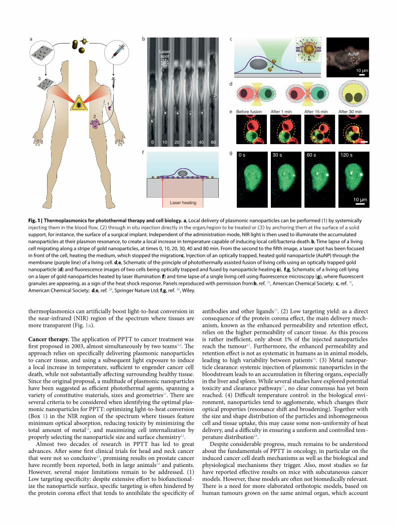
thermoplasmonics can arti�cially boost light-to-heat conversion in the near-infrared (NIR) region of the spectrum where tissues are more transparent (Fig. 1a).
Cancer therapy. �e application of PPTT to cancer treatment was �rst proposed in 2003, almost simultaneously by two teams3,4. �e approach relies on speci�cally delivering plasmonic nanoparticles to cancer tissue, and using a subsequent light exposure to induce a local increase in temperature, su�cient to engender cancer cell death, while not substantially a�ecting surrounding healthy tissue. Since the original proposal, a multitude of plasmonic nanoparticles have been suggested as e�cient photothermal agents, spanning a variety of constitutive materials, sizes and geometries11. �ere are several criteria to be considered when identifying the optimal plas-monic nanoparticles for PPTT: optimizing light-to-heat conversion (Box 1) in the NIR region of the spectrum where tissues feature minimum optical absorption, reducing toxicity by minimizing the total amount of metal12, and maximizing cell internalization by properly selecting the nanoparticle size and surface chemistry12.
Almost two decades of research in PPTT has led to great advances. A�er some �rst clinical trials for head and neck cancer that were not so conclusive13, promising results on prostate cancer have recently been reported, both in large animals14 and patients. However, several major limitations remain to be addressed. (1) Low targeting speci�city: despite extensive e�ort to biofunctional-ize the nanoparticle surface, speci�c targeting is o�en hindered by the protein corona e�ect that tends to annihilate the speci�city of
antibodies and other ligands15. (2) Low targeting yield: as a direct consequence of the protein corona e�ect, the main delivery mech-anism, known as the enhanced permeability and retention e�ect, relies on the higher permeability of cancer tissue. As this process is rather ine�cient, only about 1% of the injected nanoparticles reach the tumour11. Furthermore, the enhanced permeability and retention e�ect is not as systematic in humans as in animal models, leading to high variability between patients16. (3) Metal nanopar-ticle clearance: systemic injection of plasmonic nanoparticles in the bloodstream leads to an accumulation in �ltering organs, especially in the liver and spleen. While several studies have explored potential toxicity and clearance pathways17, no clear consensus has yet been reached. (4) Di�cult temperature control: in the biological envi-ronment, nanoparticles tend to agglomerate, which changes their optical properties (resonance shi� and broadening). Together with the size and shape distribution of the particles and inhomogeneous cell and tissue uptake, this may cause some non-uniformity of heat delivery, and a di�culty in ensuring a uniform and controlled tem-perature distribution18.
Despite considerable progress, much remains to be understood about the fundamentals of PPTT in oncology, in particular on the induced cancer cell death mechanisms as well as the biological and physiological mechanisms they trigger. Also, most studies so far have reported e�ective results on mice with subcutaneous cancer models. However, these models are o�en not biomedically relevant. �ere is a need for more elaborated orthotopic models, based on human tumours grown on the same animal organ, which account
2
1
3
AuNP
10 µm
Laser heating
0 10 20 30 40 80
Laserspot
a b
f g
c
d
e
0 s 30 s 60 s 120 s
10 µm
Before fusion After 1 min After 15 min After 30 min
Fig. 1 | Thermoplasmonics for photothermal therapy and cell biology. a, Local delivery of plasmonic nanoparticles can be performed (1) by systemically
support, for instance, the surface of a surgical implant. Independent of the administration mode, NIR light is then used to illuminate the accumulated nanoparticles at their plasmon resonance, to create a local increase in temperature capable of inducing local cell/bacteria death. b, Time lapse of a living cell migrating along a stripe of gold nanoparticles, at times 0, 10, 20, 30, 40 and 80 min. From the second to the �fth image, a laser spot has been focused in front of the cell, heating the medium, which stopped the migration. c, Injection of an optically trapped, heated gold nanoparticle (AuNP) through the membrane (purple line) of a living cell. d,e, Schematic of the principle of photothermally assisted fusion of living cells using an optically trapped gold nanoparticle (d) and �uorescence images of two cells being optically trapped and fused by nanoparticle heating (e). f,g, Schematic of a living cell lying on a layer of gold nanoparticles heated by laser illumination (f) and time lapse of a single living cell using �uorescence microscopy (g), where �uorescent granules are appearing, as a sign of the heat shock response. Panels reproduced with permission from: b, ref. 35, American Chemical Society; c, ref. 37, American Chemical Society; d,e, ref. 38, Springer Nature Ltd; f,g, ref. 30, Wiley.

for key parameters such as tumour vascularization and damage of adjacent healthy tissues and living organs. Finally, an emerging natural trend is to combine PPTT with other therapies19, such as radiotherapy, chemotherapies and immunotherapies, to achieve a synergetic therapeutic e�ect with improved overall e�cacy. Along this line, the same plasmonic nanoparticles used in PPTT have also been shown to be e�cient and stable photosensitizers capable of
locally forming — under low-intensity illumination — reactive oxy-gen species for photodynamic therapy20,21.
Beyond cancer therapy. Plasmon-based hyperthermia has recently proven to be useful beyond oncology. For example, PPTT was tested to treat atheroma, a pathology associated with an abnormal local accumulation of material (lipids, cells, tissues and so on) in artery
Box 1 | Identifying the right thermoplasmonic nanoparticles
Finding the most suitable thermoplasmonic nanoparticle for a -
ance with several constraints that can be physical, chemical or bio-logical in nature. To meet these constraints, one has three param-eters to adjust: the nanoparticle size, its shape and composition.
A common goal for all applications of thermoplasmonics is to
absorption cross-section σabs scales indeed as the volume (V) of the nanoparticle, at least for small nanoparticles in the quasistatic regime (for instance, for gold nanospheres typically smaller than 60 nm). Normalizing σabs
as it no longer depends on the nanoparticle size (at least for small
as130: Jo¼ σabsλref2πV
I, where λref = 1,240 nm is an arbitrary reference
wavelength to make Jo dimensionless. Panel a shows values of Jo
show the interest of deviating from a spherical shape to strongly
of simplicity, simulations focus here on spheroids, any deviation 131, for instance,
rods, nanoshells, nano-urchins or nanocages (panels b–g). Note that at resonance, one can show that Jo scales as the inverse of the imaginary part εʺ of the permittivity: Jo ≈ |εʹ|3/2/εʺ (ref. 130), while
the heat power density (power per unit volume) in the material is known to scale, in contrast, as εʺ|E|2
E in the latter expression scales as 1/εʺ. Remarkably, such a rationale explains
panel amaterials for both optical and thermal experiments.
compromise with other requirements and hence not always the material with the best Jo is suitable. Biological and biomedical applications require combining sizes of a few tens of nanometres, which favour cellular uptake, with low toxicity and resonances in the NIR biological transparency window. In this context, gold
chemical and physical stability is also important, rendering silver
alter the integrity of silver nanostructures132.Applications such as catalysis and thermophotovoltaics may
set other emphasis and require high structural thermal stability as temperatures of a few 100 to 1,000 °C are easily reached. Non-spherical gold nanoparticles, for example, reshape into spheres already at a temperature of 150 °C, altering their optical properties. While this problem can be overcome by transient heating (using a pulsed laser, for instance), embedding the nanoparticle within a matrix or coating it within a shell130, refractory materials (such as TiN and ZrN) can be more suitable
100 nm 50 nm
101
102
103
AuTa
CuTiN
AgZrN
Resonance wavelength (nm)500 600 700 800 900 1,000
Ellipsoids
Joul
e nu
mbe
r, Jo
1
2
3 4
56
23
4 5 6 7 8
2
2
3
4 5 6 7
1
2
3
4
5 6 7 84
5
67 8
23
4 5 6 7 60 nm 200 nm 20 nm
100 nm
g
da
e
b
f
c
Optimizing the performance of thermoplasmonic nanoparticles. a, Joule numbers calculated for ellipsoids made of di�erent materials (light polarization along the longitudinal axis), and for di�erent aspect ratios, from 1 to 8. b–g, Scanning electron micrographs of gold nanorods (b), silica–gold core–shell nanosphere (c), gold nanocages (d), gold nanostar ( e), Au–Pd nanocrystals ( f) and TiN nanoparticles ( g). Panels adapted with permission from: a, ref. 130, American Chemical Society; b, ref. 133, American Chemical Society; c, ref. 134, Dove Medical Press; d, ref. 135, American Chemical Society; e, ref. 136, American Chemical Society; f, ref. 81, American Chemical Society; g, ref. 137, RSC.

walls, leading to a restriction of blood �ow, increasing the risk of a heart attack. In 2015, successful phase II clinical trials were car-ried out, demonstrating a substantial regression of atheroma by PPTT22. Atheroma therapy does not su�er from some of the limit-ing constraints of cancer therapy. In particular, unlike for cancer-ous tumours, eliminating the whole atheroma is not a stringent requirement as partial elimination is already bene�cial as soon as it improves the blood �ow. Note that similar emerging therapies using PPTT, such as the treatment of retinal degenerative disease23, hair removal24 and acne treatment25, have also been proposed.
Sterilization and disinfection. Similar to cells, most bacteria viabil-ity is altered when experiencing a temperature greater than 45 °C. One can thus follow a similar PPTT strategy for applications to sterilization and disinfection. In 2006, a method to selectively kill
gold nanoparticles before shining them with nanosecond-pulsed laser light, was proposed26. �is concept is particularly relevant to the �eld of disinfection of surgical implants. Implant contamination has indeed become a major health issue, especially when bacterial colonization ends up forming a bio�lm making them even more resistant to antibiotic treatment. A �rst proof of principle show-
27 and this concept has recently been extended to surgical implants28. In this last study, to avoid the issues related to systemic injection of nanoparticles, such as the unspeci�c nanoparticle delivery and clearance, the authors proposed to have them directly anchored at the surface of the implant, a polymer mesh used in hernia repair,
which was chemically modi�ed to host a high-density monolayer of gold nanorods (Fig. 1a). PPTT treatment, in the form of a train of broadband light pulses, showed clinically relevant bio�lm degra-dation, associated with the alteration of the bio�lm superstructure.
Sterilization of medical instruments using a thermoplasmonic approach was proposed in 201329. In this proof-of-principle experi-ment, solar radiation was focused on a high-concentration aque-ous solution of gold nanoshells. �e formed water steam was extracted through a nozzle and sent to a sterilization module. �e authors reached a sterility level compatible with the Food and Drug Administration regulation. While plasmonic nanoparticles as absorption agents are advantageous in biomedicine, because they allow the generation of local heating through tissue using NIR illu-mination, their bene�t over black absorbers made of dyes or carbon black is not always obvious for other applications involving broad-band illumination, such as solar light, as discussed in Box 2.
Cell biologyMore fundamentally, all building blocks of living cells are highly temperature responsive: lipid layers can undergo phase transitions, proteins can unfold, DNA can melt and so on. Investigating all these
sample, which is normally achieved by combining resistive mac-roscopic heating and thermocouple measurements. Selective laser
-ful alternative approach. Along with the use of plasmonic nanopar-ticles as e�cient light absorbers, this approach o�ers three major bene�ts compared with resistive macroscopic heating. (1) Reduced
Box 2 | Plasmonic versus non-plasmonic heating
-
four properties of plasmonic materials: the absence of thermo-/ photobleaching (unlike dyes), a high density of free electrons due to their metallic nature, low losses for noble metals (see Box 1) and the existence of resonances that enhance light–matter in-
-
nanochemistry, nanosurgery, photothermal and photoacoustic imaging. Plasmonic heating is also relevant to applications re-
photoacoustic imaging), typically on the nanosecond scale, as the heating timescale varies as the square of the size of the heat source.
Conversely, when aiming for macroscale heating, the interest
cross-section σabs and the absorber size are no longer the only c also becomes an
important parameter one can play with to arbitrarily increase what only matters in this case: the absorbance Aas A = σabscl, where c is the molecular concentration (in molecules per m3) and l is the thickness of the system. In this context, the presence of plasmonic resonances is not necessary to achieve near 100% absorption. Other materials, such as chromium (used for microscale thermophoresis studies102) or even black paint or
by increasing c, near-100% absorption at low cost. Moreover, these black materials feature a high absorbance over a much larger
illumination, for instance, for solar-light-based applications.
nanoparticles for macroscale heating is never relevant, and can
be always replaced by cheaper black materials. For instance,
in cell biology (see ‘Cell biology’) or for white and colourful three-dimensional printing (see ‘Additive manufacturing’). Using
organisms for hyperthermia applications, because of their friendly biofunctionalization properties and biocompatibility. However, for applications involving broadband solar absorption, the interest of using plasmonic nanoparticles over carbon black is not evident.
4000
0.2
0.4
0.6
0.8
500 600 700 800 900 1,000
Gold nanorodsCarbon black
Wavelength (nm)
Abso
rptio
n
Absorption spectra of 0.01 g l−1 solutions of carbon black and silica-coated gold nanorods (12.5 nm × 50 nm), suspended in ethanol. Reproduced with permission from ref. 129, American Chemical Society.

spatial scale: coupling the heating laser to an optical microscope enables heating over a micrometric area, o�ering the possibility, for instance, to heat a single living cell while leaving the other adjacent cells una�ected for subsequent measurements30. In this way, a range of temperature conditions can be applied within a single multiwell plate or Petri dish. One can even heat subcellular compartments or single organelles, leading to the possibility to answer fundamental questions in thermal biology. (2) Reduced timescale: experiments at the microscale imply that the targeted temperature increase is achieved within around 1 ms, giving a well-de�ned time zero to study the dynamical response of a biological system, which can be faster than a second. In contrast, macroscopic heating with resis-tive heating usually involves timescales on the order of several min-utes, or several seconds at best. (3) Large achievable temperature: high local temperature increases under pulsed illumination can �nd application in nanosurgery or optoporation, as detailed in the next paragraph. Even under continuous-wave illumination, one can achieve high temperatures, unreachable using conventional macro-scopic heating without damaging the microscope or the objective lens, or a�ecting the immersion oil or the setup alignment31.
As mentioned above, the bene�ts of using gold nanoparticles with laser heating in biology was already recognized in 19991. Laser heating of gold nanoparticles enabled the researchers at that time to study the denaturation of chymotrypsin proteins up to 470 K, in the context of the photothermal treatment of pigmented cells. A decade later, experiments focused on a variety of other
32 (arti�cial �at mem-branes, giant unilamellar vesicles), DNA33, proteins and amyloid �brils34. Furthermore, more sophisticated experiments have been conducted in living cells, bacteria and eukaryotic cells, for funda-mental research, for example, for controlling cell migration35 (Fig. 1b), cell optoporation36 and transfection37 (Fig. 1c), cell fusion38 (Fig. 1d,e) and heat-shock response30 (Fig. 1f,g), and for applications, such as nanosurgery39. Plasmon heating can also be used to locally release molecules within cells40: biomolecules, drugs and genes can be directly loaded at the surface of a plasmonic cargo or encapsu-lated in micelles, liposomes or even red blood cells, decorated with plasmonic nanoparticles40–42. Upon illumination, the local tempera-ture increase induces a molecular release via either bond breakage or phase change. �ese approaches, however, require heating to remain below a few kelvin to avoid a�ecting cellular metabolism. Remarkably, a similar approach can also be applied to release cells anchored to a plasmonic substrate43.
All these studies represent the onset of a fast-growing �eld of research that could be named ‘single-cell thermal biology’, and where thermoplasmonics is expected to have a major role.
Photothermal and hot-electron chemistryTemperature is a common parameter to control chemical transfor-mations. According to the Arrhenius law, the rate constant K of a chemical reaction and its molar activation energy Ea are related via an exponential law: K ∝ exp(−Ea/RT), where T is the temperature and R is the gas constant. Plasmonic nanoparticles can be used to boost chemical reactions based on such a local temperature varia-tion but also with optical near-�eld enhancement and hot-carrier injection44. �is paradigm represents an important research area in plasmonics with pivotal, societal and economic impact as it could contribute to cleaner energies45. In particular, hot-electron injection involves metal electrons of a few electronvolts capable of driving reactions that could not occur otherwise via conventional heating. But in this approach, even if optical heating is not the process of interest, it has to be carefully considered to ensure the validity of the underlying catalytic mechanism. For these reasons, the rest of the section will be divided into two parts: in the �rst part, plasmonic heating is the targeted process to assist chemical reactions46; whereas the second part is oriented towards hot-carrier plasmonics45.
Photothermal chemistry. �e use of thermoplasmonics in chem-istry raises an important question: what are the speci�c bene�ts of using light-induced plasmonic heating, over regular heating with a hot plate? First, thermoplasmonics o�ers a spatial con�nement that enables micro-/nanopatterning of the chemical products on a surface47. Second, plasmonic heating can yield a high temperature increase under moderate illumination intensity up to a superheated state of the solvent. Upon pulsed illumination, the temperature increase is so brief and con�ned around each nanoparticle that no macroscopic boiling is observed even at nanoparticle temper-atures close to 300 °C, the spinodal temperature of water48. Even continuous-wave illumination can yield a superheated state of water as high as 220 °C (ref. 31). In the case of a plasmonic substrate in con-tact with a gas phase (no liquid), the temperature of the illuminated area can be locally increased by several hundreds of degrees, while the chamber itself remains at moderate temperature, making the experimental conditions less stringent and the reaction less energy consuming47. A third advantage of plasmonic nanoparticles is that they can also feature catalytic activity, especially when sized down to a few nanometres49.
Applications in photothermal chemistry can be divided in three categories: (1) plasmonic-assisted chemical vapour deposition (PACVD) in a vacuum chamber, (2) thermoplasmonic-assisted catalysis in the gas phase and (3) thermoplasmonic-assisted enhanced reactivity in solution. Chemical vapour deposition (CVD) is a thin-�lm deposition technique on a substrate at high temperature, under vacuum, from volatile molecules that react and adsorb on the substrate. As an alternative to homogeneous resis-tive heating, it was proposed to locally heat the substrate with light to spatially control the growth. PACVD was �rst reported in 2006 and demonstrated the micropatterning of TiO2 and PbO on glass (Fig. 2a,b)50. Nanoscale subdi�raction PACVD can also be achieved when single nanoparticles are heated, as corroborated in 2007 with the growth of silicon, germanium or carbon nanowires (Fig. 2c,d)47. In the gas phase, the association of gold nanoparticles and metal oxides such as titania (TiO2) has been used since 1987 to yield a sub-stantial catalytic activity even at moderate temperature increase49. In 2008, it was proposed to heat such a system with light instead of with a hot plate, bene�ting from the strong plasmonic absorp-tion of the gold nanoparticles themselves51. In that case, heating was limited to the catalytically active area, reducing the overall energy consumption. In 2010, the bene�t of laser heating over global resis-tive heating was demonstrated to yield a gain of two to three orders of magnitude52.
�is approach has been extended to the liquid phase since 2009 (Fig. 2e,f)53. Di�erent strategies have been used to achieve high temperatures that cannot be normally reached in water without boiling, for instance, using a nanosecond-pulsed laser54 and using a continuous-wave laser on a glass substrate. �is second approach can yield temperatures as high as 200 °C without boiling the �uid, opening the path for hydrothermal chemistry at ambient pressure, avoiding the use of an autoclave and a closed reaction medium (Fig. 2g,h)31,55.
Plasmonic hot-carrier chemistry. �e absorption of a photon by a metal nanoparticle promotes one electron from the conduction band by an energy that equals the photon energy, that is, of the order of a few electronvolts in the visible–NIR range. �e associ-ated electron and hole created this way are known as ballistic hot carriers. While these hot carriers only live for tens of femtosec-onds due to collisions with the other unexcited electrons of the particle48, they can, during their short lifetime, interact with sur-rounding molecules to drive chemical reactions45. In particular, they can be transferred to adsorbed molecules to induce redox pro-cesses. Along with this indirect charge transfer mechanism, a direct charge transfer mechanism has been proposed, where the excited

hot electron directly populates a state of a molecule adsorbed on the nanoparticle surface45. Originally proposed in 200456, the idea of plasmonic hot-carrier chemistry really attracted the interest of the community only from 2011, motivated by some seminal articles (refs. 57,58, among others) that stressed the colossal envisioned indus-trial and societal impacts, in particular for driving chemical reac-tions such as water splitting58 for dihydrogen production, or many redox reactions of industrial interest45. A�er more than a decade
of extensive activities, the �eld of plasmonic hot-carrier chemistry experiences heated debates regarding the actual underlying mecha-nisms8,59,60. In some seminal studies, the reaction rate enhancement by a hot-carrier mechanism was questioned in favour of a simple temperature increase, unexpectedly important due to the presence of collective thermal e�ects (see Box 3). �is awareness appeared around 2018 and since then, extensive e�orts have been made to address this question60. �ermoplasmonics is now playing an unforeseen but substantial role in this �eld of research, as it is key to better interpret experimental observations.
Solar light harvesting�e power of plasmonic particles to convert electromagnetic energy into heat is also of potential interest in solar light harvesting. �ree major �elds with two di�erent approaches arise. On the one hand, nano�uids consisting of solutions of plasmonic particles are used for volumetric heating and accordingly employed for solar energy collection and water desalination. On the other hand, thermopho-tovoltaics addresses the heat management in general and provides advanced concepts for solar energy conversion into electricity.
�ermophotovoltaics. Photovoltaics with standard p–n junc-tions is limited by the fact that, from the broad distribution of solar photon energies, only the bandgap energy contributes to generate electron–hole pairs. �e excess energy of photons above the bandgap is converted into heat and subbandgap energy is lost completely. For this reason, the photovoltaic conversion in these simple systems is limited to around 41%, known as the Shockley–Queisser limit61. �is limit can be overcome with costly multi -layer structures62 with multiple bandgaps. A di�erent and very promising approach is to get rid of thermal losses by turning all electromagnetic energy into heat63. In thermophotovoltaics, solar electromagnetic energy is �rst converted into heat by an e�cient absorber connected to a thermal emitter releasing the heat into thermal emission with an energy close to the bandgap of a pho-tovoltaic cell and thus converting almost all solar energy into electricity (Fig. 3a,b). Limits of solar energy conversion beyond 80% have been predicted63 and e�ciencies up to 29% have so far been demonstrated64. To reach theoretical e�ciencies, research requires overcoming three major challenges. First, highly e�cient absorbers for the solar spectrum have to be designed, which are able to convert the absorbed energy into heat. Second, the visible absorber has to be interfaced to an infrared thermal emitter that generates the thermal radiation for a photovoltaic cell with a low bandgap around 0.8–1.1 eV. �is energy range requires an emitter temperature between 1,000 K and 2,000 K, which sets the stron-gest limitation on the choice of possible materials.
Owing to their e�cient light-to-heat conversion (Box 1), plas-monic materials are a suitable class of materials for thermophotovol-taics. Yet, metals are not always able to withstand the high required temperatures (Box 1) although some studies reported e�cient absorber structures of gold stable up to 1,000 K owing to a dielectric coating65. �ermally stable absorber materials are cermets, a com-bination of ceramics and metal particles where the metal is respon-sible for absorptive properties66. Cermets have been fabricated from metal particles such as nickel embedded in Al2O3. �ese materi-als yield a high absorbance of the solar range but low emittance at wavelengths above 3 μm (Fig. 3c,d)67. Refractory materials may have plasmonic properties in the visible range68 but also a large imaginary part of the permittivity (W, TiN, Mo and so on), which is counter-intuitively detrimental for thermoplasmonics applications (Box 1). Well-designed metamaterial structures may be created to modify the electromagnetic density of states tailoring absorption and emission spectral shape (Fig. 3e,f)60,70. Metamaterials made of TiN (ref. 71) or W-HfO2 have shown to be stable up to 1,400 °C (ref. 72) and photonic crystals of refractory materials have been realized
Nanorod
Precursormolecule
AuNP
Laser
Substrate
Substrate
AuNP
Laser
AuNPSubstrate
Substrate
Flow
Laser
Laser
InCl3 + 3OH– In(OH)3(s) + 3Cl–200°C
a
1 µm
2 µm
b
c d
e f
g h
20 µm
200 nm
Fig. 2 | Thermoplasmonics for chemistry. a,b, PACVD. a, Schematic of a PACVD process showing a template of nanoscale gold dots (yellow spheres) exposed to a gaseous environment containing the CVD precursor (top left molecule). A laser illumination is locally heating the system to yield the formation of nanowires (brown). b, Scanning electron microscopy (SEM) image showing an example of an area of nanowires grown by PACVD. c,d, Subdi�raction PACVD. c, Schematic showing the illumination of gold nanoparticles (yellow spheres) leading to the growth of gold-catalysed nanowires along the path (dashed arrows) of the illumination beam (red beam). d, SEM image of the resulting isolated germanium nanowires. e,f, Liquid-phase plasmon assisted catalysis. e, Schematic of laser light heating a gold nanoparticle layer in contact with a liquid (light blue) between two substrates (grey) to form a gas bubble (white) that reacts on the nanoparticles acting as catalysts. f, Optical image of the formed bubble under illumination. g,h, Plasmon-assisted hydrothermal chemistry. g, Schematic showing the laser illumination of a layer of gold nanoparticles on a substrate (grey) enabling the heating of an aqueous solution at around 200 °C to allow the growth of indium(III)-hydroxide crystals (rectangles). h, SEM image of the resulting crystal products. Panels adapted with permission from: a,b, ref. 50, American Chemical Society; c,d, ref. 47, American Chemical Society; e,f, ref. 53, American Chemical Society; g,h, ref. 55, American Chemical Society.

to form thermophotovoltaic emitter structures69. An even more promising yet technologically more challenging approach is to use the near �elds of the infrared emitters73. If one manufactures tiny gaps, on the order of a wavelength, between the thermal emitter and receiver, the evanescent near �eld of the thermal excitation can strongly contribute to the electromagnetic energy transfer by opti-cal tunnelling73. Such near �elds of surface plasmon polaritons do not follow the far-�eld blackbody statistics but have a high density close to the plasmon frequency74 and can thus be highly spectrally selective75. While there is a growing number of promising theoreti-cal analysis of near-�eld thermophotovoltaics with plasmonic struc-ture, including graphene74,76, experimental realizations are still few77 but will certainly increase in the near future.
Nano�uids for solar energy harvesting. Nanoparticle solutions were �rst coined nano�uids in 1995. Since then, there has been large interest with substantial controversy regarding their thermal properties78. Most of the recent interest in nano�uids containing various metal particles stems from their absorptive properties and their use in solar thermal energy conversion for thermal power generation, desalination of sea-water or water splitting. For these applications, an absorbing material would be brought in contact with a working �uid, which is o�en a liq-uid, to transfer the heat from the absorber to the liquid. �e advantage
of nano�uids is that the absorber is now directly brought into the liq-uid, providing a volumetric and not an interfacial heating79,80. In the case of plasmonic materials, however, the absorption spectra have to extend to larger spectral ranges by using speci�c structures or materi-als (Box 2). Suggestions of using Janus particles — polymer or silica particles with a half-sided metal coating — have been made to cover a large fraction of the solar spectral range79. Other approaches involve metal nitrides81 or mixtures of di�erent gold nanoparticle sizes82 and shape83. Such immersed nano-absorbers could indeed enhance the e�ciency of solar collectors79,80; however, their applications for desali-nation84 or water splitting seem ine�ective79.
Water desalination. About 97% of water on Earth exists in form of seawater, and freshwater availability is one of the global challenges for humanity. Cost-e�ective seawater desalination could there-fore provide a sustainable source of fresh water for the future and sets a technological challenge. Techniques to desalinize water can be roughly split into two classes relying either on membranes (for example, for reverse osmosis) or on thermal processes85,86. Reverse osmosis plants are currently most e�cient, requiring less then 3 kW h energy per cubic metre of desalinized water, a factor of three above the thermodynamic limit based on the free energy change when mixing salt and water86. Desalination based on solar-thermal
Box 3 |
Frequently, the temperature increase in a system composed of plas-monic nanoparticles is estimated using the simple expression138:
δT ¼σabsI4πκReq
ð1Þ
which gives the temperature increment of a single nanoparticle, of absorption cross-section σabs, where I is the irradiance (power per unit area) of the incoming light, κ is the thermal conductivity of the surrounding medium and Req is the equivalent nanoparticle radius. However, this single-nanoparticle expression can yield wrong estimations when several nanoparticles are illuminated at once, as the temperature increase experienced by a nanoparticle j also stems from the neighbouring nanoparticles i heating their environment139,140:
δTj ¼σabs;j I4πκReq
þX
i≠j
σabs;i I4πκ ri rj
ð2Þ
Counterintuitively, the last term of equation (2) can become
δT(r) ∝ 1/r, r being
distribution of nanoparticles: at a constant irradiance I, varying the illuminated area from 300 nm to 2.5 cm leads to the appearance of
main features: (1) a strong overall temperature increase, several orders of magnitude higher than what equation (1) predicts and (2) a completely uniform temperature distribution, despite the nanoscale nature of the heat sources and their large separation distance.
estimated by calculating the dimensionless number139 ξ = p2/3RD, where p R is the radius of the nanoparticle and D is the characteristic size of the
two-dimensional nanoparticle distribution under illumination. ξ < 1. Remarkably,
not only the nanoparticle interdistance matters, but also their number (via Dno counterpart in optics. In practice, neglecting these collective
heating and misleading interpretations of experimental data60.
0
1.5 × 10 –3
δT (K)
δT (K)
0
18.4
D = 2.5 cm
I = 100 W cm –2
D = 300 nmI = 100 W cm –2
a
c d
b
Numerical simulations illustrating collective photothermal effects in plasmonics. a, Schematic of the system: a uniform, not-necessarily periodic, two-dimensional nanoparticle distribution (R = 7 nm, p = 200 nm), illuminated with a beam of irradiance of 100 W cm −2 and a diameter of D = 300 nm ( ξ ≈ 6). b, Associated temperature increase showing localized hotspot, consistent with ξ > 1. c, Schematic showing the same system of nanoparticles as before, with a beam radius of D = 2.5 cm ( ξ ≈ 10−4). d, Associated temperature increase that is fully uniform, consistent with ξ << 1.

processes involves the heating of seawater with subsequent conden-sation84. Solar-thermal desalination requires �rst of all absorber materials with a high e�ciency of converting the harvested solar photons into heat. Two materials are commonly considered — carbonaceous materials and plasmonic materials. Carbon-based materials such as carbon black, graphite, graphene or other forms of graphitic materials are o�en used as broadband absorber materi-als relying on the π-electron absorption (Box 2) with absorptivity of 80% to almost 100% over a wide range from the ultraviolet to the NIR (up to 2.5 μm wavelength)87,88. Plasmonic materials have recently gained attention as an e�cient absorber material with e� -cient light-to-heat conversion, but is not always cheap89,90. To cover the whole spectral range of solar illumination, plasmonic materi-als require a size dispersion or more complex geometries, such as particle aggregates or porous structures. Absorptivities of 95% and higher have been reached with gold, silver or aluminium-based materials (Fig. 3g–i)89–91. Using such absorbers, solar steam gen-eration and desalination has been demonstrated. Researchers have recently reported a self-assembled aluminium nanoparticle-based porous system for an e�ective desalination90. Others have decorated natural wood with plasmonic metals to create high solar conver-sion e�ciencies for steam generation91. Yet, the biggest issue for solar-thermal desalination is not primarily related to the e�cient light-to-heat conversion but rather to the thermal management. Desalination by evaporation requires a supply of the latent heat of evaporation of water. �is energy is almost three orders of mag-nitude higher than the free energy of mixing84,86. Without a recov-ery of the latent heat during the condensation process, the energy requirement would be two to three orders of magnitude higher than the reverse osmosis processes. Even with an e�cient latent heat
recovery, a speci�c energy consumption of about 40 kW h m−3 is required, which is still considerably above reverse osmosis processes driven by photovoltaic electricity86. �us, the future application of solar-thermal desalination, including approaches based on e�cient plasmonic absorbers, seems to be limited to areas of hypersaline solutions with large salinity where high water recovery is required84. Here, again, as for all applications based on solar absorption, the interest in plasmonic nanoparticles over black absorbers made of dyes or carbon black is not obvious, as discussed in Box 2.
Soft matter and fluidsMany experiments in plasmonics involve a liquid medium, the dynamics of which is highly temperature dependent. Temperature variations in liquids can yield a rich variety of processes, such as enhanced Brownian motion, thermo-viscous e�ects, convection, phase transition (such as bubble formation, polymer melting, sin-tering and so on), thermo-osmosis and thermophoresis. �is sec-tion is devoted to explaining the interest in studying so� matter in thermoplasmonics with a special focus on four e�ects: thermally induced �ows, thermophoresis, bubble formation and powder sin-tering for additive manufacturing, the latter being a rare example of industrial application of thermoplasmonics.
Temperature-induced liquid �ows. Temperature gradients in �u-ids can generate �uid convection for which the overheated parts are moving upward. �e magnitude of this e�ect depends on the tem-perature increase, the size of the heat source and the con�nement of the �uid along the vertical direction. Convection of liquids induced by optically heated plasmonic nanostructures on a substrate has been studied numerically as a function of the liquid �lm thickness92.
400 nm
Solar spectrum
Ligh
t em
issi
on (
a.u.
)
Wavelength (µm)1.00.3 10
1.0
0.5
Absorbance
Wavelength (µm)1.00.3 10
Abs
orba
nce
0
1
Solar spectrum
IR emission
b
a c
e
g h i
f
d
Part
icle
siz
e, w
(nm
)
w
50
250
300
200
150
100400 °C radiation
Au/D-NPT
Wavelength (µm)1.0 1.5 2.0 2.5
IR emitter
PV cell
Plasmonic visible absorber
Solar radiation200 nm
400 °Cradiation
200 nm
Fig. 3 | Thermoplasmonics for solar-light harvesting. a, Sketch of principle of a plasmonic-assisted thermophotovoltaic (PV) cell. b, Solar spectra irradiance and 400 °C blackbody spectral radiance, per unit wavelength (arbitrary units). c, SEM image of nickel nanochains. d, Absorbance spectrum of a Ni nanochain solution, compared with the solar spectrum and the black body spectrum at 400 °C (grey dashed lines) depicted in b. e,f, SEM image of gold nanopads of size w deposited on an Au-Al2O3 substrate ( e), along with the absorbance spectra as a function of w ( f). g–i, Self-assembled aluminium porous structure decorated with gold nanoparticles (Au/D-NPT) for broadband absorption ( g), associated SEM image ( h) and photograph of the Au/D-NPT solution ( i). Panels adapted with permission from: c,d, ref. 67, AIP; e,f, ref. 70, AIP; g–i, ref. 89, AAAS.

For liquid heights of above 100 μm, several hundred nanometres
micro�uidic channels (<10 μm high) convection is negligible. Yet, convective �ows in such settings can be boosted by combining temperature-induced electrical conductivity changes with a.c. elec-tric �elds93. �is approach, called electro-thermoplasmonics, has been exploited to load plasmonic traps as well as to overcome the di�usion limit in localized surface plasmon resonance biosensing (Fig. 4a,b)94. Temperature gradients above heated metal �lms also lead to changes in the local viscosity. Moving the heating spot on the surface generates a thermo-viscous motion of particles in the trav-elling viscosity gradient that can be used without plasmonic struc-tures to generate �ows in living cells95. Besides that, the generation of temperature gradients by plasmonic nanoparticles along solid/liquid boundaries also naturally leads to thermo-osmotic �ows96 with speeds easily reaching 100 μm s−1 being strongly con�ned to the interfacial region and the driving source of thermophoresis.
Microscale thermophoresis. �ermophoresis, also known as the Ludwig–Soret e�ect, denotes the migration of solutes (colloids, molecules) in �uids due to the existence of temperature gradients, while the �uid is at rest97,98. �e magnitude of this e�ect for a given solute, usually occurring from hot to cold regions, is given by the thermophoretic mobility DT, linking the solutes thermophoretic velocity v and the temperature gradient such that v = −DT∇T (ref. 98). �e strength of this e�ect compared with the solution di�usion coe�cient D can be better judged using the Soret coe�cient ST = DT/D. �e mechanisms of thermophoresis are well under-stood in gases and in most liquids except for water. In liquids, thermophoresis is driven by thermo-osmotic �ows arising from the perturbation of the liquid–solid interfacial interactions by the temperature gradients96.
�ermophoretic e�ects have been studied for decades, usually with infrared lasers to heat the solvent directly99,100 or other absorb-ing structures101, and applied as a tool to study binding kinetics in ensembles of proteins by just using the fact that proteins change their thermophoretic mobility upon binding102.
A chromium-coated surface as an absorbing medium was �rst used to generate microscale temperature gradients and to manip-ulate colloids103 with a reduced laser power down to 1 mW. �e opaque metal layer, however, precludes the use of optical micro-scope studies in transmission. �ey also yield weaker tempera-ture gradients. Herein lies the bene�t of plasmonic nanoparticles (Box 3). In 2013, it was proposed to apply thermoplasmonics to microscale thermophoresis experiments. Static and dynamic tem-perature �elds generated by controlled heating of plasmonic gold nanostructures at a surface allowed researchers to trap single 200 nm polymer colloids in solution104. In 2015, the trapping of even single or a well-de�ned number of multiple λ-DNA molecules in water with the help of temperature �elds generated by a feedback algo-rithm105 was achieved (Fig. 4e–g).
Other work presented a platform that is based on thermally generated electric �elds (Fig. 4h–j)106. Surfactant molecules with dissociated counterions migrate with di�erent mobilities in the temperature gradient to set up a thermoelectric �eld to move other objects. By using this e�ect with a nanoplasmonic substrate (essentially a dense nanoparticle �lm), the authors could trap gold nanoparticles of di�erent shapes, colloids, vesicles or cells107. While not all of the e�ects in these experiments are understood, they dem-onstrate the power of using thermophoresis as a tool for material science and analytics. �ese demonstrations are now at a point where thermophoresis with plasmonic structures is turned into a tool, for example, to study the aggregation of proteins, which is of foremost interest in the study of neurodegenerative diseases34.
60
0
4
8
12
12090Time (min)
Peak
shi
ft (n
m)
c
a e
f
h i
j
gb
d
5 µm
5 µm
OffOn Heating
laser spotGold layer
2 µm
20 µm
Focalposition
Withoutbubble
Withbubble
100 µmBubble
Coated particlesTem
peratureMicelles
Cl– ions–
–––
– –
–
++ +
+
++
+
+ +++
+
+ ++
ΔT
E a.c.
Fig. 4 | Thermoplasmonics in liquids. a, Electrothermoplasmonic sensors, which e�ciently transport target molecules (Y-shaped) to plasmonic sensing structures (gold disk) by a combination of thermal (colour gradient) and a.c. electric �elds ( Ea.c.; arrows). b, The plasmonic resonance shift of the sensor for di�erent operation modes, with (green) and without (brown) using the electrothermoplasmonic e�ect. c, Recon�gurable plasmo�uidic lens. A laser-induced bubble modi�es the surface plasmon propagation in a thin gold �lm. d, Marangoni �ows around a bubble generated by an optically heated gold nanoisland �lm in water. e, Thermophoretic trapping of single λ-DNA molecules in dynamic temperature �elds generated by focusing a laser on the rim of a hole in a thin gold �lm. f,g, Snapshots of the λ-DNA at di�erent times ( f) and the position statistics (blue to red colour map) in the trap ( g). The trap edge is indicated by the dashed line. h, Opto-thermoelectric tweezers based on a thermoplasmonic heating of a nanoplasmonic substrate and a thermo-electric charge separation. i,j, Optothermal assembly of colloidal superstructures based on thermal-induced depletion forces using non-spherical and spherical ( i) as well as spherical particles of di�erent size ( j), as observed in bright �eld microscopy. Panels adapted with permission from: a,b, ref. 94 , American Chemical Society; c, ref. 123, Springer Nature Ltd; d, ref. 113, Springer Nature Ltd; e–g, ref. 105, American Chemical Society; h–j, ref. 106, AAAS.

Even if there is still some way to go to reach the goal of predicting thermophoretic e�ects from the fundamental interfacial properties, its importance for micro�uidic analytical processes, the design of active matter108 and the transport of molecules in systems involving plasmon assisted chemistry and catalysis will become exponentially important in the near future.
Microbubble formation. When heating plasmonic nanoparti-cles in a liquid, a bubble is bound to appear above a certain light power. �is e�ect has been studied in detail for two decades under nano- to femtosecond-pulsed illumination109,110 for applications in biomedicine, such as cancer therapy, photoacoustic imaging or optoporation. �e interest in continuous-wave illumination is much more recent. When one or several plasmonic nanoparticles are distributed on a substrate and illuminated in a liquid, above a certain light power, a bubble can nucleate at the interface111. �is ‘surface bubble’ (SB) usually remains attached (at least in the case of water) to the liquid/solid interface, featuring a truncated spherical shape. �e beauty of this system is the rich and unexpected physics: in water, an SB forms around 200 °C (not at 100 °C)31, it is stable for several seconds to several minutes at ambient temperature31,112, it yields dramatic convection in the surrounding liquid113 (Fig. 4d) and it can be moved on the substrate at high speed by moving the heating laser. All these singular phenomena led to original funda-mental articles and some practical applications. For instance, an SB provides an interesting tool in the context of opto�uidics114,115, for microscale manipulation of beads116,117, biomolecules118, ions119 and liquids120,121; and also for the manipulation of light, such as for light modulation122 or focusing123 (Fig. 4c). Many studies have been devoted to the understanding of the underlying physics of SBs124, and of the associated e�ects. In this �eld of research, the use of plas-monic nanoparticles is not always justi�ed or necessary (Box 2). As a matter of fact, many studies of SBs have been performed on metal layers or on indium tin oxide. One can expect many other studies in this �eld as the behaviour of SBs (temperature onset, �uid convec-tion, dynamics, life time and so on) strongly depends on the sur-rounding �uid. Experiments in alcohols and alkanes have already been reported very recently125,126.
Additive manufacturing. Another �eld that can greatly bene�t from thermoplasmonics is three-dimensional printing, also known as additive manufacturing. �ree-dimensional printing has quickly developed over the past decade and is becoming the preferred pro-duction approach for many objects of our everyday life. �ere exist many three-dimensional printing approaches, each having pros and cons. One of them relies on powder sintering127, which consists of scanning a high-power CO2 laser to directly heat up polymer
particles to sintering. To overcome the use of a bulky and expensive infrared laser, it was proposed to use photothermal sensitizers, in particular, carbon black, combined with cheap and compact visible/NIR laser sources128. �e main issue of using carbon black though is the resulting dark grey–black colour of the print (Box 2), which is incompatible with colourful �nal objects (or at least would require extensive post-processing).
�ermoplasmonics o�ers an appealing way around carbon black limitations. By designing an invisible ink, made of plas-monic nanoparticles resonant in the NIR of the spectrum, one can achieve white prints, o�ering the possibility to add standard dyes to achieve colourful prints (Fig. 5)129. Albeit promising, the use of plasmonics in additive manufacturing raises several important questions. Plasmonic metals have non-zero absorption in the visible range (interband transitions and/or transverse mode in the case of nanorods), which limits how white the print can be. When project-ing market applications, there is also an issue related to production cost of the ink. For both reasons, there is a need to �nd some alter-native materials.
Outlook�ermoplasmonics turned into an asset what has long been con-sidered as a major drawback of plasmonics, namely metal losses. Atypically, this Review tackles concepts related to many �elds of science at once, namely cell biology, chemistry, medicine, material science, optics, thermodynamics, �uid dynamics, nanooptics, solar energy harvesting and more. �is diversity stems from the ubiquity of thermal-induced e�ects in all �elds of science. Such richness is the �ngerprint of thermoplasmonics, which has, over the past two decades, become a driving force to create synergies between di�er-ent scienti�c disciplines. For these reasons, we foresee that thermo-plasmonics will remain a blossoming �eld of research for decades, where the only limitation in the development of other applications will be the imagination.
Received: 27 January 2020; Accepted: 8 June 2020; Published online: 17 August 2020
References 1. Hüttmann, G. & Birngruber, R. On the possibility of high-precision
denaturation of proteins. IEEE J. Sel. Top. Quantum Electron. 5, 954–962 (1999).
2. Boyer, D., Tamarat, P., Maali, A., Lounis, B. & Orrit, M. Photothermal imaging of nanometer-sized metal particles among scatterers. Science 297, 1160–1163 (2002).
3. Pitsillides, C. M., Joe, E. K., Wei, X., Anderson, R. R. & Lin, C. P. Selective cell targeting with light-absorbing microparticles and nanoparticles. Biophys. J. 84, 4023–4032 (2003).
4. tumors under magnetic resonance guidance. Proc. Natl Acad. Sci. USA 100, 13549–13554 (2003).
5. Ghosh, P., Han, G., De, M., Kim, C. K. & Rotello, V. M. Gold nanoparticles in delivery applications. Adv. Drug Deliv. Rev. 60, 1307–1315 (2008).
6. Li, W. & Chen, X. Gold nanoparticles for photoacoustic imaging. Nanomedicine 10, 299–320 (2015).
7. as nano-sources of heat. Laser Photon. Rev. 7, 171–187 (2013).
8. (Cambridge Univ. Press, 2017). 9.
Plasmonic heating of nanostructures. Chem. Rev. 119, 8087–8130 (2019). 10. Govorov, A. O. & Richardson, H. H. Generating heat with metal
nanoparticles. Nano Today 2, 30–38 (2007). 11. Nat. Rev.
Mater. 1, 16014 (2016). 12. Morales-Dalmau, J., Vilches, C., De Miguel, I., Sanz, V. & Quidant, R.
Optimum morphology of gold nanorods for light-induced hyperthermia. Nanoscale 10, 2632–2638 (2018).
13. Pilot study of AuroLase(tm) therapy in refractory and/or recurrent tumors of the head and neck. ClinicalTrial NCT00848042 ClinicalTrials.gov https://clinicaltrials.gov/ct2/show/NCT00848042 (2017).
2 cm
a b
Fig. 5 | Thermoplasmonics in manufacturing. a, Artistic illustration of the sintering of adjacent polymer beads induced by light-to-heat conversion in gold nanorods. b, Example of a colourful sintered object printed with a PA12 powder using an invisible gold nanorod ink combined with standard dyes. Figure adapted with permission from ref. 129, American Chemical Society.

NATURE MATERIALS
14. Ali, M. R. K., Ibrahim, I. M., Ali, H. R., Selim, S. A. & El-Sayed, M. A. Treatment of natural mammary gland tumors in canines and felines using gold nanorods-assisted plasmonic photothermal therapy to induce tumor apoptosis. Int. J. Nanomed. 11, 4849–4863 (2016).
15. Chem. Soc. Rev. 45, 2440–2457 (2016).
16. Nichols, J. W. & Bae, Y. H. EPR: Evidence and fallacy. J. Control. Release 190, 451–464 (2014).
17. ACS Nano 13, 5785–5798 (2019).
18. in tissue by two-photon luminescence microscopy. Nanoscale 11, 11331–11339 (2019).
19. Coord. Chem. Rev. 387, 299–324 (2019).
20. structures by laser-activated gold nanoparticles. Nano Lett. 10, 4549–4554 (2010).
21. Vankayala, R., Huang, Y. K., Kalluru, P., Chiang, C. S. & Hwang, K. C. First demonstration of gold nanorods-mediated photodynamic therapeutic destruction of tumors via near infra-red light activation. Small 10, 1612–1622 (2014).
22. management of plaques: results of the NANOM-FIM trial. Nanoscale 7, 8003–8015 (2015).
23. using KV1.1 conjugated gold nanoparticles. Nano Lett. 18, 6981–6988 (2018).
24. Harris, T. & Kim, A. Hair removal with coated metal nanoparticles. US patent 14/471,331 (2014).
25. of sebaceous follicles with topically delivered gold plasmonic particles. Adv. Mater. TechConnect Briefs 2016 3, 177–179 (2016).
26. Zharov, V. P., Mercer, K. E., Galitovskaya, E. N. & Smeltzer, M. S. Photothermal nanotherapeutics and nanodiagnostics for selective killing of bacteria targeted with gold nanoparticles. Biophys. J. 90, 619–627 (2006).
27. gold nanorod localised surface plasmon resonance generated heat. Mater. Sci. Eng. C 80, 54–58 (2017).
28. Nano Lett. 19, 2524–2529 (2019).
29. using broadband light-harvesting nanoparticles. Proc. Natl Acad. Sci. USA 110, 11677–11681 (2013).
30. expression at the single cell level. Small 14, 42–47 (2018).
31. micro-bubble generation around plasmonic nanoparticles under cw illumination. J. Phys. Chem. C 118, 4890–4898 (2014).
32. Kyrsting, A., Bendix, P. M., Stamou, D. G. & Oddershede, L. B. Heat
vesicle cargo release. Nano Lett. 11, 888–892 (2011). 33. J. Phys. Chem.
C 114, 7401–7411 (2010). 34.
aggregation studies. Nat. Methods 16, 611–614 (2019). 35.
thermoplasmonic gold nanoarrays to manipulate cell adhesion. ACS Nano 6, 7227–7233 (2012).
36. optoporation and transfection of cancer cells. Biomaterials 33, 2345–2350 (2012).
37. Li, M., Lohmüller, T. & Feldmann, J. Optical injection of gold nanoparticles into living cells. Nano Lett. 15, 770–775 (2015).
38. Bahadori, A., Oddershede, L. B. & Bendix, P. M. Hot-nanoparticle-mediated fusion of selected cells. Nano Res. 10, 2034–2045 (2017).
39. Boulais, E., Lachaine, R., Hatef, A. & Meunier, M. Plasmonics for pulsed-laser cell nanosurgery: fundamentals and applications. J. Photochem. Photobiol. C 17, 26–49 (2013).
40. Parakhonskiy, B. V., Parak, W. J., Volodkin, D. & Skirtach, A. G. Hybrids of polymeric capsules, lipids, and nanoparticles: thermodynamics and temperature rise at the nanoscale and emerging applications. Langmuir 35, 8574–8583 (2019).
41. βvia light responsive capsules. ACS Nano 10, 4828–4834 (2016).
42. into the cytosol. J. Control. Release 159, 120–127 (2012).
43. Giner-Casares, J. J., Henriksen-Lacey, M., García, I. & Liz-Marzán, L. M. Plasmonic surfaces for cell growth and retrieval triggered by near-infrared light. Angew. Chem. Int. Ed. 55, 974–978 (2016).
44. Chem. Soc. Rev. 43, 3898–3907 (2014).
45. Shin, H. H., Koo, J. J., Lee, K. S. & Kim, Z. H. Chemical reactions driven by plasmon-induced hot carriers. Appl. Mater. Today 16, 112–119 (2019).
46. Qiu, J. & Wei, W. D. Surface plasmon-mediated photothermal chemistry. J. Phys. Chem. C 118, 20735–20749 (2014).
47. Cao, L., Barsic, D. N., Guichard, A. R. & Brongersma, M. L. Plasmon- assisted local temperature control to pattern individual semiconductor nanowires and carbon nanotubes. Nano Lett. 7, 3523–3527 (2007).
48. nanoparticles. Phys. Rev. B 84, 1–13 (2011).
49. Haruta, M., Kobayashi, T., Sano, H. & Yamada, N. Novel gold catalysts for the oxidation of carbon monoxide at a temperature far below 0 °C. Chem. Lett. 16, 405–408 (1987).
50. Boyd, D. A., Greengard, L., Brongersma, M., Ei-Naggar, M. Y. & Goodwin, D. G. Plasmon-assisted chemical vapor deposition. Nano Lett. 6, 2592–2597 (2006).
51. Chen, X., Zhu, H. Y., Zhao, J. C., Zheng, Z. F. & Gao, X. P. Visible-light-driven oxidation of organic contaminants in air with gold nanoparticle catalysts on oxide supports. Angew. Chem. Int. Ed. 47, 5353–5356 (2008).
52. Hung, W. H., Aykol, M., Valley, D., Hou, W. & Cronin, S. B. Plasmon resonant enhancement of carbon monoxide catalysis. Nano Lett. 10, 1314–1318 (2010).
53. Adleman, J. R., Boyd, D. A., Goodwin, D. G. & Psaltis, D. Heterogenous catalysis mediated by plasmon heating. Nano Lett. 9, 4417–4423 (2009).
54. Fasciani, C., Alejo, C. J. B., Grenier, M., Netto-Ferreira, J. C. & Scaiano, J. C. High-temperature organic reactions at room temperature using plasmon excitation: decomposition of dicumyl peroxide. Org. Lett. 13, 204–207 (2011).
55. plasmonic nanoparticles. ACS Omega 1, 2–8 (2016).
56. Tian, Y. & Tatsuma, T. Plasmon-induced photoelectrochemistry at metal nanoparticles supported on nanoporous TiO2. Chem. Commun. 16, 1810–1811 (2004).
57. Christopher, P., Xin, H. & Linic, S. Visible-light-enhanced catalytic oxidation reactions on plasmonic silver nanostructures. Nat. Chem. 3, 467–472 (2011).
58. Ingram, D. B. & Linic, S. Water splitting on composite plasmonic-metal/semiconductor photoelectrodes: evidence for selective plasmon-induced formation of charge carriers near the semiconductor surface. J. Am. Chem. Soc. 133, 5202–5205 (2011).
59. for plasmon-assisted photocatalysis. Chem. Sci. 11, 5017–5027 (2020).
60. procedures to distinguish photothermal from hot-carrier processes in plasmonics. Light. Sci. Appl. 9, 108 (2020).
61. junction solar cells. J. Appl. Phys. 32, 510–519 (1961).
62. IEEE J. Photovolt. 6, 343–349 (2016).
63. Zhou, Z., Sakr, E., Sun, Y. & Bermel, P. Solar thermophotovoltaics: reshaping the solar spectrum. Nanophotonics 5, 1–21 (2016).
64. Proc. Natl Acad. Sci. USA 116,
15356–15361 (2019). 65. Albrecht, G., Kaiser, S., Giessen, H. & Hentschel, M. Refractory plasmonics
without refractory materials. Nano Lett. 17, 6402–6408 (2017). 66. Chester, D., Bermel, P., Joannopoulos, J. D., Soljacic, M. & Celanovic, I.
with tungsten cermets. Opt. Express 19, A245–A257 (2011). 67. Wang, X., Li, H., Yu, X., Shi, X. & Liu, J. High-performance
solution-processed plasmonic Ni nanochain-Al2O3 selective solar thermal absorbers. Appl. Phys. Lett. 101, 203109 (2012).
68. Guler, U., Boltasseva, A. & Shalaev, V. M. Refractory plasmonics. Science 344, 263–264 (2014).
69. Nat.
Commun. 4, 2630 (2013). 70.
metamaterial. Appl. Phys. Lett. 96, 251104 (2010). 71.
broadband metamaterial absorber. Adv. Mater. 26, 7921–7921 (2014). 72.
stable up to 1400 °C. Sci. Rep. 9, 7241 (2019). 73.
energy conversion. J. Appl. Phys. 100, 063704 (2006). 74. Ilic, O., Jablan, M., Joannopoulos, J. D., Celanovic, I. & Soljačić, M.
thermophotovoltaic systems. Opt. Express 20, A366–A384 (2012).

NATURE MATERIALS
75. Appl. Phys. Lett. 105,
073903 (2014). 76. Messina, R. & Ben-Abdallah, P. Graphene-based photovoltaic cells for
Sci. Rep. 3, 1383 (2013). 77. Nat. Nanotechnol.
13, 806–811 (2018). 78.
recent developments. Adv. Colloid Interface Sci. 225, 146–176 (2015). 79. Liu, X. & Xuan, Y. Full-spectrum volumetric solar thermal conversion: via
Nanoscale 9, 14854–14860 (2017). 80.
generation. Nano Energy 17, 290–301 (2015). 81. Ishii, S., Sugavaneshwar, R. P. & Nagao, T. Titanium nitride nanoparticles as
plasmonic solar heat transducers. J. Phys. Chem. C 120, 2343–2348 (2016). 82. Chen, M., He, Y., Zhu, J. & Kim, D. R. Enhancement of photo-thermal
Energy Convers. Manag. 112, 21–30 (2016).
83. containing metallic nanoellipsoids. Sol. Energy 118, 419–425 (2015).
84. desalination. Sci. Adv. 5, eaax076 (2019).
85. technology, and the environment. Science 333, 712–717 (2011).
86. Al-Karaghouli, A. & Kazmerski, L. L. Energy consumption and water production cost of conventional and renewable-energy-powered desalination processes. Renew. Sustain. Energy Rev. 24, 343–356 (2013).
87. generation by heat localization. Adv. Mater. 27, 4302–4307 (2015).
88. Liu, Y., Chen, J., Guo, D., Cao, M. & Jiang, L. Floatable, self-cleaning, and carbon-black-based superhydrophobic gauze for the solar evaporation enhancement at the air–water interface. ACS Appl. Mater. Interfaces 7, 13645–13652 (2015).
89. absorbers for solar steam generation. Sci. Adv. 2, e1501227 (2016).
90. plasmon-enhanced solar desalination. Nat. Photon. 10, 393–398 (2016).
91. Adv. Energy Mater. 8, 1701028 (2018).
92. ACS Nano 5, 5457–5462 (2011).
93. nano-objects by a hybrid electrothermoplasmonic nanotweezer. Nat. Nanotechnol. 11, 53–59 (2016).
94. electrothermoplasmonics. ACS Photon. 5, 3673–3679 (2018).
95. physical principles of cell organization. Nat. Cell Biol. 20, 344–351 (2018).
96. Bregulla, A. P., Würger, A., Günther, K., Mertig, M. & Cichos, F. Phys. Rev. Lett. 116, 188303 (2016).
97. Rep. Prog. Phys. 73, 126601 (2010).
98. Matter 4, 1740–1744 (2008).
99. Eur. Phys. J. E 15, 277–286 (2004).
100. Phys. Rev. Lett. 107,
38301 (2011). 101.
heating for thermophoretic manipulation of DNA in polymer micro- and nanochannels. Nano Lett. 10, 826–832 (2010).
102. Wienken, C. J., Baaske, P., Rothbauer, U., Braun, D. & Duhr, S. Protein-binding assays in biological liquids using microscale thermophoresis. Nat. Commun. 1, 100 (2010).
103. Jiang, H. R., Wada, H., Yoshinaga, N. & Sano, M. Manipulation of colloids by a nonequilibrium depletion force in a temperature gradient. Phys. Rev. Lett. 102, 100–103 (2009).
104. Braun, M. & Cichos, F. Optically controlled thermophoretic trapping of single nano-objects. ACS Nano 7, 11200–11208 (2013).
105. Braun, M., Bregulla, A. P., Günther, K., Mertig, M. & Cichos, F. Single Nano
Lett. 15, 5499–5505 (2015). 106. Sci. Adv. 3,
e1700458 (2017). 107.
of colloidal particles. Chem. Commun. 53, 7357–7360 (2017). 108. Khadka, U., Holubec, V., Yang, H. & Cichos, F. Active particles bound by
Nat. Commun. 9, 3864 (2018).
109. bubble generation using plasmonic nanoparticles. J. Phys. Chem. C 119, 28586–28596 (2015).
110. generation around plasmonic nanoparticles. J. Phys. Chem. C 121, 15402–15415 (2017).
111. Xie, Y. & Zhao, C. An optothermally generated surface bubble and its applications. Nanoscale 9, 6622–6631 (2017).
112. Kim, N., Park, H. & Do, H. Evolution of cavitation bubble in tap water by continuous-wave laser focused on a metallic surface. Langmuir 35, 3308–3318 (2019).
113. Namura, K., Nakajima, K. & Suzuki, M. Quasi-stokeslet induced by Sci.
Rep. 7, 45776 (2017). 114. Adv. Opt.
Mater. 8, 1900829 (2020). 115.
nanoscale. Small 11, 4423–4444 (2015). 116. Nano Lett. 16, 701–708 (2016). 117.
manipulation via optothermally generated bubbles. Lab Chip 14, 384–391 (2014).
118. light-induced solvothermal assembly of porphyrin dimers. Sci. Rep. 8, 11108 (2018).
119. Nano Lett. 15, 776–782 (2015).
120. thermoplasmonic heating of a water vapor microbubble. Sci. Rep. 9, 4770 (2019).
121. applications. Lab Chip 11, 1389 (2011).
122. Heber, A., Selmke, M. & Cichos, F. Metal nanoparticle based all-optical photothermal light modulator. ACS Nano 8, 1893–1898 (2014).
123. Nat. Commun. 4, 2305 (2013).
124. Lombard, J., Biben, T. & Merabia, S. Nanobubbles around plasmonic nanoparticles: thermodynamic analysis. Phys. Rev. E 91, 43007 (2015).
125. Jollans, T. & Orrit, M. Explosive, oscillatory, and Leidenfrost boiling at the nanoscale. Phys. Rev. E 99, 63110 (2019).
126. n-alkanes. J. Phys. Chem. C 122, 28375–28381 (2018).
127. Simonot, A., Cassaignau, A. & Coré-Baillais, M. (Sculpteo, 2015); https://www.sculpteo.com/media/ebook/State_of_3DP_2018.pdf
128. Wagner, T., Höfer, T., Knies, S. & Eyerer, P. Laser sintering of high temperature resistant polymers with carbon black additives. Int. Polym. Process. 19, 395–401 (2004).
129. Powell, A. W., Stavrinadis, A., De Miguel, I., Konstantatos, G. & Quidant, R. White and brightly colored 3D printing based on resonant photothermal sensitizers. Nano Lett. 18, 6660–6664 (2018).
130.
conversion. J. Phys. Chem. C 119, 25518–25528 (2015). 131.
Appl. Phys. Lett. 94, 153109 (2009).
132. Wang, X., Santschi, C. & Martin, O. J. F. Strong improvement of long-term
Small 13, 1700044 (2017). 133. MacKey, M. A., Ali, M. R. K., Austin, L. A., Near, R. D. & El-Sayed, M. A.
J. Phys. Chem. B 118,
1319–1326 (2014). 134.
Int. J. Nanomed. 10, 261–271 (2015).
135. for targeted photothermal destruction of cancer cells. Nano Lett. 7, 1318–1322 (2007).
136. Chatterjee, H., Rahman, D. S., Sengupta, M. & Ghosh, S. K. Gold nanostars in plasmonic photothermal therapy: the role of tip heads in the thermoplasmonic landscape. J. Phys. Chem. C 122, 13082–13094 (2018).
137. Nanoscale 11, 19561–19570
(2019). 138.
of optical heating in complex plasmonic systems. ACS Nano 4, 709–716 (2010).

139. ACS Nano 7, 6478–6488 (2013).
140. Richardson, H. H., Carlson, M. T., Tandler, P. J., Hernandez, P. & Govorov, A. O. Experimental and theoretical studies of light-to-heat conversion and
Nano Lett. 9, 1139–1146 (2009).
AcknowledgementsWe acknowledge �nancial support from the European Research Council programme under grants ERC-CoG QnanoMECA (64790) and ERC-CoG HiPhore (772725), Fundació Privada Cellex, the CERCA programme, the Spanish Ministry of Economy and Competitiveness, through the ‘Severo Ochoa’ Programme for Centres of Excellence in R&D (SEV-2015-0522), the Deutsche Forschungsge-meinscha� (DFG, project numbers 237143019, 336492136, 203317744), the
ESF and the Free State of Saxony (Junior Research Group UniDyn, project number SAB 100382164).
Copyright © 2022 FDOKUMEN