Comparison of the structure of key variants of microcystin to vasopressin

http://vet.sagepub.com/Veterinary Pathology Online
http://vet.sagepub.com/content/28/4/259The online version of this article can be found at:
DOI: 10.1177/030098589102800401
1991 28: 259Vet PatholS. B. Hooser, V. R. Beasley, L. L. Waite, M. S. Kuhlenschmidt, W. W. Carmichael and W. M. Haschek
Microcystis aeruginosaHepatotoxin from the Blue-green Alga, Actin Filament Alterations in Rat Hepatocytes Induced In Vivo and In Vitro by Microcystin-LR, a
Published by:
http://www.sagepublications.com
On behalf of:
Pathologists.American College of Veterinary Pathologists, European College of Veterinary Pathologists, & the Japanese College of Veterinary
can be found at:Veterinary Pathology OnlineAdditional services and information for
http://vet.sagepub.com/cgi/alertsEmail Alerts:
http://vet.sagepub.com/subscriptionsSubscriptions:
http://www.sagepub.com/journalsReprints.navReprints:
http://www.sagepub.com/journalsPermissions.navPermissions:
by guest on July 10, 2011vet.sagepub.comDownloaded from
Vet Path ol 28:259-266 (1991)
Actin Filament Alterations in Rat Hepatocytes InducedIn Vivo and In Vitro by Microcystin-LR, a
Hepatotoxin from the Blue-green Alga,AJicrocystisaeruginosa
S. B. HOOSER, V. R. B EASLEY, L. L. WAITE, M. S. KUHLENSCHMIDT ,
W. W . CARMICHAEL, AN D W. M. H ASCHEK
Department s of Veterinary Pathobiology and Veterin ary Bioscienc es, Uni versity of Illinoi s,Urbana, IL; and Department of Biological Sciences, Wright State University, Dayton , O H
Abstract. The morphologic effects ofmicrocystin-LR (MCLR) were examined in vitro and in vivo to ident ifythe specific cell type(s) affected and to characterize the actin filament changes occurring in hepatocytes. MaleSprague Dawley rats were used for all studies. For in vitro studies, hepatic cells were isolated by collagena seper fusion of liver , while paren chymal cells (hepatocytes) and nonparenchymal cells were prepared by pronasedigestion and metrimazide gradient centrifugation . Cell suspensions and primary hepatocyte monolayer cultureswere treated with MCLR at dos es up to 10 Ilg/ml; cultured hepatocytes were also treated with phalloidin orcytochalasin B at a dose of 10 Ilg/ml ; and rats were treated intraperitoneally with MCLR at 180 mg/kg. Culturedhepato cyte preparations and frozen liver sectio ns were stained with rhodamine-lab eled phalloidin for filamentousactin . In cell suspensions, MCLR did not affect nonparench ymal cells but cau sed rapid , progressive, blebbingof the plasm a membrane in hepatocytes. In cultured hepatocytes, MCLR caus ed plasma membran e blebbingas well as mark ed reorganiz at ion of actin microfilaments. Th ese alterations were dose and tim e depend ent.Cultured hepatocytes treated with phall oidin or cytochalasin Balso showed extensive plasma membrane blebb ingand actin filam ent alterations; howe ver, actin filament changes were morphologically distinct from those inducedby MCLR. In vivo, MCLR-induced hepatocyte actin alterations occurred at the sam e time as, or slightly preceded,histologic changes that began 30 minutes after dosing. Th ese studies suggest that early MCLR-induced morphologic changes occurring both in vivo and in vitro are du e to alterations in hepatocyte actin filaments.
Key words: Actin filament ; blue-green algae; hepatocyte; hepatotoxin; microcystin-LR; phalloidin ; rat.
Microcystin-LR (MCLR, cyanoginosin-LR) is a cy- did not precede hepatocyte changes." It is not clear ,c1ic heptapeptide hepatotoxin (MW = 994) produced th erefore, whether the hepatocytes or th e nonparenby blue -green algae (cyanobacteria) such as Microcystis chymal hepatic cells (particularly sinusoidal endotheaerugin osa. Toxic blooms of this algae found in ponds lial cells) are the primary cells affected by MCLR.and wat er reservoirs have been associated with nu- Hepatocytes in suspension treated with MCLR unmerous livestock deaths and implicated epidemiolog- dergo rapid (5 minutes after dosing) plasma membraneically in human illness.v' -'? deformation (blebbing) without cell death, as deter-
Studies with purified MCLR in rats and mice show mined by morphologic examination, intracellular enthat it is specifically tox ic to the liver. Toxicosis is zyme release, and trypan blue uptake. 14-16 Sim ilar blebcharacterized by rapid, progressive, centrilobular he- bing has been described following hepatocyte treatmentpatocyte rounding, dissociation, and necrosis, as well with phalloidin and cytochalasin B, which specificallyas breakdown of the sinusoidal endothelium. This is affect the cytoskeleton.14.20 In vivo , we have shown thatfollowed by massive intrahepatic hemorrhage and early hepatocyte changes include plasma membranedeath;' > however, the specific cell type(s) targeted by invagination, blebbing, and loss of microvilli. ' BasedMCLR and th e mechanism of action of MCLR are on these morphologic findings , we and others haveunknown . Breakdown of sinusoidal endothelium was proposed that cytoskeletal alteration may be an earlythe earliest ultrastructural change observed in mice intracellular event following MCLR treatment.i-"intraperitoneally given an aqueous extract of M. aeru- The hepatocyte cytoskeleton consists largely ofactingi nosa.: In contrast, in rats treated intraperitoneally filaments, intermediate filaments, and microtubules,with purifi ed MCLR , injury to sinusoidal endothelium which are important in cellular structural stability, in-
259
by guest on July 10, 2011vet.sagepub.comDownloaded from

260 Hooser et al.
tracellular transport, and endocytosis. " These components interact with each other and with other ce llu larproteins, and are modulated by intracell ular conditions, suc h as pH and calcium concentration. Actinoccu rs in two forms, the monomeric (unpolymerized)form and the filamentous (F, polymerized) form . Disruption of t he equilibrium between these two forms ,suc h as occurs following phall o id in or cytochalasin Btreatment, can cause profound a lte ratio ns in ce ll morphology and function. Phalloidin bi nds directly to actinfilaments preventing depolymeri zation of actin filam ents, while cytochalasin B stops th e polym erizationof actin filaments and prevents their assembl y." Reorganization of actin filaments unassociated with acha nge in actin po lymerization, and the refore unlikethat ind uced by phalloidin or cytochalasin B, has recently be en demonstrated in iso lated suspensions ofhepatocytes treated with M CLR.2 Actin filament andcytoskeleta l organization ofhepatocytes in suspensionare d ifferent when compared to those in culture or inVIVO .
The objectives of this st udy were to I) identify theprimary hepat ic ce ll type(s) affected by M CLR, 2) determine whether alterations in th e arrangement ofactinfilaments occur in association with other MCLR-induced morphologic changes in vit ro and in v iv o, and3) com pa re M CLR-related actin filament alterationsto those caused by phalloidin and cytochalasin B.
M ateria ls and Methods
Animals
Male, 175 to 200 g Sprague-Dawley rats (Harlan SpragueDawley, Inc., Indian apolis) were acclimated for 2 weeks orlonger before use. The rats had free access to a commerciallaboratory animal ration and water and were maintained ona 12-hour light/ 12-hour dark cycle. Rats were not fasted priorto use.
To xin
Microcystin-LR (approximately 95% pure) from Microcystis aeru ginosa laborato ry strain 7820 was purified by thelaboratory of W. W. Carmichael using techniques describedpreviously. I I Microcystin -LR (MCLR) was disso lved inphosphate buffered saline (PBS) prior to use.
Hepatic parenchymal and non parenchymal cell isolation
Rat hepat ic parenchymal (hepatocytes) and nonparenchymal (primarily sinusoida l endothelial and Kupffer) cells wereisolated by a previously described two-step collagenase perfusion method.I' Centrifugation was used for hepato cyte isolat ion; pronase digestion of the supernatant and metr izamidegradient centrifugation were used to isolate nonparenchymalcells.P-" Nonparenchyrnal cells were identified by their sizeand morphology. The cell pellets were resuspended in Ham 'sF- IOmediu m containing (per 500 ml) 100 ml of fetal bovineserum, and 0.3 ml of insulin. For culturing, 1.0 ml of gen-
tamicin (50 mg/ml) and 6 ml of a combinati on of penicillin( 10,000 U /m l) and strepto mycin (10 mg/m l) were added tothe medium. Duplicate samples from three separate rats wereused to prepa re the suspensions and cultures.
Toxin exposure, cell suspension, culture and fixat ion
For cell suspension studies, 2 ml of the cell suspensions (Ix 106 cells/ ml) were placed in 5 ml cell suspension tubes towhich were add ed 2 ml of PBS (pH = 7.4) or 2 ml of MCLRin PBS at a final concentration of 0.1, 1.0, or 10.0 ttg ofMCLR/ml. The addition of PBS or MCLR was designatedtim e O. The cells were incubat ed at 37 C with mild agitati on .At 10, 20, and 30 minutes, aliqu ots of cells were removedfor imm ediat e microscopic examination or fi xed in 10% neutral butTered formal in for subsequent microscopic examination .tv' ! The percent of cells with plasma membrane blebbing was estimated and the progression of morphologicchanges determined.
For cell culture studies , hepatocytes suspended in completeHam 's F- Iamedium were add ed to Lux 8-well tissue cultureplates, each of which contained a collagen-coated 22 mmsquare glass coverslip. The cells were seeded at a subconftuentconcentration of I x 105 cells/ml , and mediurn was changeddaily. The hepatocytes were incubated in a water-jacketedincubator at 37 C in air: CO2, 95:5. 12
Sevent y-two hour monolayer cultured hepatocytes weretreated with PBS, or MCLR in PBS at a fi nal concentrationof 0. 1, 1.0 or 10.0 ttg/ml. At 6 hours (0.1 and 1.0 ttg/ml ),and at I, 2, 3, 4, 5, and 6 hours (10.0 ttg/ml ) post-t reatm ent ,the medium was removed, and the cells were washed withPBS, fixed with fresh 3.7% para forma ldehyde in PBS, andpermeabilized with 0.5% Triton x-ioo in PBS."
Twenty-four- , 48- , and 72-hour cultured hepato cytes weretreated with PBS, 10.0 ttg MCLRlml, 10.0 ttg phalloidin/mlor 10.0 ttg cytochalasin B/m!. At 6 hou rs after dosing, themedium was removed from each replicate, and the cells werewashed, fixed, and perm eabilized as above.
In vivo study
Twent y-one rats were randomly assigned to seven groupsof three rats each and given an intraperitoneal inject ion of aleth al dose of MCLR (six groups ; 180 ttg of MCLRlkg) orPBS (one group; contro ls). Rats were euthanatized by etherinhalation at 60 minutes following PBS administration (controls), or at 5, 10,20,30,45 , and 60 minutes following MCLRtreatment. Sections of liver were covered with optimal cutting temperature fluid (a cryoprotectant), frozen in liquidnit rogen, and subsequently cryosectioned. Adjacent liver sections were fixed in 10% neutral butTered form alin , routinelyprocessed, paraffin embedded, cut at 4 to 6 ttm , and stainedwith hematoxylin and eosin.
Rhoda mine-labeled phalloidin visua lization of act in
The frozen liver sections and the fixed, permeabilized cultured hepato cytes were stained with rhod amine labeled phalloidin (Molecular Probes, Eugene, OR) and examined byfluorescence (epiluminescent) microscopy and by direct luminescence light microscopy." In addition to docum entati onof qualitati ve changes, the percent of cultured hepatocytes
by guest on July 10, 2011vet.sagepub.comDownloaded from

M CLR Alters Hepatocyte Actin Filaments
with plasm a membran e blebbing was estima ted for eachtim epoint a fter treatment and for each dose of MCLR.
Photomicrograph s were taken with an Olympus BH2 fluorescent and incandescent illumina tion phot omic roscope using Kod ak T-MAX 400 black and white film.
Results
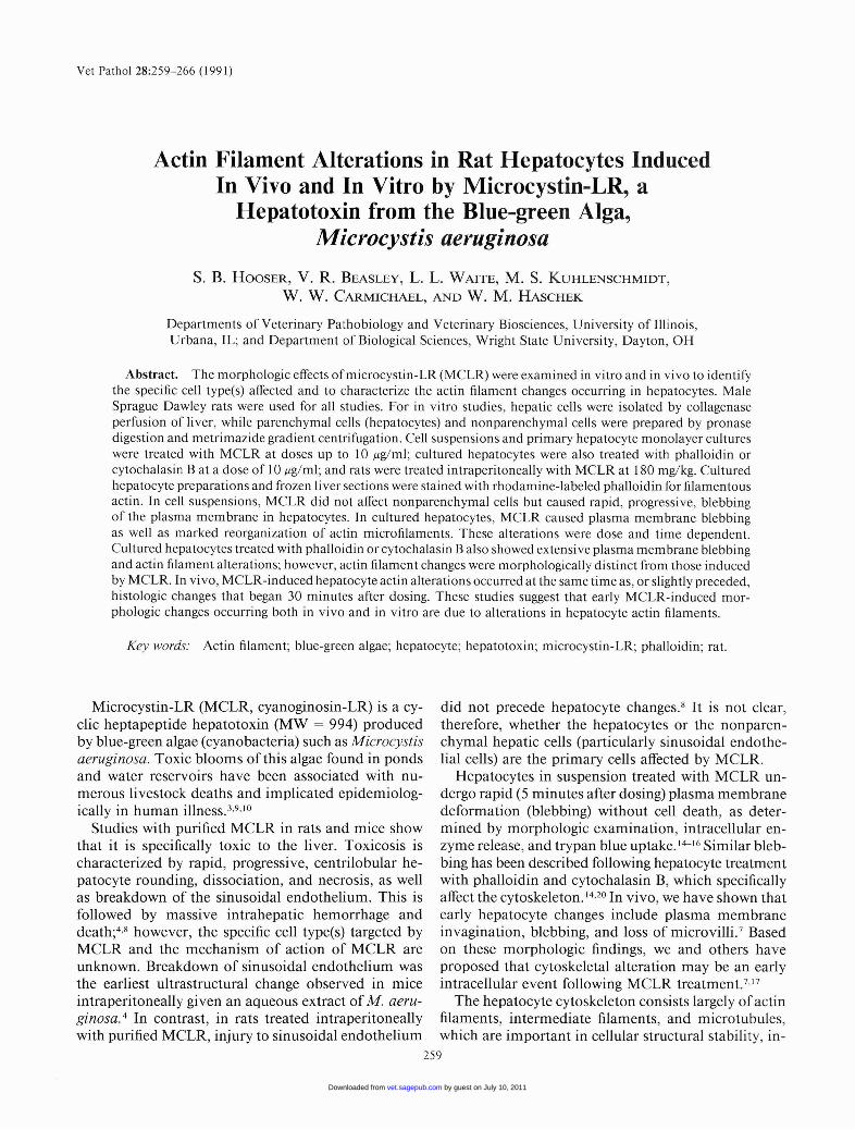
Effects of microcystin-LR (MCLR) on hepatic parenchymaland non parenchymal cells in suspension
Hepatocytes (parenchymal cells) were unaffected byphosphate buffered salin e (PBS) treatment (Fig. l a),but plasma membrane blebbing was observed following MCLR-treatment (Fig. I b). The number and sizeof blebs increased in a tim e-dependent manner unt ilat late time po ints the blebs coalesced to form small ernumbers of large bleb s. Ten minutes after treatmentwith MCLR at 10.0 j.tg/ml, greater than 90% of thecells had multiple medium to large plasma membraneblebs. By 30 minutes, these blebs had increased in size,and frequ entl y coalesced to form a rim around theoriginal cell border. Fewer and small er blebs were observed at the lower dos es of 1.0 or 0.1 j.tg of MCLRIml. Plasma membrane blebbing was not observed insimilarly treated hepatic non parenchymal cells.
/
\
26 1
Effect of MCLR on the plasma membrane and actinfilaments of cultured hepatocytes
After 24 hours in culture, hepatocytes were stillrounded and stain ed diffusely with rhodamine-labeledphalloidin, indicating that actin filaments had not yetbecome organized. After 72 hours, hepatocytes hadspread out and had well organized actin filaments. Wetherefore chose to exami ne the effects of MCLR prima rily in hepatocytes cultured for 72 hours.
Seventy-two hour cultured hepatocytes treated withPBS showed no changes in plasma membrane or act infilaments (Fig. 2a). Th e response to treatment withMCLR over time was as follows. After I hour of treatment with MCLR at 10 j.tg/ml, approximately 50% ofhepatocytes had one or several small plasma membrane bleb s. Filamentous actin was aggregated at thebase of these blebs. By 3 hours, approximately 75% ofhepato cytes had multiple plasma membrane blebs .Blebs contained filamentous actin that appeared asthi ckened linear " rays" extending to the tip of the bleband as focal aggregates at the base of the se blebs (Figs.2b, c). By 6 hours, greate r than 95% of hepatocyteswere affected . Unlike control cells, these hepatocyteswere small and rounded, with blebbed plasma membranes that occasionally coalesced to form a rim aroundthe original cell border. Th e actin in these cells wasaggregated into a single, int ensely staining locus (Fig.2d). A similar but less pronounced increase in numberof hepatocytes affected as well as the severity of effectwere observed over tim e at lower concentrations of
Fig. 1. Hepatocytes in suspension.Fig. la. Control, 30 minutes aft er treatment with phos
phat e buffered saline . An aggregate of un iforml y roundedhepatocytes is present.
Fig . 1b. Thirty minutes after treatment with 10.0 Jig ofmicrocystin-RL/ml. Hepatocytes are di saggregat ed and havemultiple medium to large pla sma membrane blebs.
MCLR. For example, at I j.tg of MCLR/ml, approximately 10% of hepatocytes were affected at I hour,20% at 3 hours, and 50% at 6 hours.
When the dose response to MCLR was exam ined,the number of hepato cytes with plasma membraneblebbing increased with dose at each of the time pointsexamined, i.e., I, 3, and 6 hours (data not shown). Forexample, at 3 hours, the number of hepatocytes affected was 0% at 0 j.tg/ml , and approximately 5% at0.1 j.tg/ml, 20% at 1.0 ug/ml, and 75% at 10 j.tg/ml.
Th e effects of MCLR were compared with those induced by phalloidin and cytochal asin B in hepatocytescultured for 24, 48, and 72 hours. With phalloidintreatment, the number ofhepatocytes develop ing plas-
by guest on July 10, 2011vet.sagepub.comDownloaded from

262 Hooser et al.
Fig. 2. Fluoresce nt micrograph s. Seve nty-two- hour cultured hepatocytes. Rhodam ine-labeled phalloidin.Fig. 2a. Control, 6 hou rs after treatm ent with phosphate buffered saline. The cells are flattened and contai n num erous,
thin, well-organi zed actin filam ent s.Fig. 2b. One hour after trea tment with 10.0 f.lg of microcyst in-LR/ml of medium . In thi s hepatocyte, seve ral plasma
membrane blebs contain large, th ick actin filaments that extend from the bases to the tips of the blebs.Fig. 2e. Three hours after treatme nt with 10.0 f.lg of microcystin-LR/ ml of medium. Actin is aggregating beneath the
plasma membrane with thickened actin filaments extending out to the tips of the blebs. As the cells und ergo blebbing, theyround up and appear smaller.
Fig. 2d. Six hours after treatm ent with 10.0 f.lg of microcystin-LR/ml of med ium . Most of the int racellular actin in eachof these hepatocytes has aggregated into a single locus and there is some blebbing of the plasma membranes.
rna membrane blebbing was related to the age of thecells: approxi mately 50% were affected in 24-hour cultures, 10 to 20% in 48-hour cultures and less than 5%in 72-hour cultures. Neith er MCLR nor cytochalasin
B showed this hepatocyte age-dependent effect. Sincephalloid in had littl e effect on hepatocytes cultured for72 hours, dat a obtained in 24-hour cultured hepatocytes are presented. Six hours after treatment with PBS,
by guest on July 10, 2011vet.sagepub.comDownloaded from

MCLR Alters Hepatocyte Actin Filaments 263
Fig. 3. Fluorescent mi crographs. Tw enty-four-hour cultured hepatocytes 6 hours after treatment. Rhodamine-lab eledphalloidin .
Fig. 3a. Treated with 10.0 /-Lg of phall oidinlml of medium. In thi s cell, intracellular actin has aggregated int o man y,variab le-sized foci locat ed th roughout the cyto plasm.
Fig.3b. Treated with 10.0 /-Lg of cytoc ha las in B/ml of medium. In thi s cell, intrace llular actin has aggregated int o man y,small foci located th rou ghout the cyto plasm.
no change was observed in 24-hour cultured hepatocytes; they remained rounded and stain ed diffusely withrhodamine-phalloidin with few linearl y organized actin fil am ents. After a 6-hour treatment of 24-hour cultured hepatocytes with 10.0 JIg of MCLR/ ml, however,cell blebbing and aggregation of filamentous actin int oa single, int ensely staining locus was identical to thatdescrib ed in 72-hour cultured hepatocytes. Twent yfour-hour cultured hepatocytes treated for 6 hours with10.0 JIg ofphalloidin1m I or with 10.0 JIg ofcytochalasinB/ml also had numerous plasma membrane blebs , butwhen stained with rhodamine-phalloidin, the actin aggregates were variable in size and scattered throughoutthe cytoplasm , thus differing from MCLR-inducedchanges (Figs. 3a, b).
Morphologic and cytoskeIetal effects of MCLR inlivers of intact rats
Morphologic changes in formalin-fixed, hematoxylin and eosin-stained sections were similar to thosereported previously." Thirt y minutes after dosing with180 JIg of M CLR/kg, there was disassociation androunding of hepatocytes immediately adjacent to central veins, with mild hem orrhage in some areas (Fig.4a). Th ese lesion s progressed in extent and severityunt il, at 60 minutes after dosing, there was hepatocytefragmentatio n and necrosis with hem orrhage involvingentire liver lobul es except for a rim of periportal hepatocytes ten to IS cells wide (Fig. 4b).
In rhodamine-phalloidin stained, frozen liver sections of control (PBS-treated) rats , actin was normallydistributed in hepatocytes with margination along theplasma membrane. Random cytoplasmic stippling wasev ident (Fig. 5a). T his was also present in MCLR-
treated animals at 5 and 10 minutes after dosing. By20 minutes after dosing, however, multiple (five to 15)discrete actin aggregates were observed in the cytoplasm of centrilobular and a few mid zonal hepatocytes.Forty-five minutes after dosing, centrilobular and midzonal hepatocytes contained distinct aggregates of filamento us actin (Fig. 5b). This lesion progressed inextent and severity until at 60 minutes the majority ofperiportal hepatocytes were also affected. At this time,the intracytoplasmic filamentous actin aggregates appeared as dense foci in most centrilobular and midzonal hepatocytes.
Discussion
These studies have provided several new insightsinto the effects of microcystin-LR (MCLR). First, themorphology of hepatocytes in suspension cultures isaffected by MCLR but that of nonparenchymal cells isnot. Second, MCLR causes time and dose-depend entalterations in the plasma membrane and actin filaments of cultured hepatocytes. Th ird , actin reorganization is associated with plasma membrane changesin cultured hepatocytes exposed to MCLR but thisreorgani zation differs markedly from that caused byeither phalloidin or cytochalasin B. Previous studieshave sho wn that phalloidin and cytochalasin B caus ea change in the polymerizat ion state of actin, whereasmicrocystin does not. 2, 16 Fourth and last, we have shownthat MCLR induces actin filament alterations in vivo,as well as in vitro, and that these alterations are associated with hepatocyte rounding and degen erationseen histologicall y.
Th e occurrence of MCLR-induced plasma membrane blebbing in hepatocytes, but not in sinusoidal
by guest on July 10, 2011vet.sagepub.comDownloaded from

264 Hooser et a1.
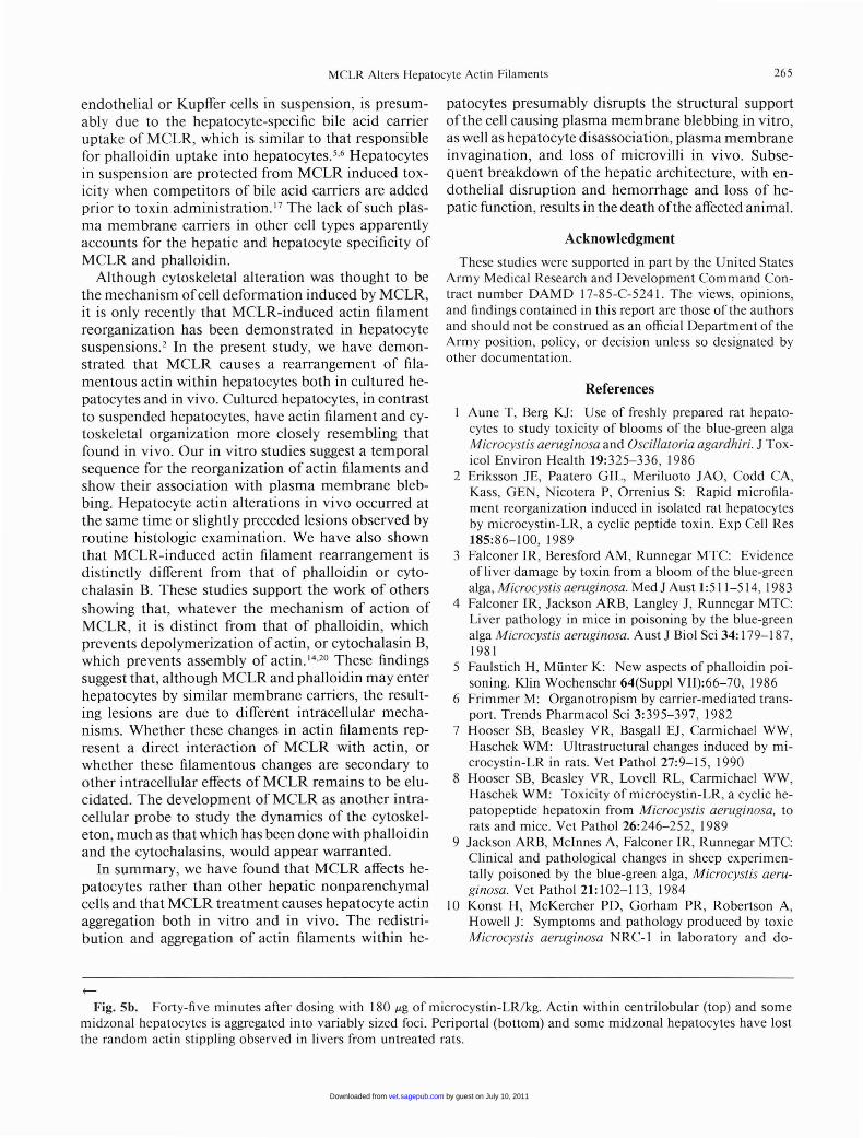
Fig. 4. Liver after dosing with 180 J.Lg of microcystin-LR/kg. HE.Fig. 4a. Thirty minutes after dosing. Th ere is rounding of hepatocytes and sinusoidal congestion adjacent to the central
vein (top right).Fig. 4b. Sixty minutes after dosing. Hepatocytes around the central vein (top ) and midzonally are disassociated and
fragmented. Small foci of hemor rhage are present. Periport al hepatocytes (botto m right) are unaffected.Fig. 5. Fluorescent micrograph s. Frozen liver sections. Rhodam ine-labeled phalloidin.Fig. Sa. Control, 60 minutes after dosing with phosphate buffered saline. There is a norm al pattern of actin sta ining at
the margins of the hepatocytes as well as randomly distributed stippling.
by guest on July 10, 2011vet.sagepub.comDownloaded from
MCL R Alte rs Hepatocyte Act in Filaments 265
endo thelia l or Kupffer cells in suspension, is presumabl y du e to the hepatocyte-specific bile acid carrierupt ake of MCLR, which is sim ilar to that respon siblefor phalloidin uptake into hepatocytes.t> Hepatocytesin suspen sion are protected from MCLR induced toxicity when competito rs of bile acid carr iers are addedpri or to toxin admini strat ion. " The lack of such plasma memb ran e car riers in other cell types apparentlyacco unts for th e hepat ic and hepatocyte spec ificity ofMCLR and phalloid in.
Altho ugh cytos keleta l alteration was thought to bethe mechan ism ofcell de formation ind uced by MCLR,it is only recently that MCLR-induced actin filam entreorgani zati on has been de monstrated in hepatocytesuspension s.' In th e present study, we have dem onstra ted th at MCLR causes a rea rrangement of filament ous actin within hepat ocytes both in cultured hepatocytes and in vivo. Cultured hepatocytes, in contras tto suspended hepatocytes, have actin filament and cytos keletal orga niza tion more closely resembling th atfound in vivo . Our in vi tro studies suggest a temporalsequence for th e reo rganization of actin filame nts andshow their associatio n with plasm a membran e blebbin g. Hepatocyte act in alterations in vivo occurred atth e same tim e or slightly preceded lesion s observed byroutine histologic exam ination. We have also shownth at MCLR-induced actin filament rearrangem ent isdi stin ctly different from that of phalloidin or cytocha lasin B. These stud ies suppo rt th e work of othersshow ing that , wha tever th e mechanism of actio n ofMCLR, it is distinct fro m that of phalloidin , whichpre ven ts depolym erizat ion of actin, or cytocha lasin B,which prevents asse mbly of act in. 14
•2o Th ese find ings
suggest th at , although MCLR and phalloidin may enterhepatocytes by sim ilar membrane carriers, th e resulting lesion s are due to different intracellular mechanism s. Whether these cha nges in actin filaments represe nt a direct interaction of MCLR with actin, orwhether th ese filamentou s changes are secondary toother int race llular effects of MC LR rem ain s to be elucida ted. The development of MCLR as ano ther intracellular pro be to study the dynamics of the cytos keleto n, m uch as that whic h has been don e with ph alloidinand th e cytochalasins , would appear warranted .
In su m mary, we have found th at MCLR affects hepatocytes rather th an other hepati c nonparenchymalcells and that MCLR treatment causes hepatocyte act inaggrega tion both in vitro and in vivo. The red istri bution and aggregat ion of acti n filaments within he-
patocytes presumabl y disrupts the struc tura l suppo rtof th e cell causing plasma membran e blebbing in vi tro ,as well as hepatocyte di sassociati on , plasma membran einvaginat ion, and loss of microvilli in vivo. Subsequ ent breakdown of the hepatic arc hitecture, with endothelial disruption and hemorrhage and loss of hepatic function , results in the death ofthe affected anima l.
Ack nowledgment
These studies were supported in part by the United State sArmy Med ical Research and Development Command Contract number DAMD 17-85-C-5241 . The views , opinions,and findi ngs conta ined in thi s report are those of the authorsand sho uld not be construed as an official Departm ent of theArmy pos ition, policy, or decision unless so designated byother docum entation .
References
Aune T , Berg KJ : Use of freshly prepared rat hepatocytes to study toxi cit y of blooms of the blue-green algaMicrocystis aeruginosa and Oscillatoria agardh iri. J Tox icol Enviro n Heal th 19:325-336, 1986
2 Eriksson JE, Paatero G IL, Meriluoto JAO, Codd CA,Kass, GEN, Nicotera P, Or renius S: Rap id micro fil amerit reorga nization ind uced in isolated rat hepatocytesby microcystin -LR , a cyclic peptide toxin . Exp Cell Res185:86-1 00 , 1989
3 Falco ner IR , Beresford AM, Runnegar MT C: Evide nceofliver dam age by toxin from a bloom of the blue-greenalga, Microcystisaeruginosa. Med J Aust 1:511-514, 1983
4 Falconer IR, Jackson ARB, Lan gley J , Ru nnegar MT C:Liver pat ho logy in mice in poisoning by the blue-greenalga Microcystis aeruginosa. Aust J BioI Sci 34: 179-1 87,1981
5 Fa ulstich H, Mu nter K: New as pects of phalloid in poi soning. Klin Wochenschr 64(Sup pl YII):66-70, 1986
6 Fri mmer M: Organotro pism by carrier-mediated transport. T rends Pharmacol Sci 3:3 95-397, 1982
7 Hooser SB, Beasley YR, Basgall EJ , Carmichael WW ,Hasc hek WM : Ultrastruc tural changes induced by microcysti n-L R in rats. Yet Path ol 27:9-1 5, 1990
8 Hooser SB, Beasley YR, Lovell RL, Car mic hael WW ,Hasc hek WM : Toxi city of microcystin-LR, a cyclic hepatopeptide hepatoxin fro m Microcystis aeruginosa, torat s and mice. Yet Pathol 26:246-252, 1989
9 Jackson ARB, Mci nnes A, Falconer IR, Runnegar MT C:Clinical and path ological cha nges in sheep experimenta lly poisoned by the blue-green alga, Microcystis aeruginosa. Yet Path oI21:1 02-113, 1984
10 Kon st H, McKercher PO , Gorh am PR , Robertson A,Howell J: Sympto ms and path ology produced by toxicMi crocystis aeruginosa NRC- I in laboratory and do-
f--
Fig. 5b. Forty-five minutes after dosi ng with 180 Jig of microcystin- LR/ kg. Actin within centri lob ular (top) and somemidzonal hepatocytes is aggregated into variab ly sized foci. Periporta l (bottom) and some midzon al hepatocy tes have lostthe random acti n stipp ling observed in livers from untrea ted rat s.
by guest on July 10, 2011vet.sagepub.comDownloaded from

266 Hooser et al.
mestic animals. Can J Comp Med Vet Sci 29:221- 228,1965
II Krish namurthy T, Carmic hae l WW , Sarver EW: Toxicpeptides from freshwater cyanobacteria (blue-green algae). 1. Isolation , purifi cat ion , and characterization ofpeptides from Microcystis aeruginosa and Anabaena jlosaquae. T oxicon 24:865-869, 1986
12 Kuhl enschmidt MS, Schmell E, Slife G W, Kuhl enschmidt TB , Seiber F, Lee Y-C, Roseman S: Studies onthe intercellul ar adhes ion of rat and chicken hepatocytes -condi tions affecting cell-cell specificity. J BioIChern 257: 3157-3164, 1982
l 3 Lafranconi WM , Glatt H, Oesc h F: Xenob iotic metabolizing enzy mes of rat liver nonparenchym al cells. Toxicol Appl Pharmacol 84:500-511 , 1986
14 Prentki M, Chaponnier C, Jeanrenand B, Gabbiani G:Actin micro filam ents, cell shape, and secretory proc essesin isolat ed rat hepatocytes, J Cell BioI 81:592-607, 1979
15 Runnegar MT C, Falcon er IR: Th e in vivo and in vitrobiological effects of the peptide hepatotoxin from theblue-green alga, Microcystis aeruginosa. S Afr J Sci 78:363- 366 , 1982
16 Runnegar MTC, Falconer IR: Effect of toxin from thecyano bacterium Microcystis aeruginosa on ultrastru ctu ral morph ology and actin polymerizat ion in isolatedhepatocytes. Toxicon 24:109-11 5,1 986
17 Runn egar MT, Falconer IR , Silver J: Deformati on of
isolated rat hepatocytes by a pept ide hepatotoxin fromthe blue-green alga, Microcystis aeruginosa. Na unynSchmiedi berg Arc h Pharmacol 317 :268- 272, 1981
18 Sager PR , Syversen TL , Clarkson T W, Cavanagh JB,Elgsaeter A, G uldberg HC, Lee SO, Lichtman MA,Mottet NK, Olmsted JB: Structure and functio n of thecytoskeleton. In: Th e Cytoskeleton: A Target for ToxicAgents, ed. Clarkso n T , Sager P, and Syverso n T , pp. 32 1. Plenu m Press, New York, NY, 1986
19 Wan son JC, Mosselm an s R: Coculture of adult rat hepatocytes and sinuso ida l cells: a new experimental modelfor the study of ultrastru ctural and functional properti esof liver cells. In : Co mmunications of Liver Cells, ed .Popper H, Gudat F, Bianchi L, and Reutter W., pp . 23925 1. MTP Press, Ltd ., Lan caster, England, 1980
20 Weiland T: Mole cular mechani sms of toxicity. In : Peptides of Poisonous Am an ita Mu shroom s, ed. Rich A, p.16 1 Springer-Verlag, New York, NY , 1986
2 1 Zacha ry JM , Cleve land G, Kwock L, Laurence T,Weissman RM , Nabell L, Fried FA, Staab EV, RisingerMA, Lin, S: Actin filam ent organization of the DunningR3327 rat prost at ic ade noca rcino ma system: correlationwith metastat ic potenti al. Cancer Res 46:926- 932, 1986
22 Zand MS, Albrecht-Beuhler G: What struc tures, besidesadhesio ns, prevent cells from rounding up? Cell Mot il13:195- 211 ,1 989
Note added in proof.
Microcystin-LR has recently been shown to be an inhibitor of protein phosphatases type I and type 2A and to causeincreased phosphorylation of cytoskeletal components. It is suggested that microc ystin-LR-induced morph ologic and cytoskeletal changes are due to the effects of phosphatase inh ibit ion (Eriksson JE, Toi vola 0 , Meriluoto JAO, Karaki H, HanY-G , Hartshorne 0 : Hepatocyte deformat ion induced by cyanobacteria l toxin s reflects inhibition of protein phosphatases.Biochem Biophys Res Comm 173:1347-1 353,1 990).
Requ est reprints from Dr. W. M. Haschek-Hock, Department of Veterin ary Pathobiology, College of Veterin ary Medicine,200 I S. Lincoln Avenue, Urbana, IL 6 180 I (USA) .
by guest on July 10, 2011vet.sagepub.comDownloaded from
Copyright © 2022 FDOKUMEN