Cyanobacterial Neurotoxin β-N-Methylamino-L-alanine (BMAA) in Shark Fins
Upload
independentCategory
view
0download
0
Mar. Drugs 2010, 8, 1650-1680; doi:10.3390/md8051650
Marine Drugs ISSN 1660-3397
www.mdpi.com/journal/marinedrugs
Review
On the Chemistry, Toxicology and Genetics of the Cyanobacterial Toxins, Microcystin, Nodularin, Saxitoxin and Cylindrospermopsin
Leanne Pearson 1, Troco Mihali 1, Michelle Moffitt 2, Ralf Kellmann 3 and Brett Neilan 1,*
1 School of Biotechnology and Biomolecular Sciences, The University of New South Wales, Sydney,
NSW, 2052, Australia; E-Mails: [email protected] (L.P.); [email protected] (T.M.) 2 School of Biomedical and Health Sciences, The University of Western Sydney, Campbelltown,
NSW, 2560, Australia; E-Mail: [email protected] (M.M.) 3 Department of Molecular Biology, The University of Bergen, P.O. Box 7803, 5020 Bergen,
Norway; E-Mail: [email protected] (R.K.)
* Author to whom correspondence should be addressed; E-Mail: [email protected];
Tel.: +61-2-9385-3235; Fax: +61-2-9385-1483.
Received: 26 March 2010; in revised form: 2 May 2010 / Accepted: 6 May 2010 /
Published: 10 May 2010
Abstract: The cyanobacteria or “blue-green algae”, as they are commonly termed,
comprise a diverse group of oxygenic photosynthetic bacteria that inhabit a wide range of
aquatic and terrestrial environments, and display incredible morphological diversity. Many
aquatic, bloom-forming species of cyanobacteria are capable of producing biologically
active secondary metabolites, which are highly toxic to humans and other animals. From a
toxicological viewpoint, the cyanotoxins span four major classes: the neurotoxins,
hepatotoxins, cytotoxins, and dermatoxins (irritant toxins). However, structurally they are
quite diverse. Over the past decade, the biosynthesis pathways of the four major
cyanotoxins: microcystin, nodularin, saxitoxin and cylindrospermopsin, have been
genetically and biochemically elucidated. This review provides an overview of these
biosynthesis pathways and additionally summarizes the chemistry and toxicology of these
remarkable secondary metabolites.
Keywords: cyanotoxin; non-ribosomal peptide; polyketide; alkaloid; toxicology
OPEN ACCESS

Mar. Drugs 2010, 8
1651
1. Microcystin
1.1. Introduction
The heptapeptide hepatotoxin, microcystin, has been isolated from multiple genera of
cyanobacteria, including Microcystis, Anabaena, Oscillatoria, Planktothrix, Chroococcus and Nostoc.
Microcystin-producing strains such as Microcystis aeruginosa have a cosmopolitan distribution and
thrive in a range of climates, making these organisms a global threat to human health. Consequently,
significant research efforts have been directed towards their identification and eradication.
1.2. Chemistry
The microcystins comprise the largest and most structurally diverse group of cyanobacterial toxins.
Around 90 microcystin isoforms varying by degree of methylation, hydroxylation, epimerization,
peptide sequence and toxicity have been identified [1,2]. Underlying the extraordinary heterogeneity
present among the microcystins is their common cyclic structure (Figure 1) and possession of several
rare, highly conserved amino acid moieties. Collectively, the microcystins may be described as
monocyclic heptapeptides containing both D- and L-amino acids plus N-methyldehydroalanine and a
unique ß-amino acid side-group, 3-amino-9-methoxy-2-6,8-trymethyl-10-phenyldeca-4,6-dienoic acid
(Adda) [3]. The microcystin isoforms differ primarily at the two L-amino acids, and secondarily on the
presence or absence of the methyl groups on D-erythro--methylaspartic acid (D-MeAsp) and/or
N-methyldehydroalanine (Mdha) [4]. However, substitutions of all moieties within microcystin have
been reported [5–7].
Figure 1. Structure of microcystin. General numbering of residues is indicated.
Microcystin is a cyclic heptapeptide. The two variable amino acids in microcystin are
indicated by X and Y. The most common isoform is microcystin-LR (MW 995.17), where
X is L-Leu and Y is L-Arg.

Mar. Drugs 2010, 8
1652
1.3. Toxicology
Acute cases of microcystin poisoning may cause rapid death in humans and other animals [8]. Upon
ingestion, microcystin is transported to the liver by organic anion transport proteins where they exert
their toxicity via inhibition of protein phosphatases 1 and 2A [9–11]. Inhibition of protein
phosphatases can lead to excessive phosphorylation of structural filaments, subsequent cyto-skeletal
degradation and breakdown of hepatic ultra structure [12,13]. Retraction of hepatocytes from
neighboring cells and sinusoidal capillaries causes blood to become pooled in the liver tissues. This
ultimately results in local tissue damage, organ failure and haemorrhagic shock [13].
Varying levels of toxicity have been reported for each microcystin isoform. For example, the LD50
of the most common isoform, microcystin-LR, is 50 µg per kilogram of body weight in mice [14],
while the rarer microcystin-RR requires a significantly higher dose of 600 µg to produce the same
lethal effect [15].
The revelation that cyanobacterial hepatotoxins cause protein phosphatase inhibition has raised the
disturbing possibility that human exposure to non-lethal doses of these compounds may contribute to
the development of cancer [16,17]. Several laboratory studies have indicated that chronic exposure to
microcystin can indeed promote skin and liver tumors in rats and mice [18,19]. Epidemiological data
suggest that similar long-term effects such as hepatocellular carcinoma may also be observed in
humans [20,21]. Such results highlight the need for sensitive and rapid detection methods and stringent
monitoring of cyanobacterial hepatotoxins in drinking water supplies.
1.4. Biosynthesis and Genetics
Microcystin is synthesized non-ribosomally by the thiotemplate function of a large multifunctional
enzyme complex containing both non-ribosomal peptide synthetase (NRPS) and polyketide synthase
(PKS) domains. The gene cluster encoding these biosynthetic enzymes, mcyS, has been sequenced and
partially characterized in several cyanobacterial species including Microcystis, Anabaena, and
Planktothrix [22–24] (Figure 2). Such fundamental studies have offered insight into the evolution of
cyanotoxin biosynthesis, and have additionally provided much of the groundwork for current
PCR-based cyanobacterial detection methods.
The microcystin biosynthesis gene cluster, mcyS, was the first complex metabolite gene cluster to
be fully sequenced from a cyanobacterium. In M. aeruginosa PCC7806, the mcyS gene cluster spans
55 kb and comprises 10 genes arranged in two divergently transcribed operons, mcyA–C and mcyD–J.
The larger of the two operons, mcyD–J, encodes a modular PKS (McyD), two hybrid enzymes
comprising NRPS and PKS modules (McyE and McyG), and enzymes putatively involved in the
tailoring (McyJ, F, and I) and transport (McyH) of the toxin. The smaller operon, mcyA–C encodes
three NRPSs (McyA–C) [22].
The formation of Adda putatively involves enzymes encoded by mcyD–G and J, based on
bioinformatic analyses and homology to related enzymes (Figure 3a). The hybrid NRPS/PKS enzyme,
McyG, constitutes the first step in Adda biosynthesis. It was initially hypothesized that the NRPS
module of McyG activates phenylacetate, however, recent biochemical characterization of the McyG
A–PCP didomain has revealed that assorted phenylpropanoids are preferentially activated and loaded

Mar. Drugs 2010, 8
1653
onto the PCP [25]. Following activation, the phenylpropanoid starter unit is extended by several
malonyl-CoA elongation steps and subsequently modified by C-methylation, reduction and
dehydration, all catalyzed by the PKS modules of McyD, E and G. The aminotransferase domain of
McyE then converts the polyketide to a -amino acid in the final step of Adda biosynthesis. The NRPS
module of the second hybrid PKS/NRPS enzyme, McyE, is thought to be involved in the activation
and condensation of D-Glu with Adda.
Figure 2. Hepatotoxin gene clusters from various cyanobacteria. Structures of the
microcystin and nodularin gene clusters of (A) N. spumigena, (B) M. aeruginosa,
(C) P. agardhii, and (D) Anabaena sp. 90, showing genes encoding polyketide synthases
(white), non-ribosomal peptide synthetases (red), tailoring enzymes (grey), and
ABC-transporters (black). Diagram not drawn to scale.
The mcyF ORF was originally predicted to encode a glutamate racemase, responsible for the
epimerization of the L-Glu residue of microcystin [22,26]. A subsequent study by [27] contended this
theory and offers evidence that McyF acts exclusively as an Asp racemase. The authors propose that
the D-Glu residue is provided by an L-Glu racemase residing outside the mcyS gene cluster.
Mutagenesis experiments in P. agardhii showed that the production of Adda also involves an
O-methylation step catalyzed by the putative monofunctional tailoring enzyme, McyJ [23].
The remaining biosynthetic enzymes in the microcystin biosynthesis pathway (NRPSs) are
putatively involved in the specific activation, modification and condensation of substrate amino acids
onto the linear peptide chain, which is then cyclized to produce microcystin. Firstly, McyA adds
L-Ser to the growing chain, followed by the addition of D-Ala. This step is followed by the addition of
L-Leu and D-MeAsp residues (McyB) followed by the addition of L-Arg (McyC), and subsequent
cyclization and release of the final peptide product (Figure 3b).
The remaining stand alone enzyme, the 2-hydroxy-acid dehydrogenase, McyI, is putatively
involved in the production of D-methylaspartate at position three within the microcystin cyclic
structure via the conversion of 3-methylmalate to 3-methyloxalacetate. It is hypothesized that a
promiscuous aspartate aminotransferase then converts 3-methyloxalacetate to methylaspartate [28].
An ABC transporter gene, mcyH, is believed to be involved in the transport of microcystin [29].
This transporter may be responsible for the thylakoid localization of the toxin [30,31] or for the
extrusion of the toxin under certain growth conditions, including exposure to high and red light [32].
Mar. Drugs 2010, 8
1654
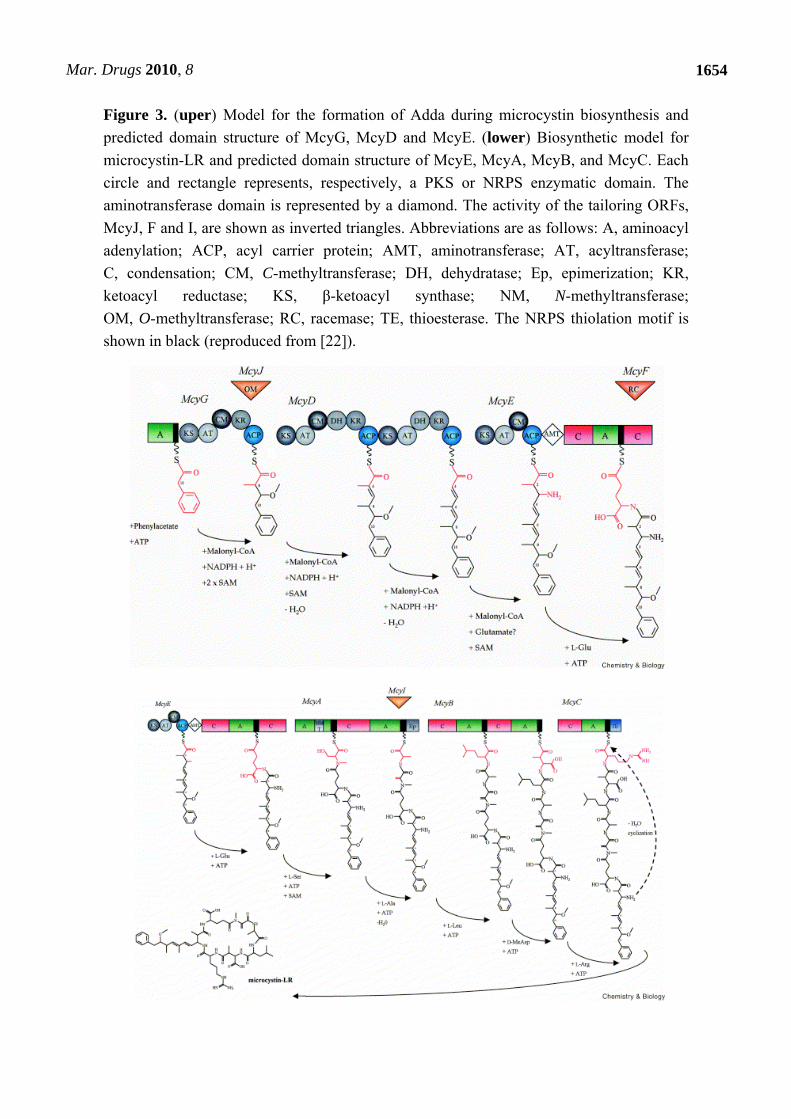
Figure 3. (uper) Model for the formation of Adda during microcystin biosynthesis and
predicted domain structure of McyG, McyD and McyE. (lower) Biosynthetic model for
microcystin-LR and predicted domain structure of McyE, McyA, McyB, and McyC. Each
circle and rectangle represents, respectively, a PKS or NRPS enzymatic domain. The
aminotransferase domain is represented by a diamond. The activity of the tailoring ORFs,
McyJ, F and I, are shown as inverted triangles. Abbreviations are as follows: A, aminoacyl
adenylation; ACP, acyl carrier protein; AMT, aminotransferase; AT, acyltransferase;
C, condensation; CM, C-methyltransferase; DH, dehydratase; Ep, epimerization; KR,
ketoacyl reductase; KS, β-ketoacyl synthase; NM, N-methyltransferase;
OM, O-methyltransferase; RC, racemase; TE, thioesterase. The NRPS thiolation motif is
shown in black (reproduced from [22]).

Mar. Drugs 2010, 8
1655
Comparative studies of the mcyS gene clusters from M. aeruginosa, P. agardhii [23], and Anabaena
sp. [24] have noted variation in the arrangement of mcyS genes between these different species of
cyanobacteria, although the proposed toxin biosynthetic processes are thought to be similar. The
M. aeruginosa and Anabaena sp. mcyS clusters are both arranged into two divergently transcribed
operons, however, the arrangement of genes within these operons differs between the two species
(Figure 2). In P. agardhii, the mcyS cluster also has a distinctive arrangement and lacks mcyF and
mcyI. Furthermore, the P. agardhii mcyS cluster contains an additional gene mcyT, upstream of the
central promoter region. This gene is thought to encode a putative type II thioesterase enzyme, which
may play an editing role by removing mis-primed amino acids from the NRPS and PKS enzymes. The
characterization of mcyS in M. aeruginosa, P. agardhii and Anabaena sp. has important implications
for understanding the origins and evolution of hepatotoxin biosynthesis in cyanobacteria. The
identification of transposases associated with the mcyS and ndaS (nodularin) gene clusters and
subsequent phylogenetic analysis has led to the theory that horizontal gene transfer and recombination
events are responsible for the sporadic distribution of the mcyS gene cluster throughout the
cyanobacteria and the various microcystin isoforms that have been identified to date [22,33,34].
Hepatotoxin production in cyanobacteria is thought to be influenced by a number of different
physical and environmental parameters, including nitrogen, phosphorous, trace metals, growth
temperature, light, and pH [35–41]. However, due to the fact that most regulatory investigations have
not been standardized, and the data have not been interpreted against the same specific growth
controls, the subject of hepatotoxin regulation remains a somewhat contentious issue. While most toxin
regulation studies have focused on direct measurements of cellular toxin, the description of the mcy
gene cluster by Tillett and co-workers [22] enabled a closer examination of microcystin regulation at
the molecular level [32]. Kaebernick et al. used the RNase protection assay to measure the
transcription of mcyB and mcyD under a variety of different light conditions. High light intensities and
red light were correlated with increased transcription, while blue light led to reduced transcript levels.
Interestingly, the authors observed two light thresholds, between dark and low light (0 and 16 µmol
photons m-2 s-1), and medium and high light (31 and 68 µmol photons m-2 s-1), at which a significant
increase in transcription occurred. The same group later found that transcription of mcy genes occurs
via two polycistronic operons, mcyABC and mcyDEFGHIJ, from a central bidirectional promoter
between mcyA and mcyD [42]. Interestingly, alternate transcriptional start sites were identified for both
operons when cells were cultured under different light intensities. For example, under low light
conditions, the polyketide and tailoring genes mcyD–J are transcribed as part of a polycistronic
message (mcyDEFGHIJ) from a central (mcyD) promoter, while under high light conditions, the genes
are transcribed from an alternative up-stream promoter. It is thought that initiation from the alternate
promoters under high light conditions may lead to increased transcription, as previously observed for
mcyB and mcyD. Many of the tailoring enzymes (mcyF, G, H, I and J) also possess their own
individual promoters [32].
Interestingly, light intensity also appears to favor the production of certain microcystin variants
over others. For example, Tonk et al. (2005) found that the cellular content of total microcystin
remained constant, independent of the irradiance. However, of the two main microcystin variants
detected in P. agardhii, the microcystin-DeRR content decreased two-fold with increased photon
irradiance, whereas the microcystin-DeLR content increased three-fold. Since microcystin-DeLR is
Mar. Drugs 2010, 8
1656
considerably more toxic than microcystin-DeRR, this implies that P. agardhii becomes more toxic at
high light intensities [40].
Other factors such as nutrient content and temperature have also been demonstrated to affect mcyS
expression and toxin biosynthesis. For example, Sevilla et al. (2008) investigated the effect of iron on
mcyS expression and toxin biosynthesis in M. aeruginosa PCC7806 [41]. Real-time PCR analysis and
HPLC were used to measure transcription of mcyD and the synthesis of microcystin-LR, respectively.
The results of this study suggested that iron starvation causes an increase in mcyD transcription,
correlative to the increase of toxin levels [41]. Davis et al. (2009) investigated the effects of
temperature on Microcystis growth and toxin genes and found that elevated temperatures yield more
toxic Microcystis cells and/or cells with more mcyD copies per cell, with either scenario potentially
yielding more toxic blooms [39].
2. Nodularin
2.1. Introduction
The cyclic pentapeptide nodularin is most commonly isolated from the filamentous, planktonic
cyanobacterium, Nodularia spumigena. This species generally forms toxic blooms in brackish and
estuarine environments. Blooms of toxic N. spumigena occur annually during summer months in the
Baltic Sea [43] resulting in nodularin being one of the most abundant naturally occurring compounds
in the Baltic Sea. Blooms are also particularly common within the estuaries and coastal lagoons of
Australia [44,45], and have been reported worldwide, including on the German North Sea coast [46],
New Zealand [47], and North America [48]. N. spumigena scums have also been reported in the fresh
to brackish/saline lakes at the mouth of the lower River Murray, South Australia, which is a major
source of both potable and irrigation water [49]. Nodularin is structurally similar to microcystin and
can induce similar toxic effects. The toxin has been reported to have detrimental effects on numerous
organisms within the ecosystem, including invertebrates and fish, but may have no effect on other
organisms [50].
The consumption of water containing toxic N. spumigena blooms has led to the death of domestic
and native animals by massive liver haemorrhage [46,47,51,52]. In sub-acute doses, nodularin, like
microcystin, is thought to act as a liver tumor initiator and promoter [53]. N. spumigena blooms are
also of importance to the seafood industry. Nodularin has been shown to accumulate in shellfish and
other seafood. Surveys of mussels, prawns, flounder both in Australia and the Baltic Sea report that
nodularin accumulated to levels of concern with the potential to cause hepatotoxicity [54–56].
2.2. Chemistry
Nodularin is a cyclic pentapeptide with a similar structure to microcystin, consisting of Adda,
D-glutamic acid (D-Glu), N-methyldehydrobutyrine (MeDhb), D-erythro--methylaspartic acid
(D-MeAsp), and L-arginine (L-Arg) (Figure 4) [57].
Seven naturally-occurring isoforms of nodularin have been reported to date. Two of these isoforms,
produced by a New Zealand Nodularia sp. bloom, have variations within the Adda residue, which
reduces or abolishes the toxicity of the compound [5]. The D-Glu residue is essential for toxicity of

Mar. Drugs 2010, 8
1657
nodularin, as esterification of its free carboxyl abolishes toxicity, however, substitution at position 1
has little effect on toxicity. The other two isoforms, nodularin-Har and motuporin, are variable at
position 2. Nodularin-Har is produced by the strain N. harveyana PCC7804, with the L-Arg, replaced
with L-Homoarginine (L-Har) [58,59]. Motuporin has been isolated from the Papua New Guinea
sponge Theonella swinhoei, and may be synthesized by an associated cyanobacterium. The L-Arg
residue of nodularin is replaced by L-Val in motuporin [60]. The L-Val residue is responsible for
additional cytotoxicity of motuporin against cancer cell lines.
Figure 4. Structure of nodularin. General numbering of residues is indicated. Nodularin is
a cyclic pentapeptide (MW 619). The L-Arg residue of nodularin may be replaced with a
homoarginine (nodularin-Har) or valine residue (motuporin).
2.3. Toxicology
Nodularin is a potent hepatotoxin in humans and other animals. Nodularin induces liver hemorrhage
in mice and has a lethal dose 50 (LD50) of 50 g.kg-1 (intra-peritoneal route of injection) in mice [61].
At doses below this concentration, nodularin may act as a carcinogen via the initiation and promotion
of liver cell division [53].
The hepatotoxicity and carcinogenicity of nodularin is associated with the inhibition of eukaryotic
protein phosphatase (PP) catalytic subunit types 1 and 2A [62]. The toxin inhibits the activity of PP2A
to a greater extent than PP1. Inhibition of PP2A by nodularin occurs at relatively the same
concentration (IC50) as that of microcystin (~0.1 nM) [62]. The hydrophobic C20 - amino acids Adda,
present in both toxins, blocks PP enzyme activity by interacting with the hydrophobic groove and
obstructing substrate access to the active site cleft [63–65]. MeDhb binds to Cys273 of PP2A in a
similar fashion to the MeDha residue in microcystin which binds to Cys273 of PP1 and Cys266 of
PP2A [64,66], however, binding of the toxin does not occur covalently and may be the reason for its
additional carcinogenic properties [67].
The toxic effects of nodularin are primarily associated with the hepatocytes due to active transport
of the toxin to the liver via the bile acid multi-specific organic anion transporters [10]. To date, studies
have been unable to identify the specific mechanism of transport.
Nodularin has been observed to accumulate on different trophic levels, in numerous organisms
including waterfowl, fish, mysid shrimp, zooplankton and benthic organisms [68]. Mesozooplankton
in particular seem to play a major role in nodularin transfer to planktivorous fish [68]. The toxin

Mar. Drugs 2010, 8
1658
appears to cause oxidative stress in the tissues in which it accumulates. In the case of the flounder
(platichthys flesus L.), this oxidative stress occurs in the liver by way of reduced GST and CAT
activities [69]. However, recent studies suggest that the toxin is rapidly detoxified and broken down or
excreted [70].
2.4. Biosynthesis and Genetics
The nodularin biosynthesis gene cluster ndaS, from Nodularia spumigena NSOR10, was sequenced
and characterized in 2004 by Moffitt and Neilan [71]. The 48 kb region of the genome consists of nine
ORFs (ndaA-I) transcribed from a bidirectional regulatory promoter region (Figure 2). While most of
the ndaS encoded genes have homologs in the mcyS cluster, their arrangement adheres more closely to
the ‘co-linearity’ rule of NRPS pathways that predicts the order of catalytic processes involved in the
biosynthesis of a non-ribosomal metabolite is generally the same as the order of the genes which
encode their catalytic enzymes [72].
The proposed pathway for nodularin biosynthesis is similar to that for microcystin. Functional
assignment of the enzymes was based on bioinformatic analysis and homology to the microcystin
synthetase enzymes. The Adda side-chain is produced via a mixed NRPS/PKS pathway from a
phenylacetate starter unit and several malonyl-CoA extensions (NdaC, D and F) (Figure 5). The NRPS
module of the hybrid NRPS/PKS, NdaF, subsequently adds D-Glu to the growing chain. Two NRPS
enzymes, NdaA and B, complete the cyclic pentapeptide by adding the final amino acid residues,
L-Thr, D-MeAsp and L-Arg. The NRPS modules responsible for the activation of D-Ala and D-Leu in
mcyS (McyA and B) are absent from ndaS as nodularin lacks these moieties. The NRPS and PKS
proteins require posttranslational modification by a phosphopantetheinyl transferase (PPT) protein.
The PPT required for activation of the Nda proteins is not clustered with the other nda genes. Recently,
degenerate PCR and subsequent functional enzymatic characterization, identified the PPT required for
nodularin biosynthesis in N. spumigena NSOR10 [73].
The ndaS cluster also encodes several putative monofunctional tailoring enzymes that may play a
role in the modification and transport of nodularin. ndaE encodes an O-methyltransferrase, ndaG
encodes a putative L-Asp/L-Glu racemase, and ndaI encodes an ABC transporter. Also encoded within
the ndaS cluster is a D-3-PGDH homolog, NdaH, which shares 71% identity with McyI. It is likely
therefore, that NdaH may be involved in the production of D-MeAsp [28].
Like mcyS, the ndaS gene cluster is transcriptionally regulated by a bi-directional promoter region.
Analysis of transcription of the ndaS cluster found that it is transcribed as two polycistronic mRNA,
ndaAB, ORF1, and ORF2, and ndaC [71]. The two genes downstream of ndaAB, ORF1 and ORF2,
encode a putative transposase and a putative high light-inducible chlorophyll-binding protein,
respectively. It is not clear why the putative transposase and the putative high light-inducible
chlorophyll-binding protein are also co-transcribed with the ndaS gene cluster. ORF2 has been
identified in all strains of toxic Nodularia and the association between ORF2 and nodularin
biosynthesis may suggest a physiological function associated with high-light stress in the cells
producing it. A putative heat shock repressor protein, encoded by the gene ORF3, was also identified
downstream of ORF2, which may be involved in the transcriptional regulation of the ndaS genes in
response to heat stress.

Mar. Drugs 2010, 8
1659
Figure 5. (uper). Model of the formation of Adda during nodularin biosynthesis and
predicted domain structure of NdaC, D and F. (lower). Biosynthetic model for nodularin
and predicted domain structure of NdaF, H, A and B. Each grey and green circle
represents, respectively, a PKS or NRPS enzymatic domain. The activities of the tailoring
ORFs, NdaE, G and H, are shown as inverted triangles. Abbreviations are as follows:
A, aminoacyl adenylation; ACP, acyl carrier protein; AMT, aminotransferase;
AT, acyltransferase; C, condensation; CM, C-methyltransferase; DH, dehydratase;
Ep, epimerization; KR, ketoacyl reductase; KS, β-ketoacyl synthase; NM,
N-methyltransferase; OM, O-methyltransferase; RC, racemase; TE, thioesterase.
(reproduced from [71]).
Mar. Drugs 2010, 8
1660
More recently, elucidation of the nda cluster has provided an opportunity to monitor transcriptional
regulation of the biosynthetic pathway [74]. The effects of ammonia and phosphate starvation were
analyzed. While expression of the nda cluster appears to be constitutive, phosphate starvation resulted
in an approximately two-fold increase in expression, while ammonia supplementation decreased
expression two-fold. Despite the changes to expression, intracellular and extracellular nodularin
concentration remained stable [74].
3. Saxitoxin
3.1. Introduction
Saxitoxin and its analogs, collectively termed paralytic shellfish poisons (PSPs), are highly potent
neurotoxins. Several freshwater species of cyanobacteria and marine dinoflagellates are known to
produce saxitoxins. Blooms of these toxic species have led to mass kills of fish, native animals and
livestock, as well as the contamination of freshwater resources [75–77]. Saxitoxins typically
accumulate through the food chain, in organisms consumed as seafood [78]. Marine shellfish are
particularly resistant to the toxins, and can therefore accumulate dangerously high levels of PSPs by
ingesting toxic plankton [79]. Saxitoxin and its analogs cause an annual estimated 2000 cases of PSP
globally, with a mortality rate of 15% [80].
The capacity to synthesize saxitoxins and other PSPs has an unusually wide phylogenetic
distribution, including both marine and freshwater organisms from two kingdoms of life (Eubacteria
and Protista). Typically, there is little convergence between the structures of secondary metabolites
from marine and freshwater organisms [81], or from organisms belonging to different phylogenetic
groups. Although several studies suggest that other bacteria are capable of synthesizing PSPs [82,83],
the findings are controversial, as the analytical methods used were not definitive and could not be
readily repeated by other researchers [84]. On the other hand, PSP biosynthesis in cyanobacteria and
dinoflagellates has been extensively verified and shown to be a consistent and heritable genetic
trait [85–90].
In the marine environment, dinoflagellate species capable of producing saxitoxins belong to the
genera Alexandrium, Pyrodinium and Gymnodinium [91–93]. While in freshwater systems, several
filamentous species of cyanobacteria, such as Anabaena circinalis, Aphanizomenon sp.,
Aphanizomenon gracile, Cylindrospermopsis raciborskii and Lyngbya wollei are also known to
produce saxitoxins [94–97]. Saxitoxin production is varied among dinoflagellate and cyanobacterial
producer genera, with not all species in a toxigenic genera being toxic, and different isolates of the
same species having differential toxicity. Furthermore, cyanobacterial isolates of the same species
from geographically distant locations provided different toxin profiles [89].
3.2. Chemistry
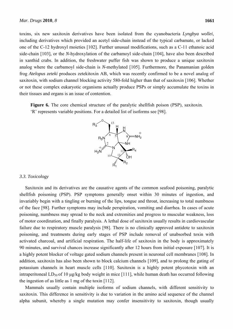
Saxitoxin is a trialkyl tetrahydropurine and the parent compound of more than 30 naturally
occurring derivatives that differ structurally at four positions [98] (Figure 6.). The variable positions
may be hydroxylated, sulfated or carbamoylated. Most of these analogs have been detected in both
cyanobacteria and dinoflagellates [95,96,98–101]. In addition to the usual carbamate and decarbamoyl
Mar. Drugs 2010, 8
1661
toxins, six new saxitoxin derivatives have been isolated from the cyanobacteria Lyngbya wollei,
including derivatives which provided an acetyl side-chain instead of the typical carbamate, or lacked
one of the C-12 hydroxyl moieties [102]. Further unusual modifications, such as a C-11 ethanoic acid
side-chain [103], or the N-hydroxylation of the carbamoyl side-chain [104], have also been described
in xanthid crabs. In addition, the freshwater puffer fish was shown to produce a unique saxitoxin
analog where the carbamoyl side-chain is N-methylated [105]. Furthermore, the Panamanian golden
frog Atelopus zeteki produces zetekitoxin AB, which was recently confirmed to be a novel analog of
saxitoxin, with sodium channel blocking activity 580-fold higher than that of saxitoxin [106]. Whether
or not these complex eukaryotic organisms actually produce PSPs or simply accumulate the toxins in
their tissues and organs is an issue of contention.
Figure 6. The core chemical structure of the paralytic shellfish poison (PSP), saxitoxin.
‘R’ represents variable positions. For a detailed list of isoforms see [98].
3.3. Toxicology
Saxitoxin and its derivatives are the causative agents of the common seafood poisoning, paralytic
shellfish poisoning (PSP). PSP symptoms generally onset within 30 minutes of ingestion, and
invariably begin with a tingling or burning of the lips, tongue and throat, increasing to total numbness
of the face [98]. Further symptoms may include perspiration, vomiting and diarrhea. In cases of acute
poisoning, numbness may spread to the neck and extremities and progress to muscular weakness, loss
of motor coordination, and finally paralysis. A lethal dose of saxitoxin usually results in cardiovascular
failure due to respiratory muscle paralysis [98]. There is no clinically approved antidote to saxitoxin
poisoning, and treatments during early stages of PSP include removal of unabsorbed toxin with
activated charcoal, and artificial respiration. The half-life of saxitoxin in the body is approximately
90 minutes, and survival chances increase significantly after 12 hours from initial exposure [107]. It is
a highly potent blocker of voltage gated sodium channels present in neuronal cell membranes [108]. In
addition, saxitoxin has also been shown to block calcium channels [109], and to prolong the gating of
potassium channels in heart muscle cells [110]. Saxitoxin is a highly potent phycotoxin with an
intraperitoneal LD50 of 10 μg/kg body weight in mice [111], while human death has occurred following
the ingestion of as little as 1 mg of the toxin [112].
Mammals usually contain multiple isoforms of sodium channels, with different sensitivity to
saxitoxin. This difference in sensitivity is due to variation in the amino acid sequence of the channel
alpha subunit, whereby a single mutation may confer insensitivity to saxitoxin, though usually
Mar. Drugs 2010, 8
1662
sacrificing speed of gating in the process [113]. The guanidinium groups and the carbon 12 hydroxyls
in STX have been shown to be critical for the binding of the sodium channel, while the carbamoyl side
chain also appears to be involved in the binding process, whereby one saxitoxin molecule binds one
channel by lodging itself in the ion-conducting pore [113–116].
The toxicity of saxitoxin derivatives varies greatly, with the carbamate toxins being 10–100-times
more potent than the N-sulfo-carbamoyl derivatives [98,117]. N-sulfo-carbamoyl analogs are,
however, labile and may easily be converted to the more toxic carbamate derivatives [118,119].
3.4. Biosynthesis and Genetics
Using a reverse genetic approach, Kellmann and co-workers [120] identified the gene cluster
putatively responsible for the biosynthesis of saxitoxin in Cylindrospermopsis raciborskii T3 (sxt)
(Figure7c). The sxt gene cluster is encoded by more than 35 kb and comparative sequence analysis
assigns 30 catalytic functions to 26 proteins. Bioinformatic analysis of this cyanobacterial saxitoxin
gene cluster, coupled with identification of novel biosynthetic intermediates enabled a revision of the
previously proposed saxitoxin biosynthesis pathway (Figure 8).
The first step in the revised saxitoxin biosynthesis pathway involves a Claisen condensation
reaction catalyzed by SxtA. This unique enzyme possesses a polyketide synthase (PKS)-like structure
composed of four catalytic domains, SxtA1-4: SxtA1 is homologous to SAM-dependant
methyltransferases; SxtA2 is related to GCN5-related N-acetyl transferases (GNAT) that transfer
acetate from acetyl-CoA to various heteroatoms [121]; SxtA3 is related to ACPs and also provides a
phosphopantetheinyl-attachment site; SxtA4 is homologous to class II aminotransferases and is most
similar to AONS (8-amino-7-oxononanoate synthase). The predicted reaction sequence of SxtA, based
on its primary structure, is the loading of the ACP (SxtA3) with acetate from acetyl-CoA, followed by
the SxtA1-catalyzed methylation of acetyl-ACP, converting it to propionyl-ACP. SxtA4, the class II
aminotransferase domain, then performs a Claisen condensation reaction between propionyl-ACP and
arginine. The putative product of SxtA is thus 4-amino-3-oxo-guanidinoheptane, which Kellmann et al.
designated compound A.
The sxtG gene encodes a putative amidinotransferase, with highest amino acid sequence similarity
to L-arginine/L-lysine amidinotransferases. SxtA is the putative substrate for SxtG, which transfers an
amidino group from arginine to the α-amino A' group, thus producing 4,7-diguanidino-3-oxoheptane
(designated compound B'). SxtB, an enzyme similar to the cytidine deaminase-like enzymes from
gammaproteobacteria, then catalyzes a retroaldol-like condensation in the conversion from B' to C'.
The putative sterol desaturase, SxtD is predicted to introduce a double bond between C-1 and C-5 of
C', resulting in the 1,2-H shift between C-5 and C-6 (compound D'). The gene product of sxtS, which
has sequence homology to nonheme iron 2-oxoglutarate-dependent dioxygenases, is predicted to
perform the consecutive epoxidation of the new double bond and opening of the epoxide to an
aldehyde with concomitant bicyclization. SxtU has sequence similarity to short-chain alcohol
dehydrogenases and is therefore predicted to reduce the terminal aldehyde group of the saxitoxin
precursor forming compound E'. The concerted action of SxtD, SxtS, and SxtU is therefore responsible
for the hydroxylation and bicyclization of compound C' to E'.

Mar. Drugs 2010, 8
1663
The gene product of sxtI is most similar to a predicted O-carbamoyltransferase from Trichodesmium
erythraeum and other cyanobacteria. Kellmann et al.’s data [120] indicate that SxtI may catalyze the
transfer of a carbamoyl group from carbamoylphosphate to the free hydroxy group of E'. Adjacent to
sxtI are two short ORFs of unknown function, sxtJ and sxtK. While sxtJ and sxtK homologs are
available in the databases, none of these genes have been functionally characterized.
sxtH and sxtT, each encode a terminal oxygenase subunit similar to those found in bacterial
phenylpropionate and related ring-hydroxylating dioxygenases. SxtH and SxtT may therefore perform
the consecutive hydroxylation of C-12, converting F' into saxitoxin. Members belonging to bacterial
phenylpropionate and related ring-hydroxylating dioxygenases are multicomponent enzymes, as they
require an oxygenase reductase for their regeneration after each catalytic cycle. The sxt gene cluster
provides a putative electron transport system, which would fulfill this function in the form of SxtV and
SxtW. SxtV, a 4Fe-4S ferredoxin, could putatively extract an electron pair from succinate, converting
it to fumarate [122]. SxtW a fumarate/reductase/succinate dehydrogenase homolog could then transfer
the electrons via ferredoxin to SxtH and SxtT.
Following synthesis of the parent molecule saxitoxin, modifying enzymes introduce various
functional groups. In addition to saxitoxin, C. raciborskii T3 produces N-1-hydroxylated (neoSTX),
decarbamoylated (dcSTX), and N-sulfurylated (GTX-5) toxins, whereas Anabaena circinalis
AWQC131C produces decarbamoylated (dcSTX) toxins and O-sulfurylated (GTX-3/GTX-2, dcGTX-
3/dcGTX-2) toxins, as well as both O- and N-sulfurylated toxins (C-1/C-2), but no N-1-hydroxylated
toxins [99].
sxtX encodes an enzyme with homology to cephalosporin hydroxylase. sxtX was detected only in C.
raciborskii T3, Aphanizomenon flos-aquae NH-5, and Lyngbya wollei, which produce
N-1-hydroxylated analogs of saxitoxin [94,108,123], such as neoSTX. This component of the gene
cluster was not present in any strain of A. circinalis, and therefore probably represents the reason why
this species does not produce N-1-hydroxylated PSP toxins [89,99]. The predicted function of SxtX is
therefore the N-1 hydroxylation of saxitoxin.
A. circinalis AWQC131C and C. raciborskii T3 also produce N- and O-sulfated analogs of
saxitoxin [GTX-5, C-2/C-3, (dc)GTX-3/GTX-4]. The activity of two 3'-phosphate 5'-phosphosulfate
(PAPS)-dependent sulfotransferases, which were specific for the N-21 of saxitoxin and GTX-3/GTX-2
and the O-22 of 11-hydroxy saxitoxin, respectively, has been described previously in studies of the
PSP toxin-producing dinoflagellate Gymnodinium catenatum [124,125]. A putative sulfotransferase
encoded by sxtN is predicted to transfer a sulfate group to either N-21 or O-22. Interestingly, the sxt
gene cluster also encodes an adenylylsulfate kinase (APSK), SxtO, putatively involved in the
formation of PAPS. Other biosynthetic gene clusters that result in sulfated secondary metabolites also
contain genes required for the production of PAPS [126].
Decarbamoylated analogs of STX could be produced via either of two hypothetical scenarios.
Enzymes that act downstream of SxtI, the carbamoyltransferase, in the biosynthesis of PSP toxins are
proposed to exhibit broad substrate specificity, processing both carbamoylated and decarbamoylated
precursors of STX. Alternatively, hydrolytic cleavage of the carbamoyl moiety from STX or its
precursors may occur. SxtL is related to GDSL lipases, which are multifunctional enzymes with
thioesterase, arylesterase, protease, and lysophospholipase activities [127]. The function of SxtL could
therefore include the hydrolytic cleavage of the carbamoyl group from STX analogs.

Mar. Drugs 2010, 8
1664
Kinetic studies of PSP toxin accumulation in producing cells and the media of cyanobacterial
cultures suggest that there is an active transport mechanism for these toxins [85]. In addition, variations
in the concentration of sodium in culture media are known to affect the accumulation of PSP toxins in
producer cells [128]. sxtF and sxtM encoded two proteins with high sequence similarity to sodium-
driven multidrug and toxic compound extrusion (MATE) proteins of the NorM family. Members of the
NorM family of MATE proteins are bacterial sodium-driven antiporters that export cationic substances
[129]. All of the PSP toxins are cationic substances, except for the C toxins, which are zwitterionic. It
is therefore probable that SxtF and SxtM are also involved in the export of PSP toxins.
Environmental factors such as nutrient (e.g., nitrogen and phosphate) content, salinity and
temperature have been reported to regulate the production of PSP toxins in dinoflagellates and
cyanobacteria [130–132]. Two transcriptional factors, sxtY and sxtZ, related to PhoU and OmpR,
respectively, as well as a two-component regulator histidine kinase proximal to the 3' end of the sxt
gene cluster in C. raciborskii T3 have been identified. PhoU-related proteins are negative regulators of
phosphate uptake [133], whereas OmpR-like proteins are involved in the regulation of a variety of
metabolisms, including nitrogen [134] and osmotic balance [135]. It is therefore likely that PSP toxin
production in C. raciborskii T3 may be regulated at the transcriptional level in response to the
availability of phosphate as well as other environmental factors.
Figure 7. Structure of the paralytic shellfish toxin biosynthesis cluster (sxt) from;
(a) Aphanizomenon sp. NH-5, (b) Anabaena circinalis AWQC131C,
(c) Cylindrospermopsis raciborskii T3. Scale indicates gene length in kilobase pairs. Full
bars and the letters A-E indicate common features between the various sxt gene clusters.

Mar. Drugs 2010, 8
1665
Following the identification and characterisation of the sxt cluster from C. raciborskii, Mihali et al.
[136] have described similar gene clusters from an Australian isolate of Anabaena circinalis and an
American isolate of Aphanizomenon sp. (Figure 7b and a, respectively). These saxitoxin gene clusters
are slightly smaller than the C. raciborskii, spanning approximately 28 kb. The topology of all three sxt
clusters is also varied which suggests the occurrence of multiple transposition events throughout the
evolution of saxitoxin biosynthesis in the cyanobacteria. Phylogenetic analysis of the sxt
O-carbamoyltransferase gene across several saxitoxin producing species indicated that the most likely
origin of the gene was an ancestral a-proteobacterium and that the entire set of genes required for
saxitoxin biosynthesis probably spread by horizontal gene transfer [137].
Figure 8. Proposed saxitoxin biosynthetic pathway in cyanobacteria based on intermediate
characterization and bioinformatic analysis. Dashed lines indicate possible alternative
reactions [see text for detailed steps].
Mar. Drugs 2010, 8
1666
4. Cylindrospermopsin
4.1. Introduction
The cyanobacterial alkaloid toxin, cylindrospermopsin, was first identified in 1979 when 148
people were hospitalized with symptoms of hepatoenteritis on Palm Island (Queensland, Australia).
This outbreak was later linked to a bloom of Cylindrospermopsis raciborskii in a drinking water
reservoir [138,139]. In addition to its impact on human health, cylindrospermopsin poisonings have
been linked to the death of domestic animals [140].
Eight cyanobacterial species have thus far been identified as cylindrospermopsin producers;
Cylindrospermopsis raciborskii, Aphanizomenon ovalisporum, Aphanizomenon flos-aquae, Umezakia
natans, Rhaphdiopsis curvata and Anabaena bergii, Anabaena lapponica, and Lygnbya wollei
[141–148]. The wide distribution of cylindrospermopsin producing species, coupled with the
invasiveness of the chief toxin producer, C. raciborskii, presents a major problem for water
management, on a global scale [149].
4.2. Chemistry
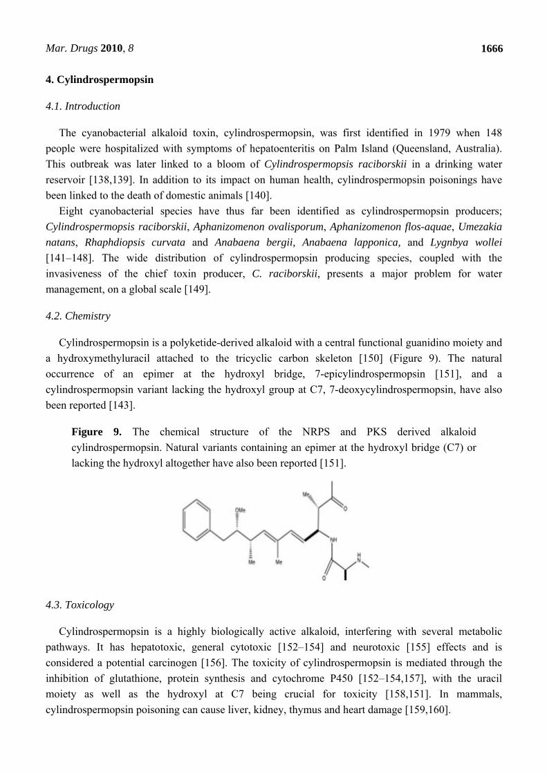
Cylindrospermopsin is a polyketide-derived alkaloid with a central functional guanidino moiety and
a hydroxymethyluracil attached to the tricyclic carbon skeleton [150] (Figure 9). The natural
occurrence of an epimer at the hydroxyl bridge, 7-epicylindrospermopsin [151], and a
cylindrospermopsin variant lacking the hydroxyl group at C7, 7-deoxycylindrospermopsin, have also
been reported [143].
Figure 9. The chemical structure of the NRPS and PKS derived alkaloid
cylindrospermopsin. Natural variants containing an epimer at the hydroxyl bridge (C7) or
lacking the hydroxyl altogether have also been reported [151].
4.3. Toxicology
Cylindrospermopsin is a highly biologically active alkaloid, interfering with several metabolic
pathways. It has hepatotoxic, general cytotoxic [152–154] and neurotoxic [155] effects and is
considered a potential carcinogen [156]. The toxicity of cylindrospermopsin is mediated through the
inhibition of glutathione, protein synthesis and cytochrome P450 [152–154,157], with the uracil
moiety as well as the hydroxyl at C7 being crucial for toxicity [158,151]. In mammals,
cylindrospermopsin poisoning can cause liver, kidney, thymus and heart damage [159,160].
Mar. Drugs 2010, 8
1667
4.4. Biosynthesis and Genetics
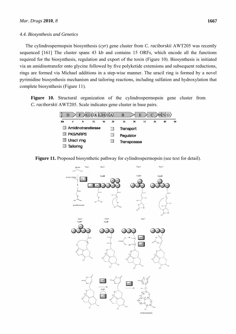
The cylindrospermopsin biosynthesis (cyr) gene cluster from C. raciborskii AWT205 was recently
sequenced [161] The cluster spans 43 kb and contains 15 ORFs, which encode all the functions
required for the biosynthesis, regulation and export of the toxin (Figure 10). Biosynthesis is initiated
via an amidinotransfer onto glycine followed by five polyketide extensions and subsequent reductions,
rings are formed via Michael additions in a step-wise manner. The uracil ring is formed by a novel
pyrimidine biosynthesis mechanism and tailoring reactions, including sulfation and hydroxylation that
complete biosynthesis (Figure 11).
Figure 10. Structural organization of the cylindrospermopsin gene cluster from
C. raciborskii AWT205. Scale indicates gene cluster in base pairs.
Figure 11. Proposed biosynthetic pathway for cylindrospermopsin (see text for detail).

Mar. Drugs 2010, 8
1668
The first step in formation of the carbon skeleton of cylindrospermopsin involves the synthesis of
guanidinoacetate via the transamidination of glycine [162–164]. An amidinotransferase encoded by
cyrA, putatively transfers a guanidino group from arginine [163], to glycine thus forming
guanidinoacetate. A mixed NRPS-PKS encoded by cyrB is thought to activate guanidinoacetate, which
is then transferred via the swinging arm of the peptidyl carrier protein (PCP) to the KS domain. The
AT domain of CyrB activates malonyl-CoA and attaches it to the ACP. This is followed by a
condensation reaction between the activated guanidinoacetate and malonyl-CoA in the KS domain.
The methyl transferase (MT) domain identified in CyrB is predicted to methylate C13. CyrB contains
two reducing modules, KR and DH. Their concerted reaction reduces the keto group to a hydroxyl
followed by elimination of H2O, resulting in a double bond between C13 and C14. A nucleophilic
attack of the amidino group at N19 onto the newly formed double bond between C13 and C14 then
putatively occurs via a ‘Michael addition’. The cyclization follows Baldwin’s rules for ring closure
[165], resulting in the formation of the first ring in cylindrospermopsin. This reaction could be
spontaneous and may not require enzymatic catalysis, as it is energetically favorable [165]. This is the
first of three ring formations and is one of the principal differences between Mihali et al.’s [161]
biosynthetic pathway and that previously proposed [163].
The third step in the biosynthesis of cylindrospermopsin involves cyrC, which encodes a PKS with
KS, AT, KR, and ACP domains. The action of these domains results in the elongation of the growing
chain by an acetate via activation of malonyl-CoA by the AT domain, its transfer to ACP and
condensation at the KS domain with the product of CyrB. The elongated chain is bound to the ACP of
CyrC and the KR domain reduces the keto group to a hydroxyl group on C12. Following the catalysis
of enzyme CyrC is CyrD, a PKS. The action of this PKS module on the product of CyrC results in the
addition of one acetate and the reduction of the keto group on C10 to a hydroxyl and dehydration to a
double bond between C9 and C10. This double bond is the site of a nucleophilic attack by the amidino
group N19 via another Michael addition that again follows Baldwin’s rules of ring closure [165],
resulting in the formation of the second ring, the first six-membered ring made in cylindrospermopsin.
The intermediate produced by CyrD is the substrate for CyrE (step 5 in Figure 1). A PKS, CyrE,
catalyzes the addition of one acetate and the formation of a double bond between C7 and C8. This
double bond is attacked by N18 via a Michael addition and the third cyclisation occurs, resulting in the
second 6-member ring. The cyrF gene encodes the final PKS module - a minimal PKS containing only
a KS, AT, and ACP. CyrF acts on the product of CyrE and elongates the chain by an acetate, leaving
C4 and C6 unreduced. Step 7 in the pathway involves the formation of the uracil ring, a reaction that
has been elusive so far and is required for the toxicity of the final cylindrospermopsin
compound [151].
The cylindrospermopsin gene cluster encodes two enzymes CyrG and CyrH that are most similar to
the enzyme family of amidohydrolases/ureases/dihydrotases, whose members catalyze the formation
and cleavage of N-C bonds. Mihali and co-workers [161] propose that these enzymes transfer a second
guanidino group from a donor molecule, such as arginine or urea, onto C6 and C4 of
cylindrospermopsin resulting in the formation of the uracil ring. The first reaction consists of the
formation of a covalent bond between the N of the guanidino donor and C6 of cylindrospermopsin
followed by an elimination of H2O forming a double bond between C5 and C6. The second reaction
catalyses the formation of a bond between the second N on the guanidino donor and C4 of

Mar. Drugs 2010, 8
1669
cylindrospermopsin, co-committently with the breaking of the thioester bond between the acyl carrier
protein of CyrF and cylindrospermopsin, causing the release of the molecule from the enzyme
complex. The third reaction - if required - would catalyze the cleavage of the guanidino group from a
donor molecule other than urea. The action of CyrG and CyrH in the formation of the uracil ring in
cylindrospermopsin describes a novel biosynthesis pathway of a pyrimidine. Mihali and
co-workers’ [161] genetic analysis shows that cyclization may happen stepwise, with successive ring
formation of the appropriate intermediate as it is synthesized. This mechanism also explains the lack of
a thioesterase or cyclization domain, which are usually associated with NRPS/PKS modules and
catalyze the release and cyclization of the final product from the enzyme complex.
The sulfation of cylindrospermopsin at C12 is likely to be carried out by the action of a
sulfotransferase. The cyrJ gene encodes a protein that is most similar to human 3'-phosphoadenylyl
sulfate (PAPS) dependent sulfotransferases. Similar enzymes have recently been implicated in the
sulfation of other cyanotoxins [120]. The cylindrospermopsin gene cluster also encodes an
adenylsulfate kinase (ASK), namely CyrN. ASKs are enzymes that catalyse the formation of PAPS,
which is the sulfate donor for sulfotransferases. Mihali and co-workers [161] propose that CyrJ sulfates
cylindrospermopsin at C12 while CyrN creates the pool of PAPS required for this reaction. Screening
of cylindrospermopsin producing and non-producing strains revealed that the sulfotransferase genes
were only present in cylindrospermopsin producing strains, further affirming the involvement of this
entire cluster in the biosynthesis of cylindrospermopsin. The cyrJ gene might therefore be a good
candidate for a toxin probe, as it is more unique than NRPS and PKS genes and would presumably
have less cross-reactivity with other gene clusters containing these genes, which are common in
cyanobacteria. The final tailoring reaction is carried out by CyrI. CyrI putatively catalyzes the
hydroxylation of C7, a residue that, along with the uracil ring, seems to confer much of the toxicity of
cylindrospermopsin [158,166].
The cylindrospermopsin gene cluster contains an ORF denoted cyrK, the product of which is most
similar to sodium ion driven multi-drug and toxic compound extrusion proteins (MATE) of the NorM
family. CyrK is hypothesized to function as a transporter for cylindrospermopsin, based on this
homology and its central location in the cluster.
Cylindrospermopsin production has been shown to be highest when fixed nitrogen is eliminated
from the growth media [167]. Flanking the cylindrospermopsin gene cluster are “hyp” gene homologs
involved in the maturation of hydrogenases. In the cyanobacterium Nostoc PCC73102 they are under
the regulation of the global nitrogen regulator NtcA, that activates transcription of nitrogen
assimilation genes [168,169]. It is plausible that the cylindrospermopsin gene cluster is under the same
regulation, as it is located wholly within the “hyp” gene cluster in C. raciborskii AWT205, and no
obvious promoter region in the cylindrospermopsin gene cluster could be identified. Finally, the
cylindrospermopsin cluster also includes an ORF at its 3’-end designated CyrO. By homology, it
encodes a hypothetical protein that appears to possess an ATP binding cassette, and is similar to WD
repeat proteins, which have diverse regulatory and signal transduction roles. CyrO may also have a
role in transcriptional regulation and DNA binding. It also shows homology to AAA family proteins
that often perform chaperone-like functions and assist in the assembly, operation, or disassembly of
protein complexes. Further insights into the role of CyrO are hindered due to low sequence homology
with other proteins in databases.

Mar. Drugs 2010, 8
1670
References and Notes
1. Sivonen, K.; Jones, G. Toxic Cyanobacteria in Water: A Guide to Their Public Health
consequences, Monitoring and Management; E and FN Spon: New York, NY, USA, 1999;
Volume 1, pp. 40–111.
2. Welker, M.; von Dohren, H. Cyanobacterial peptides - nature's own combinatorial biosynthesis.
FEMS Microbiol. Rev. 2006, 30, 530–563.
3. Botes, D.; Wessels, P.; Kruger, H.; Runnegar, M.; Santikarn, S.; Smith, R.; Barna, J.; Williams,
D. Structural studies on cyanoginosins-LR, -YR, -YA, and -YM, peptide toxins from Microcystis
aeruginosa. . J. Chem. Soc. 1985, 1, 2747–2748.
4. Namikoshi, M.; Yuan, M.; Sivonen, K.; Carmichael, W.W.; Rinehart, K.L.; Rouhiainen, L.; Sun,
F.; Brittain, S.; Otsuki, A. Seven new microcystins possessing two L-glutamic acid units, isolated
from Anabaena sp. strain 186. Chem. Res. Toxicol. 1998, 11, 143–149.
5. Rinehart, K.; Namikoshi, N.; Choi, B. Structure and biosynthesis of toxins from blue-green algae
(cyanobacteria). J. App. Phycol. 1994, 6, 159–176.
6. Sivonen, K. Cyanobacterial toxins and toxin production. Phycologia 1996, 35, 12–24.
7. Chorus, I.; Bartram, J. Toxic Cyanobacteria in Water. A Guide to their Public Health
Consequences, Monitoring and Management; E & FN Spon: London, UK, 1999.
8. Jochimsen, E.M.; Carmichael, W.W.; An, J.S.; Cardo, D.M.; Cookson, S.T.; Holmes, C.E.;
Antunes, M.B.; de Melo Filho, D.A.; Lyra, T.M.; Barreto, V.S.; Azevedo, S.M.; Jarvis, W.R.
Liver failure and death after exposure to microcystins at a hemodialysis center in Brazil. N. Engl.
J. Med. 1998, 338, 873–878.
9. Runnegar, M.T.; Gerdes, R.G.; Falconer, I.R. The uptake of the cyanobacterial hepatotoxin
microcystin by isolated rat hepatocytes. Toxicon 1991, 29, 43–51.
10. Runnegar, M.; Berndt, N.; Kaplowitz, N. Microcystin uptake and inhibition of protein
phosphatases: effects of chemoprotectants and self-inhibition in relation to known hepatic
transporters. Toxicol. Appl. Pharmacol. 1995, 134, 264–272.
11. Dawson, R.M. The toxicology of microcystins. Toxicon 1998, 36, 953–962.
12. Eriksson, J.E.; Toivola, D.; Meriluoto, J.A.; Karaki, H.; Han, Y.G.; Hartshorne, D. Hepatocyte
deformation induced by cyanobacterial toxins reflects inhibition of protein phosphatases.
Biochem. Biophys. Res. Commun. 1990, 173, 1347–1353.
13. Sahin, A.; Tencalla, F.G.; Dietrich, D.R.; Mez, K.; Naegeli, H. Enzymatic analysis of liver
samples from rainbow trout for diagnosis of blue-green algae-induced toxicosis. Am. J. Vet. Res.
1995, 56, 1110–1115.
14. Krishnamurthy, T.; Carmichael, W.W.; Sarver, E.W. Toxic peptides from freshwater
cyanobacteria (blue-green algae). I. Isolation, purification and characterization of peptides from
Microcystis aeruginosa and Anabaena flos-aquae. Toxicon 1986, 24, 865–873.
15. Watanabe, M.F.; Oishi, S.; Harda, K.; Matsuura, K.; Kawai, H.; Suzuki, M. Toxins contained in
Microcystis species of cyanobacteria (blue-green algae). Toxicon 1988, 26, 1017–1025.
16. Yoshizawa, S.; Matsushima, R.; Watanabe, M.F.; Harada, K.; Ichihara, A.; Carmichael, W.W.;
Fujiki, H. Inhibition of protein phosphatases by microcystins and nodularin associated with
hepatotoxicity. J. Cancer Res. Clin. Oncol. 1990, 116, 609–614.

Mar. Drugs 2010, 8
1671
17. Nishiwaki, S.; Fujiki, H.; Suganuma, M.; Nishiwaki-Matsushima, R.; Sugimura, T. Rapid
purification of protein phosphatase 2A from mouse brain by microcystin-affinity
chromatography. FEBS Lett. 1991, 279, 115–118.
18. Falconer, I.R. Tumor promotion and liver injury caused by oral consumption of cyanobacteria.
Environ. Toxicol. Water Qual. 1991, 6, 177–184.
19. Nishiwaki-Matsushima, R.; Ohta, T.; Nishiwaki, S.; Suganuma, M.; Kohyama, K.; Ishikawa, T.;
Carmichael, W.W.; Fujiki, H. Liver tumor promotion by the cyanobacterial cyclic peptide toxin
microcystin-LR. J. Cancer Res. Clin. Oncol. 1992, 118, 420–424.
20. Yu, S. Primary prevention of hepatocellular carcinoma. J. Gastroenterol. Hepatol. 1995, 10,
674–682.
21. Yu, S. Drinking Water and Primary Liver Cancer; China academic publishers: New York, NY,
USA, 1989.
22. Tillett, D.; Dittmann, E.; Erhard, M.; von Dohren, H.; Borner, T.; Neilan, B.A. Structural
organization of microcystin biosynthesis in Microcystis aeruginosa PCC7806: an integrated
peptide-polyketide synthetase system. Chem. Biol. 2000, 7, 753–764.
23. Christiansen, G.; Fastner, J.; Erhard, M.; Borner, T.; Dittmann, E. Microcystin biosynthesis in
planktothrix: genes, evolution, and manipulation. J. Bacteriol. 2003, 185, 564–572.
24. Rouhiainen, L.; Vakkilainen, T.; Siemer, B.L.; Buikema, W.; Haselkorn, R.; Sivonen, K. Genes
coding for hepatotoxic heptapeptides (microcystins) in the cyanobacterium Anabaena strain 90.
Appl. Environ. Microbiol. 2004, 70, 686–692.
25. Hicks, L.M.; Moffitt, M.C.; Beer, L.L.; Moore, B.; Kelleher, N.L. Structural characterisation of
in vitro and in vivo intermediates on the loading module of microcystin synthetase. ACS Chem.
Biol. 2006, 1, 93–102.
26. Nishizawa, T.; Asayama, M.; Shirai, M. Cyclic heptapeptide microcystin biosynthesis requires
the glutamate racemase gene. Microbiology 2001, 147, 1235–1241.
27. Sielaff, H.; Dittmann, E.; Tandeau De Marsac, N.; Bouchier, C.; Von Dohren, H.; Borner, T.;
Schwecke, T. The mcyF gene of the microcystin biosynthetic gene cluster from Microcystis
aeruginosa encodes an aspartate racemase. Biochem. J. 2003, 373, 909–916.
28. Pearson, L.A.; Barrow, K.D.; Neilan, B.A. Characterization of the 2-hydroxy-acid
dehydrogenase McyI, encoded within the microcystin biosynthesis gene cluster of Microcystis
aeruginosa PCC7806. J. Biol. Chem. 2007, 282, 4681–4692.
29. Pearson, L.A.; Hisbergues, M.; Borner, T.; Dittmann, E.; Neilan, B.A. Inactivation of an ABC
transporter gene, mcyH, results in loss of microcystin production in the cyanobacterium
Microcystis aeruginosa PCC 7806. Appl. Environ. Microbiol. 2004, 70, 6370–6378.
30. Shi, L.; Carmichael, W.W.; Miller, I. Immuno-gold localization of hepatotoxins in cyanobacterial
cells. Arch. Microbiol. 1995, 163, 7–15.
31. Young, F.M.; Thomson, C.; Metcalf, J.S.; Lucocq, J.M.; Codd, G.A. Immunogold localisation of
microcystins in cryosectioned cells of Microcystis. J. Struct. Biol. 2005, 151, 208–214.
32. Kaebernick, M.; Neilan, B.A.; Borner, T.; Dittmann, E. Light and the transcriptional response of
the microcystin biosynthesis gene cluster. Appl. Environ. Microbiol. 2000, 66, 3387–3392.
Mar. Drugs 2010, 8
1672
33. Mikalsen, B.; Boison, G.; Skulberg, O.M.; Fastner, J.; Davies, W.; Gabrielsen, T.M.; Rudi, K.;
Jakobsen, K.S. Natural variation in the microcystin synthetase operon mcyABC and impact on
microcystin production in Microcystis strains. J. Bacteriol. 2003, 185, 2774–2785.
34. Tooming-Klunderud, A.; Mikalsen, B.; Kristensen, T.; Jakobsen, K.S. The mosaic structure of
the mcyABC operon in Microcystis. Microbiology 2008, 154, 1886–1899.
35. Sivonen, K. Effects of light, temperature, nitrate, orthophosphate, and bacteria on growth of and
hepatotoxin production by Oscillatoria agardhii strains. Appl. Environ. Microbiol. 1990, 56,
2658–2666.
36. Lukac, M.; Aegerter, R. Influence of trace metals on growth and toxin production of Microcystis
aeruginosa. Toxicon 1993, 31, 293–305.
37. van der Westhuizen, A.J.; Eloff, J.N. Effect of temperature and light on the toxicity and growth
of the blue-green alga Microcystis aeruginosa (UV-006). Planta 1985, 163, 55–59.
38. Song, L.; Sano, T.; Li, R.; Watanabe, M.; Liu, Y.; Kaya, K. Microcystin production of
Microcystis viridis (cyanobacteria) under different culture conditions. Phycol. Res. 1998, 42, 19.
39. Davis, T.W.; Berry, D.L.; Boyer, G.L.; Gobler, C.J. The effects of temperature and nutrients on
the growth and dynamics of toxic and non-toxic strains of Microcystis during cyanobacteria
blooms. Harmful Algae 2009, 8, 715–725.
40. Tonk, L.; Visser, P.M.; Christiansen, G.; Dittmann, E.; Snelder, E.O.; Wiedner, C.; Mur, L.R.;
Huisman, J. The microcystin composition of the cyanobacterium Planktothrix agardhii changes
toward a more toxic variant with increasing light intensity. Appl. Environ. Microbiol. 2005, 71,
5177–5181.
41. Sevilla, E.; Martin-Luna, B.; Vela, L.; Bes, M.T.; Fillat, M.F.; Peleato, M.L. Iron availability
affects mcyD expression and microcystin-LR synthesis in Microcystis aeruginosa PCC7806.
Environ. Microbiol. 2008, 10, 2476–2483.
42. Kaebernick, M.; Dittmann, E.; Borner, T.; Neilan, B.A. Multiple alternate transcripts direct the
biosynthesis of microcystin, a cyanobacterial nonribosomal peptide. Appl. Environ. Microbiol.
2002, 68, 449–455.
43. Sivonen, K.; Kononen, K.; Carmichael, W.W.; Dahlem, A.M.; Rinehart, K.L.; Kiviranta, J.;
Niemela, S.I. Occurrence of the hepatotoxic cyanobacterium Nodularia spumigena in the Baltic
Sea and structure of the toxin. Appl. Environ. Microbiol. 1989, 55, 1990–1995.
44. Jones, G.J.; Blackburn, S.I.; Parker, N.S. A toxic bloom of Nodularia spumigena mertens in
Orielton Lagoon, Tasmania. Aust. J. Mar. Freshwater Res. 1994, 45, 787–800.
45. Heresztyn, T.; Nicholson, B.C. Nodularin concentrations in Lakes Alexandrina and Albert, South
Australia, during a bloom of the cyanobacterium (blue-green alga) Nodularia spumigena and
degradation of the toxin. Environ. Toxicol. Water Qual. 1997, 12, 273–282.
46. Nehring, S. Mortality of dogs associated with a mass development of Nodularia spumigena
(Cyanophyceae) in a brackish lake at the German North Sea coast. J. Plankton Res. 1993, 15,
867–872.
47. Carmichael, W.W.; Eschedor, J.T.; Patterson, G.M.; Moore, R.E. Toxicity and partial structure of
a hepatotoxic peptide produced by the cyanobacterium Nodularia spumigena Mertens emend.
L575 from New Zealand. Appl. Environ. Microbiol. 1988, 54, 2257–2263.

Mar. Drugs 2010, 8
1673
48. Galat, D.L.; Verdin, J.P.; Sims, L.L. Large-scale patterns of Nodularia spumigena blooms in
Pyramid Lake, Nevada, determined from Landsat imagery: 1972–1986. Hydrobiologia 1990,
197, 147–164.
49. Baker, P.D.; Humpage, A.R. Toxicity associated with commonly occurring cyanobacteria in
surface waters of the Murray-Darling Basin, Australia. Aust. J. Mar. Freshwater Res. 1994, 45,
773–786.
50. Karjalainen, M.; Engstrom-Ost, J.; Korpinen, S.; Peltonen, H.; Paakkonen, J.P.; Ronkkonen, S.;
Suikkanen, S.; Viitasalo, M. Ecosystem consequences of cyanobacteria in the northern Baltic
Sea. Ambio 2007, 36, 195–202.
51. Carmichael, W.W. The Toxins of Cyanobacteria. Sci. Am. 1994, 64–72.
52. Francis, G. Poisonous Australian lake. Nature 1878, 18, 11–12.
53. Ohta, T.; Sueoka, E.; Iida, N.; Komori, A.; Suganuma, M.; Nishiwaki, R.; Tatematsu, M.; Kim,
S.J.; Carmichael, W.W.; Fujiki, H. Nodularin, a potent inhibitor of protein phosphatases 1 and
2A, is a new environmental carcinogen in male F344 rat liver. Cancer Res. 1994, 54, 6402–6406.
54. Van Buynder, P.G.; Oughtred, T.; Kirkby, B.; Phillips, S.; Eaglesham, G.; Thomas, K.; Burch,
M. Nodularin uptake by seafood during a cyanobacterial bloom. Environ. Toxicol. 2001, 16,
468–471.
55. Falconer, I.R.; Choice, A.; Hosja, W. Toxicity of edible mussels Mytilus edulis growing naturally
in an estuary during a water bloom of the blue-green alga Nodularia spumigena. Environ.
Toxicol. Water Qual. 1992, 7, 119–123.
56. Mazur-Marzec, H.; Tyminska, A.; Szafranek, J.; Plinski, M. Accumulation of nodularin in
sediments, mussels, and fish from the Gulf of Gdansk, southern Baltic Sea. Environ. Toxicol.
2007, 22, 101–111.
57. Rinehart, K.L.; Harada, K.; Namikoshi, M.; Chen, C.; Harvis, C.A.; Munro, M.H.G.; Blunt, J.W.;
Mulligan, P.E.; Beasley, V.R.; Dahlem, A.M.; Carmicheal, W.W. Nodularin, microcystin, and
the configuration of Adda. J. Am. Chem. Soc. 1988, 110, 8557–8558.
58. Saito, K.; Konno, A.; Ishii, H.; Saito, H.; Nishida, F.; Abe, T.; Chen, C. Nodularin-Har: a new
nodularin from Nodularia. J. Nat. Prod. 2001, 64, 139–141.
59. Beattie, K.A.; Kaya, K.; Codd, G.A. The cyanobacterium Nodularia PCC7804, of freshwater
origin, produces [L-Har2]nodularin. Phytochemistry 2000, 54, 57–61.
60. deSilva, E.D.; Williams, D.E.; Anderson, R.J.; Klix, H.; Holmes, C.F.B.; Allen, T.M. Motuporin,
a potent protein phosphatase inhibitor isolated from the Papua New Guinea sponge Theonella
swinhoei Gray. Tetrahedron Lett. 1992, 33, 1561–1564.
61. Eriksson, J.E.; Meriluoto, J.A.; Kujari, H.P.; Osterlund, K.; Fagerlund, K.; Hallbom, L.
Preliminary characterization of a toxin isolated from the cyanobacterium Nodularia spumigena.
Toxicon 1988, 26, 161–166.
62. Honkanen, R.E.; Dukelow, M.; Zwiller, J.; Moore, R.E.; Khatra, B.S.; Boynton, A.L.
Cyanobacterial nodularin is a potent inhibitor of type 1 and type 2A protein phosphatases. Mol.
Pharmacol. 1991, 40, 577–583.
63. An, J.; Carmichael, W.W. Use of a colorimetric protein phosphatase inhibition assay and enzyme
linked immunosorbent assay for the study of microcystins and nodularins. Toxicon 1994, 32,
1495–1507.

Mar. Drugs 2010, 8
1674
64. MacKintosh, R.W.; Dalby, K.N.; Campbell, D.G.; Cohen, P.T.; Cohen, P.; MacKintosh, C. The
cyanobacterial toxin microcystin binds covalently to cysteine-273 on protein phosphatase 1.
FEBS Lett. 1995, 371, 236–240.
65. Kelker, M.S.; Page, R.; Peti, W. Crystal structures of protein phosphatase-1 bound to nodularin-
R and tautomycin: a novel scaffold for structure-based drug design of serine/threonine
phosphatase inhibitors. J. Mol. Biol. 2009, 385, 11–21.
66. Runnegar, M.; Berndt, N.; Kong, S.M.; Lee, E.Y.; Zhang, L. In vivo and in vitro binding of
microcystin to protein phosphatases 1 and 2A. Biochem. Biophys. Res. Commun. 1995, 216,
162–169.
67. Bagu, J.R.; Sykes, B.D.; Craig, M.M.; Holmes, C.F. A molecular basis for different interactions
of marine toxins with protein phosphatase-1. Molecular models for bound motuporin,
microcystins, okadaic acid, and calyculin A. J. Biol. Chem. 1997, 272, 5087–5097.
68. Karjalainen, M.; Pääkkönen, J.; Peltonen, H.; Sipiä, V.; Valtonen, T.; Viitasalo, M. Nodularin
concentrations in Baltic Sea zooplankton and fish during a cyanobacterial bloom. Mar. Biol.
2008, 155, 483–491.
69. Persson, K.; Legrand, C.; Olsson, T. Detection of nodularin in European flounder (Platichthys
flesus) in the west coast of Sweden: Evidence of nodularin mediated oxidative stress. Harmful
Algae 2009, 8, 832–838.
70. Vuorinen, P.J.; Sipia, V.O.; Karlsson, K.; Keinanen, M.; Furey, A.; Allis, O.; James, K.; Perttila,
U.; Rimaila-Parnanen, E.; Meriluoto, J.A. Accumulation and effects of nodularin from a single
and repeated oral doses of cyanobacterium Nodularia spumigena on flounder (Platichthys flesus
L.). Arch. Environ. Contam. Toxicol. 2009, 57, 164–173.
71. Moffitt, M.C.; Neilan, B.A. Characterization of the nodularin synthetase gene cluster and
proposed theory of the evolution of cyanobacterial hepatotoxins. Appl. Environ. Microbiol. 2004,
70, 6353–6362.
72. Kleinkauf, H.; Von Dohren, H. A nonribosomal system of peptide biosynthesis. Eur. J. Biochem.
1996, 236, 335–351.
73. Copp, J.N.; Roberts, A.A.; Marahiel, M.A.; Neilan, B.A. Characterization of PPTNs, a
cyanobacterial phosphopantetheinyl transferase from Nodularia spumigena NSOR10. J.
Bacteriol. 2007, 189, 3133–3139.
74. Jonasson, S.; Vintila, S.; Sivonen, K.; El-Shehawy, R. Expression of the nodularin synthetase
genes in the Baltic Sea bloom-former cyanobacterium Nodularia spumigena strain AV1. FEMS.
Microbiol. Ecol. 2008, 65, 31–39.
75. Negri, A.P.; Jones, G.J.; Hindmarsh, M. Sheep mortality associated with paralytic shellfish
poisons from the cyanobacterium Anabaena circinalis. Toxicon 1995, 33, 1321–1329.
76. Reyero, M.; Cacho, E.; Martínez, A.; Vázquez, J.; Marina, A.; Fraga, S.; Franco, J. Evidence of
saxitoxin derivatives as causative agents in the 1997 mass mortality of monk seals in the Cape
Blanc Peninsula. Nat. Toxins 1999, 7, 311–315.
77. Sawyer, P.J.; Gentile, J.H.; Sasner, J.J.J. Demonstration of a toxin from Aphanisomenon flos-
aquae (L.) Ralfs. Can. J. Microbiol. 1968, 14, 1199–1204.
78. Shumway, S.E. Phycotoxin-related shellfish poisoning: Bivalve molluscs are not the only
vectors. Rev. Fish. Sci. 1995, 3, 1–31.

Mar. Drugs 2010, 8
1675
79. Shumway, S.E.; Cucci, T.L.; Yentsch, C.M.; Newell, R.C.; Gainey, L. The effects of the toxic
dinoflagellate, Protogonyaulax tamarensis, on the physiology and behavior of marine molluscs.
J. Shellfish Res. 1988, 7, 132–133.
80. Hallegraeff, G.M. Harmful algal blooms: A global overview. In Manual on Harmful Marine
Microalgae; Hallegraeff, G.M.; Anderson, D.M., Cembella, A.D., Eds.; UNESCO: Paris, France,
1995; pp. 1–22.
81. Daly, J.W. Marine toxins and nonmarine toxins: convergence or symbiotic organisms? J. Nat.
Prod. 2004, 67, 1211–1215.
82. Baker, T.R.; Doucette, G.J.; Powell, C.L.; Boyer, G.L.; Plumley, F.G. GTX(4) imposters:
characterization of fluorescent compounds synthesized by Pseudomonas stutzeri SF/PS and
Pseudomonas/Alteromonas PTB-1, symbionts of saxitoxin-producing Alexandrium spp. Toxicon
2003, 41, 339–347.
83. Kodoma, M.; Ogata, T.; Sakamoto, S.; Sato, S.; Honda, T.; Miwatani, T. Production of paralytic
shellfish toxins by a bacterium Moraxella sp. isolated from Protogonyaulax tamarensis. Toxicon
1990, 28, 707–714.
84. Martins, C.A.; Alvito, P.; Tavares, M.J.; Pereira, P.; Doucette, G.; Franca, S. Reevaluation of
production of paralytic shellfish toxin by bacteria associated with dinoflagellates of the
Portuguese coast. Appl. Environ. Microbiol. 2003, 69, 5693–5698.
85. Castro, D.; Vera, D.; Lagos, N.; Garcia, C.; Vasquez, M. The effect of temperature on growth
and production of paralytic shellfish poisoning toxins by the cyanobacterium Cylindrospermopsis
raciborskii C10. Toxicon 2004, 44, 483–489.
86. Negri, A.; Stirling, D.; Quilliam, M.; Blackburn, S.; Bolch, C.; Burton, I.; Eaglesham, G.;
Thomas, K.; Walter, J.; Willis, R. Three novel hydroxybenzoate saxitoxin analogues isolated
from the dinoflagellate Gymnodinium catenatum. Chem. Res. Toxicol. 2003, 16, 1029–1033.
87. Sako, Y.; Kim, C.H.; Ishida, Y. Mendelian inheritance of paralytic shellfish poisoning toxin in
the marine dinoflagellate Alexandrium. Biosci. Biotechnol. Biochem. 1992, 56692–56694.
88. Sako, Y.; Naya, N.; Yoshida, Y.; Kim, C.H.; Ushida, A.; Ishida, Y. Studies on the stability and
heredity of PSP toxin composition in the toxic dinoflagellate Alexandrium. In Harmful Marine
Algal Blooms; Lassus, P., Arzul, G., Erard, P., Gentien, C., Marcaillou-Le Baut, C., Eds.;
London-Paris-New York: Nantes, France, 1995; pp. 345–350.
89. Velzeboer, R.M.A.; Baker, P.D.; Rositano, J.; Heresztyn, T.; Codd, G.A.; Raggett, S.L.
Geographical patterns of occurrence and composition of saxitoxins in the cyanobacterial genus
Anabaena (Nostocales, Cyanophyta) in Australia. Phycologia 2000, 39, 395–407.
90. Mahmood, N.A.; Carmichael, W.W. Paralytic shellfish poisons produced by the freshwater
cyanobacterium Aphanizomenon flos-aquae NH-5. Toxicon 1986, 24, 175–186.
91. Harada, T.; Oshima, Y.; Yasumoto, T. Structure of two paralytic shellfish toxins, gonyautoxins V
and VI, isolated from a tropical dinoflagellate Pyrodinium bahamense var. compressa. Agric.
Biol. Chem. 1982, 46, 1861–1864.
92. Oshima, Y.; Hasegawa, M.; Yasumoto, T.; Hallegaeff, G.; Blackburn, S. Dinoflagellate
Gimnodium catenatum as the source of paralytic shellfishtoxins in Tasmanian shellfish. Toxicon
1987, 25, 1105–1111.

Mar. Drugs 2010, 8
1676
93. Shimizu, Y. Chemistry and Distribution of Deleterious Dinoflagellate Toxins; Faulkner, D.,
Fenical, W., Eds.; Plenum: New York, NY, USA, 1977; pp. 261–269.
94. Carmichael, W.W.; Evans, W.R.; Yin, Q.Q.; Bell, P.; Moczydlowski, E. Evidence for paralytic
shellfish poisons in the freshwater cyanobacterium Lyngbya wollei (Farlow ex Gomont) comb.
nov. Appl. Environ. Microbiol. 1997, 63, 3104–3110.
95. Humpage, A.R.; Rositano, J.; A., B.; Brown, R.; Baker, P.; Nicholson, B.C.; Steffensen, D.A.
Paralytic shellfish poisons from australian cyanobacterial blooms. Aust. J. Mar. Freshwater Res.
1994, 45, 761–771.
96. Lagos, N.; Onodera, H.; Zagatto, P.A.; Andrinolo, D.; Azevedo, S.M.; Oshima, Y. The first
evidence of paralytic shellfish toxins in the fresh water cyanobacterium Cylindrospermopsis
raciborskii, isolated from Brazil. Toxicon 1999, 37, 1359–1373.
97. Ballot, A.; Fastner, J.; Wiedner, C. Paralytic shellfish poisoning toxin-producing cyanobacterium
Aphanizomenon gracile in northeast Germany. Appl. Environ. Microbiol. 2010, 76, 1173–1180.
98. Llewellyn, L.E. Saxitoxin, a toxic marine natural product that targets a multitude of receptors.
Nat. Prod. Rep. 2006, 23, 200–222.
99. Llewellyn, L.E.; Negri, A.P.; Doyle, J.; Baker, P.D.; Beltran, E.C.; Neilan, B.A. Radioreceptor
assays for sensitive detection and quantitation of saxitoxin and its analogues from strains of the
freshwater cyanobacterium, Anabaena circinalis. Environ. Sci. Technol. 2001, 35, 1445–1451.
100. Oshima, Y. Postcolumn derivatization liquid chromatographic method for paralytic shellfish
toxins. J. AOAC Int. 1995, 78, 528–532.
101. Oshima, Y.; Blackburn, S.; Hallegraeff, G.M. Comparative study on paralytic shellfish toxin
profiles of the dinoflagellate Gymnodinium catenatum from three different countries. Mar. Biol.
1993, 116, 471–476.
102. Onodera, H.; Satake, M.; Oshima, Y.; Yasumoto, T.; Carmichael, W.W. New saxitoxin
analogues from the freshwater filamentous cyanobacterium Lyngbya wollei. Nat. Toxins 1997, 5,
146–151.
103. Arakawa, O.; Nishio, S.; Noguchi, T.; Shida, Y.; Onoue, Y. A new saxitoxin analogue from a
xanthid crab Atergatis floridus. Toxicon 1995, 33, 1577–1584.
104. Arakawa, O.; Noguchi, T.; Shida, Y.; Onoue, Y. Occurrence of carbamoyl-N-hydroxy derivatives
of saxitoxin and neosaxitoxin in a xanthid crab Zosimus aeneus. Toxicon 1994, 32, 175–183.
105. Zaman, L.; Arakawa, O.; Shimosu, A.; Shida, Y.; Onoue, Y. Occurrence of a methyl derivative
of saxitoxin in Bangladeshi freshwater puffers. Toxicon 1998, 36, 627–630.
106. Yotsu-Yamashita, M.; Kim, Y.H.; Dudley, S.C. Jr., ; Choudhary, G.; Pfahnl, A.; Oshima, Y.;
Daly, J.W. The structure of zetekitoxin AB, a saxitoxin analog from the golden frog Atelopus
zeteki: a potent sodium channel blocker. Proc. Natl. Acad. Sci. USA 2004, 101, 4346–4351.
107. Kao, C.Y. Paralytic shellfish poisoning. In Algal Toxins in Seafood and Drinking Water;
Falconer, I.R., Ed.; Academic Press: London, UK, 1993; pp. 75–86.
108. Kao, C.Y.; Levinson, S.R. Tetrodotoxin, Saxitoxin, and the Molecular Biology of the Sodium
Channel; The New York Academy of Science: New York, NY, USA, 1986; Volume 479.
109. Su, Z.; Sheets, M.; Ishida, H.; Li, F.; Barry, W.H. Saxitoxin blocks L-type ICa. J. Pharmacol.
Exp. Ther. 2004, 308, 324–329.

Mar. Drugs 2010, 8
1677
110. Wang, J.; Salata, J.J.; Bennett, P.B. Saxitoxin is a gating modifier of HERG K+ channels. J. Gen.
Physiol. 2003, 121, 583–598.
111. Halstead, B.W.; Schantz, E.J. Paralytic shellfish poisoning. WHO Offset Publ. 1984, 1–59.
112. Evans, M.H. Mechanism of saxitoxin and tetrodotoxin poisoning. Br. Med. Bull. 1969, 25,
263–267.
113. Satin, J.; Kyle, J.W.; Chen, M.; Bell, P.; Cribbs, L.L.; Fozzard, H.A.; Rogart, R.B. A mutant of
TTX-resistant cardiac sodium channels with TTX-sensitive properties. Science 1992, 256,
1202–1205.
114. Strichartz, G. Structural determinabts of the affinity of saxitoxin for neuronal sodium channels. J.
Gen. Physiol. 1984, 84, 281–305.
115. Tikhonov, D.B.; Zhorov, B.S. Modeling P-loops domain of sodium channel: homology with
potassium channels and interaction with ligands. Biophys. J. 2005, 88, 184–197.
116. Jiang, Y.; Lee, A.; Chen, J.; Cadene, M.; Chait, B.T.; MacKinnon, R. The open pore
conformation of potassium channels. Nature 2002, 417, 523–526.
117. Hall, S.; Strichartz, G.; Moczydlowski, E.; Ravindran, A.; Reichardt, P.B. The saxitoxins:
sources, chemistry and pharmacology. In Marine Toxins. Origin, Structure and Pharmacology;
Hall, S., Reichardt, P.B., Eds.; American Chemical Society: Washington DC, USA, 1990;
pp. 29–69.
118. Cembella, A.D.; Shumway, S.E.; Larocque, R. Sequestering and putative biotransformation of
paralytic shellfish toxins by the sea scallop Placopecten magellanicus - seasonal and spatial
scales in natural populations. J. Exp. Mar. Biol. Ecol. 1994, 180, 1–22.
119. Cembella, A.D.; Shumway, S.E.; Lewis, N.I. Anatomical distribution and spatiotemporal
variation in paralytic shellfish toxin composition in two bivalve species from the Gulf of Maine.
J. Shellfish Res. 1993, 12, 389–403.
120. Kellmann, R.; Mihali, T.K.; Jeon, Y.J.; Pickford, R.; Pomati, F.; Neilan, B.A. Biosynthetic
intermediate analysis and functional homology reveal a saxitoxin gene cluster in cyanobacteria.
Appl. Environ. Microbiol. 2008, 74, 4044–4053.
121. Neuwald, A.F.; Landsman, D. GCN5-related histone N-acetyltransferases belong to a diverse
superfamily that includes the yeast SPT10 protein. Trends Biochem. Sci. 1997, 22, 154–155.
122. Hederstedt, L.; Rutberg, L. Succinate dehydrogenase--a comparative review. Microbiol. Rev.
1981, 45, 542–555.
123. Mahmood, N.A.; Carmichael, W.W. The pharmacology of anatoxin-a(s), a neurotoxin produced
by the freshwater cyanobacterium Anabaena flos-aquae NRC 525–17. Toxicon 1986, 24,
425–434.
124. Sako, Y.; Yoshida, T.; Uchida, A.; Arakawa, O.; Noguchi, T.; Ishida, Y. Purification and
characterization of a sulfotransferase specific to N-21 of saxitoxin and gonyautoxin 2+3 from the
toxic dinoflagellate Gymnodinium catenatum (Dinophyceae). J. Phycol 2001, 37, 1044–1051.
125. Yoshida, T.; Sako, Y.; Uchida, A.; Kakutani, T.; Arakawa, O.; Noguchi, T.; Ishida, Y.
Purification and characterization of sulfotransferase specific to O-22 of 11-hydroxy saxitoxin
from the toxic dinoflagellate Gymnodinium catenatum (dinophyceae). Fish. Sci. 2002, 68,
634–642.

Mar. Drugs 2010, 8
1678
126. Schwedock, J.S.; Long, S.R. Rhizobium meliloti genes involved in sulfate activation: the two
copies of nodPQ and a new locus, sua. Genetics 1992, 132, 899–909.
127. Akoh, C.C.; Lee, G.C.; Liaw, Y.C.; Huang, T.H.; Shaw, J.F. GDSL family of serine
esterases/lipases. Prog. Lipid Res. 2004, 43, 534–552.
128. Pomati, F.; Rossetti, C.; Manarolla, G.; Burns, B.P.; Neilan, B.A. Interactions between
intracellular Na+ levels and saxitoxin production in Cylindrospermopsis raciborskii T3.
Microbiology 2004, 150, 455–461.
129. Brown, M.H.; Paulsen, I.T.; Skurray, R.A. The multidrug efflux protein NorM is a prototype of a
new family of transporters. Mol. Microbiol. 1999, 31, 394–395.
130. Anderson, D.M.; Kulis, D.M.; Sullivan, J.J.; Hall, S.; Lee, C. Dynamics and physiology of
saxitoxin production by the dinoflagellates Alexandrium spp. Mar. Biol. 1990, 104, 511–524.
131. Dias, E.; Pereira, P.; Franca, S. Production of paralytic shellfish toxins by Aphanizomenon sp.
LMECYA 31 (cyanobacteria). J. Phycol. 2002, 38, 705–712.
132. Gedaria, A.I.; Luckas, B.; Reinhardt, K.; Azanza, R.V. Growth response and toxin concentration
of cultured Pyrodinium bahamense var. compressum to varying salinity and temperature
conditions. Toxicon 2007, 50, 518–529.
133. Steed, P.M.; Wanner, B.L. Use of the rep technique for allele replacement to construct mutants
with deletions of the pstSCAB-phoU operon: evidence of a new role for the PhoU protein in the
phosphate regulon. J. Bacteriol. 1993, 175, 6797–6809.
134. Ehria, S.; Ohmori, M. NrrA, a nitrogen-responsive response regulator facilitates heterocyst
development in the cyanobacterium Anabaena sp. strain PCC 7120. Mol. Microbiol. 2006, 59,
1692–1703.
135. Forst, S.; Delgado, J.; Inouye, M. Phosphorylation of OmpR by the Osmosensor EnvZ Modulates
Expression of the ompF and ompC Genes in Escherichia coli. Proc. Natl. Acad. Sci. USA 1989,
86, 6052–6056.
136. Mihali, T.K.; Kellmann, R.; Neilan, B.A. Characterisation of the paralytic shellfish toxin
biosynthesis gene clusters in Anabaena circinalis AWQC131C and Aphanizomenon sp. NH-5.
BMC Biochem. 2009, 10, 8.
137. Kellmann, R.; Mihali, T.K.; Neilan, B.A. Identification of a saxitoxin biosynthesis gene with a
history of frequent horizontal gene transfers. J. Mol. Evol. 2008, 67, 526–538.
138. Bourke, A.T.C.; Hawes, R.B.; Neilson, A.; Stallman, N.D. An outbreak of hepato-enteritis (the
Palm Island mystery disease) possibly caused by algal intoxication. Toxicon 1983, 21, 45–48.
139. Byth, S. Palm Island mystery disease. Med. J. Aust. 1980, 2, 40–42.
140. Saker, M.L.; Thomas, A.D.; Norton, J.H. Cattle mortality attributed to the toxic cyanobacterium
Cylindrospermopsis raciborskii in an outback region of North Queensland. Environ. Toxicol.
Water Qual. 1999, 14, 179–182.
141. Banker, R.; Carmeli, S.; Hadas, O.; Teltsch, B.; Porat, R.; Sukenik, A. Identification of
cylindrospermopsin in Aphanizomenon ovalisporum (Cyanophyceae) isolated from Lake
Kinneret, Israel. J. Phycol. 1997, 33, 613–616.
142. Harada, K.-I.; Ohtani, I.; Iwamoto, K.; Suzuki, M.; Watanabe, M.F.; Watanabe, M.; Terao, K.
Isolation of cylindrospermopsin from a cyanobacterium Umezakia natans and its screening
method. Toxicon 1994, 32, 73–84.

Mar. Drugs 2010, 8
1679
143. Li, R.; Carmichael, W.W.; Brittain, S.; Eaglesham, G.K.; Shaw, G.R.; Liu, Y.; Watanabe, M.M.
First report of the cyanotoxins cylindrospermopsin and deoxycylindrospermopsin from
Raphidiopsis curvata (Cyanobacteria). J. Phycol. 2001, 37, 1121–1126.
144. Li, R.; Carmichael, W.W.; Brittain, S.; Eaglesham, G.K.; Shaw, G.R.; Mahakhant, A.;
Noparatnaraporn, N.; Yongmanitchai, W.; Kaya, K.; Watanabe, M.M. Isolation and identification
of the cyanotoxin cylindrospermopsin and deoxy-cylindrospermopsin from a Thailand strain of
Cylindrospermopsis raciborskii (Cyanobacteria). Toxicon 2001, 39, 973–980.
145. Preussel, K.; Stuken, A.; Wiedner, C.; Chorus, I.; Fastner, J. First report on cylindrospermopsin
producing Aphanizomenon flos-aquae (Cyanobacteria) isolated from two German lakes. Toxicon
2006, 47, 156–162.
146. Schembri, M.A.; Neilan, B.A.; Saint, C.P. Identification of genes implicated in toxin production
in the cyanobacterium Cylindrospermopsis raciborskii. Environ. Toxicol. 2001, 16, 413–421.
147. Spoof, L.; Berg, K.A.; Rapala, J.; Lahti, K.; Lepisto, L.; Metcalf, J.S.; Codd, G.A.; Meriluoto, J.
First observation of cylindrospermopsin in Anabaena lapponica isolated from the boreal
environment (Finland). Environ. Toxicol. 2006, 21, 552–560.
148. Seifert, M.; McGregor, G.; Eaglesham, G.; Wickramasinghe, W.; Shaw, G. First evidence for the
production of cylindrospermopsin and deoxy-cylindrospermopsin by the freshwater benthic
cyanobacterium, Lyngbya wollei (Farlow ex Gomont) Speziale and Dyck. Harmful Algae 2007,
6, 73–80.
149. Neilan, B.A.; Saker, M.L.; Fastner, J.; Torokne, A.; Burns, B.P. Phylogeography of the invasive
cyanobacterium Cylindrospermopsis raciborskii. Mol. Ecol. 2003, 12, 133–140.
150. Ohtani, I.; Moore, R.E.; Runnegar, M.T.C. Cylindrospermopsin: A potent hepatotoxin from the
blue-green alga Cylindrospermopsis raciborskii. J. Am. Chem. Soc. 1992, 114, 7941–7942.
151. Banker, R.; Carmeli, S.; Werman, M.; Teltsch, B.; Porat, R.; Sukenik, A. Uracil Moiety is
Required for Toxicity of the Cyanobacterial Hepatotoxin Cylindrospermopsin. J. Toxicol.
Environ. Health Part A 2001, 62, 281–288.
152. Runnegar, M.T.; Kong, S.M.; Zhong, Y.Z.; Lu, S.C. Inhibition of reduced glutathione synthesis
by cyanobacterial alkaloid cylindrospermopsin in cultured rat hepatocytes. Biochem. Pharmacol.
1995, 49, 219–225.
153. Runnegar, M.T.; Kong, S.M.; Zhong, Y.Z.; Ge, J.L.; Lu, S.C. The role of glutathione in the
toxicity of a novel cyanobacterial alkaloid cylindrospermopsin in cultured rat hepatocytes.
Biochem. Biophys. Res. Commun. 1994, 201, 235–241.
154. Runnegar, M.T.; Xie, C.; Snider, B.B.; Wallace, G.A.; Weinreb, S.M.; Kuhlenkamp, J. In vitro
Hepatotoxicity of the Cyanobacterial Alkaloid Cylindrospermopsin and Related Synthetic
Analogues. Toxicol. Sci. 2002, 67, 81–87.
155. Kiss, T.; Vehovszky, A.; Hiripi, L.; Kovacs, A.; Voros, L. Membrane effects of toxins isolated
from a cyanobacterium, Cylindrospermopsis raciborskii, on identified molluscan neurones.
Comp. Biochem. Physiol. C: Toxicol. Pharmacol. 2002, 131, 167–176.
156. Humpage, A.R.; Fenech, M.; Thomas, P.; Falconer, I.R. Micronucleus induction and
chromosome loss in transformed human white cells indicate clastogenic and aneugenic action of
the cyanobacterial toxin, cylindrospermopsin. Mutat. Res. 2000, 472, 155–161.

Mar. Drugs 2010, 8
1680
157. Froscio, S.M.; Humpage, A.R.; Burcham, P.C.; Falconer, I.R. Cylindrospermopsin-induced
protein synthesis inhibition and its dissociation from acute toxicity in mouse hepatocytes.
Environ. Toxicol. Water Qual. 2003, 18, 243–251.
158. Norris, R.L.; Eaglesham, G.K.; Pierens, P.; Shaw, G.R.; Smith, M.J.; Chiswell, R.K.; Seawright,
A.A.; Moore, M.R. Deoxycylindrospermopsin, an analog of cylindrospermopsin from
Cylindrospermopsis raciborskii. Environ. Toxicol. Water Qual. 1999, 14, 163–165.
159. Terao, K.; Ohmori, S.; Igarashi, K.; Ohtani, I.; Watanabe, M.F.; Harada, K.I.; Ito, E.; Watanabe,
M. Electron microscopic studies on experimental poisoning in mice induced by
cylindrospermopsin isolated from blue-green alga Umezakia natans. Toxicon 1994, 32, 833–843.
160. Wiegand, C.; Pflugmacher, S. Ecotoxicological effects of selected cyanobacterial secondary
metabolites: a short review. Toxicol. Appl. Pharmacol. 2005, 203, 201–218.
161. Mihali, T.K.; Kellmann, R.; Muenchhoff, J.; Barrow, K.D.; Neilan, B.A. Characterization of the
gene cluster responsible for cylindrospermopsin biosynthesis. Appl. Environ. Microbiol. 2008,
74, 716–722.
162. Burgoyne, D.L.; Hemscheidt, T.K.; Moore, R.E.; Runnegar, M.T. Biosynthesis of
cylindrospermopsin. J. Org. Chem. 2000, 65, 152–156.
163. Kellmann, R.; Mills, T.; Neilan, B.A. Functional modeling and phylogenetic distribution of
putative cylindrospermopsin biosynthesis enzymes. J. Mol. Evol. 2006, 62, 267–280.
164. Shalev-Alon, G.; Sukenik, A.; Livnah, O.; Schwarz, R.; Kaplan, A. A novel gene encoding
amidinotransferase in the cylindrospermopsin producing cyanobacterium Aphanizomenon
ovalisporum. FEMS Microbiol. Lett. 2002, 209, 87–91.
165. Baldwin, J.E.; Thomas, R.C.; Kruse, L.I.; Silberman, L. Rules for ring closure: ring formation by
conjugate addition of oxygen nucleophiles. J. Org. Chem. 1977, 42, 3846–3852.
166. Norris, R.L.; Seawright, A.A.; Shaw, G.R.; Senogles, P.; Eaglesham, G.K.; Smith, M.J.;
Chiswell, R.K.; Moore, M.R. Hepatic xenobiotic metabolism of cylindrospermopsin in vivo in
the mouse. Toxicon 2002, 40, 471–476.
167. Saker, M.L.; Neilan, B.A.; Griffiths, D.J. Two morphological forms of cylindrospermopsis
raciborskii (cyanobacteria) isolated from Solomon dam, palm island, Queensland. J. Phycol.
1999, 35, 599–606.
168. Hansel, A.; Axelsson, R.; Lindberg, P.; Troshina, O.Y.; Wunschiers, R.; Lindblad, P. Cloning
and characterisation of a hyp gene cluster in the filamentous cyanobacterium Nostoc sp. strain
PCC 73102. FEMS Microbiol. Lett. 2001, 201, 59–64.
169. Tamagnini, P.; Axelsson, R.; Lindberg, P.; Oxelfelt, F.; Wünschiers, R.; Lindblad, P.
Hydrogenases and Hydrogen Metabolism of Cyanobacteria. Microbiol. Mol. Biol. Rev. 2002, 66,
1–20.
© 2010 by the authors; licensee MDPI, Basel, Switzerland. This article is an open-access article
distributed under the terms and conditions of the Creative Commons Attribution license
(http://creativecommons.org/licenses/by/3.0/).
Copyright © 2022 FDOKUMEN