High levels of structural diversity observed in microcystins from Microcystis CAWBG11 and...
24
Mar. Drugs 2014, 12, 5372-5395; doi:10.3390/md12115372 marine drugs ISSN 1660-3397 www.mdpi.com/journal/marinedrugs Article High Levels of Structural Diversity Observed in Microcystins from Microcystis CAWBG11 and Characterization of Six New Microcystin Congeners Jonathan Puddick 1, *, Michè le R. Prinsep 2 , Susanna A. Wood 1,3 , Sangata A. F. Kaufononga 2 , Stephen Craig Cary 3 and David P. Hamilton 4 1 Cawthron Institute, Private Bag 2, Nelson 7010, New Zealand; E-Mail: [email protected] 2 Chemistry Department, School of Science, University of Waikato, Private Bag 3105, Hamilton 3240, New Zealand; E-Mails: [email protected] (M.R.P.); [email protected] (S.A.F.K.) 3 Biology Department, School of Science, University of Waikato, Private Bag 3105, Hamilton 3240, New Zealand; E-Mail: [email protected] 4 Environmental Research Institute, University of Waikato, Private Bag 3105, Hamilton 3240, New Zealand; E-Mail: [email protected] * Author to whom correspondence should be addressed; E-Mail: [email protected]; Tel.: +64-3-548-2319; Fax: +64-3-546-9464. External Editor: Orazio Taglialatela-Scafati Received: 10 September 2014; in revised form: 21 October 2014 / Accepted: 23 October 2014 / Published: 13 November 2014 Abstract: Microcystins (MCs) are cyclic peptides produced by cyanobacteria, which can be harmful to humans and animals when ingested. Differences in the coding of the non-ribosomal peptide synthetase/polyketide synthase enzyme complex responsible for microcystin production have resulted in more than 100 microcystin variants being reported to date. The microcystin diversity of Microcystis CAWBG11 was investigated using matrix-assisted laser desorption/ionization-time of flight mass spectrometry and liquid chromatography-mass spectrometry. This revealed that CAWBG11 simultaneously produced 21 known microcystins and six new congeners: [Asp 3 ] MC-RA, [Asp 3 ] MC-RAba, [Asp 3 ] MC-FA, [Asp 3 ] MC-WA, MC-FAba and MC-FL. The new congeners were putatively characterized by tandem mass spectrometry and chemical derivatization. A survey of the microcystin congeners produced by 49 cyanobacterial strains documented in OPEN ACCESS
Transcript of High levels of structural diversity observed in microcystins from Microcystis CAWBG11 and...
Mar. Drugs 2014, 12, 5372-5395; doi:10.3390/md12115372
marine drugs ISSN 1660-3397
www.mdpi.com/journal/marinedrugs
Article
High Levels of Structural Diversity Observed in Microcystins
from Microcystis CAWBG11 and Characterization of Six New
Microcystin Congeners
Jonathan Puddick 1,*, Michèle R. Prinsep 2, Susanna A. Wood 1,3, Sangata A. F. Kaufononga 2,
Stephen Craig Cary 3 and David P. Hamilton 4
1 Cawthron Institute, Private Bag 2, Nelson 7010, New Zealand;
E-Mail: [email protected] 2 Chemistry Department, School of Science, University of Waikato, Private Bag 3105,
Hamilton 3240, New Zealand; E-Mails: [email protected] (M.R.P.);
[email protected] (S.A.F.K.) 3 Biology Department, School of Science, University of Waikato, Private Bag 3105, Hamilton 3240,
New Zealand; E-Mail: [email protected] 4 Environmental Research Institute, University of Waikato, Private Bag 3105, Hamilton 3240,
New Zealand; E-Mail: [email protected]
* Author to whom correspondence should be addressed; E-Mail: [email protected];
Tel.: +64-3-548-2319; Fax: +64-3-546-9464.
External Editor: Orazio Taglialatela-Scafati
Received: 10 September 2014; in revised form: 21 October 2014 / Accepted: 23 October 2014 /
Published: 13 November 2014
Abstract: Microcystins (MCs) are cyclic peptides produced by cyanobacteria, which can
be harmful to humans and animals when ingested. Differences in the coding of the
non-ribosomal peptide synthetase/polyketide synthase enzyme complex responsible for
microcystin production have resulted in more than 100 microcystin variants being reported
to date. The microcystin diversity of Microcystis CAWBG11 was investigated using
matrix-assisted laser desorption/ionization-time of flight mass spectrometry and liquid
chromatography-mass spectrometry. This revealed that CAWBG11 simultaneously
produced 21 known microcystins and six new congeners: [Asp3] MC-RA, [Asp3]
MC-RAba, [Asp3] MC-FA, [Asp3] MC-WA, MC-FAba and MC-FL. The new congeners
were putatively characterized by tandem mass spectrometry and chemical derivatization. A
survey of the microcystin congeners produced by 49 cyanobacterial strains documented in
OPEN ACCESS
Mar. Drugs 2014, 12 5373
scientific literature showed that cyanobacteria generally produce four microcystin
congeners, but strains which produce up to 47 microcystin congeners have been reported.
Microcystis CAWBG11 (which produces at least 27 congeners) was positioned in the top
ten percentile of the strains surveyed, and showed fluidity of the amino acids incorporated
into both position two and position four.
Keywords: microcystins; Microcystis CAWBG11; microcystin diversity; mass spectrometry
1. Introduction
Cyanobacteria (blue-green algae) are a group of ancient prokaryotic organisms which use
chlorophyll-a to harness energy from the sun with water as the reductant [1]. Cyanobacteria primarily
use carbon dioxide as a carbon source, making their need for nutrients minimal and consequently they
have been reported in a wide range of environments, including extreme habitats such as geothermal
springs, desert soils and Polar regions [2]. Anthropogenic eutrophication of lakes, ponds and oceans,
has increased the nutrient composition of these environments and created conditions favorable for the
rapid growth of some cyanobacterial species (causing blooms). The cyanobacteria comprising these
blooms can produce foul tastes and odors [3], but they can also synthesize toxic secondary metabolites
which are poisonous to humans and animals upon ingestion [4,5].
The most notable of these toxins are the microcystins (MCs) on account of their common
production by bloom-forming cyanobacteria and their high toxicity. The microcystins are a
family of cyclic heptapeptides produced via microcystin synthase, a combination non-ribosomal
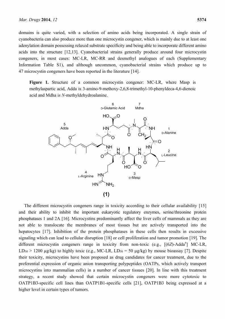
peptide synthetase/polyketide synthase (NRPS/PKS). As is evident in the most common variant,
MC-LR (1; Figure 1), microcystins are generally composed of the unique β-amino acid Adda
(3S-amino-9S-methoxy-2S,6,8S-trimethyl-10-phenyldeca-4E,6E-dienoic acid), D-glutamic acid (Glu),
N-methyl dehydroalanine (Mdha), D-alanine (Ala), D-erythro-β-methylaspartic acid (Masp) and two
variable L-amino acids. To date, there have been at least 100 different microcystin congeners
characterized [6], mostly due to substitutions of the variable L-amino acids in positions two and four,
although modifications have been reported for all of the amino acids [7].
Microcystins are produced by numerous cyanobacterial genera when the necessary microcystin
synthase genes are present. These NRPS and PKS genes produce a multi-enzyme complex which
sequentially adds components from malonyl-CoA, S-adenosyl-L-methionine and amino acids onto
phenylacetate and transforms them to produce a modified peptide chain, which is condensed to form the
cyclic microcystin [8–11]. The occurrence of different microcystin congeners is due to variability in the
coding of the microcystin synthase genes among cyanobacterial strains. For example, at times the
McyA1 N-methyltransferase domain can be inactive, which results in position seven desmethyl
variants [10]. Furthermore, the amino acid specificity of the numerous adenylation domains is
dependent upon their genetic coding and changes to this sequence can cause the domains to be specific
for different amino acids or to recognize more than one amino acid [12]. Between cyanobacterial
species, the substrate specificity for many of the adenylation domains is relatively conserved (positions
one, three, six and seven), whilst the substrate specificity of the position two and four adenylation
Mar. Drugs 2014, 12 5374
domains is quite varied, with a selection of amino acids being incorporated. A single strain of
cyanobacteria can also produce more than one microcystin congener, which is mainly due to at least one
adenylation domain possessing relaxed substrate specificity and being able to incorporate different amino
acids into the structure [12,13]. Cyanobacterial strains generally produce around four microcystin
congeners, in most cases: MC-LR, MC-RR and desmethyl analogues of each (Supplementary
Information Table S1), and although uncommon, cyanobacterial strains which produce up to
47 microcystin congeners have been reported in the literature [14].
Figure 1. Structure of a common microcystin congener: MC-LR, where Masp is
methylaspartic acid, Adda is 3-amino-9-methoxy-2,6,8-trimethyl-10-phenyldeca-4,6-dienoic
acid and Mdha is N-methyldehydroalanine.
The different microcystin congeners range in toxicity according to their cellular availability [15]
and their ability to inhibit the important eukaryotic regulatory enzymes, serine/threonine protein
phosphatases 1 and 2A [16]. Microcystins predominantly affect the liver cells of mammals as they are
not able to translocate the membranes of most tissues but are actively transported into the
hepatocytes [17]. Inhibition of the protein phosphatases in these cells then results in excessive
signaling which can lead to cellular disruption [18] or cell proliferation and tumor promotion [19]. The
different microcystin congeners range in toxicity from non-toxic (e.g., [(6Z)-Adda5] MC-LR,
LD50 > 1200 µg/kg) to highly toxic (e.g., MC-LR, LD50 = 50 µg/kg) by mouse bioassay [7]. Despite
their toxicity, microcystins have been proposed as drug candidates for cancer treatment, due to the
preferential expression of organic anion transporting polypeptides (OATPs, which actively transport
microcystins into mammalian cells) in a number of cancer tissues [20]. In line with this treatment
strategy, a recent study showed that certain microcystin congeners were more cytotoxic to
OATP1B3-specific cell lines than OATP1B1-specific cells [21], OATP1B3 being expressed at a
higher level in certain types of tumors.
Mar. Drugs 2014, 12 5375
A Microcystis species (CAWBG11) isolated from Lake Hakanoa (Huntly, New Zealand) in
2005 [22] was investigated as it produced a large number of oligopeptides including 27 microcystin
congeners, six of which were new variants. Whilst cyanobacterial strains have been noted to produce
more microcystin congeners [14], the diversity of microcystin variants produced by Microcystis
CAWBG11 is not commonly observed.
2. Results and Discussion
2.1. Oligopeptide Diversity of Microcystis CAWBG11
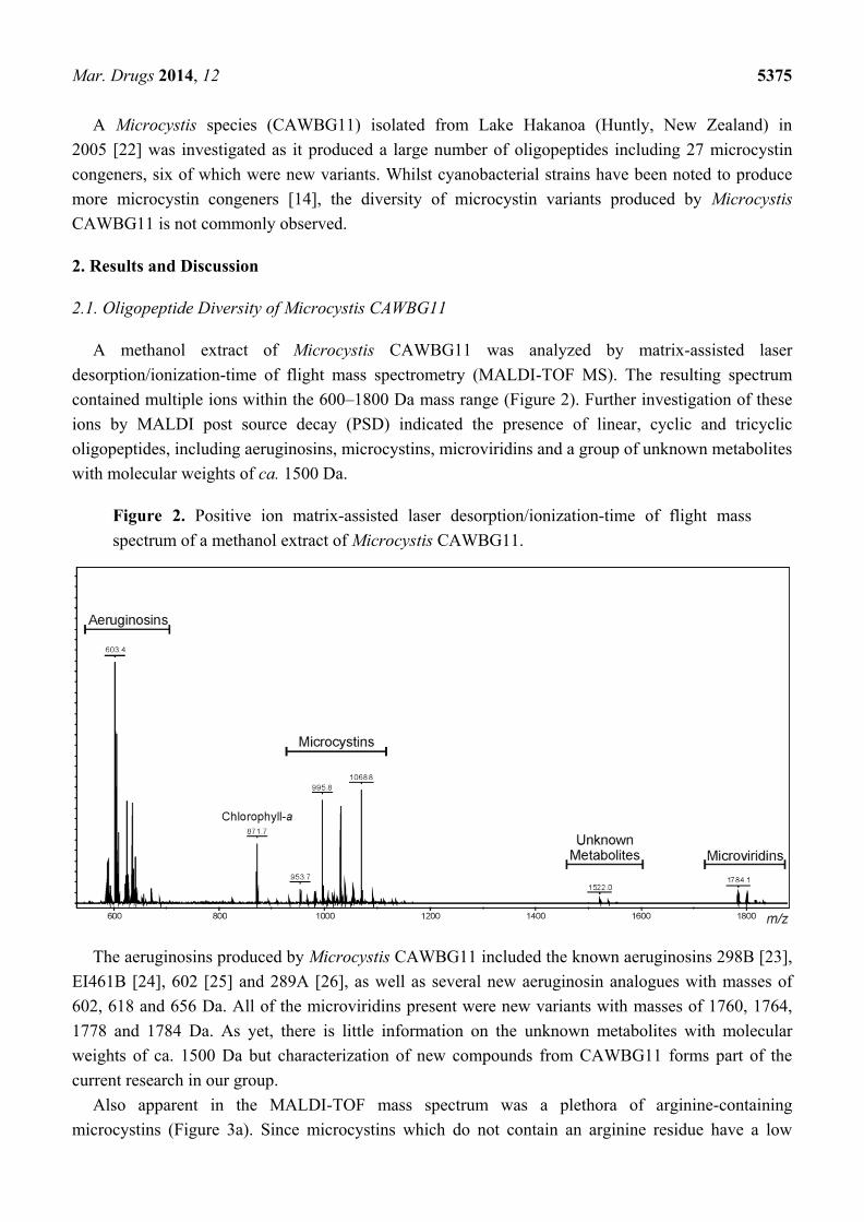
A methanol extract of Microcystis CAWBG11 was analyzed by matrix-assisted laser
desorption/ionization-time of flight mass spectrometry (MALDI-TOF MS). The resulting spectrum
contained multiple ions within the 600–1800 Da mass range (Figure 2). Further investigation of these
ions by MALDI post source decay (PSD) indicated the presence of linear, cyclic and tricyclic
oligopeptides, including aeruginosins, microcystins, microviridins and a group of unknown metabolites
with molecular weights of ca. 1500 Da.
Figure 2. Positive ion matrix-assisted laser desorption/ionization-time of flight mass
spectrum of a methanol extract of Microcystis CAWBG11.
The aeruginosins produced by Microcystis CAWBG11 included the known aeruginosins 298B [23],
EI461B [24], 602 [25] and 289A [26], as well as several new aeruginosin analogues with masses of
602, 618 and 656 Da. All of the microviridins present were new variants with masses of 1760, 1764,
1778 and 1784 Da. As yet, there is little information on the unknown metabolites with molecular
weights of ca. 1500 Da but characterization of new compounds from CAWBG11 forms part of the
current research in our group.
Also apparent in the MALDI-TOF mass spectrum was a plethora of arginine-containing
microcystins (Figure 3a). Since microcystins which do not contain an arginine residue have a low
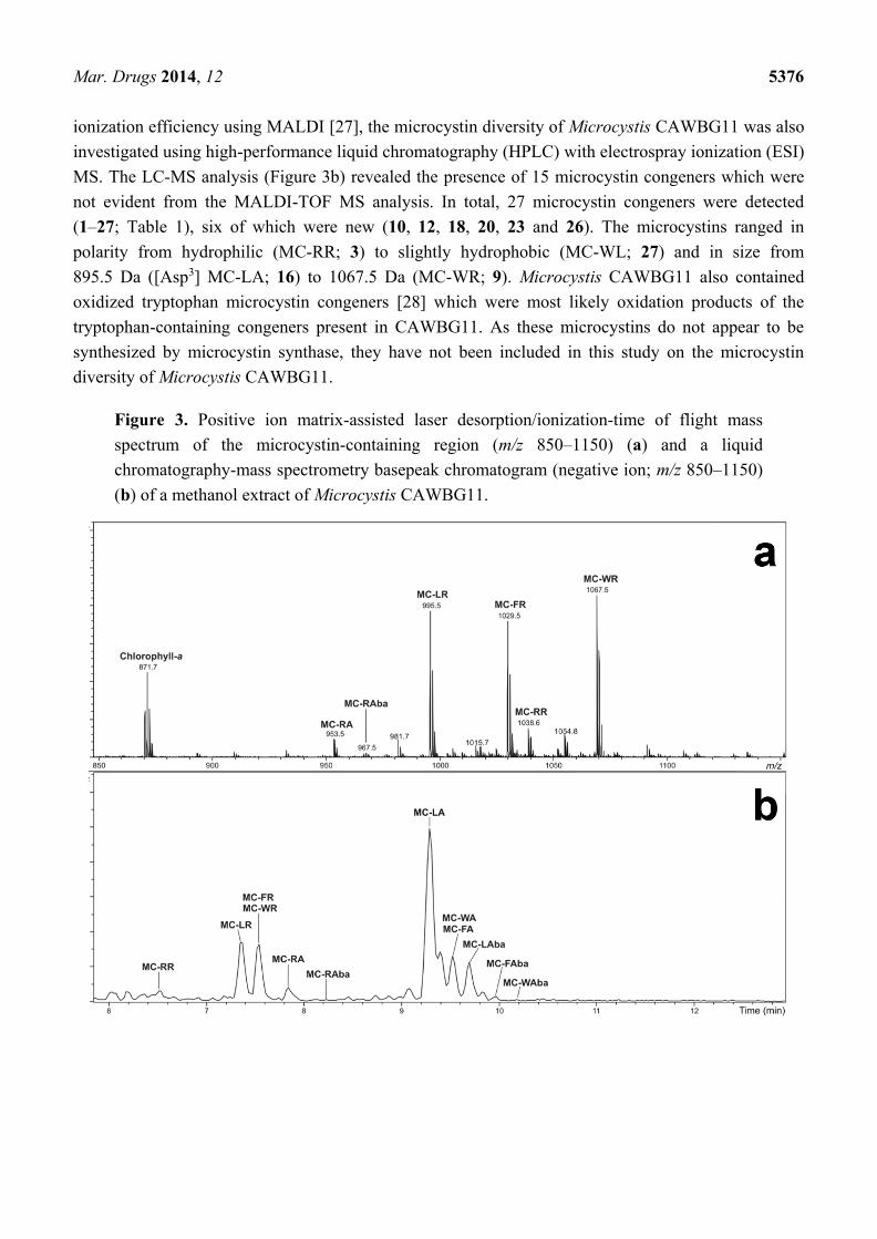
Mar. Drugs 2014, 12 5376
ionization efficiency using MALDI [27], the microcystin diversity of Microcystis CAWBG11 was also
investigated using high-performance liquid chromatography (HPLC) with electrospray ionization (ESI)
MS. The LC-MS analysis (Figure 3b) revealed the presence of 15 microcystin congeners which were
not evident from the MALDI-TOF MS analysis. In total, 27 microcystin congeners were detected
(1–27; Table 1), six of which were new (10, 12, 18, 20, 23 and 26). The microcystins ranged in
polarity from hydrophilic (MC-RR; 3) to slightly hydrophobic (MC-WL; 27) and in size from
895.5 Da ([Asp3] MC-LA; 16) to 1067.5 Da (MC-WR; 9). Microcystis CAWBG11 also contained
oxidized tryptophan microcystin congeners [28] which were most likely oxidation products of the
tryptophan-containing congeners present in CAWBG11. As these microcystins do not appear to be
synthesized by microcystin synthase, they have not been included in this study on the microcystin
diversity of Microcystis CAWBG11.
Figure 3. Positive ion matrix-assisted laser desorption/ionization-time of flight mass
spectrum of the microcystin-containing region (m/z 850–1150) (a) and a liquid
chromatography-mass spectrometry basepeak chromatogram (negative ion; m/z 850–1150)
(b) of a methanol extract of Microcystis CAWBG11.
Mar. Drugs 2014, 12 5377
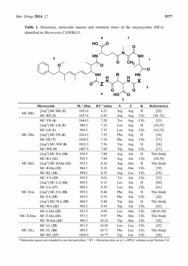
Table 1. Structures, molecular masses and retention times of the microcystins (MCs)
identified in Microcystis CAWBG11.
NH
O
Z
HN
HN X
HNN
NH
HN
O
O
O
O O
O
O
HO O
OHO
R
CH2
2
1
3
4
76
5
Microcystin Mr a (Da) RT b (min) X Z R Reference(s)
MC-RRs [Asp3] MC-RR (2) 1023.6 6.32 Arg Arg H [29]
MC-RR (3) 1037.6 6.45 Arg Arg CH3 [30–32]
MC-XRs
MC-YR (4) 1044.5 7.20 Tyr Arg CH3 [33]
[Asp3] MC-LR (5) 980.5 7.32 Leu Arg H [34,35]
MC-LR (1) 994.5 7.35 Leu Arg CH3 [16,33]
[Asp3] MC-FR (6) 1014.5 7.52 Phe Arg H [36]
MC-FR (7) 1028.5 7.54 Phe Arg CH3 [37]
[Asp3] MC-WR (8) 1053.5 7.56 Trp Arg H [36]
MC-WR (9) 1067.5 7.60 Trp Arg CH3 [37]
MC-RZs
[Asp3] MC-RA (10) 938.5 7.80 Arg Ala H This Study
MC-RA (11) 952.5 7.84 Arg Ala CH3 [38,39]
[Asp3] MC-RAba (12) 952.5 8.16 Arg Aba H This Study
MC-RAba (13) 966.5 8.18 Arg Aba CH3 [39]
MC-RL (14) 994.5 8.35 Arg Leu CH3 [39]
MC-XAs
MC-YA (15) 959.5 9.01 Tyr Ala CH3 [33]
[Asp3] MC-LA (16) 895.5 9.12 Leu Ala H [40]
MC-LA (17) 909.5 9.29 Leu Ala CH3 [41]
[Asp3] MC-FA (18) 929.5 9.46 Phe Ala H This Study
MC-FA (19) 943.5 9.52 Phe Ala CH3 [42]
[Asp3] MC-WA (20) 968.5 9.48 Trp Ala H This Study
MC-WA (21) 982.5 9.54 Trp Ala CH3 [42]
MC-XAbas
MC-LAba (22) 923.5 9.69 Leu Aba CH3 [40,43,44]
MC-FAba (23) 957.5 9.97 Phe Aba CH3 This Study
MC-WAba (24) 996.5 10.24 Trp Aba CH3 [28]
MC-XLs
MC-LL (25) 951.5 10.50 Leu Leu CH3 [45]
MC-FL (26) 985.5 10.71 Phe Leu CH3 This Study
MC-WL (27) 1024.5 10.77 Trp Leu CH3 [28]
a Molecular masses are rounded to one decimal place; b RT = Retention time on a C18 HPLC column as per Section 3.4.

Mar. Drugs 2014, 12 5378
2.2. Characterization of Microcystins from Microcystis CAWBG11
In order to characterize the microcystins produced by Microcystis CAWBG11, multiple methods of
analysis were undertaken including a recently described thiol derivatization technique, tandem MS
(MS/MS), high-resolution MS (HRMS) and amino acid analysis (Advanced Marfey’s method).
However, many of the microcystins were present in low quantities allowing only analysis by MS/MS
and thiol derivatization, including each of the new congeners reported here (10, 12, 18, 20, 23 and 26).
The position seven amino acid in microcystins is frequently Mdha, although the isometric amino
acid, dehydrobutyrine (Dhb), has also been reported on several occasions [46–48]. Previously, NMR
analysis of purified material (~1 mg) has been required to confirm the identity of this amino acid in
microcystins. However, a recently described thiol derivatization technique has been shown to be
effective for the discrimination of these two moieties [49]. When using this technique, a microcystin
containing a terminal alkene, as is found in Mdha and dehydroalanine (Dha), will readily react with
β-mercaptoethanol under alkaline conditions [39,50]. When Dhb is present, this reaction rate is at least
two orders of magnitude slower [49]. A β-mercaptoethanol derivatization of the Microcystis
CAWBG11 extract underwent rapid reaction (t½ < 18 min) and all of the congeners were fully
derivatized within four hours indicating that all microcystins present contained either Dha or Mdha.
Tandem MS analysis of each congener showed that an 83 Da moiety was present at position seven
(Supplementary Information Tables S2–S7); therefore, each congener produced by CAWBG11 was
designated as containing Mdha.
Many of the microcystins identified in Microcystis CAWBG11 have been reported previously
(see Table 1) and therefore an in-depth description of their characterization is not presented here.
However, tables of MS/MS fragment assignments used to confirm the structure of each microcystin
detected in CAWBG11 are available in Supplementary Information Tables S2–S7.
The MC-RR microcystins (2–3) were predominantly observed as [M+2H]2+ ions by ESI MS,
although low intensity signals for the singly-protonated ions were also observed by MALDI MS. The
resulting ESI MS/MS fragment ions were a combination of singly- and doubly-charged ions
(Supplementary Information Table S2). During the MS/MS analysis of the -RR congeners,
doubly-protonated fragment ions were distinguished from singly-protonated ions by assessing the
isotopic peak pattern. When a singly-protonated ion was present there was a difference of +1 m/z between
the isotopic peaks and a difference of +0.5 m/z was observed when the ion was doubly-protonated.
The -XR and -RZ microcystins yielded predominantly [M+H]+ ions by both MALDI MS and ESI
MS (although there were also sodium- and potassium-adducts present in the ESI mass spectra).
The -XR microcystins were characterized using a combination of the MALDI PSD and ESI CID
fragment ions (Supplementary Information Table S3) where the MALDI PSD spectra provided lower
mass fragment ions and the ESI CID spectra provided higher mass fragments. These fragments
included commonly observed elements such as a fragment of the Adda sidechain (m/z 135), the
Adda′-Glu-Mdha fragment ion (m/z 375) and the Arg-Adda-Glu or Masp-Arg-Adda fragments
(m/z 599) [43,51]. The -RZ microcystins were also characterized using both MALDI PSD and ESI CID
data (Supplementary Information Table S4). Since the position of the arginine residue in the structure
is inverted in the -RZ microcystins (and the guanidinium group provides the major ionization point),
several of the MS/MS fragment ions observed were different from those seen in the -XR congeners.
Mar. Drugs 2014, 12 5379
For example, while the m/z 599 ion (Arg-Adda-Glu or Masp-Arg-Adda) [51] is one of the major
fragment ions in a -XR microcystin congener, a m/z 440 fragment ion (Glu-Mdha-Ala-Arg and
Mdha-Ala-Arg-Masp) was present in the -RZ congeners [52].
The -XA, -XAba and -XL microcystins did not ionize efficiently by MALDI MS and yielded
predominantly [M+Na]+ and [M+K]+ adduct ions by ESI MS. However, there were low levels
of [M+H]+ ions present which were used for the MS/MS characterization. The -XA microcystins
(Supplementary Information Table S5), -XAba microcystins (Supplementary Information Table S6)
and -XL microcystins (Supplementary Information Table S7) produced many of the fragment ions
observed for the other microcystin congeners analyzed but also possessed an additional ammonia-loss
fragment ion series which started from Adda-Glu-Mdha minus NH3 (m/z 509). The -XL microcystins
produced several intense fragment ions that could not be assigned during the course of this study, for
example, an m/z 535 was present in MS/MS spectra of these congeners (Supplementary Information
Table S7). As this fragment had the same m/z in each of the -XL microcystin congeners (25–27), it is
most likely from a common portion of the structure. The mass of the other unassigned fragment ion
differed in each of the microcystins: m/z 440 in MC-LL (25), m/z 474 in MC-FL (26) and m/z 513 in
MC-WL (27). The mass increments between each of these fragments were the same as those between
leucine, phenylalanine and tryptophan and are therefore most likely to result from a portion of the
structure which contains the position two amino acid.
Semi-pure mixtures of the microcystins were analyzed by HRMS to determine accurate masses and
confirm the proposed molecular formulae of the compounds. The CAWBG11 microcystins which were
present in sufficient quantities to be detected using the HRESIMS instrumentation available yielded
accurate masses which were consistent with their proposed structures (±5 ppm; Supplementary
Information Table S8). The accurate mass for MC-RR was determined from the doubly-protonated
ion, the -XR and -RZ congeners were determined from the singly-protonated ions, whilst the -XA
and -XAba congeners were determined using the sodium adduct ions.
Several of the microcystins in CAWBG11 were present in sufficient quantities to isolate and
perform NMR and amino acid analysis to further confirm their structures and determine the
stereochemistry of certain structural elements. The full characterization of MC-FA (19) and MC-WA
(21) from Microcystis CAWBG11 was recently reported [42] and determined that these microcystins
contained D-Ala, D-Glu, D-Masp, Mdha, 3S,4E,6E-Adda and two L-amino acids. Hydrolyzates of
purified MC-RA (11) and MC-RAba (12) from Microcystis CAWBG11 were also subjected to
Advanced Marfey’s amino acid analysis which yielded complimentary results (Supplementary
Information Figures S1–S2): that the microcystins contained D-Glu, D-Masp, D-Ala, 3S-Adda, two
L-amino acids and N-methylamine (which is indicative of Mdha [53]). It also confirmed that the
MC-RAba found in Microcystis CAWBG11 contained L-2-aminobutanoic acid (Aba) and not the
isomeric version (2-amino-iso-butanoic acid; Aib). This was consistent with the only other study of an
Aba-containing microcystin, where differentiation was made between Aba and Aib [54]. It was
assumed that the other Aba-containing microcystins in CAWBG11 also contain this amino
acid variant.

Mar. Drugs 2014, 12 5380
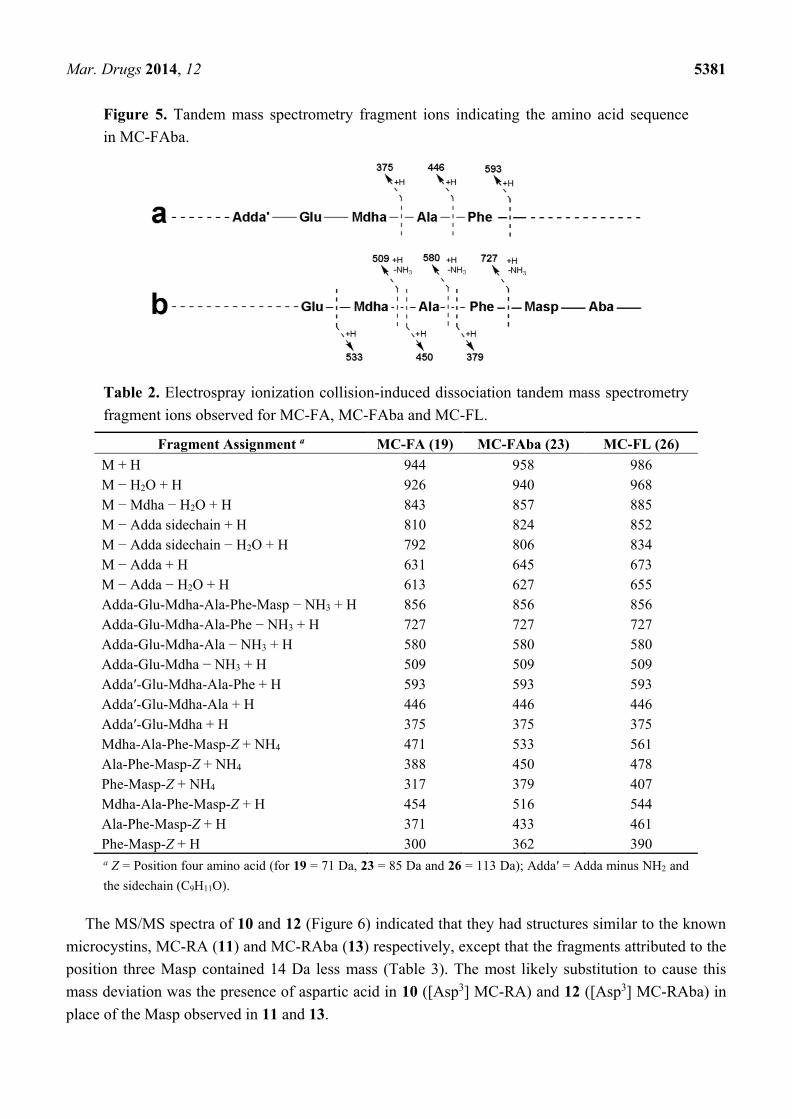
2.3. Tandem Mass Spectrometry Characterization of New Microcystins from Microcystis CAWBG11
The MS/MS spectra of 23 and 26 (Figure 4) indicated that they were very similar in structure to
MC-FA (19), except that the fragments attributed to the position four amino acid contained either 14 or
42 Da additional mass (Table 2). The most likely substitution to cause these mass deviations was the
incorporation of Aba into 23 (MC-FAba) and leucine into 26 (MC-FL), in place of the alanine
observed in 19 (MC-FA).
For MC-FAba (23), the fragment ion series starting with Adda′-Glu-Mdha (m/z 375) was extended
to include Ala and Phe (Figure 5a). This sequence was supported by the ion series containing Adda
minus NH3 (m/z 509, 580 and 727; Figure 5b). A fragment ion series which began with Phe-Masp-Aba
(m/z 379; Figure 5b) and extended in the opposite direction to include Ala and Mdha gave the
complete amino acid sequence of Adda-Glu-Mdha-Ala-Phe-Masp-Aba. A fragment resulting from the
loss of Mdha and water (m/z 857; Table 2) indicated that Adda and the Aba residue were joined and
that the structure was cyclic. The amino acid sequence in MC-FL (26) was similarly established.
Figure 4. Electrospray ionization collision-induced dissociation tandem mass spectra and
putative structures of MC-FAba (a) and MC-FL (b).
334.2
375.2
390.2
413.2
431.1
474.2
509.2
535.2
593.2 690.2 727.1 751.3
775.3799.5
835.1
852.3
893.1
925.3 954.3
968.3
+MS2(986.5), 10.6-10.8min #(323-328)
0.0
0.5
1.0
1.5
2.0
4x10
Intens.
300 400 500 600 700 800 900 m/z
320.2
375.1
450.1 516.1 593.2
628.2 662.2695.3
727.1
751.8
789.2
806.2
824.2
870.6
926.2
941.2
+MS2(958.5), 9.9-10.3min #(300-312)
0
1
2
3
4x10
Intens.
300 400 500 600 700 800 900 m/z
b
a
N H
O
HN
HN
H NN
N H
H N
C H 2O
O
O
O O
O
O
H O O
OH O
( 2 3 )
N H
O
HN
HN
H NN
N H
H N
C H 2O
O
O
O O
O
O
H O O
OH O
( 2 6 )
Mar. Drugs 2014, 12 5381
Figure 5. Tandem mass spectrometry fragment ions indicating the amino acid sequence
in MC-FAba.
Table 2. Electrospray ionization collision-induced dissociation tandem mass spectrometry
fragment ions observed for MC-FA, MC-FAba and MC-FL.
Fragment Assignment a MC-FA (19) MC-FAba (23) MC-FL (26)
M + H 944 958 986
M − H2O + H 926 940 968
M − Mdha − H2O + H 843 857 885
M − Adda sidechain + H 810 824 852
M − Adda sidechain − H2O + H 792 806 834
M − Adda + H 631 645 673
M − Adda − H2O + H 613 627 655
Adda-Glu-Mdha-Ala-Phe-Masp − NH3 + H 856 856 856
Adda-Glu-Mdha-Ala-Phe − NH3 + H 727 727 727
Adda-Glu-Mdha-Ala − NH3 + H 580 580 580
Adda-Glu-Mdha − NH3 + H 509 509 509
Adda′-Glu-Mdha-Ala-Phe + H 593 593 593
Adda′-Glu-Mdha-Ala + H 446 446 446
Adda′-Glu-Mdha + H 375 375 375
Mdha-Ala-Phe-Masp-Z + NH4 471 533 561
Ala-Phe-Masp-Z + NH4 388 450 478
Phe-Masp-Z + NH4 317 379 407
Mdha-Ala-Phe-Masp-Z + H 454 516 544
Ala-Phe-Masp-Z + H 371 433 461
Phe-Masp-Z + H 300 362 390
a Z = Position four amino acid (for 19 = 71 Da, 23 = 85 Da and 26 = 113 Da); Adda′ = Adda minus NH2 and
the sidechain (C9H11O).
The MS/MS spectra of 10 and 12 (Figure 6) indicated that they had structures similar to the known
microcystins, MC-RA (11) and MC-RAba (13) respectively, except that the fragments attributed to the
position three Masp contained 14 Da less mass (Table 3). The most likely substitution to cause this
mass deviation was the presence of aspartic acid in 10 ([Asp3] MC-RA) and 12 ([Asp3] MC-RAba) in
place of the Masp observed in 11 and 13.

Mar. Drugs 2014, 12 5382
Figure 6. Matrix-assisted laser desorption/ionization post-source decay mass spectrum
of [Asp3] MC-RA (a) and electrospray ionization collision-induced dissociation tandem
mass spectra with putative structures of [Asp3] MC-RA (b) and [Asp3] MC-RAba (c).
311.2
329.3
354.2412.2
426.2
440.1
510.2
528.2
546.2
581.1
595.1635.2
683.2
729.0
754.3
785.2807.4
824.3851.3
881.3
925.3
+MS2(953.6), 7.7-8.1min #(233-244)
0.0
0.5
1.0
1.5
2.0
2.5
4x10
Intens.
300 400 500 600 700 800 900 m/z
311.1
354.3
368.1
426.2
440.1
497.2
528.2
581.0
595.1618.2 682.0 718.2
740.2
754.2 811.3
829.6
867.3
921.3
+MS2(939.5), 7.6-7.8min #(229-236)
0
1000
2000
3000
4000
5000
Intens.
300 400 500 600 700 800 900 m/z
135
70
112
87213
155127 200 375
100 330174 294239120 311269 427 441357283 471185 226 485255 455
CYN06 MS-MS\PSD-CID of m-z=939.LIFT\fast
0.0
0.2
0.4
0.6
0.8
1.0
4x10
Inte
ns. [a
.u.]
100 150 200 250 300 350 400 450
m/z
b
a
c
N H
O
HN
HN
H NN
N H
H N
C H 2
O
O
O
O
O O
O
O
H O
O H
OH N
N H
N H 2
( 1 2 )
N H
O
HN
HN
H NN
N H
H N
C H 2
O
O
O
O
O O
O
O
H O
O H
OH N
N H
N H 2
( 1 0 )
Mar. Drugs 2014, 12 5383
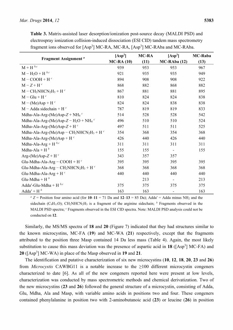
Table 3. Matrix-assisted laser desorption/ionization post-source decay (MALDI PSD) and
electrospray ionization collision-induced dissociation (ESI CID) tandem mass spectrometry
fragment ions observed for [Asp3] MC-RA, MC-RA, [Asp3] MC-RAba and MC-RAba.
Fragment Assignment a [Asp3]
MC-RA (10)
MC-RA
(11)
[Asp3]
MC-RAba (12)
MC-Raba
(13)
M + H b,c 939 953 953 967
M − H2O + H b,c 921 935 935 949
M − COOH + H c 894 908 908 922
M − Z + H c 868 882 868 882
M − CH2NHCN2H3 + H c 867 881 881 895
M − Glu + H c 810 824 824 838
M − (Me)Asp + H c 824 824 838 838
M − Adda sidechain + H c 787 819 819 833
Mdha-Ala-Arg-(Me)Asp-Z + NH4 c 514 528 528 542
Mdha-Ala-Arg-(Me)Asp-Z − H2O + NH4 c 496 510 510 524
Mdha-Ala-Arg-(Me)Asp-Z + H c 497 511 511 525
Mdha-Ala-Arg-(Me)Asp − CH2NHCN2H3 + H c 354 368 354 368
Mdha-Ala-Arg-(Me)Asp + H c 426 440 426 440
Mdha-Ala-Arg + H b,c 311 311 311 311
Mdha-Ala + H b 155 155 - 155
Arg-(Me)Asp-Z + H c 343 357 357
Glu-Mdha-Ala-Arg − COOH + H c 395 395 395 395
Glu-Mdha-Ala-Arg − CH2NHCN2H3 + H c 368 368 368 368
Glu-Mdha-Ala-Arg + H c 440 440 440 440
Glu-Mdha + H b 213 - 213
Adda′-Glu-Mdha + H b,c 375 375 375 375
Adda′ + H b 163 163 - 163
a Z = Position four amino acid (for 10–11 = 71 Da and 12–13 = 85 Da); Adda′ = Adda minus NH2 and the
sidechain (C9H11O); CH2NHCN2H3 is a fragment of the arginine sidechain; b Fragments observed in the
MALDI PSD spectra; c Fragments observed in the ESI CID spectra. Note: MALDI PSD analysis could not be
conducted on 12.
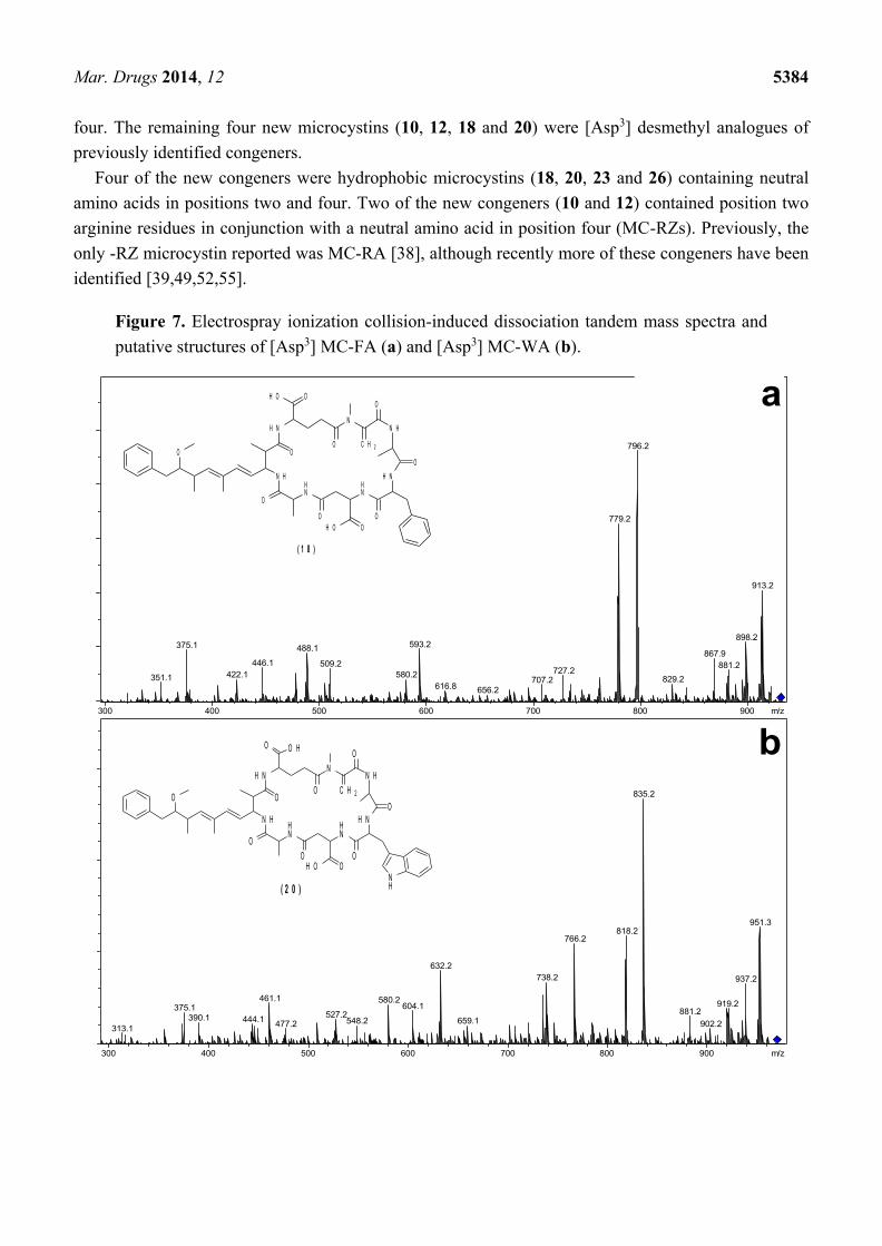
Similarly, the MS/MS spectra of 18 and 20 (Figure 7) indicated that they had structures similar to
the known microcystins, MC-FA (19) and MC-WA (21) respectively, except that the fragments
attributed to the position three Masp contained 14 Da less mass (Table 4). Again, the most likely
substitution to cause this mass deviation was the presence of aspartic acid in 18 ([Asp3] MC-FA) and
20 ([Asp3] MC-WA) in place of the Masp observed in 19 and 21.
The identification and putative characterization of six new microcystins (10, 12, 18, 20, 23 and 26)
from Microcystis CAWBG11 is a notable increase to the ≥100 different microcystin congeners
characterized to date [6]. As all of the new congeners reported here were present at low levels,
characterization was conducted by mass spectrometric methods and chemical derivatization. Two of
the new microcystins (23 and 26) followed the general structure of a microcystin, consisting of Adda,
Glu, Mdha, Ala and Masp, with variable amino acids in positions two and four. These congeners
contained phenylalanine in position two with 2-aminobutanoic acid (23) or leucine (26) in position
Mar. Drugs 2014, 12 5384
four. The remaining four new microcystins (10, 12, 18 and 20) were [Asp3] desmethyl analogues of
previously identified congeners.
Four of the new congeners were hydrophobic microcystins (18, 20, 23 and 26) containing neutral
amino acids in positions two and four. Two of the new congeners (10 and 12) contained position two
arginine residues in conjunction with a neutral amino acid in position four (MC-RZs). Previously, the
only -RZ microcystin reported was MC-RA [38], although recently more of these congeners have been
identified [39,49,52,55].
Figure 7. Electrospray ionization collision-induced dissociation tandem mass spectra and
putative structures of [Asp3] MC-FA (a) and [Asp3] MC-WA (b).
351.1
375.1
422.1
446.1
488.1
509.2
580.2
593.2
616.8 656.2
707.2727.2
779.2
796.2
829.2
867.9
881.2
898.2
913.2
+MS2(930.5), 9.1-9.2min #(275-279)
0.0
0.2
0.4
0.6
0.8
1.0
5x10
Intens.
300 400 500 600 700 800 900 m/z
313.1
375.1
390.1 444.1
461.1
477.2527.2
548.2
580.2604.1
632.2
659.1
738.2
766.2818.2
835.2
881.2
902.2
919.2
937.2
951.3
+MS2(969.5), 9.0-9.2min #(274-278)
0.0
0.5
1.0
1.5
5x10
Intens.
300 400 500 600 700 800 900 m/z
b
a
N H
O
HN
HN
H NN
N H
H N
C H 2
O
O
O
O
O O
O
O
H O
O H
ONH( 2 0 )
N H
O
HN
HN
H NN
N H
H N
C H 2O
O
O
O O
O
O
H O O
OH O
( 1 8 )
Mar. Drugs 2014, 12 5385
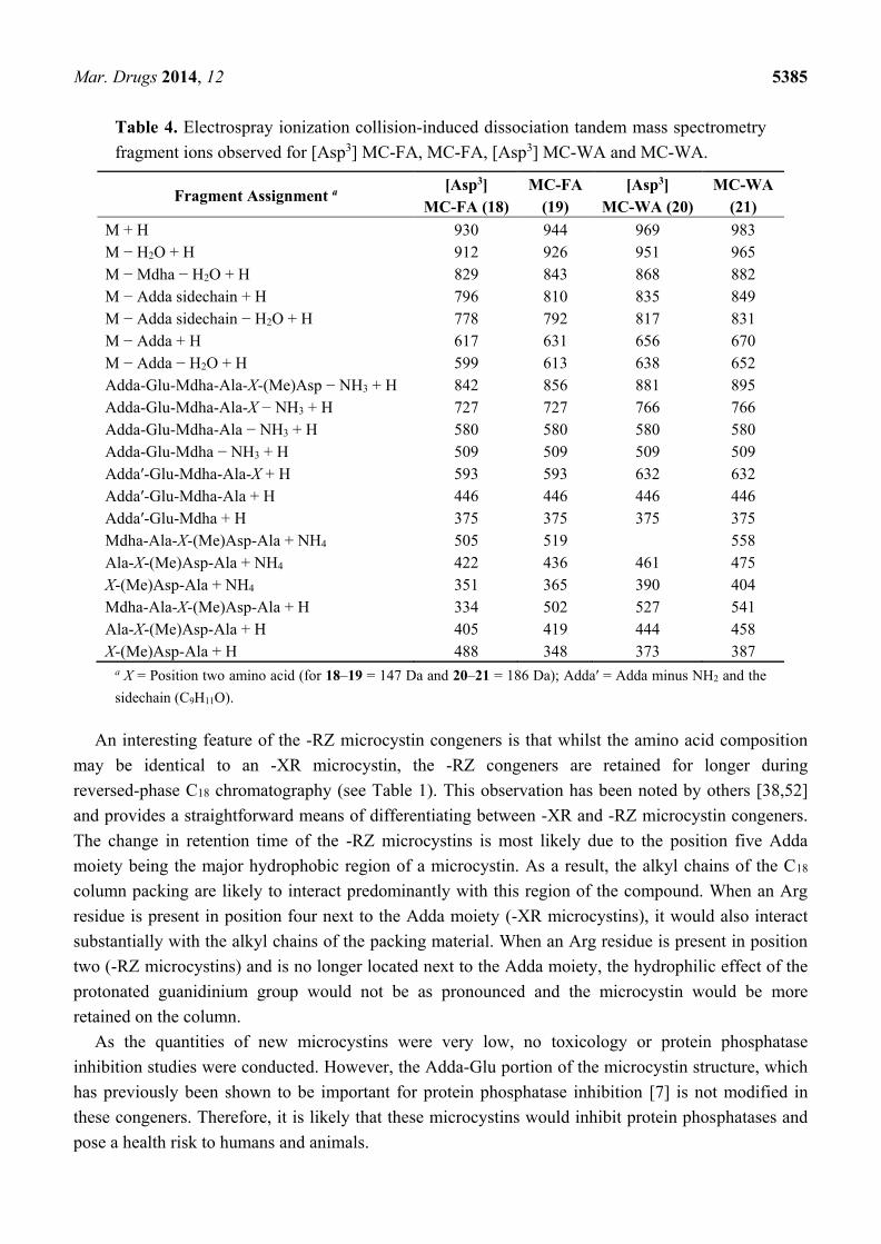
Table 4. Electrospray ionization collision-induced dissociation tandem mass spectrometry
fragment ions observed for [Asp3] MC-FA, MC-FA, [Asp3] MC-WA and MC-WA.
Fragment Assignment a [Asp3]
MC-FA (18)
MC-FA
(19)
[Asp3]
MC-WA (20)
MC-WA
(21)
M + H 930 944 969 983
M − H2O + H 912 926 951 965
M − Mdha − H2O + H 829 843 868 882
M − Adda sidechain + H 796 810 835 849
M − Adda sidechain − H2O + H 778 792 817 831
M − Adda + H 617 631 656 670
M − Adda − H2O + H 599 613 638 652
Adda-Glu-Mdha-Ala-X-(Me)Asp − NH3 + H 842 856 881 895
Adda-Glu-Mdha-Ala-X − NH3 + H 727 727 766 766
Adda-Glu-Mdha-Ala − NH3 + H 580 580 580 580
Adda-Glu-Mdha − NH3 + H 509 509 509 509
Adda′-Glu-Mdha-Ala-X + H 593 593 632 632
Adda′-Glu-Mdha-Ala + H 446 446 446 446
Adda′-Glu-Mdha + H 375 375 375 375
Mdha-Ala-X-(Me)Asp-Ala + NH4 505 519 558
Ala-X-(Me)Asp-Ala + NH4 422 436 461 475
X-(Me)Asp-Ala + NH4 351 365 390 404
Mdha-Ala-X-(Me)Asp-Ala + H 334 502 527 541
Ala-X-(Me)Asp-Ala + H 405 419 444 458
X-(Me)Asp-Ala + H 488 348 373 387
a X = Position two amino acid (for 18–19 = 147 Da and 20–21 = 186 Da); Adda′ = Adda minus NH2 and the
sidechain (C9H11O).
An interesting feature of the -RZ microcystin congeners is that whilst the amino acid composition
may be identical to an -XR microcystin, the -RZ congeners are retained for longer during
reversed-phase C18 chromatography (see Table 1). This observation has been noted by others [38,52]
and provides a straightforward means of differentiating between -XR and -RZ microcystin congeners.
The change in retention time of the -RZ microcystins is most likely due to the position five Adda
moiety being the major hydrophobic region of a microcystin. As a result, the alkyl chains of the C18
column packing are likely to interact predominantly with this region of the compound. When an Arg
residue is present in position four next to the Adda moiety (-XR microcystins), it would also interact
substantially with the alkyl chains of the packing material. When an Arg residue is present in position
two (-RZ microcystins) and is no longer located next to the Adda moiety, the hydrophilic effect of the
protonated guanidinium group would not be as pronounced and the microcystin would be more
retained on the column.
As the quantities of new microcystins were very low, no toxicology or protein phosphatase
inhibition studies were conducted. However, the Adda-Glu portion of the microcystin structure, which
has previously been shown to be important for protein phosphatase inhibition [7] is not modified in
these congeners. Therefore, it is likely that these microcystins would inhibit protein phosphatases and
pose a health risk to humans and animals.
Mar. Drugs 2014, 12 5386
2.4. The Microcystin Diversity of Microcystis CAWBG11
In this study, 27 different microcystin congeners were detected in cultures of Microcystis
CAWBG11, due to relaxed substrate specificity of the microcystin synthase at positions two, three and
four (Table 1). This indicates that the microcystin synthase in Microcystis CAWBG11 has relaxed
substrate specificity at McyB1 as well as at McyC (see [8] for more details). The presence of position
three desmethyl congeners indicates that the adenylation domain of McyB2 recognizes both Masp and
Asp and results in approximately 2.5% of each microcystin congener containing Asp in position three
instead of the usual Masp. Five different amino acids were observed to be incorporated into position
two of the structure, two amino acids were observed in position three and four amino acids were
observed in position four. This equates to a potential 40 microcystins which could be produced by
CAWBG11. This value would increase if the CAWBG11 microcystin synthase were shown to be able
to incorporate more amino acids than reported in the present study. As the various amino acid
substitutions occur at different frequencies, not all of the possible congeners were observed in the
present study, namely, [Asp3] analogues of MC-RL, and the -XAba, -XL and -YZ microcystin
congeners. Tyrosine-containing analogues of the -XAba and -XL congeners were also not observed.
In order to better understand whether the number of microcystin congeners observed in Microcystis
CAWBG11 was unique, relevant literature was surveyed to assess the diversity of congeners produced
by isolated cyanobacterial strains (Supplementary Information Table S1). As this survey was
formulated from multiple studies by different researchers, it is difficult to ascertain the level of
characterization performed on each reported strain which could bias the results towards a lower
microcystin diversity. In several cases, researchers were not able to identify all of the microcystins
present and listed these as “unidentified microcystins” [14,43,49,56]. These “unidentified microcystins”
skew the analysis slightly as it cannot be predicted whether these congeners were similar analogues to
those identified or whether they were structurally different.
The investigation revealed that the median number of microcystin congeners produced by a
cyanobacterial strain was between four and five (Supplementary Information Figure S3). When
assessing the number of congeners identified (within the study), the median was slightly lower
(Median = 4) than assessments based on the number of congeners observed (including “unidentified
microcystins”; Median = 5). Besides Microcystis CAWBG11, the range for the number of microcystin
congeners identified in a single cyanobacterial strain spanned from 1–16 (Supplementary Information
Table S1), but could be extended to 47 when the unidentified congeners were taken into account.
When there were no “unidentified microcystins” present in a cyanobacterial strain, the potential
number of congeners which could be produced was calculated. This took account of all of the
combinations of the amino acids incorporated into positions two and four, as well as the variable
modifications observed. Values for the potential microcystin production of an isolated cyanobacterial
strain ranged from 1–72, with a median of four congeners. As expected, the range for the potential
number of microcystins was higher than that based on the microcystins identified/observed, since some
congeners occur at lower levels and may not be detected.
The number of microcystins identified in Microcystis CAWBG11 (27 congeners) and the potential
number of microcystins which could be produced (40 congeners), positioned CAWBG11 in the top ten
percentile in the distribution of strains assessed. Other strains in the top ten percentile included
Mar. Drugs 2014, 12 5387
Anabaena 66A [14], Microcystis viridis NIES102 [14], Nostoc 152 [14,57–59] and Nostoc
IO-102-1 [14,60]. These cyanobacterial strains were able to incorporate an array of amino acids into
position two and produced variable modifications at other positions in the structure (Supplementary
Information Table S1). However, these strains did not show much variability at the position four amino
acid, as Anabaena 66A, Microcystis viridis NIES102 and Nostoc IO-102-1 could only incorporate
arginine whilst Nostoc 152 was able to utilize arginine and homoarginine. The microcystins
produced by Microcystis CAWBG11 contained arginine, alanine, aminobutanoic acid and leucine in
position four.
Overall, amongst the 49 cyanobacterial strains assessed in the survey, there was a low degree of
variability in the amino acids incorporated into position four of the microcystin structure. The majority
of the strains assessed were only able to incorporate one or two amino acids into this position
(Supplementary Information Table S1). Interestingly, when strains did exhibit high levels of amino
acid variability at position four, they tended to exhibit a high degree of substrate specificity at position
two. Microcystis CAWBG11, however, showed relaxed substrate specificity at both positions two and
four. As a result, CAWBG11 produced microcystin congeners which contained no arginine residues, a
single arginine residue and two arginine residues. Most of the other cyanobacterial strains assessed
produced congeners containing one or two arginine residues or congeners that contained one or no
arginine residues. Recently, three cyanobacterial strains which produced the same nine microcystins
were reported: Planktothrix agardhii CYA 56/3, CYA 137 and CYA 532 [49]. Like CAWBG11, these
strains were able to simultaneously produce microcystins which contained one, two and no arginine
residues, although they exhibited a lesser degree of structural variability than CAWBG11. As CAWBG11
produces such a diverse range of microcystin congeners, it could be a useful cyanobacterial strain for
investigation of the parameters which cause modulation of microcystin congener concentrations.
3. Experimental Section
3.1. Microcystis CAWBG11
Microcystis species CAWBG11 was isolated from a bloom sample obtained from Lake Hakanoa
(Huntly, New Zealand) in 2005. A culture was initiated from a small colony (<10 cells) [22]. The
culture is maintained alive and cryopreserved in the Cawthron Institute micro-algae culture
collection. Based on morphological features, the strain most closely matches the descriptions of
Microcystis aeruginosa. The cells are sub-spherical in shape and very densely aggregated in large
colonies which are irregular in shape and have colorless, homogeneous mucilage that does not extend
beyond colony edges (Supplementary Information Figure S4). Cells are bright green, with aerotopes.
The 16S ribosomal RNA gene partial sequence and full 16S-23S rRNA intergenic spacer sequence are
available on GenBank (EF634465). CAWBG11 has a high (>99%) sequence homology to other
Microcystis aeruginosa strains (e.g., AM778951 or HF678510). Although not initiated from a single
cell, the toxin profile has remained stable despite nearly 10 years of sub-culturing, thus we believe the
original colony comprised a single strain.
Mar. Drugs 2014, 12 5388
3.2. General Experimental Procedures
The HRESIMS analysis was performed on a Bruker MicrOTOF mass spectrometer. Data for
MALDI-TOF MS and PSD was collected on a Bruker AutoFlex II mass spectrometer. The LC-MS and
LC-MS/MS analyses were performed on a Bruker AmaZon X ESI mass spectrometer coupled to a
Dionex UltiMate 3000 HPLC system. Ultraviolet (UV) absorption data for purified microcystins was
determined using a Cary 100 Scan UV/visible spectrophotometer (Varian) over a wavelength range of
200–800 nm. Optical rotations of purified microcystins were determined using an AUTOPOL IV
polarimeter (Rudolph Research Analytical). Reversed-phased C18 separations were conducted using
YMC-gel ODS-A (YMC) and size exclusion chromatography was conducted using Sephadex LH-20
(Pharmacia Fine Chemicals). HPLC purification was performed using Waters 515 HPLC pumps
coupled to a photodiode array detector (200–400 nm; Waters 2996) and an Econosil C18 Column
(250 × 10 mm, 10-μm; Alltech).
3.3. Matrix-Assisted Laser Desorption/Ionization-Time of Flight Mass Spectrometry Analysis
Samples were prepared for MALDI-TOF MS analysis by mixing an aliquot (0.5 µL) 1:1 with a
saturated solution of α-cyano-4-hydroxycinnamic acid in 2:1:1 methanol/acetonitrile/0.1% trifluoroacetic
acid, directly on a 600-μm anchorchip. The dried spot was washed by pipetting wash solution (5 µL;
0.1% trifluoroacetic acid in 10 mM NH4H2PO4) onto the spot for 5 s, then withdrawing the liquid.
Spectra were acquired over various m/z ranges, with an acceleration voltage of 19 kV and a reflector
voltage of 20 kV. Pulsed ion extraction of 60 ns was used to build up the concentration of ions in the
ion source and ions below 500 m/z were suppressed to avoid detector saturation from matrix ions.
Mass calibration was performed using a peptide calibration standard (Bruker Daltonics) which was
prepared in the same manner as the samples. Post-source decay spectra were obtained from the
singly-protonated ions of the target compounds.
3.4. Liquid Chromatography-Mass Spectrometry Analysis
Samples (20 µL) for LC-MS and LC-MS/MS were separated on a C18 column (Ascentis Express
C18, 100 × 2.1 mm, 2.7-μm; Supleco Analytical) at a flow of 200 µL/min using a gradient of 2%
acetonitrile + 0.1% formic acid (v/v; solvent A) and 98% acetonitrile + 0.1% formic acid (v/v;
solvent B) with the following gradient program; the sample was loaded in 10% B; 10% B was held for
1 min and increased to 100% B over 12 min; 100% B was held for 2 min; the solvent composition was
returned to 10% B in 1 min and the column re-equilibrated for 4 min. The eluting compounds were
ionized using a capillary voltage of 3.5 kV and a nebulizer pressure of 3.0 bar. Desolvation was
accomplished with a nitrogen flow of 8 L/min at 220 °C. Mass spectra were acquired for positive or
negative ions over a range of m/z 100–2000. Daughter ion scans were obtained from the
singly-protonated ions of the target compounds by CID (collision amplitude of 1.0).
3.5. β-mercaptoethanol Derivatization for Mdha/Dhb Determination
A recently developed thiol derivatization technique [49] was used to determine which of the
isometric amino acids, Mdha or Dhb, was present in CAWBG11 microcystins. A methanol extract of
Mar. Drugs 2014, 12 5389
CAWBG11 (1.42 mL) was mixed with 200 mM sodium bicarbonate (pH 9.7; 360 µL) in a
septum-capped vial and left to equilibrate at 30 °C. Following LC-MS analysis of the original extract,
β-mercaptoethanol (20 µL) was added to the extract and the vial inverted to mix. The reaction mixture
was maintained at 30 °C in the sample tray of the LC-MS and injections were made periodically over a
6 h period.
3.6. Isolation of MC-RA and MC-RAba
Microcystis sp. CAWBG11 was grown in 20 × 20 L plastic carboys, each containing 16 L of MLA
media [61]. Cultures were grown at 18 °C under a 12:12 h light/dark cycle with a photon-flux of
100 μE·m−2·s−1. After 40 days, the cultures were harvested using plankton netting (11-µm mesh).
The concentrated cell material was lyophilized and stored at −20 °C until extracted.
Freeze-dried cells (76.9 g) were extracted in 7:3 ethanol/water (5 × 800 mL). The remaining cell
pellet was extracted in methanol (5 × 250 mL). A voucher of the cellular material extracted
(JP2-033-05) is held at the Department of Chemistry, University of Waikato, Hamilton, New Zealand.
The crude extracts (5.6 g and 0.45 g respectively) were evaporated and individually fractionated by
reversed-phase C18 chromatography (50 g) using a steep stepped gradient from water to methanol to
dichloromethane, where 10–14 eluted between 3:7 and 1:1 methanol/water.
These fractions were combined (130 mg) and separated on a reversed-phase C18 column (20 g)
acidified with 0.1% formic acid (v/v) using a steep stepped gradient from acidified water to acidified
methanol to methanol to dichloromethane, where 10–14 eluted with 13:7 methanol/water + 0.1%
formic acid (v/v).
The fraction containing 10–14 (36.2 mg) was dissolved in methanol and subjected to size exclusion
chromatography to remove residual pigments, before the -RZ microcystins (12.5 mg) were separated
by isocratic HPLC using acetonitrile:10 mM ammonium acetate (13:37). The dried samples were
lyophilized then residual ammonium acetate was removed by passing the sample (dissolved in 10%
methanol; v/v), through a plug of C18 material (200 mg) and eluting with 70% methanol (v/v) to yield
10 (<0.1 mg), 11 (0.2 mg), 12 (<0.1 mg), 13 (0.1 mg) and 14 (<0.1 mg).
MC-RA (11): White amorphous solid (0.2 mg, 2.60 × 10⁻⁴%); [α]20D −100° (c 0.02 g/100 mL,
methanol); UV (methanol) λmax (log ɛ) 205 (4.26), 238 (4.26) nm; HRESIMS m/z 953.5122 (calculated
for C46H69N10O12, 953.5091, Δ + 3.28 ppm).
MC-RAba (13): White amorphous solid (0.1 mg, 1.30 × 10⁻⁴%); [α]20D −60° (c 0.013 g/100 mL,
methanol); UV (methanol) λmax (log ɛ) 207 (4.29), 238 (4.18) nm; HRESIMS m/z 967.5259 (calculated
for C47H71N10O12, 967.5247, Δ + 1.15 ppm).
3.7. Advanced Marfey’s Amino Acid Analysis
Purified microcystins (11 and 13) were subjected to amino acid analysis using the Advanced
Marfey’s method [53,62]. 1-Fluoro-2,4-dinitrophenyl-5-leucine (FDLA) was synthesized according to
the method of Marfey [63], but using leucinamide (Bachem) instead of alaninamide. Both the D- and
L- forms of the reagent were synthesized from the respective stereoisomers of leucinamide.
Microcystins (100 µg) were dried at 35 °C under a stream of nitrogen gas, resuspended in 6 N

Mar. Drugs 2014, 12 5390
hydrochloric acid (0.5 mL) and incubated at 110 °C for 16 h. The hydrochloric acid was removed at
35 °C under a stream of nitrogen gas. Hydrolyzates were resuspended in water (105 µL) and aliquots
were placed in two microcentrifuge tubes (50 µL each), to which 1 M sodium bicarbonate (20 µL) and
1% L- or DL-FDLA (w/v; 100 µL) was added. The tubes were incubated at 40 °C for 1 h, before being
quenched with 1 N hydrochloric acid (20 µL). The derivatized hydrolyzates were diluted with MeOH
(810 µL), centrifuged (14,000× g, 5 min) and the supernatant transferred to a septum capped LC vial.
The derivatized sample (20 µL) was analyzed by LC-MS using an Econosil C18 column (250 × 3.2 mm,
5-µm; Alltech) and a gradient of 25%–75% (v/v) acetronitrile + 0.1% (v/v) formic acid over 30 min.
Eluting derivatives were detected by UV absorption (250–500 nm) and ESI MS (negative ion mode,
m/z 300–1100).
Retention times of the L-FDLA derivatives were as follows: 11: D-Glu (14.3 min), L-Ala (15.8 min),
D-Masp (15.9 min), D-Ala (18.6 min), N-methylamine (19.6 min), L-Arg (27.2 min), 3(S)-Adda (32.9 min);
13: D-Glu (14.3 min), D-Masp (15.9 min), L-Aba (17.4 min), D-Ala (18.6 min), N-methylamine (19.6 min),
L-Arg (27.2 min), 3(S)-Adda (32.9 min).
4. Conclusions
Assessment of Microcystis CAWBG11 indicated the presence of numerous oligopeptides including
aeruginosins, microviridins and microcystins. Further investigation of the microcystin diversity
indicated that CAWBG11 produced at least 27 microcystin congeners, of which six have not been
reported previously. The putative structures of [Asp3] MC-RA (10), [Asp3] MC-RAba (12), MC-FAba
(18), MC-FL (20), [Asp3] MC-FA (23) and [Asp3] MC-WA (26) were determined by MS/MS and thiol
derivatization. The number of microcystin congeners produced by CAWBG11 was in the upper 10th
percentile of the cyanobacterial strains assessed. Uniquely, CAWBG11 showed combined fluidity of
the amino acids incorporated into positions two and four which allows it to simultaneously produce
congeners containing no arginine residues, a single arginine residue and two arginine residues. This
has only recently been reported in other cyanobacterial strains [49].
Acknowledgments
The authors thank Wendy Jackson, Pat Gread, Colin Monk and Lynne Parker (University of
Waikato, Hamilton, New Zealand) for their valued technical assistance, Brent Copp and Jiayi Wang
(University of Auckland, Auckland, New Zealand) for conducting the polarimetry of these compounds,
Christopher Miles (Norwegian Veterinary Institute, Oslo, Norway) for helpful discussions and the
anonymous peer-reviewers for their comments which have improved this paper. This research was
supported by the New Zealand Ministry of Business, Innovation and Employment (UOWX0505; Lake
Biodiversity Restoration), the Marsden Fund of the Royal Society of New Zealand (12-UOW-087) and
a University of Waikato Doctoral Scholarship (Jonathan Puddick).
Author Contributions
The majority of the work presented here constituted a portion of Jonathan Puddick’s Ph.D. research
for which Michèle R. Prinsep, Stephen Craig Cary and David P. Hamilton formed the supervisory
Mar. Drugs 2014, 12 5391
panel. Purification and characterization of the microcystins in CAWBG11 was completed by
Jonathan Puddick under advisement of Michèle R. Prinsep, Susanna A. Wood, Stephen Craig Cary and
David P. Hamilton. Bulk culturing of cyanobacteria was undertaken by Susanna A. Wood and
identification of the aeruginosins/microviridins in CAWBG11 was undertaken by Sangata A. F.
Kaufononga. The manuscript and cyanobacterial strain survey was prepared by Jonathan Puddick and
reviewed by all authors.
Conflicts of Interest
The authors declare no conflict of interest.
References
1. Holt, J.G.; Krieg, N.R.; Sneath, P.H.A.; Staley, J.T.; Williams, S.T. Oxygenic photosynthetic
bacteria. In Bergey’s Manual of Determinative Bacteriology, 9th ed.; Lippincott Williams and
Wilkins: Philadelphia, PA, USA, 2000; pp. 377–426.
2. Whitton, B.A.; Potts, M. The Ecology of Cyanobacteria, Their Diversity in Time and Space;
Kluwer Academic Publishers: Dordrecht, The Netherlands, 2000.
3. Jones, G.J.; Korth, W. In situ production of volatile odour compounds by river and reservoir
phytoplankton populations in Australia. Water Sci. Technol. 1995, 31, 145–151.
4. Sivonen, K.; Jones, G. Cyanobacterial toxins. In Toxic Cyanobacteria in Water: A Guide to Their
Public Health Consequences, Monitoring and Management; Chorus, I., Bartram, J., Eds.; E & FN
Spon: London, UK, 1999; pp. 55–124.
5. Van Apeldoorn, M.E.; van Egmond, H.P.; Speijers, G.J.A.; Bakker, G.J.I. Toxins of
cyanobacteria. Mol. Nutr. Food Res. 2007, 51, 7–60.
6. Niedermeyer, T. Microcystin Congeners Described in the Literature, 2013. Available online:
http://dx.doi.org/10.6084/m9.figshare.880756 (accessed on 21 October 2014).
7. Rinehart, K.; Namikoshi, M.; Choi, B. Structure and biosynthesis of toxins from blue-green algae
(cyanobacteria). J. Appl. Phycol. 1994, 6, 159–176.
8. Tillett, D.; Dittmann, E.; Erhard, M.; von Döhren, H.; Börner, T.; Neilan, B.A. Structural
organization of microcystin biosynthesis in Microcystis aeruginosa PCC7806: An integrated
peptide-polyketide synthetase system. Chem. Biol. 2000, 7, 753–764.
9. Moore, R.E.; Chen, J.L.; Moore, B.S.; Patterson, G.M.L.; Carmichael, W.W. Biosynthesis of
microcystin-LR: Origin of the carbons in the Adda and Masp units. J. Am. Chem. Soc. 1991, 113,
5083–5084.
10. Nishizawa, T.; Asayama, M.; Fujii, K.; Harada, K.-I.; Shirai, M. Genetic analysis of the peptide
synthetase genes for a cyclic heptapeptide microcystin in Microcystis spp. J. Biochem. 1999, 126,
520–529.
11. Nishizawa, T.; Ueda, A.; Asayama, M.; Fujii, K.; Harada, K.-I.; Ochi, K.; Shirai, M.
Polyketide synthase gene coupled to the peptide synthetase module involved in the biosynthesis of
the cyclic heptapeptide microcystin. J. Biochem. 2000, 127, 779–789.
Mar. Drugs 2014, 12 5392
12. Börner, T.; Dittmann, E. Molecular biology of cyanobacterial toxins. In Harmful Cyanobacteria;
Huisman, J., Matthijs, H.C.P., Visser, P.M., Eds.; Springer: Dordrecht, The Netherlands, 2005;
pp. 25–40.
13. Mikalsen, B.; Boison, G.; Skulberg, O.M.; Fastner, J.; Davies, W.; Gabrielsen, T.M.; Rudi, K.;
Jakobsen, K.S. Natural variation in the microcystin synthetase operon mcyABC and impact on
microcystin production in Microcystis strains. J. Bacteriol. 2003, 185, 2774–2785.
14. Fewer, D.; Rouhiainen, L.; Jokela, J.; Wahlsten, M.; Laakso, K.; Wang, H.; Sivonen, K.
Recurrent adenylation domain replacement in the microcystin synthetase gene cluster.
BMC Evol. Biol. 2007, 7, 1–11.
15. Fischer, A.; Hoeger, S.J.; Stemmer, K.; Feurstein, D.J.; Knobeloch, D.; Nussler, A.; Dietrich, D.R.
The role of organic anion transporting polypeptides (OATPs/SLCOs) in the toxicity of
different microcystin congeners in vitro: A comparison of primary human hepatocytes and
OATP-transfected HEK293 cells. Toxicol. Appl. Pharmacol. 2010, 245, 9–20.
16. An, J.; Carmichael, W.W. Use of a colorimetric protein phosphatase inhibition assay and enzyme
linked immunosorbent assay for the study of microcystins and nodularins. Toxicon 1994, 32,
1495–1507.
17. Runnegar, M.T.; Falconer, I.R.; Silver, J. Deformation of isolated rat hepatocytes by a peptide
hepatotoxin from the blue-green alga Microcystis aeruginosa. Naunyn-Schmiedeberg’s
Arch. Pharmacol. 1981, 317, 268–272.
18. Falconer, I.R.; Yeung, D.S.K. Cytoskeletal changes in hepatocytes induced by Microcystis toxins
and their relation to hyperphosphorylation of cell proteins. Chem. Biol. Interact. 1992, 81, 181–196.
19. Fujiki, H.; Suganuma, M. Tumor promotion by inhibitors of protein phosphatases 1 and 2A: The
okadaic acid class of compounds. Adv. Cancer Res. 1993, 61, 143–194.
20. Sainis, I.; Fokas, D.; Vareli, K.; Tzakos, A.; Kounnis, V.; Briasoulis, E. Cyanobacterial
cyclopeptides as lead compounds to novel targeted cancer drugs. Mar. Drugs 2010, 8, 629–657.
21. Niedermeyer, T.H.J.; Daily, A.; Swiatecka-Hagenbruch, M.; Moscow, J.A. Selectivity and
potency of microcystin congeners against OATP1B1 and OATP1B3 expressing cancer cells.
PLoS One 2014, 9, e91476.
22. Rueckert, A.; Wood, S.A.; Cary, S.C. Development and field assessment of a quantitative
PCR for the detection and enumeration of the noxious bloom-former Anabaena planktonica.
Limnol. Oceanogr. Methods 2007, 5, 474–483.
23. Ishida, K.; Okita, Y.; Matsuda, H.; Okino, T.; Murakami, M. Aeruginosins, protease inhibitors
from the cyanobacterium Microcystis aeruginosa. Tetrahedron 1999, 55, 10971–10988.
24. Ploutno, A.; Carmeli, S. Modified peptides from a water bloom of the cyanobacterium Nostoc sp.
Tetrahedron 2002, 58, 9949–9957.
25. Welker, M.; Marsálek, B.; Sejnohová, L.; von Döhren, H. Detection and identification of
oligopeptides in Microcystis (cyanobacteria) colonies: Toward an understanding of metabolic
diversity. Peptides 2006, 27, 2090–2103.
26. Murakami, M.; Okita, Y.; Matsuda, H.; Okino, T.; Yamaguchi, K. Aeruginosin 298-A,
a thrombin and trypsin inhibitor from the blue-green alga Microcystis aeruginosa (NIES-298).
Tetrahedron Lett. 1994, 35, 3129–3132.
Mar. Drugs 2014, 12 5393
27. Howard, K.L.; Boyer, G.L. Quantitative analysis of cyanobacterial toxins by matrix-assisted laser
desorption ionization mass spectrometry. Anal. Chem. 2007, 79, 5980–5986.
28. Puddick, J.; Prinsep, M.R.; Wood, S.A.; Miles, C.O.; Rise, F.; Cary, S.C.; Hamilton, D.P.;
Wilkins, A.L. Structural characterization of new microcystins containing tryptophan and oxidized
tryptophan residues. Mar. Drugs 2013, 11, 3025–3045.
29. Meriluoto, J.A.O.; Sandström, A.; Eriksson, J.E.; Remaud, G.; Grey Graig, A.; Chattopadhyaya, J.
Structure and toxicity of a peptide hepatotoxin from the cyanobacterium Oscillatoria agardhii.
Toxicon 1989, 27, 1021–1034.
30. Kusumi, T.; Ooi, T.; Watanabe, M.M.; Takahashi, H.; Kakisawa, H. Cyanoviridin RR, a
toxin from the cyanobacterium (blue-green alga) Microcystis viridis. Tetrahedron Lett. 1987,
28, 4695–4698.
31. Painuly, P.; Perez, R.; Fukai, T.; Shimizu, Y. The structure of a cyclic peptide toxin,
cyanogenosin-RR from Microcystis aeruginosa. Tetrahedron Lett. 1988, 29, 11–14.
32. Watanabe, M.F.; Oishi, S.; Harada, K.-I.; Matsuura, K.; Kawai, H.; Suzuki, M. Toxins contained
in Microcystis species of cyanobacteria (blue-green algae). Toxicon 1988, 26, 1017–1025.
33. Botes, D.P.; Wessels, P.L.; Kruger, H.; Runnegar, M.T.C.; Santikarn, S.; Smith, R.J.; Barna,
J.C.J.; Williams, D.H. Structural studies on cyanoginosins-LR, -YR, -YA, and -YM, peptide
toxins from Microcystis aeruginosa. Org. Bioorg. Chem. 1985, 12, 2747–2748.
34. Krishnamurthy, T.; Szafraniec, L.; Hunt, D.F.; Shabanowitz, J.; Yates, J.R.; Hauer, C.R.;
Carmichael, W.W.; Skulberg, O.; Codd, G.A.; Missler, S. Structural characterization of toxic
cyclic peptides from blue-green algae by tandem mass spectrometry. Proc. Natl. Acad. Sci. USA
1989, 86, 770–774.
35. Harada, K.-I.; Ogawa, K.; Kimura, Y.; Murata, H.; Suzuki, M.; Thorn, P.M.; Evans, W.R.;
Carmichael, W.W. Microcystins from Anabaena flos-aquae NRC 525-17. Chem. Res. Toxicol.
1991, 4, 535–540.
36. Lee, T.-H.; Chou, H.-N. Isolation and identification of seven microcystins from a cultured
M.TN-2 strain of Microcystis aeruginosa. Bot. Bull. Academ. Sinica 2000, 41, 197–202.
37. Namikoshi, M.; Rinehart, K.L.; Sakai, R.; Stotts, R.R.; Dahlem, A.M.; Beasley, V.R.;
Carmichael, W.W.; Evans, W.R. Identification of 12 hepatotoxins from a Homer Lake bloom of
the cyanobacteria Microcystis aeruginosa, Microcystis viridis, and Microcystis wesenbergii: Nine
new microcystins. J. Org. Chem. 1992, 57, 866–872.
38. Lee, T.-H.; Chen, Y.-M.; Chou, H.-N. First report of microcystins in Taiwan. Toxicon 1998, 36,
247–255.
39. Miles, C.O.; Sandvik, M.; Nonga, H.E.; Rundberget, T.; Wilkins, A.L.; Rise, F.; Ballot, A.
Thiol derivatization for LC-MS identification of microcystins in complex matrices.
Environ. Toxicol. 2012, 46, 8937–8944.
40. Diehnelt, C.W.; Dugan, N.R.; Peterman, S.M.; Budde, W.L. Identification of microcystin toxins
from a strain of Microcystis aeruginosa by liquid chromatography introduction into a hybrid
linear ion trap-fourier transform ion cyclotron resonance mass spectrometer. Anal. Chem. 2006,
78, 501–512.
Mar. Drugs 2014, 12 5394
41. Botes, D.P.; Tuinman, A.A.; Wessels, P.L.; Viljoen, C.C.; Kruger, H.; Williams, D.H.;
Santikarn, S.; Smith, R.J.; Hammond, S.J. The structure of cyanoginosin-LA, a cyclic
heptapeptide toxin from the cyanobacterium Microcystis aeruginosa. Org. Bioorg. Chem. 1984,
10, 2311–2318.
42. Puddick, J.; Prinsep, M.R.; Wood, S.A.; Cary, S.C.; Hamilton, D.P.; Wilkins, A.L. Isolation and
structure determination of two new hydrophobic microcystins from Microcystis sp. (CAWBG11).
Phytochem. Lett. 2013, 6, 575–581.
43. Del Campo, F.F.; Ouahid, Y. Identification of microcystins from three collection strains of
Microcystis aeruginosa. Environ. Pollut. 2010, 158, 2906–2914.
44. Gathercole, P.S.; Thiel, P.G. Liquid chromatographic determination of the cyanoginosins, toxins
produced by the cyanobacterium Microcystis aeruginosa. J. Chromatogr. A 1987, 408, 435–440.
45. Craig, M.; McCready, T.L.; Luu, H.A.; Smillie, M.A.; Dubord, P.; Holmes, C.F.B. Identification
and characterization of hydrophobic microcystins in Canadian freshwater cyanobacteria. Toxicon
1993, 31, 1541–1549.
46. Beattie, K.A.; Kaya, K.; Sano, T.; Codd, G.A. Three dehydrobutyrine-containing microcystins
from Nostoc. Phytochemistry 1998, 47, 1289–1292.
47. Sano, T.; Beattie, K.A.; Codd, G.A.; Kaya, K. Two (Z)-dehydrobutyrine-containing microcystins
from a hepatotoxic bloom of Oscillatoria agardhii from Soulseat Loch, Scotland. J. Nat. Prod.
1998, 61, 851–853.
48. Sano, T.; Kaya, K. Two new (E)-2-amino-2-butenoic acid (Dhb)-containing microcystins isolated
from Oscillatoria agardhii. Tetrahedron 1998, 54, 463–470.
49. Miles, C.O.; Sandvik, M.; Haande, S.; Nonga, H.; Ballot, A. First use of LC-MS analysis with
thiol derivatization to differentiate [Dhb7]- from [Mdha7]-microcystins: Analysis of
cyanobacterial blooms, Planktothrix cultures and European crayfish from Lake Steinsfjorden,
Norway. Environ. Sci. Technol. 2013, 47, 4080–4087.
50. Smith, J.L.; Boyer, G.L. Standardization of microcystin extraction from fish tissues: A novel
internal standard as a surrogate for polar and non-polar variants. Toxicon 2009, 53, 238–245.
51. Frias, H.V.; Mendes, M.A.; Cardozo, K.H.M.; Carvalho, V.M.; Tomazela, D.; Colepicolo, P.;
Pinto, E. Use of electrospray tandem mass spectrometry for identification of microcystins during a
cyanobacterial bloom event. Biochem. Biophys. Res. Commun. 2006, 344, 741–746.
52. Okello, W.; Portmann, C.; Erhard, M.; Gademann, K.; Kurmayer, R. Occurrence of
microcystin-producing cyanobacteria in Ugandan freshwater habitats. Environ. Toxicol. 2010, 25,
367–380.
53. Fujii, K.; Ikai, Y.; Oka, H.; Suzuki, M.; Harada, K.-I. A nonempirical method using LC/MS for
determination of the absolute configuration of constituent amino acids in a peptide: Combination
of Marfey’s method with mass spectrometry and its practical application. Anal. Chem. 1997, 69,
5146–5151.
54. Mahakhant, A.; Sano, T.; Ratanachot, P.; Tong-a-ram, T.; Srivastava, V.C.; Watanabe, M.M.;
Kaya, K. Detection of microcystins from cyanobacterial water blooms in Thailand fresh water.
Phycol. Res. 1998, 46, 25–29.
Mar. Drugs 2014, 12 5395
55. Okello, W.; Ostermaier, V.; Portmann, C.; Gademann, K.; Kurmayer, R. Spatial isolation favours
the divergence in microcystin net production by Microcystis in Ugandan freshwater lakes.
Water Res. 2010, 44, 2803–2814.
56. Gademann, K.; Portmann, C.; Blom, J.F.; Zeder, M.; Juttner, F. Multiple toxin production in the
cyanobacterium Microcystis: Isolation of the toxic protease inhibitor cyanopeptolin 1020.
J. Nat. Prod. 2010, 73, 980–984.
57. Namikoshi, M.; Rinehart, K.L.; Sakai, R.; Sivonen, K.; Carmichael, W.W. Structures of three new
cyclic heptapeptide hepatotoxins produced by the cyanobacterium (blue-green alga) Nostoc sp.
strain 152. J. Org. Chem. 1990, 55, 6135–6139.
58. Sivonen, K.; Namikoshi, M.; Evans, W.R.; Fardig, M.; Carmichael, W.W.; Rinehart, K.L.
Three new microcystins, cyclic heptapeptide hepatotoxins, from Nostoc sp. strain 152.
Chem. Res. Toxicol. 1992, 5, 464–469.
59. Sivonen, K.; Carmichael, W.W.; Namikoshi, M.; Rinehart, K.L.; Dahlem, A.M.; Niemela, S.I.
Isolation and characterization of hepatotoxic microcystin homologs from the filamentous
freshwater cyanobacterium Nostoc sp. strain 152. Appl. Environ. Microbiol. 1990, 56, 2650–2657.
60. Oksanen, I.; Jokela, J.; Fewer, D.P.; Wahlsten, M.; Rikkinen, J.; Sivonen, K. Discovery of rare
and highly toxic microcystins from lichen-associated cyanobacterium Nostoc sp. strain IO-102-I.
Appl. Environ. Microbiol. 2004, 70, 5756–5763.
61. Bolch, C.; Blackburn, S. Isolation and purification of Australian isolates of the toxic
cyanobacterium; Microcystis aeruginosa; Kütz. J. Appl. Phycol. 1996, 8, 5–13.
62. Fujii, K.; Ikai, Y.; Mayumi, T.; Oka, H.; Suzuki, M.; Harada, K.-I. A nonempirical method using
LC/MS for determination of the absolute configuration of constituent amino acids in a peptide:
Elucidation of limitations of Marfey’s method and of its separation mechanism. Anal. Chem.
1997, 69, 3346–3352.
63. Marfey, P. Determination of d-amino acids. II. Use of a bifunctional reagent,
1,5-difluoro-2,4-dinitrobenzene. Carlsberg Res. Commun. 1984, 49, 591–596.
© 2014 by the authors; licensee MDPI, Basel, Switzerland. This article is an open access article
distributed under the terms and conditions of the Creative Commons Attribution license
(http://creativecommons.org/licenses/by/4.0/).