Bacterial Secondary Metabolites as Biopigments for Textile ...

Development of a microtiter plate based assay for lipid linkedglycosyl transferase products using the mycobacterial cell wallrhamnosyl transferase, WbbL
Anna E. Grzegorzewicza, Yufang Maa,b, Victoria Jonesa, Dean Cricka, Avraham Liava, andMichael R. McNeila,*a Department of Microbiology, Colorado State University, Fort Collins, CO 80523b Dalian Medical University, Dalian China
SummaryIn Mycobacterium tuberculosis a rhamnosyl transferase (WbbL) catalyzes the transfer of an α-L-Rhap residue from dTDP-Rha to decaprenyldiphosphoryl-α-D-N-acetyl glucosamine (GlcNAc-P-P-DP) to form α-L-Rhap-(1→3)-α-D-GlcNAc-P-P-DP, which is then further elongated with Galf andAraf units and finally mycolylated and attached to the peptidoglycan. This enzyme is essential forM. tuberculosis viability and at the same time absent in eukaryotic cells, and therefore it is a goodtarget for the development of new antituberculosis therapeutics. Here we report a microtiter platebased method for the assay for this enzyme using a crude membrane preparation from an E. colistrain overexpressing wbbL as an enzyme source, and the natural acceptor substrate GlcNAc-P-P-decaprenyl. Initial characterization of the enzyme included unequivocal identification of the productRha-GlcNAc-P-P-DP by LC/MS and the facts that WbbL shows an absolute requirement for divalentcations and its activity is stimulated by β-mercaptoethanol. Its pH optimum and basic kineticparameters were also determined and the kinetic analysis showed that WbbL uses a ternary complexmechanism reaction. The microtiter plate based assay for this enzyme was developed by takingadvantage of the lipophilic nature of the product. This assay should be readily transferable to otherglycosyl transferases which use lipid based acceptors and aid greatly in obtaining inhibitors of suchglycosyl transferases for the purposes of new drug development.
Keywordsglycosyl transferase assay; rhamnosyl transferase; WbbL; tuberculosis; drug targeting; tuberculosis
IntroductionMany bacterial glycosyl transferases involved in the cell wall biosynthesis transfer a sugarfrom a sugar nucleotide to a lipid (often polyisoprene) containing acceptor. The N-
*To whom correspondence should be addressed; E-mail: E-mail: [email protected]; Ph: +1-970 491 1784; Fax: +1-970 491 1784.This is an author manuscript that has been accepted for publication in Microbiology, copyright Society for General Microbiology, buthas not been copy-edited, formatted or proofed. Cite this article as appearing in Microbiology. This version of the manuscript may notbe duplicated or reproduced, other than for personal use or within the rule of ‘Fair Use of Copyrighted Materials’ (section 17, Title 17,US Code), without permission from the copyright owner, Society for General Microbiology. The Society for General Microbiologydisclaims any responsibility or liability for errors or omissions in this version of the manuscript or in any version derived from it by anyother parties. The final copy-edited, published article, which is the version of record, can be found at http://mic.sgmjournals.org, and isfreely available without a subscription.Contents category: Biochemistry and Molecular Biology
NIH Public AccessAuthor ManuscriptMicrobiology. Author manuscript; available in PMC 2009 December 1.
Published in final edited form as:Microbiology. 2008 December ; 154(Pt 12): 3724–3730. doi:10.1099/mic.0.2008/023366-0.
NIH
-PA Author Manuscript
NIH
-PA Author Manuscript
NIH
-PA Author Manuscript
acetylglucosamine (GlcNAc) transferase, MurG, is a key peptidoglycan synthetic enzyme thatutilizes a lipid containing acceptor (Menginlecreulx et al., 1991). Another important examplein gram positive bacteria is TagA (Zhang et al., 2006b) where a N-acetylmannosamine isattached to an undecaprenyl diphosphate linked GlcNAc. In the mycobacterial cell wall thebiosynthesis of the polymer, arabinogalactan which connects the peptidoglycan and mycolicacids layers occurs on the lipid decaprenyl phosphate (DP) (Mikusova et al., 1996). Here arhamnosyl residue is attached to a decaprenyl diphosphate linked GlcNAc by the rhamnosyltransferase WbbL (Mills et al., 2004). Convenient and inexpensive assays to search forinhibitors of such enzymes are lacking and using WbbL we describe the development of suchan assay herein.
The wbbL gene is found in the genome of all mycobacteria. The protein sequence of this enzymedoes not show homology to other rhamnosyl transferases in mycobacteria that synthesizevarious glycolipids. Previous studies with the temperature sensitive mutant of Mycobacteriumsmegmatis mc2155 (Mills et al., 2004) have shown that WbbL is essential for mycobacterialviability. The incubation of the temperature sensitive mutant at the non permissive temperatureresulted in bacteria which could not be recovered at the lower permissive temperaturesuggesting that the inactivation of WbbL is a lethal defect (Mills et al., 2004). Therefore afacile assay for its activity is particularly important in the development of new TB drugs.
The substrates for WbbL are dTDP-Rha and decaprenyldiphosphoryl-D-N-acetyl glucosamine(GlcNAc-P-P-DP) (Fig. 1). GlcNAc-P-P-DP has hitherto not been available and all assays forWbbL have been done using crude membranes that contain the GlcNAc-1-P transferase, WecA,and endogenous DP which allows the in situ preparation of the GlcNAc-P-P-DP acceptor whenUDP-GlcNAc is provided. Although such assays were used to identify the gene encodingWbbL (Mills et al., 2004) they are clearly inadequate to characterize the enzyme and to developspecific assays for its activity. Using WbbL substrates directly, we first describe thecharacterization of WbbL in terms of pH optimum, ion dependence, kinetics and then go onto describe the development of a microtiter plate based assay for its activity that takes advantageof the lipid nature of the enzyme product.
MethodsPCR, Cloning, and expression
The DNA sequence of M. tuberculosis wbbL gene (906 bp) was acquired from M.tuberculosis genome database (http://genolist.pasteur.fr/TubercuList/). M. tuberculosis wbbLwas amplified from M. tuberculosis H37Rv genomic DNA by Vent DNA polymerase (NewEngland Biolabs) using CATATGGTAGCGGTGACCTAC andGGATCCTCAGTGCCGCCCTTC primers. NdeI and BamHI sites in primers facilitatedcloning into pSTBlue 1 palsmid. The resulting pSTB-Mtb wbbL plasmid was digested by NdeIand BamHI and the wbbL gene was ligated into the NdeI and BamHI sites of plasmid pET16b,yielding an expression vector pET16b-Mtb wbbL. For expression, pET16b-Mtb wbbL wastransformed into the following E.coli strains: BL21[DE3](Novagen), BL21[DE3]pLysS(Novagen), BL21[DE3] codon plus (Novagen), C41DE3(Miroux & Walker, 1996),ArcticExpressTMRP (Stratagene), ER2566 (New England Biolabs, Beverly, MA) and BL21[DE3] containing pKJE7 (TakaraMirus BioInc., Madison WI). Expression from pET16b in E.coli (ER2566) produced substantial WbbL in the membrane fraction and was used in furtherstudies. The cells (E. coli, ER2566) were grown in LB medium containing 100 μg ml−1 ofampicillin at 37°C until the culture reached an OD600 0.6 and then induced with 0.5 mM IPTGfor 4 hours at RT. The cells were harvested, then resuspended in 100 mM TAPS (3-[2-hydroxy-1,1-bis(hydroxymethyl)ethyl]amino-1-propanesulfonic acid) buffer pH 8.6containing 5 mM β-mercaptoethanol and 10 mM MgCl2 (buffer 1) and broken by French press.The broken cells were centrifuged at 20,000 × g for 10 min and the obtained supernatant was
Grzegorzewicz et al. Page 2
Microbiology. Author manuscript; available in PMC 2009 December 1.
NIH
-PA Author Manuscript
NIH
-PA Author Manuscript
NIH
-PA Author Manuscript

ultracentrifuged at 100,000 × g for 2 h. The pellet containing membranes fractions washomogenized in buffer 1 and store at −80°C.
SDS-PAGESDS-PAGE was run using 12% gels and proteins were visualized with Coomassie brilliantblue and analyzed by Western blot using monoclonal anti-polyhistidine antibody (mouse IgG2aisotype; Sigma) as the first antibody and anti-mouse-IgG-alkaline phosphatase conjugateantibody (Sigma) as the second antibody.
dTDP-Rha and dTDP-[14C]Rha preparationdTDP-Rha was made from dTDP-Glc (Sigma-Aldrich) using recombinant RmlB (conversionto dTDP-4-keto-6-deoxy-D-xylohexulose), RmlC (di epimerization to form dTDP-4-keto-6-deoxyl-L-lyxo-hexulose) and RmlD (reduces the 4-keto group to form dTDP-Rha (Mills etal., 2004b). The standard 1 ml reaction mixture contained 1.2 mM dTDP-Glc, 1.8 mM NADPH,20 mM MgCl2, 53 μg of RmlB from Salmonella typhimurium, 13 μg of RmlC (TB) and 5 μgof RmlD (TB) in 200 mM Tris-HCl pH 7.5. After 1 h incubation at 30°C the reaction mixturewas centrifuged using Amicon Microcon with 10 kDa cut off membranes to remove protein.dTDP-Rha was purified by HPLC on Dionex column (CarboPacTm, PA-100). The HPLC flowrate was 2 ml min−1 and a gradient from 0–500 mM triethylammonium acetate over 30 minwas applied. The fractions were monitored by reading absorbance at 260 nm and checked byanalytical HPLC (isocratic gradient of 200 mM KH2PO4). The retention times for dTDP-Rhaand dTDP-Glc were 5.74 and 6.52 respectively. The fractions containing dTDP-Rha werepooled and lyophilized. For quantification of dTDP-Rha, alditol acetates were prepared andanalyzed by GC-MS. dTDP-[14C]Rha was made as previously described (Mills et al.,2004a). Briefly, [14C] sucrose (American Radiolabelled Chemicals) was converted to [14C]Glc-1-P by commercially available sucrose phosphorylase. Subsequently [14C]Glc-1-P andTTP were converted to dTDP-[14C] Glc by RmlA. The conversion of dTDP-[14C] Glc to dTDP-[14C]-Rha and purification was performed as described above for non-radioactive dTDP-Rha.
Enzymatic synthesis and purification GlcNAc-P-P-DPEnzymatic synthesis of GlcNAc-P-P-DP was adopted from (Rush et al., 1997). GlcNAc-P-P-DP was synthesized from DP (Indofine) and UDP-GlcNAc using WecA enzyme. Themembranes, overexpressing WecA enzyme, were prepared as previously described (Hyland &Anderson, 2003). The reaction mixture contained 0.5 mg of membrane protein, 50 mM TrispH 8.0, 5 mM β-mercaptoethanol, 40 mM MgCl2,0.5% Chaps, 1 mM sodium orthovanadate,4 mM UDP-GlcNAc and 2 mM DP in a total volume of 0.5 ml. After incubation for 1.5 h at30°C the reaction was quenched by the addition of 7 ml of chloroform:methanol (2:1, v/v) andshaken at room temperature for 20 min. After addition of 500 μl of water, the organic layerwas dried. The organic fraction was dissolved in 0.2 ml of 0.2 M NaOH in CH3OH andincubated for 20 min at 37°C (Mikusova, Mikus, Besra, Hancock, & Brennan, 1996). Afterneutralization of NaOH with 5 μl of CH3COOH the reaction mixture was dried, resuspendedin 1.75 ml of chloroform:methanol:water (4:2:1, v/v/v). After extraction the organic layer wasdried and applied to aluminum oxide column (0.5 × 5 cm). The column was developed with 6column volumes of chloroform:methanol:ammonium hydroxide:water (6.5:2.5:0.2:0.2, v/v/v/v), followed by 8 volumes of chloroform: methanol:ammonium hydroxide:water(10:10:0.046:3, v/v/v/v). The fractions were checked by TLC(chloroform:methanol:ammonium hydroxide:water ; 65:25:0.5:3.5, v/v/v/v). The fractionscontaining GlcNAc-P-P-DP were pooled and dried. The yield, after quantification of GlcNAc-P-P-DP by preparation of alditol acetates followed by GC/MS analysis, was 0.25 mg providingenough substrate for 5 microtiter plates using the microtiter plate assay described below.
Grzegorzewicz et al. Page 3
Microbiology. Author manuscript; available in PMC 2009 December 1.
NIH
-PA Author Manuscript
NIH
-PA Author Manuscript
NIH
-PA Author Manuscript

Deprotection of per-O-acetylated GlcNAc-P-P-DPPer-O-acetylated GlcNAc-P-P-DP was provided as a gift by Drs. Michio Kurosu and Kai Liand prepared by them as described (Kurosu & Li, 2008) using acetylated GlcNAc-1-phosphate(Imperiali & Zimmerman, 1990) in the coupling reaction with DP. The O-acetylated productwas purified by preparative TLC (RP-18F254s, Merck) using 0.05M NH4HCO3:THF (2:3, v/v). Deprotection was accomplished by treatment with 0.2 M anhydrous sodium methoxide inmethanol for 30 min at room temperature. The mixture was neutralized with Dowex 50 (H+)and dried. The final product was stored at −80°C in chloroform:methanol:ammoniumhydroxide mixture (2:1:0.003, v/v/v). The identity of product was verified by negative ion massspectroscopy (ESI-MS). The mass spectrum was dominated by the M-1 ion m/z of 1060.640,which corresponds to calculated mass of 1060.641.
Non-microtiter plate enzyme assayThe reaction mixture contained 100 mM TAPS pH 8.6, 5 mM β-mercaptoethanol, 10 mMMgCl2, 30 μM GlcNAc-P-P-DP, 30 μM dTDP-Rha, 1.2 μM dTDP-[14C]Rha (20,000 cpm),0.1% n-octyl- β-glucopyranoside and 5 μg of membrane proteins in a total volume of 25 μl.GlcNAc-P-P-DP was dried in a test tube and resuspended in 1% n-octyl-β-glucopyranoside byultrasounds (ultrasound bath, 1 min) prior to the addition of other compounds. The reactionwas initiated by adding membrane proteins. After incubation at 30°C for 30 min the reactionwas quenched by the addition of 1 ml of chloroform:methanol (2:1, v/v) and 141 μl of water.After separation of the organic layer from the aqueous layer a portion of the organic layer wassubjected to scintillation counting. For TLC, the organic layer was dissolved inchloroform:methanol (2:1, v/v) and TLC plates were run in chloroform:methanol:ammoniumhydroxide:water (65:25:0.5:3.5, v/v/v/v) solvent system. Autoradiography was carried usinga phosphoimager system.
LC-MS of Rha-GlcNAc-P-P-DPLC-MS was performed using Agilent 6220 TOF spectrometer. A C-18 X-bridge (Waters)HPLC column was used at a flow rate was 0.32 ml min−1. A gradient was run starting with5mM ammonium acetate in methanol and progressing linearly to 5 mM ammonium acetate in20% hexanes in n-propanol over 45 minutes. The column temperature was 45°C. Theinstrument was operated in an electrospray negative ionization mode (ESI−) and scanned from180 to 3200 amu. The sample for analysis was prepared as described above with a minormodification. The total volume of the reaction mixture was increased to 100 μl and only 2 μgof membrane protein was used in the reaction. The incubation time was extended to 1 h. Thenegative control sample was exactly the same as the positive sample except that GlcNAc-P-P-DP was omitted.
Kinetic studiesFor the determination of apparent Km, substrates were added at various concentrations andprotein concentration was reduced to 2 μg. Incubation times were chosen to obtain initial ratedata with conversion to product less than 20%. The apparent Km’s were obtained by nonlinearregression method using the program Grafit 5.0. To determine if WbbL follows a ternarycomplex reaction mechanism or a double displacement (ping-pong), the reaction velocity overa range of dTDP-Rha or GlcNAc-P-P-DP concentrations at several fixed concentration of bothsubstrates was measured.
Microtiter plate based assayThe assay for rhamnosyl transferase was performed in 96-well scintillation plates (MicroBeta1450). The reaction mixture contained 100 mM TAPS pH 8.6, 5 mM β-mercaptoethanol, 10mM MgCl2, 18 μM GlcNAc-P-P-DP, 35 μM dTDP-Rha, dTDP-[14C]Rha (6,000 to 25,000
Grzegorzewicz et al. Page 4
Microbiology. Author manuscript; available in PMC 2009 December 1.
NIH
-PA Author Manuscript
NIH
-PA Author Manuscript
NIH
-PA Author Manuscript

cpm depending on the experiment), 0.1% n-octyl-β-glucopyranoside and 1 μg of membraneproteins in a total volume of 25 μl. GlcNAc-P-P-DP was dried in a test tube and resuspendedin 1% n-octyl-β-glucopyranoside by ultrasound (ultrasound bath, 10 min) prior to the additionof 100 mM TAPS pH 8.6, 5 mM β-mercaptoethanol, 10 mM MgCl2 and 1 μg of membraneprotein. This mixture (15 μl) was applied to the wells of the microtiter plate. The reaction wasinitiated by adding the mixture of dTDP-Rha and dTDP-[14C]Rha in buffer (10 μl) and wasperformed for 15 min at 30°C. The reaction was stopped by adding 100 μl of water saturatedbutanol and thoroughly mixed 20 times using a multichannel pipette. Then 75 μl of “OrganicReady” scintillation cocktail was added.
The plates were counted using Perkin-Elmer Life Sciences Microbeta Trilux scintillationsystem.
ResultsCloning and expression of M. tuberculosis Rv3265c
The analysis of the sequence of Rv3265c by Sosui(http://bp.nuap.nagoyau.ac.jp/sosui/sosui_submit.html) indicated that WbbL is a solubleprotein but our data suggested that it is a very tightly associated peripheral membrane protein.After investigation of several systems, expression from pET16b in E. coli (ER2566) producedsubstantial WbbL in the membrane fraction (Fig. 2). The recombinant protein showedmolecular weight consistent with that predicted for Rv3265c-Histag (~34 kDA). A smallamount of enzyme was also expressed in a soluble form (Fig. 2). Soluble and membrane boundenzyme was assayed using dTDP-[14C]-Rha and GlcNAc-P-P-DP in the non-microtiter plateassay as described in methods (the synthetic form of GlcNAc-P-P-DP was used in mostexperiments given its greater yield). Both soluble and membrane bound enzyme were activebut the soluble fraction could not be purified via a Ni column and was not a practical enzymesource. There was no enhancement of soluble enzyme after expression in chaperones-producing strains like ArcticExpressTMRP or BL21[DE3]:pKJE7. Cloning Rv3265c usingGST-gene fusion system did not improve solubility in a significant way nor did solubilizationfrom membranes using detergents and/or salts. Thus the enzyme was used directly in itsmembrane associated form.
The identity of the lipid product produced by the membrane associated enzyme was checkedby TLC. A single band migrating the same as a standard of Rha-GlcNAc-P-P-DP was present(Fig. 3a). Analysis of WbbL lipid product was performed by negative ion mass spectrometry(ESI-MS). The standard reaction sample showed the presence of a M-1 ion at m/z 1206.6921,which corresponds to the calculated Rha-GlcNAc-P-P-DP (minus a proton) mass of 1206.6986(Fig. 3d and Fig. 3e). There was no presence of this ion in a control sample lacking the acceptorsubstrate GlcNAc-P-P-DP (Fig. 3b and Fig. 3c).
Determining pH and temperature optima and divalent cations requirementsPreliminary experiments were carried out to determine suitable conditions for studying thekinetics and developing a microtiter plate based assay for WbbL. The enzyme showed optimumactivity at pH 8.6. and was absolutely dependent on the presence of divalent cation. Theaddition of 5 mM EDTA abolished the enzyme activity almost completely. The enzyme activitywas restored after adding to buffer containing 5mM EDTA, 20 mM Mg++ or 20 mM Mn++,but not 20 mM Ca++. The optimum temperature for the activity was determined to be 30°C. Itwas also found that β-mercaptoethanol is required for the enzyme activity (data not presented).
Grzegorzewicz et al. Page 5
Microbiology. Author manuscript; available in PMC 2009 December 1.
NIH
-PA Author Manuscript
NIH
-PA Author Manuscript
NIH
-PA Author Manuscript

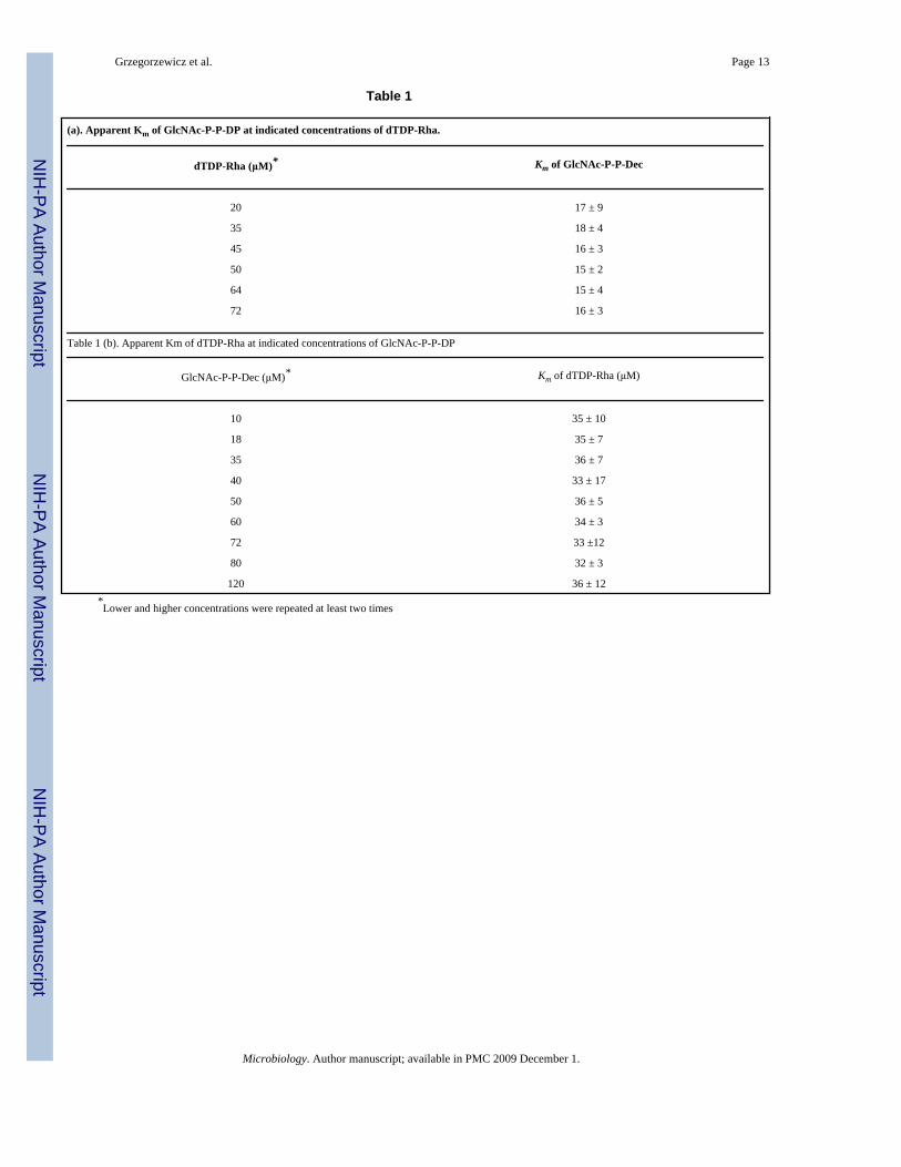
Kinetic studiesThe kinetic parameters of Rv3265c were investigated using different concentration ofsubstrates. The apparent Km for GlcNAc-P-P-DP was determined to be 18 μM at 35 μM dTDP-Rha and the apparent Km for dTDP-Rha was found to be 35 μM at 18 μM GlcNAc-P-P-DP.
To determine if WbbL follows a ternary complex reaction mechanism or a double displacement(ping-pong), the reaction velocity over a range of dTDP-Rha or GlcNAc-P-P-DPconcentrations at several fixed concentrations of the other substrate were measured (Table 1aand Table 1b). The Km value of both substrates remained almost the same over a wide rangeof the other substrate concentration, indicating that the enzyme utilizes a ternary complexmechanism in which the two substrates must bind together to the enzyme. Since the substrateinhibition was observed above 60 μM for both substrates, kinetic studies were limited to lowerconcentrations.
Development of a microtiter plate based assay for rhamnosyl transferaseWe have taken advantage of the organic nature of product to determine enzymatic activity bymeasuring the incorporation of radioactive product [14C]Rha-GlcNAc-P-P-DP into ascintillation cocktail (Ready Organic) that is immiscible with aqueous material. We found thatby adding butanol to the microtiter plate wells followed by Ready Organic scintillation cocktailwe could get reasonable (~ 67%) counting efficiency of the Rha-GlcNAc-P-P-decaprenylproduct as determined by comparing the Ready Organic/butanol microtiter plate counting withcounting the product prepared by the standard extraction protocol. The efficiency of countinga known amount of radioactivity in dTDP-[14C]-Rha (determined by standard liquidscintillation counting) in the Ready Organic scintillation cocktail mixed with butanol was verylow at only ~ 1.5%.
The microtiter plate based assay was run using substrates at their Km concentrations. To stayin the linear range it was decided to perform a microtiter plate based assay at 20% conversionof dTDP-[14C]-Rha. We then determined that DMSO had no negative effect up to at least 2%v/v and in fact increased the counting efficiency to 80% (data not presented). We nextdetermined the Z’ value (Zhang et al., 1999) for the assay using positive (all componentsincluding 1% DMSO) and background (same as positive except lacking enzyme). This data(Fig. 4a) yielded a Z’ value of be 0.83 indicating an excellent assay. The assay was furthervalidated by showing inhibition of WbbL activity in a dose dependent manner by dTDP (Fig.4b).
DiscussionWbbL has been classified into family 2 of the GTs with a characteristic fold of superfamilyGT-A (http://cazy.org/tools/search.html). GT-2 enzymes use inverting mechanism, as doesWbbL which utilizes dTDP-β-rhamnose as a substrate and generates an α-rhamnosyl product.Given its inverting mechanism it would be expected to catalyze the attachment of the rhamnosylresidue in an SN2 like fashion. Our kinetic evidence is consistent with this mechanism as itclearly shows that WbbL forms a ternary complex with both substrates being bound. Howeverdetailed kinetic studies were hampered by substrate inhibition. A similar phenomenon wasobserved by (Zhang et al., 2006a) in working with Bacillus subtilis TagA, a glycosyltransferasethat catalyzes a very similar reaction, i.e. the formation of a β-glycosidic bond between N-acetylmannosamine and undecaprenyldiphosphoryl-D-N-acetyl glucosamine (also inversion—in this case an αsugar nucleotide to a β-linkage).
An earlier assay for enzymes with lipid linked acceptors uses a solid-liquid bead-basedseparation system to selectively adsorb the highly hydrophobic products of the reaction
Grzegorzewicz et al. Page 6
Microbiology. Author manuscript; available in PMC 2009 December 1.
NIH
-PA Author Manuscript
NIH
-PA Author Manuscript
NIH
-PA Author Manuscript

(Hyland & Anderson, 2003). In contrast, the microtiter plate based assay herein is based onthe organic solubility of the WbbL product, Rha-GlcNAc-P-P-DP in distinction to the aqueoussolubility of the dTDP-Rha donor and, importantly, no scintillation beads are required. Thekey component to transferring from an extraction method to direct and selective counting inan organic scintillation cocktail was the addition of butanol to the reaction mixtures; otherwiselittle radioactivity was detected even when radioactive product was present. The microtiterplate assay shows good reproducibility and should aid dramatically in the effort to detect WbbLinhibitors in new TB drug discovery efforts. Furthermore this methodology is readilyapplicable to other enzymes such as TagA that transfer a glycosyl residue from a sugarnucleotide to a lipid linked substrate.
AcknowledgmentsWe gratefully acknowledge the gift of synthetic GlcNAc-P-P-decaprenyl from Kai Li and Michio Kurosu. We alsowould like to thank Dr. Matt Anderson for providing pET 11a-WecA plasmid, Dr. Vara Vissa for help in constructingtruncated WbbL and Donald Dick for running LCMS. This work was supported by U.S. Public Health Service GrantNIH-NIAID AI 33706.
AbbreviationsAG
arabinogalactan
dTDP-Rha dTDP-L rhamnose
ESI electrospray ionization
GC-MS gas chromatography-mass spectrometry
GlcNAc-P-P-DP decaprenyldiphosphoryl-α-D-N-acetyl glucosamine
GT glycosyltransferase
IPTG isopropyl-β-D-thiogalactopyranose
LC liquid chromatography
TAPS 3-[2-hydroxy-1,1-bis(hydroxymethyl)ethyl]amino-1-propanesulfonic acid
TB tuberculosis
Reference ListHyland SA, Anderson MS. A high-throughput solid-phase extraction assay capable of measuring diverse
polyprenyl transferases as exemplified by the phosphate: sugar-1-phosphate WecA, MraY, and MurGproteins. Analytical Biochemistry 2003;317:156–164. [PubMed: 12758253]
Imperiali B, Zimmerman JW. Synthesis of Dolichypyrophosphate-linked Oligosaccharides. TetrahedronLett 1990;31:6485–6488.
Grzegorzewicz et al. Page 7
Microbiology. Author manuscript; available in PMC 2009 December 1.
NIH
-PA Author Manuscript
NIH
-PA Author Manuscript
NIH
-PA Author Manuscript

Kurosu M, Li K. Synthetic studies on Mycobacterium tuberculosis specific fluorescent Park’s nucleotideprobe. Heterocycles 2008;76In Press
Menginlecreulx D, Texier L, Rousseau M, Vanheijenoort J. The Murg Gene of Escherichia-Coli Codesfor the Udp-N-Acetylglucosamine - N-Acetylmuramyl-(Pentapeptide) Pyrophosphoryl-UndecaprenolN-Acetylglucosamine Transferase Involved in the Membrane Steps of Peptidoglycan Synthesis.Journal of Bacteriology 1991;173:4625–4636. [PubMed: 1649817]
Mikusova K, Mikus M, Besra G, Hancock I, Brennan PJ. Biosynthesis of the linkage region of themycobacterial cell wall. J Biol Chem 1996;271:7820–7828. [PubMed: 8631826]
Mills JA, Motichka K, Jucker M, et al. Inactivation of the mycobacterial rhamnosyltransferase, which isneeded for the formation of the arabinogalactan-peptidoglycan linker, leads to irreversible loss ofviability. J Biol Chem 2004b;279:43540–43546. [PubMed: 15294902]
Mills JA, Motichka K, Jucker M, et al. Inactivation of the mycobacterial rhamnosyltransferase, which isneeded for the formation of the arabinogalactan-peptidoglycan linker, leads to irreversible loss ofviability. J Biol Chem 2004a;279:43540–43546. [PubMed: 15294902]
Miroux B, Walker JE. Over-production of proteins in Escherichia coli: mutant hosts that allow synthesisof some membrane proteins and globular proteins at high levels. J Mol Biol 1996;260:289–298.[PubMed: 8757792]
Rush JS, Rick PD, Waechter CJ. Polyisoprenyl phosphate specificity of UDP-GlcNAc:undecaprenylphosphate N-acetylglucosaminyl 1-P transferase from E.coli. Glycobiology 1997;7:315–322.[PubMed: 9134438]
Zhang JH, Chung TDY, Oldenburg KR. A simple statistical parameter for use in evaluation and validationof high throughput screening assays. Journal of Biomolecular Screening 1999;4:67–73. [PubMed:10838414]
Zhang YH, Ginsberg C, Yuan YQ, Walker S. Acceptor substrate selectivity and kinetic mechanism ofBacillus subtilis TagA. Biochemistry 2006a;45:10895–10904. [PubMed: 16953575]
Zhang YH, Ginsberg C, Yuan YQ, Walker S. Acceptor substrate selectivity and kinetic mechanism ofBacillus subtilis TagA. Biochemistry 2006b;45:10895–10904. [PubMed: 16953575]
Grzegorzewicz et al. Page 8
Microbiology. Author manuscript; available in PMC 2009 December 1.
NIH
-PA Author Manuscript
NIH
-PA Author Manuscript
NIH
-PA Author Manuscript

Fig. 1.The reaction catalyzed by rhamnosyl transferase (WbbL).
Grzegorzewicz et al. Page 9
Microbiology. Author manuscript; available in PMC 2009 December 1.
NIH
-PA Author Manuscript
NIH
-PA Author Manuscript
NIH
-PA Author Manuscript

Fig. 2.SDS-PAGE analysis of subcellular fractions from E. coli expressing Rv 3265c (WbbL)visualized by (a) Coomassie brilliant blue and (b) by Western blot. For both (a) and (b) thelanes are: molecular weight markers (1); membranes (2); cytosol (3).
Grzegorzewicz et al. Page 10
Microbiology. Author manuscript; available in PMC 2009 December 1.
NIH
-PA Author Manuscript
NIH
-PA Author Manuscript
NIH
-PA Author Manuscript
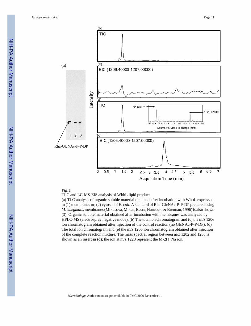
Fig. 3.TLC and LC-MS-EIS analysis of WbbL lipid product.(a) TLC analysis of organic soluble material obtained after incubation with WbbL expressedin (1) membranes or, (2) cytosol of E. coli. A standard of Rha-GlcNAc-P-P-DP prepared usingM. smegmatis membranes (Mikusova, Mikus, Besra, Hancock, & Brennan, 1996) is also shown(3). Organic soluble material obtained after incubation with membranes was analyzed byHPLC-MS (electrospray negative mode). (b) The total ion chromatogram and (c) the m/z 1206ion chromatogram obtained after injection of the control reaction (no GlcNAc-P-P-DP). (d)The total ion chromatogram and (e) the m/z 1206 ion chromatogram obtained after injectionof the complete reaction mixture. The mass spectral region between m/z 1202 and 1238 isshown as an insert in (d); the ion at m/z 1228 represent the M-2H+Na ion.
Grzegorzewicz et al. Page 11
Microbiology. Author manuscript; available in PMC 2009 December 1.
NIH
-PA Author Manuscript
NIH
-PA Author Manuscript
NIH
-PA Author Manuscript

Fig. 4.A validation of a microtiter plate based assay for WbbL(a) Z’ factor determination for microtiter plate based assay was done with 20 positive and 20negative controls (no enzyme). (b) Titration of dTDP.The assay was performed using 18 μM GlcNAc-P-P-DP, 35 μM dTDP-Rha, 1.5 μM dTDP-[14C]Rha (25,000 cpm) and 1 μg of membrane proteins.
Grzegorzewicz et al. Page 12
Microbiology. Author manuscript; available in PMC 2009 December 1.
NIH
-PA Author Manuscript
NIH
-PA Author Manuscript
NIH
-PA Author Manuscript
NIH
-PA Author Manuscript
NIH
-PA Author Manuscript
NIH
-PA Author Manuscript
Grzegorzewicz et al. Page 13
Table 1
(a). Apparent Km of GlcNAc-P-P-DP at indicated concentrations of dTDP-Rha.
dTDP-Rha (μM)* Km of GlcNAc-P-P-Dec
20 17 ± 9
35 18 ± 4
45 16 ± 3
50 15 ± 2
64 15 ± 4
72 16 ± 3
Table 1 (b). Apparent Km of dTDP-Rha at indicated concentrations of GlcNAc-P-P-DP
GlcNAc-P-P-Dec (μM)* Km of dTDP-Rha (μM)
10 35 ± 10
18 35 ± 7
35 36 ± 7
40 33 ± 17
50 36 ± 5
60 34 ± 3
72 33 ±12
80 32 ± 3
120 36 ± 12*Lower and higher concentrations were repeated at least two times
Microbiology. Author manuscript; available in PMC 2009 December 1.
Copyright © 2022 FDOKUMEN