Diversity and ecology of algae and cyanobacteria in the Aragvi River, Georgia
Upload
independentCategory
view
1download
0
186 Journal of Basic Microbiology 2008, 48, 186–194
© 2008 WILEY-VCH Verlag GmbH & Co. KGaA, Weinheim www.jbm-journal.com
Research Paper
Evaluation of fungicidal activity of extracellular filtrates of cyanobacteria – possible role of hydrolytic enzymes
Radha Prasanna, Lata Nain, Ravindramani Tripathi, Vishal Gupta, Vidhi Chaudhary, Sheetal Middha, Monica Joshi, Radhika Ancha and Brahma D. Kaushik
Division of Microbiology and Centre for Conservation & Utilization of Blue-Green Algae (CCUBGA), Indian Agricultural Research Institute (IARI), New Delhi, India
A set of seventy axenised and unicyanobacterial isolates belonging to the genus Anabaena were
evaluated for biocidal activity against a set of phytopathogenic fungi. Among them, 35
Anabaena strains showed zone of inhibition against one or more fungi. The extracellular
filtrates from 4 and 8 weeks old cultures of these Anabaena strains were further evaluated in
terms of hydrolytic enzymes, proteins and IAA employing standard methods. Significant
differences were also observed among the strains in terms of their FPase, chitosanase and
xylanase activity, while low and relatively similar values of CMCase, cellobiase and protease
activity were recorded in the strains analyzed. IAA production was also observed in all the
strains. Comparative evaluation of activity of hydrolytic enzymes and antifungal activity
revealed that such enzymes may contribute to the fungicidal activity of the cyanobacterial
strains, besides other bioactive compounds, including IAA, which are established promising
traits for biocontrol agents. This study is a first time report on the production of hydrolytic
enzymes by these oxygenic photosynthetic prokaryotes, which can be potential candidates for
the development of biocontrol agent(s) against selected phytopathogenic fungi.
Keywords: Cyanobacteria / Fungicidal activity / Hydrolytic enzymes / Phytopathogenic fungi / IAA
Received: June 19, 2007; accepted: January 13, 2008
DOI 10.1002/jobm.200700199
Introduction*
Cyanobacteria represent a small taxonomic group of
prokaryotes, which are equipped with the power to
harm and help plants, animals and humankind, and
possess a tremendous potential for producing a wide
range of secondary metabolites [1]. It was in the 1930s
that De [2] pointed out that the preponderance of
cyanobacteria was responsible for the inherent and
sustained fertility of rice fields which led to the cyano-
bacterial populations in soil being evaluated in terms of
their diversity and utility as biofertilizers, not only for
rice, but for other crops including wheat [3–5]. Cyano-
bacteria are also found commonly growing as blooms
in eutrophic lakes, reservoirs and as floating assem-
Correspondence: Dr. Radha Prasanna, Division of Microbiology & Centre for Conservation and Utilization of Blue-Green Algae (CCUBGA), Indian Agricultural Research Institute (IARI), New Delhi – 110 012, India E-mail: [email protected] Phone: +91-011-25847649 Fax: +91-011-25846420
blages in marine ecosystems. Such blooms have been
notorious and associated with the production of tox-
ins/allelopathic compounds, which provide a competi-
tive advantage to these organisms [6–9]. This is one of
the critical factors responsible for their abundance in
diverse environments especially eutrophic water bod-
ies. However, the chemical potential of these ubiqui-
tous prokaryotes, widely distributed in diverse soil
types, aquatic environments and ecologies, in terms of
production of metabolites has not been much investi-
gated.
Agriculture requires the extensive use of chemical
pesticides to protect crops against pests and diseases.
Several of these chemicals pollute our ground water
and drinking water and therefore some governments
have decided to reduce these chemical inputs substan-
tially. This urges the need for alternative crop protec-
tants. One of these alternatives is the use of biological
control agents, among which are microorganisms that
can protect the plants against diseases [10]. The coloni-

Journal of Basic Microbiology 2008, 48, 186–194 Fungicidal activity of cyanobacterial strains 187
© 2008 WILEY-VCH Verlag GmbH & Co. KGaA, Weinheim www.jbm-journal.com
zation and defensive retention of rhizosphere niche by
microorganisms is enabled by the production of al-
lelochemicals, antibiotics, biocidal volatiles and lytic/
detoxification enzymes.
Terrestrial cyanobacteria such as Anabaena laxa are
known to produce antifungal cyclic peptides such as
laxaphycin A and B [11]. Casamatta and Wickstrom [12]
reported that the exudates of Microcystis aeruginosa were
inhibitory towards bacterial plankton communities. A
number of nucleosides – tubercidin, toyocamycin and
their corresponding derivatives isolated from Scytone-
mataceae members were observed to be toxic towards
Aspergillus oryzae, Candida albicans, Penicillium notatum and
Saccharomyces cerevisiae [13]. A diverse range of com-
pounds are also known to exhibit a bioregulatory role
as a result of their cytotoxic, immunosuppressive and
enzyme-inhibiting activities which are of tremendous
pharmaceutical significance [14, 15]. However, the role
of hydrolytic enzymes, which are implicated in fungi-
cidal activity of several biocontrol strains, has not been
explored in these photosynthetic prokaryotes.
The present investigation describes the characteriza-
tion of cyanobacterial metabolites from a set of Ana-
baena strains exhibiting biocidal activity, as a prelude to
their utilization as biocontrol agent(s) in agriculture.
Materials and methods
Organisms The available germplasm of seventy cyanobacterial
strains, belonging to the genus Anabaena (isolates from
rice fields, Azolla fronds and water bodies [5]) was
screened for the biocidal activity against selected phy-
topathogenic fungi obtained from the Indian Fungal
Type Culture Collection, Division of Plant Pathology,
IARI, New Delhi.
Growth and maintenance The cyanobacterial strains were axenised by standard
procedures employing a set of antibiotics [16]. Such
cultures were used for further work, after repeated sub
culturing and grown under L: D (light: dark cycles –
16:18), white light (50–55 µmol photons m–2
s–1
) and
29 ± 1 °C in nitrogen free BG-11 medium [17] in Haf-
fkine flasks. Potato dextrose medium was used for the
growth and maintenance of the fungal cultures used.
Incubation was done at 30 °C in a BOD incubator.
Screening of the cultures for biocidal activity All the seventy strains were tested for fungicidal activ-
ity utilizing double layer technique to obtain a uniform
lawn of test organisms. Sterile filter paper discs of
5 mm diameter soaked in 100 µl filtrates (0.22 µm filter
drawn) of cyanobacterial cultures (4/8 weeks old) were
placed on the lawn of test organism. The phytopatho-
genic fungi, causing diseases of various crops, were
used as test organisms. The fungus to be tested against
was placed in the center as a disc; inhibition zone was
measured around each well after incubation period of
7–8 d at 30 °C. The presence of inhibition zone was
taken as positive indication, and the diameter of the
zones measured. Antifungal sensitivity discs containing
Nystatin (100 Units) obtained from Hi Media Laborato-
ries, India were used as standard. The culture filtrates
were also partitioned using two solvents – Ethyl Acetate
and Dichloromethane and sterile discs soaked in the
aqueous phase were placed in a similar manner for
evaluating the water soluble nature of the toxic com-
ponent of the filtrate.
Total proteins The amount of proteins was determined spectropho-
tometrically according to [18] with Bovine serum albu-
min (BSA) as standard.
Chitosanase activity The cell free culture filtrates were analysed for chito-
sanase activity (EC 3.2.1.4) by the spectrophotometric
method using glycol chitosan as the assay substrate.
One unit of chitosanase activity was defined as micro-
moles of N-Acetyl glucosamine released per min under
the assay conditions [19].
Protease activity Protease activity was assayed using casein as a substrate
and measured by liberation of tyrosine from the incu-
bation mixture [20]. The intensity of blue color was
measured at 660 nm using spectrophotometer against
the standard curve of Tyrosine, and expressed as Inter-
national unit (IU), which was calculated as µmoles of
tyrosine liberated min–1
.
Filter paperase/FPase (exo-β-1, 4-glucanase; EC 3.2.1.91) and CMCase activity (EC 3.2.1.4) The enzyme activities of both enzymes were assayed
spectrophotometrically using filter paper and car-
boxymethyl cellulose as substrates respectively [21].
Reducing sugars liberated were estimated at 575 nm
against the standard curve of Glucose. One unit of en-
zyme represents I µmole of glucose liberated per ml of
culture filtrate per min.
Xylanase activity The xylanase activity was measured spectrophotometri-
cally at 575 nm using xylan as substrate [22] against the

188 R. Prasanna et al. Journal of Basic Microbiology 2008, 48, 186–194
© 2008 WILEY-VCH Verlag GmbH & Co. KGaA, Weinheim www.jbm-journal.com
standard curve of xylose. One unit of xylanase activity
represents I µmole of xylose liberated per ml of culture
filtrate per min.
Cellobiase (β-D-glucosidase; EC 3.2.1.21) activity The cellobiase activity was determined spectropho-
tometrically at 430 nm by the method outlined by [23]
against the standard curve of p-nitrophenol.
IAA activity IAA activity was measured by the method described by
[24]. The intensity of pink color was measured at
530 nm and values were compared with those of the
standard curve of IAA.
The activity of all the hydrolytic enzymes is ex-
pressed as standard International Unit (IU). The culture
filtrates of the cultures of the cyanobacterial strains
were also scanned in the UV-VIS range using Specord
model spectrophotometer, and the peaks recorded for
identification of novel compounds.
Statistical analyses The data recorded in triplicate for the parameters in
various strains was subjected to ANOVA (analysis of
variance) in accordance with the experimental design
(completely randomized block design) using MSTAT-C
statistical package to quantify and evaluate the sources
of variation. Duncan’s Multiple Range Test (DMRT) was
employed to compare the mean performances of differ-
ent strains for the specific parameters under study and
the rankings are denoted by superscripts in the rele-
vant tables.
Results and discussion
Soil microorganisms are known to be an important
determinant of allelopathic activity, besides leading to
severe monetary losses as a result of soil borne patho-
gens of vegetables, fruits and field crops. Among them,
fungal pathogens are a major problem in agriculture, as
most of the fungicides employed exhibit lower potency
under field conditions and have been a source of
chemical pollution, poisoning the fruits and vegetables,
which form an essential component of human diet.
Cyanobacteria are known to produce a large number
of compounds with varying bioactivities, including the
hepato- and neurotoxins, which are a cause for much
concern in aquatic environments [6]. Extensive screen-
ing programs for bioactive molecules in these organ-
isms have revealed the presence of protease inhibitors,
cytotoxins to human and animal cells and compounds
with antibacterial, antifungal and antiviral properties
[25]. Despite the availability of a few patents [26] the
industrial exploitation of cyanotoxins as fungicidal
agents is limited and a need exists for identification of
novel metabolites for developing commercial products.
In this context, it would be valuable to analyse the
bioactive compounds/enzymes from cyanobacteria for a
better understanding of the mechanism of fungicidal
activity.
Preliminary analyses of the culture filtrates of a set
of 70 cyanobacterial strains against the selected phyto-
pathogenic fungi (as measured by disc diffusion assay)
revealed that only 35 strains produced a zone of inhibi-
tion of varying diameter, ranging from 10–20 mm, and
the average size of the zone was measured as 10 mm
(Fig. 1, Table 1). Comparative evaluation of filtrates
from 4 and 8 weeks cultures (data not shown) had
shown that the latter stage produced larger zones of
inhibition. It is well known that biocidal compounds
are secondary metabolites, produced at late log-
stationary phase stage (more than 3 weeks of growth);
hence, further evaluation was done with 8 weeks old
culture filtrates. Among these, 23 strains of Anabaena
inhibited the growth of Fusarium moniliforme while 17
strains inhibited Alternaria solani and Aspergillus candida
was inhibited by 15 strains. Three strains each inhib-
ited the growth of Drechslera oryzae and Pythium apha-
nidermatum. Three cyanobacterial strains – RP 8, 9 and
69 exhibited a broad spectrum of activity, inhibiting
the growth of six phytopathogenic fungi. Strains RP 1
Figure 1. Zone of inhibition by filtrates of cyanobacterial cultures on the lawn of selected phytopathogenic fungi. (a) Aspergillus candida (b) Pythium aphanidermatum (c) Alternaria solani (d) Fusarium solani.

Journal of Basic Microbiology 2008, 48, 186–194 Fungicidal activity of cyanobacterial strains 189
© 2008 WILEY-VCH Verlag GmbH & Co. KGaA, Weinheim www.jbm-journal.com
Table 1. Fungicidal activity of cyanobacterial culture filtrates in terms of zone of inhibition (values in parentheses indicate the diameter of inhibition zone (mm).
Phytopathogenic fungi tested Anabaena strains
Macro- phomina phaseolina
Fusarium monoliforme
Alternaria solani
Pythium aphanidermatum
Aspergillus candida
Drechslera oryzae
Fusarium solani
RP 1 – + (20) + (10) – – – + (16) RP 3 – + (11) + (11) – + (10) – – RP 4 + (12) + (10) – + (10) – + (10) RP 5 – – + (10) – + (10) – + (13) RP 6 + (10) + (11) + (11) – + (10) – + (11) RP 7 – + (11) + (10) – – – + (10) RP 8 – + (12) + (11) – + (10) – + (10) RP 9 – + (12) + (11) + (12) + (10) – – RP 11 – – + (10) – – – – RP 12 – + (11) – – – – – RP 14 – + (13) – – – – + (11) RP 16 – + (10) + (11) – – – – RP 17 – – – – – – – RP 20 – – – – – – – RP 21 – – – – – – + (10) RP 25 – – – – + (10) + (10) – RP 26 – – + (27) – + (12) + (10) – RP 33 – – – + (12) + (14) – – RP 34 – + (12) – + (14) – – – RP 35 – – – – – – – RP 45 – + (10) – – – – + (10) RP 47 – – + (10) – – – – RP 49 – + (10) + (10) – + (10) – – RP 50 – + (16) + (10) – + (12) – – RP 52 – + (13) + (12) – – – – RP 53 – + (12) + (28) – + (10) – + (14) RP 56 – + (14) – – – – – RP 57 – + (10) – – + (10) – – RP 58 – – – – – – – RP 59 – + (10) + (40) – – – + (10) RP 63 – + (10) – – – – + (10) RP 68 – + (11) – – – – + (11) RP 69 – + (12) – – + (10) + (10) + (11) RP 70 – – – – – + (11) RP 71 – + (10) – – + (12) – + (11) Nystatin + (16) + (30) + (20) + (14) + (14) + (16) + (16)
and 59 were observed to be highly potent in terms of
their fungicidal activity, as they produced a much lar-
ger zone of inhibition than those of standard antifungal
agent – nystatin. The largest diameter of zone of inhibi-
tion of 40 mm was recorded by strain RP 59, followed
by RP 53 (28 mm) against Alternaria solani. Other phyto-
pathogenic fungi – Pythium debaryanum, Rhizopus oryzae,
Fusarium oxysporum pisi, Ustilago tritici, Fusarium graminea-
rum, Rhizoctonia solani, Drechslera sorkiniana, Sclerotium
oryzae and Alternaria triticiana were also tested, however,
no inhibition zone was observed.
The basis of antibiosis as a biocontrol mechanism has
become increasingly better understood over the past
two decades [27, 28]. A variety of antibiotics have been
identified, including compounds such as amphisin,
2,4-di acetylphloroglucinol (DAPG), hydrogen cyanide,
oomycin A, phenazine, pyoluteorin, pyrrolnitrin, ten-
sin, troplone, and cylic lipopeptides produced by pseu-
domonads, and oligomycin A, kanosamine, zwitter-
micin A, Xanthobaccin produced by Bacillus and Steno-
trophomonas spp. [29]. However, a variety of microorga-
nisms also exhibit hyperparasitic activity, attacking
pathogens by excreting cell wall hydrolases, which has
been a subject of intensive research in recent years. Lim
and coworkers [30] isolated a strain of Psuedomonas
stutzeri that produced extracellular chitosanase and
laminarase, which could digest the mycelia of Fusarium
solani. However, cyanobacteria are generally believed to

190 R. Prasanna et al. Journal of Basic Microbiology 2008, 48, 186–194
© 2008 WILEY-VCH Verlag GmbH & Co. KGaA, Weinheim www.jbm-journal.com
be obligate phototrophs, not requiring other C sources
for their growth; therefore, no focused efforts have
been undertaken on production of hydrolytic enzymes.
The filtrates from four and eight weeks old cultures
were analyzed in terms of proteins, IAA and hydrolytic
enzymes. A two-fold enhancement was observed in
terms of proteins in the filtrates from 8 weeks old cul-
tures, as compared to four weeks old cultures, indica-
tive of release of proteinaceous molecules by the cells
(Table 2). Extensive screening programs for bioactive
compounds in cyanobacteria has revealed the presence
of unique peptides with cyclic structures (cyanopepto-
lins, depsipeptides), besides linear peptides isolated
from a number of strains belonging to genera – Micro-
cystis, Nostoc and Anabaena [25]. The analyses of the
culture filtrates are currently in progress as UV scans
revealed the presence of novel compounds. Although
toxins were the main subject of research on toxic
cyanobacterial blooms, isolation of bioactive com-
pounds other than biotoxins has now been recently
stressed [1].
The production of phytohormones, earlier consid-
ered, as a trait of the plant kingdom is also widespread
among soil and plant associated prokaryotes [31], espe-
cially those involved in plant microbe symbiotic or
associative interactions or plant pathogenesis. Microor-
ganisms inhabiting rhizosphere of various plants are
likely to synthesize and release auxin as secondary
metabolite because of rich supplies for substrates ex-
uded from the roots compared with non-rhizosphere
soils. IAA production was also observed in the cyano-
bacterial strains, which enhanced several folds with
increasing age of the culture. Strains RP 53, 57 and 68
exhibited a 7–10 folds increase, indicative of the possi-
ble role in their interactions with the fungal pathogens.
It has been suggested up to 80% of bacteria isolated
from rhizosphere can produce IAA. The capacity for
IAA biosynthesis was found in representative of free
living and symbiotic cyanobacteria of the genera Nostoc,
Chlorogloeopsis, Calothrix, Plectonema, Gloeothece, Anabaena,
Cylindrospermum and Anabaenopsis [32, 33]. The trypto-
phan independent pathway, more common in plants, is
also found in microorganisms – Azospirilla [31] and
cyanobacteria [33]. Out of 34 free-living and symbioti-
cally competent cyanobacterial strains, auxin like sub-
stances were detected in 38% of the free living and 83%
of symbiotic isolates in the presence and absence of
tryptophan. The endogenous accumulation of IAA was
confirmed with immunological techniques (ELISA) and
GC-MS techniques. Genes involved in IAA production
via indole pyruvic acid pathway, such as ipdC, has also
been sequenced in Nostoc PCC 73102 strain [33].
Table 2. Protein accumulation and IAA production in cyano-bacterial strains at different stages of growth (4 and 8 weeks old cultures)*.
Strain no. Protein (µg ml–1) IAA (µg ml–1)
4 weeks 8 weeks 4 weeks 8 weeks
RP 1 80.001 130.0017 0.36125 1.92019
RP 3 66.003 140.0016 0.42124 5.4408
RP 4 31.0011 190.0010 0.53223 4.20011
RP 5 12.0019 170.0013 0.56223 3.45114 RP 6 14.0018 143.0015 1.69018 1.05623
RP 7 15.0018 120.0019 2.39414 1.57321
RP 8 20.0016 220.009 2.44113 1.76920
RP 9 12.0019 128.0018 0.36725 3.21615
RP 11 8.0020 410.001 1.17322 3.44014
RP 12 75.002 380.003 3.5216 2.06018
RP 14 4.0020 280.005 3.2867 2.58016
RP 16 8.0020 230.006 3.6124 1.94819
RP 17 42.005 171.0013 2.58211 0.91524
RP 20 8.0020 34.0025 3.0998 0.99323
RP 21 26.0012 400.002 3.5605 1.36422
RP 25 38.008 210.008 2.11315 3.18013
RP 26 62.004 220.007 0.11327 1.90019
RP 33 23.0015 50.0024 2.48812 3.25615
RP 34 25.0013 192.009 1.31421 6.5506
RP 35 2.0022 130.0017 3.0998 3.23915
RP 45 7.0020 60.0023 3.8973 0.19126
RP 47 15.0018 27.0027 2.91110 4.34310
RP 49 25.0013 67.0022 3.5685 4.25011
RP 50 37.009 76.0021 3.8503 5.8507
RP 52 7.0020 34.0025 3.0059 7.2305
RP 53 39.007 150.0014 0.28226 7.9803
RP 56 19.0017 100.0020 3.2397 2.44817
RP 57 31.0011 210.008 1.97216 10.782
RP 58 35.0010 310.004 4.3192 5.3788
RP 59 43.005 180.0012 4.5071 0.37525
RP 63 40.006 182.0011 4.5341 2.41017
RP 68 32.0011 140.0016 1.54919 11.201
RP 69 26.0012 75.0021 1.45520 3.99212
RP 70 24.0014 400.002 1.83117 7.8404
RP 71 20.0016 30.0026 2.39414 5.1889
SEM 0.820 0.816 0.0025 0.0365
* Superscripts denote DMRT ranking in descending order
In our investigation it was interesting to observe that
the cyanobacterial strains exhibiting fungicidal activity
produced one or more hydrolytic enzymes. The hydro-
lytic enzymes such as protease, CMCase, Cellobiase and
Fpase also showed a wide range of values in the cyano-
bacterial strains tested (Table 3). The activity of CMCase
and FPase enzymes were enhanced in 8 weeks old
cultures. Protease activity in the culture filtrates of the
4 weeks old cultures ranged from 0.01–0.362 Uml–1
,
which however dropped to 0.001–0.012 Uml–1
in
8 weeks old cultures. A similar pattern was observed
for cellobiase activity, which was in general, very low.
Chitosanase activity was remarkably high in all the
strains, especially at 4 weeks stage, with values ranging
from 0.040 to 0.482 Uml–1
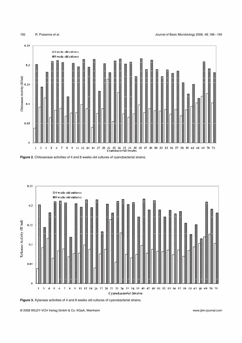
(Fig. 2). Xylanase activity

Journal of Basic Microbiology 2008, 48, 186–194 Fungicidal activity of cyanobacterial strains 191
© 2008 WILEY-VCH Verlag GmbH & Co. KGaA, Weinheim www.jbm-journal.com
showed an almost two folds enhancement in activity in
eight weeks old culture filtrates (Fig. 3). Strains RP 69,
70 and 71 were observed to show much higher values
for these parameters, especially in the 4 weeks old cul-
tures, however, strains RP 9 and 45 were the highest
ranked strains, when 8 weeks old cultures were ana-
lysed.
On analyzing the overall performance of the strains
in terms of biocontrol traits, strain RP 1 was observed
to be among the five top ranked strains in terms of
diameter of zone of inhibition and chitosanase activity,
while RP 9 was top ranked in terms of chitosanase ac-
tivity, besides exhibiting high levels of activity of other
hydrolytic enzymes and inhibited the growth of five
out of seven fungi tested. The top ranked strains in
terms of their hydrolytic enzyme activity were com-
pared with respect to the antifungal activity (inhibition
zone) shown by culture filtrates and aqueous phase
after partitioning with Ethyl acetate and Dichloro-
methane as solvents, which revealed the significant
role of the enzymes.
The excretion of hydrolytic enzymes is known to be a
common trait of plant pathogens/symbionts, which
promotes a closer association with plant roots/target
organisms and improve the stability of such associa-
tions. Chitosanases are known to selectively degrade
chitosan/chitin by hydrolysis of the β-1, 4-glycosidic
bonds that link N-acetyl glucosamine residues of chitin
and form the basis for antifungal activity. The chito-
sanase/chitinase produced by S. plymuthica C48 inhibited
Table 3. Screening the activity of selected hydrolytic enzymes in the 4 and 8 weeks old culture filtrates of cyanobacterial strains*.
Strain no. Protease (IU uml–1) CMCase (IU ml–1) Cellobiase (IU ml–1) FPase (IU ml–1)
4 weeks 8 weeks 4 weeks 8 weeks 4 weeks 8 weeks 4 weeks 8 weeks
RP 1 0.2632 0.0051 0.0071 0.0353 0.0082 0.0031 0.0324 0.10210
RP 3 1.1211 0.0041 0.0101 0.0672 0.0082 0.0041 0.0374 0.1268
RP 4 0.2982 0.0051 0.0261 0.1171 0.0092 0.0031 0.0344 0.08412
RP 5 0.2522 0.0031 0.0061 0.0692 0.0042 0.0031 0.0543 0.1069
RP 6 0.2202 0.0121 0.0081 0.0433 0.0072 0.0031 0.0404 0.10010
RP 7 0.3412 0.0091 0.0101 0.0173 0.0062 0.0051 0.0334 0.07315
RP 8 0.2902 0.0031 0.0291 0.0313 0.0072 0.0021 0.0334 0.1377
RP 9 0.3622 0.0021 0.0181 0.0373 0.0092 0.0031 0.0833 0.1465
RP 11 0.3622 0.0011 0.0131 0.0313 0.0092 0.0061 0.0613 0.1682
RP 12 0.3312 0.0031 0.0041 0.0812 0.0092 0.0041 0.0324 0.1514
RP 14 0.3412 0.0051 0.0151 0.0253 0.0082 0.0031 0.0274 0.1129
RP 16 0.3512 0.0021 0.0101 0.0363 0.0092 0.0061 0.1132 0.07613
RP 17 0.3312 0.0051 0.0081 0.0223 0.0771 0.0041 0.0274 0.09510
RP 20 0.2922 0.0051 0.0071 0.0323 0.0102 0.0051 0.0234 0.3351
RP 21 0.2922 0.0051 0.0071 0.0393 0.0132 0.0031 0.0394 0.09011
RP 25 0.3332 0.0051 0.0101 0.0333 0.0122 0.0041 0.0484 0.09610
RP 26 0.3112 0.0081 0.0041 0.0413 0.0082 0.0031 0.0384 0.09310
RP 33 0.0242 0.0061 0.0031 0.0373 0.0082 0.0021 0.0563 0.08512
RP 34 0.0202 0.0061 0.0041 0.0313 0.0132 0.0061 0.0753 0.07414
RP 35 0.0262 0.0061 0.0031 0.0353 0.0092 0.0071 0.0813 0.82013
RP 45 0.0282 0.0051 0.0061 0.0393 0.0082 0.0081 0.0593 0.07315
RP 47 0.0202 0.0071 0.0031 0.0323 0.0062 0.0071 0.0464 0.07713
RP 49 0.0222 0.0061 0.0061 0.0233 0.0082 0.0031 0.0703 0.07414
RP 50 0.0242 0.0071 0.0031 0.0602 0.0062 0.0031 0.0783 0.09510
RP 52 0.0302 0.0081 0.0041 0.0433 0.0082 0.0031 0.0563 0.06216
RP 53 0.0212 0.0061 0.0031 0.0483 0.0102 0.0051 0.0603 0.08013
RP 56 0.0122 0.0071 0.0161 0.0313 0.0082 0.0031 0.0404 0.08113
RP 57 0.0222 0.0061 0.0051 0.0353 0.0092 0.0051 0.0374 0.1079
RP 58 0.0262 0.0071 0.0081 0.0353 0.0092 0.0021 0.0494 0.09710
RP 59 0.0252 0.0011 0.0051 0.0403 0.0062 0.0031 0.0374 0.1099
RP 63 0.0182 0.0071 0.0171 0.0283 0.0082 0.0031 0.0553 0.1308
RP 68 0.0222 0.0061 0.0041 0.0323 0.0132 0.0071 0.0294 0.1563
RP 69 0.0222 0.0061 0.0041 0.0363 0.0092 0.0031 0.0504 0.1513
RP 70 0.0212 0.0081 0.0181 0.0373 0.0192 0.0051 0.1381 0.1396
RP 71 0.0262 0.0051 0.0021 0.0363 0.0092 0.0041 0.0633 0.1553
SEM 0.2016 0.0025 0.0025 0.0025 0.0025 0.0025 0.0025 0.0025
* Superscripts denote DMRT ranking in descending order.
192 R. Prasanna et al. Journal of Basic Microbiology 2008, 48, 186–194
© 2008 WILEY-VCH Verlag GmbH & Co. KGaA, Weinheim www.jbm-journal.com
Chi
tosa
nase
Act
ivity
(IU
/ml)
Chi
tosa
nase
Act
ivity
(IU
/ml)
Figure 2. Chitosanase activities of 4 and 8 weeks old cultures of cyanobacterial strains.
Figure 3. Xylanase activities of 4 and 8 weeks old cultures of cyanobacterial strains.

Journal of Basic Microbiology 2008, 48, 186–194 Fungicidal activity of cyanobacterial strains 193
© 2008 WILEY-VCH Verlag GmbH & Co. KGaA, Weinheim www.jbm-journal.com
Table 4. Comparative evaluation of hydrolytic enzyme activity with inhibition zone produced by selected cyanobacterial strains on the lawn of phytopathogenic fungi.
Hydrolytic enzymes evaluated Phytopathogenic fungi tested
Anabaena strains*
FPase Cellobiase CMCase Protease Chitosanase Xylanase Fusarium moniliforme
Alternaria solani
Aspergillus candida
RP 1 – – – – 0.302 0.202 AP-DCMb, AP-EAc, CFa
CFa AP-DCMb, AP-EAc
RP 4 – – 0.117 – – 0.182 CFa AP-DCMb CFa RP 6 – – – 0.012 – 0.212 CFa CFa CFa RP 9 0.146 – – – 0.318 0.205 CFa CFa CFa RP 11 0.168 – – – 0.286 0.196 AP-EAc – RP 12 0.151 0.081 0.293 0.215 AP-EAc, CFa – AP-EAc, CF RP 16 – – – – 0.306 0.215 CFa CFa AP-DCMb, CFa RP 20 0.335 – – – – 0.204 AP-EAc – – RP 26 – – – 0.008 – 0.216 – CFa CFa RP 34 – – – – 0.313 0.208 CFa AP-DCMb,
AP-EAc, CF –
RP 45 – 0.008 – – – 0.217 CFa CFa AP-DCMb, AP-EAc RP 47 – – – – 0.292 0.189 AP-DCMb, CFa CFa AP-DCMb, CFa RP 50 – – 0.060 – – 0.189 CFa AP-DCMb, CFa
–
RP 53 – – – – 0.295 0.188 CFa CFa – RP 57 – – – – 0.301 0.185 CFa – – RP 68 0.156 0.007 – – 0.278 AP-DCMb,
AP-EAc, CFa – –
RP 69 0.151 – – – 0.282 0.209 CFa CFa – RP 71 0.155 – – – – 0.181 CFa – –
* Top ranked strains on the basis of DMRT analyses of activity of hydrolytic enzymes a denotes Culture Filtrate b denotes Aqueous Phase obtained using Dichloromethane as solvent c denotes Aqueous Phase obtained using Ethyl acetate as solvent
spore germination and germ-tube elongation in Botrytis
cinerea. The ability to produce extracellular chitosana-
ses/chitinase is considered crucial for Serratia marcescens
to act as antagonist against Sclerotium rolfsii, and for
Paenibacillus sp. Strain 300 and Streptomyces sp. strain
385 to suppress Fusarium oxysporum f.sp. cucumerinum
[34]. The regulation of siderophores and antibiotics,
lytic enzyme production (proteases and chitosanases in
particular) is known to involve the GacA/GacS regula-
tory systems and colony phase variation [35]. Similar
studies on the genes involved need to be undertaken in
cyanobacteria, for a better understanding of their rela-
tionship with biocidal activity.
Although the role of cyanotoxins/peptides/phenolic
molecules cannot be ruled out in terms of biocidal ac-
tivity, it becomes evident from this study that hydro-
lytic enzymes are definitely contributing to the inhibi-
tion of phytopathogenic fungi.
Conclusions
The present investigation, for the first time, illustrates
the activity of hydrolytic enzymes in these ubiquitous
photosynthetic prokaryotes and their possible role in
biocidal activity against phytopathogenic fungi. The
potential of these strains in developing biocontrol
agents is immense – as these strains possess abilities for
production of IAA and hydrolytic enzymes, besides the
UV absorbing compounds (which are presently being
characterized). Such multifaceted strains would possess
a competitive edge over other rhizosphere microflora,
against phytopathogenic fungi and need to be explored
for the developing biocontrol agents.
Acknowledgements
The study was undertaken as a part of the AMAAS Net-
work Project on Microorganisms, granted by the Indian
Council of Agricultural Research (ICAR), New Delhi. We
thank the authorities of the Division of Microbiology
and CCUBGA, IARI, New Delhi, for providing necessary
facilities for undertaking this study.
References
[1] Mundt, S., Kreitlow, S., Nowotny, A. and Effmert, U., 2001. Biochemical and Pharmacological investigations of selected cyanobacteria. Intl. J. Hygeine Envl. Health., 203, 327–334.

194 R. Prasanna et al. Journal of Basic Microbiology 2008, 48, 186–194
© 2008 WILEY-VCH Verlag GmbH & Co. KGaA, Weinheim www.jbm-journal.com
[2] De, P.K., 1939. The role of blue green algae in nitrogen fixation in rice fields. Proc. Royal Soc. (London) Ser. B., 127, 129–139.
[3] Venkataraman, G.S., 1981. Blue-green algae for rice pro-duction – a manual for its promotion, FAO Soils bulletin No. 46 FAO, Rome, 102p.
[4] Karthikeyan, N. Prasanna, R., Lata, Nair and Kaushik, B.D., 2007. Evaluating the potential of plant growth pro-moting cyanobacteria as inoculants for wheat. Eur. J. Soil Biol. 43, 23–30.
[5] Prasanna, R. and Nayak, S., 2007. Influence of diverse rice soil ecologies on cyanobacterial diversity and abundance. Wetlands Ecol. Managmt. (In press; DOI 101007/s11273-006-9018-2).
[6] Carmichael, W.W., 1994. The toxins of cyanobacteria. Sci. Amer, 270, 78–86.
[7] Nagle, D.G. and Wedge, D.E., 2002. Antifungal properties of cyanobacteria and algae: ecological and agricultural implications. In: Inderjit, Malik A.U. (Eds.).Chemical eco-logy of plants: Allelopathy in Aquatic and Terrestrial eco-systems. Birkhauser Verlag/Switzerland. p. 7–32.
[8] Jaiswal, P., Prasanna, R. and Singh, P.K., 2005. Isolation and Screening of rice field cyanobacteria exhibiting algi-cidal activity. Asian J. Microbiol. Biotechnol. Environ. Sci., 7(4), 367–373.
[9] Jaiswal, P., Prasanna, R. and Singh, P.K., 2006. Factors influencing algicide production by Microcystis sp and its effect on selected cyanobacteria In: Gupta RK, Pandey VK, editors. Advances in Applied Phycology, Daya Publishing House, India, pp. 6–12.
[10] Akkopru, A. and Demir, S., 2005. Biological control of Fusarium wilts in tomato caused by Fusarium oxysporum ly-copersici by AMF Glomus intraradices and some rhizobacte-ria. J. Phytopathol., 153, 544–550.
[11] Frankmolle, W.P., Larsen, L.K., Caplan, F.R., Patterson, G.M.L., Knubel, G. and Moore, R.E., 1992. Antifungal Cyc-lic Peptides from the terrestrial Blue Green Alga Anabaena laxa. J. Ant. 45(9), 1451–1457.
[12] Casamatta, D.A. and Wickstrom, C.E., 2000. Sensitivity of two disjunct bacterioplankton communities to exudates from the cyanobacterium Microcystis aeruginosa. Microb. Ecol., 41, 64–73.
[13] Kulik, M.M, 1995. The potential for using cyanobacteria (blue green algae) and algae in the biological control of plant pathogenic bacteria and fungi. Eur. J. Plant Pathol., 101, 585–599.
[14] Asthana, R.K., Srivastava, A., Kayastha, A.M., Nath, G. and Singh, S.P., 2006. Antibacterial potential of γ-linolinic acid from Fischerella sp colonizing neem bark. J. Appl. Phycol., 22, 443–448.
[15] Bagchi, S.N. and Ray, S., 2001. Extraction and purification of an algicidal metabolite from a cyanobacterium Oscilla-toria latevirens. Indian J. Microbiol., 41, 163–167.
[16] Kaushik, B.D., 1987. Laboratory methods for blue green algae. Associated Publishing Company, New Delhi.
[17] Stanier, R.Y., Kunisawa, R., Mandal, M. and Cohen-Bazire, G., 1971. Purification and properties of unicellular blue green algae (Order: Chroococcales). Bacteriol. Rev., 35, 171–305.
[18] Herbert, D., Phipps, P. J. and Strange, R.E., 1971. Chemical analysis of microbial cells. In: Norris J.R, Ribbons D.W, (Eds.). Methods in Microbiology. Academic Press, New York, pp. 209–344.
[19] Ohtakara, A., 1988. Chitosanase and β-N-acetyl hexosami-ne from Pycnosporus cinnabarinus. Methods Enzymol., 168, 464–468.
[20] Ong, P.S. and Gaucher, G.M., 1973. Protease activity by thermophilic fungi. Can. J. Microbiol., 19, 129–133.
[21] Ghosh, T.K., Bailey, H.J., Bisaria, V.S. and Enari, T.M., 1983. Measurement of cellulase activities – Final recom-mendations, commission of Biotechnology. International Union of Pure Applied Chemistry, 59, 1–13.
[22] Bailey, M.J., Biely, P. and Poutamen, K., 1992. Interlabora-tory testing method for assay of xylanase activity. J. Bio-technol., 23, 257–270.
[23] Wood, T.M. and Bhat, K.M., 1988. Methods for measure-ment of cellulose activity. Methods Enzymol., 160, 87–112.
[24] Gordon, A.S. and Weber, R.P., 1951. Colorimetric estima-tion of indole acetic acid. Plant Physiol., 26, 192–195.
[25] Namikoshi, M. and Rinehart, K.L., 1996. Bioactive com-pounds produced by cyanobacteria. J. Indus. Microbiol. Bio-technol., 17, 373–384.
[26] Patterson, G.M.L., Moore, R.E., Carmeli, S., Smith, C.D. and Kimura, L.H., 1995. Scytophycin compounds, compo-sition and methods for their production and use. US Pat-ent No 5,493,933.
[27] Anjaiah, V., Cornelis P. and Koedam, N., 2003. Effect of genotype and root colonization in biological control of Fusarium wilts in pigeonpea and chickpea by Pseudomonas aeruginosa PNA1. Can. J. Microbiol., 49(2), 85–91.
[28] Ballone, E.J. and Peluso, R.W., 2003. Production of Bacillus pumilus (MSH) of an antifungal compound that is active against Mucoraceae and Aspergillus species: preliminary report. J. Med. Microbiol., 52, 69–74.
[29] Kim, B.S., Moon, S.S. and Hwang, B.K., 1999. Isolation, identification and antifungal activity of a macrolide anti-biotic, oligomycin A, produced by Streptomyces libani. Can. J. Bot., 77, 850–858.
[30] Lim, H.S., Kim, Y.S. and Kim, S.D., 1991. Pseudomonas stutzeri YPL-1 genetic transformation and antifungal me-chanism against Fusarium solani, an agent of plant root rot. Appl. Environ. Microbiol., 57, 510–516.
[31] Costacurta, A. and Vanderleyden, J., 1995. Synthesis of phytohormones by plant–associated bacteria. Crit. Rev. Microbiol., 21, 1–18.
[32] Misra, S. and Kaushik, B.D., 1989. Growth promoting substances of cyanobacteria II Detection of amino acids, sugars and auxins. Proc. Indian Sci. Acad. B55, 499–504.
[33] Sergeeva, E., Liaimer, A. and Bergman, B., 2002. Evidence for production of the phytohormone indole-3-acetic acid by cyanobacteria. Planta., 215, 229–238.
[34] Singh, P.P., Shin, Y.C., Park, C.S. and Chung, Y.R., 1999. Biological control of Fusarium wilts of cucumber by chi-tinolytic bacteria. Phytopathology, 89, 92–99.
[35] Lugtenberg, B.J.J., Dekkers, L. and Bloemberg, G.V., 2001. Molecular determinants of rhizosphere colonization by Pseudomonas. Annu. Rev. Phytopathol., 39, 461–490.
((Funded by:
• Indian Council of Agricultural Research (ICAR),
New Delhi))
Copyright © 2022 FDOKUMEN