Rediscovering cyanobacteria as valuable sources of bioactive compounds (Review
16
ISSN 00036838, Applied Biochemistry and Microbiology, 2010, Vol. 46, No. 2, pp. 119–134. © Pleiades Publishing, Inc., 2010. Published in Russian in Prikladnaya Biokhimiya i Mikrobiologiya, 2010, Vol. 46, No. 2, pp. 133–147. 119 1 Cyanobacteria are a remarkable group of photo synthetic prokaryotes, comprising more than 150 gen era and 2000 species, which play diverse yet significant roles in aquatic and terrestrial ecosystems. Broadly classified as oxygenic phototrophs containing chloro phyll a and accessory pigments, cyanobacteria consti tute one of the 11 phyla of the Bacteria [1] and inhabit most of earth’s environments. This phylum comprises more than 1000 species, whose members are classified using both botanical and bacteriological taxonomic codes [2]. The significance of cyanobacteria as biofer tilizers for rice, where they are known to contribute 20–25 kg N/ha cropping season and also improve soil and crop yields is well established [3, 4]. However, recent screening programs for discovery of bioactive compounds from algae (including cyanobacteria) have shown that cyanobacteria represent an untapped bioresource for a diverse range of secondary metabo lites, which show unique similarities with plant and animal products [5]. In the global programs, the focus has been on identifying their antifungal, antiviral, antibacterial, antimitotic, antihelminthic, anticoag ulating, hemagglutinating and toxic metabolites [6–8]. Cyanobacterial metabolites are also now being explored as important sources of pharmacologically active compounds useful in diagnostics or pigments as fluorescent probes and as nutraceuticals and food/feed supplements. Allelopathic interactions involving cyanobacteria and algae also have tremendous implications in inter 1 The article is published in the original. active biology [9]. The chemicals excreted/secreted by these organisms may also to provide them a competi tive advantage and permit proliferation in specific environments, especially eutrophic lakes or vast expanses of undisturbed marine habitats. The produc tion of water blooms is a widespread phenomenon reported from different parts of world, presents con siderable threat to the flora and fauna and risk to health and welfare of human beings, with definite socioeconomic impact. But these blooms are also a rich source of secondary metabolites with novel chem ical and molecular structures [10]. Many of these have pharmaceutical value—such as hepatotoxins (liver damaging), neurotoxin (nerve damaging), cytotoxins (cell damaging) and toxins responsible for allergic reactions. Over the past decade, cyanobacteria have also been recognized as a major source of novel classes of pharmacologically active natural products with potential therapeutic applications in the treatment of cancer and HIV related diseases [11, 12]. Diversity in Pigments Cyanobacterial pigments comprise the most colourful and attractive components in these microor ganisms. Screening programs all over the world have further confirmed the diversity and rich repertoire of pigments, which can revolutionize the industrial uses of “colours” with their nutraceutical and pharmaceu tical value [13]. The extensively utilized pigments in bioindustry, are the phycobiliproteins (comprising phycocyanin, Rediscovering Cyanobacteria as Valuable Sources of Bioactive Compounds (Review) 1 R. Prasanna a , A. Sood b , P. Jaiswal a , S. Nayak a , V. Gupta a , V. Chaudhary a , M. Joshi a , and C. Natarajan a a Division of Microbiology and Centre for Conservation and Utilization of BlueGreen Algae Indian Agricultural Research Institute, New Delhi, 110012, India b Department of Botany, University of Delhi, Delhi, 110007, India email: [email protected], [email protected] Received January 16, 2009 Abstract—Cyanobacteria are a simple, but primitive and diverse group of microorganisms, with characteris tics in common to both bacteria and algae. Their success as a group in a wide range of habitats has been attrib uted to their unique physiological characters and high adaptive ability under a wide range of environmental conditions. The potential of cyanobacteria as a source of a variety of compounds such as polysaccharides, lip ids, proteins, vitamins, sterols, enzymes, pharmaceuticals and other fine chemicals is well recognized, and their demand is now on an increasing trend. This compilation reviews the salient advances in the discovery of bioactive compounds from cyanobacteria and their significance in agriculture and industry. DOI: 10.1134/S0003683810020018
-
Upload
independent -
Category
Documents
-
view
0 -
download
0
Transcript of Rediscovering cyanobacteria as valuable sources of bioactive compounds (Review

ISSN 0003�6838, Applied Biochemistry and Microbiology, 2010, Vol. 46, No. 2, pp. 119–134. © Pleiades Publishing, Inc., 2010.Published in Russian in Prikladnaya Biokhimiya i Mikrobiologiya, 2010, Vol. 46, No. 2, pp. 133–147.
119
1 Cyanobacteria are a remarkable group of photo�synthetic prokaryotes, comprising more than 150 gen�era and 2000 species, which play diverse yet significantroles in aquatic and terrestrial ecosystems. Broadlyclassified as oxygenic phototrophs containing chloro�phyll a and accessory pigments, cyanobacteria consti�tute one of the 11 phyla of the Bacteria [1] and inhabitmost of earth’s environments. This phylum comprisesmore than 1000 species, whose members are classifiedusing both botanical and bacteriological taxonomiccodes [2]. The significance of cyanobacteria as biofer�tilizers for rice, where they are known to contribute20–25 kg N/ha cropping season and also improve soiland crop yields is well established [3, 4]. However,recent screening programs for discovery of bioactivecompounds from algae (including cyanobacteria) haveshown that cyanobacteria represent an untappedbioresource for a diverse range of secondary metabo�lites, which show unique similarities with plant andanimal products [5]. In the global programs, the focushas been on identifying their antifungal, antiviral,antibacterial, antimitotic, antihelminthic, anti�coag�ulating, hemagglutinating and toxic metabolites [6–8].Cyanobacterial metabolites are also now beingexplored as important sources of pharmacologicallyactive compounds useful in diagnostics or pigments asfluorescent probes and as nutraceuticals and food/feedsupplements.
Allelopathic interactions involving cyanobacteriaand algae also have tremendous implications in inter�
1 The article is published in the original.
active biology [9]. The chemicals excreted/secreted bythese organisms may also to provide them a competi�tive advantage and permit proliferation in specificenvironments, especially eutrophic lakes or vastexpanses of undisturbed marine habitats. The produc�tion of water blooms is a widespread phenomenonreported from different parts of world, presents con�siderable threat to the flora and fauna and risk tohealth and welfare of human beings, with definitesocioeconomic impact. But these blooms are also arich source of secondary metabolites with novel chem�ical and molecular structures [10]. Many of these havepharmaceutical value—such as hepatotoxins (liverdamaging), neurotoxin (nerve damaging), cytotoxins(cell damaging) and toxins responsible for allergicreactions. Over the past decade, cyanobacteria havealso been recognized as a major source of novel classesof pharmacologically active natural products withpotential therapeutic applications in the treatment ofcancer and HIV related diseases [11, 12].
Diversity in Pigments
Cyanobacterial pigments comprise the mostcolourful and attractive components in these microor�ganisms. Screening programs all over the world havefurther confirmed the diversity and rich repertoire ofpigments, which can revolutionize the industrial usesof “colours” with their nutraceutical and pharmaceu�tical value [13].
The extensively utilized pigments in bioindustry,are the phycobiliproteins (comprising phycocyanin,
Rediscovering Cyanobacteria as Valuable Sources of Bioactive Compounds (Review)1
R. Prasannaa, A. Soodb, P. Jaiswala, S. Nayaka, V. Guptaa, V. Chaudharya, M. Joshia, and C. Natarajana
a Division of Microbiology and Centre for Conservation and Utilization of Blue�Green AlgaeIndian Agricultural Research Institute, New Delhi, 110012, Indiab Department of Botany, University of Delhi, Delhi, 110007, India
e�mail: [email protected], [email protected] January 16, 2009
Abstract—Cyanobacteria are a simple, but primitive and diverse group of microorganisms, with characteris�tics in common to both bacteria and algae. Their success as a group in a wide range of habitats has been attrib�uted to their unique physiological characters and high adaptive ability under a wide range of environmentalconditions. The potential of cyanobacteria as a source of a variety of compounds such as polysaccharides, lip�ids, proteins, vitamins, sterols, enzymes, pharmaceuticals and other fine chemicals is well recognized, andtheir demand is now on an increasing trend. This compilation reviews the salient advances in the discovery ofbioactive compounds from cyanobacteria and their significance in agriculture and industry.
DOI: 10.1134/S0003683810020018

120
APPLIED BIOCHEMISTRY AND MICROBIOLOGY Vol. 46 No. 2 2010
PRASANNA et al.
phycoerythrin and allophycocyanin), which accountfor about 20% of total dry weight [13–15] of manycyanobacteria. The phycobiliproteins represent themajor photosynthetic accessory pigments in cyano�bacteria, along with chlorophyll a (Table 1). Phycobil�iproteins are a family of highly soluble and reasonablystable fluorescent proteins derived from cyanobacte�ria. There are three basic types of biliproteins—phy�coerythrin (PE, λmax 560 nm), phycocyanin (PC,λmax 615 nm, blue pigment) and allophycocyanin(APC, λmax 652 nm, bluish green pigment).
Cyanobacteria have all three types of phycobilins:APC and PC are always present and PE is found insome organisms and not in others [16], but forms themost spectroscopically variable class of phycobilipro�teins. Light energy absorbed by PE migrates first toPC, then to APC and finally to chlorophyll a. Theinformation generated over 30 years from electronmicroscopy, biochemical and biophysical studies, X�ray crystallography and molecular analysis of genesencoding (phycobiliproteins, PBS) polypeptides hasled to the development of detailed models of PBSstructure [17] and this structure is fundamentally sim�ilar in most cyanobacteria. The PBS are organized intomacromolecular aggregates known as phycobilisomes.PC is the major constituent of the phycobilisomes,while APC functions as bridge pigment between phy�cobilisomes and photosynthetic lamellae [18].
Kaushik [19] reported that among various cyano�bacterial strains, heterocystous forms – Anabaena vari�abilis, Aulosira fertilissima, Hapalosiphon sp. and Tolypo�thrix tenuis can be exploited for phycobilins, as theirPBS content varies between 14.72 to 17.52%. A set ofTolypothrix strains were screened in terms of inter andintra specific diversity in qualitative and quantitativeaspects of PBS. Four strains belonging to T. tenuis andone T. ceylonica were identified as potential sources ofPBS [20].
Carotenoids are the most common, naturallyoccurring terpenoid pigments. They are of great inter�est in many scientific disciplines because of their widedistribution, diverse nature and interesting properties
[21, 22]. The colour of these carotenoids range fromyellow, orange to red with variations of brown and pur�ple and several types of carotenoids are accumulated asa part of their response to various stresses. They carry outimportant functions in photosynthesis, nutrition andprotection against oxidative damage. Most common car�otenoids in cyanobacteria are β�carotene, zeaxanthin,ketocarotenoid, echinenone and myxoxanthophyll.The carotenoids are grouped into two groups: car�otenes—that do not contain oxygen atom and xan�thophylls—the oxygenated derivatives of carotene[23]. During exponential�phase growth, cells of Syn�echococcus sp. strain PCC7942 were found to have acarotenoid distribution of 52% β�carotene and 38%zeaxanthin, with minor amounts of caloxanthin, nos�toxanthin and cryptoxanthin [24]. Cyanobacteria haveacquired two homologous desaturase genes crtP andcrtQ that mediate the formation of ζ�carotene andlycopene, respectively [22, 25]. Recently, Steiger et al.[26] reported that Gloeobacter violaceus PCC4721, acyanobacterium�lacking thylakoids has single bacte�rial�type 4 step desaturase pathway catalyzed by two�cyanobacterial desaturases and an isomerase.
Among the various environmental factors thataffect pigment production—light (intensity and qual�ity) plays the most critical role and this has beenexploited for enhancing pigment production.
Cyanobacteria grow in a range of different lightenvironments, which has led them to evolve mecha�nisms to detect the incident illumination and modifytheir photosynthetic apparatus. To accommodaterapid fluctuations in light quality and intensity, thephotosynthetic apparatus can be modified throughshort�term modifications or “state transition”, whichdoes not require protein synthesis, or through long�term acclimation responses, which requires the induc�tion of genes and de novo synthesis of proteins.Cyanobacterial phycobilisomes adapt to quality oflight, through a complex process that involves changesin the ratio of PC to PE and enables some cyanobacte�ria to efficiently absorb prevalent wavelength of light.This control of pigmentation in cyanobacteria by light
Table 1. General types and characteristics of pigments in cyanobacteria
Chemical nature Major group of pigment Types Predominant pigment Predominant colour
Tetrapyrole Porphyrin derivatives Chlorophyll Chlorophyll a Green
Phycobilin Phycoerythrin, phycocyanin and allophycocyanin Bluish green
Tetera�terpenoids Carotenoids Carotene, ε�carotene, lycopene, γ�carotene, β�carotene Yellow to red or orange
Xanthophyll Astaxanthin, canthaxanthin, β�cryptoxanthin, echinenone, myxoanthophyll, oscillaxanthin

APPLIED BIOCHEMISTRY AND MICROBIOLOGY Vol. 46 No. 2 2010
REDISCOVERING CYANOBACTERIA AS VALUABLE SOURCES 121
is termed as complementary chromatic adaptation(CCA) [27].
Takano and co�workers [28] recorded maximumPC and biomass productivities (24 mg l–1 day–1 and130 mg l–1 day–1 respectively) in Synechococcus sp.NKBG 042902 when illuminated with light�emittingdiodes of 55�μmol m–2 s–1. Ranjitha and Kaushik [29]reported that high light intensity (13–28 klux) stim�ulated carotenoid production while low light inten�sity (1 klux) favoured maximum phycobiliproteinaccumulation in Nostoc muscorum. Calothrix elenke�
nii was observed to accumulate high levels of β�caro�tene and chlorophyll when grown in the presence oflight�dark cycles and 30 mM glucose, indicative of itsphotoheterotrophic potential and commercial signifi�cance as a source of pigments [30]. Also, increased fre�quency of hormogonium and/or hormocyst like struc�tures within and outside the filaments was observed(Fig. 1).
The interplay between light�dark regimes, continu�ous light/darkness combined with aerobic, microaero�bic and airtight environments were investigated in
H
Ho
(а) (b)
H
Fig. 1. Filaments of Calothrix elenkenii under aerobic conditions (H, heterocyst, a) and filaments showing hormogonia/hor�mocyst (Ho, b) like structures under airtight conditions of incubation [30].
Table 2. Qualitative and quantitative changes in phycobiliprotein composition under different environmental conditions
Treat�ment*
Calothrix elenkenii Synechocysitis sp. Lyngbya sp.
PC PE TPBS %TPBS/proteins PC PE TPBS %TPBS/
proteins PC PE TPBS %TPBS/proteins
ANCL 4.98d 6.74c 17.24c 9.09 4.29c 3.313c 12.15c 10.63 5.76c 3.31c 13.36c 18.93
ANLD 6.62c 9.54b 24.93b 39.14 4.77c 2.457cd 10.60cd 18.84 6.60c 2.63cd 14.26c 26.61
ANCD 2.98e 3.50d 9.96d 13.22 2.35e 2.120d 7.05ef 13.18 3.54de 2.56cd 9.57cd 22.29
ATCL 4.58d 6.11c 16.08c 14.91 3.98c 2.490cd 9.96cd 17.41 5.22cd 2.86cd 13.68c 21.23
ATLD 5.52cd 6.13c 17.35c 16.06 3.77cd 2.600cd 10.20cd 11.36 6.58c 3.08c 14.14c 34.17
ATCD 1.60f 1.77e 5.22e 8.32 2.90de 2.553cd 8.14de 34.35 3.71de 2.93cd 10.41cd 30.69
ACL 11.06a 12.7a 34.99a 8.11 13.34a 6.82a 28.18a 10.81 38.44a 10.33a 68.33a 26.84
ALD 8.75b 11.64a 29.27b 16.53 8.68b 10.13b 4.90b 23.52b 14.62
ACD 2.48ef 2.46de 7.64de 84.98 2.26e 2.21e 2.12d 7.69d 14.01
CD at 5% 0.948 1.34 3.58 0.822 0.707 2.19 1.574 0.611 3.68
* Anaerobic continuous light (ANCL), Anaerobic light�dark (ANLD), Anaerobic continuous dark (ANCD), Air�tight continuous light(ATCL), Air�tight light�dark (ATLD), Air�tight continuous dark (ATCD), Aerobic continuous light (ACL), Aerobic light�dark (ALD),Aerobic continuous dark (ACD) in Calothrix elenkenii, Lyngbya sp. and Synechocysitis sp. All values are expressed in mg ml–1. TPBS – totalPBS. Alphabetical superscripts denote ranking in descending order i.e. a denotes the highest value according to Duncan’s Multiple RangeTest (DMRT), b, c, d, e, f denote significantly lower values. Compiled from [35, 36].

122
APPLIED BIOCHEMISTRY AND MICROBIOLOGY Vol. 46 No. 2 2010
PRASANNA et al.
three cyanobacterial strains (Table 2). Aerobicallygrown cultures of this organism grown under continu�ous illumination accumulated much higher phycobil�iproteins than under light�dark cycles [31]. β�Caroteneaccumulation (Fig. 2) was highest under argon enrichedand air�tight environments (ANCL and ATCL respec�tively). Maximum accumulation of chlorophyll, phyco�biliproteins and β�carotene was observed in cultures ofSynechocystis sp. and Lyngbya sp. grown under aerobicconditions, while interestingly, microaerobic environ�ment (especially airtight conditions) induced maximumconversion of proteins to phycobiliproteins in both thestrains [32]. Such strains merit attention for industrialutilization for mass production in fermentors.
A set of Anabaena strains was characterized in termsof pigment accumulation [33, 34] and further in thepresence of glucose ± DCMU (Photosystem II inhib�itor). Two strains of Anabaena cylindrica (ATCC 29414)and Anabaena azollae exhibited significant enhance�ment in phycobilins, in the presence of light [35]. Fur�ther evaluation using other sugars (sucrose and jaggery±DCMU, in the presence/absence of different lightintensities/darkness) recorded higher levels of PC, PEand % PBS/total proteins [36, 37]. SDS�PAGE pro�files of whole cell proteins revealed the presence ofunique bands under such conditions (Fig. 3).
Detailed molecular analyses of light colourresponses in Fremyella diplosiphon have focused onCCA and primarily on the expression of genes encod�ing PBS component [17]. The cpcB1A1 (cpc1) operonencodes constitutive PC and is expressed at similarlevels in both RL and GL [38] the cpcB2A2 (cpc2) genesencodes inducible PC (Pci). Stowe�Evans and cowork�
ers [39] reported that the levels of nearly 80 proteins arealtered in the cells growing in GL vs. RL and identified17 new genes that were regulated by light colour. InCalothrix, CCA interacts with nitrogen metabolismand hormogonium differentiation in a complex regu�latory network. The environmental sources of nitrogenmodulate pigment content to favour PE when nitrateis supplied or PC if ammonium is supplied [40].Chemical mutagenesis (using NTG) and penicillinwere utilized to isolate a phenotypically alteredmutant of Synechocystis with enhanced PBS [41].
Several types of microbes such as algae, fungi andbacteria have been reported to produce carotenoidsbut none of cyanobacterial genera have been exploitedcommercially to date [42, 43]. Lagrade et al. [44]reported that in cyanobacteria, carotenoid contentcan be altered significantly with a combination of overexpression and deletion of particular genes. In Syn�echocystis sp. strain PCC 6803, over expression of crtRinduced 2.5�fold increase in zeaxanthin accumula�tion. Changes in carotenoid content and compositionthat occur in response to light intensity is also an adap�tive mechanism in acclimatized cyanobacteria. Whencells of Microcystis aeruginosa were grown under low,standard and high irradiance intensities (20, 40 and70 μmol photon m–2 s–1), it was found that the cellsexposed to the standard irradiance treatment had sub�stantially higher amounts of carotenoids, chlorophylla and total proteins after 15 days of growth comparedwith cells exposed to either the high or low intensities[15]. Lakatos et al. [45] concluded that there is adjust�ment of the ratio of canthaxanthin/β�carotene in manyterrestrial cyanobacterial species as a response to irra�diance. Cyanobacteria also exhibit a drastic responseunder limited supply of nutrients, such as S, N, P, C orFe, which can lead to reduced abundance of pigmentmolecules in the cell. Sendersky et al. [46] identified anovel component, NblC, which mediates phycobili�some degradation under nitrogen, sulphur and phos�
Pigments, mg ml–1
7
6
5
4
3
2
1
0ANCL
ANLDANCD
ATCLATLD
ATCDACL
ALDACD
Treatments
1
2
Fig. 2. Influence of different environments on accumula�tion of chlorophyll (1) and β crotene (2) in C. Elenkenii.Abbreviations as given in Table 2 [31].
M 1 2 3 4 5 6 7
kDa9766
4329
23
20
14
Fig. 3. Effect of high light intensity + sugars ± DCMU onprotein profiles of Anabaena azollae; 1—control; 2–4—Anabaena grown with different sugars + light; 5–7—Ana�baena grown with different sugars + light + DCMU [36].

APPLIED BIOCHEMISTRY AND MICROBIOLOGY Vol. 46 No. 2 2010
REDISCOVERING CYANOBACTERIA AS VALUABLE SOURCES 123
phorus starvation in Synechococcus PCC7942. Theyalso reported that efficient expression of nblA, anessential component of the degradation pathway,requires NblC.
Mycosporine�like amino acids (MAAs) are a typeof pigments, having a role in protection from absorb�ing harmful UV radiation in cyanobacteria [47, 48].They are water soluble substances characterized by acyclohexenone or cyclohexenimine chromophoreconjugated with the nitrogen substituent of an aminoacid or its imino alcohol, having absorption maximaranging from 310 to 360 nm [49–51]. MAAs has beenidentified in a number of taxonomically diverse organ�isms [whitehead et al.]. MAAs such as asterina�330,mycosporine�gly, porphyra�334, and shinorine arecommon in diverse types of organisms. Most of thecyanobacteria reported to contain MAAs have beenidentified from terrestrial habitats (Table 3).
Bohm and co�workers [52] reported the MAAs inNostoc commune to be located extracellularly andlinked to oligosaccharides in the sheath. Sinha and co�workers [53] reported the presence of MAAs and theinducibility of MAA synthesis by UV radiation in Ana�baena sp. The MAA was identified as shinorine, abisubstituted MAA containing both glycine and serinegroups. There was an increase in the amount of MAAwhen the cultures were exposed to PAR + UV in com�parison to the cultures exposed to PAR only. Thisshows that UV stress induces the synthesis of MAA inthis cyanobacterium which may protect the organismagainst deleterious high solar radiation particularlyduring summer season in the tropics. Sinha et al. [54]also tested in three filamentous and heterocystous N2�fixing cyanobacteria, Anabaena sp., Nostoc communeand Scytonema sp. for the presence of ultraviolet�absorbing mycosporine�like amino acids (MAAs) andtheir induction by solar ultraviolet�B (UV�B) radia�tion. Volkmann and coworkers [55] investigated thatthe unicellular cyanobacterium Euhalothece sp. con�tains high concentrations of two microsporine�likeamino acids, microsporine�2�glycine and euhaloth�ece�362.
Exploitation of Pigments in Industry
Natural colourants such as PC are gaining impor�tance over synthetic colours as they are environmentfriendly, non toxic and non�carcinogenic. By choiceof particular algal species and culture conditions, alarge array of attractive colours can be produced.These colorants have been employed in preparation oftoiletries �shampoo, bath foam, colored powder; makeup formulations, including lipstick in German labora�tories/industries. A number of patents have been filedfor use of these in such bioindustries. The JapaneseCompany Dai Nippon Ink and Chemical Companyextracts the blue PC from Spirulina platensis and sells itas a natural blue pigment for use in health foods andcosmetic products. It is also used in fish feeds for fancy
Koi Carps. Other applications of the biliproteinsinclude confectionaries, candied ices and sherbets[56]. The use of carotenoids containing plant extractsfor colouring foods has been practiced for centuries;β�carotene, astaxanthin and canthaxanthin areaccepted as colour additive in food in USA [42].
PBS are soluble in water; therefore they can be eas�ily isolated as protein pigment complexes. Severalmethods have been employed in the past to extractthem from cyanobacteria [57], but appropriate controlof pH and ionic strength during extraction procedureis crucial for complex stability.
Pure PBS from crude algal extracts are usuallyobtained by a combination of different chromato�graphic methods and ammonium sulphate precipita�tion [58–60]. Minkova et al. 58] recorded the yield ofC�phycocyanin to be approximately 46% from crudeextract of Spirulina (Arthrospira) fusiformis via rivanol�sulfate method. Reis et al. [[61] purified PE from Nos�toc sp. using ultracentrifugation, (NH4)2SO4 precipita�tion followed by gel filtration and ion�exchange chro�matography. They reported that cost of production ofhigh purity phycoerythrin is 30 US $ per g. Theprozyme USA based company is producing variousPBS for commercial use (http://www.prozyme.com).PBS, particularly PC and carotenoids have beenreported to exhibit a variety of pharmacological prop�erties. Romay et al. [62] reported the antioxidant,anti�inflammatory, neuroprotective and hepatopro�tective effects of C�phycocyanin (C�PC), when it wasevaluated as an antioxidant in vitro as it was able toscavenge alkoxyl, hydroxyl and peroxy radicals andinhibited microsomal lipids peroxidation in vitro. C�PC isused for the treatment of diseases such as Alzheimer’s
Table 3. Some of UV absorbing pigments reported in cyano�bacteria
UV absorbing pigments Cyanobacteria References
Porphyra 334, Shinorine Nodularia spumigena [47, 48, 52]
Mycosporine�alanine Oscillatoria sp.
Mycosporine�glutaminol Leptolyngbya sp.
Mycosporin�2�glycine Euhalothece sp. [50]
Euhalothece�362 Euhalothece sp. [51]
Shinorine Anabaena sp. [54]
Mycosporine�glutami�nol�glucoside
Leptolyngbya foveo�larum
[55]
124
APPLIED BIOCHEMISTRY AND MICROBIOLOGY Vol. 46 No. 2 2010
PRASANNA et al.
and Parkinson’s [63] and prevents experimental oraland skin cancers. Strong antioxidant properties andanti�inflammatory properties have been ascribed tothe blue colour pigment present in the extract of Apha�nizomenon flos�aquae and Spirulina platensis [64–66].
The most extensive use of algal pigments has beenin diagnostics as fluorescent tags or “phycoflourprobes”. Phycoflours are conjugates of intensely fluo�rescent algal phycobiliproteins with molecules thatconfer biological specificity. PBS are also valuablecandidates in the design and characterization of lightsensing elements in biosensors [67]. A novel on�linefluorescence monitoring system for marine cyanobac�terial cultivation based on phycocyanin fluorescencewas developed by Sode et al. [68]. The property thatPBS retain their colour during electrophoretic move�ment can be an added advantage for their use as inter�val protein markers [69]. The possible use of PE as atime temperature integrator to predict the destructionof microorganisms during cooking of food productswas described by Ramirez et al. [70]. They found thatby manipulation of chemical environment, the z valueof phycoerythrin might be adjusted to approximate thez values of several pathogenic microbes. Techniqueshave also been developed for chemical conjugation ofPBS to antibodies and other proteins eg. Zenonimmunolabeling technology, developed by MolecularProbes, which increases the possibility of detection ofdiverse combinations of wavelengths in multicolourexperiments. The Zenon antibody labeling kits pro�vide numerous advantages, especially when preparingPBS—labeled antibodies, as the process is quick(labeling and purification of the complex take onlyminutes), irreversible and can be done with multipleantibodies using submicrogram quantities.
Carotenoids act as antioxidants, hence, protect cellsfrom damage by unstable oxygen molecules. These pig�ments can boost the immune system and possibly lowerthe risk of heart disease, prevent onset of cancers and pro�tect against age related diseases such as cataracts andmacular degradation, multiple sclerosis [71].
Extracellular Growth Promoting Substances
A large number of freshwater and marine cyano�bacteria are known to excrete organic and inorganicsubstances in the surrounding medium in which theygrow. The ability to form plant hormones is believed tobe a major property of rhizospheric, epiphytic andsymbiotic bacteria that stimulate and facilitate plantgrowth (the so called PGPR strains). On the otherhand, some free�living microorganisms are also capa�ble of synthesizing phytohormones [72].
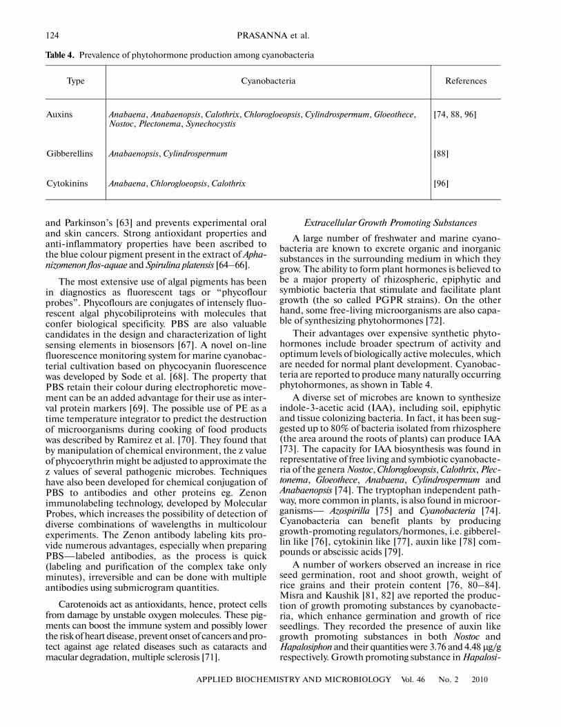
Their advantages over expensive synthetic phyto�hormones include broader spectrum of activity andoptimum levels of biologically active molecules, whichare needed for normal plant development. Cyanobac�teria are reported to produce many naturally occurringphytohormones, as shown in Table 4.
A diverse set of microbes are known to synthesizeindole�3�acetic acid (IAA), including soil, epiphyticand tissue colonizing bacteria. In fact, it has been sug�gested up to 80% of bacteria isolated from rhizosphere(the area around the roots of plants) can produce IAA[73]. The capacity for IAA biosynthesis was found inrepresentative of free living and symbiotic cyanobacte�ria of the genera Nostoc, Chlorogloeopsis, Calothrix, Plec�tonema, Gloeothece, Anabaena, Cylindrospermum andAnabaenopsis [74]. The tryptophan independent path�way, more common in plants, is also found in microor�ganisms— Azospirilla [75] and Cyanobacteria [74].Cyanobacteria can benefit plants by producinggrowth�promoting regulators/hormones, i.e. gibberel�lin like [76], cytokinin like [77], auxin like [78] com�pounds or abscissic acids [79].
A number of workers observed an increase in riceseed germination, root and shoot growth, weight ofrice grains and their protein content [76, 80–84].Misra and Kaushik [81, 82] ave reported the produc�tion of growth promoting substances by cyanobacte�ria, which enhance germination and growth of riceseedlings. They recorded the presence of auxin likegrowth promoting substances in both Nostoc andHapalosiphon and their quantities were 3.76 and 4.48 μg/grespectively. Growth promoting substance in Hapalosi�
Table 4. Prevalence of phytohormone production among cyanobacteria
Type Cyanobacteria References
Auxins Anabaena, Anabaenopsis, Calothrix, Chlorogloeopsis, Cylindrospermum, Gloeothece, Nostoc, Plectonema, Synechocystis
[74, 88, 96]
Gibberellins Anabaenopsis, Cylindrospermum [88]
Cytokinins Anabaena, Chlorogloeopsis, Calothrix [96]

APPLIED BIOCHEMISTRY AND MICROBIOLOGY Vol. 46 No. 2 2010
REDISCOVERING CYANOBACTERIA AS VALUABLE SOURCES 125
phon extract was found to be IAA acid and the possibil�ity of presence of indole�3�propionic acid or 3�methylindole also existed. Avena growth test also indicatedthe presence of auxins in the growth medium of Nostocand Hapalosiphon. Physiological attributes of a set ofcyanobacterial strains, isolated from the rhizosphereof wheat (var. HD 2687), identified as belonging to thegenera— Calothrix, Westiellopsis, Hapalosiphon andNostoc were analyzed [84]. The concentrated culturefiltrates of three cyanobacterial strains— Calothrixghosei, Hapalosiphon intricatus and Nostoc sp. were ableto enhance germination percentage, radicle andcoleoptile length in imbibition studies with wheatseeds. IAA production was recorded in light and dark(+0.5% glucose) incubated cultures. Incubation ofcultures in the presence of tryptophan significantlyenhanced IAA production. Acetylene reducing activ�ity was higher in light incubated cultures of Nostoc sp.followed by Calothrix ghosei, while in dark, Calothrixghosei recorded highest values. TLC analyses of the fil�trates revealed the presence of several amino acids,such as histidine and auxinlike compounds. Co�cul�turing experiments with selected cyanobacterialstrains recorded significant enhancement in plantchlorophyll. Root sections of wheat seedlings co�cul�tured with Calothrix ghosei, revealed the presence ofshort filaments inside the root hairs and cortical region[84]. Such strains can be promising candidates fordeveloping plant growth promoting associations forwheat crop, besides serving as model systems forunderstanding the metabolic interactions of cyano�bacteria with host plant, such as wheat.
IAA formation via indole�3�pyruvic acid andindole�3�acetic aldehyde is found in the majority ofmicroorganisms studied, such as the symbiotic nitro�gen fixing cyanobacterium Nostoc sp. Sergeeva andco�workers [74] in 2002 screened 34 free�living andsymbiotically competent cyanobacteria, that representall morphotypes from the unicellular to the highly dif�ferentiated and showed that auxin�like compoundswere released by about 38% of the free�living as com�pared to 83% of the symbiotic isolates. For recordingthe authenticity of IAA, verification by chemical char�acterization using gas chromatography�mass spec�trometry in Nostoc PCC 9229 (isolated from theangiosperm Gunnera) and in Nostoc 268 (free�living)was done. Addition of the putative IAA precursor tryp�tophan enhanced IAA accumulation in cell extractsand supernatants. Genes responsible for IAA synthesisin bacteria may have plasmid or chromosomal local�ization [85]. As the genome of the symbiotically com�petent Nostoc PCC 73102 was found to contain homo�logues of key enzymes of the indole�3�pyruvic acidpathway, a transaminase and indolepyruvate decar�boxylase (IpdC), the putative ipdC gene from thiscyanobacterium was cloned and used in Southern blotanalysis. Out of 11 cyanobacterial strains respondingpositively in the Salkowski/ELISA test, ipdC homo�logues were found in four strains. A constitutive and
possibly tryptophan�dependent production of IAA viathe indole�3�pyruvic acid pathway is therefore sug�gested. The conversion of tryptophan into indole�3�acetic aldehyde may involve an alternative pathway inwhich tryptamine is formed and believed to operate inthe cyanobacterium Chlorogloea fritschii [78].
The ability to synthesize gibberellins is inherent inall groups of microorganisms. Gupta and Lata [86]observed that cyanobacteria accelerated seed germi�nation and promoted seedling growth. In addition,they also observed that both yield and the quality of thegrains was improved because they were richer in pro�teins. It seems very likely that the beneficial effect ofthe algae on the rice crop may not be restricted to theircapacity to fix atmospheric nitrogen alone, especiallyin fields where nitrogen�fixing algae may not bepresent in appreciable quantities. A gibberellin�likesubstance has been isolated from the cyanobacteriumPhormidium foveolarum and this is active in GA�bioas�says 87]. Mohon and Mukerji [88] reported synthesisof gibberellins in Cylindrospermum spp. and in Ana�baenopsis spp. Extracts of marine cyanobacteria stimu�lated somatic embryogenesis in carrot cell cultures ofDaucus carota [89]. The growth promotion activity ofmarine cyanobacterial extracts on plant regenerationmay not be due to a nutrient effect, and Wake and co�workers [90] tentatively concluded that a protein richfraction was the active component on plantlet forma�tion.
Extracellular amino acids have been reported in anumber of cyanobacteria. The production of com�bined nitrogen in soluble form by nitrogen fixingcyanobacteria has been recorded by several authors[91, 92]. Fogg [92] investigated the nature and originof nitrogenous substance in filtrates from the nitrogenfixing blue green algae Anabaena cylindrica Lemm.Watanabe [93] recorded the presence of aspartic acid,glutamic acid and alanine the only free amino acids inthe culture solutions of Calothrix brevissima. The liber�ation of these aminoacids has also been reported inNostoc sp. and Anabaena azollae [94]. Whitton [95]examined the soluble extracellular materials producedby Anabaena cylindrica, Nostoc sp., Anacystis nidulansand Oscillatoria planctonica and concluded that samplesof filtrates contained polypeptide of rather similaramino acid composition, with prominence of alanine,glycine and serine and the presence of aspartic acid,glutamic acid, leucine, threonine and valine. Singhand Trehan [76] also reported production of aminoacids like aspartic acid, glutamic acid, proline andvaline from Aulosira fertilissima and Anacystis nidulansand they found that the maximum liberation of theamino acids in the culture medium took place duringthe lag and stationary phase of algal growth. Mohonand Mukerji [88] in their study attempted to detect theamino acid secretion from various N2�fixing and nonN2�fixing cyanobacteria and concluded that in nonN2�fixing blue green algae, Anacystis nidulans, the typeof amino acids in culture filtrates were different from

126
APPLIED BIOCHEMISTRY AND MICROBIOLOGY Vol. 46 No. 2 2010
PRASANNA et al.
those of N2�fixing i.e. Cylindrospermum sp. and Ana�baenopsis raciborskii. Misra and Kaushik [82] in hisstudy reported that a variety of amino acids were liber�ated during the growth phase of cyanobacteria. The pres�ence of threonine, glutamic acid, proline and valine werewell established in external growth medium of Nostocmuscorum. In addition to this glycine, serine was also lib�erated. They also reported the presence of lysine, cystineand isoleucine from Hapalosiphon fontinalis in additionto that observed in Nostoc.
Selyakh and Semenova [96] reported the presenceof cytokinins in culture liquids of Chlorogloeopsis andCalothrix. Rodriguez and co�workers [97] investigatedthe effect of the cyanobacterium Scytonema hofmanniextracellular products on the growth of rice seedlingsinhibited by NaCl and observed that this was compa�rable with the effect of the gibberellic acid in the samestress condition. Several reports have indicated thatapplication on crops of growth regulators, such as GA3and cytokinin, produced some benefit in alleviatingthe adverse effects of salt stress [98] and cyanobacteriacan be possible candidates in this context.
Cyanobacteria (blue�green algae) are an excellentsource of nutrition. Spirulina supplies all most all thevitamins like the B�series vitamins, tocopherol, nia�cin; inositol and folic acid that all living beings need tocarry on metabolic processes [99, 100]. Like otheralgae, blue green algae/cyanobacteria also require andproduce vitamins. There have been only a few studieson the vitamin content and their excretion into thegrowth medium (Table 5).
The vitamin content of a cyanobacterium is depen�dent upon the genetic constitution, the stage ofgrowth, nutritional status of the organism, besidesother factors such as light intensity, which affectgrowth and metabolism. Many of these species canmake Vitamin B12, which plants do not make [100,101].Many water�soluble vitamins have been observedin the supernatants of cyanobacterial cultures, whichmay be a product of active release or due to death/dis�integration of the organism. The proportion of analga’s vitamin production that is excreted to themedium may be quite high. Kopteva et al. [102]reported that the blue green alga Anabaena bassaliexcretes up to 94% of its biotin. However, commercialexploitation of cyanobacteria for vitamin productionis not reported. Most of the available processes employChlorella or Porphyridium.
Exopolysaccharides
Many species of unicellular and filamentouscyanobacteria produce large quantities of extracellularpolymeric substances consisting mainly polysaccha�rides; especially edaphic forms which produce extra�cellular polymers of diverse chemical composition,especially exopolysaccharides (EPS) that enhancemicrobial growth and as a consequence, improve soilstructure and fertility. They also produce a range of
carbohydrates, both as storage products and asosmotic effectors. Some cyanobacteria excrete slimeor mucilage that becomes dispersed around the organ�ism and, to an extent, partially dissolves in the culturemedium or in the soil solution [103]. Nostoc muscorumis known to improve the aggregate stability of a saline�sodic soil, where the increase in soil aggregation ismainly due to exopolysaccharides secreted by micro�organisms or EPS added to soil after death and cellularlysis [104]. Cyanobacterial EPS are thought to play akey role in allowing the survival and growth of cyano�bacteria under extreme conditions [105]. Misra andKaushik [82] reported the production of extracellularpolysaccharides composed of galactose, xylose, fruc�tose and a series of unidentified sugars from Nostocmuscorum and Hapalosiphon fontinalis.
Cyanobacteria can be incorporated into soil asorganic matter and also as a source of enzymes as theyproduce acid and alkaline extracellular phosphatasesthat are active in solution or located in the periplasmicspace of the cell wall. Both biomass and EPS incorpo�rated into soil, induced a growth promotion of othermicroorganisms and increased the activity of soilenzymes that participate in the liberation of nutrientsrequired by plants [104].
Allelochemicals from Cyanobacteria
The chemical potential of cyanobacteria for pro�ducing structurally novel and biologically active natu�ral products is now being realized and exploited glo�bally [10, 106]. Cyanobacteria and eukaryotic algaeare known to excrete bioactive compounds into theenvironment, which are important determinants ofallelopathic activity in water and soil. Allelochemicalsare secondary metabolites or non—nutritional pri�mary metabolites that affect growth, reproduction orbehaviour of individuals other than the ones produc�ing them or influence the structure and dynamics ofpopulations or communities of either plants or ani�mals or microbes. Allelopathic chemicals play a role inthe interactions between the emitter organisms andtheir direct competitors or predators; they are catego�rized according to their toxic stimulatory effect onseveral organisms, including some that may not bepresent in their immediate environment. In recentyears there has been an increasing focus on the pros�pects of exploiting allelopathy as an alternative strat�egy for controlling weeds, insects and diseases.
Schlegel et al. [107] isolated cyanobacterial strainsform diverse habitats, spanning continents of Australiaand Asia, which exhibited antibiotic activity againstgreen algae and cyanobacteria. Gross [108] reportedthe production of allelochemicals with strong phyto�toxicity against several algae (including cyanobacte�ria). Nodularia harveyana, a Mediterranean cyano�bacterium produced lipophilic bioactive compoundsagainst cyanobacteria, eubacteria, rotifers and crusta�ceans. Most of the known cyanotoxins are photosyn�

APPLIED BIOCHEMISTRY AND MICROBIOLOGY Vol. 46 No. 2 2010
REDISCOVERING CYANOBACTERIA AS VALUABLE SOURCES 127
thetic inhibitors [109]. Blinkova and co�workers [110]in their review provided interesting information onSpirulina platensis (SP), having diverse biologicalactivity. Due to the high content of highly valuableproteins, indispensable amino acids, vitamins, beta�carotene and other pigments, mineral substances,indispensable fatty acids and polysaccharides, it hasbeen found suitable for use as bioactive additive. SPproduces an immunostimulating effect by enhancingthe resistance of humans, mammals, chickens and fishto infections, the capacity of influencing hemopoiesis,stimulating the production of antibodies and cytok�ines. Under the influence of SP macrophages, T and Bcells are activated. SP sulfolipids have proved to beeffective against HIV. Shinohara and co�workers [111]observed that the growth�promoting substances (bilip�roteins) in a non�dialyzable extract of Synechococcuselongatus var. promoted the growth of RPMI 8226 cells(a human myeloma cell line). A patent was granted inthe USA [112] for the use of extracts of Spirulina as areplacement for serum used in animal cell tissue cul�ture growth medium.
Cyanobacteria are proving to be a rich source of alarge number of novel compounds with biologicalactivity, Table 6 [8]. The microcystins are a group ofcyclic heptapeptide (7 amino acids) hepatotoxins(liver toxins) produced by a number of cyanobacterialgenera, the most notable of which is the widespreadMicrocystis from which the toxins take their name.Approximately 70 different structural analogues ofmicrocystins have been identified to date [113].Microcystins consist of a seven�membered peptidering, which is made up of five non�protein amino acidsand two protein amino acids. It is these two proteinamino acids that distinguish microcystins from oneanother, while the other amino acids are more or lessconstant between variant microcystins. The structureof nodularin is related to the potent cyclic heptapep�tide hepatotoxins, the microcystins, but differs in thatnodularin is composed of only five amino acids in thepeptide ring. The anatoxins are a group of neurotoxicalkaloids produced by a number of cyanobacterialgenera including Anabaena, Oscillatoria and Aphani�zomenon. The toxicity of these compounds (LD50) var�ies from 20 μg kg–1 (by weight, i.p. mouse) for ana�toxin�a(S) to 200–250 μg kg–1 for anatoxin�a andhomoanatoxin�a, making them more toxic than manymicrocystins. Like anatoxins, the saxitoxins (STX) areneurotoxic alkaloids, which are also known as PSP’s(paralytic shelfish poisons) due to their occurrenceand association with seafood. They block sodiumchannels in nerve cells, thus causing neurotoxiceffects. There are a number of STX variants generallydivided into groups based on their structure or organ�ism of origin. STX’s are highly toxic with LD50 s as lowas 10 μg kg–1 (i.p.) in mice. Although originallydescribed from Cylindrospermopsis raciborskii [114],cylindrospermopsin can be found also in Aphanizome�non ovalisporum and Umezakia natans. The toxin is a
cyclic alkaloid and like microcystins, primarily affectsthe liver, although causes considerable damage toother major organs. Its LD50 (i.p., mice) of 200 μg kg–
1 ranks it as a relatively potent cyanobacterial toxin.Aplysiatoxins, which exhibit dermatotoxic activitycausing inflammation of the skin, are produced bymarine cyanobacteria such as Lyngbya. They are alsopotent tumour promoters. Aplysiatoxins and debro�moaplysiatoxin have been found associated with fila�mentous cyanobacterial species including Schizothrixcalcicola and Oscillatoria nigroviridis.
Toxin production by cyanobacteria is known to beinfluenced by various nutritional and environmentalfactors. The age of culture has significant effect ontoxin production and many reports are availableregarding optimum toxin production by M. aeruginosain late exponential phase of growth [6, 115]. Similarresults were obtained by Ray and Bagchi [116] onOscillatoria and Dias et al. [117] in Anabaena sp. Diaset al. [117] reported an increase in amount of extracel�lular toxin with culture time, indicating that toxins arereleased in water through cell lysis and may beexpected to remain in water upon the collapse of toxicbloom or after cells removal by water treatment. Coddand Poon [115] found 10 times less toxin than refer�ence cell by removing nitrogen source in M. aerugi�nosa cultures. On the other hand, Oh et al. [118]showed more P in the culture medium stimulatesgrowth and toxin production by M. aeruginosa. Simi�lar results were obtained by Repka et al. [119]. Maxi�mum toxin production was found in late log phase cul�ture and with highest phosphate concentration (2.6and 3.4 mg/l respectively) in Anabaena strains. Partialpurification using TLC, followed by GLC revealed asingle peak [120, 121]. Ray and Bagchi [116] reportedalgicide production by Oscillatoria laetevirens was neg�atively regulated with phosphate. Dias et al. [117]reported changing phosphate levels result in change intype of toxin produced by cyanobacterium Aphani�zomenon sp. Evidently P level does influence the toxin
Table 5. Vitamins reported to be excreted by cyanobacteria
Cyanobacteria Vitamins reported References
Anabaena flos�aquae B12 [101]
Chroococcus minutus B12 [100]
Oscillatoria jasorvensis Pantothenate, B12 [100]
Spirulina spp. B12, pantothenate, inositol
[99]
Nostoc spp. B12, B1, biotin, nico�tinic acid
[100]

128
APPLIED BIOCHEMISTRY AND MICROBIOLOGY Vol. 46 No. 2 2010
PRASANNA et al.
Table 6. Cyanotoxins – source and chemical properties [8, 106]
Cyanobacterial genera Toxin produced Type of toxin Structure LD50
Anabaena Anatoxin Neurotoxins Secondary amine 250
Microcystins Hepatotoxins Cyclic peptide 45–1000
Saxitoxins Neurotoxins Carbamate alkaloids 10–30
LPS’s Endotoxins Lipopolysaccharides –
Anabaenopsis Microcystins Hepatotoxins Cyclic peptide 45–1000
LPS’s Endotoxins Lipopolysaccharides –
Anacystis LPS’s Endotoxins Lipopolysaccharides –
Aphanizomenon Saxitoxins Neurotoxins Carbamate alkaloids –
LPS’s Endotoxins Lipopolysaccharides –
Cylindrospermopsins Cytotoxin Cylindrospermopsin alkaloid 2100 in one day & 200 in 5–6 days
Cylindrospermopsis Cylindrospermopsins Cytotoxin Cylindrospermopsin alkaloid 2100 in one day & 200 in 5–6 days
Saxitoxins Neurotoxins Carbamate alkaloids –
LPS’s Endotoxins Lipopolysaccharides –
Hapalosiphon Microcystins Hepatotoxins Cyclic peptide 45–1000
LPS’s Endotoxins Lipopolysaccharides –
Lyngbya Aplysiatoxins Cytotoxin Alkaloid –
Lyngbiatoxin�a Cytotoxin Phorbol Ester –
LPS’s Endotoxins Lipopolysaccharides –
Microcystis Microcystins Hepatotoxins Pentapeptide –
LPS’s Endotoxins Lipopolysaccharides –
Nodularia Nodularin Hepatotoxins Cyclic peptide 45–1000
LPS’s Endotoxins Lipopolysaccharides 30–50
Nostoc Microcystins Neurotoxins Secondary amine –
LPS’s Endotoxins Lipopolysaccharides 45–1000
Phormidium(Oscillatoria)
Anatoxins Neurotoxins Secondary amine –
LPS’s Endotoxins Lipopolysaccharides 250
Planktothrix(Oscillatoria)
Anatoxins Neurotoxins Secondary amine –
Aplysiatoxins Cytotoxin Alkaloid 250
Microcystin Hepatotoxins Cyclic peptide 45–1000
Saxitoxin Neurotoxins Carbamate alkaloids 10–30
LPS’s Endotoxins Lipopolysaccharides –
Schizothrix Aplysiatoxins Cytotoxin Alkaloid –
LPS’s Endotoxins Lipopolysaccharides –
Trichodesmium Cylindrospermopsin Cytotoxin Cylindrospermopsin alkaloid 2100 in one day & 200 in 5–6 days
LPS’s Endotoxins Lipopolysaccharides –

APPLIED BIOCHEMISTRY AND MICROBIOLOGY Vol. 46 No. 2 2010
REDISCOVERING CYANOBACTERIA AS VALUABLE SOURCES 129
production by cyanobacteria but there is a differentialresponse of different cyanobacterial strains towardsvarying levels of phosphate.
Watanabe and Oishi [122] observed a fourfoldincrease in M. aeruginosa toxicity when light intensitywas increased from 7.53 to 30.1 microeinsteins m–2 s–1.On the contrary Codd and Poon [115] reported thatlight intensity of 5–50 microeinsteins m–2 s–1 had noinfluence on toxin production in a strain of M. aerugi�nosa. Utkilen and Gjolme [123] reported an increasein toxin production rate upto 40 μE m–2 s–1 thatdecreased at higher light intensities. These contradic�tory reports regarding effect of light intensities wereexplained by Sivonen [124] as due to differences inlight sources, different behaviour of strain and species,different culture media used and different toxin detec�tion method used.
Toxin production by cyanobacteria is also reportedto be influenced by pH of the medium [116, 125]. Vander Westhuizen and Eloff [125] reported that althoughmaximum growth rate of cyanobacteriaum Microcystisaeruginosa was observed at pH 9 but cells were moretoxic at higher and lower pH values, where they grewmore slowly. Ray and Bagchi [116] observed a negativeregulation of pH and toxin production in Oscillatorialatevirens. However, Jaiswal et al. [126, 127] andRadhakrishnan [120] showed that there is no signifi�cant effect of enhancing or decreasing pH of mediumon secondary metabolite production by M. aeruginosaand Calothrix respectively and maximum activity wasobserved under routine growth conditions. Such stud�ies highlight the importance of environmental factorsin aggravating or reducing the toxic effects of theseharmful cyanobacteria.
Microscopic identification of cyanobacteria aloneor in combination with direct analysis of toxin hasbeen used generally for the detection of toxic cyano�bacteria. Although, some cyanobacteria are geneti�cally capable of producing toxins, they do not exhibittoxicity under different environmental/nutritionalconditions, and some do not produce toxins at all. Therelationship between toxigenicity and phylogenywithin the various cyanobacterial genera is alsounclear. Several morphological and molecular studieshave attempted to resolve the ambiguous relationshipof Microcystis toxigenicity to its population structure[128]. Molecular approaches have included thosebased on allozyme polymorphisms [129], 16s rRNAgenes [130, 131], the phycocyanin intergenic spacers(PC�IGS) region [130], DNA�DNA hybridization[132], nucleotide base composition [133, 134], the16S–23S rRNA internal transcribes spacers region[135], the rbc L gene [136] and rpo D homologs [137].Neilan et al. [130] using restriction length polymor�phism, probed a short tandemly repeated DNAsequence found in the chromosome of Anabaena sp.PCC 7120 and found that it can distinguish hepato�toxic Anabaena isolate from neurotoxin producingstrains. Availability of this kind of gene probe can be a
very useful tool in screening of toxic cyanobacteria.Fergusson and Saint [138] reported that Anabaena cir�cinalis specific PCR assay targeting the rpoCI gene todetect the organism directly from environmental sam�ple. Wilson et al. [139] developed a PCR test targetinga region of rpoC1 gene unique to C. raciborskii for spe�cific identification of strain from both purifiedgenomic DNA and environmental sample. Baker et al.[140] showed from their studies that bloom compo�nent can be identified and monitored for toxigenicityby PCR amplification of phycocyanin intergenicspacer region between the genes for β and α subunit,more effectively than by other methods like micros�copy and mouse bioassay. Microcystin is synthesizednonribosomally via a mixed polyketide synthase/non�ribosomal peptide synthetase system called microcys�tin synthetase [141]. The 55 kb gene cluster (mcyABCDEFGHIJ) encoding the microcystin synthetasecomplex has been identified and sequenced [142–144]. This cluster consists of six open reading frames(ORFs) with a mixed nonribosomal peptide syn�thetase/polyketide synthase nature (mcy A to mcy Eand mcy G) and four smaller ORFs with putative pre�cursor and tailoring functions (mcy F and mcyH to mcyJ Fand mcyH to mcyJ). The mcy ABCDEFGHIJ genesare transcribed as two polycistronic operons, mcy ABCand mcy DEFGHIJ from a central bidirectional pro�moter between mcy A and mcy D cluster. Kaebernicket al. [145] were able to identify the start sites for all themcy genes except for mcy B and mcy C. Transcriptionalknowledge of mcy gene cluster will help to increase ourunderstanding of microcystin synthetase regulationand toxin biosynthesis and provide useful insight intoother nonribosomal systems.
Applications of Allelochemicals
Cyanobacteria have been recognized as candidatesfor the discovery of novel antibiotics and use as bio�control agents of bacterial and fungal pathogens. Inaddition to best�known neurotoxins and hepatotoxins,they are known to produce antibacterial compounds[6], antifungal compounds [146], anticancerous/anti�neoplastic agents and compounds useful in treatmentof HIV [11].
Terrestrial cyanobacteria such as Anabaena laxaproduce antifungal cyclic peptides such as laxaphycinA and B [146]. Cassamata and Wickstrom [147]observed that the exudates of M. aeruginosa wereinhibitory towards bacterial plankton communities.Kulik [148] reported that extracts from the cyanobac�terium Nostoc muscorum Agardh inhibited the in vitrogrowth of the fungal plant pathogens such as Sclero�tinia sclerotiorum (Cottony rot of vegetables and flow�ers) and Rhizoctonia solani (root and stem rots). Anumber of compounds exhibiting fungicidal activityagainst specific agriculturally important fungi havebeen identified [120] and patented [114, 149–153].Several compounds isolated from fresh water cyano�

130
APPLIED BIOCHEMISTRY AND MICROBIOLOGY Vol. 46 No. 2 2010
PRASANNA et al.
bacteria have been identified as having potent algicidaland fungicidal properties. Hagmann and Juttner in1996 [151] reported about a secondary metabolite Fis�cherellin from Fischerella muscicola, while Papke et al.[154] reported Fischerellin B from the same Fischer�ella muscicola. Hagmann and Juttner [151] found thatfischerellin A had differential antifungal activityagainst several plant pathogenic fungi'. It showed100% inhibition of Uromyces appendiculatus (brownrust) at 25 μg ml–1, at 1000 μg ml–1, fischerellin Acaused 100% inhibition of Erysiphe graminis (powderymildew), 80% inhibition of Phytophthora infestans andPyricularia oryzae (rice blast) and was less potentagainst Monilinia fructigena (brown rot) and Pseudocer�cosporella herpotrichoides (stem break).
The extracts of cyanobacteria are known to reducethe incidence of Botrytis cinerea on strawberries andErysiphe polygoni causing powdery mildew on turnipsand damping off in tomato seedlings, besides reducingthe growth of saprophytes—Chaetomium globosum,Cunninghamella blakesleeana and Aspergillus oryzae andplant pathogens such as Rhizoctonia solani and Scleroti�ana sclerotium [148]. An investigation was undertakento explore the biocidal efficacy of fungicidal com�pounds) produced by Calothrix elenkenii against damp�ing�off disease, in three vegetable crops—Tomato,Chilly and Brinjal. The observations taken after aperiod of four weeks revealed the superiority of seedtreatment with ethyl acetate extracts, in terms of percent mortality and plant parameters. Chilly recordedhighest percent survivors and responded best to theseed treatment with ethyl acetate extract of Calothrixelenkenii [155]. The excretion of hydrolytic enzymes isknown to be a common trait of plant pathogens/sym�bionts, which promotes a closer association with plantroots/target organisms and improve the stability ofsuch associations. The extracellular filtrates of a set ofseventy axenized cyanobacterial isolates belonging tothe genus Anabaena exhibited inhibition of phyto�pathogenic fungi under laboratory conditions. Analy�ses of their chemical nature revealed the activity ofhydrolytic enzymes. The high values of chitosanaseand xylanase activity in selected cultures may contrib�ute to the fungicidal activity of the cyanobacterialstrains, as a significant correlation was observedbetween the diameter of zone of inhibition andenzyme activity [156]. Sequence homologues of chito�sanase and endoglucanase have been recently identi�fied in two Anabaena strains and are being analyzedusing bioinformatic tools, for their evolutionary andphylogenetic relationships with rhizobacterial/fungalstrains employed in biocontrol of agriculturally rele�vant phytopathogenic fungi. This is a first time reporton the production of hydrolytic enzymes and relatedallele mining in these oxygenic photosyntheticprokaryotes, which can be potential candidates for thedevelopment of biocontrol agent(s) against selectedphytopathogenic fungi.
With the realization of their chemical potential ofcyanobacteria in recent years, there has been a tre�mendous increase in their biological significance, andproducts being commercialized. These include PC,carotenoids, fatty acids, polysaccharides, vitamins,sterols and other biologically active compounds, manyof which have proved to be exciting molecules withimmuno�modulatory, bioregulatory and therapeuticpotential. With the advent of bioassay�directed screen�ing and efficacy testing techniques, there is a definiteneed to target significant pathogens pests/diseases,include relevant standards (commercial pesticides),environmental hazards testing and adherence to sta�tistically sound principles. An improved understand�ing of the biosynthetic pathways leading to productionof these novel compounds and finally their release tothe environment is very essential. Molecular dissec�tion of these processes will help in exploiting thesemicroorganisms, which are considered as nuisanceorganisms, for the betterment of mankind. This canprovide novel inputs, not only for environmentally safe“organic” and “green” agriculture, but also tools fortargeting emerging human and animal diseases.
ACKNOWLEDGMENTS
The authors are thankful to Indian Council of Agri�cultural Research, New Delhi for the AP Cess fundproject, Network Project on Microorganisms(AMAAS) and Council of Scientific and IndustrialResearch (CSIR), New Delhi, India for providingfinancial assistance. The facilities required for carry�ing out this investigation provided by the Division ofMicrobiology and Centre for Conservation and Utili�zation of Blue Green Algae, IARI, New Delhi.
REFERENCES
1. Woese, C.R., Microbiol. Rev., 1987, vol. 51, no. 2,pp. 221–271.
2. Castenholz, R.W., in Bergey’s Manual of SystematicBacteriology, 2nd ed., Boone, D.R., andCastenholz, R.W., Eds., Berlin: Springer., 2001,pp. 473–599.
3. Venkataraman, G.S., Algal Biofertilizers and Rice Cul�tivation, New Delhi: Today and Tomorrow Printersand Publishers, 1972.
4. Kaushik, B.D., in Cyanobacterial Biotechnology, Sub�ramanian, G., Kaushik, B.D., andVenkataraman, G.S., Eds., USA: Science Publs. Inc.,2002, pp. 211–222.
5. Namikoshi, M. and Rinehart, K.L., J. Ind. Microbiol.,1996, vol. 17, nos. 5�6, 373–384.
6. Carmichael, W.W., Sci. Amer., 1994, vol. 270, no. 1,pp. 78–86.
7. Kumar, K., Lakshmanan, A., and Kannaiyan, S.,Indian J. Microbiol., 2005, vol. 43, no. 1, pp. 9–16.
8. Jaiswal, P., Prasanna, R., and Singh, P.K., Can.J. Microbiol., 2008, vol. 54, pp. 701–717.

APPLIED BIOCHEMISTRY AND MICROBIOLOGY Vol. 46 No. 2 2010
REDISCOVERING CYANOBACTERIA AS VALUABLE SOURCES 131
9. Nagle, D.G. and Inderjit, in Chemical Ecology ofPlants: Allelopathy in Aquatic and Terrestrial Ecosys�tems, Inderjit, and A.U., Malik, Eds., Birkhauser,Switzerland: Verlag, 2002.
10. Mundt, S., Kreitlow, S., Nowotny, A., and Effmert, U.,Int. J. Hygiene Environ. Health., 2001, vol. 203, no. 4,pp. 327–334.
11. Smith, C.D., Zhang, X., Mooberry, S.L., andMoore, R.E., Cancer Res., 1994, vol. 54, no. 14,pp. 3779–3784.
12. Harrigan, G.G., Luesch, H., Yoshida, W.Y.,Moore, R.E., Nagle, D.G., Paul, V.J., Mooberry, S.L.,Corbett, T.H., and Valeriote, F.A., J. Nat. Prod., 1998,vol. 61, no. 9, pp. 1075–1077.
13. Prasanna, R., Sood, A., Suresh, A., Nayak, S., andKaushik, B.D., Acta Botanica Hungarica, 2007,vol. 49, nos. 1–2, pp. 131–156.
14. Borowitzka, M.A., in Cyanobacterial Biotechnology,Subramanian, G., Kaushik, B.D., andVenkatamaran, G.S., Eds., New Delhi: Oxford IBHPublishing Co., 1998, pp. 249–257.
15. Walsh, K., Jone, G.J., and Dustan, H., Phytochemis�try, 1997, vol. 44, no. 5, pp. 817–824.
16. Samsonoff, W.A., and MacColl, Arch. Microbiol.,2001, vol. 176, no. 6, pp. 400–405.
17. Grossman, A.R., Photosynth. Res., 2003, vol. 76,nos. 1–3, pp. 207–215.
18. MacColl, R., J. Struct. Biol., 1998, vol. 124, no. 2–3,pp. 311–334.
19. Kaushik, B.D., Indian Hydrobiol., 2000, vol. 3, no. 2,pp. 81–84.
20. Prasanna, R., Prasanna, B.M., Mohammadi, S.A.,and Singh, P.K., Folia Microbiol., 2003a, vol. 48, no. 1,pp. 59–64.
21. Bhosale, P., Appl. Microbiol. Biotechnol., 2004, vol. 63,no. 4, pp. 351–361.
22. Cheng, Q., J. Ind. Microbiol. Biotechnol., 2006,vol. 33, no. 7, pp. 552–559.
23. Britton, G., Liaaen�Jensen, S., and Pfander, H., Car�otenoids Handbook, Basel: Birkhaser Verlag, 2004.
24. Gombos, Z. and Vigh, L., Plant Physiol., 1986, vol. 80,no. 2, pp. 415–419.
25. Sandmann, G., Chembiochem., 2002, vol. 3, no. 7,pp. 629–635.
26. Steiger, S., Jackisch, Y., and Sandmann, G., Arch.Microbiol., 2005, vol. 184, no. 4, pp. 207–214.
27. Gaidukov, N., Ber. Deutsch Bot. Ges., 1903, vol. 21,pp. 517–522.
28. Takano, H., Arai, T., Hirano, M., and Matsunga, T.,Appl. Microbiol. Biotechnol., 1995, vol. 43, no. 6,pp. 1014–1018.
29. Ranjitha, K. and Kaushik, B.D., Indian J. Microbiol.,2005b, vol. 45, no. 1, pp. 67–70.
30. Prasanna, R., Pabby, A., and Singh, P.K., Folia Micro�biol., 2004, vol. 49, no. 1, pp. 26–30.
31. Prasanna, R., Pabby, A., Saxena, S., and Singh, P.K.,J. Plant Physiol., 2004, vol. 161, no. 10, pp. 1125–1132.
32. Prasanna, R., Sood, A., Saxena, S., and Kaushik, B.D.,Indian Hydrobiol., 2005, vol. 8, no. 1, pp. 89–98.
33. Prasanna, R., Kumar, R., Sood, A., Prasanna, B.M.,and Singh, P.K., Microbiol. Res., 2006, vol. 161, no. 3,pp. 187–202.
34. Nayak, S., Prasanna, R., Prasanna, B.M., andSahoo, D.B., World J. Microbiol. Biotechnol., 2007,vol. 23, no. 11, pp. 1575–1584.
35. Venugopal, V., Prasanna, R., Sood, A., andKaushik, B.D., Indian Hydrobiol., 2005, vol. 8, no. 2,pp. 157–165.
36. Venugopal, V., Prasanna, R., Sood, A., Jaiswal, P., andKaushik, B.D., Foila Microbiol., 2006, vol. 51, no. 1,pp. 50–56.
37. Vasudevan, V., Prasanna, R., Sood, A., andKaushik, B.D., Acta Botanica Hungarica, 2007,vol. 49, nos. 1–2, pp. 187–198.
38. Mazel, D., Houmard, J., and de Marsac, N., Mol. Gen.Genet., 1988, vol. 211, no. 2, pp. 296–304.
39. Stowe�Evans, L.E., Ford, J., and Kohoe, D.M.,J. Bacteriol., 2004, vol. 186, no. 13, pp. 4338–4349.
40. Liotenberg, S., Campbell, D., Rippka, R.,Houmard, J., and de Marsac, N., Microbiology, 1996,vol. 142, no. 2, pp. 611–622.
41. Prasanna, R., Dhar, D.W., Dominic, T.K., Tiwari,O.N., Singh, P.K., and Acta, Biol., Hungarica, 2003b,vol. 54, no. 1, pp. 113–120.
42. Nelis, H.J. and DeLeenheer, A.P., J. Appl. Bacteriol.,1991, vol. 70, no. 3, pp. 181–191.
43. Johnson, E.A. and Schroeder, W.A., Adv. Biochem.Eng. Biotechnol., 1996, vol. 53, pp. 119–178.
44. Langarde, D., Benf, L., and Vermas, W., Appl. Environ.Microbiol., 2000, vol. 66, no. 1, pp. 64–72.
45. Lakatos, M., Wolfgang, B., and Budel, B., Eur. J. Phy�col., 2001, vol. 36, no. 4, pp. 367–375.
46. Sendersky, E., Lahmi, R., Shaltiel, J., Perelman, A.,and Schwarz, R., Mol. Microbiol., 2005, vol. 58, no. 3,pp. 659–668.
47. Bandaranayake, W.M., Nat. Prod. Rep., 1998, vol. 15,no. 2, pp.159–172.
48. Garcia�Pichel, F., Wingard, C.E., and Castenholz, R.W.,Appl. Environ. Microbiol., 1993, vol. 59, no. 1,pp. 170–176.
49. Nakamura, H., Kobayashi, J., and Hirata, Y., J. Chro�matogr., 1982, vol. 250, pp.113–118.
50. Kedar, L., Kashman, Y., and Oren, A., FEMS Micro�biol. Lett., 2002, vol. 208, no. 2, pp. 233–237.
51. Volkmann, M. and Gorbushina, A.A., FEMS Micro�biol. Lett., 2006, vol. 255, no. 2, pp. 286–295.
52. Bohm, G.A., Pfleiderer, W., Boger, P., and Scherer, S.,J. Biol. Chem., 1995, vol. 270, no. 15, pp. 8536–8539.
53. Sinha, R.P., Klisch, M., Groniger, A., and Hader, D.P.,J. Photochem. Photobiol. B Biol., 1998, vol. 47, nos. 2–3,pp. 83–94.
54. Sinha, R.P., Klisch, M., Helbling, E.W., andHader, D.P., J. Photochem. Photobiol. B Biol., 2001,vol. 60, nos. 2–3, pp. 129–135.
55. Volkmann, M., Gorbushina, A.A., Kedar, L., andOren, A., FEMS Microbiol. Lett., 2006, vol. 258, no. 1,pp. 50–54.
132
APPLIED BIOCHEMISTRY AND MICROBIOLOGY Vol. 46 No. 2 2010
PRASANNA et al.
56. Branen, L.A., Davidson, M.P., Salmine, N.S., andThorngate, H.J., Food Additives, New York: MarcelDekker, 2002.
57. Rowan, K.S., Photosynthetic Pigments in Algae, Cam�bridge: Cambridge Univ. Press, 1989.
58. Minkova, K.M., Tchernov, A.A., Tchorbadjieva, M.I.,Fournadjieva, R.E., and Busheva, M.C., J. Biotech�nol., 2003, vol. 102, no. 1, pp. 55–59.
59. Ranjitha, K. and Kaushik, B.D., J. Sci. Ind. Res.,2005a, vol. 64, no. 5, pp. 372–375.
60. Patel, A., Mishra, S., Pawar, R., and Ghosh, P.K., Pro�tein Expr. Purif., 2005, vol. 40, no. 2, pp. 181–187.
61. Reis, A., Mendes, A., Lobo�Fernandes, H.,Empis, J.A., and Novais, J.M., Bioresource Technol.,1998, vol. 66, no. 3, pp. 181–187.
62. Romay, C.P., Gonazelz, R., Ledon, N., Ramirez, D.,and Vimbare, V., Curr. Protein Peptide Sci., 2003,vol. 4, no. 3, pp. 207–216.
63. Rimbau, V., Camins, A., Pubill, D., Sureda, F.X.,Romay, C., Gonzalez, R., Jimenez, A., Escubedo, E.,Camarasa, J., and Pallas, M., Arch. Pharmacol., 2001,vol. 364, no. 2, pp. 96–104.
64. Benedetti, S., Benvenuti, F., Pagliarani, S.,Francogli, S., Scoglio, S., and Canestrari, F., Life Sci.,2004, vol. 75, no. 18, pp. 2353–2362.
65. Subhashini, J., Mahipal, S.V., Reddy, M.C., Mallikar�juna, Reddy, M., Rachamallu, A., and Reddanna, P.,Biochem. Pharmacol., 2004, vol. 68, no. 3, pp. 453–462.
66. Bhaskar, S.U., Gopalswamy, G., and Raghu, R.,Indian J. Exp. Biol., 2005, vol. 43, no. 3, pp. 277–279.
67. Ayyagari, M., Pande, R., Kamtekar, S., Gao, H.,Marx, K., Kumar, J., Tripathy, S., Akkara, J., andKaplan, D., Biotechnol. Bioeng., 1995, vol. 45, no. 2,pp. 116–121.
68. Sode, K.J., Horikoshi, K., Takeyama, J.,Nakamura, N., and Matsunga, T., J Biotechnol., 1991,vol. 21, no. 3, pp. 209–218.
69. Araoz, R., Lebert, M., and Hader, D.P., Electrophore�sis, 1998, vol. 19, no. 2, pp. 215–219.
70. Ramirez, A.O., Merrill, J.E., and Smith, D.E., J. FoodProtection, 2001, vol. 64, no. 11, pp. 1806–1811.
71. Rissanen, T., Voutilainen, S., Nyyssonen, K., andSalonen, J.T., Exp. Biol. Med., 2002, vol. 227, no. 3,pp. 900–907.
72. Tsavkelova, E.A., Klimova, S.Y., Cherdyntseva, T.A.,and Netrusov, A.I., Appl. Biochem. Microbiol., 2006,vol. 42, no. 2, pp. 133–143.
73. Loper, J.E. and Schroth, N., Phytopathology, 1986,vol. 76, no. 4, pp. 386–389.
74. Sergeeva, E., Liaimer, A., and Bergman, B., Planta,2002, vol. 215, no. 2, pp. 229–238.
75. Costacurta, A. and Vanderleyden, J., Crit. Rev. Micro�biol., 1995, vol. 21, no. 1, pp. 1–18.
76. Singh, V.P. and Trehan, T., Plant Soil, 1973, vol. 38,no. 2, pp. 457–464.
77. Rodgers, G.A., Bergman, B., Henriksson, U., andUdris, M., Plant Soil, 1979, vol. 52, no. 1, pp. 99–107.
78. Ahmad, M.R. and Winter, A., Planta, 1968, vol. 78,no. 3, pp. 277–286.
79. Marsalek, B., Zahradnickova, H., and Hronkova, M.,J. Plant Physiol., 1992, vol. 139, no. 4, pp. 506–508.
80. Venktaraman, G.S. and Neelakanantan, S., J. Gen.Appl. Microbiol., 1967, vol. 13, no. 1, pp. 53–62.
81. Misra, S. and Kaushik, B.D., Proc. Indian Natn. Sci.Acad., 1989a, vol. B 55, no. 4, pp. 295–300.
82. Misra, S. and Kaushik, B.D., Proc. Indian Natn. Sci.Acad., 1989b, vol. B 55, no. 6, pp. 499–504.
83. Karthikeyan, N. and Prasanna, R., Lata, andKaushik, B.D., European J. Soil Biol., 2007, vol. 43,no. 1, pp. 23–30.
84. Karthikeyan, N., Prasanna, R., Sood, A., Jaiswal, P.,Nayak, S., and Kaushik, B.D., Folia Microbiol., 2009,vol. 54, no. 1, pp. 43–51.
85. Patten, C.L. and Glick, B.R., Can. J. Microbiol., 1996,vol. 42, no. 3, pp. 207–220.
86. Gupta, A.B. and Lata, K., Hydrobiologia, 1964,vol. 24, nos. 1–2, pp. 430–434.
87. Gupta, A.B. and Gupta, K.K., Hydrobiologia, 1972,vol. 41, no. 1, pp. 127–132.
88. Mohan, M. and Mukerji, K.G., Phykos, 1978, vol. 18,no. 2, pp. 73–82.
89. Wake, H., Akasata, A., Umetsu, H., Ozeki, Y., Shimo�mura, K., and Matsunaga, T., Plant Cell Rep., 1991,vol. 9, no. 12, pp. 655–658.
90. Wake, H., Akasata, A., Umetsu, H., Ozeki, Y., Shimo�mura, K., and Matsunaga, T., Plant Cell Rep., 1992,vol. 11, no. 2, pp. 62–65.
91. De, P.K., Proc. R. Soc. London, 1939, vol. 127,no. 846, pp. 121–139.
92. Fogg, F.E., Proc. R. Soc. London, 1952, vol. B139,pp. 372–397.
93. Watanabe, A., Arch. Biochem. Biophys., 1951, vol. 34,no. 1, pp. 50–55.
94. Venkataraman, G.S., Saxena, H.K., and Indian, J.,Agric. Sci., 1963, vol. 33, no. 1, pp. 21–25.
95. Whitton, B.A., J. Gen. Microbiol., 1965, vol. 40, no. 1,pp. 1–11.
96. Selyakh, I.O. and Semenova, L.R., Problems of Ecol�ogy and Physiology of Microorganisms, Moscow: Dia�log�MGU, 2000, p. 94.
97. Rodriguez, A.A., Stella, A.M., Storni, M.M.,Zulpa, G., and Zaccaro, M.C., Saline, Sys., 2006,vol. 2, no. 1, p. 7.
98. Xiong, L. and Zhu, J.K., in The Arabidopsis Book,Somerville, C. and Meyerowitz, E., Eds., Rockville,MD: American Society of Plant Biologists, 2002,pp. 1–22.
99. Carlucci, A.F. and Bowes, P.M., J. Phycol., 1970,vol. 6, no. 4, pp. 351–357.
100. Shah, A.K. and Vaidya, B.S., Biologia Plant., 1977,vol. 19, no. 6, pp. 426–429.
101. Grieco, E. and Desrochers, R., Can. J. Microbiol.,1978, vol. 24, no. 12, pp. 1562–1566.
102. Kopteva, Z.H.P., Tantsiurenko, O.V., andSmirnova, M.M., Mikrobiol. Zh. (Kiev), 1970, vol. 32,no. 6, pp. 709–714.
103. Rogers, S.L. and Burns, R.G., Biol. Fertil. Soils, 1994,vol. 18, no. 3, pp. 209–215.

APPLIED BIOCHEMISTRY AND MICROBIOLOGY Vol. 46 No. 2 2010
REDISCOVERING CYANOBACTERIA AS VALUABLE SOURCES 133
104. Caire, G., Storni de Cano, M., Zaccaro deMule, M.C., Palma, R.M., and Colombo, K., J. Appl.Phycol., 1997, vol. 9, no. 3, pp. 249–253.
105. De Philippis, R., Margheri, M.C., Materassi, R., andVincenzini, M., Appl. Environ. Microbiol., 1998,vol. 64, no. 3, pp.1130–1132.
106. Leflaive, J. and Ten�Hage, L., Freshwater Biol., 2007,vol. 52, no. 2, pp. 199–214.
107. Schlegel, I., Doan, N.T., de Chazal, N., andSmith, G.D., J. Appl. Phycol., 1999, vol. 10, no. 5,pp. 471–479.
108. Gross, E., in Principles and Practices in Plant Ecology:Allelochemical Interactions, Inderjit, Dakshini, K.M.M.,and Foy, C.L., Eds., Boca Raton: CRC Press, 1999,pp.179–199.
109. Smith, G.D. and Doan, N.T., J .Appl. Phycol., 1999,vol. 11, no. 4, pp. 337–344.
110. Blinkova, L.P., Gorobets, O.B., and Baturo, A.P., Zh.Mikrobiol. Epidemiol. Immunobiol., 2001, vol. 2, no. 6,suppl., pp. 114–118.
111. Shinohara, K., Okura, Y., Koyano, T., Murakami, H.,and Omura, H., In Vitro Cell Dev. Biol., 1988, vol. 24,no. 10, pp. 1057–1060.
112. Kumamoto, S., Methods of Human Cell Culture:Microalgae, US Patent No. 4 468 460, 1984.
113. Sivonen, K. and Jones, G., in Toxic Cyanobacteria inWater, Guide to Their Public Health Consequences,Monitoring and Management, Chorus, I. andBatram, J.A., Eds., London: E. F.N. Spon, 1999.
114. Moore, R.E., Furusawa, E., Norton, T.R.,Patterson, G.M.L., and Mynderse, J.S., Scytophycins,U.S. Patent No. 4 863 955, 1989.
115. Codd, G.A. and Poon, G.K., in Biochemistry of theAlgae and Cyanobacteria, Rogers, L.J. and Gallon, J.R.,Eds., in Proceedings of the Phytochemistry Society ofEurope, Oxford: Oxford University Press, 1988,vol. 28, pp. 283–296.
116. Ray, S. and Bagchi, S.N., New Phytol., 2001, vol. 149,no. 3, pp. 455–460.
117. Dias, E., Pereira, P., and Franca, S., J. Phycol., 2002,vol. 38, no. 4, pp. 705.–712.
118. Oh, H.M., Lee, S.J., Jang, M.H., and Yoon, B.D.,Appl. Environ. Microbiol., 2000, vol. 66, no. 1,pp. 176–179.
119. Repka, S., Meyerhofer, M., Von Brockel, K., andSivonen, K., Microbial Ecol., 2004, vol. 47, no. 4,pp. 350–358.
120. Radhkrishan, B., Potential of Cyanotoxin As AntifungalAgent. Thesis, Division of Microbiology, New Delhi:Indian Agricultural Research Institute, 2006.
121. Radhakrishnan, B., Prasanna, R., Jaiswal, P.,Nayak, S., and Dureja, P., Biologia, 2009, vol. 64,no. 5, pp. 881–889.
122. Watanabe, M.F. and Oishi, S., Appl. Environ. Micro�biol., 1985, vol. 49, no. 5, pp. 1342–1344.
123. Utkilen, H. and Gjolme, N., Appl. Environ. Microbiol.,1992, vol. 58, no. 4, pp. 1321–1325.
124. Sivonen, K., Appl. Environ. Microbiol., 1990, vol. 56,no. 9, pp. 2658–2666.
125. Van der Westhuizen, A.J. and Eloff, J.N., Z. Pflanzen�physiol., 1983, vol. 110, no. 1, pp. 157–163.
126. Jaiswal, P., Prasanna, R., Singh, P.K., and Asian, J.,Microbiol. Biotechnol. Environ. Sci., 2005, vol. 7, no. 4,pp. 367–370.
127. Jaiswal, P., Prasanna, R., and Singh, P.K., in Advancesin Applied Phycology, Gupta, R.K. and Pandey, V.D.,Eds., New Delhi: Daya Publishing House, 2007,pp. 75–84.
128. Kleinkauf, H. and Von Dohren, H., Eur. J. Biochem.,1990, vol. 192, no. 1, pp. 1–15.
129. Kato, T., Watanabe, M.F., and Watanabe, M., Algol.Stud., 1991, vol. 64, no. 1, pp. 129–140.
130. Neilan, B.A., Jacobs, D., Del Dot, T., Blackall, L.L.,Hawkins, P.R., Cox, P.T., and Goodman, A.E., Int.J. Syst. Bacteriol., 1997, vol. 47, no. 3, pp. 693–697.
131. Rinehart, K.L., Harada, K., Namikoshi, M.,Chen, C., Harvis, C.A., Munro, M.H.G., Blunt, J.W.,Muilligen, P.E., Bearley, V.R., Dahlem, A.M., andCarmichael, W.W., J. Amer. Chem. Soc., 1988, vol. 110,no. 25, pp. 8557–8558.
132. Wilmotte, A., in The Molecular Biology of the Cyano�bacteria, Bryantm, D.A., Ed., The Netherlands, Dor�drecht: Kluwer, 1994, pp. 1–25.
133. Fahrenkrug, P.M., Bett, M.B., and Parker, D.L., Int.J. Syst. Bacteriol., 1992, vol. 42, no. 1, pp. 182–184.
134. Nishizawa, T., Asayama, M., Fujii, K., Harada, K.,and Shirai, M., J. Biochem., 1999, vol. 126, no. 3,pp. 520–529.
135. Otsuka, S., Suda, S., Li, R., Watanabe, M.,Oyaizer, H., Matsumoto, S., and Watanabe, M.M.,FEMS Microbiol. Lett., 1999, vol. 164, no. 1, pp. 119–124.
136. Rudi, K., Skulberg, O.M., Larsen, F., andJakobsen, K.S., Appl. Environ. Microbiol., 1997,vol. 63, no. 7, pp. 2593–2599.
137. Sakamoto, T., Shirai, M., Asayama, M., Aida, T.,Sato, A., Tanaka, K., Takahashi, H., and Nakano, M.,Int. J. Syst. Bacteriol., 1993, vol. 43, no. 4, pp. 844–847.
138. Fergusson, K.M. and Saint, C.P., Appl. Environ.Microbiol., 2000, vol. 66, no. 9, pp. 4145–4148.
139. Wilson, K.M., Schembri, A., Baker, P.D., andSaint, C.P., Appl. Environ. Microbiol., 2000, vol. 66,no. 1, pp. 332–338.
140. Baker, J.A., Neilan, B.A., Entsch, B., andMcKay, D.B., Environ. Toxicol., 2001, vol. 16, no. 6,pp. 472–482.
141. Arment, A.R. and Carmichael, W.W., J. Phycol., 1996,vol. 32, no. 4, pp. 591–597.
142. Nishizawa, T., Asayama, M., Fujii, K., Harada, K.,and Shirai, M., J. Biochem., 1999, vol. 126, no. 3,pp. 520–529.
143. Tillet, D., Dittmann, E., and Erhard, M., Chem. Biol.,2000, vol. 7, no. 10, pp. 753–764.
144. Tillet, D., Parker, D.L., and Neilan, B.A., Appl. Envi�ron. Microbiol., 2001, vol. 67, no. 6, pp. 2810–2818.
145. Kaebernick, M., Dittmann, E., Berner, T., and Neil�kan, B.A., Appl. Environ. Microbiol., 2002, vol. 68,no. 2, pp. 449–455.
146. Frankmolle, W.P., Larsen, L.K., Caplan, F.R., Patter�son, M.L., Knubel, G., and Moore, R.E., J. Antibiot.,1992, vol. 45, no. 9, pp. 1451–1457.

134
APPLIED BIOCHEMISTRY AND MICROBIOLOGY Vol. 46 No. 2 2010
PRASANNA et al.
147. Casamatta, D.A. and Wickstrom, C.E., Microbial.Ecol., 2000, vol. 40, no. 1, pp. 64–73.
148. Kulik, M.M., Eur. J. Plant Path., 1995, vol. 101, no. 6,pp. 585–599.
149. Patterson, G.M.L., Moore, R.E., Carmeli, S.,Smith, C.D., and Kimura, L.H., Scytophycin Com�pounds, Compositions and Methods for Their Productionand Use, US Patent No. 5 493 933, 1995.
150. Moore, R.E., J. Ind. Microbiol. Biotechnol., 1996,vol. 16, no. 2, pp. 134–143.
151. Hagmann, L. and Juttner, F., Tetrahedron Lett., 1996,vol. 37, pp. 6539–6542.
152. Nagle, D.G. and Wedge, D.E., in Chemical Ecology ofPlants: Allelopathy in Aquatic and Terrestrial Ecosys�
tems, Inderjit and Malik, A.U., Eds., Birkhauser: Ver�lag/Switzerland, 2002, pp. 7–32.
153. Volk, R.B. and Furkert, F.H., Microbiol. Res., 2006,vol. 161, no. 2, pp. 180–186.
154. Papke, U., Gross, E.M., and Francke, W., TetrahedronLett., 1997, vol. 38, no. 3, pp. 379–382.
155. Manjunath, M., Prasanna, R., Lata, Dureja, P.,Singh, R., Kumar, A., Jaggi, S., and Kaushik, B.D.,Arch. Phytopathol. Plant Protection, 2009, vol. 42.no. 12.
156. Prasanna, R., Lata, Tripathi, R., Gupta, V.,Middha, S., Joshi, M., Ancha, R., and Kaushik, B.D.,J. Basic Microbiol., 2008, vol. 48, no. 3, pp. 186–194.