Evidence-Based Framework to Manage Cyanobacteria ... - MDPI
24
Citation: Jalili, F.; Moradinejad, S.; Zamyadi, A.; Dorner, S.; Sauvé, S.; Prévost, M. Evidence-Based Framework to Manage Cyanobacteria and Cyanotoxins in Water and Sludge from Drinking Water Treatment Plants. Toxins 2022, 14, 410. https://doi.org/10.3390/ toxins14060410 Received: 9 May 2022 Accepted: 13 June 2022 Published: 15 June 2022 Publisher’s Note: MDPI stays neutral with regard to jurisdictional claims in published maps and institutional affil- iations. Copyright: © 2022 by the authors. Licensee MDPI, Basel, Switzerland. This article is an open access article distributed under the terms and conditions of the Creative Commons Attribution (CC BY) license (https:// creativecommons.org/licenses/by/ 4.0/). toxins Review Evidence-Based Framework to Manage Cyanobacteria and Cyanotoxins in Water and Sludge from Drinking Water Treatment Plants Farhad Jalili 1 , Saber Moradinejad 1, *, Arash Zamyadi 2 , Sarah Dorner 1 ,Sébastien Sauvé 3 and Michèle Prévost 1 1 Department of Civil, Mineral and Mining Engineering, Polytechnique Montréal, Montréal, QC H3C 3A7, Canada; [email protected] (F.J.); [email protected] (S.D.); [email protected] (M.P.) 2 Faculty of Engineering and Information Technology, University of Melbourne, Melbourne, VIC 3010, Australia; [email protected] 3 Department of Chemistry, University of Montréal, Montréal, QC H3C 3J7, Canada; [email protected] * Correspondence: [email protected] Abstract: Freshwater bodies and, consequently, drinking water treatment plants (DWTPs) sources are increasingly facing toxic cyanobacterial blooms. Even though conventional treatment processes including coagulation, flocculation, sedimentation, and filtration can control cyanobacteria and cell- bound cyanotoxins, these processes may encounter challenges such as inefficient removal of dissolved metabolites and cyanobacterial cell breakthrough. Furthermore, conventional treatment processes may lead to the accumulation of cyanobacteria cells and cyanotoxins in sludge. Pre-oxidation can enhance coagulation efficiency as it provides the first barrier against cyanobacteria and cyanotoxins and it decreases cell accumulation in DWTP sludge. This critical review aims to: (i) evaluate the state of the science of cyanobacteria and cyanotoxin management throughout DWTPs, as well as their associated sludge, and (ii) develop a decision framework to manage cyanobacteria and cyanotoxins in DWTPs and sludge. The review identified that lab-cultured-based pre-oxidation studies may not represent the real bloom pre-oxidation efficacy. Moreover, the application of a common exposure unit CT (residual concentration × contact time) provides a proper understanding of cyanobacteria pre- oxidation efficiency. Recently, reported challenges on cyanobacterial survival and growth in sludge alongside the cell lysis and cyanotoxin release raised health and technical concerns with regards to sludge storage and sludge supernatant recycling to the head of DWTPs. According to the review, oxidation has not been identified as a feasible option to handle cyanobacterial-laden sludge due to low cell and cyanotoxin removal efficacy. Based on the reviewed literature, a decision framework is proposed to manage cyanobacteria and cyanotoxins and their associated sludge in DWTPs. Keywords: cyanobacteria; cyanotoxins; pre-oxidation; sludge; accumulation; management; water treatment plant Key Contribution: Cyanobacteria and cyanotoxins management in DWTPs is a triangular activity that includes monitoring, treatment, and sludge handling and interrelation. 1. Introduction A cyanobacterial bloom occurrence may result in metabolite (cyanotoxins and taste and odor agents) production and release, which is considered a widespread problem in drinking water resources around the world [1–10]. Conventional treatment processes, including coagulation, flocculation, sedimentation, and filtration, are widely applied to remove cyanobacterial cells and cell-bound cyan- otoxins [11–14]. However, conventional treatment processes may not be able to remove Toxins 2022, 14, 410. https://doi.org/10.3390/toxins14060410 https://www.mdpi.com/journal/toxins
-
Upload
khangminh22 -
Category
Documents
-
view
0 -
download
0
Transcript of Evidence-Based Framework to Manage Cyanobacteria ... - MDPI

Citation: Jalili, F.; Moradinejad, S.;
Zamyadi, A.; Dorner, S.; Sauvé, S.;
Prévost, M. Evidence-Based
Framework to Manage
Cyanobacteria and Cyanotoxins in
Water and Sludge from Drinking
Water Treatment Plants. Toxins 2022,
14, 410. https://doi.org/10.3390/
toxins14060410
Received: 9 May 2022
Accepted: 13 June 2022
Published: 15 June 2022
Publisher’s Note: MDPI stays neutral
with regard to jurisdictional claims in
published maps and institutional affil-
iations.
Copyright: © 2022 by the authors.
Licensee MDPI, Basel, Switzerland.
This article is an open access article
distributed under the terms and
conditions of the Creative Commons
Attribution (CC BY) license (https://
creativecommons.org/licenses/by/
4.0/).
toxins
Review
Evidence-Based Framework to Manage Cyanobacteria andCyanotoxins in Water and Sludge from Drinking WaterTreatment PlantsFarhad Jalili 1 , Saber Moradinejad 1,*, Arash Zamyadi 2, Sarah Dorner 1, Sébastien Sauvé 3
and Michèle Prévost 1
1 Department of Civil, Mineral and Mining Engineering, Polytechnique Montréal,Montréal, QC H3C 3A7, Canada; [email protected] (F.J.); [email protected] (S.D.);[email protected] (M.P.)
2 Faculty of Engineering and Information Technology, University of Melbourne,Melbourne, VIC 3010, Australia; [email protected]
3 Department of Chemistry, University of Montréal, Montréal, QC H3C 3J7, Canada;[email protected]
* Correspondence: [email protected]
Abstract: Freshwater bodies and, consequently, drinking water treatment plants (DWTPs) sourcesare increasingly facing toxic cyanobacterial blooms. Even though conventional treatment processesincluding coagulation, flocculation, sedimentation, and filtration can control cyanobacteria and cell-bound cyanotoxins, these processes may encounter challenges such as inefficient removal of dissolvedmetabolites and cyanobacterial cell breakthrough. Furthermore, conventional treatment processesmay lead to the accumulation of cyanobacteria cells and cyanotoxins in sludge. Pre-oxidation canenhance coagulation efficiency as it provides the first barrier against cyanobacteria and cyanotoxinsand it decreases cell accumulation in DWTP sludge. This critical review aims to: (i) evaluate the stateof the science of cyanobacteria and cyanotoxin management throughout DWTPs, as well as theirassociated sludge, and (ii) develop a decision framework to manage cyanobacteria and cyanotoxinsin DWTPs and sludge. The review identified that lab-cultured-based pre-oxidation studies may notrepresent the real bloom pre-oxidation efficacy. Moreover, the application of a common exposure unitCT (residual concentration × contact time) provides a proper understanding of cyanobacteria pre-oxidation efficiency. Recently, reported challenges on cyanobacterial survival and growth in sludgealongside the cell lysis and cyanotoxin release raised health and technical concerns with regards tosludge storage and sludge supernatant recycling to the head of DWTPs. According to the review,oxidation has not been identified as a feasible option to handle cyanobacterial-laden sludge due tolow cell and cyanotoxin removal efficacy. Based on the reviewed literature, a decision framework isproposed to manage cyanobacteria and cyanotoxins and their associated sludge in DWTPs.
Keywords: cyanobacteria; cyanotoxins; pre-oxidation; sludge; accumulation; management; watertreatment plant
Key Contribution: Cyanobacteria and cyanotoxins management in DWTPs is a triangular activitythat includes monitoring, treatment, and sludge handling and interrelation.
1. Introduction
A cyanobacterial bloom occurrence may result in metabolite (cyanotoxins and tasteand odor agents) production and release, which is considered a widespread problem indrinking water resources around the world [1–10].
Conventional treatment processes, including coagulation, flocculation, sedimentation,and filtration, are widely applied to remove cyanobacterial cells and cell-bound cyan-otoxins [11–14]. However, conventional treatment processes may not be able to remove
Toxins 2022, 14, 410. https://doi.org/10.3390/toxins14060410 https://www.mdpi.com/journal/toxins

Toxins 2022, 14, 410 2 of 24
dissolved metabolites (e.g., cyanotoxins) efficiently [12,15–22]. Moreover, toxic cyanobac-terial breakthrough has been reported in the effluent of conventional treatment processesand even after post-oxidation [18]. Therefore, additional treatment such as oxidation orpowdered (PAC)/granular activated carbon (GAC) may be required to control dissolvedmetabolites [23–26].
Pre-oxidation enhances cyanobacteria cell removal during the coagulation/sedimentationprocess [27–34] and may decrease the cell breakthrough potential from the downflowprocesses. However, it is reported that pre-oxidation may cause cyanobacteria cell damage(decrease in cell viability) and cell-bound cyanotoxin release [35–37]. The level of celllysis/damage and cyanotoxin degradation/release following pre-oxidation depends onthe oxidation exposure (CT as residual concentration x contact time), and it is the driver tofind the best pre-oxidation practice against cyanobacteria and cyanotoxins [38,39].
Furthermore, conventional treatment processes cause cell accumulation in drink-ing water treatment plants’ (DWTPs) sludge, even in DWTPs with low cyanobacterialcell numbers in the intake water [13,18,40–44]. Several studies have demonstrated thatcyanobacterial cells could survive in the stored sludge and release cyanotoxins for up to12 days [13,40,45–52]. Recent studies revealed a new challenge on the probability of ex-tended survival time and even cyanobacterial growth during sludge storage [53–55]. Thus,recycling the supernatant of stored cyanobacteria-laden sludge to the head of the DWTPscan increase health-related concerns [55,56]. Such challenges highlight the importance ofthe treatment and management of cyanobacteria-laden sludge [57–59].
The objectives of this study are to: (1) critically review shreds of evidence of the pre-oxidation impact on the cultured-based and natural bloom studies, (2) perform a criticalreview of the fate of cyanobacteria and cyanotoxins in conventional treatment plants’sludge and during sludge storage, and (3) develop an operational decision framework todetermine the best practice to minimize risks associated with cyanobacteria and cyanotoxinpresence in DWTPs.
This critical review provides insight into the fate of cyanobacteria and their associ-ated metabolites throughout DWTPs and their sludge; furthermore, a practical decisionframework to mitigate health and operational risks is developed.
2. Impact of Conventional Treatment on Cyanobacteria and Cyanotoxin Accumulationin Sludge
Different studies have reported that conventional treatment processes can remove 62–99% of the cyanobacterial cells in DWTPs [13,18,40–42,56]. It has been demonstrated thatpotential toxic cyanobacterial cells such as Microcystis, Dolichospermum, and Aphanocapsacan be removed using conventional processes [12,56,60,61].
The long-term monitoring of a high-risk DWTP (Lake Champlain—Quebec) duringcyanobacterial bloom seasons from 2008 to 2011 showed an extreme accumulation ofcyanobacteria cells (up to 107 cells/mL) and cyanotoxins (up to 60 µg/L microcystin-LR (MC-LR)) in the sludge of the clarifier [12,18]. Monitoring of the same DWTP in2017 showed that cyanobacteria cell accumulation in the sludge holding tank was up to31-fold higher than taxonomic cell counts in the intake water [56]. An investigation of fourDWTPs in the Great Lakes (Ontario) with low cyanobacterial cell influx (<1000 cells/mL)revealed that cyanobacterial cells and cyanotoxins accumulated in the sludge by up to100 and 12 times higher than the raw water, respectively [44]. Zamyadi, et al. [57] reporteda 406% and 2600% cell count increase in the thickened and centrifuged sludge, respectively,in a DWTP equipped with dissolved air flotation (DAF). A similar high accumulation wasalso reported for the backwash of the direct filtration process [45].
Pre-oxidation may decrease the risk of cyanobacterial accumulation in the clarifierand sludge [43]. Two DWTPs (the same source for intake water with low cyanobacte-ria cells; maximum < 500 cells/mL) with chemically enhanced conventional treatmentprocesses were studied, but only one of the DWTPs was equipped by pre-ozonation [43].Pre-ozonation (initial concentration: 0.3–0.8 mg/L, contact time: 6.3 min) decreased cell

Toxins 2022, 14, 410 3 of 24
accumulation in the surface of the clarifier and filters by up to 1450 times as compared tothe DWTP without pre-ozonation. Accordingly, an up to 7 times lower cell accumulationwas observed in the sludge of the DWTP with pre-ozonation (Figure 1).
Toxins 2022, 14, x FOR PEER REVIEW 3 of 30
ozonation (initial concentration: 0.3–0.8 mg/L, contact time: 6.3 min) decreased cell accu-mulation in the surface of the clarifier and filters by up to 1450 times as compared to the DWTP without pre-ozonation. Accordingly, an up to 7 times lower cell accumulation was observed in the sludge of the DWTP with pre-ozonation (Figure 1).
In many DWTPs, the supernatant of the stored sludge is recycled to the head of the plant as the spent filter backwash water [45,57,62]. A full-scale study on a low-risk DWTP (3400 cells/mL at the intake water) documented that cyanobacterial cell counts in intake water increased by up to 43% after recycling the supernatant. Surprisingly, 80% of the transferred cells from the supernatant water were viable [60]. A recent laboratory investi-gation on intake water that contained 1 × 10⁶ cells/mL of cultured M. aeruginosa reported that although conventional treatment maintained the treated effluent parameters at below WHO and USEPA guidelines, recycling of the sludge supernatant resulted in an addi-tional increase in cells and cyanotoxins levels in the influent by up to 7 × 10⁴ cells/mL and 0.26 μg/L MC-LR, respectively [63].
Figure 1. Cyanobacterial accumulation in two low risk DWTPs (maximum influx cell: <500 cells/mL). Only DWTP1 had pre-ozonation. The average values are from August to October 2011, adapted from [43].
3. Pre-Oxidation Impact on Cyanobacteria Cells, Viability, and Cyanotoxins 3.1. Impact of Pre-Oxidation on Cyanobacteria Cell Counts
Cyanobacterial entry into DWTPs can be dampened by using pre-oxidation. Pre-ox-idation may cause cell lysis, damage, and cyanotoxin release and degradation. Several studies were conducted to evaluate the pre-oxidation impact on cyanobacteria (e.g., cell viability and lysis) and cyanotoxins (release and degradation). A recent study tried to map the treatment barriers against cyanobacteria cells and cyanotoxins in drinking water facil-ities [10]. The results showed that the efficiency of the multi-barrier approach depends on the species present, metabolite concentration, and pre-oxidation dose [10]. Tables 1–4 summarize the literature on the impact of pre-oxidation on cyanobacteria (cultured-based and natural blooms) for four common oxidants (chlorine, ozone, potassium
Figure 1. Cyanobacterial accumulation in two low risk DWTPs (maximum influx cell: <500 cells/mL).Only DWTP1 had pre-ozonation. The average values are from August to October 2011, adaptedfrom [43].
In many DWTPs, the supernatant of the stored sludge is recycled to the head ofthe plant as the spent filter backwash water [45,57,62]. A full-scale study on a low-riskDWTP (3400 cells/mL at the intake water) documented that cyanobacterial cell counts inintake water increased by up to 43% after recycling the supernatant. Surprisingly, 80%of the transferred cells from the supernatant water were viable [60]. A recent laboratoryinvestigation on intake water that contained 1 × 10⁶ cells/mL of cultured M. aeruginosareported that although conventional treatment maintained the treated effluent parametersat below WHO and USEPA guidelines, recycling of the sludge supernatant resulted in anadditional increase in cells and cyanotoxins levels in the influent by up to 7 × 10⁴ cells/mLand 0.26 µg/L MC-LR, respectively [63].
3. Pre-Oxidation Impact on Cyanobacteria Cells, Viability, and Cyanotoxins3.1. Impact of Pre-Oxidation on Cyanobacteria Cell Counts
Cyanobacterial entry into DWTPs can be dampened by using pre-oxidation. Pre-oxidation may cause cell lysis, damage, and cyanotoxin release and degradation. Severalstudies were conducted to evaluate the pre-oxidation impact on cyanobacteria (e.g., cellviability and lysis) and cyanotoxins (release and degradation). A recent study tried tomap the treatment barriers against cyanobacteria cells and cyanotoxins in drinking waterfacilities [10]. The results showed that the efficiency of the multi-barrier approach dependson the species present, metabolite concentration, and pre-oxidation dose [10]. Tables 1–4summarize the literature on the impact of pre-oxidation on cyanobacteria (cultured-basedand natural blooms) for four common oxidants (chlorine, ozone, potassium permanganate,

Toxins 2022, 14, 410 4 of 24
and hydrogen peroxide). Tables 1–4 show that CT (residual concentration × contact time)is a main driver of cyanobacterial cell lysis, damage, cyanotoxin release, and degradation.
Although some studies have reported a reduction of more than 90% in taxonomic cellcounts following pre-oxidation in the lab-cultured cells, Zamyadi, et al. [35] reported a cellreduction of 70% at high chlorine exposure (CT 296 mg min/L). Fan, et al. [37] showeda limited impact of chlorine exposure (CT 104 mg min/L) on the taxonomic cell countsof Microcystis aeruginosa (logarithmic phase). These observations might be related to thecyanobacteria stage of life and agglomeration. Furthermore, comparing the taxonomiccell count percentage in cultured-based and natural bloom studies demonstrates the lowerimpact of pre-oxidation during natural blooms. Figure 2 exhibits a lower impact of pre-ozonation (2 mg/L) on cell number reduction in a natural bloom in comparison withlab-cultured cyanobacteria.
Toxins 2022, 14, x FOR PEER REVIEW 4 of 30
permanganate, and hydrogen peroxide). Tables 1–4 show that CT (residual concentration × contact time) is a main driver of cyanobacterial cell lysis, damage, cyanotoxin release, and degradation.
Although some studies have reported a reduction of more than 90% in taxonomic cell counts following pre-oxidation in the lab-cultured cells, Zamyadi, et al. [35] reported a cell reduction of 70% at high chlorine exposure (CT 296 mg min/L). Fan, et al. [37] showed a lim-ited impact of chlorine exposure (CT 104 mg min/L) on the taxonomic cell counts of Microcystis aeruginosa (logarithmic phase). These observations might be related to the cyanobacteria stage of life and agglomeration. Furthermore, comparing the taxonomic cell count percentage in cultured-based and natural bloom studies demonstrates the lower impact of pre-oxidation during natural blooms. Figure 2 exhibits a lower impact of pre-ozonation (2 mg/L) on cell number reduction in a natural bloom in comparison with lab-cultured cyanobacteria.
Tables 1–4 and Figure 2 show that pre-oxidation, even at high CTs, may not be able to cause complete cell lysis. Consequently, it is important to clarify how far the pre-oxida-tion can cause viability loss and cyanotoxin release.
Figure 2. Comparison of cell count reduction following ozonation (2 mg/L) in the (a) cultured Doli-chospermum, Microcystis [64], (b) Natural bloom [65].
0
20
40
60
80
100
0 1 3 5 10
Cell
redu
ctio
n %
Contact time (min)
Dolichospermum Aphanizomenon Microcystis Pseudanabaena
0
20
40
60
80
100
0 1 3 5 10
Cell
redu
ctio
n %
Contact time (min)
Dolichospermum Aphanizomenon Microcystis Pseudanabaena
a
b
Figure 2. Comparison of cell count reduction following ozonation (2 mg/L) in the (a) culturedDolichospermum, Microcystis [64], (b) Natural bloom [65].
Tables 1–4 and Figure 2 show that pre-oxidation, even at high CTs, may not be able tocause complete cell lysis. Consequently, it is important to clarify how far the pre-oxidationcan cause viability loss and cyanotoxin release.

Toxins 2022, 14, 410 5 of 24
Table 1. Summary of the literature on the impact of pre-chlorination on cyanobacteria (cultured andnatural blooms). HV: high viability, LV: low viability, DV: development stage, MA: maintenance stage.
DominantCyanobacteria(Cell Density)
Lab/Field Cl2 Dose(mg/L)
ContactTime (min)
CT (mgmin/L)
Cell CountReduction %
CellViability % Toxins Reference Comment
Microcystis(2 × 10⁶
cells/mL)Lab 1–2 - min. 15
max. 90 - min. 83max. 18.4
99% degra-dation [66]
Saline solution;exact dose and
contact timewere not
provided; noresidual; CTevaluationweak; no
cell-bound
D. circinalis(46,000 cells/mL) Lab 2
3 0–60 min. 1.8max. 50 - min. 15%
0 for CT 5.8
>100%release (CT
5.8)>90 degra-dation (CT
50)
[67]
River water;using
fluoresceindiacetate (FDA)
for viability
Microcystis(6 × 10⁴ cells/mL
(2.5 × 10⁵
cells/mL)(5 × 105
cells/mL)
Lab2
4.510
0–60 min. 3max. 296.1 max. 76% -
>100%release (CT
5)>90 degra-dation (CT
35)
[35]
River water,ultrapurewater; no
viability wasreported
Microcystis(7 × 10⁵
cells/mL)Lab 3, 4, 5 1, 2, 5, 10, 20,
30, 60min. 2.8max. 104 Limited impact <5% (CT 4)
25% degra-dation (CT
2.8)Completedegrada-tion (CT
104)
[37,68] Ultrapurewater
Microcystis(2 × 10⁶
cells/mL)Lab
0.50.71.5
5, 11, 50, 60,120
min. 2.5max 180 - <5% (CT 180)
10% degra-dation
40%increase in
released
[69] Lake water; noCT reported
Microcystis(106 cells/mL)
Lab 0.2, 0.4, 0.8 Range 0–480 min. 12max. 396 -
18% (at CT12)
0.1% (at CT396)
- [70]
Lake water; noCT reported; no
cell count; notoxin
Microcystis(106 cells/mL)
Lab 1, 2, 4, 8 1, 2, 4, 8, 16,32, 60
HVmin. 0.98max. 361
LVmin. 0.98max 200
-
HV95–0% (CT >
15)LV
44–0% (CT >15)
HVCT↑—
degradation↑Complete(CT 108)
CT↑—degradation↑
> 50%release CT
> 7> 62%
degrada-tion at
highest CT
[71]Ultrapurewater; two
viability range
Microcystis(1 × 10⁶ cells/mL)
(2 × 10⁶
cells/mL)
Lab 1, 2, 4, 8 1, 2, 4, 8, 16,32, 60
DVmin. 3.8max 356
MAmin 3.7max 293
>95% reduction(CT > 13.3)
>95% reduction(CT > 11.9)
No cellviability after
oxidation
Same ascell death [72]
Ultrapurewater; twostage of life
Microcystis-Colony
(105 cells/mL)Lab 0.3, 0.5, 1,
2Range 0–20
minmin. 0.97max. 52 -
Depends oncolony size
(0–95%)
Releaseand degra-
dationColony-
size-dependent
[73]
Lake water;different colony
size; no cellcount
Natural bloom Field Cl2/DOC:0.05–3.6 0–20 min min. 0.15
max 6.8>80% increase
(CT 6.8)88%
reduction
Completerelease
CT:4(Cl2/DOC:
0.3)
[74]
No CTprovided; CT
estimated;Chl-a measuredas cell damage
surrogate
Natural bloomUS: (3 × 10⁶
cells/mL) -Planktothrix
agardhiiCanada: (3 × 10⁵
cells/mL) -D. spiroides
FieldCl2/DOC:0.05, 025,
0.15, 0.1, 10–20 min
USmin 0.13max 15
CAmin 0.3 max
21
- Completedegradation
Completedegrada-
tionCT 11 (US),
CT 7.5(CA)
[75]
No cellviability; no cell
count; Chl-ameasured ascell damage
surrogate

Toxins 2022, 14, 410 6 of 24
Table 1. Cont.
DominantCyanobacteria(Cell Density)
Lab/Field Cl2 Dose(mg/L)
ContactTime (min)
CT (mgmin/L)
Cell CountReduction %
CellViability % Toxins Reference Comment
Natural bloom(3.3 × 10⁵
cells/mL)D. spiroides(5.4 × 10⁴
cells/mL)M. aeruginosa
Field 0.2, 0.6 0–120 min min 0.15max 3.84
min. CT 5%decrease
max. CT 34%decrease
min CT: 82%max CT:55%
CT 3.84:23%
decrease[76]
Softchlorination(low dose)
Natural bloom Field 2, 5 0–60 min min 1.14max 14.8
min. < 5%reduction
max. > 50%reduction
-
2 mg, CT10, >200%
release5 mg, CT20, >200%
release
[77] No cell viability
Table 2. Summary of the literature on the impact of pre-ozonation on the cyanobacteria (culturedand natural bloom).
DominantCyanobacteria(Cell Density)
Lab/Field O3 Dose(mg/L)
ContactTime (min)
CT (mgmin/L)
Cell CountReduction %
Cell Viability% (for CT) Toxins Reference Comment
Microcystis(2 × 10⁶ cells/mL)
Lab 12 - min. 12
max 16 - CT > 54,complete loss
CT = 12complete
degradation[66]
Saline solution;exact dose and
contact timewere not
provided; noresidual; CT
evaluation weak
Microcystis(7 × 10⁵ cells/mL)
Lab 2, 4, 6 5 min. < 0.22max. 2.29 - Min CT: 50%
Max CT: 8.5%
>100%release
(high CT)50% degra-
dation
[37,68] Ultrapure water
MicrocystisD. flos-Aquae
(2.5 × 10⁴ cells/mL(1.5 × 105 cells/mL)
Lab 0.5, 2, 4 0.5–10 min. < 0.2max. 22
32% for 2 mg/L41% for 4 mg/L
Completeloss,
CT < 0.2- [64] Ultrapure; no
flow cytometry
Microcystis(2 × 10⁵ cells/mL)
Oscillatoria(2800 cells/mL)
Lyngbya sp.(1600 cells/mL)
Lab 0.63–5 24 h min. 0.5max 17
100% reduction(CT 0.5)
Completeloss,
(CT > 2)- [78]
River water;Chl-a measuredas cell damagesurrogate; no
toxinmeasurement
Microcystis,Dolichospermum
(4 × 10⁵ cells/mL)Lab 0.5, 1, 2 5, 10 max. 2.5 >95% reduction Complete loss - [79]
Natural water;no toxin
measurement
Microcystis,Dolichospermum(1.2 × 10⁵–2 ×10⁶ cells/mL)
Field 2, 3, 4, 5 0–10 min. 1.4max 16.8
75% reduction(CT 16.8)
CT 3.2: 45%CT 16.8: 15%
CT < 2,more than
100%release
[65] Natural bloom
Natural bloomUS (3 × 10⁶ cells/mL)—
Planktothrix agardhiiCA (3 ×
105 cells/mL)—D. spiroides
Field O3/DOC:0.05—0.75 0–20
US-min. 1.5max. 3
CA-min 0.2max. 4.1
- ->80%
degrada-tion
CT 4.1(CA)[75]
No cell viability;no cell count;
Chl-a measuredas cell damage
surrogate
Natural bloom(3.3 × 10⁵ cells/mL)
D. spiroides(5.4 × 10⁴ cells/mL)
M. aeruginosa
Field 0.1, 0.3 0–10 max: 0.86 max CT 14%decrease max CT: 79%
14% degra-dation
No release[76] Soft ozonation
(low dose)
Table 3. Summary of the literature on the impact of potassium permanganate on cyanobacteria(cultured and natural bloom).
DominantCyanobacteria(Cell Density)
Lab/FieldKMnO4
Dose(mg/L)
ContactTime (h)
CT (mgmin/L)
Cell CountReduction %
Cell Viability% (for CT) Toxins Reference Comment
Microcystis(2 × 10⁶ cells/mL)
Lab 1–2 - min. 15max. 600 -
min. CT: 60%,CT > 60:
complete loss
CT: 30Completedissolveddegrada-
tion
[66]
Saline solution;exact dose and
contact timewere not
provided; noresidual; CT
evaluation weak

Toxins 2022, 14, 410 7 of 24
Table 3. Cont.
DominantCyanobacteria(Cell Density)
Lab/FieldKMnO4
Dose(mg/L)
ContactTime (h)
CT (mgmin/L)
Cell CountReduction %
Cell Viability% (for CT) Toxins Reference Comment
Microcystis(7 × 10⁵ cells/mL)
Lab 1, 5, 10 0.25–7 min. 28.7max. 2642
14% cell num-ber reduction
(CT max)CT 2600:
complete loss
Release atCT > 70
Completedegrada-tion CT
2600
[37,68] Ultrapure water
Microcystis,Dolichospermum
(4 × 10⁵ cells/mL)Lab 2, 5 20 max. 456 10% reduction
at highest CTCT 456: 18%
viability - [80]Natural water;
no toxinmeasurement
MicrocystisBloom from Lake Erie
LabField 0.5–8 1–5 min. 120
max. 1920 -Cell, CT 1920:
2%Bloom, CT1920: 40%
- [81]
No cell countand toxin; noCT; CT with
lower doses wasunable todecreaseviability
Table 4. Summary of the literature on the impact of hydrogen peroxide on cyanobacteria (culturedand natural bloom). h: hour, d: day.
DominantCyanobacteria(Cell Density)
Lab/Field H2O2 Dose (mg/L) Contact Time CT (mg h/L) Cell CountReduction %
Cell Viability% (for CT) Toxins Reference Comment
Microcystis(3.7 × 10⁶ cells/mL)
Lab 3.4, 17 4 h, 2 d, 4 d min. 13.6max. 1632
min. CT: 8%reduction
max. CT: 89%reduction
K+ releasemin. CT: 81%max. CT: 5%
CT > 81626% MCrelease
[82]
K release as asurrogate for celldamage; no CT
provided
Microcystis(7 × 10⁵ cells/mL)
Lab 10.2, 51, 102 0.1 d–7 h min. 189.3max. 17,678 Limited change min. CT: 86%
CT 4770: 7%
No release,CT 364:>95%
degradation[37,68] Ultrapure water
Pseudanabaena(107 cells/mL) Lab 3, 5, 10, 20 2 h, 4 h, 8 h, 2
d, 4 dmin. 6
max. 960
min. CT: Nochange
max. CT: >90%reduction
CT 120: 2% - [83] Reservoir water;no toxins
Microcystis(6 × 10⁶ cells/mL)
Lab 1–15 0.1 d–7 d min. 2.4max. 2520
CT 1680: 95%reduction
max. CT3% viability
CT > 1512,82%
degradation[84] Culture; no CT
provided
Microcystis,Dolichospermum
(4 × 10⁵ cells/mL)Lab 5, 10 6 h min. 13.9
max. 96.1 <5% reduction min. CT: 39%max. CT: 30% - [79] Natural water
Natural bloom:(3.3 × 10⁵ Cells/mL)
D. spiroides:(5.43 × 10⁴
cells/mL)M. aeruginosa
Field 10 6 h–1 d min. 47max. 140.7
max. CT 52%reduction
min. CT: 60%max. CT: 40%
No releasemax.; 15%
MCdegradation
[76] -
3.2. Chlorination
Figure 3 is a reconstructed graph from the cell viability results following pre-chlorinationbased on the oxidant exposure (CT). Parameters such as background water quality (e.g.,pH and dissolved organic carbon (DOC)) which have an impact on the oxidant demand areincluded in the CT concept. Therefore, a comparable level of damage should be found bycomparing cell viability results using oxidant exposure (CT) for lab-cultured and naturalblooms. Figure 3 demonstrates that at the same level of chlorine exposure, the naturalbloom is more resistant to pre-chlorination as compared to lab-cultured cells. In otherwords, lab-cultured studies are not representative of natural bloom pre-chlorination. Fan,et al. [73] reported that the level of cell damage and toxin release depends on the colonysize. Figure 3b shows the cultured-based studies fitted with the Chick–Watson equation.Although the results from different unicellular studies are aligned with each other, thecolonial Microcystis chlorination shows a different cell damage rate. This could be relatedto the agglomeration of cyanobacteria cells and increasing the mucilage sheath in colonialcyanobacteria [73]. Despite using the CT calculation to compare the results, Figure 3cdemonstrates different cell damage rates for each study of real bloom after chlorination,and the same level of chlorine exposure may not result in the same level of cell damage.Figure 3c shows that cyanobacterial bloom oxidation could be site- and bloom-specific,depending on the agglomeration, cyanobacteria (bloom) stage of life, and metabolic func-tions. Higher cell damage following pre-oxidation (especially with higher CTs) can lead to

Toxins 2022, 14, 410 8 of 24
higher cyanotoxin release, which cannot be removed during conventional treatment. Softchlorination showed cell damage by up to 45% and total microcystin (MC) degraded byup to 23%, while no cyanotoxin release was observed [76]. In addition, soft chlorinationmay cause lower disinfection by-products as a lower oxidant concentration is used inthis approach.
1
Figure 3. (a) Comparison of the cell viability results of cultured-based Microcystis and Dolichosper-mum [37,68,71,72,85] and natural cyanobacterial blooms [74,76,77] following pre-chlorination. [74]used Chl-a as a proxy for cell viability. (b) Cell viability experimental data and fitted model of unicel-lular [37,68,71,72,85] and colonial Microcystis [73] following pre-chlorination. (c) Cell viability experi-mental data and fitted model for three different cyanobacterial blooms following pre-chlorination.
3.3. Ozonation
Figure 4 shows the impact of pre-ozonation on cyanobacteria cell damage for cultured-based and natural bloom studies. Figure 4a demonstrates lower cyanobacteria cell damagefor a specific ozone exposure for natural blooms as compared to the lab-cultured cyanobac-teria. The model fit results (Figure 4b) show a higher cell damage rate for the lab-culturedcyanobacteria in comparison to the natural bloom. As per soft oxidation, soft pre-ozonationwas reported to cause up to 21% of cell damage and 14% of MC degradation, while no MCrelease was observed simultaneously [76]. Such an observation implies the effectiveness ofsoft pre-ozonation to damage the cells without cyanotoxin release.
3.4. Potassium Permengeanate
Figure 5 demonstrates that the viability loss of the lab-cultured studies harvested inthe logarithmic phase is lower than those that harvested in the stationary phase. Thisobservation implies the impact of the cyanobacteria stage of life on pre-oxidation efficiency.A comparison of the cell viability results of the lab-cultured with natural bloom studiesfollowing potassium permanganate pre-oxidation confirms the higher resistance of realcyanobacterial bloom cells (Figure 5). In addition, the degradation rate constant of dissolvedMCs was higher than that released by MCs for high potassium permanganate doses [36,81].

Toxins 2022, 14, 410 9 of 24
Toxins 2022, 14, x FOR PEER REVIEW 11 of 30
used Chl-a as a proxy for cell viability. (b) Cell viability experimental data and fitted model of uni-cellular [37,68,71,72,85] and colonial Microcystis [73] following pre-chlorination. (c) Cell viability ex-perimental data and fitted model for three different cyanobacterial blooms following pre-chlorination.
3.3. Ozonation Figure 4 shows the impact of pre-ozonation on cyanobacteria cell damage for cul-
tured-based and natural bloom studies. Figure 4a demonstrates lower cyanobacteria cell damage for a specific ozone exposure for natural blooms as compared to the lab-cultured cyanobacteria. The model fit results (Figure 4b) show a higher cell damage rate for the lab-cultured cyanobacteria in comparison to the natural bloom. As per soft oxidation, soft pre-ozonation was reported to cause up to 21% of cell damage and 14% of MC degradation, while no MC release was observed simultaneously [76]. Such an observation implies the effectiveness of soft pre-ozonation to damage the cells without cyanotoxin release.
Figure 4. (a) Comparison of the cell viability results of cultured-based Microcystis, Dolichospermum, Oscillatoria, Lyngby asp. [37,64,68,78,79], and natural blooms [65,76] following pre-ozonation. (b) Cell viability experimental data and fitted model for cultured-based and natural bloom samples following pre-ozonation.
3.4. Potassium Permengeanate Figure 5 demonstrates that the viability loss of the lab-cultured studies harvested in
the logarithmic phase is lower than those that harvested in the stationary phase. This ob-servation implies the impact of the cyanobacteria stage of life on pre-oxidation efficiency. A comparison of the cell viability results of the lab-cultured with natural bloom studies following potassium permanganate pre-oxidation confirms the higher resistance of real cyanobacterial bloom cells (Figure 5). In addition, the degradation rate constant of dis-solved MCs was higher than that released by MCs for high potassium permanganate doses [36,81].
Figure 4. (a) Comparison of the cell viability results of cultured-based Microcystis, Dolichospermum,Oscillatoria, Lyngby asp. [37,64,68,78,79], and natural blooms [65,76] following pre-ozonation. (b) Cellviability experimental data and fitted model for cultured-based and natural bloom samples followingpre-ozonation.Toxins 2022, 14, x FOR PEER REVIEW 12 of 30
Figure 5. (a) Comparison of the cell viability results of cultured-based and natural bloom samples for different studies following potassium permanganate peroxidation: Microcystis [37,68], Micro-cystis, Dolichospermum [79], and natural blooms [76]. (b) Comparison of the cell viability results of cultured-based samples (Microcystis) following potassium permanganate peroxidation adapted from [81]. (c) Comparison of the cell viability results of natural bloom samples (from Lake Erie) adapted from [81].
3.5. Hydrogen Peroxide Matthijs, et al. [86] reported that a concentration of 2 mg/L H2O2 was able to decrease
cyanobacteria (natural bloom) by two logs within 3 days. In addition, cyanobacteria re-mained at a low abundance level for 7 weeks following H2O2 addition. Figure 6 demon-strates that natural blooms are more resistant to H2O2 than the lab-cultured cyanobacteria, as observed for other oxidants (Figures 2–5). Foo, et al. [87] reported that the impact of H2O2 on cyanobacteria is dependent on the residual concentration (C) and contact time (T). In addition, the authors concluded that toxic and non-toxic Microcystis aeruginosa are impacted by H2O2 with the same trend. Zhou, et al. [84] stated that a low dose of H2O2 (<5 mg/L) would have a low and recoverable impact on the lab-cultured Microcystis. On the other hand, the higher the H2O2 dose (> 8 mg/L), the higher necrosis, cell death, and con-sequent cyanotoxin release. A medium dosage of H2O2 with low to medium contact time can activate apoptosis-like programmed cell death (AL-PCD) [84]. The cellular energy re-quired for AL-PCD is provided from the transcriptional, biochemical, and structural changes. Zhou, et al. [84] documented the maximum cell death with low MC production by AL-PCD activation. Zamyadi, et al. [17] studied the impact of H2O2 on blooms and lab-cultured cyanobacteria (Microcystis aeruginosa). The results highlighted a delayed impact of H2O2 on cyanobacteria cells after complete depletion of H2O2 during stagnation (up to one week) [17]. Chl-a and phycocyanin (PC) fluorescence were significantly declined by
0
20
40
60
80
100
120
0 200 400 600 800
Cell
Viab
ility
%
CT (mg.min/L)
Cultured cells, Fan et al., (2013, 2014), Logarithmic PhaseBloom cells, Moradinejad (2021)Cultured cells, Ding et al., (2010), Moradinejad et al., (2019)
0
20
40
60
80
100
120
0 50 100 150 200 250
Viab
ility
%
Time (min)
0.5 mg/L, Cultured cell s 1 mg/l , Cultured cells 2 mg/l , Cultured cells
4 mg/l , Cultured cells 8 mg/l , Cultured cells
0
20
40
60
80
100
120
0 50 100 150 200 250
Viab
ility
%
Time (min)
0.5 mg/L, Cultured cell s 1 mg/l , Cultured cells 2 mg/l , Cultured cells
4 mg/l , Cultured cells 8 mg/l , Cultured cells
cb
a
Figure 5. (a) Comparison of the cell viability results of cultured-based and natural bloom samplesfor different studies following potassium permanganate peroxidation: Microcystis [37,68], Micro-cystis, Dolichospermum [66,79], and natural blooms [76]. (b) Comparison of the cell viability resultsof cultured-based samples (Microcystis) following potassium permanganate peroxidation adaptedfrom [81]. (c) Comparison of the cell viability results of natural bloom samples (from Lake Erie)adapted from [81].
3.5. Hydrogen Peroxide
Matthijs, et al. [86] reported that a concentration of 2 mg/L H2O2 was able to de-crease cyanobacteria (natural bloom) by two logs within 3 days. In addition, cyanobacteriaremained at a low abundance level for 7 weeks following H2O2 addition. Figure 6 demon-strates that natural blooms are more resistant to H2O2 than the lab-cultured cyanobacteria,as observed for other oxidants (Figures 2–5). Foo, et al. [87] reported that the impact ofH2O2 on cyanobacteria is dependent on the residual concentration (C) and contact time(T). In addition, the authors concluded that toxic and non-toxic Microcystis aeruginosa areimpacted by H2O2 with the same trend. Zhou, et al. [84] stated that a low dose of H2O2(<5 mg/L) would have a low and recoverable impact on the lab-cultured Microcystis. On

Toxins 2022, 14, 410 10 of 24
the other hand, the higher the H2O2 dose (>8 mg/L), the higher necrosis, cell death, andconsequent cyanotoxin release. A medium dosage of H2O2 with low to medium contacttime can activate apoptosis-like programmed cell death (AL-PCD) [84]. The cellular energyrequired for AL-PCD is provided from the transcriptional, biochemical, and structuralchanges. Zhou, et al. [84] documented the maximum cell death with low MC productionby AL-PCD activation. Zamyadi, et al. [17] studied the impact of H2O2 on blooms and lab-cultured cyanobacteria (Microcystis aeruginosa). The results highlighted a delayed impactof H2O2 on cyanobacteria cells after complete depletion of H2O2 during stagnation (up toone week) [17]. Chl-a and phycocyanin (PC) fluorescence were significantly declined by93% and 74% in natural bloom and lab-cultured samples, respectively. Additionally, thelab-cultured results revealed delayed MC release during stagnation [17].
Toxins 2022, 14, x FOR PEER REVIEW 13 of 30
93% and 74% in natural bloom and lab-cultured samples, respectively. Additionally, the lab-cultured results revealed delayed MC release during stagnation [17].
Besides the current oxidants, peracetic acid (PAA) has been used in wastewater treat-ment facilities as a disinfection alternative for chlorine [88]. Almuhtaram and Hofmann [89] studied the impact of PAA and PAA/UV on cyanobacteria and cyanotoxin removal. The results show that 10 mg/L of PAA with 60 min contact time was able to degrade MC-LR by 80% (3.46 M−1 s−1 lower reaction rate as compared to HOCl 1.2 × 10² M−1s−1). In addi-tion, the results elaborated that PAA alone can barely remove cyanobacteria, except at a high dose (10 mg/L) and with lower cyanobacterial cell counts (105 cells/mL).
Figure 6. Comparison of the cell viability results of cultured-based cyanobacterial cells (Microcystis, Pseudanabaena) [37,68,83,84] and natural bloom cells [76,86] after oxidation by hydrogen peroxide.
3.6. Considerations on the Impact of Pre-Oxidation on Downflow Processes The impact of pre-oxidation on downflow processes should also be considered as it
may influence the removal of cyanobacteria by coagulation, flocculation, and sedimenta-tion. Previous studies have been reported that pre-oxidation has a positive impact on en-hancing cyanobacterial removal through coagulation/flocculation and sedimentation [27,28,31,32,34,90]. Pre-oxidation can cause morphological deformation [82] and changes in the surface charge of the cells, leading to increased cell removal efficiency during coag-ulation/flocculation [37].
KMnO4 increases the binding potential to the coagulant by oxidizing organic matter (extracellular and released cell-bound) to lower molecular weight fractions, as well as forming colloids (by MnO2) to be adsorbed to the cyanobacterial cells and forming larger flocs [32,34,81]. Xie, et al. [27] reported that KMnO4 exposure (CT: 10 mg min/L, estimated) could increase cyanobacteria cell removal by 22% during coagulation/flocculation. In ad-dition, pre-ozonation with CT: 4, 10, and 20 mg min/L (estimated) led to an increase in cyanobacteria cell removal during coagulation by 14%, 20%, and 24%, respectively [27]. Cyanobacteria cell removal during coagulation was improved in a full-scale DWTP equipped by pre-ozonation systems (CT: 2.52–3.78 mg min/L (estimated)) [43]. Pre-oxida-tion may cause metabolite release (organic matter and cell-bound cyanotoxins) following cyanobacterial cell damage. Besides the challenge to remove dissolved cyanotoxins, coag-ulation efficiency can be compromised by high algal organic matter release following pre-oxidation. Xie, et al. [27] showed that due to pre-ozonation with CT > 4 mg min/L (esti-mated), cyanobacteria cell viability was completely degraded, and consequently, organic matter concertation increased. Further, Barešová, et al. [91] demonstrated that pre-ozona-tion (CT < 40 mg min/L (estimated)) could interrupt the coagulation (Al/Fe-based)
0
20
40
60
80
100
0 100 200 300 400 500
Cell
Viab
ility
%
CT (mg.h/L)
Culture-Based Natural Bloom
Figure 6. Comparison of the cell viability results of cultured-based cyanobacterial cells (Microcystis,Pseudanabaena) [37,68,83,84] and natural bloom cells [76,86] after oxidation by hydrogen peroxide.
Besides the current oxidants, peracetic acid (PAA) has been used in wastewater treat-ment facilities as a disinfection alternative for chlorine [88]. Almuhtaram and Hofmann [89]studied the impact of PAA and PAA/UV on cyanobacteria and cyanotoxin removal. Theresults show that 10 mg/L of PAA with 60 min contact time was able to degrade MC-LRby 80% (3.46 M−1 s−1 lower reaction rate as compared to HOCl 1.2 × 10² M−1s−1). Inaddition, the results elaborated that PAA alone can barely remove cyanobacteria, except ata high dose (10 mg/L) and with lower cyanobacterial cell counts (105 cells/mL).
3.6. Considerations on the Impact of Pre-Oxidation on Downflow Processes
The impact of pre-oxidation on downflow processes should also be considered as itmay influence the removal of cyanobacteria by coagulation, flocculation, and sedimen-tation. Previous studies have been reported that pre-oxidation has a positive impacton enhancing cyanobacterial removal through coagulation/flocculation and sedimen-tation [27,28,31,32,34,90]. Pre-oxidation can cause morphological deformation [82] andchanges in the surface charge of the cells, leading to increased cell removal efficiencyduring coagulation/flocculation [37].
KMnO4 increases the binding potential to the coagulant by oxidizing organic matter(extracellular and released cell-bound) to lower molecular weight fractions, as well asforming colloids (by MnO2) to be adsorbed to the cyanobacterial cells and forming largerflocs [32,34,81]. Xie, et al. [27] reported that KMnO4 exposure (CT: 10 mg min/L, esti-mated) could increase cyanobacteria cell removal by 22% during coagulation/flocculation.In addition, pre-ozonation with CT: 4, 10, and 20 mg min/L (estimated) led to an in-crease in cyanobacteria cell removal during coagulation by 14%, 20%, and 24%, respec-tively [27]. Cyanobacteria cell removal during coagulation was improved in a full-scaleDWTP equipped by pre-ozonation systems (CT: 2.52–3.78 mg min/L (estimated)) [43].Pre-oxidation may cause metabolite release (organic matter and cell-bound cyanotoxins)following cyanobacterial cell damage. Besides the challenge to remove dissolved cyan-otoxins, coagulation efficiency can be compromised by high algal organic matter releasefollowing pre-oxidation. Xie, et al. [27] showed that due to pre-ozonation with CT > 4 mg

Toxins 2022, 14, 410 11 of 24
min/L (estimated), cyanobacteria cell viability was completely degraded, and consequently,organic matter concertation increased. Further, Barešová, et al. [91] demonstrated that pre-ozonation (CT < 40 mg min/L (estimated)) could interrupt the coagulation (Al/Fe-based)efficiency of DOC removal (in comparison with higher CTs) due to the degradation of highmolecular weight algal organic matter to low molecular weight compounds.
It is noteworthy to recall that H2O2 can have a delayed impact on cyanobacteriaand, potentially, cyanotoxin release after complete degradation of the oxidant [17]. Thisdelayed cyanotoxin release should be considered in the downstream processes, as well asin sludge handling.
The oxidant exposure must be adjusted to maximize cell damage and cyanobacteriacell removal (directly or after coagulation) and minimize cyanotoxin release and cellaccumulation in the sludge, simultaneously. Figure 7 summarizes the pre-oxidation (softand normal) advantages/disadvantages of cyanobacteria and cyanotoxins during watertreatment. In fact, soft pre-oxidation (low CT of Cl2 and O3) can (1) partially degradecyanobacteria cells, (2) cause low cyanotoxin release, (3) improve coagulation efficiency toremove cells, and (4) cause low cell accumulation in the downflow processes.
Toxins 2022, 14, x FOR PEER REVIEW 14 of 30
efficiency of DOC removal (in comparison with higher CTs) due to the degradation of high molecular weight algal organic matter to low molecular weight compounds.
It is noteworthy to recall that H2O2 can have a delayed impact on cyanobacteria and, potentially, cyanotoxin release after complete degradation of the oxidant [17]. This de-layed cyanotoxin release should be considered in the downstream processes, as well as in sludge handling.
The oxidant exposure must be adjusted to maximize cell damage and cyanobacteria cell removal (directly or after coagulation) and minimize cyanotoxin release and cell ac-cumulation in the sludge, simultaneously. Figure 7 summarizes the pre-oxidation (soft and normal) advantages/disadvantages of cyanobacteria and cyanotoxins during water treatment. In fact, soft pre-oxidation (low CT of Cl2 and O3) can (1) partially degrade cya-nobacteria cells, (2) cause low cyanotoxin release, (3) improve coagulation efficiency to remove cells, and (4) cause low cell accumulation in the downflow processes.
Figure 7. Summary of pre-oxidation (low and medium-high CT) impact on cyanobacte-ria/cyanotoxins and downflow processes (#: very low impact, +: increase, and -: decrease). Low CTfor Cl2 = CT < 4 mg min/L, low CT for O3 = CT < 1 mg min/L, low CT for KMnO4 = CT < 50 mgmin/L, and low CT for H2O2 = CT < 50 mg h/L.

Toxins 2022, 14, 410 12 of 24
4. Sludge Storage, Oxidation, and Handling
Cyanobacteria and cyanotoxins (cell-bound) accumulate in the sludge of clarifiersthroughout the flocculation/coagulation/sedimentation processes. This cyanobacteria-laden sludge remains in the sludge holding tank before disposal. In addition, potentialoptions to treat cyanobacteria-laden sludge need to be considered. Furthermore, safe(healthy, both operationally and environmentally) cyanobacteria-laden sludge handlingapproaches are required.
4.1. Fate of Cyanobacteria and Cyanotoxins during Sludge Storage
Several studies (Table 5) demonstrated that cyanobacteria cells could stay viable within2–12 days in the stored sludge. The loss of viability and consequent cyanotoxin release caused anincrease in dissolved cyanotoxin concentrations during sludge storage [13,40,45–52]. However,dissolved cyanotoxins in stored sludge can be adsorbed onto the remained PAC injectedinto the intake water [56], flocs [50] or it can be biodegraded by cyanotoxin degraderspecies [58,92].
Besides cell survival potential during sludge storage, some studies have hypothesizedthat cyanobacteria can also grow in stored sludge [53–55]. Water Research Foundation(WRF) and Water Research Australia [53] documented that concentrations of DOC, MC-LR, and cylindrospermopsin in stored coagulated sludge contained M. aeruginosa and C.raciborskii exceeded the expected concentrations by 4–10-fold based on the cell quota (ifall cell-bound metabolites are released) within 7–16 days, respectively. Dreyfus, et al. [55]studied the fate of stored sludge that contained cultured M. aeruginosa, D. circinale, and C.raciborskii within 18 days. The authors demonstrated that DOC, MC-LR, MC-LA, and CYNconcentrations increased by up to 5-, 2.2-, 1.2-, and 2.5-fold during storage, respectively.Another investigation on stored sludge containing cultured M. aeruginosa and D. circinalereported that taxonomic cell counts increased by up to 4.2-fold in sludge stored in a lagoonwithin 7 days [54]. The authors also reported that the concentrations of cyanobacterialmetabolites increased by up to 5 times in the sludge supernatant within 20 days. In theworst case, cyanobacteria could survive by up to 35 days in the stored sludge [54]. Despitethe important findings of the previous studies on cell survival and metabolite release duringsludge storage, cyanobacterial cell growth during sludge storage is yet to be explored indetail. In these studies, cell and metabolite increase during sludge storage might be due tothe cell growth or to either the (1) underestimation of cell quota, (2) increase of metaboliteproduction per cell during storage, or (3) additional cell settling from the supernatant tothe sludge during the storage [53–55].
Our recent study on the cyanobacteria-laden sludge of a DWTP documented celldepletion, survival, and growth in different sludge samples [58]. Cell growth was observedin four out of eight sludge samples (different sampling dates) stored in the dark for7–38 days. In the worst-case scenario, taxonomic cell counts increased from 2.7 × 10⁶ to5.3 × 106 cells/mL within 16 days (96% cell growth). Cell growth was also confirmed byincreasing cyanobacterial biomarkers such as the “Pentose phosphate pathway” marker,which is responsible for the heterotrophic growth of cyanobacteria [93].
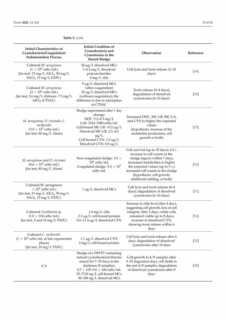
Table 5. Impact of sludge storage on cyanobacteria and cyanotoxins. STX: saxitoxin, PACl: colyalu-minium chloride, CTSAC: Chitosan-aluminum chloride.
Initial Characteristics ofCyanobacteria/Coagulation/
Sedimentation Process
Initial Condition ofCyanobacteria andCyanotoxins in the
Stored Sludge
Observation Reference
Cultured M. aeruginosa(1 × 10⁶ cells/mL)
(Jar test, 70 mg/L alum)
8 × 10⁶ cells/mL,2500 µg MC-LR/L
Cell survival (2 days); cell lysisand cyanotoxin release (2 days);
degradation of dissolvedcyanotoxins (8–10 days)
[13]

Toxins 2022, 14, 410 13 of 24
Table 5. Cont.
Initial Characteristics ofCyanobacteria/Coagulation/
Sedimentation Process
Initial Condition ofCyanobacteria andCyanotoxins in the
Stored Sludge
Observation Reference
Cultured D. circinale and C. raciborskii(1.0 × 10⁵ cells/mL)
(Jar test, 40 mg/L alum)
Sludge supernatant:D. circinale:
1300 cells/mLSTX: 0.4 µg/L
Cells remained viable up to 7days; cell lysis and toxin release
within 3 days[45]
Cultured M. aeruginosa(2 × 10⁶ cells/mL)
(Jar test, 15 mg/L AlCl3)18 µg/L dissolved MCs Cell lysis and cyanotoxin release
after 6 days [40]
Cultured M. aeruginosa(1 × 10⁶ cells/mL)
(Jar test, 4 mg/L PACl-optimumdose)
20 µg/L dissolved MCs Cell lysis and cyanotoxin releasewithin 6–12 days [46]
Microcystis flos aquae(5.2 × 10⁵ cells/mL)
(Jar test, 100 mg/L alum)
Sludge supernatant:MC-RR, MC-YR: < 2 µg/L
Cell survival (5 days); cell lysisand cyanotoxin release (5–10
days); degradation of dissolvedcyanotoxins (up to 15 days)
[62]
Cultured M. aeruginosa(1 × 10⁶ cells/mL)
(Jar test, 15 mg/L ALCl3, 4 mg/LPACl)
−0.9 bar vacuum pressure fordewatering the sludge
23 µg/L total MCs
Cell lysis and cyanotoxin releasewithin 4–6 days; optimum sludgestorage time for AlCl3 and PACl
was suggested to be 4 and 2 days,respectively.
[47]
Cultured M. aeruginosa(1 × 10⁶ cells/mL)
(Jar test, 0–70 mg/L FeCl3)~1 µg/L dissolved MCs
Cell lysis and cyanotoxin release(2–8 days); degradation of
dissolved cyanotoxins (> 10 days)[48]
Myponga reservoirCultured M. aeruginosa (2.3 × 10⁵
cells/mL)Cell-bound MC-LR: 4.7 µg/LDissolved MC-LR: 2.0 µg/L
(Jar test-80 mg/L alum)
Sludge supernatant after 1 daystorage:
Cells: 4300 cells/mLCell-bound MC-LR: 0.5 µg/LDissolved MC-LR: 2.5 µg/L
Cell survival (4 days); cell lysisand cyanotoxin release (4–7 days);
degradation of dissolvedcyanotoxins (> 4 days)
[53]
Myponga reservoirCultured M. aeruginosa (3.1 × 10⁵
cells/mL)DOC: 10.1 mg/L
Cell-Bound MC-LR: 5.0 µg/LDissolved MC-LR: 2.9 µg/L
(Jar test-80 mg/L alum)
Sludge supernatant after 1 daystorage:
DOC: 5.2 mg/LCell: 2760 cells/mL
Cell-bound MC-LR: <DLDissolved MC-LR: 4.0 µg/L
Cell growth (within 7–16 days)confirmed by DOC and MC-LR
cell quota
Myponga reservoirCultured C. raciborskii (3.1 × 10⁵
cells/mL)DOC: 10 mg/L
Cell-bound CYN: 2.5 µg/LDissolved CYN: 0.7 µg/L(Jar test-80 mg/L alum)
Sludge supernatant after 1 daystorage:
DOC: 6.0 mg/LCell: 7080 cells/mL
Cell-bound CYN: 1.0 µg/LDissolved CYN: 0.8 µg/L
Cell growth (within 7–23 days)confirmed by DOC and CYN cell
quota
River MuraryCultured C. raciborskii (3.1 × 10⁵
cells/mL)DOC: 8.63 mg/L
Cell-bound CYN: 2.7 µg/LDissolved CYN: 0.3 µg/L(Jar test-80 mg/L alum)
Sludge supernatant after 1 daystorage:
DOC: 4.9 mg/LCell: 4140 cells/mL
Cell-bound CYN: 0.3 µg/LDissolved CYN: 0.9 µg/L
Cell growth (within 15–23 days)confirmed by DOC and CYN cell
quota
Toxins 2022, 14, 410 14 of 24
Table 5. Cont.
Initial Characteristics ofCyanobacteria/Coagulation/
Sedimentation Process
Initial Condition ofCyanobacteria andCyanotoxins in the
Stored Sludge
Observation Reference
Cultured M. aeruginosa(1 × 10⁶ cells/mL)
(Jar test- 15 mg/L AlCl3, 50 mg/LFeCl3, 15 mg/L PAFC)
20 µg/L dissolved MCs1–4.2 mg/L dissolved
polysaccharides4 mg/L chla
Cell lysis and toxin release (2–10days) [49]
Cultured M. aeruginosa(2 × 10⁶ cells/mL)
(Jar test, 2.6 mg/L chitosan- 7.5 mg/LAlCl3 (CTSAC)
9 µg/L dissolved MCs(after coagulation)
18 µg/L dissolved MCs(without coagulation); the
difference is due to adsorptionin CTSAC
Toxin release (0–4 days);degradation of dissolvedcyanotoxins (6–10 days)
[50]
M. aeruginosa, D. circinale, C.raciborskii
(3.0 × 10⁵ cells/mL)(Jar test, 80 mg/L Alum)
Sludge supernatant after 1 daystorage:
DOC: 5.2–6.5 mg/LCell: 2162–7080 cells/mL
Cell-bound MC-LR: <0.5 µg/LDissolved MC-LR: 2.5–4.0
µg/LCell-bound CYN: 1.0 µg/LDissolved CYN: 0.8 µg/L
Increased DOC, MC-LR, MC-LA,and CYN to higher the expected
values(hypothesis: increase of themetabolite production, cell
growth or both)
[55]
M. aeruginosa and D. circinale(8.6 × 10⁴ cells/mL)
(Jar test, 80 mg/L Alum)
Non-coagulated sludge: 5.0 ×10⁶ cells/mL
Coagulated sludge: 5.4 × 10⁵
cells/mL
Cell survival (up to 35 days); 4.2×increase in cell counts in thesludge lagoon within 7 days;
increased metabolites to higherthe expected values (up to 5×);
increased cell counts in the sludge(hypothesis: cell growth,
additional settling, or both)
[54]
Cultured M. aeruginosa× 105 cells/mL)
(Jar test, 15 mg/L AlCl3, 50 mg/LFeCl3, 15 mg/L PAFC)
1 µg/L dissolved MCs Cell lysis and toxin release (4–6days); degradation of dissolved
cyanotoxins (6–10 days)[51]
Cultured Oscillatoria sp.(1.0 × 10⁴ cells/mL)
(Jar test, 5 and 10 mg/L PAFC)
1.0 mg/L chla2.3 µg/L cell-bound protein
8.6–11.4 µg/L dissolved CYN
Increase in chla level after 4 days,suggesting cell growth; loss of cellintegrity after 2 days, while cells
remained viable up to 8 days;increase in dissolved CYN,
showing toxin release within 4days
[94]
Cultured C. raciborskii(1 × 10⁶ cells/mL at late exponential
phase)(Jar test, 10 mg/L PAFC)
1.1 µg/L dissolved CYN2 mg/L cell-bound protein
Cell lysis and toxin release after 6days; degradation of dissolved
cyanotoxins after 10 days[52]
n/a
Sludge of a DWTP containingnatural cyanobacterial blooms
stored for 7–35 days in thedarkness (8 samples).
0.7 × 10⁵–5.6 × 10⁶ cells/mL25–7130 ng/L cell-bound MCs38–349 ng/L dissolved MCs
Cell growth in 4/8 samples after9–35 stagnation days; cell death inthe rest 4/8 samples; degradationof dissolved cyanotoxins after 8
days
[58]

Toxins 2022, 14, 410 15 of 24
4.2. Cyanobacteria-Laden Sludge Treatment
A summary of studies on the treatment of cyanobacteria-laden sludge is presentedin Table 6. The available data demonstrated that sludge oxidation could not completelyremove cyanobacteria cells and metabolites from the sludge [57,59]. Sludge is often storedafter oxidation, while its supernatant can be recycled to the head of the DWTP. Thus, theimpact of oxidation on sludge storage should be investigated. Recent findings showedno remarkable benefits in sludge oxidation followed by sludge storage as compared toonly sludge storage [59]. The maximum additional taxonomic cell count decreased by acombination of oxidation (KMnO4 or H2O2) and storage was 32% as compared to stor-age only. However, oxidation/storage could cause a remarkable cell growth (by up to145%) and toxic gene copy numbers of mcyD increase (by up to 13.0×) in some sludgesamples [59]. This phenomenon can be attributed to gene expression regulation due tothe presence of oxidative stresses [58,59,95,96]. Similarly, sludge oxidation could not com-pletely remove cyanobacteria and cyanotoxins from the supernatant sludge [59]. Finally,the costs and by-product formation during the oxidation of organic-matter-rich sludgeshould be considered [58].
Table 6. Data of cyanobacteria-laden sludge treatment. MIB: 2-Methylisoborneol.
Source ofSludge Scale Treatment
Agent/DosageContact
TimeInitial
ConditionsCell CountReduction
MetaboliteReduction Reference
Sludgethickener
Laboratory 3 mg/LKMnO4
2 h5.0 × 104
cells/mLPseudanabaena
>95% -
[57]
Laboratory 10–100 mg/LPAC 1 h 100/L MIB - 42–100% MIB
Full-scale 10 mg/LKMnO4
15 h (max.)4.3 × 10⁵
cells/mL(natural blooms)
13–98% totaland Pseudan-
abaena cellcounts
-
Full-scale
10 mg/LKMnO420 mg/L
PAC
KMnO4:24–72 hPAC: 1 h
3.7 × 10⁵
cells/mL120 ng/L MIB
(natural blooms)
40–52% intotal andPseudan-
abaena cellcounts
20–22% MIB
Sludgeholding tank
Laboratory
5 mg/LKMnO4 60 min
2.3–2.7 × 10⁶
cells/mL63–161 ng/L
MCs(natural blooms)
46–55% totalcell counts 0.3–24% MCs
[59]
10 mg/LKMnO4
59–62% totalcell counts 2–32% MCs
10 mg/LH2O2 24 h
58% total cellcounts 27% MCs
20 mg/LH2O2
77% total cellcounts 41% MCs
Full-scale(shock
oxidation)
10 mg/LKMnO4
24–72 h
2.4 × 10⁶
cells/mL88–1083 ng/L
MCs(natural blooms)
24–43% totalcell counts(31% cell
countincrease after
48 h in onesample)
MCs:3–25%
decrease inone sample
37–589%increase inone sample
4.3. Sludge Handling Challenges
In general, the sludge supernatant is recycled to the head of the DWTP or is dischargedinto the source [13,56,97]. The solid phase either can be transferred to the WWTP or is ap-

Toxins 2022, 14, 410 16 of 24
plied for landfilling [98–100]. Less environmentally friendly approaches such as untreatedresidual discharge into lakes or ponds can be also applied in some circumstances [101]. There-use of DWTPs’ residuals is growing [99,102–104]. However, an investigation demon-strated that the half-life of MC analogs varies from 8 to 18 days in soil [105]. Since there is arisk of soil and groundwater contamination, landfill and field applications of cyanobacteria-laden sludge should be avoided. Overall, cyanobacteria-laden sludge should be treatedbefore disposal either in situ or via sending it to wastewater treatment plants.
Flocs may have a protective role during sludge storage for M. aeruginosa [40,46–48,52].In contrast, Li, et al. [52] documented that polyaluminium ferric chloride (PAFC) canstimulate the lysis of C. raciborskii and CYN release by up to 94% during sludge storage.This may occur in the sludge and lead to cyanotoxin release. However, all studies havebeen conducted in laboratory conditions and on cultured-based cyanobacteria. In fact, dueto complex parameters such as the presence of various cyanobacterial cells in various forms(aggregated, multicellular), ages, and viabilities, the design of such experiments in fullscale is complex.
Stresses such as oxidation and storage can shift cyanobacterial communities towardsresistant genera (e.g., Microcystis and Aphanocapsa), which can produce MCs [56,58,59].Thus, the survival probability of MC producer species can increase during sludge oxidationor storage. The fate of cyanotoxins in the sludge is complex due to the simultaneousoccurrence of various phenomena such as cell survival, growth, lysis, cell-bound cyanotoxinrelease, and released cyanotoxin degradation [53–56,59,106,107]. Based on the increasedrisk of cell lysis and cyanotoxin release during sludge storage, some studies have suggestedthat cyanobacteria-laden sludge should be disposed of prior to 4 days to avoid metaboliterelease [47,51,108]. However, these studies only focused on metabolite release and not oncell survival/growth phenomena. Additionally, the possibility of sludge disposal can be atechnical and financial challenge in large DWTPs.
5. Decision Framework to Manage Cyanobacteria and Cyanotoxins in Drinking WaterTreatment Plants5.1. Framework Basis
Since cyanobacterial cells and their associated metabolites, including cyanotoxins, aswell as taste and odor agents such as geosmin and 2-Methylisoborneol (MIB), affect waterand sludge quality, monitoring should be applied for the evaluation of the water treatmentchain and sludge handling.
Microscopy taxonomic cell count techniques have been widely applied to evaluate thewater and sludge in previous studies [12,18,43,44,56,77]. Previous cyanobacterial monitor-ing guidelines were prepared based on taxonomic cell counts and biovolumes [109–111]. Arecent study suggested 0.3 mm3/L biovolumes as the vigilance level [109]. However, biasrelated to human error [112], the negative impact of Lugol’s iodine on biovolumes [113],cell underestimation/overestimation due to the presence of aggregated cells [114], andthe presence of debris and sediments, especially in the sludge samples [56], may affect theresults. More importantly, the significant time required for taxonomic cell counts is a majorbarrier in using them for a real-time/practical approach.
In situ fluorometry using on-line probes is a compromising technique for measurementof PC based on relative fluorescence units (RFU) in water resources [18,115–119]. How-ever, the correlation between RFU and biovolume is complex and site-specific [119–121].Previous investigations have reported that various RFUs ranging from 0.7 to 1.8 couldcorrelate with a 0.3 mm3/L biovolume in different sources and bloom events [116,119].Therefore, it is recommended to perform a correlation between on-line probe readings(RFU) and biovolumes for each water resource. It is noteworthy that the limits of detectionand quantification of on-line probes should be considered [116,118].
MC concentration has been introduced in several guidelines such as those of theWHO (1.0 µg/L MC-LR) [122,123] and Health Canada (1.5 µg/L) [124]. Geosmin andMIB negatively affect water quality, raise complaints about taste and odor, and decrease

Toxins 2022, 14, 410 17 of 24
the public’s confidence about the treated water safety [125–129]. Thus, they should bemonitored throughout the treatment chain and the recycled sludge supernatant. Usingenzyme-linked immunoassay (ELISA) tests for cyanotoxins measurement has been acceptedfor cyanotoxin monitoring [44,130–132]. The reported thresholds for geosmin and MIB are1.3–4.0 ng/L and 6.3–15 ng/L, respectively [133–135]. Since the taste and odor agents are notharmful, but increase complaints and concerns about the water quality [126,127,129,134],olfactory detection can be considered for monitoring and detection [128,136].
5.2. Decision Framework
A decision framework to manage cyanobacteria and cyanotoxins in DWTPs is pre-sented in Figure 8. The objective of this framework is to minimize cell breakthroughand accumulation throughout DWTPs and sludge. The three steps, (i) source water riskassessment, (ii) treatment breakthrough assessment and management, and (iii) sludgeand supernatant risk assessment and management, should be taken for cyanobacteriaand cyanotoxin control in DWTPs and sludge. This framework can help water utilitiesto understand appropriate approaches/strategies against cyanobacteria and cyanotoxinsin DWTPs.
Toxins 2022, 14, x FOR PEER REVIEW 23 of 30
Figure 8. Decision framework for cyanobacterial bloom management in drinking water treatment utilities.
Figure 8. Decision framework for cyanobacterial bloom management in drinking water treatment utilities.
Overall, taxonomic cell counts, MCs, and taste and odor agents should be monitoredin the (i) intake water, (ii) treatment chain, and (iii) sludge supernatant. These points aresubjected to cyanobacteria and cyanotoxin accumulation, leading to a negative impact onwater quality.
The optimization of conventional processes may include coagulant dose adjustment,applying aid-flocculants, and lowering the sedimentation and filtration rate during cellbreakthrough [12,137–139]. Secondary barriers such as pre-oxidation, PAC injection, andGAC, in case of metabolite breakthrough, should be applied [15,25,39,65,140–144]. Theimpact of supernatant recycling or discharging on the source/intake water quality shouldbe considered during toxic cyanobacterial blooms. Supernatant treatment may be requiredin the presence of cyanotoxins or taste and odor agents. MC concentration levels shouldbe monitored in the sludge (solids) in case of landfilling or land application. In the case ofelevated concentrations of MCs, sludge treatment is required.

Toxins 2022, 14, 410 18 of 24
6. Conclusions
# Using the exposure unit (CT) is recommended for cyanobacteria and cyanotoxinsoxidation studies, rather than using dose or contact time individually.
# Regardless of the oxidant type, lab-cultured studies cannot depict the complete pictureof natural cyanobacterial bloom behavior during oxidation and may overestimatethe oxidation efficiency. In addition, cyanobacterial bloom oxidation is site- andbloom-specific, which could be related to the level of agglomeration, cyanobacteria(bloom) stage of life, and metabolic functions.
# Soft pre-chlorination and pre-ozonation can compromise cell viability with no orlimited cyanotoxin release. Overall, soft pre-oxidation may cause lower disinfectionby-products compared to normal pre-oxidation.
# The cyanobacteria in stored sludge can not only survive, but also grow and releasecyanotoxins, even in the dark. Although dissolved cyanotoxins can be degradedduring sludge storage, the potential risk of growth and cyanotoxin release shouldbe considered. In fact, the cell growth/depletion in stored sludge is complex andnot easy to predict. Therefore, the worst-case scenario should be considered duringsludge handling.
# Due to the low efficacy of sludge oxidation as compared to only stored sludge, as well asthe occurrence of cell growth, and gene expression regulation during oxidation/storage,oxidation cannot be a reliable approach in sludge treatment and management.
# Management of cyanobacteria and cyanotoxins in sludge should be initiated with theminimization of cyanobacteria and cyanotoxin accumulation throughout DWTPs.
# To control the negative impacts of cyanobacterial accumulation in DWTPs, recyclingsludge supernatant to the head of the DWTPs should be regulated during cyanobacte-rial seasons. Suitable treatment and disposal approaches should be set into guidanceand regulations for sludge-containing cyanotoxins.
Author Contributions: Conceptualization, F.J., S.M., S.D., A.Z., S.S. and M.P.; methodology, F.J.,S.M., S.D. and M.P.; validation, F.J., S.M., A.Z., S.D. and M.P.; formal analysis, F.J., S.M. and M.P.;investigation, F.J., S.M., S.D., A.Z. and M.P.; writing—original draft preparation, F.J. and S.M.; writing—review and editing, F.J., S.M., S.D., A.Z., S.S. and M.P.; visualization, F.J. and S.M.; supervision, S.D.,S.S. and M.P.; project administration, S.S., S.D. and M.P.; funding acquisition, S.D. and S.S. All authorshave read and agreed to the published version of the manuscript.
Funding: The authors sincerely acknowledge the financial supports of Algal Blooms, Treatment, RiskAs-sessment, Prediction, and Prevention through Genomics (ATRAPP) projects, Génome Québec,Genome Canada and the NSERC Industrial Chair on Drinking Water at Polytechnique Montréal(grant number ALLRP 560764-20).
Institutional Review Board Statement: Not applicable.
Informed Consent Statement: Not applicable.
Conflicts of Interest: The authors declare no conflict of interest.
References1. Churro, C.; Dias, E.; Valério, E. Risk assessment of cyanobacteria and cyanotoxins, the particularities and challenges of Planktothrix
spp. monitoring. In Novel Approaches and Their Applications in Risk Assessment; IntechOpen: London, UK, 2012; pp. 59–85.2. Kosten, S.; Huszar, V.L.M.; Bécares, E.; Costa, L.S.; Donk, E.; Hansson, L.-A.; Jeppesen, E.; Kruk, C.; Lacerot, G.; Mazzeo, N.; et al.
Warmer climates boost cyanobacterial dominance in shallow lakes. Glob. Chang. Biol. 2012, 18, 118–126. [CrossRef]3. Paerl, H.W.; Paul, V.J. Climate change: Links to global expansion of harmful cyanobacteria. Water Res. 2012, 46, 1349–1363.
[CrossRef] [PubMed]4. Chorus, I.; Fastner, J.; Welker, M. Cyanobacteria and Cyanotoxins in a Changing Environment: Concepts, Controversies,
Challenges. Water 2021, 13, 2463. [CrossRef]5. Mokoena, M.M.; Mukhola, M.S. Current Effects of Cyanobacteria Toxin in Water Sources and Containers in the Hartbeespoort
Dam Area, South Africa. Int. J. Environ. Res. Public Health 2019, 16, 4468. [CrossRef]6. Huisman, J.; Codd, G.A.; Paerl, H.W.; Ibelings, B.W.; Verspagen, J.M.H.; Visser, P.M. Cyanobacterial blooms. Nat. Rev. Microbiol.
2018, 16, 471–483. [CrossRef]

Toxins 2022, 14, 410 19 of 24
7. O’Keeffe, J. Cyanobacteria and Drinking Water: Occurrence, Risks, Management and Knowledge Gaps for Public Health; NationalCollaborating Centre for Environmental Health: Vancouver, BC, Canada, 2019.
8. Mishra, S.; Stumpf, R.P.; Schaeffer, B.A.; Werdell, P.J.; Loftin, K.A.; Meredith, A. Measurement of Cyanobacterial Bloom Magnitudeusing Satellite Remote Sensing. Sci. Rep. 2019, 9, 18310. [CrossRef]
9. Akyol, C.; Ozbayram, E.G.; Accoroni, S.; Radini, S.; Eusebi, A.L.; Gorbi, S.; Vignaroli, C.; Bacchiocchi, S.; Campacci, D.; Gigli,F.; et al. Monitoring of cyanobacterial blooms and assessing polymer-enhanced microfiltration and ultrafiltration for microcystinremoval in an Italian drinking water treatment plant. Environ. Pollut. 2021, 286, 117535. [CrossRef]
10. Zamyadi, A.; Glover, C.M.; Yasir, A.; Stuetz, R.; Newcombe, G.; Crosbie, N.D.; Lin, T.-F.; Henderson, R. Toxic cyanobacteria inwater supply systems: Data analysis to map global challenges and demonstrate the benefits of multi-barrier treatment approaches.H2Open J. 2021, 4, 47–62. [CrossRef]
11. Westrick, J.A.; Szlag, D.C.; Southwell, B.J.; Sinclair, J. A review of cyanobacteria and cyanotoxins removal/inactivation in drinkingwater treatment. Anal. Bioanal. Chem. 2010, 397, 1705–1714. [CrossRef]
12. Zamyadi, A.; Dorner, S.; Ellis, D.; Bolduc, A.; Bastien, C.; Prévost, M. Species-dependence of cyanobacteria removal efficiency bydifferent drinking water treatment processes. Water Res. 2013, 47, 2689–2700. [CrossRef]
13. Drikas, M.; Chow, C.W.K.; House, J.; Burch, M.D. Using coagulation, flocculation, and settling to remove toxic cyanobacteria. J.Am. Water Work. Assoc. 2001, 93, 100–111. [CrossRef]
14. Szlag, D.C.; Sinclair, J.L.; Southwell, B.; Westrick, J.A. Cyanobacteria and Cyanotoxins Occurrence and Removal from FiveHigh-Risk Conventional Treatment Drinking Water Plants. Toxins 2015, 7, 2198–2220. [CrossRef] [PubMed]
15. Newcombe, G.; Nicholson, B. Water treatment options for dissolved cyanotoxins. Water Supply Res. Technol.-Aqua 2004, 53,227–239. [CrossRef]
16. Wert, E.C.; Rosario-Ortiz, F.L. Intracellular organic matter from cyanobacteria as a precursor for carbonaceous and nitrogenousdisinfection byproducts. Environ. Sci. Technol. 2013, 47, 6332–6340. [CrossRef] [PubMed]
17. Zamyadi, A.; Greenstein, K.E.; Glover, C.M.; Adams, C.; Rosenfeldt, E.; Wert, E.C. Impact of hydrogen peroxide and coppersulfate on the delayed release of microcystin. Water 2020, 12, 1105. [CrossRef]
18. Zamyadi, A.; MacLeod, S.; Fan, Y.; McQuaid, N.; Dorner, S.; Sauvé, S.; Prévost, M. Toxic cyanobacterial breakthrough andaccumulation in a drinking water plant: A monitoring and treatment challenge. Water Res. 2012, 46, 1511–1523. [CrossRef][PubMed]
19. Henderson, R.K.; Parsons, S.A.; Jefferson, B. The impact of differing cell and algogenic organic matter (AOM) characteristics onthe coagulation and flotation of algae. Water Res. 2010, 44, 3617–3624. [CrossRef]
20. Newcombe, G. International Guidance Manual for the Management of Toxic Cyanobacteria; Global Water Research Coalition and WaterQuality Research Australia: London, UK, 2009; p. 44.
21. Gonzalez-Torres, A.; Putnam, J.; Jefferson, B.; Stuetz, R.M.; Henderson, R.K. Examination of the physical properties of Microcystisaeruginosa flocs produced on coagulation with metal salts. Water Res. 2014, 60, 197–209. [CrossRef]
22. Pivokonsky, M.; Naceradska, J.; Brabenec, T.; Novotna, K.; Baresova, M.; Janda, V. The impact of interactions between algalorganic matter and humic substances on coagulation. Water Res. 2015, 84, 278–285. [CrossRef]
23. Merel, S.; Walker, D.; Chicana, R.; Snyder, S.; Baures, E.; Thomas, O. State of knowledge and concerns on cyanobacterial bloomsand cyanotoxins. Environ. Int. 2013, 59, 303–327. [CrossRef]
24. He, X.; Liu, Y.-L.; Conklin, A.; Westrick, J.; Weavers, L.K.; Dionysiou, D.D.; Lenhart, J.J.; Mouser, P.J.; Szlag, D.; Walker, H.W. Toxiccyanobacteria and drinking water: Impacts, detection, and treatment. Harmful Algae 2016, 54, 174–193. [CrossRef] [PubMed]
25. Abbas, T.; Kajjumba, G.W.; Ejjada, M.; Masrura, S.U.; Marti, E.J.; Khan, E.; Jones-Lepp, T.L. Recent Advancements in The Removalof Cyanotoxins from Water Using Conventional and Modified Adsorbents—A Contemporary review. Water 2020, 12, 2756.[CrossRef]
26. Villars, K.; Huang, Y.; Lenhart, J.J. Removal of the Cyanotoxin Microcystin-LR from Drinking Water Using Granular ActivatedCarbon. Environ. Eng. Sci. 2020, 37, 585–595. [CrossRef]
27. Xie, P.; Ma, J.; Fang, J.; Guan, Y.; Yue, S.; Li, X.; Chen, L. Comparison of permanganate preoxidation and preozonation on algaecontaining water: Cell integrity, characteristics, and chlorinated disinfection byproduct formation. Environ. Sci. Technol. 2013, 47,14051–14061. [CrossRef]
28. Lapsongpon, T.; Leungprasert, S.; Yoshimura, C. Pre-chlorination contact time and the removal and control of Microcystisaeroginosa in coagulation. IOP Conf. Ser. Earth Environ. Sci. 2017, 67, 012011. [CrossRef]
29. Petrusevski, B.; van Breeman, A.N.; Alaerts, G.J. Effect of permanganate pre-treatment and coagulation with dual coagulants onalgae removal in direct filtration. Water Supply Res. Technol.-Aqua 1996, 45, 316–326.
30. Chu, W.; Yao, D.; Deng, Y.; Sui, M.; Gao, N. Production of trihalomethanes, haloacetaldehydes and haloacetonitriles duringchlorination of microcystin-LR and impacts of pre-oxidation on their formation. J. Hazard. Mater. 2017, 327, 153–160. [CrossRef]
31. Lin, J.L.; Hua, L.C.; Hung, S.K.; Huang, C. Algal removal from cyanobacteria-rich waters by preoxidation-assisted coagulation-flotation: Effect of algogenic organic matter release on algal removal and trihalomethane formation. J. Environ. Sci. 2018, 63,147–155. [CrossRef]
32. Naceradska, J.; Pivokonsky, M.; Pivokonska, L.; Baresova, M.; Henderson, R.K.; Zamyadi, A.; Janda, V. The impact of pre-oxidationwith potassium permanganate on cyanobacterial organic matter removal by coagulation. Water Res. 2017, 114, 42–49. [CrossRef]

Toxins 2022, 14, 410 20 of 24
33. Wang, H.; Masters, S.; Falkinham, J.O., 3rd; Edwards, M.A.; Pruden, A. Distribution system water quality affects responses ofopportunistic pathogen gene markers in household water heaters. Environ. Sci. Technol. 2015, 49, 8416–8424. [CrossRef]
34. Xie, P.; Chen, Y.; Ma, J.; Zhang, X.; Zou, J.; Wang, Z. A mini review of preoxidation to improve coagulation. Chemosphere 2016, 155,550–563. [CrossRef] [PubMed]
35. Zamyadi, A.; Fan, Y.; Daly, R.I.; Prévost, M. Chlorination of Microcystis aeruginosa: Toxin release and oxidation, cellular chlorinedemand and disinfection by-products formation. Water Res. 2013, 47, 1080–1090. [CrossRef] [PubMed]
36. Fan, J.; Daly, R.; Hobson, P.; Ho, L.; Brookes, J. Impact of potassium permanganate on cyanobacterial cell integrity and toxinrelease and degradation. Chemosphere 2013, 92, 529–534. [CrossRef] [PubMed]
37. Fan, J.; Ho, L.; Hobson, P.; Brookes, J. Evaluating the effectiveness of copper sulphate, chlorine, potassium permanganate,hydrogen peroxide and ozone on cyanobacterial cell integrity. Water Res. 2013, 47, 5153–5164. [CrossRef] [PubMed]
38. Moradinejad, S.; Trigui, H.; Maldonado, J.F.G.; Shapiro, B.J.; Terrat, Y.; Sauvé, S.; Fortin, N.; Zamyadi, A.; Dorner, S.; Prévost,M. Metagenomic study to evaluate functional capacity of a cyanobacterial bloom during oxidation. Chem. Eng. J. Adv. 2021,8, 100151. [CrossRef]
39. Moradinejad, S.; Trigui, H.; Guerra Maldonado, J.F.; Shapiro, J.; Terrat, Y.; Zamyadi, A.; Dorner, S.; Prévost, M. Diversityassessment of toxic cyanobacterial blooms during oxidation. Toxins 2020, 12, 728. [CrossRef]
40. Sun, F.; Pei, H.-Y.; Hu, W.-R.; Ma, C.-X. The lysis of Microcystis aeruginosa in AlCl3 coagulation and sedimentation processes.Chem. Eng. J. 2012, 193–194, 196–202. [CrossRef]
41. Teixeira, M.R.; Rosa, M.J. Comparing dissolved air flotation and conventional sedimentation to remove cyanobacterial cells ofMicrocystis aeruginosa. Part II. The effect of water background organics. Sep. Purif. Technol. 2007, 53, 126–134. [CrossRef]
42. Chorus, I.; Bartram, J. Chapter 6. Situation assessment, planning and management. In Toxic Cyanobacteria in Water: A Guide toTheir Public Health Consequences, Monitoring and Management; WHO: Geneva, Switzerland, 1999; p. 28.
43. Zamyadi, A.; Dorner, S.; Ndong, M.; Ellis, D.; Bolduc, A.; Bastien, C.; Prévost, M. Low-risk cyanobacterial bloom sources: Cellaccumulation within full-scale treatment plants. J. Am. Water Work. Assoc. 2013, 102, E651–E663. [CrossRef]
44. Almuhtaram, H.; Cui, Y.; Zamyadi, A.; Hofmann, R. Cyanotoxins and cyanobacteria cell accumulations in drinking watertreatment plants with a low risk of bloom formation at the source. Toxins 2018, 10, 430. [CrossRef]
45. Ho, L.; Dreyfus, J.; Boyer, J.e.; Lowe, T.; Bustamante, H.; Duker, P.; Meli, T.; Newcombe, G. Fate of cyanobacteria and theirmetabolites during water treatment sludge management processes. Sci. Total Environ. 2012, 424, 232–238. [CrossRef] [PubMed]
46. Sun, F.; Pei, H.-Y.; Hu, W.-R.; Li, X.-Q.; Ma, C.-X.; Pei, R.-T. The cell damage of Microcystis aeruginosa in PACl coagulation and flocstorage processes. Sep. Purif. Technol. 2013, 115, 123–128. [CrossRef]
47. Sun, F.; Hu, W.; Pei, H.; Li, X.; Xu, X.; Ma, C. Evaluation on the dewatering process of cyanobacteria-containing AlCl3 and PACldrinking water sludge. Sep. Purif. Technol. 2015, 150, 52–62. [CrossRef]
48. Li, X.; Pei, H.; Hu, W.; Meng, P.; Sun, F.; Ma, G.; Xu, X.; Li, Y. The fate of Microcystis aeruginosa cells during the ferric chloridecoagulation and flocs storage processes. Environ. Technol. 2015, 36, 920–928. [CrossRef] [PubMed]
49. Xu, H.; Pei, H.; Xiao, H.; Jin, Y.; Li, X.; Hu, W.; Ma, C.; Sun, J.; Li, H. Behaviors of Microcystis aeruginosa cells during floc storagein drinking water treatment process. Sci. Rep. 2016, 6, 34943. [CrossRef]
50. Ma, C.; Pei, H.; Hu, W.; Xu, H.; Jin, Y. The lysis and regrowth of toxic cyanobacteria during storage of achitosan–aluminiumchloride composite coagulated sludge: Implications for drinking water sludge treatment. RSC Adv. 2016, 6, 112756–112764.[CrossRef]
51. Xu, H.; Pei, H.; Jin, Y.; Xiao, H.; Ma, C.; Sun, J.; Li, H. Characteristics of water obtained by dewatering cyanobacteria-containingsludge formed during drinking water treatment, including C-, N-disinfection byproduct formation. Water Res. 2017, 111, 382–392.[CrossRef]
52. Li, H.; Pei, H.; Xu, H.; Jin, Y.; Sun, J. Behavior of Cylindrospermopsis raciborskii during coagulation and sludge storage-higherpotential risk of toxin release than Microcystis aeruginosa? J. Hazard. Mater. 2018, 347, 307–316. [CrossRef]
53. Water Research Foundation (WRF); Water Research Australia. Optimizing Conventional Treatment for the Removal of Cyanobacteriaand Toxins; Water Research Foundation (WRF): Denver, CO, USA, 2015; p. 185. ISBN 978-1-60573-216-9.
54. Pestana, C.J.; Reeve, P.J.; Sawade, E.; Voldoire, C.F.; Newton, K.; Praptiwi, R.; Collingnon, L.; Dreyfus, J.; Hobson, P.; Gaget,V.; et al. Fate of cyanobacteria in drinking water treatment plant lagoon supernatant and sludge. Sci. Total Environ. 2016, 565,1192–1200. [CrossRef]
55. Dreyfus, J.; Monrolin, Y.; Pestana, C.J.; Reeve, P.J.; Sawade, E.; Newton, K.; Ho, L.; Chow, C.W.K.; Newcombe, G. Identificationand assessment of water quality risks associated with sludge supernatant recycling in the presence of cyanobacteria. J. WaterSupply Res. Technol.-Aqua 2016, 65, 441–452. [CrossRef]
56. Jalili, F.; Trigui, H.; Guerra Maldonado, J.F.; Dorner, S.; Zamyadi, A.; Shapiro, B.J.; Terrat, Y.; Fortin, N.; Sauvé, S.; Prévost, M. Cancyanobacterial diversity in the source predict the diversity in sludge and the risk of toxin release in a drinking water treatmentplant? Toxins 2021, 13, 25. [CrossRef] [PubMed]
57. Zamyadi, A.; Henderson, R.K.; Stuetz, R.; Newcombe, G.; Newtown, K.; Gladman, B. Cyanobacterial management in full-scalewater treatment and recycling processes: Reactive dosing following intensive monitoring. Environ. Sci. Water Res. Technol. 2016, 2,362–375. [CrossRef]
58. Jalili, F. Optimal Treatment Strategies to Prevent and Manage Cyanobacteria and Cyanotoxins in Drinking Water Sludge. Ph.D.Thesis, Polytechnique Montréal, Montréal, QC, Canada, 2022.

Toxins 2022, 14, 410 21 of 24
59. Jalili, F.; Trigui, H.; Maldonado, J.F.G.; Dorner, S.; Zamyadi, A.; Shapiro, B.J.; Terrat, Y.; Fortin, N.; Sauvé, S.; Prévost, M. Oxidationto Control Cyanobacteria and Cyanotoxins in Drinking Water Treatment Plants: Challenges at the Laboratory and Full-ScalePlants. Water 2022, 14, 537. [CrossRef]
60. Zamyadi, A.; Romanis, C.; Mills, T.; Neilan, B.; Choo, F.; Coral, L.A.; Gale, D.; Newcombe, G.; Crosbie, N.; Stuetz, R.; et al.Diagnosing water treatment critical control points for cyanobacterial removal: Exploring benefits of combined microscopy,next-generation sequencing, and cell integrity methods. Water Res. 2019, 152, 96–105. [CrossRef]
61. Water Research Foundation (WRF); United States Environmental Protection Agency (US EPA); Veolia Water Indianapolis.Strategies for Controlling and Mitigating Algal Growth within Water Treatment Plants; Water Research Foundation (WRF): Denver, CO,USA, 2009; p. 312.
62. Ho, L.; Barbero, A.; Dreyfus, J.; Dixon, D.R.; Qian, F.; Scales, P.J.; Newcombe, G. Behaviour of cyanobacterial bloom materialfollowing coagulation and/or sedimentation. J. Water Supply Res. Technol.-Aqua 2013, 62, 350–358. [CrossRef]
63. Pinkanjananavee, K.; Teh, S.J.; Kurobe, T.; Lam, C.H.; Tran, F.; Young, T.M. Potential Impacts on Treated Water Quality ofRecycling Dewatered Sludge Supernatant during Harmful Cyanobacterial Blooms. Toxins 2021, 13, 99. [CrossRef]
64. Coral, L.A.; Zamyadi, A.; Barbeau, B.; Bassetti, F.J.; Lapolli, F.R.; Prévost, M. Oxidation of Microcystis aeruginosa and Anabaenaflos-aquae by ozone: Impacts on cell integrity and chlorination by-product formation. Water Res. 2013, 47, 2983–2994. [CrossRef]
65. Zamyadi, A.; Coral, L.A.; Barbeau, B.; Dorner, S.; Lapolli, F.R.; Prévost, M. Fate of toxic cyanobacterial genera from natural bloomevents during ozonation. Water Res. 2015, 73, 204–215. [CrossRef]
66. Ding, J.; Shi, H.; Timmons, T.; Adams, C. Release and removal of microcystins from microcystis during oxidative-, physical-, andUV-based disinfection. J. Environ. Eng. 2010, 136, 2–11. [CrossRef]
67. Zamyadi, A.; Ho, L.; Newcombe, G.; Daly, R.I.; Burch, M.; Baker, P.; Prévost, M. Release and oxidation of cell-bound saxitoxinsduring chlorination of Anabaena circinalis cells. Environ. Sci. Technol. 2010, 44, 9055–9061. [CrossRef]
68. Fan, J.J.; Hobson, P.; Ho, L.; Daly, R.; Brookes, J. The effects of various control and water treatment processes on the membraneintegrity and toxin fate of cyanobacteria. J. Hazard. Mater. 2014, 264, 313–322. [CrossRef] [PubMed]
69. Zhang, H.; Dan, Y.; Adams, C.D.; Shi, H.; Ma, Y.; Eichholz, T. Effect of oxidant demand on the release and degradation ofmicrocystin-LR from Microcystis aeruginosa during oxidation. Chemosphere 2017, 181, 562–568. [CrossRef] [PubMed]
70. Qi, J.; Lan, H.; Liu, R.; Miao, S.; Liu, H.; Qu, J. Prechlorination of algae-laden water: The effects of transportation time on cellintegrity, algal organic matter release, and chlorinated disinfection byproduct formation. Water Res. 2016, 102, 221–228. [CrossRef][PubMed]
71. Li, X.; Chen, S.; Zeng, J.; Song, W.; Yu, X. Comparing the effects of chlorination on membrane integrity and toxin fate of high-andlow-viability cyanobacteria. Water Res. 2020, 177, 115769. [CrossRef]
72. Li, X.; Chen, S.; Zeng, J.; Chabi, K.; Song, W.; Xian, X.; Yu, X. Impact of chlorination on cell inactivation, toxin release anddegradation of cyanobacteria of development and maintenance stage. Chem. Eng. J. 2020, 397, 125378. [CrossRef]
73. Fan, J.; Rao, L.; Chiu, Y.T.; Lin, T.F. Impact of chlorine on the cell integrity and toxin release and degradation of colonial Microcystis.Water Res. 2016, 102, 394–404. [CrossRef]
74. He, X.; Wert, E.C. Colonial cell disaggregation and intracellular microcystin release following chlorination of naturally occurringMicrocystis. Water Res. 2016, 101, 10–16. [CrossRef]
75. Greenstein, K.E.; Zamyadi, A.; Glover, C.M.; Adams, C.; Rosenfeldt, E.; Wert, E.C. Delayed Release of Intracellular MicrocystinFollowing Partial Oxidation of Cultured and Naturally Occurring Cyanobacteria. Toxins 2020, 12, 335. [CrossRef]
76. Moradinejad, S. Characterization of Phenomena Governing Oxidation of Cyanobacteria through Metagenomics. Ph.D Thesis,Polytechnique Montréal, Montreal, QC, Canada, 2021.
77. Zamyadi, A.; Dorner, S.; Ndong, M.; Ellis, D.; Bolduc, A.; Bastien, C.; Prévost, M. Breakthrough and accumulation of toxiccyanobacteria in full scale clarification and filtration processes. In Proceedings of the American Water Works Association-WaterQuality Technology Conference and Exposition (WQTC), Toronto, ON, Canada, 4–8 November 2012; p. 27.
78. Wert, E.C.; Dong, M.M.; Rosario-Ortiz, F.L. Using digital flow cytometry to assess the degradation of three cyanobacteria speciesafter oxidation processes. Water Res. 2013, 47, 3752–3761. [CrossRef]
79. Moradinejad, S.; Glover, C.; Mailly, J.; Zadfatollah-Seighalani, T.; Peldszus, S.; Barbeau, B.; Dorner, S.; Prévost, M.; Zamyadi, A.Using advanced spectroscopy and organic matter characterization to evaluate the impact of oxidation on cyanobacteria. Toxins2019, 11, 278. [CrossRef]
80. Moradinejad, S.; Glover, C.; Dorner, S.; Barbeau, B.; Prévost, M.; Zamyadi, A. Using advanced spectroscopy and visualizationtechniques to assess the impact of oxidation on cyanobacteria cell morphology. In Proceedings of the WQTC, Dallas, TX, USA,3–7 November 2019.
81. Piezer, K.; Li, L.; Jeon, Y.; Kadudula, A.; Seo, Y. The Application of Potassium Permanganate to Treat Cyanobacteria-Laden Water.Process Saf. Environ. Prot. 2020, 148, 400–414. [CrossRef]
82. Zhou, S.; Shao, Y.; Gao, N.; Deng, Y.; Qiao, J.; Ou, H.; Deng, J. Effects of different algaecides on the photosynthetic capacity, cellintegrity and microcystin-LR release of Microcystis aeruginosa. Sci. Total Environ. 2013, 463, 111–119. [CrossRef] [PubMed]
83. Xu, H.; Brookes, J.; Hobson, P.; Pei, H. Impact of copper sulphate, potassium permanganate, and hydrogen peroxide onPseudanabaena galeata cell integrity, release and degradation of 2-methylisoborneol. Water Res. 2019, 157, 64–73. [CrossRef][PubMed]
Toxins 2022, 14, 410 22 of 24
84. Zhou, T.; Cao, H.; Zheng, J.; Teng, F.; Wang, X.; Lou, K.; Zhang, X.; Tao, Y. Suppression of water-bloom cyanobacterium Microcystisaeruginosa by algaecide hydrogen peroxide maximized through programmed cell death. J. Hazard. Mater. 2020, 393, 122394.[CrossRef] [PubMed]
85. Daly, R.I.; Ho, L.; Brookes, J.D. Effect of chlorination on Microcystis aeruginosa cell integrity and subsequent microcystin releaseand degradation. Environ. Sci. Technol. 2007, 41, 4447–4453. [CrossRef]
86. Matthijs, H.C.P.; Visser, P.M.; Reeze, B.; Meeuse, J.; Slot, P.C.; Wijn, G.; Talens, R.; Huisman, J. Selective suppression of harmfulcyanobacteria in an entire lake with hydrogen peroxide. Water Res. 2012, 46, 1460–1472. [CrossRef]
87. Foo, S.C.; Chapman, I.J.; Hartnell, D.M.; Turner, A.D.; Franklin, D.J. Effects of H2O2 on growth, metabolic activity and membraneintegrity in three strains of Microcystis aeruginosa. Environ. Sci. Pollut. Res. 2020, 27, 38916–38927. [CrossRef]
88. Luukkonen, T.; Teeriniemi, J.; Prokkola, H.; Rämö, J.; Lassi, U. Chemical aspects of peracetic acid based wastewater disinfection.Water Sa 2014, 40, 73–80. [CrossRef]
89. Almuhtaram, H.; Hofmann, R. Evaluation of ultraviolet/peracetic acid to degrade M. aeruginosa and microcystins-LR and-RR. J.Hazard. Mater. 2022, 424, 127357. [CrossRef]
90. Wang, H.-Q.; Mao, T.-G.; Xi, B.-D.; Zhang, L.-Y.; Zhou, Q.-H. KMnO4 pre-oxidation for Microcystis aeruginosa removal by a lowdosage of flocculant. Ecol. Eng. 2015, 81, 298–300. [CrossRef]
91. Barešová, M.; Naceradská, J.; Novotná, K.; Cermáková, L.; Pivokonský, M. The impact of preozonation on the coagulationof cellular organic matter produced by Microcystis aeruginosa and its toxin degradation. J. Environ. Sci. 2020, 98, 124–133.[CrossRef] [PubMed]
92. Kormas, K.A.; Lymperopoulou, D.S. Cyanobacterial toxin degrading bacteria: Who are they? Biomed. Res. Int. 2013, 2013, 463894.[CrossRef] [PubMed]
93. Min, H.; Golden, S.S. A new circadian class 2 gene, opcA, whose product is important for reductant production at night inSynechococcus elongatus PCC 7942. J. Bacteriol. 2000, 182, 6214–6221. [CrossRef] [PubMed]
94. Sun, J.; Xu, H.; Pei, H.; Jin, Y.; Li, H.; Ma, C. Worse than cell lysis: The resilience of Oscillatoria sp. during sludge storage indrinking water treatment. Water Res. 2018, 142, 405–414. [CrossRef]
95. Pimentel, J.S.; Giani, A. Microcystin production and regulation under nutrient stress conditions in toxic microcystis strains. Appl.Environ. Microbiol. 2014, 80, 5836–5843. [CrossRef]
96. Schatz, D.; Keren, Y.; Vardi, A.; Sukenik, A.; Carmeli, S.; Börner, T.; Dittmann, E.; Kaplan, A. Towards clarification of the biologicalrole of microcystins, a family of cyanobacterial toxins. Environ. Microbiol. 2007, 9, 965–970. [CrossRef]
97. Chow, C.W.K.; Drikas, M.; House, J.; Burch, M.D.; Velzeboer, R.M.A. The impact of conventional water treatment processes oncells of the cyanobacterium Microcystis aeruginosa. Water Res. 1999, 33, 3253–3262. [CrossRef]
98. United States Environmental Protection Agency (USEPA). Drinking Water Treatment Plant Residuals Management Technical Report,Summary of Residuals Generation, Treatment, and Disposal at Large Community Water Systems; United States Environmental ProtectionAgency (USEPA): Washington, DC, USA, 2011; p. 378.
99. Turner, T.; Wheeler, R.; Stone, A.; Oliver, I. Potential Alternative Reuse Pathways for Water Treatment Residuals: RemainingBarriers and Questions—A Review. Water Air Soil Pollut. 2019, 230, 227. [CrossRef]
100. Qrenawi, L.I.; Rabah, F.K.J. Sludge management in water treatment plants: Literature review. Int. J. Environ. Waste Manag. 2021,27, 93. [CrossRef]
101. Ahmad, T.; Ahmad, K.; Alam, M. Characterization of Water Treatment Plant’s Sludge and its Safe Disposal Options. ProcediaEnviron. Sci. 2016, 35, 950–955. [CrossRef]
102. Jung, K.W.; Hwang, M.J.; Park, D.S.; Ahn, K.H. Comprehensive reuse of drinking water treatment residuals in coagulation andadsorption processes. J. Environ. Manag. 2016, 181, 425–434. [CrossRef] [PubMed]
103. Xu, Y.; Chen, T.; Xu, R.; He, L.; Cui, F. Impact of recycling alum sludge on coagulation of low-turbidity source waters. DesalinationWater Treat. 2015, 57, 6732–6739. [CrossRef]
104. Suman, A.; Ahmad, T.; Ahmad, K. Dairy wastewater treatment using water treatment sludge as coagulant: A novel treatmentapproach. Environ. Dev. Sustain. 2017, 20, 1615–1625. [CrossRef]
105. Chen, W.; Song, L.; Gan, N.; Li, L. Sorption, degradation and mobility of microcystins in Chinese agriculture soils: Risk assessmentfor groundwater protection. Environ. Pollut. 2006, 144, 752–758. [CrossRef] [PubMed]
106. Clemente, A.; Wilson, A.; Oliveira, S.; Menezes, I.; Gois, A.; Capelo-Neto, J. The role of hydraulic conditions of coagulation andflocculation on the damage of cyanobacteria. Sci. Total Environ. 2020, 740, 139737. [CrossRef] [PubMed]
107. Pestana, C.J.; Capelo-Neto, J.; Lawton, L.; Oliveira, S.; Carloto, I.; Linhares, H.P. The effect of water treatment unit processes oncyanobacterial trichome integrity. Sci. Total Environ. 2019, 659, 1403–1414. [CrossRef] [PubMed]
108. Pei, H.; Xu, H.; Wang, J.; Jin, Y.; Xiao, H.; Ma, C.; Sun, J.; Li, H. 16S rRNA Gene amplicon sequencing reveals significant changesin microbial compositions during cyanobacteria-laden drinking water sludge storage. Environ. Sci. Technol. 2017, 51, 12774–12783.[CrossRef]
109. Chorus, I.; Welker, M. Toxic Cyanobacteria in Water: A Guide to Their Public Health Consequences, Monitoring and Management, 2nded.; CRC Press: Boca Raton, FL, USA, 2021; p. 859.
110. Chorus, I.; Bartram, J. Toxic Cyanobacteria in Water: A Guide to Their Public Health Consequences, Monitoring, and Management; WHO:London, UK; New York, NY, USA, 1999.

Toxins 2022, 14, 410 23 of 24
111. Ellis, D. Guide D’intervention Pour les Propriétaires, les Exploitants ou les Concepteurs de Stations de Production D’eau Potablemunicipalesaux Prises avec une Problématique de Fleurs D’eau de Cyanobactéries; Ministère du Développement durable, Environnement et Parcs:Québec, QC, Canada, 2009; p. 51.
112. America Water Works Association (AWWA). Algae Source to Treatment. Manual of Water Supply Practices—M57, 1st ed.; AmericaWater Works Association (AWWA): Denver, CO, USA, 2010; p. 481.
113. Hawkins, P.R.; Holliday, J.; Kathuria, A.; Bowling, L. Change in cyanobacterial biovolume due to preservation by Lugol’s Iodine.Harmful Algae 2005, 4, 1033–1043. [CrossRef]
114. Park, J.; Kim, Y.; Kim, M.; Lee, W.H.; Park, J.; Kim, Y.; Kim, M.; Lee, W.H. A novel method for cell counting of Microcystis coloniesin water resources using a digital imaging flow cytometer and microscope. Environ. Eng. Res. 2018, 24, 397–403. [CrossRef]
115. Zamyadi, A.; McQuaid, N.; Prévost, M.; Dorner, S. Monitoring of potentially toxic cyanobacteria using an online multi-probe indrinking water sources. J. Environ. Monit. 2012, 14, 579–588. [CrossRef]
116. McQuaid, N.; Zamyadi, A.; Prévost, M.; Bird, D.F.; Dorner, S. Use of in vivo phycocyanin fluorescence to monitor potentialmicrocystin-producing cyanobacterial biovolume in a drinking water source. J. Environ. Monit. 2011, 13, 455–463. [CrossRef][PubMed]
117. Gregor, J.; Marsalek, B.; Sipkova, H. Detection and estimation of potentially toxic cyanobacteria in raw water at the drinkingwater treatment plant by in vivo fluorescence method. Water Res. 2007, 41, 228–234. [CrossRef] [PubMed]
118. Zamyadi, A.; Choo, F.; Newcombe, G.; Stuetz, R.; Henderson, R.K. A review of monitoring technologies for real-time managementof cyanobacteria: Recent advances and future direction. TrAC Trends Anal. Chem. 2016, 85, 83–96. [CrossRef]
119. Bowling, L.C.; Zamyadi, A.; Henderson, R.K. Assessment of in situ fluorometry to measure cyanobacterial presence in waterbodies with diverse cyanobacterial populations. Water Res. 2016, 105, 22–33. [CrossRef] [PubMed]
120. Thomson-Laing, G.; Puddick, J.; Wood, S.A. Predicting cyanobacterial biovolumes from phycocyanin fluorescence using ahandheld fluorometer in the field. Harmful Algae 2020, 97, 101869. [CrossRef] [PubMed]
121. Cotterill, V.; Hamilton, D.P.; Puddick, J.; Suren, A.; Wood, S.A. Phycocyanin sensors as an early warning system for cyanobacteriablooms concentrations: A case study in the Rotorua lakes. N. Z. J. Mar. Freshw. Res. 2019, 53, 555–570. [CrossRef]
122. World Health Organization (WHO). Cyanobacterial Toxins: Microcystin-LR in Drinking-Water (Background Document for Developmentof WHO Guidelines for Drinking Water Quality); WHO/SDE/WSH/03.04/57; 2nd Edition Addendum to Volume 2; Health Critiriaand Other Supporting Information; WHO: Geneva, Switzerland, 1998; p. 18.
123. WHO. Cyanobacterial Toxins: Microcystins. Background Document for Development of WHO Guidelines for Drinking-Water Qualityand Guidelines for Safe Recreational Water Environments; WHO/HEP/ECH/WSH/2020.6; World Health Organization: Geneva,Switzerland, 2020.
124. Health Canada; Federal-Provincial-Territorial Commitee on Drinking Water. Cyanobacterial toxins in drinking water. Documentfor public consultation. Guidelines for Canadian Drinking Water Quality: Guideline Technical Document. 2016; 177.
125. Ridal, J.; Brownlee, B.; McKenna, G.; Levac, N. Removal of taste and odour compounds by conventional granular activatedcarbon filtration. Water Qual. Res. J. Can. 2001, 36, 43–54. [CrossRef]
126. Smith, K.M. Characterization of Activated Carbon for Taste and Odour Control. Master’s Thesis, University of Toronto, Toronto,ON, Canada, 2011.
127. Hobson, P.; Fazekas, C.; House, J.; Daly, R.I.; Kildea, T.; Giglio, S.; Burch, M.; Lin, T.-F.; Chen, Y.-M. Tastes and Odours in Reservoirs;Water Quality Research Australia: Adelaide, Australia, 2010; p. 98.
128. Kim, K.T.; Park, Y.-G. Geosmin and 2-MIB Removal by Full-Scale Drinking Water Treatment Processes in the Republic of Korea.Water 2021, 13, 628. [CrossRef]
129. Zamyadi, A.; Henderson, R.; Stuetz, R.; Hofmann, R.; Ho, L.; Newcombe, G. Fate of geosmin and 2-methylisoborneol in full-scalewater treatment plants. Water Res. 2015, 83, 171–183. [CrossRef]
130. Sanseverino, I.; Conduto António, D.; Loos, R.; Lettieri, T. Cyanotoxins: Methods and Approaches for Their Analysis and Detection;EUR 28624; European Commission: Brussels, Belgium, 2017; p. 70.
131. Picardo, M.; Filatova, D.; Nuñez, O.; Farré, M. Recent advances in the detection of natural toxins in freshwater environments.TrAC Trends Anal. Chem. 2019, 112, 75–86. [CrossRef]
132. United States Environmental Protection Agency (USEPA). Method 546: Determination of Total Microcystins and Nodularins inDrinking Water and Ambient Water by Adda Enzyme-Linked Immunosorbent Assay; United States Environmental Protection Agency(USEPA): Washington, DC, USA, 2016.
133. Young, W.F.; Horth, H.; Crane, R.; Ogden, T.; Arnott, M. Taste and odour threshold concentrations of potential potable watercontaminants. Water Res. 1996, 30, 331–340. [CrossRef]
134. Watson, S.B. Aquatic taste and odor: A primary signal of drinking-water integrity. J. Toxicol. Environ. Health Part A 2004, 67,1779–1795. [CrossRef] [PubMed]
135. Wert, E.C.; Korak, J.A.; Trenholm, R.A.; Rosario-Ortiz, F.L. Effect of oxidant exposure on the release of intracellular microcystin,MIB, and geosmin from three cyanobacteria species. Water Res. 2014, 52, 251–259. [CrossRef] [PubMed]
136. Chong, S.; Lee, H.; An, K.-G. Predicting Taste and Odor Compounds in a Shallow Reservoir Using a Three–DimensionalHydrodynamic Ecological Model. Water 2018, 10, 1396. [CrossRef]
137. Bernhardt, H.; Clasen, J. Eutrophication control as an essential condition for an optimum disinfection. Water Supply 1993, 11,89–108.

Toxins 2022, 14, 410 24 of 24
138. Agence Française de Sécurité Sanitaire des Aliments (AFSSA); Agence Française de Sécurité Sanitaire de l’Environnement et duTravail (AFSSET). Avis relatif à l’évaluation des risques sanitaires liés à la présence de cyanobactéries dans les plans et cours d’eau destinés àla baignade et/ou à d’autres usages, Saisine AFSSET no 2004/008; France. Juillet 2006; 2.
139. Pietsch, J.; Bornmann, K.; Schmidt, W. Relevance of intra- and extracellular cyanotoxins for drinking water treatment. ActaHydrochim. Hydrobiol. 2002, 30, 7–15. [CrossRef]
140. Newcombe, G.; Brooke, S.; Cullum, P.; Nicholson, B.; Slyman, N. Oxidation, adsorption and biological treatment for algal toxinremoval. In Proceedings of the American Water Works Association-Water Quality Technology Conference Seattle, Washington,DC, USA, 10–14 November 2002; p. 5.
141. Newcombe, G. Removal of Algal Toxins from Drinking Water Using Ozone and GAC; 90904; American Water Works AssociationResearch Foundation and American Water Works Association: Denver, CO, USA, 2002; p. 133.
142. Newcombe, G.; Drikas, M. MIB removal: Adsorption capacity and kinetics of eight activated carbons. In Proceedings of theAustralian Water and Wastewater Association WaterTECH Conference, Sydney, Australia, 27–28 May 1996; pp. 615–620.
143. United States Environmental Protection Agency (USEPA). Cyanobacteria and Cyanotoxins: Information for Drinking Water Systems;United States Environmental Protection Agency (USEPA): Washington, DC, USA, 2019; p. 11.
144. Lusty, M.W.; Gobler, C.J. The efficacy of hydrogen peroxide in mitigating cyanobacterial blooms and altering microbial communi-ties across four lakes in NY, USA. Toxins 2020, 12, 428. [CrossRef]