
In search of non-photosynthetic Cyanobacteria - UQ eSpace
244
In search of non-photosynthetic Cyanobacteria Rochelle Melissa Soo BCA, BBMedSc, Victoria University of Wellington MSc (Hons I), University of Waikato A thesis submitted for the degree of Doctor of Philosophy at The University of Queensland in 2015 School of Chemistry and Molecular Biosciences
-
Upload
khangminh22 -
Category
Documents
-
view
1 -
download
0
Transcript of In search of non-photosynthetic Cyanobacteria - UQ eSpace

In search of non-photosynthetic Cyanobacteria
Rochelle Melissa Soo BCA, BBMedSc, Victoria University of Wellington
MSc (Hons I), University of Waikato
A thesis submitted for the degree of Doctor of Philosophy at
The University of Queensland in 2015
School of Chemistry and Molecular Biosciences

i
Abstract One of the major evolutionary events that occurred on our planet is the establishment of organisms
capable of performing oxygenic photosynthesis. This changed the earth’s atmosphere from an
anoxic to an oxic environment which likely contributed to the development of more complex
organisms. Cyanobacteria are the only known prokaryotes capable of performing oxygenic
photosynthesis and until recently, it was believed that all Cyanobacteria carry out this process. With
the advent of culture-independent molecular techniques, a number of basal lineages of
Cyanobacteria have been detected and classified as 4C0d-2 and ML635J-21. Many representatives
of these lineages have been found in aphotic environments, raising the possibility that they are non-
photosynthetic. The main aim of this thesis was to use metagenomics to obtain genomes belonging
to the cyanobacterial basal lineage 4C0d-2, determine if they contain the photosynthetic apparatus
required for oxygenic photosynthesis and whether they should be classified as Cyanobacteria or as a
separate phylum.
In Chapter Two, amplicon pyrosequencing was used to identify environments that contain members
of the basal lineage, 4C0d-2. Positive habitats were shotgun sequenced and six genomes were
extracted from 4C0d-2 populations using differential coverage binning; three from koala faeces, one
from human faeces, one from a lab-scale (EBPR), and one from a full-scale (UASB). No
photosynthetic genes were identified in any of the 4C0d-2 genomes suggesting that this lineage is
indeed non-photosynthetic. Genome-based phylogenetic trees confirmed that 4C0d-2 shares a
common ancestor with photosynthetic cyanobacteria. An independent study by Di Rienzi et al
(2013) concluded that 4C0d-2 is a sister phylum of the Cyanobacteria for which they proposed the
name Melainabacteria. Based on the robust phylogenetic association and a number of inferred
common traits between the two groups, I proposed that the Melainabacteria should be reclassified
as a class within the Cyanobacteria, comprising four orders represented by genome sequences;
Gastranaerophilales, Obscuribacterales, Caenarcaniphilales and Vampirovibrionales.
During analysis of Melainabacteria 16S rRNA genes, the sequence of a possible cultured
representative, Vampirovibrio chlorellavorus, was discovered (after which I named one of the
orders above). In the 1970’s, V. chlorellavorus was observed preying upon the microalga, Chlorella
vulgaris and was initially classified as a Bdellovibrio (member of the Deltaproteobacteria) based on
its predatory nature and cell shape. In Chapter Three, the DNA from 36 year-old lyophilised cells of
V. chlorellavorus and C. vulgaris were extracted and the genome of V. chlorellavorus was shotgun

ii
sequenced and assembled into a near-complete draft genome. Genome-based trees confirmed that
V. chlorellavorus is a member of the Melainabacteria and not the Deltaproteobacteria, expanding
the number of known phyla containing predatory bacteria to five. The molecular machinery used by
V. chlorellavorus for predation was inferred from the annotated genome.
Representatives from the order Obscuribacterales were detected in a sample collected from intact
permafrost (palsa) as part of another study. In Chapter Four, two genomes from this order were
extracted from ultra-deep metagenomic sequencing and metabolic reconstructions were created to
provide an in-depth insight into their functionality. Metatranscriptomic analysis of one palsa sample
was performed to determine which genes are actively expressed by the permafrost
Obscuribacterales.
The findings presented in this thesis provide a useful basis for understanding the newly discovered
non-photosynthetic cyanobacterial lineage, the Melainabacteria, and increases the number of known
phyla containing predatory bacteria. For example, the Melainabacteria genomes may be used to
understand the evolutionary origins of oxygenic photosynthesis, design probes for visualisation or
identify potential media for culturing.

iii
Declaration by author
This thesis is composed of my original work, and contains no material previously published or
written by another person except where due reference has been made in the text. I have clearly
stated the contribution by others to jointly-authored works that I have included in my thesis.
I have clearly stated the contribution of others to my thesis as a whole, including statistical
assistance, survey design, data analysis, significant technical procedures, professional editorial
advice, and any other original research work used or reported in my thesis. The content of my thesis
is the result of work I have carried out since the commencement of my research higher degree
candidature and does not include a substantial part of work that has been submitted to qualify for
the award of any other degree or diploma in any university or other tertiary institution. I have
clearly stated which parts of my thesis, if any, have been submitted to qualify for another award.
I acknowledge that an electronic copy of my thesis must be lodged with the University Library and,
subject to the policy and procedures of The University of Queensland, the thesis be made available
for research and study in accordance with the Copyright Act 1968 unless a period of embargo has
been approved by the Dean of the Graduate School.
I acknowledge that copyright of all material contained in my thesis resides with the copyright
holder(s) of that material. Where appropriate I have obtained copyright permission from the
copyright holder to reproduce material in this thesis.

iv
Publications during candidature
Published peer-reviewed papers
Soo, R.M., Skennerton, C.T., Sekiguchi, Y., Imelfort, M., Paech, S.J., Dennis P,G., Steen, J.A.,
Parks, D.H., Tyson, G.W., and Hugenholtz, P. 2014. An Expanded Genomic Representation of the
Phylum Cyanobacteria. Genome Biology and Evolution. 6 (5): 1031-1045.
Soo, R.M., Woodcroft, B.J., Parks, D.H., Tyson, G.W., and Hugenholtz, P. 2015. Back from the
dead; the curious tale of the predatory Cyanobacterium Vampirovibrio chlorellavorus. PeerJ.
3:e968.
Publications included in this thesis
Soo, R.M., Skennerton, C.T., Sekiguchi, Y., Imelfort, M., Paech, S.J., Dennis P,G., Steen, J.A.,
Parks, D.H., Tyson, G.W., and Hugenholtz, P. 2014. An Expanded Genomic Representation of the
Phylum Cyanobacteria. Genome Biology and Evolution. 6 (5): 1031-1045 - incorporated as Chapter
2.
Contributor Statement of contribution
Soo, R.M. (Candidate) Designed experiments (30%)
Performed experiment (28%)
Analysed data (70%)
Wrote and edited paper (60%)
Skennerton, C.T. Designed experiments (10%)
Performed experiment (35%)
Analysed data (20%)
Wrote and edited paper (2%)
Sekiguchi, Y. Designed experiments (10%)
Performed experiment (35%)
Analysed data (20%)
Wrote and edited paper (1%)
Imelfort, M. Analysed data (2%)
Paech, S.J. Analysed data (2%)

v
Dennis, P.G. Performed experiment (2%)
Steen, J.A. Analysed data (1%)
Parks, D.H. Analysed data (5%)
Wrote and edited the paper (2%)
Tyson, G.W. Designed experiments (20%)
Wrote and edited the paper (15%)
Hugenholtz, P. Designed experiments (30%)
Wrote and edited paper (20%)
Soo, R.M., Woodcroft, B.J., Parks, D.H., Tyson, G.W., and Hugenholtz, P. 2015. Back from the
dead; the curious tale of the predatory Cyanobacterium Vampirovibrio chlorellavorus. PeerJ.
3:e968 - incorporated as Chapter 3.
Contributor Statement of contribution
Soo, R.M. (Candidate) Designed experiments (60%)
Analysed data (75%)
Wrote and edited paper (60%)
Woodcroft, B.J. Designed experiments (5%)
Analysed data (10%)
Wrote and edited paper (1%)
Parks, D.H. Designed experiments (5%)
Analysed data (15%)
Wrote and edited paper (2%)
Tyson, G.W. Designed experiments (10%)
Wrote and edited paper (2%)
Hugenholtz, P. Designed experiments (20%)
Wrote and edited paper (35%)

vi
Contributions by others to the thesis Chapter 4 – Population genomics and transcriptomics of two Obscuribacterales populations
recovered from palsa in Stordalen Mire, northern Sweden
Contributor Statement of contribution
Soo, R.M. (Candidate) Analysed data (75%)
Wrote and edited chapter (93%)
Woodcroft, B.J. Designed experiments (10%)
Analysed data (10%)
Singleton, C. Analysed data (15%)
Hugenholtz, P. Wrote and edited chapter (5%)
Tyson, G.W. Designed experiments (90%)
Wrote and edited chapter (2%)
Statement of parts of the thesis submitted to qualify for the award of another
degree None

vii
Acknowledgements Firstly, I would like to express my sincere gratitude to my advisor, Phil Hugenholtz, who took me
under his wing as a PhD student after a Skype conversation. He has taught me to think critically and
pushed me to think outside the box. Thank you for all the countless hours that were spent discussing
different parts of my thesis and going through the manuscripts. I think at times it has been a bit of a
roller coaster ride but we finally made it! I would also like to thank my co-supervisor, Gene Tyson,
for his support, advice and guidance.
Thank you to my thesis committee panel members, Scott Beatson, Al McKinnon and Bernie
Degnan for their helpful advice and questions during my PhD milestones. I thanks my fellow lab
mates at ACE, especially Nancy Lachner, Caitlin Singleton, Dyana Rahman, Serene Lowe, Josh
Daly, Fauzi Haroon, Connor Skennerton, Steve Robbins and Inka Vanwonterghem. It was great to
have other PhD students to discuss the lows and highs of writing a thesis and know that we were all
going through the same thing. A big thank you to Donovan Parks and Ben Woodcroft for their
advice and guidance. Thanks to my friends Ana Cano Gomez and Katie Glover for their support.
I’d like to thank my family, especially my husband Brett, who was willing to move to Brisbane with
me so I could continue my study. He has been my rock during all the ups and downs. Last but not
least, I would like to thank my family: my parents and my brother, who have supported me
throughout writing this thesis. Also thank you to all my extended family for the enjoyable small
breaks during the last 3 and a half years.
I’m also grateful to the ARC for providing me with an APA scholarship that financially supported
me during my PhD and for the grant that was awarded to Phil to carry out the research.

viii
Keywords
Cyanobacteria, Melainabacteria, comparative genomics, photosynthesis, metabolism, phylogenetics
Australian and New Zealand Standard Research Classifications
(ANZSRC)
ANZSRC code: 060408, Genomics, 50%
ANZSRC code: 060309, Phylogeny and Comparative Analysis, 20%
ANZSRC code: 060104, Cell Metabolism, 30%
Fields of Research (FoR) Classification
FoR code: 0605, Microbiology, 70%
FoR code: 0601, Biochemistry and cell biology, 30%

ix
Table of Contents Chapter 1: Literature Review ................................................................................................................ 1
1.1 Cyanobacteria ............................................................................................................................. 1 1.2 Photosynthesis ............................................................................................................................ 2
1.2.1 The origin and evolution of photosynthesis ......................................................................... 4 1.3 Classification of Cyanobacteria .................................................................................................. 7
1.3.1 Uncultured basal Cyanobacteria .......................................................................................... 8 1.4 Challenging the dogma that all Cyanobacteria are photosynthetic ............................................ 8 1.5 Culture-independent molecular approaches ............................................................................... 9
1.5.1 Community profiling using 16S rRNA genes ..................................................................... 9 1.5.2 Metagenomics .................................................................................................................... 10
1.5.2.1 Read trimming and assembly ...................................................................................... 11 1.5.2.2 Binning ....................................................................................................................... 11 1.5.2.3 Annotation (gene calling) ........................................................................................... 12 1.5.2.4 Comparative analysis .................................................................................................. 13
1.5.3 Metatranscriptomics .......................................................................................................... 15 1.5.3.1 Sample collection and RNA extraction ...................................................................... 15 1.5.3.2 Enrichment of mRNA, cDNA synthesis and high throughput sequencing ................ 15 1.5.3.3 Metatranscriptomic data analysis ............................................................................... 16 1.5.3.4 Differential gene expression analysis ......................................................................... 17
1.6 Summary of chapters ................................................................................................................ 18 1.7 References ................................................................................................................................ 18
Chapter 2: An Expanded Genomic Representation of the Phylum Cyanobacteria ........... 31
2.1 Abstract .................................................................................................................................... 31 2.2 Introduction .............................................................................................................................. 31 2.3 Materials and Methods ............................................................................................................. 32
2.3.1 Sample collection and DNA extraction ............................................................................. 33 2.3.2 Community profiling of koala faeces and EBPR samples ................................................. 34 2.3.3 Community profiling of UASB samples ........................................................................... 34 2.3.4 Paired end sequencing ....................................................................................................... 35 2.3.5 Sequence assembly and population genome binning ........................................................ 35 2.3.6 Population genome completeness and contamination ....................................................... 36 2.3.7 Taxonomic assignment of population genomes ................................................................ 36 2.3.8 Mate pair sequencing for Melainabacteria genome improvement .................................... 36 2.3.9 16S rRNA gene reconstruction .......................................................................................... 37 2.3.10 16S rRNA phylogeny ...................................................................................................... 38 2.3.11 Whole genome phylogeny ............................................................................................... 38

x
2.3.12 Melainabacteria genome annotation and metabolic reconstruction ................................. 39 2.3.13 Protein family analysis .................................................................................................... 40
2.4 Results and Discussion ............................................................................................................. 40 2.4.1 Recovery of Melainabacteria population genomes ............................................................ 41 2.4.2 An expanded phylogenetic classification of the phylum Cyanobacteria ........................... 42 2.4.3 Inferred metabolism of Melainabacteria genomes ............................................................ 46 2.4.4 Emergence of photosynthesis in the Cyanobacteria .......................................................... 50
2.5 Conclusion ................................................................................................................................ 54 2.6 Acknowledgements .................................................................................................................. 54 2.7 References ................................................................................................................................ 55
Chapter 3: Back from the dead; the curious tale of the predatory cyanobacterium
Vampirovibrio chlorellavorus ............................................................................................................. 61
3.1 Abstract .................................................................................................................................... 61 3.2 Introduction .............................................................................................................................. 61 3.3 Materials and Methods ............................................................................................................. 62
3.3.1 Sample collection ............................................................................................................... 62 3.3.2 Genomic DNA extraction .................................................................................................. 63 3.3.3 Genome assembly, completeness and contamination ........................................................ 63 3.3.4 Genome annotation ............................................................................................................ 64 3.3.5 Phylogenetic tree ............................................................................................................... 64 3.3.6 Phylogenetic trees for virB4 and fliI genes ........................................................................ 65 3.3.7 Comparison of V. chlorellavorus to other predatory bacteria ........................................... 66 3.3.8 Comparison of V. chlorellavorus to other Melainabacteria genomes ............................... 66
3.4 Results and Discussion ............................................................................................................. 66 3.4.1 Genome summary .............................................................................................................. 66 3.4.2 Phylogeny and taxonomy .................................................................................................. 68 3.4.3 Cell shape and envelope .................................................................................................... 69 3.4.4 Core metabolism ................................................................................................................ 69 3.4.5 The predatory lifestyle of Vampirovibrio chlorellavorus .................................................. 72
3.4.5.1 Phase i: Prey location .................................................................................................. 73 3.4.5.2 Phase ii: Attachment and formation of a conjugative secretion apparatus ................. 73 3.4.5.3 Phase iii: Ingestion ...................................................................................................... 74 3.4.5.4 Phase iv: Binary fission .............................................................................................. 76 3.4.5.5 Phase v: Release ......................................................................................................... 76
3.4.6 Comparison of V. chlorellavorus to other predatory bacteria ........................................... 77 3.4.7 Comparison of V. chlorellavorus to other Melainabacteria genomes ............................... 77
3.5 Conclusions .............................................................................................................................. 78

xi
3.6 Acknowledgements .................................................................................................................. 78 3.7 References ................................................................................................................................ 79
Chapter 4: Population genomics and transcriptomics of two Obscuribacterales
populations recovered from palsa in Stordalen Mire, northern Sweden ............................ 86
4.1 Abstract .................................................................................................................................... 86 4.2 Introduction .............................................................................................................................. 86 4.3 Materials and Methods ............................................................................................................. 87
4.3.1 Sample collection ............................................................................................................... 87 4.3.2 DNA and RNA extraction and sequencing ........................................................................ 88 4.3.3 Determining relative abundance and binning the Melainabacteria genomes .................... 88 4.3.4 Phylogenetic tree ............................................................................................................... 89 4.3.5 Genome annotation ............................................................................................................ 89 4.3.6 Metatranscriptomics .......................................................................................................... 90
4.4 Results and Discussion ............................................................................................................. 90 4.4.1 Metagenome and metatranscriptome data summary ......................................................... 90 4.4.2 Obscuribacterales population genomes and gene expression ............................................ 90 4.4.3 Phylogeny and taxonomy .................................................................................................. 91 4.4.4 Cell wall and shape ............................................................................................................ 95 4.4.5 Metabolism of Obscuribacterales genomes ....................................................................... 95
4.4.5.1 Energy metabolism ..................................................................................................... 95 4.4.5.2 Carbohydrate metabolism ........................................................................................... 96 4.4.5.3 Amino acid metabolism ............................................................................................ 100 4.4.5.4 Nucleotide, coenzyme and cofactor biosynthesis ..................................................... 100 4.4.5.5 Fatty acid biosynthesis and beta-oxidation ............................................................... 100
4.4.6 Chemotaxis and motility .................................................................................................. 101 4.4.7 Antibiotics and secondary metabolites ............................................................................ 101 4.4.8 Secretory systems ............................................................................................................ 101 4.4.9 Drug and antibiotics resistance ........................................................................................ 102 4.4.10 Potential adaptations to a cold climate .......................................................................... 102
4.4.10.1 Sigma factors .......................................................................................................... 102 4.4.10.2 Chaperones and stress proteins ............................................................................... 102 4.4.10.3 Transcription and translation .................................................................................. 102 4.4.10.4 Carbon and energy reserves .................................................................................... 103 4.4.10.5 Cryoprotectants ....................................................................................................... 103 4.4.10.6 Oxidative stress ....................................................................................................... 103 4.4.10.7 Cell membrane adaptations ..................................................................................... 104
4.5 Conclusion .............................................................................................................................. 104 4.6 Acknowledgements ................................................................................................................ 104

xii
4.7 References .............................................................................................................................. 104
Chapter 5: Conclusion and future directions .............................................................................. 111
5.1 Overview ................................................................................................................................ 111 5.2 Definition of a phylum ........................................................................................................... 112 5.3 The evolution of photosynthesis in Cyanobacteria ................................................................ 113 5.4 The evolution of respiration in Melainabacteria .................................................................... 114 5.5 Future directions ..................................................................................................................... 115 5.6 References .............................................................................................................................. 116
Appendix A: Supplementary figures and tables for Chapter 2 .............................................. 120
Appendix B: Supplementary figures and tables for Chapter 3 .............................................. 165
Appendix C: Supplementary figures and tables for Chapter 4 .............................................. 191
Appendix D: Screening and visualising Melainabacteria ........................................................ 209

xiii
List of Figures Chapter 1
Figure 1.1: Maximum likelihood phylogenetic tree of phyla with photosynthetic and non-
photosynthetic representatives
Figure 1.2: Overview of reaction centre types in Proteobacteria (purple bacteria), Cyanobacteria
and (oxygenic phototrophs) and Chlorobi (green sulphur bacteria)
Figure 1.3: Overview of binning a bacterial genome from a metagenomic dataset and constructing a
metabolic schema
Figure 1.4: Overview of genome-guided transcriptomics
Chapter 2
Figure 2.1: Concatenated gene tree of the phylum Cyanobacteria and 16S rRNA gene tree of class
Melainabacteria
Figure 2.2: Metabolic reconstruction of Melainabacteria representatives
Figure 2.3: Distribution of key traits across the Cyanobacteria and other bacterial phyla
Chapter 3
Figure 3.1: Phylogenetic position of Vampirovibrio chlorellavorus in the phylum Cyanobacteria
Figure 3.2: Metabolic reconstruction of Vampirovibrio chlorellavorus
Figure 3.3: Proposed predatory life cycle of Vampirovibrio chlorellavorus informed by genome
annotations
Figure 3.4: Proposed conjugative mechanism
Chapter 4
Figure 4.1: Maximum likelihood concatenated gene tree using 83 single copy marker genes
Figure 4.2: Metabolic reconstruction of P3DObs1
Figure 4.3: Metabolic reconstruction of P3DObs2

xiv
List of Tables
Chapter 2
Table 2.1: Summary statistics for population genomes belonging to the class Melainabacteria
Chapter 3
Table 3.1. Features of the Vampirovibrio chlorellavorus genome
Chapter 4
Table 4.1 Genome statistics for Melainabacteria representatives

xv
List of Abbreviations used in the thesis
ANI Average Nucleotide Identity
ATCC American Type Culture Collection
EBPR Enhanced Biological Phosphorus Removal
EMP Embden-Meyerhof-Parnas pathway
IMG/ER Integrated Microbial Genomes/Expert Review
KEGG Kyoto Encyclopedia of Genes and Genomes
NCIMB National Collection of Industrial, Marine and Food Bacteria
PCR Polymerase Chain Reaction
PS Photosystems
RC Reaction Centres
TCA Tricarboxylic acid cycle
TFP Type IV pili
T4SS Type IV secretion system
UASB Upflow Anaerobic Sludge Blanket

Chapter 1: Literature Review
1.1 Cyanobacteria As one of the most abundant and diverse groups of microorganisms, members of the phylum
Cyanobacteria play a major role in the production of oxygen through the process of oxygenic
photosynthesis. These organisms changed the Earth’s atmosphere from a reducing to an oxidising
one, which led to the creation of more complex organisms [1]. In addition to performing
photosynthesis, Cyanobacteria also play a key role in carbon and nitrogen cycles [2]. They are a
major contributor towards CO2 sequestration with the products from photosynthesis, ATP and
NADPH fixing atmospheric CO2 via the Calvin cycle into carbon skeletons for the synthesis of
starch and sucrose [3]. Many Cyanobacteria are capable of fixing nitrogen using either heterocysts
(microoxic cells that provide an environment in oxic environments) or akinetes (reproductive
spores), which protect the highly oxygen-sensitive nitrogenase [4]. Nitrogen-fixing Cyanobacteria
that lack heterocysts or akinetes have the ability to alternate their carbon metabolism between
oxygenic photosynthesis and an anoxygenic form using sulphide as an electron donor, instead of
water to allow nitrogen fixation [5]. In the case of cyanobacterium Candidatus
Atelocyanobacterium thalass [6], an absence of photosystem II (the protein complex required for
oxygenic photosynthesis) allows the bacteria to express nitrogenase genes during photosynthesis
[7]. Cyanobacteria can be found in almost every aquatic and terrestrial habitat including freshwater [8],
marine [9], desert crusts [10] and endolithic borings in rocks [11]. They are morphologically
diverse and can range from single-cells to filaments [12] and they have an unusual Gram-negative
cell wall which is thicker (10 to > 700 nm) in comparison with other Gram-negative bacteria (2 to 6
nm). Although they do not possess flagella, some are able to utilise a peculiar type of motility,
called gliding [13].
Genome sizes can vary by almost an order of magnitude, ranging from a minimum of 1.44 Mbp for
the streamlined marine cyanobacterium Candidatus Atelocyanobacterium thalassa [7] to Scytonema
hofmanni PCC 7110, with 11.96 Mbp [14]. The number of genes found in Cyanobacteria ranges
from 1,241 to 12,356 from S. hofmanni PCC 7110, which is the most gene-rich prokaryote currently
known [14]. Furthermore, most contain circular genomes with additional plasmids [4].
Cyanobacteria can also differ in ploidy (genome copy number) with a real-time PCR study showing
that Synechococcus PCC 7942 and WH7803 have 3 to 4 genome copies per cell, whereas
1

Synechocystis PCC 6803 contains 218 genome copies in exponential phase and 42 genome copies
in linear and stationary growth phase [15].
1.2 Photosynthesis Photosynthesis is the conversion of solar energy by plants, most algae and certain bacteria for the
synthesis of complex organic molecules needed to power life. There are two types of
photosynthesis, oxygenic and anoxygenic [16]. In the oxygenic process, water is used as the
electron donor with oxygen as a byproduct, whereas in anoxygenic photosynthesis, organic or
sulphur compounds, nitrite, arsenite, and molecular hydrogen can be used as electron donors,
however oxygen is not produced [17].
Within the bacteria, photosynthetic organisms are found in the following phyla: Cyanobacteria,
Proteobacteria, Chloroflexi, Firmicutes, Chlorobi, Acidobacteria and the recently added
Gemmatimonadetes [18] (Figure 1.1). The core of the photosynthetic apparatus called the
photosystems (PS), contain the reaction centres (RC) in which the chlorophylls or
bacteriochlorophylls (the principal antennae pigments), as well as other cofactors such as quinone
or iron sulphur centres are located and charge separation occurs. Chlorophyll, required for oxygenic
photosynthesis, and bacteriochlorophyll, required for anoxygenic photosynthesis are used to absorb
light, which is then channelled to RCs where they perform photochemical charge separation and
send electrons through the electron transport chain. In addition, all photosynthetic organisms
contain different chlorophylls that also differ in structure, allowing them to absorb light at different
frequencies [19]. The RCs initiate light-driven electron transport and are classified into two
categories, RC1 and RC2, depending on the electron acceptor [17]. RC1 has an iron-sulfur (FeS)
type electron acceptor and is found in photosynthetic Chlorobi, Firmicutes, Acidobacteria and PSI
of Cyanobacteria. RC1 contains chlorophyll A molecules as a fundamental part of the charge
separation and electron transfer to ferredoxin. RC2 has a pheophytin-quinone type (Q-type) electron
acceptor and is found in photosynthetic Proteobacteria, Chloroflexi, Gemmatimonadetes and PSII
of Cyanobacteria (Figure 1.2). Cyanobacteria are the only bacteria to possess both RC1 and RC2
and can therefore uniquely perform oxygenic photosynthesis, whereas the other photosynthetic
bacteria contain RC1 or RC2 and perform anoxygenic photosynthesis [17].
2

Figure 1.1. Maximum likelihood phylogenetic tree of phyla with photosynthetic and non-
photosynthetic representatives
Species in red encode genes for RC1, species in blue encode genes for RC2 and species in green
encode genes for RC1 and RC2. p__ represents phylum, c__ represents class and o__ represents
order. Black circles in the tree represents nodes with >90% bootstrap support, grey circles represent
nodes with >80% bootstrap support and white circles in the tree represent nodes with >70%
bootstrap support.
c__Oxyphotobacteria
c__Melainabacteria
p__Tenericutes
p__Fusobacteria
c__Bacilli
c__ClostridiaHeliobacterium modesticaldum Ice1
c__Negativicutes
p__Actinobacteria
p__Deinococci
c__AnaerolineaeChloroflexus aurantiacus J−10−flChloroflexus sp. Y−400−fl
Chloroflexus aggregans DSM 9485Oscillochloris trichoides DG6
Candidatus Chlorothrix halophilaRoseiflexus sp. RS−1Roseiflexus castenholzii HLO8, DSM 13941
Herpetosiphon aurantiacus DSM 785bacterium JKG1
c__Thermomicrobia
p__Synergistetes
o__EnterobacterialesMoritella marina
o__PseudomonadalesCongregibacter litoralis KT71
Melitea salexigens DSM 19753gamma proteobacterium sp. HdN1
Wohlfahrtiimonas chitiniclastica SH04gamma proteobacterium SCGC AAA076−P13
c__Zetaproteobacteria
Phyllobacterium unclassifiedBradyrhizobium sp. ORS278Bradyrhizobium sp. ORS285
Bradyrhizobium sp. STM 3809Bradyrhizobium sp. BTAi1
Rhodopseudomonas palustris CGA009Rhodopseudomonas palustris TIE−1Rhodopseudomonas palustris DX−1
Rhodopseudomonas palustris BisB5Rhodopseudomonas palustris HaA2Rhodopseudomonas palustris BisB18Rhodopseudomonas palustris BisA53
Cucumibacter marinus DSM 18995Phaeobacter daeponensisRuegeria mobilis F1926
Roseobacter denitrificans OCh 114Roseobacter litoralis Och 149
Rhodobacterales sp. HTCC2083Roseovarius sp. 217Roseobacter sp. AzwK−3bLoktanella vestfoldensis
Dinoroseobacter shibae DFL−12, DSM 16493Rhodobacter sphaeroides 2.4.1Rhodobacter sphaeroides KD131Rhodobacter sphaeroides ATCC 17025
Erythrobacter sp. NAP1Sphingopyxis alaskensis RB2256
Sandarakinorhabdus limnophilaRhodospirillum rubrum S1, ATCC 11170
c__Deltaproteobacteria
p__AcidobacteriaCandidatus Chloracidobacterium thermophilum Bc__Solibacteres
c__Holophagae
p__Nitrospirae
p__Epsilonmicrobia
p__Thermotogae
p__Aquificae
p__Deferribacteres
p__Bacteroidetes
Melioribacter roseus P3MIgnavibacterium album Mat9−16, JCM 16511
Pelodictyon phaeoclathratiforme BU−1Chlorobium ferrooxidans DSM 13031
Chlorobium chlorochromatii CaD3Chlorobium phaeobacteroides DSM 266
Chlorobium limicola DSM 245Pelodictyon luteolum DSM 273
Chlorobium tepidum TLSChlorobaculum limnaeum DSM 1677
Chlorobaculum parvum NCIB 8327Chlorobium phaeobacteroides BS1
Prosthecochloris aestuarii SK413, DSM 271Chloroherpeton thalassium ATCC 35110
p__Marinimicrobia
p__Cloacimonetes
p__Chlamydiae
p__Verrucomicrobia
p__Planctomycetes
p__Spirochaetes
0.10
≥ 90% (ML)≥ 80% (ML)≥ 70% (ML)
c__Cloroflexi
c__Betaproteobacteria
p__Proteobacteria
p__Chlorobi
p__Ignavibacteria
Gemmatimonas aurantiaca T-27Gemmatimonas sp. A464 p__Gemmatimonadetes
p__Cyanobacteria
p__Firmicutes
p__Chloroflexi
3

Figure 1.2. Overview of reaction centre types in Proteobacteria (purple bacteria),
Cyanobacteria (oxygenic phototrophs) and Chlorobi (green sulphur bacteria)
The Proteobacteria contain Q-type RC (RC2), the Chlorobi contain Fe-S type RCs (RC1) and
Cyanobacteria contain both a Q-type (RC2) and a FeS-type (RC1). Image from Hohmann-Marriott
and Blankenship, 2011.
1.2.1 The origin and evolution of photosynthesis
The origin of photosynthesis has long been debated and in particular the question of the earliest
ancestral photosynthetic organism [20]. Isotopic evidence, fossils, geochemical analysis and
biochemical markers have been used to try to predict the origin of photosynthesis [21]. Isotopic
evidence for carbon fixation date back to 3.8 billion years ago (or Gig-annum, Ga) [22], however,
this evidence has been questioned by many as Fischer-Tropsch synthesis of organic compounds at
hydrothermal systems can produce molecules with a similar C isotope ratio [23]. The earliest
Cyanobacteria-like microfossil shows that these organisms were likely present 3.2 to 3.5 Ga in
stromatolites, layered structures consisting of mat-forming organisms and sediment. Notably, the
stromatolite findings remain controversial because it is impossible to determine cell physiology
from microfossils [24]. The oldest cyanobacterial fossils generally accepted as Cyanobacteria, date
back to ~1.9 Ga [25]. Other fossils that have been identified include those described by Schopf who
identified fossils preserved in Early Archean Apex Basalt but these have been widely dismissed
[26]; filaments preserved in sulphide described by Rasmussen and others and organic walled fossils
discovered in the Moodies by Javaux et al. [27]. Chemical biomarkers (2-methylhopanoids) found
in ancient rock suggest that cyanobacteria-like organisms existed before 2.5 Ga, however it is not
possible to determine if these are from Cyanobacteria [28, 29] and contamination is a huge issue in
4

this field [30]. The most prominent evidence for phototrophy comes from Tice and Lowe who
showed evidence of photosynthetic carbon fixation by filamentous microbial mats found in 3.4 Ga
sedimentary rocks in anoxic environments. Their geochemical analysis identified hydrogen as the
primary electron source, suggesting that early photosynthesis was carried out by anoxygenic
photosynthetic organisms [31].
Three lines of evidence have shown that the atmosphere was increasing in oxygen by 2.4 Ga: the
nitrogen-oxygen redox cycle establishes oxygen increasing by 2.7 Ga; the chromium signatures
suggest the presence of oxygen by 2.8 Ga and the sulphur fractionation data suggests oxygen
accumulation by 2.5 to 2.45 Ga. It has been predicted that the accumulation of oxygen occurred in
two phases. The first phase occurred between 2.4 and 2.0 Ga during the “Great Oxidation Event”
(GOE), with a gradual increase in atmospheric oxygen to 1-2%. Oxygen layers may have been
stable before rising to the 20% levels found in today’s atmosphere. The second rise occurred with
the emergence of photosynthetic eukaryotes [17]. However, indications of oxygen being produced
before this time have been shown by banded iron formations (BIFs), which suggest that the
oxidation of Fe2+ to Fe3+ may have occurred 3.5 Ga, with biogenic oxygen being liberated for ~1
billion years before it started to accumulate in the atmosphere. These data suggest that
Cyanobacteria have been performing oxygenic photosynthesis since 3.5 Ga. An alternative to this
theory is that BIFs may have been produced by anoxygenic photosynthetic organisms that utilise
ferrous iron as an electron donor [16], which would suggest that oxygenic Cyanobacteria may only
be 2.5 Ga. Most of the geological evidence for the rise of oxygen remains controversial. However,
the S isotopes and redox sensitive detritral grains [32] are thought to be the most reliable evidence,
showing that the rise of oxygen occurred ~2.4-2.3 Ga.
The presence of RC1 and RC2 in Cyanobacteria has led many to question how both RCs arose in
this organism (Figure 1.2). Two main models have been put forward for the evolution of the RCs,
the selective loss model and the fusion model. In the selective loss model, it is suggested that both
RCs were present in a single organism and with the exception of the Cyanobacteria, photosynthetic
organisms lost either the FeS or Q-type RC. In the fusion model, it has been proposed that both RCs
developed in different organisms and a bacterium that gave rise to the cyanobacterial line already
contained one RC but was provided with an additional RC through lateral gene transfer [17]. To
date, the most widely accepted model is the fusion model due to the greater simplicity of the
subunit composition of the RCs in anoxygenic photosynthetic organisms [33]. However, Sousa and
colleagues argue against the fusion model as their study suggests the presence of two serially linked
photosystems at the origin of water-splitting photosynthesis [34]. Other studies have suggested a
5

duplication event [35, 36]. Whether RC1 or RC2 organisms were the first to photosynthesise is still
under debate, although a number of studies have suggested that RC1-containing Chlorobi were the
original photosynthetic organisms [37, 38]. Perhaps declaring whether RC1 or RC2 was first may
be too simplistic as arguments against both models can be made.
Another question that has been hotly debated is how did photosynthesis evolve in diverse lineages?
Photosynthetic organisms represent mosaics of genes with different evolutionary histories so
identifying genes that can be used to reliably infer the evolution of photosynthesis is challenging
[39]. Despite this issue, studies of chlorophyll and bacteriochlorophyll, as well as RCs have been
used to try to answer this question. The evolution of chlorophyll has been used as an indicator for
the evolution of photosynthesis as a whole. The most popular school of thought about how
photosynthesis arose in multiple divergent lineages is the Granick hypothesis, which states that the
evolution of the chlorophyll biosynthetic pathway followed the sequential inventions of new
enzymes to generate more stable products [40]. However the topic is still under debate with Xiong
and colleagues providing evidence through phylogeny for the bacteriochlorophyll/chlorophyll genes
that chlorophyll-a biosynthesis evolved from a more complex bacteriochlorophyll biosynthetic
pathway with Proteobacteria identified as the earliest emerging photosynthetic lineage [37]. On the
other hand, these results remain controversial because the bacteriochlorophyll/chlorophyll proteins
provide different results depending on the evolutionary model and alignments used and the proteins
are small, providing little evolutionary information. In addition, there is also a large amount of
reticulate evolution and paralogous evolution in the pigment pathway, making it difficult to
reconstruct the right taxonomy history. Over 100 genes are needed for the synthesis and regulation
of the photosynthetic apparatus, however the pattern of the gene order differs amongst
photosynthetic taxa. In Proteobacteria and Firmicutes, most of the photosynthetic genes are
clustered in large contiguous photosynthetic gene clusters, whereas in Chloroflexi, Chlorobi and
Cyanobacteria, small clusters of two to four genes are conserved in linkage. In addition, the gene
organisation in the Proteobacteria suggests lateral gene transfer. It is thought that the divergent
clustering patterns of photosynthetic genes in Cyanobacteria have emerged as a result of
progressive operon splitting, which may lead to different gene recombinations [41].
Non-photosynthetic markers have also been used to predict the earliest photosynthetic organism.
One study used the conservative 16S rRNA gene to create phylogenetic trees and identified
Chloroflexi as the earliest photosynthetic lineage, followed by Heliobacteria, Chlorobi,
Cyanobacteria and Proteobacteria [19]. In another study, Heliobacteria is identified as the earliest
branching of the photosynthetic bacteria based on 16S rRNA [36], perhaps highlighting the limited
6

resolution of inter-phylum branching orders in the bacterial domain [42]. Gupta and colleagues used
heat shock proteins to construct phylogenetic trees of photosynthetic organisms, and the presence of
shared insertions and deletions (indels) to infer the order of evolutionary events. They concluded
that Heliobacteria were the most ancient, followed by Chloroflexi, Cyanobacteria, Chlorobi and
Proteobacteria. A study by Mulkidjanian and colleagues analysed 15 cyanobacterial genomes and
their photosynthetic genes, comparing them to other photosynthetic organisms and concluded that
Cyanobacteria were the most ancestral phototroph due to their enlarged photosynthetic core gene
set. They suggested that a group termed ‘procyanobacteria’, an ancestor of the present day
Cyanobacteria, was the most ancient phototroph and they spread their photosynthetic genes to other
phyla by horizontal gene transfer (HGT) [43]. Based on these disparate lines of evidence for the
origin of photosynthesis, the jury is clearly still out on the identity of the earliest photosynthetic
ancestor.
1.3 Classification of Cyanobacteria Cyanobacteria were first defined in the early 19th century as a class or division of algae due to their
photosynthetic capabilities. It was not until 1962 when the differences between eukaryotes and
prokaryotes were defined that the Cyanobacteria were reclassified as prokaryotes [44].
Cyanobacteria have historically been classified according to the Botanical code, which is based on
morphology and ecology, due to their perceived relationship to algae. However, this was challenged
in the 1970’s by Stanier and colleagues who advocated that since Cyanobacteria are bacteria, their
nomenclature should be governed by the Bacteriological Code [45]. In 1979, Rippka and colleagues
proposed to categorise Cyanobacteria into five subgroups based on cell organisation to simplify
assignment for cultures and attempted to use generic nomenclature and definitions used by
phycologists [46]. Section I and II are unicellular with section I reproducing by binary fission and
section II reproducing by multiple fission. Section III, IV and V are filamentous. Section III and IV
divide in one plane, whereas section V has the ability to form branching filaments. Section IV and
V may also form akinetes or homogonia, with section V having a further ability to form branching
filaments [47].
More recently, molecular methods have been used to classify Cyanobacteria, most notably the 16S
rRNA genes and the internal transcribed spacer sequences (ITS) [47, 48]. Phylogenetic analysis of
various protein coding sequences have also been used including the phycocyanin operon and its ITS
[49], the β-subunit of RNA polymerase (RpoB) [50], ribulose bisphosphate carboxylase/oxygenase
(RbcLX) [51] and other marker genes [38]. Another approach that has been used for cyanobacterial
classification is the identification of synapomorphies that are specific for different phylogenetically
7

defined clades. In one study, >40 conserved indels were identified in proteins that were present in
either all Cyanobacteria or its major clades [51]. Martin et al. (2003) identified 181 proteins in 7 out
of 8 Cyanobacteria that were not found in other bacteria, supporting the monophyly of the phylum.
However, at lower taxonomic ranks there is extensive discord between molecular and morphology-
based classifications [52, 53]. Comparative analysis of whole genomes is likely to provide a
definitive basis for classification of Cyanobacteria [53-60].
Recently, Shih and colleagues sequenced the genomes of 54 phylogenetically and phenotypically
diverse cyanobacterial species enormously improving the genomic coverage of the phylum. They
generated a species tree using a concatenation of 31 conserved proteins and grouped the
Cyanobacteria into seven major subclades which they labelled A to G. This tree largely supports
single gene-based classifications and contradicts morphology-based classification in numerous
instances. These contradictions suggest that several morphological attributes such as filament cell
type and the ability to form baeocyes evolved independently several times and are therefore not
sound indicators of common ancestry in the Cyanobacteria [2].
1.3.1 Uncultured basal Cyanobacteria
In additional to cyanobacterial isolates, the 16S rRNA gene has been extensively applied to survey
microbial communities without the need for microbial cultivation (see Section 1.5). Within the 16S
rRNA-defined Cyanobacteria, two class-level groups of uncultured basal Cyanobacteria called
4C0d-2 and ML635J-21 have been identified in culture-independent studies [61, 62]. Four orders
have been described for class 4C0d-2: YS2, MLE1-12, SM1D11 and SM2F09 [63-65]. Although
members of this class have not been cultured, they have been identified in multiple 16S rRNA gene
clone libraries and amplicon analyses. YS2 are the most widely reported and have been identified in
mammal guts, including mice, yaks, gayals and swamp buffaloes [64, 66-70], human guts [71-73]
and anaerobic digestors [74]. The order MLE1-12 has been found in rapeseed [63], deep marine
sediments [75], drinking water [76], bioreactors [77] and arctic snow and meltwater [78]. SM1D11
have been detected in soil [65, 79, 80] and earthworm intestines [81] and SM2F09 representatives
have been identified in hot springs and short-tailed shearwater but these data have not been
published as yet.
Uncultivated microorganisms can also be characterised directly from environmental samples using
shotgun sequencing (metagenomics; see Section 1.5). Recently, Di Rienzi and colleagues obtained
six draft genomes [82] of members of class 4C0d-2 from metagenomes of human gut and aquifer
samples. Importantly, these genomes did not contain any detectable photosynthetic genes. They
reclassified class 4C0d-2 as a new phylum, Melainabacteria, based on their basal position relative to
8

photosynthetic Cyanobacteria and their lack of photosynthetic genes, categorising the
representatives within two groups, environmental (non-gut) and gut [81]. However, the
phylogenetic positioning of this lineage remains debatable as to whether it should be classified as a
new phylum or a class within the Cyanobacteria.
1.4 Challenging the dogma that all Cyanobacteria are photosynthetic The distribution of Melainabacteria includes many aphotic habitats and the study by Di Rienzi
confirmed that genomes from this lineage lack photosynthesis genes (see above). If the
Melainabacteria are confirmed to be part of the Cyanobacteria than the dogma that all
Cyanobacteria are photosynthetic would be challenged. The fact that some Cyanobacteria are non-
photosynthetic should not be controversial because all other phyla that contain photosynthetic
representatives also contain non-photosynthetic representatives, except for the Chlorobi (Figure
1.2). However, the Chlorobi form a robust monophyletic grouping with the non-photosynthetic
phyla Bacteroidetes, Ignavibacteria and Marinimicrobia, so it could be contested whether the
Chlorobi should include non-photosynthetic members if these were to be included in the phyla
(Figure 1.2). Within the Proteobacteria and Firmicutes, photosynthetic representatives are sparsely
spread suggesting either late acquisition(s) of photosynthesis or many independent losses [34]. Two
of the seven classes of the phylum Chloroflexi, Anaerolineae and Chloroflexi, contain
photosynthetic representatives and although there are only a few cultured Gemmatimonadetes, both
photosynthetic [18] and non-photosynthetic [83] members have been reported. The phylum
Acidobacteria, only has one recognised photosynthetic representative, Candidatus
Chloroacidobacterium thermophilum [84].
1.5 Culture-independent molecular approaches It is commonly cited that less than 1% of microorganisms in Nature are able to be obtained in pure
culture leading to the conclusion that microbiology has been limited by a “great plate-count
anomaly” [85]. Over the past four decades a plethora of molecular tools have been developed
allowing us to bypass this cultivation bottleneck, resulting in the discovery of new organisms.
1.5.1 Community profiling using 16S rRNA genes
The use of the universally conserved 16S rRNA gene to classify microorganisms was pioneered by
Carl Woese (1977) providing the first objective evolutionary framework for microbial taxonomy. In
the mid-1980’s Norm Pace and colleagues developed methods to use the 16S rRNA gene to identify
microorganisms in environmental samples without the need for cultivation, bypassing the
cultivation bottleneck. These pioneering advances have revolutionised our understanding of
9

microbial evolution and ecology [42, 87, 88]. PCR-based fingerprinting methods that target the 16S
rRNA gene, including Terminal Restriction Fragment Length Polymorphism (T-RFLP) [89],
Amplified Ribosomal DNA Restriction Analysis (ARDRA) [90], Denaturing Gradient Gel
Electrophoresis (DGGE) [91] and Automated Ribosomal Intergenic Spacer Analysis (ARISA) [92]
are laborious, costly and time-consuming, limiting the number of samples that can be processed.
Recently a number of high-throughput DNA sequencing technologies (or next-generation
sequencing) has become available, such as the 454 GS FLX and GS Junior (Roche), MiSeq and
HiSeq2000 (Illumina), SOLiD (Applied Biosystems/Life-Technologies), IonTorrent PGM (Life
Technologies) and the PacBio RS from Pacific Biosciences [93-96]. These platforms provide large
amounts of sequence data and many have been applied to sequencing the 16S rRNA gene to profile
microbial communities. They are less time consuming, more cost effective and have much greater
resolution than traditional fingerprinting methods. A large number of samples can be run in parallel
by multiplexing and samples can be split based on unique sample-specific barcodes [97]. Although
the use of the 16S rRNA gene has led to the discovery many new microbial lineages, the data is
only able to provide a profile of the community (membership) and does not give insight into the
genetics, physiology and biochemistry of the members [98].
1.5.2 Metagenomics
Metagenomics, here defined as the shotgun (random) sequencing and analysis of bulk DNA
extracted from environmental samples can be used to determine both the identity and putative
function of a microbial community [99]. Basic bioinformatic steps in metagenomics are read
trimming and assembly, which produce contigs (overlapping sequence data (reads)), binning
(grouping contigs together based on high statistical support), annotation and comparative analysis,
which are described in turn below (Figure 1.3). Gene absence and partial pathways can be difficult
to interpret but overall metagenomics can be useful for developing physiological hypotheses and
testing evolutionary ideas. Metagenomics was first used to identify gene families across the entire
microbial community, a process termed gene-centric analysis (GCA), due to initial difficulties in
extracting genomes from individual populations. GCA identifies different relative abundances of
gene families between microbial communities, thus providing clues as to important functionalities
for a given habitat [100]. With increasing sequencing depth and computational power, it is now
possible to separate (bin) near complete genomes from bulk metagenomic data to explore the
metabolic potential of individual populations.
10
1.5.2.1 Read trimming and assembly
Short-insert paired-end read sequences (reads with <1kb inserts) are typically trimmed using tools
such as cutadapt [101] or Trimmomatic [102] so that only high quality sequences are used for
metagenome assembly. The trimmed reads are assembled into contiguous sequences called contigs
using a metagenome assembler such as MetaVelvet [103], Ray Meta [104], Minimus [105],
Newbler or CLC Genomics Workbench (http://www.clcbio.com). MetaVelvet and Ray Meta use a
de Bruijn graph algorithm, in which the sequencing reads are cut into shorter k-mers (DNA
sequences consisting of a fixed number (k) of bases) which form the de Bruijn graph and infer the
genome sequence. Both Minimus and Newbler use an Overlap-Layout-Consensus method, where
the assembler overlaps all the reads, carries out a layout of all reads and infers a consensus
sequence [106]. CLC Genomics Workbench is a popular commercial assembler with its own
proprietary algorithm but is likely a de Bruijn graph assembler. Contigs can be joined using paired
end or mate pair information (links), a process termed scaffolding. Scaffolding tools such as
SSPACE (SSAKE-based Scaffolding of Pre-Assembled Contigs after Extension) [107] or Bambus
2 [108] scaffold contigs by mapping long-insert mate-pair reads (reads with 2-5kb inserts) to the
contigs, potentially resolving repetitive structures. Both SSPACE and Bambus use a greedy
algorithm either joining together contigs with the most links first and ignoring subsequent edges
that conflict with an existing join or building the first scaffold from the longest contig and
continuing to make joins as long as the majority of read pairs support the join [109].
1.5.2.2 Binning
Contigs and scaffolds can be grouped (binned) together into component populations based on a
number of features including similarity/homology to reference genomes [110, 111], sequence
composition, e.g. GC content [112, 113], and sequencing coverage in single or multiple samples
[114, 115]. MEGAN (Metagenome Analyzer) [110] is a homology-based binning method that
compares scaffolds against a database of known sequences. It then estimates and explores the
content of the dataset to summarise and order the results and uses a simple algorithm to assign each
read to the lowest common ancestor. CARMA [111] is a phylogenetic algorithm that searches for
conserved Pfam (protein family) domains and protein families in unassembled sequencing reads.
The gene fragments are classified based on reconstruction of phylogenetic trees of each matching
Pfam family. Studies on cultivated organisms have shown that each species has its own unique
nucleotide composition. The frequency at which these nucleotides occur are unique between species
but are conserved throughout the genome [116]. Genome signatures in the form of k-mer
frequencies (most commonly tetramers) can be used to bin scaffolds into their component genomes.
Two examples of methods that use sequence composition-based binning is PhyloPythia [112] and
11

Emergent self-organising maps (ESOMs) [113]. Phylophythia uses the relative frequencies of 4 to
6-mers as features to train support vector machine classifiers, which then assign the query sequence
to a bin. ESOMs are unsupervised neural network algorithms that cluster sequencing fragments
(contigs that are cut into fragments) containing tetranucleotides into two-dimensional borderless
maps. The sequencing fragments that cluster closer together are more similar and are potentially
from the same population genome [113]. Read depth of assembled contigs (coverage) has been used
extensively to group contigs together which is based on the fact that higher relative abundance
populations are represented by more reads in a metagenome than lower abundance populations
[117]. A recent modification of this approach that is gaining popularity uses coverage information
from multiple related metagenomes to create a coverage profile for a given population. This
approach termed differential coverage binning, is capable of recovering low abundance populations
(<1% relative abundance) given enough sequencing depth [114]. An automated binning tool based
primarily on differential coverage binning, GroopM [115], has recently been described and requires
a minimum of three related metagenomes to bin populations.
1.5.2.3 Annotation (gene calling)
Putative genes, ribosomal and transfer RNAs (rRNAs and tRNAs respectively) encoded on the
binned scaffolds can be obtained by annotation, the process of calling open reading frames (ORFs)
and assigning function using a database of characterised genes. Bacterial genome annotation can be
processed by annotation tools such as DIYA (Do-It-Yourself Annotator) [118], the Integrated
Microbial Genomes (IMG) system [119], RAST (Rapid Annotation using Subsystem Technology)
[120] or Prokka [121]. These annotation tools rely on gene recognition software, such as Glimmer
(Gene Locator and Interpolated Markov ModelER) [122], GeneMark [123] or Prodigal (Prokaryotic
Dynamic Programming Genefinding Algorithm) [124] that identify coding regions using different
algorithms. The annotated data can be mapped onto KEGG (Kyoto Encyclopedia of Genes and
Genomes) pathways, which can be used for biological interpretation of higher-level functions. In
addition, the MetaCyc Database [125] can be used to identify metabolic pathways used by an
organism that may not be present in the KEGG database. Specialist databases can also be used to
identify specific attributes within a genome, for example the CAZy (Carbohydrate-Active
enZYmes) database describes the families of structurally-related catalytic and carbohydrate-binding
molecules of enzymes, and TransportDB [126] is a database of cytoplasmic membrane transport
systems and outer membrane channels. The genome annotations, KEGG maps and specialist
databases, can be used to reconstruct the overall metabolism available to a given organism
(genome).
12

1.5.2.4 Comparative analysis
Comparative genomics, in which the genomic features of different organisms are compared, can be
used to identify sequences that share a common ancestry called orthologous sequences (orthologs),
paralogs (duplicated genes) and co-orthologs (sequences that share a common ancestry and have
been duplicated). Orthologs are of interest because it is expected that microbes maintain at least part
of their (ancestral) biological function [127]. Tools such as OrthoMCL-DB [128], Ensembl [129],
Proteinortho [127] and eggNOG (evolutionary genealogy of genes: Non-supervised Orthologous
Groups) [130] can be used to identify orthologs, paralogs and co-orthologs through reciprocal best
alignment heuristics to protein sequences found in multiple genomes of interest. The results from
these analyses can be used to describe the pan-genome of a given set of genomes, which comprises
the core genome (genes that are shared by all genomes being compared), accessory genome (genes
that are present in two or more genomes) and unique genes (genes that are only present in one
genome) [131]. The core genome typically includes genes responsible for core functions (e.g.
transcription, translation), whereas the accessory genome usually encodes genes that confer a
selective advantage to a given species or mobilisable elements [132]. Such comparative analyses
have been used extensively on microbial isolate genomes [132-134], but are increasingly being
applied on sets of population genomes derived from metagenomic datasets [135].
13

Figure 1.3. Overview of binning a bacterial genome from a metagenomic dataset and
constructing a metabolic schema
Paired-end reads
Contigs Depth of coverage
Mate-pair reads
1. Sample collection
2. DNA extraction
3. Library construction
4. Sequencing
5. Sequence trimming
6. Assembly
de Brujn graph
Scaffolds
Sequencing adaptors
7. Binning
8. Annotation
9. Metabolic reconstruction
10. Comparative genomics20 8579560
935 864Genome 1
Genome 3
Genome 2
Paired-end reads
AGCTGAGGTCCGAATCATGACTGATACTGCTACCTGCATCG Tetramer
DNA
14

1.5.3 Metatranscriptomics
Metagenomics provides only the potential functionality of a community or population, as it
interrogates the genomic blueprints and not the expressed products of those blueprints [136]. One
bioinformatic proxy, predicted highly expressed (PHX) gene analysis can be used to predict gene
expression levels based on codon usage differences [137]. However, gene expression data is
preferable as it directly confirms that products are formed and provides context-dependent
information on gene expression, such as expression at different pH or temperatures. “Omics”
approaches that provide information on gene expression include metaproteomics which identifies
proteins present in microbial communities by referencing peptides to metagenomic datasets [138]
and metabolomics which identifies some of the collection of small molecules produced by cells
[139]. However, the most widely used expression based omic technology is metatranscriptomics
which identifies which genes encoded in a metagenome are transcribed and therefore which
metabolic pathways are active [140]. Metatranscriptomics provides a snapshot of the total RNA
present in a microbial community at a given time and comprises coding (messenger) RNA (mRNA)
and non-coding RNA (rRNA, tRNA, regulatory RNA and other RNA species) [141]. A typical
metatranscriptomic workflow is shown in Figure 1.4, with the major steps described below in more
detail.
1.5.3.1 Sample collection and RNA extraction
Environmental samples collected for RNA extraction should be snap-frozen immediately and stored
in an RNA preserving buffer (e.g. Lifeguard), as mRNAs have a half-life typically ranging from a
fraction of a minute to hours [142-144]. During RNA extraction, DNA is degraded with DNAse and
an inhibitor of RNases can be added to avoid RNA degradation. RNA can be extracted using
several methods including guanidinium thiocyanate-phenol-chloroform extraction [145] or standard
laboratory kits such as NucleoSpin RNA (Macherey-Nagel) or the RNA PowerSoil Total RNA
Isolation Kit (Mo Bio) which use either guanidinium thiocyanate or a bead-beating method with a
phenol-chloroform extraction respectively.
1.5.3.2 Enrichment of mRNA, cDNA synthesis and high throughput sequencing
Messenger RNA only contributes 1-5% of the total RNA in a typical bacterial cell, with rRNA
comprising most of the remainder (up to 90% of total RNA) [146]. Therefore, rRNAs are typically
depleted prior to sequencing, for example, through subtractive hybridisation using antisense rRNA
probes bound to magnetic beads that hybridise to conserved regions of rRNA molecules and
remove them by drawing down the beads [147] or by using exonucleases that preferentially digest
rRNA by targeting 5’-monophosphate ends [148]. Once the rRNA is depleted, the remaining RNA
15

is fragmented and used to synthesise single-stranded complementary DNA (cDNA) by reverse
transcription with random hexamers. A second round of PCR is used to amplify the second strand
of cDNA and to attach sample-specific barcodes which allows multiple samples to be pooled and
sequenced together and bioinformatically separated during data analysis. The pooled cDNAs are
purified prior to sequencing (see Section 1.5.1). Messenger RNA is strand-specific and
unidirectional, therefore terminal tagging at each PCR step can be used to identify which strand
corresponds to the mRNA strand. This also allows for detection of contaminating DNA, as these
sequences will be bi-directional.
1.5.3.3 Metatranscriptomic data analysis
As for the metagenomic sequencing reads, the metatranscriptomic reads are trimmed so that only
high quality sequences are used and sequencing adaptors are removed. Paired-end reads are merged
into single longer reads and any residual rRNA can be removed using tools such as rRNASelector,
which uses a Hidden Markov Model (HMM) to sort reads against a pre-built database [149].
Another tool for removing rRNA reads is SortMeRNA [146], which uses an algorithm to filter
sequencing reads that are similar to a user-provided set of rRNA sequences. There are two main
ways typically used to reconstruct a transcriptome: ‘genome-guided’ and ‘genome-independent’.
Genome-guided methods rely on mapping cDNA reads to a reference genome or metagenome,
whereas genome-independent methods de novo assemble reads into transcripts without the use of a
reference [150]. In the genome-guided method, the trimmed RNA paired-end reads are mapped
back to the genomic contigs to identify which genes are being expressed. Genome-guided
alignment can be achieved using a seed method, such as MAQ (mapping and assembly with
quality) or Stampy, or the Burrows-Wheeler transform method with alignment tools such as BWA
(Burrows-Wheeler Aligner; [151]), Bowtie2 [152] or SOAP3-dp (Short Oligonucleotide Analysis
Package; [153]). Seed methods find matches for short subsequences, called ‘seeds’, where it is
assumed that at least one seed will perfectly match the reference. Each seed is used to find an area
that closely matches and then more sensitive methods can be used to extend seeds to full
alignments. In the Burrows-Wheeler transform methods, the genomes are compacted into a data
structure which is efficient for searching for perfect matches but becomes slower as it allows for
mismatches [150]. Genome-guided and -independent assemblies can be visualised using tools such
as Geneious [154] or Tablet [155] to confirm that reads are unidirectional (usually bi-directional
paired reads indicate DNA contamination) and the level of transcription based on read coverage.
16

1.5.3.4 Differential gene expression analysis
Read coverage is standardly normalised to allow unbiased comparisons of metatranscriptomic data.
Normalisation enables accurate comparisons between and within samples and adjusts for systematic
and technical biases, such as gene length and GC-content [156]. Fragments (paired-end sequences)
per kilobase of transcript per million mapped (FPKM) is one of the most used methods for
normalisation. It takes into account the length and total number of mapped fragments in a sample as
more reads will map to larger fragments than shorter fragments of the same abundance, skewing
expression levels. Other methods include total count, upper quartile and median of gene counts and
Trimmed Mean of M-values (TMM), in which a TMM factor is computed for each lane with one
lane used as a reference [156].
Figure 1.4. Overview of genome-guided transcriptomics
1. Sample collection
2. RNA extraction
3. Fragment RNA
4. Synthesize 1st strand cDNA
with tagging seqeunce
5. Synthesize 2nd strand cDNA
with terminal-tagging Oligo
(TTO) 3’end blocked and barcode
6. Sequencing
7. Removal of adapter sequences,
barcodes and rRNA
8. Mapping of mRNA to contigs
Barcode
2. Removal of rRNA mRNA rRNA
3’ NNNN
NNNN5’
Modified from ScriptSeq manual
9. Normalisation
Rea
d co
unt
FPK
M
1 33 22 1
17

1.6 Summary of chapters Chapter Two describes the sequencing and analysis of five near-complete Melainabacteria
population genomes. Environmental samples containing Melainabacteria representatives were
identified by 16S rRNA community profiling and metagenomes were prepared from these samples.
Melainabacteria population genomes were recovered from these samples using differential coverage
binning which were then used for phylogeny and comparative analyses. From these analyses it was
proposed that the Melainabacteria is a class within the phylogenetically defined Cyanobacteria and
not a sister phylum as previously concluded. Four new orders within the class Melainabacteria
were also proposed based on the population genomes. During analysis of Melainabacteria 16S
rRNA genes, the sequence of a putative cultured representative, Vampirovibrio chlorellavorus, was
discovered. Chapter Three describes the sequencing and comparative analysis of a near-complete
draft genome of this predatory bacterium obtained directly from 36 year-old lyophilised cells co-
cultured with its host. A detailed schema of how V. chlorellavorus attaches and attacks its
microalgae prey, Chlorella vulgaris, is proposed. In Chapter Four, two Melainabacteria population
genomes belonging to the order Obscuribacterales were recovered from a Swedish permafrost
sample primarily via sequence composition and coverage analysis. Metabolic reconstruction of the
two genomes was used to identify potential adaptations for living in the cold environment.
Metatranscriptomics data from the same samples was used to identify which genes are expressed in
the two genomes. Chapter Five summarises the findings in this thesis and suggests future directions
for understanding the Melainabacteria.
1.7 References 1. Martin, W., et al., Evolutionary analysis of Arabidopsis, cyanobacterial, and chloroplast
genomes reveals plastid phylogeny and thousands of cyanobacterial genes in the nucleus.
Proceedings of the National Academy of Sciences of the United States of America, 2002.
99(19): p. 12246-51.
2. Shih, P.M., et al., Improving the coverage of the cyanobacterial phylum using diversity-
driven genome sequencing. Proceedings of the National Academy of Sciences of the United
States of America, 2013. 110(3): p. 1053-8.
3. Raines, C., The Calvin cycle revisited. Photosynthesis Research, 2003. 75(1): p. 1-10.
4. Hess, W.R., Cyanobacterial genomics for ecology and biotechnology. Current Opinion in
Microbiology, 2011. 14(5): p. 608-14.
5. Cohen, Y., et al., Sulphide-dependent anoxygenic photosynthesis in the cyanobacterium
Oscillatoria limnetica. Nature, 1975. 257(5526): p. 489-492.
18

6. Thompson, A.W., et al., Unicellular Cyanobacterium Symbiotic with a Single-Celled
Eukaryotic Alga. Science, 2012. 337(6101): p. 1546-1550.
7. Zehr, J.P., et al., Globally Distributed Uncultivated Oceanic N2-Fixing Cyanobacteria Lack
Oxygenic Photosystem II. Science, 2008. 322(5904): p. 1110-1112.
8. Zurawell, R.W., et al., Hepatotoxic Cyanobacteria: A Review of the Biological Importance
of Microcystins in Freshwater Environments. Journal of Toxicology and Environmental
Health, Part B, 2005. 8(1): p. 1-37.
9. Rocap, G., et al., Genome divergence in two Prochlorococcus ecotypes reflects oceanic
niche differentiation. Nature, 2003. 424(6952): p. 1042-1047.
10. Garcia-Pichel, F., A. López-Cortés, and U. Nübel, Phylogenetic and Morphological
Diversity of Cyanobacteria in Soil Desert Crusts from the Colorado Plateau. Applied and
Environmental Microbiology, 2001. 67(4): p. 1902-1910.
11. Warren-Rhodes, K., et al., Hypolithic Cyanobacteria, Dry Limit of Photosynthesis, and
Microbial Ecology in the Hyperarid Atacama Desert. Microbial Ecology, 2006. 52(3): p.
389-398.
12. Kauff, F. and B. Büdel, Phylogeny of cyanobacteria: an overview, in Progress in Botany 72.
2011, Springer. p. 209-224.
13. Hoiczyk, E. and A. Hansel, Cyanobacterial Cell Walls: News from an Unusual Prokaryotic
Envelope. Journal of Bacteriology, 2000. 182(5): p. 1191-1199.
14. Dagan, T., et al., Genomes of Stigonematalean Cyanobacteria (Subsection V) and the
Evolution of Oxygenic Photosynthesis from Prokaryotes to Plastids. Genome Biology and
Evolution, 2013. 5(1): p. 31-44.
15. Griese, M., C. Lange, and J. Soppa, Ploidy in cyanobacteria. FEMS Microbiology Letters,
2011. 323(2): p. 124-131.
16. Blankenship, R., Origin and early evolution of photosynthesis. Photosynthesis Research,
1992. 33(2): p. 91-111.
17. Hohmann-Marriott, M.F. and R.E. Blankenship, Evolution of Photosynthesis. Annual
Review of Plant Biology, 2011. 62(1): p. 515-548.
18. Zeng, Y., et al., Functional type 2 photosynthetic reaction centers found in the rare bacterial
phylum Gemmatimonadetes. Proceedings of the National Academy of Sciences of the
United States of America, 2014. 111(21): p. 7795-7800.
19. Xiong, J., Photosynthesis: what color was its origin? Genome Biology, 2006. 7(12): p. 245.
20. Xiong, J. and C.E. Bauer, Complex evolution of photosynthesis. Annual Review of Plant
Biology, 2002. 53: p. 503-21.
19

21. Olson, J.M. and R.E. Blankenship, Thinking about the evolution of photosynthesis.
Photosynthesis Research, 2004. 80(1-3): p. 373-86.
22. Schidlowski, M., A 3,800-million-year isotopic record of life from carbon in sedimentary
rocks. Nature, 1988. 333(6171): p. 313-18.
23. McCollom, T.M. and J.S. Seewald, Carbon isotope composition of organic compounds
produced by abiotic synthesis under hydrothermal conditions, 2006. 243(1-2): p. 74-84.
24. Des Marais, D.J., When Did Photosynthesis Emerge on Earth? Science, 2000. 289(5485): p.
1703-1705.
25. Hofmann, H.J. and G.D. Jackson, Proterozoic minstromatolites with radial-fibrous fabric.
Sedimentology, 1987. 34(6): p.963-71.
26. Schopf, J.W. Microfossils of the Early Archean Apex Chert: New Evidence of the Antiquity
of Life, 1993. 260(5108): p. 640-46.
27. Javaux, E.J., Marshall, C.P. and A. Bekker, Organic-walled microfossils in 3.2-billion-year-
old shallow-marine siliciclastic deposits. Nature, 2010. 463:p. 934-38.
28. Summons, R. E., Jahnke, L. L., Hope, J. M. and G.A. Logan, 2-Methylhopanoids as
biomarkers for cyanobacterial oxygenic phostosynthesis. Nature, 1999. 400: p.554-57.
29. Brocks, J.J., Logan, G.A., Buick, R. and R.E. Summons, Archean Molecular Fossils and the
Early Rise of Eukaryotes. Science, 1999. 285(5430): p.1033-36.
30. French, K.L., Hallmann, C., Hope, J.M., Schoon, P.L., Zumberge, J.A., Hoshino, Y., Peters,
C.A., George, S.C., Love, G.D., Brocks, J.J., Buick, R. and R.E. Summons, Reappraisal of
hydrocarbon biomarkers in Archean rocks. Proceedings of the Naitonal Academy of
Sciences of the United States of America, 2015. 112(19): p5915-20.
31. Tice, M.M. and D.R. Lowe, Photosynthetic microbial mats in the 3,416-Myr-old ocean.
Nature, 2004. 431(7008): p. 549-552.
32. Johnson, J.E., Gerpheide, A., Lamb, M.P. and W.W. Fischer, O2 constraints from
Paleoproterozoic detrital pyrite and uraninite. Geological Society of America Bulletin, 2014.
126(5-6): p. 813-30.
33. Raymond, J. and R.E. Blankenship, Biosynthetic pathways, gene replacement and the
antiquity of life. Geobiology, 2004. 2(4): p. 199-203.
34. Sousa, F.L., et al., Chlorophyll biosynthesis gene evolution indicates photosystem
gene duplication, not photosystem merger, at the origin of oxygenic photosynthesis.
Genome Biology and Evolution, 2013. 5(1): p. 200-216.
35. Larkum, A.D., The Evolution of Chlorophylls and Photosynthesis, in Chlorophylls and
Bacteriochlorophylls, B. Grimm, et al., Editors. 2006, Springer Netherlands. p. 261-282.
20

36. Cardona, T., A fresh look at the evolution and diversification of photochemical reaction
centers. Photosynthesis Research, 2014: p. 1-24.
37. Xiong, J., et al., Molecular Evidence for the Early Evolution of Photosynthesis. Science,
2000. 289(5485): p. 1724-1730.
38. Gupta, R.S., Evolutionary relationships among photosynthetic bacteria. Photosynthesis
Research, 2003. 76(1-3): p. 173-83.
39. Raymond, J., et al., Whole-genome analysis of photosynthetic prokaryotes. Science, 2002.
298(5598): p. 1616-20.
40. Granick, S., Evolution of Heme and Chlorophyll, in Evolving Genes and Proteins, V.
Bryson and H.J. Vogel, Editors. 1965, Academic Press. p. 67-88.
41. Shi, T., et al., Protein Interactions Limit the Rate of Evolution of Photosynthetic Genes in
Cyanobacteria. Molecular Biology and Evolution, 2005. 22(11): p. 2179-2189.
42. Hugenholtz, P., B.M. Goebel, and N.R. Pace, Impact of culture-independent studies on the
emerging phylogenetic view of bacterial diversity. Journal of Bacteriology, 1998. 180(18):
p. 4765-74.
43. Mulkidjanian, A.Y., et al., The cyanobacterial genome core and the origin of photosynthesis.
Proceedings of the National Academy of Sciences of the United States of America, 2006.
103(35): p. 13126-13131.
44. Stanier, R.Y. and G. Cohen-Bazire, Phototrophic prokaryotes: the cyanobacteria. Annual
Review of Microbiology, 1977. 31: p. 225-74.
45. Wilmotte, A., Molecular Evolution and Taxonomy of the Cyanobacteria, in The Molecular
Biology of Cyanobacteria, D. Bryant, Editor. 2004, Springer Netherlands. p. 1-25.
46. Rippka, R., et al., Generic Assignments, Strain Histories and Properties of Pure Cultures of
Cyanobacteria. Journal of General Microbiology, 1979. 111(1): p. 1-61.
47. Honda, D., A. Yokota, and J. Sugiyama, Detection of Seven Major Evolutionary Lineages in
Cyanobacteria Based on the 16S rRNA Gene Sequence Analysis with New Sequences of
Five Marine Synechococcus Strains. Journal of Molecular Evolution, 1999. 48(6): p. 723-
739.
48. Rocap, G., et al., Resolution of Prochlorococcus and Synechococcus Ecotypes by Using
16S-23S Ribosomal DNA Internal Transcribed Spacer Sequences. Applied and
Environmental Microbiology, 2002. 68(3): p. 1180-1191.
49. Berrendero, E., E. Perona, and P. Mateo, Genetic and morphological characterization of
Rivularia and Calothrix (Nostocales, Cyanobacteria) from running water. International
Journal of Systematic and Evolutionary Microbiology, 2008. 58(2): p. 447-460.
21

50. Rajaniemi, P., et al., Phylogenetic and morphological evaluation of the genera Anabaena,
Aphanizomenon, Trichormus and Nostoc (Nostocales, Cyanobacteria). International Journal
of Systematic and Evolutionary Microbiology, 2005. 55(1): p. 11-26.
51. Gupta, R.S., Protein signatures (molecular synapomorphies) that are distinctive
characteristics of the major cyanobacterial clades. International Journal of Systematic and
Evolutionary Microbiology, 2009. 59(10): p. 2510-2526.
52. Martin, K.A., et al., Cyanobacterial signature genes. Photosynth Res, 2003. 75(3): p. 211-21.
53. Schirrmeister, B., A. Antonelli, and H. Bagheri, The origin of multicellularity in
cyanobacteria. BMC Evolutionary Biology, 2011. 11(1): p. 45.
54. SÁNchez-Baracaldo, P., P.K. Hayes, and C.E. Blank, Morphological and habitat evolution
in the Cyanobacteria using a compartmentalization approach. Geobiology, 2005. 3(3): p.
145-165.
55. Ciccarelli, F.D., et al., Toward Automatic Reconstruction of a Highly Resolved Tree of Life.
Science, 2006. 311(5765): p. 1283-1287.
56. Zhaxybayeva, O., et al., Phylogenetic analyses of cyanobacterial genomes: Quantification of
horizontal gene transfer events. Genome Research, 2006. 16(9): p. 1099-1108.
57. Shi, T. and P.G. Falkowski, Genome evolution in cyanobacteria: the stable core and the
variable shell. Proceedings of the National Academy of Sciences of the United States of
America, 2008. 105(7): p. 2510-5.
58. Swingley, W.D., R.E. Blankenship, and J. Raymond, Integrating Markov Clustering and
Molecular Phylogenetics to Reconstruct the Cyanobacterial Species Tree from Conserved
Protein Families. Molecular Biology and Evolution, 2008. 25(4): p. 643-654.
59. Larsson, J., J. Nylander, and B. Bergman, Genome fluctuations in cyanobacteria reflect
evolutionary, developmental and adaptive traits. BMC Evolutionary Biology, 2011. 11(1): p.
187.
60. Beck, C., et al., The diversity of cyanobacterial metabolism: genome analysis of multiple
phototrophic microorganisms. BMC Genomics, 2012. 13(1): p. 56.
61. McDonald, D., et al., An improved Greengenes taxonomy with explicit ranks for ecological
and evolutionary analyses of bacteria and archaea. ISME J, 2012. 6(3): p. 610-8.
62. Quast, C., et al., The SILVA ribosomal RNA gene database project: improved data
processing and web-based tools. Nucleic Acids Research, 2013. 41(D1): p. D590-D596.
63. Kaiser, O., A. Puhler, and W. Selbitschka, Phylogenetic Analysis of Microbial Diversity in
the Rhizoplane of Oilseed Rape (Brassica napus cv. Westar) Employing Cultivation-
Dependent and Cultivation-Independent Approaches. Microbial Ecology, 2001. 42(2): p.
136-149.
22

64. Ley, R.E., et al., Obesity alters gut microbial ecology. Proceedings of the National Academy
of Sciences of the United States of America, 2005. 102(31): p. 11070-11075.
65. Elshahed, M.S., et al., Novelty and Uniqueness Patterns of Rare Members of the Soil
Biosphere. Applied and Environmental Microbiology, 2008. 74(17): p. 5422-5428.
66. Ley, R.E., et al., Microbial ecology: Human gut microbes associated with obesity. Nature,
2006. 444(7122): p. 1022-1023.
67. Stecher, B., et al., Salmonella enterica serovar typhimurium exploits inflammation to
compete with the intestinal microbiota. PLoS Biol, 2007. 5(10): p. 2177-89.
68. Tajima, K., et al., Influence of high temperature and humidity on rumen bacterial diversity
in Holstein heifers. Anaerobe, 2007. 13(2): p. 57-64.
69. Monteils, V., et al., Potential core species and satellite species in the bacterial community
within the rabbit caecum. FEMS Microbiology Ecology, 2008. 66(3): p. 620-9.
70. Yang, S., et al., Bacterial diversity in the rumen of Gayals (Bos frontalis), Swamp buffaloes
(Bubalus bubalis) and Holstein cow as revealed by cloned 16S rRNA gene sequences.
Molecular Biology Reports, 2010. 37(4): p. 2063-73.
71. Dethlefsen, L., et al., The Pervasive Effects of an Antibiotic on the Human Gut Microbiota,
as Revealed by Deep 16S rRNA Sequencing. PLoS Biology, 2008. 6(11): p. e280.
72. Ley, R.E., et al., Evolution of Mammals and Their Gut Microbes. Science, 2008. 320(5883):
p. 1647-1651.
73. Turnbaugh, P.J., et al., A core gut microbiome in obese and lean twins. Nature, 2009.
457(7228): p. 480-4.
74. Riviere, D., et al., Towards the definition of a core of microorganisms involved in anaerobic
digestion of sludge. ISME J, 2009. 3(6): p. 700-714.
75. Reed, D.W., et al., Microbial Communities from Methane Hydrate-Bearing Deep Marine
Sediments in a Forearc Basin. Applied and Environmental Microbiology, 2002. 68(8): p.
3759-3770.
76. Williams, M.M., et al., Phylogenetic diversity of drinking water bacteria in a distribution
system simulator. Journal of Applied Microbiology, 2004. 96(5): p. 954-64.
77. Liu, B., et al., Thauera and Azoarcus as functionally important genera in a denitrifying
quinoline-removal bioreactor as revealed by microbial community structure comparison.
FEMS Microbiology Ecology, 2006. 55(2): p. 274-86.
78. Larose, C., et al., Microbial sequences retrieved from environmental samples from seasonal
Arctic snow and meltwater from Svalbard, Norway. Extremophiles, 2010. 14(2): p. 205-212.
79. Lesaulnier, C., et al., Elevated atmospheric CO2 affects soil microbial diversity associated
with trembling aspen. Environmental Microbiology, 2008. 10(4): p. 926-41.
23

80. Cruz-Martinez, K., et al., Despite strong seasonal responses, soil microbial consortia are
more resilient to long-term changes in rainfall than overlying grassland. ISME J, 2009. 3(6):
p. 738-44.
81. Rattray, R.M., et al., Microbiomic comparison of the intestine of the earthworm Eisenia
fetida fed ergovaline. Current Microbiology, 2010. 60(3): p. 229-35.
82. Di Rienzi, S.C., et al., The human gut and groundwater harbor non-photosynthetic bacteria
belonging to a new candidate phylum sibling to Cyanobacteria. eLife, 2013. 2: p. e01102.
83. Zhang, H., et al., Gemmatimonas aurantiaca gen. nov., sp. nov., a Gram-negative, aerobic,
polyphosphate-accumulating micro-organism, the first cultured representative of the new
bacterial phylum Gemmatimonadetes phyl. nov. International Journal of Systematic and
Evolutionary Microbiology, 2003. 53(4): p. 1155-1163.
84. Bryant, D.A., et al., Candidatus Chloracidobacterium thermophilum: An Aerobic
Phototrophic Acidobacterium. Science, 2007. 317(5837): p. 523-526.
85. Staley, J.T. and A. Konopka, Measurement of in situ activities of nonphotosynthetic
microorganisms in aquatic and terrestrial habitats. Annual Review of Microbiology, 1985.
39: p. 321-46.
86. Woese, C.R. and G.E. Fox, Phylogenetic structure of the prokaryotic domain: The primary
kingdoms. Proceedings of the National Academy of Sciences of the United States of
America, 1977. 74(11): p. 5088-5090.
87. Olsen, G.J., et al., Microbial Ecology and Evolution: A Ribosomal RNA Approach. Annual
Review of Microbiology, 1986. 40(1): p. 337-365.
88. Tringe, S.G. and P. Hugenholtz, A renaissance for the pioneering 16S rRNA gene. Current
Opinion in Microbiology, 2008. 11(5): p. 442-446.
89. Osborn, A.M., E.R. Moore, and K.N. Timmis, An evaluation of terminal-restriction
fragment length polymorphism (T-RFLP) analysis for the study of microbial community
structure and dynamics. Environmental Microbiology, 2000. 2(1): p. 39-50.
90. De Baere, T., et al., Evaluation of amplified rDNA restriction analysis (ARDRA) for the
identification of cultured mycobacteria in a diagnostic laboratory. BMC Microbiology,
2002. 2(1): p. 4.
91. Muyzer, G., DGGE/TGGE a method for identifying genes from natural ecosystems. Current
Opinion in Microbiology, 1999. 2(3): p. 317-322.
92. Fisher, M.M. and E.W. Triplett, Automated approach for ribosomal intergenic spacer
analysis of microbial diversity and its application to freshwater bacterial communities.
Applied and Environmental Microbiology, 1999. 65(10): p. 4630-6.
24

93. Caporaso, J.G., et al., Ultra-high-throughput microbial community analysis on the Illumina
HiSeq and MiSeq platforms. ISME J, 2012. 6(8): p. 1621-1624.
94. Carneiro, M.O., et al., Pacific biosciences sequencing technology for genotyping and
variation discovery in human data. BMC Genomics, 2012. 13: p. 375.
95. Jünemann, S., et al., Bacterial Community Shift in Treated Periodontitis Patients Revealed
by Ion Torrent 16S rRNA Gene Amplicon Sequencing. PLoS ONE, 2012. 7(8): p. e41606.
96. Pilloni, G., et al., Testing the Limits of 454 Pyrotag Sequencing: Reproducibility,
Quantitative Assessment and Comparison to T-RFLP Fingerprinting of Aquifer Microbes.
PLoS ONE, 2012. 7(7): p. e40467.
97. Sogin, M.L., et al., Microbial diversity in the deep sea and the underexplored “rare
biosphere”. Proceedings of the National Academy of Sciences of the United States of
America, 2006. 103(32): p. 12115-12120.
98. Handelsman, J., Metagenomics: Application of Genomics to Uncultured Microorganisms.
Microbiology and Molecular Biology Reviews, 2004. 68(4): p. 669-685.
99. Hugenholtz, P. and G.W. Tyson, Microbiology: Metagenomics. Nature, 2008. 455(7212): p.
481-483.
100. Tringe, S.G., et al., Comparative Metagenomics of Microbial Communities. Science, 2005.
308(5721): p. 554-557.
101. Martin, M., Cutadapt removes adapter sequences from high-throughput sequencing reads.
EMBnet.journal, 2011. 17(1).
102. Bolger, A.M., M. Lohse, and B. Usadel, Trimmomatic: a flexible trimmer for Illumina
sequence data. Bioinformatics, 2014. 30(15): p. 2114-2120.
103. Namiki, T., et al., MetaVelvet: an extension of Velvet assembler to de novo metagenome
assembly from short sequence reads. Nucleic Acids Research, 2012. 40(20): p. e155.
104. Boisvert, S., et al., Ray Meta: scalable de novo metagenome assembly and profiling.
Genome Biology, 2012. 13(12): p. R122.
105. Sommer, D., et al., Minimus: a fast, lightweight genome assembler. BMC Bioinformatics,
2007. 8(1): p. 64.
106. Li, Z., et al., Comparison of the two major classes of assembly algorithms: overlap-layout-
consensus and de-bruijn-graph. Briefings in Functional Genomics, 2012. 11(1): p. 25-37.
107. Boetzer, M., et al., Scaffolding pre-assembled contigs using SSPACE. Bioinformatics, 2011.
27(4): p. 578-9.
108. Koren, S., T.J. Treangen, and M. Pop, Bambus 2: scaffolding metagenomes. Bioinformatics,
2011. 27(21): p. 2964-2971.
25

109. Hunt, M., et al., A comprehensive evaluation of assembly scaffolding tools. Genome
Biology, 2014. 15(3): p. R42.
110. Huson, D.H., et al., MEGAN analysis of metagenomic data. Genome Research, 2007. 17(3):
p. 377-386.
111. Krause, L., et al., Phylogenetic classification of short environmental DNA fragments.
Nucleic Acids Research, 2008. 36: p. 2230 - 2239.
112. McHardy, A.C., et al., Accurate phylogenetic classification of variable-length DNA
fragments. Nature Methods, 2007. 4(1): p. 63-72.
113. Dick, G., et al., Community-wide analysis of microbial genome sequence signatures.
Genome Biology, 2009. 10(8): p. R85.
114. Albertsen, M., et al., Genome sequences of rare, uncultured bacteria obtained by differential
coverage binning of multiple metagenomes. Nature Biotechnology, 2013. 31(6): p. 533-538.
115. Imelfort, M., et al., GroopM: an automated tool for the recovery of population genomes
from related metagenomes. PeerJ, 2014. 2: p. e603.
116. Karlin, S., J. Mrázek, and A.M. Campbell, Compositional biases of bacterial genomes and
evolutionary implications. Journal of Bacteriology, 1997. 179(12): p. 3899-913.
117. Tyson, G.W., et al., Community structure and metabolism through reconstruction of
microbial genomes from the environment. Nature, 2004. 428: p. 37 - 43.
118. Stewart, A.C., B. Osborne, and T.D. Read, DIYA: a bacterial annotation pipeline for any
genomics lab. Bioinformatics, 2009. 25(7): p. 962-963.
119. Markowitz, V.M., et al., IMG: the integrated microbial genomes database and comparative
analysis system. Nucleic Acids Research, 2012. 40(D1): p. D115-D122.
120. Overbeek, R., et al., The SEED and the Rapid Annotation of microbial genomes using
Subsystems Technology (RAST). Nucleic Acids Research, 2014. 42: p. D206-D214.
121. Seemann, T., Prokka: rapid prokaryotic genome annotation. Bioinformatics, 2014.
122. Delcher, A.L., et al., Improved microbial gene identification with GLIMMER. Nucleic
Acids Research, 1999. 27(23): p. 4636-4641.
123. Besemer, J. and M. Borodovsky, GeneMark: web software for gene finding in prokaryotes,
eukaryotes and viruses. Nucleic Acids Research, 2005. 33: p. W451-W454.
124. Hyatt, D., et al., Prodigal: prokaryotic gene recognition and translation initiation site
identification. BMC Bioinformatics, 2010. 11(1): p. 119.
125. Caspi, R., et al., The MetaCyc database of metabolic pathways and enzymes and the BioCyc
collection of pathway/genome databases. Nucleic Acids Research, 2012. 40: p. D742-D753.
26

126. Ren, Q., K. Chen, and I.T. Paulsen, TransportDB: a comprehensive database resource for
cytoplasmic membrane transport systems and outer membrane channels. Nucleic Acids
Research, 2007. 35: p. D274-D279.
127. Lechner, M., et al., Proteinortho: Detection of (Co-)orthologs in large-scale analysis. BMC
Bioinformatics, 2011. 12(1): p. 124.
128. Fischer, S., et al., Using OrthoMCL to Assign Proteins to OrthoMCL-DB Groups or to
Cluster Proteomes Into New Ortholog Groups, in Current Protocols in Bioinformatics. 2002,
John Wiley & Sons, Inc.
129. Hubbard, T., et al., The Ensembl genome database project. Nucleic Acids Research, 2002.
30(1): p. 38-41.
130. Powell, S., et al., eggNOG v3.0: orthologous groups covering 1133 organisms at 41
different taxonomic ranges. Nucleic Acids Research, 2012. 40: p. D284-D289.
131. Medini, D., et al., The microbial pan-genome. Current Opinion in Genetics & Development,
2005. 15(6): p. 589-594.
132. Tettelin, H., et al., Genome analysis of multiple pathogenic isolates of Streptococcus
agalactiae: Implications for the microbial “pan-genome”. Proceedings of the National
Academy of Sciences of the United States of America, 2005. 102(39): p. 13950-13955.
133. Lefébure, T. and M.J. Stanhope, Evolution of the core and pan-genome of Streptococcus:
positive selection, recombination, and genome composition. Genome Biology, 2007. 8(5): p.
R71.
134. Lukjancenko, O., T. Wassenaar, and D. Ussery, Comparison of 61 Sequenced Escherichia
coli Genomes. Microbial Ecology, 2010. 60(4): p. 708-720.
135. Skennerton, C.T., et al., Expanding our view of genomic diversity in Candidatus
Accumulibacter clades. Environmental Microbiology, 2015. 17(5): p.1574-1585.
136. Verberkmoes, N.C., et al., Shotgun metaproteomics of the human distal gut microbiota.
ISME J, 2008. 3(2): p. 179-189.
137. Karlin, S. and J. Mrázek, Predicted Highly Expressed Genes of Diverse Prokaryotic
Genomes. Journal of Bacteriology, 2000. 182(18): p. 5238-5250.
138. VerBerkmoes, N.C., et al., Systems Biology: Functional analysis of natural microbial
consortia using community proteomics. Nature Reviews Microbiology, 2009. 7(3): p. 196-
205.
139. Patti, G.J., O. Yanes, and G. Siuzdak, Innovation: Metabolomics: the apogee of the omics
trilogy. Nat Rev Mol Cell Biol, 2012. 13(4): p. 263-269.
27

140. Frias-Lopez, J., et al., Microbial community gene expression in ocean surface waters.
Proceedings of the National Academy of Sciences of the United States of America, 2008.
105(10): p. 3805-3810.
141. Wade, J.T. and D.C. Grainger, Pervasive transcription: illuminating the dark matter of
bacterial transcriptomes. Nature Reviews Microbiology, 2014. 12(9): p. 647-653.
142. Régnier, P. and C.M. Arraiano, Degradation of mRNA in bacteria: emergence of ubiquitous
features. BioEssays, 2000. 22(3): p. 235-244.
143. Bernstein, J.A., et al., Global analysis of mRNA decay and abundance in Escherichia coli at
single-gene resolution using two-color fluorescent DNA microarrays. Proceedings of the
National Academy of Sciences of the United States of America, 2002. 99(15): p. 9697-9702.
144. Belasco, J.G., All things must pass: contrasts and commonalities in eukaryotic and bacterial
mRNA decay. Nature Reviews Molecular Cell Biology, 2010. 11(7): p. 467-478.
145. Chomczynski, P. and N. Sacchi, The single-step method of RNA isolation by acid
guanidinium thiocyanate-phenol-chloroform extraction: twenty-something years on. Nature
Protocols, 2006. 1(2): p. 581-585.
146. Kopylova, E., L. Noe, and H. Touzet, SortMeRNA: fast and accurate filtering of ribosomal
RNAs in metatranscriptomic data. Bioinformatics, 2012. 28(24): p. 3211-7.
147. Stewart, F.J., E.A. Ottesen, and E.F. DeLong, Development and quantitative analyses of a
universal rRNA-subtraction protocol for microbial metatranscriptomics. ISME J, 2010. 4(7):
p. 896-907.
148. He, S., et al., Validation of two ribosomal RNA removal methods for microbial
metatranscriptomics. Nature Methods, 2010. 7(10): p. 807-12.
149. Lee, J.-H., H. Yi, and J. Chun, rRNASelector: A computer program for selecting ribosomal
RNA encoding sequences from metagenomic and metatranscriptomic shotgun libraries. The
Journal of Microbiology, 2011. 49(4): p. 689-691.
150. Garber, M., et al., Computational methods for transcriptome annotation and quantification
using RNA-seq. Nature Meth, 2011. 8(6): p. 469-477.
151. Li, H. and R. Durbin, Fast and accurate short read alignment with Burrows–Wheeler
transform. Bioinformatics, 2009. 25(14): p. 1754-1760.
152. Langmead, B. and S.L. Salzberg, Fast gapped-read alignment with Bowtie 2. Nature Meth,
2012. 9(4): p. 357-359.
153. Luo, R., et al., SOAPdenovo2: an empirically improved memory-efficient short-read de
novo assembler. Gigascience, 2012. 1(1): p. 18.
28

154. Kearse, M., et al., Geneious Basic: An integrated and extendable desktop software platform
for the organization and analysis of sequence data. Bioinformatics, 2012. 28(12): p. 1647-
1649.
155. Milne, I., et al., Using Tablet for visual exploration of second-generation sequencing data.
Briefings in Bioinformatics, 2013. 14(2): p. 193-202.
156. Dillies, M.-A., et al., A comprehensive evaluation of normalization methods for Illumina
high-throughput RNA sequencing data analysis. Briefings in Bioinformatics, 2013. 14(6): p.
671-683.
29

30

Chapter 2: An Expanded Genomic Representation of the Phylum
Cyanobacteria
Rochelle M. Soo1, Connor T. Skennerton1,2, Yuji Sekiguchi3, Michael Imelfort1, Samuel J. Paech1,
Paul G. Dennis1†, Jason A. Steen1, Donovan H. Parks1, Gene W. Tyson1,2, and Philip Hugenholtz1,4*
1Australian Centre for Ecogenomics, School of Chemistry and Molecular Biosciences, The
University of Queensland, St Lucia, QLD 4072, Australia. 2Advanced Water Management Centre,
The University of Queensland, St Lucia, QLD 4072, Australia. 3Biomedical Research Institute,
National Institute of Advanced Industrial Science and Technology (AIST), AIST Tsukuba Central
6, Ibaraki 305-8566, Japan4. Institute for Molecular Biosciences, The University of Queensland, St
Lucia, QLD 4072, Australia.
2.1 Abstract Molecular surveys of aphotic habitats have indicated the presence of major uncultured lineages
phylogenetically classified as members of the Cyanobacteria. One of these lineages has recently
been proposed as a non-photosynthetic sister phylum to the Cyanobacteria, the Melainabacteria,
based on recovery of population genomes from human gut and groundwater samples. Here, we
expand the phylogenomic representation of the Melainabacteria through sequencing of six diverse
population genomes from gut and bioreactor samples supporting the inference that this lineage is
non-photosynthetic, but not the assertion that they are strictly fermentative. We propose that the
Melainabacteria is a class within the phylogenetically defined Cyanobacteria based on robust
monophyly and shared ancestral traits with photosynthetic representatives. Our findings are
consistent with theories that photosynthesis occurred late in the Cyanobacteria and involved
extensive lateral gene transfer, and extends the recognised functionality of members of this phylum.
2.2 Introduction Cyanobacteria are recognised primarily for oxygenic photosynthesis [1], a feature that is thought to
be common to all members of this phylum. However, oxygenic photosynthesis is widely thought to
have originated after anoxygenic photosynthesis, likely well after the primary diversification of
bacterial phyla [2]. This suggests that the Cyanobacteria as a primary bacterial line of descent
predate oxygenic photosynthesis. Consistent with this inference is the relatively shallow
31

phylogenetic depth circumscribed by photosynthetic cyanobacteria compared to other bacterial
phyla based on comparative rRNA analyses [3].
Cultured Cyanobacteria are categorised into five subsections on phenotypic grounds according to
the botanical code [4]. However, over the past decade, 16S rRNA-based culture-independent
molecular surveys have greatly increased our awareness of the phylogenetic breadth of the
Cyanobacteria with the identification of additional major lines of descent such as YS2/4C0d-2,
mle1-12, SM2F09, SM1D11 and ML635J-21 [5, 6]. These deep-branching Cyanobacteria have
been found in numerous environments, including drinking water [7], grassland soil [8], wastewater
treatment plants [9], and human and animal guts [10]. Many of these habitats are aphotic, which
suggests that a large number of organisms phylogenetically defined as Cyanobacteria are non-
photosynthetic [5, 6].
Recently, Di Rienzi et. al., obtained five near complete genomes of members of the YS2/4C0d-2
lineage and confirmed the absence of photosynthetic machinery in these representatives.
Additionally, comparative analysis of a 16S rRNA gene sequence (genbank acc. HM038000)
obtained from freeze-dried cells of Vampirovibrio chlorellavorus ATCC 29753 [11] indicates that
this bacterium is a member of the SM1D11 lineage. The original description of this organism
provided no indication of photosynthetic capability further supporting absence of photosynthesis in
basal cyanobacterial lineages. Di Rienzi and colleagues proposed a new phylum, Melainabacteria
(Greek “nymph of dark waters”), for YS2/4C0d-2 and related basal lineages, given their deeply
branching position relative to photosynthetic cyanobacteria and due to their lack of photosynthetic
genes [12]. To further explore the inferred properties of the Melainabacteria group and to assess
whether they should be excluded from the cyanobacterial phylum, we obtained six near-complete
genomes representing a broader phylogenetic coverage of the Melainabacteria (representing
YS2/4C0d-2, mle1-12 and SM2F09). Comparative analyses of these genomes corroborate the
assertion that the Melainabacteria is a non-photosynthetic lineage, however they are robustly
monophyletic with the photosynthetic cyanobacteria with which they share inferred common
ancestral traits, such as cell envelope structure. We therefore suggest that the Melainabacteria
represent a class within the Cyanobacteria extending the recognised metabolic capacity of this
phylum.
2.3 Materials and Methods A schematic of the workflow used in this study is presented in Figure S2.1 with details as follows:
32

2.3.1 Sample collection and DNA extraction
Faecal samples from a 12 year old make koala (Phascolarctos cereus) named Zagget was collected
in sterile 50ml falcon tubes on 12 May, 2011 (Zag_T1), 28 July, 2011 (Zag_T2) and 24 November,
2011 (Zag_T3), at Lone Pine Koala Sanctuary, Brisbane, Australia. Ethics approval for the
collection of koala faeces was obtained from the Animal Welfare Unit, The University of
Queensland, under ANRFA/074/11 A study into Koala hindgut microbiology. Samples were snap
frozen in dry ice mixed with ethanol at the time of sampling and then transferred to -80°C until
further processing. Genomic DNA was extracted from faeces using a MP-BIO FASTSPIN® spin
kit for soil (MP Biomedicals, Solon, OH) according to manufacturer’s instructions with the
exception of two extra ethanol washes (Table S2.1).
Activated sludge was sampled from two, 4 L enhanced biological phosphorus removal (EBPR)
sequencing batch reactors seeded from Thornside Wastewater Treatment Plant, Queensland, on 16
February, 2011. The first reactor (EBPR1) was operated on 6h reaction cycles of 120 min anaerobic
phase, 180 min aerobic phase, 20 min settling, 20 min decant (1 L volume removed from the reactor
supernatant) and 14 min pre-feed oxygen purge. At the end of each cycle, 1 L of nutrient solution
containing 800 mg/L acetate and 40 mg/L phosphate (20:1 COD/P) was added over a period of 6
min (described in detail in [13]). The second reactor (EBPR2) was operated under identical
conditions with the exception that the anaerobic phase lasted 60 min and nutrient solution was
added over a period of 60 min compared to 6 min for EBPR1. Mixed liquor was collected 90 min
into the aerobic phase and microbial biomass was concentrated by centrifugation at 4000 rpm for 2
min. Samples were collected at six timepoints from EBPR1 (EBPR1_T1 – EBPR1_T6) and at three
timepoints from EBPR2 (EBPR2_T1 – EBPR2_T3) (Table S2.1). DNA was extracted from ca. 500
mg (wet-weight) of biomass with the MP-BIO FASTSPIN® spin kit for soil according to the
manufacturer’s instructions (MP Biomedicals, Solon, OH).
Methanogenic sludge samples were taken from a full-scale upflow anaerobic sludge blanket
(UASB) reactor treating a high-strength organic wastewater discharged from a food-processing
factory (Yamada et al. 2011). Two samples of the UASB sludge (A1 and A2) were taken on
different dates (A1, 25, December, 2012; A2, 16 September, 2010). The A1 sample was further
subsampled into two parts (flocculant sludge [F1] and granular sludge [G1]) by gravimetric
settlement. Four samples in total (A1, A2, G1, F1) were used for sequencing. DNA was extracted
by a bead-beating method as described previously [13](Table S2.1).
33
2.3.2 Community profiling of koala faeces and EBPR samples
The V6 to V8 regions of the 16S rRNA gene was amplified using fusion primers containing 454
adaptor sequences ligated to the primers 926F (5’-AAACTYAAAKGAATTGRCGG-3’) and
1392R (5’-ACGGGCGGTGTGTRC-3’) [14]. Multiplex identifiers consisting of five nucleotides
were incorporated in the 1392R primer to allow for multiplexing. Fifty-microlitre PCR reactions
were prepared containing 20 ng of template DNA, 5 µL of 10x buffer (Fisher Biotec, Wembley,
Australia), 1 µL of 10 mM dNTP mix (Fisher Biotec) 1.5 µL BSAI (Fisher Biotech), 4 µL 25 mM
MgCl2 (Fisher Biotec), 1 µL of each 10 µM primer, and 1 unit of Taq polymerase (Fisher Biotec).
Cycling conditions were 95°C for 3 min, followed by 30 cycles of 95°C for 30 s, 55°C for 30 s and
74°C for 30 s followed by a final extension of 74°C for 10 min. Following amplification, PCR
products for each sample were purified using the Agencourt AMPure XP PCR purification system
(Beckman-Coultier) and quantified using the Qubit Fluorometer (Invitrogen, Carlsbad, CA, USA).
Amplicons were sequenced from the reverse primers using the Roche 454 GS-FLX Titanium
platform at the Australian Centre for Ecogenomics, University of Queensland, Australia (ACE,
UQ). Sequence data generated were demultiplexed and processed using a modified version of the
QIIME pipeline [15], which uses Acacia 1.50 (app v 2.0.0) [16] to correct homopolymer errors
(modified pipeline is available at https://github.com/Ecogenomics/APP). Sequences were clustered
at 97% sequence identity and the taxonomy of the representatives from each OTU was assigned
using blastn v. 2.2.26 [17] against the Greengenes database, version 12_10) [18].
2.3.3 Community profiling of UASB samples
For microbial community structure analysis of the UASB samples, the V4 regions of the 16S rRNA
gene were amplified using fusion primers for the Illumina sequencing platform [19]. Multiplex
identifiers of twelve nucleotides were incorporated in the M806R primer to allow for multiplexing.
PCR conditions were described elsewhere [13]. Following amplification, PCR products for each
sample were purified using the Agencourt AMPure XP PCR purification system (Beckman-
Coultier) and quantified using the Qubit Fluorometer (Invitrogen, Carlsbad, CA, USA). Amplicons
were sequenced from both the forward and reverse primers using the Illumina MiSeq platform and
the MiSeq 500 cycles reagent kit v2 (Illumina Inc.). Sequence data generated were demultiplexed
and processed using the QIIME pipeline [15]. Sequences were clustered at 97% sequence identity
and the taxonomy of the representatives from each OTU was assigned using blastn [20] against the
Greengenes database [18].
34

2.3.4 Paired end sequencing
The genomic DNA from Zag_T2, EBPR1_T1, EBPR1_T3, EBPR1_T5, EBPR2_T1, EBPR2_T2,
EBPR2_T3 and were sent to Aalborg University, Denmark where DNA libraries were prepared for
sequencing using TruSeq DNA Sample Preparation Kits v2 (Illumina, San Diego, CA, USA) with 2
µg of DNA following the manufacturer’s instructions with nebuliser fragmentation. Library DNA
concentration was measured using the QuantIT kit (Molecular probes, Carlsbad, CA, USA) and
paired-end sequenced (2 x 150 bp with an average 250 bp fragment size) on an Illumina HiSeq2000
using the TruSeq PE Cluster Kit v3-cBot-HS and (Illumina). The Zag_T2 sample was sequenced on
a whole lane of a flowcell and the EBPR1_T1, EBPR1_T3 and EBPR1_T5 were sequenced on a
third of a flowcell lane each.
Zag_T1 and Zag_T3, EBPR1_T2, EBPR1_T4, EBPR1_T6, were sequenced at the Institute for
Molecular Bioscience, The University of Queensland (IMB, UQ) generating paired-end 150 bp
reads (with an average fragment size of 320) using the Nextera DNA Sample Prep kit (Illumina)
and the Illumina HiSeq2000 platform. Each Zag sample was sequenced on a quarter of a flowcell
lane each and the EBPR samples was sequenced on a third of a flowcell lane each (Table S2.1).
DNA extracts from the four UASB sludge samples (A1, A2, F1, F2) were fragmented to 250-400bp
using a Covaris S2 (Covaris, Woburn, MA, USA), and were used for library preparation with a
TruSeq sequencing kit (Illumina). Library DNA concentration was measured using the QuantIT kit
(Molecular probes) and paired-end sequenced (2 x 250 bp with an approximate average fragment
size of 300 bp) on an Illumina MiSeq system using the MiSeq 500 cycles reagent kit v2 (Illumina
Inc.). Each of the four DNA libraries was sequenced in a single MiSeq run (Table S2.1).
Raw paired end 2 x 75 bp Illumina data for two male and five female gut metagenome datasets
were downloaded from the public MetaHIT database. Details relating to the collection, sequencing,
and analysis of the MetaHIT data are provided at http://www.metahit.eu/ [21] (Table S2.1).
2.3.5 Sequence assembly and population genome binning
Paired end reads for the koala faeces, MetaHIT, EBPR, and UASB samples were quality trimmed
using CLC workbench v6 (CLC Bio, Taipei, Taiwan) with a quality score threshold of 0.01 (phred
score 20) and minimum read lengths as follows; 100 bp for the koala faecal and EBPR and samples,
125 bp for the UASB samples, and 50 bp for the MetaHIT samples, in accordance with the read
length for each dataset. No ambiguous nucleotides were accepted and Illumina sequencing adapters
were trimmed if found.
35

Trimmed sequences for each biome were assembled using CLC’s de novo assembly algorithm, with
a kmer size of 63 for koala faecal, EBPR, and UASB samples and a kmer size of 45 for the
MetaHIT data.
Population genomes were recovered from the paired-end assemblies using GroopM, version 1.0
with default settings [22]. Briefly, reads from each sample were mapped onto their corresponding
co-assemblies (scaffolds ≥ 500 bp; Table S2.1) and coverage patterns for each scaffold were
calculated, transformed and projected onto a 3-dimensional plot in which scaffolds from the same
population genome would cluster. Integrity of bins was initially confirmed using the GroopM
visualisation tool.
2.3.6 Population genome completeness and contamination
All contigs in each population genome bin were translated into six open reading frames and a set of
105 single copy marker genes (a subset of the 111 single copy marker genes widely conserved in
Bacteria from [23]) were identified in the translated dataset using HMMER3 [24] with default
settings and the model-specific PFAM [25] and TIGRFAM [26] thresholds. Completeness was
estimated as the percentage of the 105 markers identified in any given population bin, and
contamination as the percentage of markers found in >1 copy in a population bin (Table 2.1). The
marker gene identification and completeness/contamination calculation functions are combined in
the software tool CheckM version 0.5.0 [27].
2.3.7 Taxonomic assignment of population genomes
To identify putative representatives of the Melainabacteria amongst the population bins of a
minimum quality threshold (>60% completeness, <10% contamination), we constructed a
maximum likelihood tree based on a concatenated set of 83 marker genes with known reference
genomes (see Whole genome phylogeny below) including previously reported standard draft
Melainabacteria genomes [12].
2.3.8 Mate pair sequencing for Melainabacteria genome improvement
Genomic DNA extracted from Zag_T1, Zag_T2 and Zag_T3 were multiple strand-displacement
amplified in triplicates using the Illustra Genomiphi V2 DNA amplification kit (GE Healthcare) as
per manufacturer’s instructions. 1 µg of DNA was used for Mate-pair libraries using the Illumina
MiSeq sequencing protocol and a gel-free protocol (2-15 kbp inserts).
36

EBPR1_T1, EBPR1_T6, EBPR2_T1 and EBPR2_T3 were sequenced using long-insert mate-pair
sequencing according to Illumina’s Nextera protocol. DNA was size selected for 3.5 kbp fragments
with a standard deviation of 300bp and further sequenced at IMB, UQ with the Illumina HiSeq
platform. Raw mate-pair reads were processed using Prepmate 0.2, removing read pairs where less
than 30 bp remained after trimming the adaptor sequence
(https://github.com/ctSkennerton/prepmate). Approximately 50% of the raw reads were
retained as valid mate-pairs (reads correctly oriented in the reverse-forward direction) resulting in
between 16 to 19 million read pairs per sample (Table S2.1).
For the UASB samples, one out of the four samples, A1, was sequenced at the National Institute of
Advanced Industrial Science and Technology, Japan (AIST) using the Mate Pair Library
Preparation Kit v2 (Illumina) and the MiSeq 500 cycles reagent kit v2 (Illumina) on an Illumina
MiSeq system.
Mate-pair sequence data for each sample type (except the public MetaHIT data) was used to
scaffold contigs in identified Melainabacteria population genome bins (Table S2.1) using the
default settings of SSPACE v2.0 [28]. Scaffolded assemblies were then checked from completeness
and contamination using CheckM (Table 2.1).
2.3.9 16S rRNA gene reconstruction
16S rRNA genes are often difficult to recover via differential coverage binning due to coassembly
of rRNA genes present in multiple copies, which distorts their coverage statistics. Therefore, 16S
rRNA genes were independently reconstructed from the metagenomic data by extracting read pairs
that matched an HMM model of the 16S rRNA gene built using HMMER v3.1b1 from 42,363
bacterial and 1,119 archaeal sequences within the 94% dereplicated Greengenes database released
on October, 2012 [5]. These extracted read pairs were then mapped to the Greengenes database
using BWA-MEM v0.7.5a-r405 [29]. A read was considered reliably mapped if at least 85% of the
read aligned to the reference sequence, and the edit distance of the alignment was at most 10% of
the length of the read (e.g., less than 10 for 100 bp reads. Pairs were further filtered to remove any
pair where both reads did not properly map to reference sequences within a branch length of 0.03 as
measured over the Greengenes phylogeny.
The remaining pairs were clustered in a greedy manner in order to identify pairs mapping to similar
Greengenes reference sequences. Reference sequences were put in ascending order according to the
number of pairs mapped to them. Starting with the reference sequence with the highest number of
37

assigned pairs, any pairs assigned to a reference sequence within a branch length of 0.03 to this
reference sequence were clustered together and removed from further consideration. This process
was repeated until all pairs were assigned to a cluster. Each cluster of 16S pairs was then
independently assembled using CLC Workbench v6.5. Using this technique an additional 16S
rRNA gene was recovered from Zag_221 (1,403 bp).
2.3.10 16S rRNA phylogeny
16S rRNA genes from Melainabacteria population genomes were aligned to the standard
Greengenes alignment with PyNAST [5]. Aligned sequences and a Greengenes reference
alignment, version gg_13_5 (ftp://greengenes.microbio.me/ greengenes_release; [5]) were imported
into ARB [30] and the Melainabacteria sequence alignments were manually corrected using the
ARB EDIT tool. For constructing the alignment data of different taxon configurations,
representative taxa (>1,300 nt) were selected and their alignment data were exported from ARB
with the Lane mask filtering, resulting in totals of 402 and 67 taxa for two data sets. Neighbour
joining trees were calculated from the masked alignments with LogDet distance estimation using
PAUP*4.0 [31]. A further analysis was run with 100 bootstrap replicates. Maximum parsimony
trees were calculated using PAUP*4.0 [31]. A heuristic search was used with a random stepwise
addition sequence of 10 replicates and nearest-neighbour-interchange swapping. A further analysis
was run with 100 bootstrap replicates. Maximum likelihood trees were calculated from the masked
alignments using the Generalised Time-Reversible model with Gamma and I options in RAxML
version 7.7.8 [32] (raxmlHPC-PTHREADS -f a -k -x 12345 -p 12345 -N 100 -T 4 -m
GTRGAMMAI). Bootstrap resampling data (100 replicates) were generated with SEQBOOT in the
phylip package [33], and were used for 100 bootstrap resamplings. Generated trees were re-
imported into ARB for visualisation (Figure 2.1B). A BLASTN analysis was performed on the 16S
rRNA genes extracted from the koala bins and the 16S rRNA gene from Eucalyptus grandis
chloroplasts to check for chloroplast contamination.
2.3.11 Whole genome phylogeny
Two sets of ubiquitous single copy marker genes were obtained and aligned from seven high-
quality and four standard draft Melainabacteria population genomes (Table 2.1) and up to 434
complete bacterial and archaeal reference genomes obtained from IMG (v4.0) [34] using HMMER3
[24]. The first set of 38 markers [32] is found in nearly all Bacteria and Archaea and the second set
of 83 markers (Table S2.2) was derived from a subset of the 111 bacterial marker set [23] based on
congruent taxonomic signal as follows. Individual gene trees were constructed using FastTree
v2.1.7 [35] and compared to the IMG taxonomy [34]. We used the following measure to quantify
38

the agreement of each node in an unrooted gene tree with a specific clade c (e.g., Bacteria,
Firmicutes) within the IMG taxonomy:
consistency = max(NL(c) / (T(c) + IL(c)) , NR(c) / (T(c) + IR(c))
where T(c) is the total number of genomes from clade c, the subscripts R and L indicate the subset
of genomes to the ‘right’ and ‘left’ of the node under consideration, Nx(c) is the number of genomes
in subset x from clade c, and Ix(c) is the number of genomes in subset x not from clade c. The
consistency of a clade c was assigned the highest consistency found over all nodes. Average
consistencies were then determined over all clades with at least five genomes independently at the
domain, phylum, and class ranks. Gene trees where the average consistency over these three ranks
was less than 0.86 were discarded as a sharp drop-off in consistency was observed beyond this
threshold.
Ambiguous and uninformative alignment positions were removed from aligned sets of concatenated
marker genes using gblocks [36] under default settings with the exception that a conserved position
was not allowed to have gaps in more than half of the sequences. Phylogenetic trees were
reconstructed from the two filtered marker gene alignments with outgroup configurations as
detailed in Tables S3 and S4. All tree topologies were tested for robustness using the maximum
likelihood methods from FastTree version 2.1.7 (JTT model, CAT approximation) [35], RAxML
version 7.7.8 with a JTT and Gamma models [37] (raxmlHPC-PTHREADS -f a -k -x 12345 -p
12345 -N 100 -T 8 -m PROTGAMMAJTT), and maximum parsimony method using PAUP*4.0
with heuristic search, a random stepwise addition sequence of 10 replicates, and nearest-neighbour-
interchange swapping. Generated trees were imported into ARB where they were rooted, beautified
and grouped for display purposes.
2.3.12 Melainabacteria genome annotation and metabolic reconstruction
The draft Melainabacteria genomes were submitted to IMG/ER [38] for automated annotation and
manual analysis. KEGG maps and gene annotations were used to reconstruct the metabolism of the
Melainabacteria representatives and a composite metabolic cartoon was prepared in Adobe
Illustrator CS6 (Figure 2.2 and Table S2.5).
Average nucleotide identity was calculated using the ANI calculator with default settings
(http://enve-omics.ce.gatech.edu/ani/).
39

2.3.13 Protein family analysis
The presence of Pfams and TIGRfams for maximally differentiating cell wall types, as previously
described in [39] and flagella assembly [40], were identified in the draft Melainabacteria genomes
and 2,363 representative phyla using complete bacterial genomes obtained from IMG (v4.0) [34]
(Table S2.6). Photosynthesis and (bacterio)chlorophyll biosynthesis genes as described in [41]
(Table S2.6) were also identified as present or absent by using the BLASTP module [42] in IMG
with an e-value of >1e-10 and amino acid identities of ≥ 25%. Paralogs from the cobalamin
pathway or later steps in the bacteriochlorophyll c pathway were removed. The colour key
represents the number of species within a certain phylum that have the Pfams, TIGRfams or genes
versus the total number of complete bacterial genomes obtained from IMG or the draft
Melainabacteria genomes. A heat map was constructed in RStudio v0.95.265 [43] using gplots [44]
and RColorBrewer [45] (Figure 2.3).
COG profiles for each genome were constructed through homology search between ORFs predicted
with Prodigal v2.60 [46] and the 2003 COG database [47]. Homology was assessed with BLASTP
v2.2.26+ [20] using an e-value threshold of 1e-5 and a percent identity threshold of 30%. The
relative percentage of each COG was calculated in relation to the total number of ORFs predicted
for each genome. All statistical plots and analyses were conducted using STAMP v2.0.1 [48].
2.4 Results and Discussion During ongoing culture-independent molecular surveys, 16S rRNA phylotypes belonging to basal
cyanobacterial lineages were identified in a number of habitats. These included faecal samples
collected from a geriatric male koala (Phascolarctos cinereus; Zagget), a lab-scale sequencing
batch reactor performing enhanced biological phosphorous removal (EBPR) and an upflow
anaerobic sludge blanket (UASB) reactor treating a high-strength organic wastewater discharged
from a food processing factory (see sampling details below). In parallel, public metagenomic
datasets of human faecal samples containing members of YS2/4C0d-2 [21] were re-analysed with
the goal of obtaining additional genomes from this lineage.
Three distantly related (88% 16S rRNA gene identity) phylotypes belonging to the YS2/4C0d-2
lineage were detected in the koala faeces, a representative of mle1-12 was identified in the EBPR
bioreactor and a representative of SM2F09 was identified in the UASB reactor (Figure 2.1).
Although Melainabacteria are typically found in low abundance, these phylotypes comprised up to
6.7%, 4.2% and 1.7% of the koala faecal, EBPR and UASB microbial communities respectively.
Samples with the highest relative abundance of Melainabacteria were chosen for deep metagenomic
40

sequencing to improve the likelihood of obtaining near-complete population genomes for
comparative analysis. The relative abundance of the Melainabacteria in the MetaHIT shotgun
datasets was estimated to be up to 2.6% by direct classification of 16S rRNA genes in the shotgun
datasets.
2.4.1 Recovery of Melainabacteria population genomes
Koala faecal samples were collected from three timepoints from the same koala over a period of six
months and sequenced to produce 90.7 Gbp of metagenomic data. Two EBPR reactors were
sampled six times and three times respectively over a period of seven months, producing 211.6
Gbp, and the UASB reactor was sampled twice producing 31.5 Gbp of metagenomic data (Table
S2.1). Human faecal metagenomes from healthy Danish individuals (two male and five female)
were obtained from the public repository (http://www.ebi.ac.uk/ena/home, study accession number
ERP000108) comprising a total of 21.6 Gbp. Multiple datasets from the same sample types were
co-assembled which produced between 3,139 and 148,338 contigs with an N50 of 1.4 kbp to 4.6
kbp. Population genomes were extracted from the assemblies using differential coverage binning of
each set of related metagenomes. This approach leverages differences in relative abundance of
target populations between two or more related samples to identify contigs with co-varying
coverage [39, 49]. Population genomes obtained using this method were taxonomically assigned by
placement in concatenated gene trees comprising all finished IMG reference genomes [34] (Table
S2.2; see below). Six population genomes were found to form a monophyletic lineage together with
the reference cyanobacterial genomes (Figure 2.1A). These comprised one mle1-12 representative
from the EBPR (EBPR_351), one SM2F09 representative from the UASB reactor (UASB_169),
and four YS2/4C0d-2 representatives from koala and human faeces (Zag_1, Zag_111, Zag_221 and
MH_37) consistent with 16S rRNA gene amplicon community profiling. Analysis of 16S rRNA
genes recovered from four of the six population genomes also confirmed that they are members of
the Melainabacteria (Table 2.1 and Figure 2.1B) and not chloroplast contamination (79% identity
with the 16S rRNA gene from Eucalyptus grandis chloroplast). Further sequencing using long-
insert (2-15 kb) libraries of the EBPR, UASB reactor and koala faeces were used to improve the
quality of the draft population genomes (Table S2.1). The completeness and degree of
contamination of the improved genomes was estimated by determining the presence and number of
copies of 105 conserved single-copy bacterial marker genes [23]. All population genomes had
>90% estimated completeness (>97 of the 105 conserved single-copy bacterial marker genes) and
<10% contamination (multiple copies of genes expected to be present as single copies) (Table 2.1).
41

2.4.2 An expanded phylogenetic classification of the phylum Cyanobacteria
We began analysis of the Melainabacteria genomes by constructing phylogenetic trees based on two
concatenated alignments of broadly conserved single copy marker genes (see Methods; Table S2.2
and [32]). The ingroup comprised 81 reference cyanobacterial genomes and 11 Melainabacteria
genomes; six determined in the present study and the five most complete genomes obtained in the
Di Rienzi et. al. (2013) study (Figure 2.1A and Table S2.3). We evaluated the monophyly of the
photosynthetic Cyanobacteria and Melainabacteria groups using up to 377 outgroup genomes
representing 28 phyla (Figures S2.2 to S2.4 and Table S2.4). In all cases, the evolutionary
association between the two groups was reproducibly resolved with >80% bootstrap support (node
1, Figure 2.1A). Di Rienzi et. al. (2013) concluded that the two groups are sister phyla rather than a
single phylum based on a combined divergence slightly greater than a recommended threshold of
85% 16S rRNA gene sequence identity for distinguishing new phyla [50]. However, the primary
criterion for defining a new phylum is not satisfied in this instance, i.e. that the lineage is
reproducibly unaffiliated with existing phyla [50] according to both 16S rRNA and genome-level
phylogenies. There are well known examples of reproducibly associated (sister) phyla in the
bacterial domain grouped into superphyla, such as the PVC (Planctomycetes, Verrucomicrobia,
Chlamydiae) superphylum [52], that arguably should be consolidated into single phyla according to
the Hugenholtz et al. (1998) definition. We suggest that the PVC and other superphyla are historical
artifacts that should be consolidated into phyla to provide a more naturalistic taxonomy based on
reproducible divergence points in evolutionary trees.
Several inferred features beyond the evolutionarily conserved core set of genes used to construct the
genome tree are consistent with a common ancestry between the photosynthetic cyanobacteria and
Melainabacteria (see below). Therefore, we propose that the phylum Melainabacteria should be
reclassified as the class Melainabacteria within the phylum Cyanobacteria. Me.lai.na.bac.te.ria. Gr.
n. Melaina, a nymph in Greek mythology, who presides over dark subterranean aspects; N.L. masc.
n. bacter (from Gr. n. baktron), a rod; suff -ia ending to denote a class; N.L. fem. pl. n.
Melainabacteria class of bacteria found in the dark.
The node defining the Melainabacteria in the concatenated gene alignment tree (node 2, Figure
2.1A) was supported in all analyses with >80% confidence consistent with the genome-based
analysis of Di Rienzi et. al. (2013). The population genomes formed three primary lines of descent
within the Melainabacteria, with the human and koala gut genomes and groundwater genome
ACD20 [12] forming a monophyletic cluster. 16S rRNA-based inference provides only modest
42

Figure 2.1. Concatenated gene tree of the phylum Cyanobacteria and 16S rRNA gene tree of class Melainabacteria
0.05
Group B
Group A G
roup D G
eitle
rinem
a sp
. PCC
740
7Gr
oup E
Group C
Group F
Group G
Gloeobacter violaceus PCC 7421
MH_37MEL_C1Zag_1MEL_A1Zag_111
MEL_B1
MEL_B2
ACD20
Ca. Obscuribacter phosphatis
Ca. Caenarcanum bioreactoricola
Ca. Gastranaerophilus phascolarctosicola Vampirovibrionales (SM1D11)
Gastranaerophilales (YS2/4C0d-2)
Obscuribacterales (mle1-12)
Caenarcaniphilales (SM2F09)
>90% (MLx2/MP/NJ)>80% (MLx2/MP/NJ)
a Class Oxyphotobacteria
b Class Melainabacteria
outgr
oups
>90% (MLx2/MP)>80% (MLx2/MP)
Orders
V201-46(SM2F09)
SHAS531(SM2F09)
Node 1 Node 2
Node 3
human feces genome MEL_B1rumen clone NED4H4, EF445288
beef cattle fecal clone 5−8E22, FJ681745human feces genome MEL_A1
gastrointestinal specimen clone ELU0102−T240−, HQ784942human feces genome MEL_C1
human stool clone JQ189529rat feces clone R-9591, FJ879949Ca. Gastranaerophilus phascolarctosicola (Zag_221)
koala feces genome Zag111human stool clone BD09289, JQ190810beef cattle colon ingesta clone Hdb2−84, JX095658termite hindgut clone 290cost002−P3, EF453956
flying fox feces clone FF_b03_1, EU469690human feces genome MEL_B2
anaerobic digester clone QEDT2AA10, CU920275beef cattle fecal clone 6−8N17, FJ684721
rat feces clone R−7915, FJ880408drinking water distribution system clone DSSD30, AY328729Ca. Obscuribacter phosphatis (EBPR_351)arsenic contaminated aquifer clone NMG18, GU183623drinking water distribution system clone HOClCi9, AY328558
membrane bioreactor clone 5.17m26, JN679197deep sea sediment clone D5, AY375144municipal wastewater treatment plant clone CU466697arsenic contaminated aquifer clone CMF35, GU183589
outcrop clone S1−49, HQ674905petroleum-contaminated soil clone EK_An805, JN038282
anaerobic digester clone, CU921797rice root endosphere clone SHCB0781, JN697943
anaerobic digester clone QEDS3BG03, CU926712yellow microbial mat clone GB926O3eO9, JN616036
activated sludge clone DMS08, FJ536878grassland soil clone FCPN395, EF516003
soil clone FFCH9162, EU134277drinking water clone Ivry13, FJ236041
drinking water clone Joinville9, FJ236036anaerobic sludge clone mle1-12, AF280849wood chip-decomposing anaerobic sludge clone SHUX536, JQ624979
gold mine biofilm clone G03−2, FM253671soil clone MK42, EF540417
hot spiring clone SM1D11, AF445677cucumber rhizosphere soil cloneHG−B02128, JN409206Vampirovibrio chlorellavorus ICPB 3707, HM038000
karst spring water clone, M6A−99, AM991180soil clone FFCH18662, EU134279
geothermal power station drain clone ST01−SN2C, AY222299soil clone OTU80−73, JQ311872potassium mine soil clone B77, JF833569
deep ocean clone PRTBB8659, HM799060iron oxidizing biofilm clone 3BH−D8, EU937941
white microbial mat clone GP28373hO7, HM445383coal tar waste-contaminated groundwater clone JMYB12−55, FJ810552
vermifilter biofilm clone V201−46, HQ114061cave pool clone CC0262f02, JQ675521
ocean clone 2C228604, EU800487Trichodesmium colonies associated clone SR530HS31, JN791037Ca. Caenarcanum bioreactoricola (UASB_169)
hot spring clone SM2H06, AF445744hot spring clone SM2F09, AF445731
hypersaline mat clone SBYX_588, JN489913ocean clone SHAS531, GQ349422
freshwater clone LiUU−9−48.2, HQ386626fish aquaria clone T0−Ps−25C−72, JX105663
coral -associated clone MD3.50, FJ4256380.05
human feces clone RL204 aaj58g01, DQ824055
Putative acquisition of photosystems
Putative acquisition of flagella
Putative loss of flagella
43
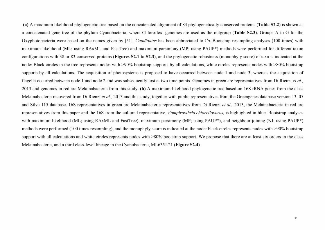
(a) A maximum likelihood phylogenetic tree based on the concatenated alignment of 83 phylogenetically conserved proteins (Table S2.2) is shown as
a concatenated gene tree of the phylum Cyanobacteria, where Chloroflexi genomes are used as the outgroup (Table S2.3). Groups A to G for the
Oxyphotobacteria were based on the names given by [51]. Candidatus has been abbreviated to Ca. Bootstrap resampling analyses (100 times) with
maximum likelihood (ML; using RAxML and FastTree) and maximum parsimony (MP; using PAUP*) methods were performed for different taxon
configurations with 38 or 83 conserved proteins (Figures S2.1 to S2.3), and the phylogenetic robustness (monophyly score) of taxa is indicated at the
node: Black circles in the tree represents nodes with >90% bootstrap supports by all calculations, white circles represents nodes with >80% bootstrap
supports by all calculations. The acquisition of photosystems is proposed to have occurred between node 1 and node 3, whereas the acquisition of
flagella occurred between node 1 and node 2 and was subsequently lost at two time points. Genomes in green are representatives from Di Rienzi et al.,
2013 and genomes in red are Melainabacteria from this study. (b) A maximum likelihood phylogenetic tree based on 16S rRNA genes from the class
Melainabacteria recovered from Di Rienzi et al., 2013 and this study, together with public representatives from the Greengenes database version 13_05
and Silva 115 database. 16S representatives in green are Melainabacteria representatives from Di Rienzi et al., 2013, the Melainabacteria in red are
representatives from this paper and the 16S from the cultured representative, Vampirovibrio chlorellavorus, is highlighted in blue. Bootstrap analyses
with maximum likelihood (ML; using RAxML and FastTree), maximum parsimony (MP; using PAUP*), and neighbour joining (NJ; using PAUP*)
methods were performed (100 times resampling), and the monophyly score is indicated at the node: black circles represents nodes with >90% bootstrap
support with all calculations and white circles represents nodes with >80% bootstrap support. We propose that there are at least six orders in the class
Melainabacteria, and a third class-level lineage in the Cyanobacteria, ML635J-21 (Figure S2.4).
44

Table 2.1. Summary statistics for population genomes belonging to the class Melainabacteria Population genome
# of scaffolds
Estimated genome size (Mbp)
%GC Number of genesa
rRNAsb Estimated completen-ess (%)c
Estimated contamin- ation (%)c
Proposed candidatus name
Study
Zag_221 14 1.8 38.5 1838 (1799) 16S 100.0 1.0 Gastranaerophilus phascolarctosicola
Present study
Zag_1 322 2.0 34.9 2194 (2160) - 94.3 1.9 Present study Zag_111 65 2.2 36.7 2313 (2257) 5S, 16S 23S 98.1 5.7 Present study MH_37 157 2.2 34.1 2402 (2360) - 100.0 1.0 Present study MEL_A1 1 1.9 33.0 1879 (1832) 5S, 16S, 23S 100.0 2.9 Di Rienzi et
al., 2013 MEL_B1 21 2.3 35.4 2269 (2219) 5S, 16S, 23S 100.0 1.0 Di Rienzi et
al., 2013 MEL_B2 26 2.3 36.3 2262 (2215) 5S, 16S, 23S 100.0 1.9 Di Rienzi et
al., 2013 MEL_C1 4 2.1 34.1 2162 (2120) 5S, 16S, 23S 100.0 1.9 Di Rienzi et
al., 2013 ACD20d 185 2.7 33.5 2455 (2325) 5S, 23S 100.0 2.9 Di Rienzi et
al., 2013 EBPR_351 8 5.5 49.4 4392 (4342) 5S, 16S, 23S 99.1 7.6 Obscuribacter phosphatis Present study UASB_169 67 1.8 27.5 1917 (1870) 16S, 23S 94.3 0.0 Caenarcanum
bioreactoricola Present study
aNumbers in brackets for number of genes is the number of protein coding genes. b16S rRNA lengths are >1,000 bp. cEstimated completeness and estimated contamination is based on the 105 single copy marker genes (a subset of the 111 single copy marker set from
Dupont et al., 2012). dACD20 is the corrected genome from Albertsen et al., (2013) as the original completeness for ACD20 was 100.0% and original contamination was 107.6%.
45

support for the monophyly of the Melainabacteria (Figure 2.1B and Figure S2.5) and indeed
several of the robustly monophyletic groups therein are classified as primary cyanobacterial lines of
descent (classes) in Greengenes and Silva [5, 6]. The higher resolution afforded by the genome
sequences suggests that these lineages should be classified as orders within the class
Melainabacteria (Figure 2.1). We propose names for four of these orders based on habitat and
analysis of the population genomes (see below), and the recognition of Vampirovibrio
chlorellavorus in the SM1D11 lineage (Figure 2.1). The location of the ACD20 genome could not
be determined within the 16S rRNA gene tree (Figure 2.1B) as it lacks a 16S rRNA gene sequence,
but genome trees based on a refined binning of this population [39] (Table 2.1) indicate that it is
basal and monophyletic with the order, Gastranaerophilales (YS2/4C0d2; Figure 2.1A and Figures
S2.2 and S2.3). An additional distantly related cyanobacterial lineage ML635J-21 [5, 6] currently
not represented by a sequenced genome may represent another class-level lineage within the
Cyanobacteria (Figure S2.4) highlighting the need for further genomic exploration of the
cyanobacterial phylum.
Our analyses show that the photosynthetic cyanobacteria are robustly monophyletic within the
expanded context of the phylum Cyanobacteria (node 3, Figure 2.1A). We therefore propose to
reinstigate the name Oxyphotobacteria [53] to describe all photosynthetic cyanobacteria (including
chloroplasts) in a single class. Gr. adj. oxus, acid or sour and in combined words indicating oxygen;
Gr. n. phos photos, light; Gr. n. baktêria, staff, cane; suff. -ia, ending to denote a class; N.L. neut.
pl. n. Oxyphotobacteria, light-requiring bacteria that produce oxygen. The name implies that the
class is able to photosynthesise. We denote the order-level groupings within this class as A to G
(Figure 2.1A) in accordance with a recent genome-based analysis [51]. Oxyphotobacteria are still
classified primarily on morphological grounds into five subsections [4] despite clear incongruencies
between phylogenetic reconstructions and morphological complexity [51]. Therefore, it is likely
that the order- and family-level groupings within the Oxyphotobacteria will be reclassified on
phylogenetic grounds with a concomitant widespread reclassification of cyanobacterial strains once
this group is no longer under the jurisdiction of the Botanical Code [54].
2.4.3 Inferred metabolism of Melainabacteria genomes
Di Rienzi et, al. inferred metabolic properties of the class Melainabacteria based on comparative
analysis of draft population genomes belonging to only one order, the Gastranaerophilales (Figure
2.1). We substantially increase the phylogenetic coverage of the Melainabacteria in the present
study by recovery of population genomes spanning three of the six identified orders (Figure 2.1B).
Expanded genomic representation should provide a more balanced overview of the metabolic
46

properties of this class including features in common with, or distinct from, the Oxyphotobacteria.
We begin by proposing Candidatus species for the most complete genomes in each of the three
orders obtained in this study and describe their inferred metabolic properties below.
The most complete Gastranaerophilales genome with the least number of scaffolds, Zag_221, was
selected as the Candidatus species representative of the group, Candidatus Gastranaerophilus
phascolarctosicola (Table 2.1 and Figure 2.1). Gastranaerophilus phascolarctosicola
(Gas.tra.nae.ro.phi’lus. Gr. n. gaster stomach; Gr. pref. an-, not; Gr. masc. n. aer, air; L. masc. adj.
philus [from Gr. adj.philos], friend, loving; Gastranaerophilus a bacterium loving anaerobic gastric
environments. ‘phas.co.larc.to.si.co.la’. N.L. Phascolarctos the name of koala; L. suffix -cola
inhabitant, dweller; N.L. masc. n. phascolarctosicola hiding in the belly of a koala). The genome
size and GC content range of the four Gastranaerophilales genomes were in accord with the Di
Rienzi et. al. population genomes from this order (Table 2.1). Members of this group have small
streamlined genomes ranging in size from 1.8 to 2.3 Mb, with the exception of ACD20, which is
2.7 Mb after binning refinement [39]. The Gastranaerophilales genomes recovered from the koala
and human faeces in the present study support the assertion [12] that this lineage comprises obligate
fermenters missing the genes necessary for aerobic and anaerobic respiration, as well as the
tricarboxylic acid (TCA) cycle (Figure 2.2 and Table S2.5). Instead, all Gastranaerophilales
genomes contain the Embden-Meyerhof-Parnas (EMP) pathway, capable of converting glucose,
mannose, starch or glycogen into lactate, ethanol and/or formate (Figure 2.2). All representative
genomes have the potential to produce riboflavin, nicotinamide, biotin, dehydrofolate and pantoate
as found previously [12].
Di Rienzi et. al. highlighted the presence of FeFe hydrogenases in their human gut population
genomes speculating that these organisms are hydrogen-producing anaerobes in syntrophic
interactions with hydrogenotrophic methanogens or acetogens. We also identified FeFe
hydrogenases in the MH_37 genome obtained from the human gut, but in contrast found Fe-only or
NiFe hydrogenases in koala gut Gastranaerophilales genomes (Figure S2.6). It is possible that the
less oxygen sensitive NiFe hydrogenase would allow members of this order to colonize the jejunum
as well as the more anaerobic colon [55].
Di Rienzi et. al. also reported that the Melainabacteria are flagellated based on the presence of a
complete set of flagella genes in three of their draft Gastranaerophilales genomes (MEL_B1,
MEL_B2 and ACD20). Of the nine Gastranaerophilales genomes available (Table 2.1), only these
three had complete flagella gene sets, the remainder having only a subset of genes that would not
47

encode a functional flagellum (Figure S2.7). We infer that flagella were present in the ancestor of
the class Melainabacteria and subsequently lost on at least two occasions based on monophyly of
flagella genes common to the Gastranaerophilales and Caenarcaniphilales (Figure 2.1B and Figure
S2.7). Moreover, there appears to have been a subsequent loss of functional flagella in ‘G.
phascolarctocola’ and relatives (Figure S2.7) indicating the presence of non-motile members of
this order in animal gut habitats.
Based on a suggested species threshold of 95% average nucleotide identity [56], three of the
Gastranaerophilus genomes can be considered to belong to the same species (Table 2.1 and Figure
S2.8). Two of these genomes were recovered from humans (MH37 and MEL_C1) and the third
from a koala (Zag_1) despite the structural and physiological differences between the human and
koala gut. The majority of genes that differ between these genomes are hypothetical proteins or
phage associated (Figure S2.8), typical of differences seen between strains belonging to the same
species.
Candidatus Obscuribacter phosphatis (Ob.scur.i.bac.ter. L. adj. obscurus dark; N.L. masc. n. bacter
(from Gr. n. baktron), a rod; Obscuribacter a bacterium found in the dark. ‘phos.pha.tis’. N.L. n.
phosphatis, phosphate; N.L. phosphatis accumulating phosphate) EBPR_351 (‘O. phosphatis’)
representing the order Obscuribacterales (Figure 2.1 and Table 2.1) is conspicuous among the
Melainabacteria genomes because of its larger size (5 Mb) and associated metabolic versatility. ‘O.
phosphatis’ contains the genes necessary for polyphosphate metabolism, including a low affinity
inorganic phosphate transporter (PiT), polyphosphate kinase 1 (used to synthesise or degrade polyP
while consuming or generating respectively ATP directly), polyphosphate kinase 2 (degrades polyP
producing GTP from GDP), exopolyphosphatase (degrades polyP in a non-reversible reaction that
does not generate ATP directly), polyphosphate:AMP phosphotransferase and adenylate kinase
(Seviour & Nielsen 2010). ‘O. phosphatis’ has the capacity for aerobic and anaerobic respiration,
and fermentation, allowing it to function during both the oxic and anoxic phases of EBPR [57]. It
contains genes encoding a complete respiratory chain including Complexes I, II, III and IV and an
F-Type ATPase (Figure 2.2). Di Rienzi et. al. concluded that the Melainabacteria lack electron
transport chains and are therefore incapable of respiration. This highlights the dangers of inferring
phylum- or class-level functionality based on limited phylogenetic sampling of the lineage.
Like the Gastranaerophilales, ‘O. phosphatis’ has the capability to metabolise a wide range of
simple carbohydrates via the EMP pathway, and also fatty acids via the beta-oxidation pathway
48

Figure 2.2. Metabolic reconstruction of Melainabacteria representatives
Metabolic predictions for Gastranaerophilales, Obscuribacterales and Caenarcaniphilales representatives based on genome annotations. Names of
pathways are italicised and fermentation products are shown in rectangles. Features identified in one or more of the orders are highlighted by colour.
Missing pathways are shown in grey. The Melainabacteria are missing photosystems I and II, and the Calvin Cycle, which are found in
Oxyphotobacteria. All Melainabacteria use the Embden-Mayerhoff-Parnas pathway to produce fermentation products, whereas the Obscuribacterales
representative is capable of using both aerobic and anaerobic respiration to produce energy.
Obscuribacterales
CaenarcanophilalesGastranaerophilales
PitH+ Mg2+ Pi
Trehalose
Oxaloacetate Citrate
Isocitrate
Oxoglutarate
SuccinylCoA
Succinate
Fumarate
Malate Glycoxylate
Pyruvate
Glycogen
F6P
G6P
CS
acn
IDH
OGDC
SCS
SDH
ICLmalS
Ethanol
Acetylaldehyde
ALDH
ADH
CalvinCycle
Starch
Mannose
Fructose
Glucose
F1,6P
GAP
3PGA
2PGA
PEP
Acetyl-CoA
Lactate
Formate
Pi
Polyphosphate
ppx
ATP
ADP
AMP
ppk1
pap adk
fumC
malDH
β-G6P G1,5LP 6GP
Ru5P
Ri5PX5P
GAP
S7PE4P
C16 C14 C12 C10 C8 C6 C4
Fatty Acid Synthesis
C16C14C12C10C8C6C4
Beta Oxidation
Fe Fe
Fe
Ni
Fe
H +H
2
TCA Cycle
EMP
Glyc
olys
is
Pentose PhosphatePathway
Fermentation
Polyphosphate Metabolism
H+ H2
H+ H2
FeFe hydrogenase
Fe only hydrogenase
H+ H2
Amino acid transporters Carbohydrate
ABC transporter
Sulfate permease
Na+/H+ dicarboxylate
symporter
Cation/multidrug efflux pump
Mg2+/Co2+ transporter
Heavy metal efflux pump
FeNi
Flagella
Photosynthesis
PSII
CytB6/f
PSI
NADH
NAD+
3H+
ADP + Pi
ATP
Complex I
Complex II
Complex IV
F-Type ATPase
UQH2
4H+
2H+
3H+
Electron Transport Chain
NO3 -
NO2 -
NAR
NO3 -
NO2 -
2H+
MQ+
MQH2
NiFe hydrogenase I
NiFe hydrogenase III
UQ
2 x 2H+
cytcComplex III
49

(Figure 2.2). Under oxic conditions, we predict that ‘O. phosphatis’ will fully oxidise one or more
of these substrates via the TCA cycle, feeding NADH into the electron transport chain with a cbb3-
type cytochrome as the terminal oxidase. This family of cytochromes is typically used in
microaerophilic conditions [58] suggesting that ‘O. phosphatis’ may be found within flocs where
oxygen concentrations are lower [39]. Under anoxic conditions, we predict that it performs either
respiration with nitrate as the terminal electron acceptor or, in the absence of nitrate, mixed-acid
fermentation with the potential to produce ethanol, lactate, formate, succinate, CO2 and H2 (Figure
2.2). The presence of these metabolic pathways suggests that ‘O. phosphatis’ has adapted to more
dynamic environments (requiring greater metabolic plasticity) with ‘feast-famine’ nutrient cycles
such as those artificially imposed on EBPR bioreactors.
Candidatus Caenarcanum bioreactoricola (‘Caen.arc.an.um’ L. neut. n. caenum mud, sludge; L.
neut. n. arcanum secret, hidden; N.L. neut. n. Caenarcanum a bacterium hidden in sludge.
‘bio.re.ac.to.ri.co.la’. L. suffix -cola inhabitant, dweller; N.L. masc. n. bioreactericola living in a
bioreactor) UASB_169 (‘C. bioreactoricola’) representing the order Caenarcaniphilales, has an
estimated genome size of ~2 Mb and a remarkably low GC content of 27.7%, the lowest GC
content reported for Cyanobacteria. Similar to the Gastranaerophilales genomes, ‘C.
bioreactoricola’ lacks the genes necessary for aerobic and anaerobic respiration, as well as the TCA
cycle, suggesting that ‘C. bioreactoricola’ has a streamlined metabolism only producing energy via
fermentation with ethanol and lactate as the main fermentation products. ‘C. bioreactoricola’
contains the subunits for Fe-only hydrogenase and the potential to produce hydrogen as a by-
product from the fermentation process. Like the Gastranaerophilales, ‘C. bioreactoricola’ may also
be a hydrogen producer living in syntrophy with methanogens or acetogens in the bioreactor, as
microcolonies of syntrophic bacteria are often observed in the granules from UASB systems and
electron transfer in these microcolonies is thought to mostly occur through interspecies hydrogen
transfer [59].
2.4.4 Emergence of photosynthesis in the Cyanobacteria
Melainabacteria resemble Oxyphotobacteria in their cell envelope gene complement comprising
genes indicative of a Gram-negative (diderm) cell wall (Figure 2.3). This includes genes for the
biosynthesis of Lipid A for the production of lipopolysaccharide (LPS) as previously reported for
members of the Gastranaerophilales [12]. Oxyphotobacteria also have unusual cell envelope
components for Gram-negative bacteria including porins (somA, somB), which are thought to help
anchor the outer membrane to the peptidoglycan layer [60, 61]. All Melainabacteria have closely
50

related homologs to the oxyphotobacterial somA and somB genes suggesting that their cell
envelopes comprise similar porins.
Di Rienzi et. al. highlighted the presence of putative circadian rhythm (rpaA and rpaB) and light
response (nblS) regulators in the Gastranaerophilales, which are diagnostic of Cyanobacteria. We
identified orthologs of these genes in all three orders of the Melainabacteria suggesting that these
are uniquely ancestral features of the phylum. Together with the unambiguous phylogenetic
placement of the Melainabacteria within the cyanobacterial radiation based on comparative analysis
of highly conserved marker genes (Figure 2.1), the conservation of features characteristic of
Oxyphotobacteria in the Melainabacteria further support a common ancestry and the proposal for a
single phylum.
The most conspicuous difference between the Melainabacteria and Oxyphotobacteria is the absence
of chlorophyll biosynthesis genes in the former (Figure 2.3) consistent with previous findings [12].
All subunits for photosystems I and II, and the electron transport chain were absent from the
Melainabacteria genomes sequenced in this study. Another trait characteristic of photosynthetic
cyanobacteria, carbon fixation, is similarly absent in the Melainabacteria (Figure 2.2) indicating
that these organisms do not engage in a photoautotrophic lifestyle. Instead, members of this lineage
appear to be chemoheterotrophs with diverse functionality.
The idea of non-photosynthetic Cyanobacteria is contrary to the prevailing dogma that all members
of this phylum are photosynthetic [51]. However, this should not be a controversial conclusion
given that photosynthesis is only found in sub-lineages of several other phyla such as the
Proteobacteria, Firmicutes, Acidobacteria and Chloroflexi [62]. The complete absence of
photosynthetic apparatus in the Melainabacteria suggests that the Oxyphotobacteria acquired
photosystems after diverging from the common ancestor of the Melainabacteria (Figure 2.1). This
is consistent with the inferences that photosynthesis genes have an extensive history of lateral
transfer [2] and that photosynthesis developed late in the Cyanobacteria [63].
The acquisition of oxygenic photosynthesis in the Oxyphotobacteria had profound impact not only
on the biosphere [2] but left imprints in their genomes that are now apparent by contrasting with
their newly sequenced non-photosynthetic relatives. For example, there was a great expansion of
ATP-driven transport systems in the Oxyphotobacteria likely for acquiring bicarbonate
(COG0600/0715/1116) and iron (COG0609/1629/0735) necessary for photosynthesis and
respiration (Figure S2.9). The additional energy available to Oxyphotobacteria via oxygenic
51

Figure 2.3. Distribution of key traits across the Cyanobacteria and other bacteria phyla
Color Key
0.20 0.6 1
Cell envelope Flagella assembly (Bacterio)chlorophyll biosynthesis
LPS Cell envelope related Other
Glyc
os_tr
ansf_
NLp
xKLp
xBLp
xCLp
xDEx
pDSu
rf_Ag
_VNR
YjgP
_YjgQ
Bac_
surfa
ce_A
gSe
cretin OEP
OstA
TonB
_dep
_Rec
TonB
_C Plug
PdxA
LptC
PdxJ
LolA
TolB
_NOs
tA_C
DegV
DivI
VAHT
H_W
hiAW
hiA_N
DUF5
52So
rtase
LytR
_cps
A_ps
rDU
F196
7R3
H
(B)C
hlH(B
)ChlD
(B)C
hlI(B
)ChlM
(B)C
hlE acsF
bciA
bciB
LPOR
(B)C
hlN
MotA
_Exb
BM
otB_p
lug
FliG
_C
FliM
SpoA
FlgH FlgI
FHIP
EPBa
c_ex
port_
2
FliP
Bac_
expo
rt_3
Bac_
expo
rt_1
FliD
_N/F
liD_C
FlgD
Flag
ellin_
N/Fl
agell
in_C
YscJ
_FliF
/Ysc
J_Fl
iF_C
Flg_
bb_r
od/F
lg_bb
r_C
FliS
FliE
FliH FliJ
M
M
M
D
M
D(A)?
DA
DA
DA
DA
D(L)
DL
DL
DL
DL
DL
DL
DL
DL
Tenericutes
Firmicutes
Actinobacteria
Thermi
Chloroflexi
Melainabacteria
Oxyphotobacteria
Thermotogae
Synergistetes
Fusobacteria
Spirochaetes
Chlamydiae
Verrucomicrobia
Planctomycetes
Bacteroidetes
Chlorobi
Aquificae
Acidobacteria
Proteobacteria
0.10 (B)C
hlB(B
)ChlL
>90% (MLx2/MP)
52

A maximum likelihood genome tree of the bacterial domain constructed using a concatenated alignment of 38 conserved proteins is shown at the left of
the figure for phylogenetic ordering of traits shown in the heat map to the right. Black circles on interior nodes represent affiliations with >90%
bootstrap support. Columns in the heat map represent individual gene families (Pfams or GIs; Table S2.6) grouped into three subsystems of interest;
cell envelope, flagella and (bacterio)chlorophyll biosynthesis. Increasing representation of each gene family in a given phylum (percentage of
genomes) is shown by increasing depth of color. Cell envelope classification is indicated by the abbreviations to the right of the phylum names:
Monoderm (M), Diderm (D), Diderm-LPS (DL), Diderm-Atypical (DA) from Albertsen et al., 2013. Note that it is not possible to determine if the
Melainabacteria are diderms or atypical diderms based on sequence data only.
53

photosynthesis may also explain the widespread acquisition of energy-intensive biosynthetic
pathways such as secondary metabolite synthesis [51] (Figure S2.10).
2.5 Conclusion Our findings expand the recognised phylogenomic boundaries of the phylum Cyanobacteria. We
infer that the cyanobacterial ancestor was a non-photosynthetic chemoheterotroph and that
photosystems were acquired after divergence of the classes Melainabacteria and Oxyphotobacteria.
We suggest that the acquisition of oxygenic photosynthesis resulted in an increase in genome
complexity within the Oxyphotobacteria (followed by a subsequent reduction and streamlining in
the Prochlorococcus lineage; [64]) while the Melainabacteria mostly retained a simpler ancestral
metabolism. Consistent with the phylogenetic depth of the Melainabacteria, members of this class
occupy a wide range of environmental niches with varied metabolic properties, mostly centred
around fermentative lifestyles, that nonetheless extend the known metabolic diversity of the
Cyanobacteria. These include respiratory nitrate reduction (‘O. phosphatis’) and flagella-based
motility (in some Gastranaerophilales [12]). If the inclusion of Vampirovibrio chlorellavorus [65] in
the Melainabacteria is confirmed by genomic sequencing, then parasitism can also be added to the
known phenotypes of Cyanobacteria. The availability of eleven high quality draft genomes
representing multiple orders within the Melainabacteria (Table 2.1) provides a sound basis for
further investigations into this fascinating group, for example, via spatial visualisation [66] and
genome-directed isolation [67, 68].
2.6 Acknowledgements We thank Norman Pace, Tal Dagan and Michael Galperin for providing valuable perspective on the
study and Karen Nilsson and Jacqui Brumm at Lone Pine Koala Sanctuary for facilitating collection
of koala faecal samples, Adam Skarshewski for implementing the concatenation of single copy
marker genes, Serene Low and Margaret Butler for preparing samples for Illumina sequencing,
Fiona May for 454 pyrotags, Queensland Centre for Medical Genomics, UQ and Aalborg
University for Illumina paired end and mate pair sequencing. We also thank Akiko Ohashi and
Satoko Matsukura at AIST for Illumina shotgun and 16S amplicon sequencing. We thank Tim
Lilburn for information on the ATCC strain of Vampirovibrio chlorellavorus. We thank Satoshi
Hanada and Aharon Oren for etymological advice and clarification of nomenclature versus
classification issues (we are classifying here). This work is supported by the Australian Research
Council (ARC) through project DP120103498, strategic funds from the Australian Centre for
Ecogenomics; G.W.T. is supported by an ARC Queen Elizabeth II fellowship [DP1093175]; R.M.S
and C.T.S is supported by an Australian Postgraduate Award (APA).
54

2.7 References 1. Nelson, N. and A. Ben-Shem, The complex architecture of oxygenic photosynthesis. Nature
Reviews Molecular Cell Biology, 2004. 5(12): p. 971-982.
2. Hohmann-Marriott, M.F. and R.E. Blankenship, Evolution of Photosynthesis. Annual
Review of Plant Biology, 2011. 62(1): p. 515-548.
3. Dojka, M.A., J.K. Harris, and N.R. Pace, Expanding the Known Diversity and
Environmental Distribution of an Uncultured Phylogenetic Division of Bacteria. Applied
and Environmental Microbiology, 2000. 66(4): p. 1617-1621.
4. Rippka, R., et al., Generic Assignments, Strain Histories and Properties of Pure Cultures of
Cyanobacteria. Journal of General Microbiology, 1979. 111(1): p. 1-61.
5. McDonald, D., et al., An improved Greengenes taxonomy with explicit ranks for ecological
and evolutionary analyses of bacteria and archaea. ISME J, 2012. 6(3): p. 610-8.
6. Quast, C., et al., The SILVA ribosomal RNA gene database project: improved data
processing and web-based tools. Nucleic Acids Research, 2013. 41(D1): p. D590-D596.
7. Williams, M.M., et al., Phylogenetic diversity of drinking water bacteria in a distribution
system simulator. Journal of Applied Microbiology, 2004. 96(5): p. 954-964.
8. Cruz-Martinez, K., et al., Despite strong seasonal responses, soil microbial consortia are
more resilient to long-term changes in rainfall than overlying grassland. ISME J, 2009. 3(6):
p. 738-44.
9. Liu, B., et al., Thauera and Azoarcus as functionally important genera in a denitrifying
quinoline-removal bioreactor as revealed by microbial community structure comparison.
FEMS Microbiology Ecology, 2006. 55(2): p. 274-86.
10. Ley, R.E., et al., Microbial ecology: Human gut microbes associated with obesity. Nature,
2006. 444(7122): p. 1022-1023.
11. Gromov, B. and K. Mamkaeva, Proposal of a new genus Vampirovibrio for chlorellavorus
bacteria previously assigned to Bdellovibrio. Mikrobiologia, 1980. 49: p. 165-167.
12. Di Rienzi, S.C., et al., The human gut and groundwater harbor non-photosynthetic bacteria
belonging to a new candidate phylum sibling to Cyanobacteria. eLife, 2013. 2: p. e01102.
13. Lu, H., et al., Obtaining highly enriched cultures of Candidatus Accumulibacter phosphates
through alternating carbon sources. Water Research, 2006. 40(20): p. 3838-48.
14. Matsuki, T., et al., Development of 16S rRNA-Gene-Targeted Group-Specific Primers for
the Detection and Identification of Predominant Bacteria in Human Feces. Applied and
Environmental Microbiology, 2002. 68(11): p. 5445-5451.
15. Caporaso, J.G., et al., QIIME allows analysis of high-throughput community sequencing
data. Nature Methods, 2010. 7(5): p. 335-336.
55

16. Bragg, L., et al., Fast, accurate error-correction of amplicon pyrosequences using Acacia.
Nature Methods, 2012. 9(5): p. 425-426.
17. Johnson, M., et al., NCBI BLAST: a better web interface. Nucleic Acids Research, 2008.
36: p. W5-9.
18. DeSantis, T.Z., et al., Greengenes, a chimera-checked 16S rRNA gene database and
workbench compatible with ARB. Applied and Environmental Microbiology, 2006. 72(7):
p. 5069-72.
19. Caporaso, J.G., et al., Ultra-high-throughput microbial community analysis on the Illumina
HiSeq and MiSeq platforms. ISME J, 2012. 6(8): p. 1621-1624.
20. Camacho, C., et al., BLAST+: architecture and applications. BMC Bioinformatics, 2009.
10(1): p. 421.
21. Qin, J., et al., A human gut microbial gene catalogue established by metagenomic
sequencing. Nature, 2010. 464(7285): p. 59-65.
22. Imelfort, M., et al., GroopM: an automated tool for the recovery of population genomes
from related metagenomes. PeerJ, 2014. 2: p. e603.
23. Dupont, C.L., et al., Genomic insights to SAR86, an abundant and uncultivated marine
bacterial lineage. ISME J, 2012. 6(6): p. 1186-99.
24. Finn, R.D., J. Clements, and S.R. Eddy, HMMER web server: interactive sequence
similarity searching. Nucleic Acids Research, 2011. 39: p. W29-37.
25. Punta, M., et al., The Pfam protein families database. Nucleic Acids Research, 2012.
40(D1): p. D290-D301.
26. Haft, D.H., J.D. Selengut, and O. White, The TIGRFAMs database of protein families.
Nucleic Acids Research, 2003. 31(1): p. 371-3.
27. Parks, D.H., et al., CheckM: assessing the quality of microbial genomes recovered from
isolates, single cells, and metagenomes. PeerJ PrePrints, 2014. 2: p. e554v1.
28. Boetzer, M., et al., Scaffolding pre-assembled contigs using SSPACE. Bioinformatics, 2011.
27(4): p. 578-9.
29. Li, H. and R. Durbin, Fast and accurate long-read alignment with Burrows-Wheeler
transform. Bioinformatics, 2010. 26(5): p. 589-95.
30. Ludwig, W., et al., ARB: a software environment for sequence data. Nucleic Acids
Research, 2004. 32(4): p. 1363-1371.
31. Swofford, D., PAUP*. Phylogenetic Analysis Using Parsimony (* and Other Methods).
Version 4. 2003.
32. Darling, A.E., et al., PhyloSift: phylogenetic analysis of genomes and metagenomes. PeerJ,
2014. 2: p. e243.
56

33. Felsenstein, J., PHYLIP - Phylogeny Inference Package (Version 3.2). Cladistics, 1989. 5:
p. 164-166.
34. Markowitz, V.M., et al., IMG: the integrated microbial genomes database and comparative
analysis system. Nucleic Acids Research, 2012. 40(D1): p. D115-D122.
35. Price, M.N., P.S. Dehal, and A.P. Arkin, FastTree 2 – Approximately Maximum-Likelihood
Trees for Large Alignments. PLoS ONE, 2010. 5(3): p. e9490.
36. Castresana, J., Selection of conserved blocks from multiple alignments for their use in
phylogenetic analysis. Molecular Biology and Evolution, 2000. 17(4): p. 540-52.
37. Stamatakis, A., RAxML-VI-HPC: maximum likelihood-based phylogenetic analyses with
thousands of taxa and mixed models. Bioinformatics, 2006. 22(21): p. 2688-90.
38. Markowitz, V.M., et al., IMG ER: a system for microbial genome annotation expert review
and curation. Bioinformatics, 2009. 25(17): p. 2271-2278.
39. Albertsen, M., et al., Genome sequences of rare, uncultured bacteria obtained by differential
coverage binning of multiple metagenomes. Nature Biotechnology, 2013. 31(6): p. 533-538.
40. Pallen, M.J. and N.J. Matzke, From The Origin of Species to the origin of bacterial flagella.
Nature Reviews Microbiology, 2006. 4(10): p. 784-790.
41. Sousa, F.L., et al., Chlorophyll biosynthesis gene evolution indicates photosystem
gene duplication, not photosystem merger, at the origin of oxygenic photosynthesis.
Genome Biology and Evolution, 2013. 5(1): p. 200-216.
42. Altschul, S.F., et al., Gapped BLAST and PSI-BLAST: a new generation of protein database
search programs. Nucleic Acids Research, 1997. 25(17): p. 3389-402.
43. Racine, J.S., RStudio: A Platform-Independent IDE for R and Sweave. Journal of Applied
Econometrics, 2012. 27(1): p. 167-172.
44. Warnes, G.R., et al., gplots: Various R programming tools for plotting data. 2013.
45. Neuwirth, E., RColorBrewer: ColorBrewer palettes. 2011.
46. Hyatt, D., et al., Prodigal: prokaryotic gene recognition and translation initiation site
identification. BMC Bioinformatics, 2010. 11(1): p. 119.
47. Tatusov, R.L., et al., The COG database: an updated version includes eukaryotes. BMC
Bioinformatics, 2003. 4: p. 41.
48. Parks, D.H., et al., STAMP: statistical analysis of taxonomic and functional profiles.
Bioinformatics, 2014. 30(21): p. 3123-4.
49. Sharon, I., et al., Time series community genomics analysis reveals rapid shifts in bacterial
species, strains, and phage during infant gut colonization. Genome Research, 2013. 23(1): p.
111-20.
57

50. Hugenholtz, P., B.M. Goebel, and N.R. Pace, Impact of culture-independent studies on the
emerging phylogenetic view of bacterial diversity. Journal of Bacteriology, 1998. 180(18):
p. 4765-74.
51. Shih, P.M., et al., Improving the coverage of the cyanobacterial phylum using diversity-
driven genome sequencing. Proceedings of the National Academy of Sciences of the United
States of America, 2013. 110(3): p. 1053-8.
52. Wagner, M. and M. Horn, The Planctomycetes, Verrucomicrobia, Chlamydiae and sister
phyla comprise a superphylum with biotechnological and medical relevance. Current
Opinion in Biotechnology, 2006. 17(3): p. 241-9.
53. Gibbons, N.E. and R.G.E. Murray, Validation of Cyanobacteriales Stanier in Gibbons and
Murray 1978 as a New Order of the Kingdom Procaryotae Murray 1968, and of the Use of
Neuter Plural Endings for Photobacteria and Scotobacteria classes nov. Gibbons and
Murray 1978: Request for an Opinion. International Journal of Systematic Bacteriology,
1978. 28(2): p. 332-333.
54. Komárek, J., Recent changes (2008) in cyanobacteria taxonomy based on a combination of
molecular background with phenotype and ecological consequences (genus and species
concept). Hydrobiologia, 2010. 639(1): p. 245-259.
55. Quigley, E.M. and R. Quera, Small intestinal bacterial overgrowth: roles of antibiotics,
prebiotics, and probiotics. Gastroenterology, 2006. 130: p. S78-90.
56. Goris, J., et al., DNA-DNA hybridization values and their relationship to whole-genome
sequence similarities. International Journal of Systematic and Evolutionary Microbiology,
2007. 57: p. 81-91.
57. Blackall, L.L., et al., A review and update of the microbiology of enhanced biological
phosphorus removal in wastewater treatment plants. Antonie Van Leeuwenhoek, 2002.
81(1-4): p. 681-91.
58. Kulajta, C., et al., Multi-step assembly pathway of the cbb3-type cytochrome c oxidase
complex. Journal of Molecular Biology, 2006. 355(5): p. 989-1004.
59. Schmidt, J.E. and B.K. Ahring, Granular sludge formation in upflow anaerobic sludge
blanket (UASB) reactors. Biotechnology and Bioengineering, 1996. 49(3): p. 229-46.
60. Hansel, A., et al., Cloning and characterization of the genes coding for two porins in the
unicellular cyanobacterium Synechococcus PCC 6301. Biochimica et Biophysica Acta,
1998. 1399(1): p. 31-9.
61. Hoiczyk, E. and A. Hansel, Cyanobacterial Cell Walls: News from an Unusual Prokaryotic
Envelope. Journal of Bacteriology, 2000. 182(5): p. 1191-1199.
58

62. Bryant, D.A. and N.U. Frigaard, Prokaryotic photosynthesis and phototrophy illuminated.
Trends in Microbiology, 2006. 14(11): p. 488-96.
63. Xiong, J., et al., Molecular Evidence for the Early Evolution of Photosynthesis. Science,
2000. 289(5485): p. 1724-1730.
64. Partensky, F. and L. Garczarek, Prochlorococcus: advantages and limits of minimalism.
Annual Review of Marine Science, 2010. 2: p. 305-31.
65. Coder, D.M. and L.J. Goff, The host range of the Chlorellavorus bacterium ("Vampirovibrio
chlorellavorus"). Journal of Phycology, 1986. 22(4): p. 543-546.
66. Moter, A. and U.B. Göbel, Fluorescence in situ hybridization (FISH) for direct visualization
of microorganisms. Journal of Microbiological Methods, 2000. 41(2): p. 85-112.
67. Pope, P.B., et al., Isolation of Succinivibrionaceae implicated in low methane emissions
from Tammar wallabies. Science, 2011. 333(6042): p. 646-8.
68. Tyson, G.W., et al., Genome-directed isolation of the key nitrogen fixer Leptospirillum
ferrodiazotrophum sp. nov. from an acidophilic microbial community. Applied and
Environmental Microbiology, 2005. 71(10): p. 6319-24.
59

60

Chapter 3: Back from the dead; the curious tale of the predatory
cyanobacterium Vampirovibrio chlorellavorus
Rochelle M. Soo1, Ben J. Woodcroft1, Donovan H. Parks1, Gene W. Tyson1,2 and Philip
Hugenholtz1,3* 1Australian Centre for Ecogenomics, School of Chemistry and Molecular Biosciences, The
University of Queensland, St Lucia, QLD 4072, Australia
2Advanced Water Management Centre, The University of Queensland, St Lucia, QLD 4072,
Australia
3Institute for Molecular Bioscience, The University of Queensland, St Lucia, QLD 4072, Australia
3.1 Abstract An uncultured non-photosynthetic basal lineage of the Cyanobacteria, the Melainabacteria, was
recently characterised by metagenomic analyses of aphotic environmental samples. However, a
predatory bacterium, Vampirovibrio chlorellavorus, originally described in 1972 appears to be the
first cultured representative of the Melainabacteria based on a 16S rRNA sequence recovered from a
lyophilised co-culture of the organism. Here, we sequenced the genome of V. chlorellavorus directly
from 36 year-old lyophilised material that could not be resuscitated confirming its identity as a
member of the Melainabacteria. We identified attributes in the genome that likely allow V.
chlorellavorus to function as an obligate predator of the microalga Chlorella vulgaris, and predict
that it is the first described predator to use an Agrobacterium tumefaciens-like conjugative type IV
secretion system to invade its host. V. chlorellavorus is the first cyanobacterium recognised to have a
predatory lifestyle and further supports the assertion that Melainabacteria are non-photosynthetic.
3.2 Introduction Predatory microorganisms attack and digest their prey, which can be either bacteria or microbial
eukaryotes [1, 2]. They have been found in a range of environments, including terrestrial, freshwater,
estuaries, oceans, sewages and animal faeces [3]. Microbial predators have been classified as obligate
(unable to grow in the absence of prey) or facultative (able to grow as a pure culture without the
presence of prey). In addition they can be periplasmic (penetrate and attach to the inner membrane),
epibiotic (attach to the outside), endobiotic (penetrate the cytoplasm) or wolf-pack (swarming as a
61

‘wolf-pack’ towards prey, which they kill and degrade) [4, 5]. To date, four bacterial phyla harbour
microbial predators; the Proteobacteria, Actinobacteria, Bacteroidetes and Chloroflexi [2, 6, 7].
In 1972, Gromov and Mamkaeva first described the predatory nature of Bdellovibrio chlorellavorus
towards the microalgae Chlorella vulgaris in a Ukrainian freshwater reservoir [8]. They reported that
co-inoculation of the alga and bacterium resulted in clumping and colour change of algal cells,
formation of refractile bodies and finally algal cell death. However, unlike other Bdellovibrio species
that invade the periplasm of Gram-negative bacteria, B. chlorellavorus only attached to the surface of
C. vulgaris, producing peripheral vacuoles in the alga followed by a gradual dissolution of the
infected cell contents [9]. This distinct mode of predation called into question the classification of B.
chlorellavorus as a Bdellovibrio [1] resulting in its reclassification as Vampirovibrio chlorellavorus
in 1980, although its higher level assignment to the Deltaproteobacteria was retained [10].
Co-cultures of V. chlorellavorus and C. vulgaris were deposited in three culture collections in 1978
[1]. However, to the best of our knowledge there are no reports of successful resuscitation of the
organism from lyophilised material. The only subsequent studies of V. chlorellavorus were based on
co-cultures obtained directly from the investigators who originally enriched the bacterium [9, 11].
The American Type Culture Collection (ATCC) was able to successfully extract DNA from one of
the 32 year-old lyophilised co-cultures and sequence the 16S rRNA gene of V. chlorellavorus
(Genbank acc. no. HM038000). Comparative analyses of this sequence indicate that V.
chlorellavorus is actually a member of the phylum Cyanobacteria rather than the Proteobacteria
according to the Greengenes [12] and Silva [13] taxonomies. This may explain why the culture could
not be revived as Cyanobacteria are notoriously difficult to resuscitate from lyophilised material [14].
More specifically, V. chlorellavorus is a member of a recently described basal lineage of non-
photosynthetic Cyanobacteria, the class Melainabacteria ([15], originally classified as a separate
phylum [16]). Here, we report the near-complete genome of V. chlorellavorus sequenced directly
from a 36-year-old vial of co-cultured lyophilised cells, confirm its phylogenetic position in the
Cyanobacteria, and infer the molecular underpinnings of its predatory life cycle.
3.3 Materials and Methods 3.3.1 Sample collection
Co-cultured Vampirovibrio chlorellavorus and Chlorella vulgaris (NCIB 11383) (deposited in 1978
by Coder and Starr) were obtained as lyophilised cells from the National Collections of Industrial,
Food and Marine Bacteria (NCIMB), Aberdeen, Scotland.
62

3.3.2 Genomic DNA extraction
Genomic DNA (gDNA) was extracted from lyophilised cells using a MoBio Soil Extraction kit
(MoBio Laboratories, Carlsbad, CA). gDNA was quantified using a Qubit 2.0 fluorometer (Life
technologies). 1ng of the gDNA was used to construct a paired-end library with the Illumina Nextera
XT DNA Sample Preparation kit according to protocol but with double size selection to obtain an
insert size of 300-800 bp [17]. The library was sequenced on an Illumina Miseq system using the
Miseq Reagent Kit v3 at the Institute of Molecular Bioscience, University of Queensland.
3.3.3 Genome assembly, completeness and contamination
Sequencing reads were processed with FastQC to check for quality
(http://www.bioinformatics.babraham.ac.uk/projects/fastqc/) and Illumina Nextera adaptors were
removed using FASTX-Toolkit (http://hannonlab.cshl.edu/fastx_toolkit/). Reads were parsed through
GraftM (https://github.com/geronimp/graftM) version r2439db using the May, 2013 version of the
Greengenes database 97% OTUs (operational taxonomic units) as a reference [12] to identify those
containing parts of 16S or 18S rRNA genes using default parameters. The 5’ end of all reads was
trimmed (~20bp) to remove low-quality sequence and paired reads were assembled into contigs with
a kmer size of 63 using CLC Genomics Workbench v7.0 (CLC bio). The statistical package R with
ggplot2 (https://github.com/hadley/ggplot2) was used to plot GC content against coverage allowing
contigs belonging to the V. chlorellavorus genome to be identified. A discrete cluster of contigs with
>180x coverage and a GC range of 42-54% were identified as belonging to V. chlorellavorus, while
contigs with <180x coverage were assigned to C. vulgaris (Figure S3.1). BLASTN [18] (v2.2.29+)
using default settings was used to verify that contigs with >180x coverage had homology to bacterial
sequences with NCBI’s non-redundant database. Additionally, the 16S rRNA gene was identified
using Prokka v1.8 [19] and a BLASTN search was used to identify the closest neighbour in the May,
2013 version of the Greengenes database [12]. The completeness and contamination of the genome
belonging to V. chlorellavorus was examined using CheckM v0.9.5 [20] with a set of 104 conserved
bacterial single-copy marker genes [15]. IslandViewer was used to identify genomic islands [21] with
the SIGI-HMM programme [22].
Plasmids were identified using the ‘roundup’ mode of FinishM git version 5664703
(https://github.com/wwood/finishm), using raw reads as input, a kmer length of 51bp and a coverage
cutoff of 15. A combination of manual inspection of the assembly graph generated using the
‘visualise’ mode and automated assembly with the ‘assemble’ mode confirmed that the contig ends
unambiguously joined together (i.e. they joined together and to no other contig ends) and that the two
plasmid contigs originally assembled with CLC were otherwise free of mis-assemblies. Plasmids
63

were also confirmed by the annotation of multiple transfer (tra) genes by the Integrated Microbial
Genomics Expert Review (IMG/ER) system (see below).
3.3.4 Genome annotation
The V. chlorellavorus genome was submitted to IMG/ER for annotation [23]. The V. chlorellavorus
genome has been deposited at JGI [JGI IMG-ER:2600254900]. The genome was also annotated with
Prokka v.1.8 [19] and the Uniref 90 database [24]. KEGG maps [25] and gene annotations were used
to reconstruct the metabolism of the V. chlorellavorus genome. Individual genes that were annotated
as ‘hypothetical protein’ or had been potentially misannotated based on the annotation of surrounding
genes were further explored through BLASTN searches against the NCBI-nr database. A metabolic
cartoon was prepared in Adobe Illustrator CS6.
The methyl-accepting chemotaxis proteins identified by IMG-ER were submitted to InterProScan5
[26] to determine chemotaxis protein domains. Putative genes were annotated with the dbCAN web
server [27] to identify glycoside hydrolases and checked against the IMG annotations and BLAST
results. The MEROPS server [28] was used to identify putative peptidases in V. chlorellavorus using
batch BLAST.
A Genbank file for V. chlorellavorus was generated through the xBASE website [29]. The ribosomal
proteins, chaperones and transcriptional and translational proteins of V. chlorellavorus were used as
representatives of recognised highly expressed genes to identify other putatively highly expressed
genes in the genome using PHX (predicted highly expressed) analysis using the standard genetic
code (http://www.cmbl.uga.edu/software/phxpa.html; [30, 31]. Putatively horizontally transferred
(alien) genes were identified by their atypical codon usage from the genome average also using PHX
analysis.
3.3.5 Phylogenetic tree
A bacterial genome tree was inferred in order to establish the phylogenetic relationship of the V.
chlorellavorus genome. A set of 5,449 bacterial genomes previously identified as being of
exceptional quality were used to establish a set of bacterial marker genes suitable for phylogenetic
inference [20]. An initial set of 178 single copy genes present exactly once in >90% of the trusted
genomes (found in >90% of the genomes) was identified using the PFAM [32] and TIGRFAM [33]
annotations provided by the Integrated Microbial Genomes (IMG; [34]). The same protein family
may be represented in both PFAM and TIGRFAM. Families from these two databases were
considered redundant if they matched the same genes in >90% of the trusted genomes, in which case
64

preference was given to the TIGRFAM families. Genes present multiple times within a genome were
considered to have congruent phylogenetic histories if all copies of the gene were situated within a
single conspecific clade within its gene tree. From the 178 initial genes, 69 were removed from
consideration as they exhibited divergent phylogenetic histories in >1% of the trusted genomes
(Table S3.1). The remaining 109 genes were identified across an expanded set of 7732 bacterial
genomes, including all known Melainabacteria genomes along with an outgroup of 169 archaeal
genomes using Prodigal v2.60 [35] to identify call genes and HMMER v3.1b1
(http://hmmer.janelia.org) to assign genes to PFAM and TIGRFAM families. Gene assignment was
performed using model specific cutoff values for both the PFAM (-cut_gc) and TIGRFAM (-cut_tc)
HMMs. For both the individual gene trees and concatenated genome tree, genes were aligned with
HMMER v3.1b1 and phylogenetic inference performed with FastTree v2.1.7 [36] under the
WAG+GAMMA model. Support values for the bacterial genome tree were determined by applying
FastTree to 100 bootstrapped replicates [37]. The 16S rRNA gene tree was constructed as previously
described [15]. Briefly, the 16S rRNA gene from V. chlorellavorus was aligned to the standard
Greengenes alignment with PyNAST [12]. Aligned sequences and a Greengenes reference alignment,
version gg_13_5 were imported into ARB and the V. chlorellavorus sequence alignment was
corrected using the ARB EDIT tool. Representative taxa (>1,300 nt) were selected for constructing
the alignments, which were exported from ARB [38] with Lane mask filtering. Neighbour joining
trees were calculated from the mask alingments with LogDet distance estimation using PAUP*4.0
[39] with 100 bootstrap replicates. Maximum parsimony trees were calculated using PAUP*4.0 [39]
with 100 bootstrap replicates. Maximum likelihood trees were calculated from the masked
alignments using the Generalised Time-Reversible model with Gamma and I options in RAxML
version 7.7.8 [40] (raxmlHPC-PTHREADS -f a -k -x 12345 -p 12345 -N 100 -T 4 -m
GTRGAMMAI). Bootstrap resampling data (100 replicates) were generated with SEQBOOT in the
phylip package [41] and used for 100 bootstrap resamplings. Generated trees were re-imported into
ARB for visualisation.
3.3.6 Phylogenetic trees for virB4 and fliI genes
VirB4 sequences were obtained from Guglielmini et al. 2012. The phylip file
(figure3_mafft_alignment.phy) obtained from the DRYAD database was converted to an HMM using
HMMer v3.1b1 (http://hmmer.janelia.org) and the VirB4 sequences from V. chlorellavorus was
aligned to the HMM. The aligned sequences were used to construct a phylogenetic tree with phyml
(v3.1) [42] using default settings [43].
65
The HMM for TIGR03496 (FliI_clade 1) was used to identify fliI genes from 2,256 finished genomes
in the IMG database v4 and the 12 Melainabacteria genomes, including V. chlorellavorus. A
phylogenetic tree of the fliI genes was constructed using FastTree (version 2.1.7) with default settings
[36].
3.3.7 Comparison of V. chlorellavorus to other predatory bacteria
The presence of orthologues for differentiating predatory and non-predatory bacteria as described in
Pasternak et al. (2013) were identified in the V. chlorellavorus genome using BLASTP [18] against
the OrthoMCL DB v4 [44] with an e-value threshold of 1e-5.
3.3.8 Comparison of V. chlorellavorus to other Melainabacteria genomes
Eleven Melainabacteria genomes were compared to the V. chlorellavorus genome [15, 16]. COG
profiles were constructed using homology search between putative genes predicted with Prodigal
v2.60 [45] and the 2003 COG database [46]. Genes were assigned to COGs using BLASTP (v2.2.22)
with an e-value threshold of 1e-2, an alignment length threshold of 70% and a percent identity
threshold of 30%. The relative percentage of a COG category was calculated in relation to the total
number of putative genes predicted for each genome. STAMP v2.0.8 [47] was used to explore the
resulting COG profiles and create summary plots.
3.4 Results and Discussion 3.4.1 Genome summary
A total of 701.2 Mbp of shotgun sequence data (2 x 300 bp paired-end Illumina) was obtained from
DNA extracted from a co-culture of Vampirovibrio chlorellavorus and Chlorella vulgaris (NCIB
11384). A search of the unassembled dataset for 16S rRNA sequences revealed 333 reads mapping to
V. chlorellavorus (16 chloroplast, 3 mitochondria). No matches to other microorganisms were
identified. Sequence reads were assembled into 113 contigs comprising 3.2 Mbp. Ordination of the
data by GC content and mapping read depth revealed a high coverage cluster of contigs comprising
~94% of the data (Figure S3.1). These contigs were inferred to belong to V. chlorellavorus by the
presence of a 16S rRNA gene on one of the contigs (see below) and low coverage contigs were
inferred to belong to the C. vulgaris by best matches to reference Chlorella genomes. Inspection of
the assemblies showed no evidence for microheterogeneity (SNPs, indels) in the V. chlorellavorus
contigs suggesting that it was a pure bacterial strain. After manual curation, the genome of V.
chlorellavorus was represented by 26 contigs comprising a total of 2.91 Mbp with an average GC
content of 51.4% and two plasmids comprising ~72 Kbp and ~50 Kbp were identified which
66

contained genes for conjugative gene transfer (see below). These plasmids had mapping coverage
similar to the genomic contigs suggesting that they are low-copy. The genome was estimated to be
near-complete with low contamination according to CheckM [20] suggesting that the fraction of
missed genes in contig gaps was minimal. The protein coding density of the genome is 87.1% and
predicted to encode 2,847 putative genes, 41 tRNA genes which represent all 20 amino acids and one
rRNA operon (only the 16S and 23S rRNA genes were identified). Approximately two thirds
(69.9%) of the putative genes can be assigned to a putative function and half (53.2%) can be assigned
to a COG category. V. chlorellavorus contains 13 transposases and 18 genomic islands (genomic
regions that are thought to have horizontal origins) (Table 3.1).
Table 3.1. Features of the Vampirovibrio chlorellavorus genome
Isolate name Vampirovibrio chlorellavorus Closest 16S rRNA gene clonea HG-B02128 (JN409206) Number of contigs 26 Number of plasmids 2 Total length (bp) 3,030,230 N50 217,646 GC (%) 51.4 tRNA genes 41 rRNA genes found in genome 16S, 23S Putative genes 2,844 Genomic islandsb 18 Mobile genetic elements 13 transposases CDS coding for hydrolytic enzymes 106 proteases/peptidases 0 DNases 0 RNases 0 glycanases 3 lipases/esterases 2 lysophospholipase Genome completenessc 100% (104/104) Genome contaminationc 0.95% (1/104) Proposed class Melainabacteria Proposed order Vampirovibrionales a BLASTN search was used to identify the closest neighbour in the May, 2013 version of the
Greengenes database [12]. b IslandViewer was used to identify genomic islands [21] with the SIGI-HMM programme [22]. c The completeness and contamination of the population genome bin belonging to V. chlorellavorus
was examined using CheckM v0.9.5 [20].
67

3.4.2 Phylogeny and taxonomy
The 16S rRNA gene obtained from the draft genome is identical to the reference sequence for V.
chlorellavorus ATCC 29753 (acc. HM038000) and comparative analysis confirmed its placement as
a deep-branching member of the Cyanobacteria phylum within the class Melainabacteria and order
Vampirovibrionales ([15]; Figure 3.1B). Importantly, a concatenated gene tree of 109 conserved
single copy genes produced a robust topology consistent with the 16S rRNA tree, also placing V.
chlorellavorus in the class Melainabacteria (Figure 3.1A: Figure S3.2). These phylogenetic
inferences clearly indicate that V. chlorellavorus is not a member of the Deltaproteobacteria as first
suggested [8].
Figure 3.1. Phylogenetic position of Vampirovibrio chlorellavorus in the phylum Cyanobacteria
A) A maximum likelihood (ML) phylogenetic tree of the phylum Cyanobacteria inferred from a
concatenated alignment of 109 single copy marker genes conserved across the bacterial domain.
Black circles represent branch nodes with >90% bootstrap support by ML analysis. Class
Oxyphotobacteria group names are according to Shih et al. 2013. The blue and red arrow indicate
putative acquisition and loss of flagella respectively in the class Melainabacteria. Representatives of
32 bacterial phyla were used as outgroups in the analysis (Figure S3.2). Ca = Candidatus. B) A ML
tree of the order Vampirovibrionales [15] based on aligned 16S rRNA gene sequences from the May,
2013 Greengenes database [12]. Black circles represent nodes with >90% ML, maximum parsimony
(MP) and neighbour joining (NJ) bootstrap support values.
A.
Group CGeitlerinema sp. PCC 7407
Group G
Group F
Group E
Gloeobacter violaceus PCC 7421Ca. Obscuribacter phosphatis
MEL_B1MEL_B2
ACD20
Ca. Caenarcanum bioreactoricola
0.10
Ca. Gastranaerophilus phascolarctosicola and non-motile Gastranaerophilales
> 90% (ML)
Vampirovibrio chlorellavorus
soil clone MK42, EF540417hot spring clone SM1D11, AF445677
cucumber rhizosphere soil clone HG-B02128, JN409206Vampirovibrio chlorellavorus ATCC29753, HM038000
karst spring water clone, M6A-99, AM991180soil clone FFCH18662, EU134279
geothermal power station drain clone ST01-SN2C, AY222299soil clone OTU80-73, JQ311872potassium mine soil clone B77, JF833569
deep ocean clone PRTBB8659, HM799060iron oxidizing biofilm clone 3BH-D8, EU937941
white microbial mat clone GP28373hO7, HM4453830.10
B.
> 90% (ML/MP/NJ)
Group CGeitlerinema sp. PCC 7407
Group G
Group F
Group E
Gloeobacter violaceus PCC 7421
0.10
Ca. Obscuribacter phosphatis
MEL_B1MEL_B2
ACD20
Ca. Caenarcanum bioreactoricola
Ca. Gastranaerophilus phascolarctosicolaand non-motile Gastranaerophilales
Loss of flagella
Gain of flagella
Melainabacteria
Oxyphotobacteria
soil clone MK42, EF540417hot spring clone SM1D11, AF445677
cucumber rhizosphere soil clone HG-B02128, JN409206Vampirovibrio chlorellavorus ATCC29753, HM038000
karst spring water clone, M6A-99, AM991180soil clone FFCH18662, EU134279
geothermal power station drain clone ST01-SN2C,, AYAYY22222222222999soil clone OTU80-73, JQ311872potassium mine soil clone B77, JF8F833333356565699
deep ocean cloonene PPPRTRTRTBBB8659, HM799060iron oxidizing biofilmm ccclololonene 33BH-D8, EU937941
white microbiaal l mamamattt clclone GP28373hO7, HM4453830.10
B.
0% (ML/MP/NJ)> 90Vampirovibrio chlorellavorus
68

3.4.3 Cell shape and envelope
Microscopy studies revealed that V. chlorellavorus has a pleomorphic life cycle, being cocci during
its free-living phase and vibrioid once attached to its host [1]. The V. chlorellavorus genome contains
a gene that may function to spatially organise peptidoglycan synthesis (mreB) and a key cell division
protein (ftsZ), which have been shown to be necessary for the maintenance of cell shape in
Caulobacter crescentus and Eschericia coli [49, 50]. The bacterium also contains the genes
indicative of a Gram-negative cell envelope including those for the production of lipopolysaccharide
(LPS), Lipid A and O-antigen [51]. This is consistent with prior ultrastructural imaging of V.
chlorellavorus which showed this bacterium has a typical Gram-negative cell envelope [1].
Interestingly, the genome also contains surface layer homology (SLH) domains, suggesting that the
cell has the capacity to produce an S-layer, although no such structures were observable in
transmission electron microscopy (TEM) images [1, 11]. This does not preclude their presence,
however, because the samples were not processed optimally for S-layer visualisation; and under
unfavourable laboratory cultivation conditions, the formation of the S-layer may be lost [52, 53]. S-
layers have been observed in at least 60 strains of Cyanobacteria [53] and SLH domains have also
been found in other Melainabacterial genomes.
3.4.4 Core metabolism
The V. chlorellavorus genome encodes a complete glycolysis pathway utilising glucose-6-phosphate,
glycerol and mannose, the pentose phosphate pathway and a tricarboxylic acid (TCA) cycle. The
genome also contains a complete set of genes for an electron transport chain comprising Complexes I
to IV and an F-type ATPase. It has two terminal oxidases; a bd-type quinol and a cbb3-type
cytochrome (Complex IV), both of which are used for microaerobic respiration [54]. According to
PHX (predicted highly expressed) analysis [31], many of the genes in the glycolysis pathway, TCA
cycle and electron transport chain are predicted to be highly expressed (Figure 3.2; Table S3.2)
suggesting oxidative metabolism is central to the predatory lifestyle of V. chlorellavorus despite the
inference of adaptation to low oxygen conditions. However, the genome also contains lactate
dehydrogenase suggesting that it is able to ferment pyruvate to lactate under anaerobic conditions
(Figure 3.2). The bacterium contains genes for fatty acid biosynthesis and β-oxidation, which leads
to the production of acetyl-CoA. Consistent with other described members of the class
Melainabacteria, and in contrast to oxygenic photosynthetic cyanobacteria, V. chlorellavorus lacks
genes for photosynthesis and carbon fixation [15]. V. chlorellavorus can synthesise its own
nucleotides and several cofactors and vitamins including lipoate, nicotinate, heme, riboflavin and
69

Figure 3.2. Metabolic reconstruction of Vampirovibrio chlorellavorus
Oxaloacetate Citrate
Isocitrate
Oxoglutarate
SuccinylCoA
Succinate
Fumarate
Malate
Pyruvate
F6P
G6P
CS
IDH
OGDC
SCS
SDH
Mannose
F1,6P
GAP
3PGA
2PGA
PEP
Acetyl-CoALactate
Aspartate
Glutamate
Alanine
AsparagineGlutamine
Threonine
Valine
Glycerol
Acetate
Purine
Tryptophan
IPP
LysineSerine
Proline
IsoleucineLeucine
acnAspartate
fumC
Ru5P
Ri5PX5P
GAP
S7PE4P
C16 C14 C12 C10 C8 C6 C4Fatty Acid Synthesis
Fermentation
Non-mevalonatepathway
TCA Cycle
EMP
pathw
ay (g
lycoly
sis)
Pentose PhosphatePathway
Amino acid transportersCarbohydrate
transporter, CUT1
Multidrug transport
Sugar transport
Resistance to organic solvents
Na+/H+ dicarboxylate
symporter
Cation/multidrug efflux pump
Mg2+/Co2+ transporter
Heavy metal efflux pump
C16C14C12C10C8C6C4
Beta-oxidation
Phosphate/sulphate permease
Multidrug resistance efflux
pump Polyketide cyclase/dehydrase and lipid transport
Arabinose efflux permease
Sulphite exporter
Glycerol uptake facilitator
PRPP
Ca2+/Na+ antiporter
Cation diffusion facilitator
Potassium/proton antiporter CPA2
Cation efflux
APC family amino acid-polyamine-organocation
transporter
Conductance mechanosensitive channel protein
MCP
DMAPP
Cysteine
Glycine
Methionine
malDH
Ammonium transporter
Lipoate
Nicotinate
Heme
ThDP
Riboflavin
Purine
Pyrimidine
Chemotaxis
Flagella
Type IVpili
Type IVsecretion
Type Vsecretion
Sec pathway
NADH
NAD+
3H+
ADP + Pi
ATP
4H+
2H+
3H+
Electron Transport Chain
Complex III
2 x 2H+
UQ
UQH2
cytccheWcheA
cheRcheB
cheY
cheX
cheD
bphP
F-Type ATPase
Complex IV
Complex II
Complex I
70

Metabolic predictions for V. chlorellavorus based on genes annotated by IMG/ER [23]. Solid and dashed lines represent single or multiple steps in a
pathway respectively. Black ovals indicate substrates that enter the glycolysis pathway. Fermentation end-products are indicated as black rectangles. V.
chlorellavorus is capable of oxidative phosphorylation as it contains a complete TCA cycle and electron transport chain. Biosynthetic products are
shown in green (amino acids), red (co-factors and vitamins), purple (nucleotides), and orange (non-mevalonate pathway products). Serine (highlighted in
blue) is not able to be synthesised and is presumably transported into the cell. ATP-binding cassette transporters are highlighted in yellow and
permeases, pumps and transporters are highlighted in orange. The direction of substrate transport across the membrane is shown with arrows. Putatively
highly expressed genes and complexes are bolded. V. chlorellavorus is missing all recognised photosynthesis genes including those for Photosystems I
and II, chlorophyll and antennae proteins.
71

thiamine-diphosphate, but only 15 amino acids: alanine, asparagine, aspartate, cysteine, glutamate,
glutamine, glycine, isoleucine, leucine, lysine, methionine, proline, threonine, tryptophan and valine.
Although V. chlorellavorus does not have the genes necessary to synthesise the remaining five amino
acids or their polyamine derivatives, it contains amino acid and polyamine transporters (Figure 3.2)
that would allow it to obtain these organic compounds from external sources, most likely C. vulgaris.
3.4.5 The predatory lifestyle of Vampirovibrio chlorellavorus
Based on genomic inference and electron microscopy images obtained by Coder & Starr (1978), we
divide the predatory life cycle of V. chlorellavorus into five phases comprising i) prey location, ii)
attachment, iii) formation of secretion apparatus and ingestion, iv) binary division and v) release
(Figure 3.3).
Figure 3.3. Proposed predatory life cycle of Vampirovibrio chlorellavorus informed by genome
annotations
i) V. chlorellavorus seeks out C. vulgaris cells via chemotaxis and flagella. ii) it attaches to prey cells
via a type IV secretion system (T4SS). iii) plasmid DNA and hydrolytic enzymes are transferred to
the prey cells via the T4SS where they degrade algal cell contents (see Figure 3.4 for details). iv)
algal cell exudates are ingested by V. chlorellavorus allowing it to replicate by binary division. v)
progeny are released completing the cycle. S - starch granule, M - mitochondria and N - nucleus.
i. Prey location
ii. Attachment and formation of secretion apparatus
iii. Ingestion iv. Binary division
v. Release
S
M
S
N
M
N
Hydrolytic enzymes
Type IV secretion system
S N
72

3.4.5.1 Phase i: Prey location
The V. chlorellavorus genome encodes two-component regulatory systems including the well-known
CheA-CheY signal transduction pathway that couples to flagella rotation or pili extension,
attachment and retention (Figure 3.2) allowing the cell to move towards chemoattractants or away
from chemorepellents [55]. Coder and Starr (1978) showed that V. chlorellavorus is able to swim
towards its prey using a single, polar unsheathed flagellum possibly assisted by pili visible as thick
bundles in proximity to the flagellum. All of the genes necessary to produce a functional flagellum
and type IV pili (TFP) are present in the V. chlorellavorus genome ([56]; Table S3.3). In
Cyanobacteria, Synechocystis strain PCC 6803 uses TFP for motility and it has also been speculated
that TFP can drive motility in Nostoc punctiforme [30, 57]. It is likely that V. chlorellavorus uses
chemotaxis to help it locate prey, but based on genome inference alone, it is not possible to determine
which gradients V. chlorellavorus is detecting and responding to. However, the genome does contain
one globin-coupled sensor inferred to be used for aerotaxis ([58]; Figure S3.3) and one putative
light-activated kinase (bacteriophytochrome; [59]; BphP in Figure 3.2) that may enable V.
chlorellavorus to move towards oxic and illuminated regions of its habitat that have a higher
likelihood of containing Chlorella cells.
3.4.5.2 Phase ii: Attachment and formation of a conjugative secretion apparatus
V. chlorellavorus has a number of cellular features that likely facilitate its observed attachment to
Chlorella cells: TFP (described above), an outer membrane protein (OmpA) and von Willebrand
domain-containing proteins. While there are no reports of bacteria adhering to unicellular microbial
eukaryotes using these structures, there are a number of examples for adherence to animal tissues.
TFP are known to be involved in adhesion of pathogenic Escherichia coli and Neisseria meningitidis
to human epithelial cells as a key virulence mechanism [60, 61]. OmpA porins are outer membrane
proteins that assemble into an eight stranded β-barrel structure with four surface-exposed loops. Shin
et al. (2005), showed that OmpA surface loops are critical for adhesion of E. coli to brain
microvascular endothelial cells leading to neonatal meningitis [62]. Furthermore, OmpA is involved
in the binding of Acinetobacter baumanii and Pasteurella multocida to fibronectin from human lung
carcinoma [63]. The von Willebrand factor A (VWA) domains are found predominantly in cell
adhesion and extracellular matrix molecules, including integrins, hemicentins and matrilins [64].
Enterococcus faecalis VWA domains are able to mediate protein-protein adhesion through a metal
ion-dependent adhesion site [65].
Ultrastructural studies have shown that V. chlorellavorus forms a discrete pad of unknown
composition during attachment to Chlorella cells [8, 11]. Similar pads are involved in the attachment
73

of the uncultured predatory bacterium Vampirococcus to its bacterial prey, Chromatium [66]. Spikes
of electron dense material have been observed to extend from the V. chlorellavorus pad into the
Chlorella cell through the algal cell envelope [1]. We propose that the attachment pad and spike are a
type IV secretion system (T4SS) fully encoded in the V. chlorellavorus genome in three operons
(Figure 3.2 and Figure S3.4). Phylogenetic analysis of the VirB4 ATPase (gene trbE), a highly
conserved component of the T4SS used to classify these secretion systems [43] showed that the V.
chlorellavorus orthologue is most closely related to a T-type conjugation system in Nitrosomonas
eutropha (Figure S3.5). T-type conjugation T4SS are best known in Agrobacterium tumefaciens
which form a secretion channel through which the T-strand (the strand destined for transfer) is passed
into plant cells causing crown gall disease [67]. More generally, T-type conjugation systems can pass
single stranded DNA and proteins into recipient cells [68]. Two of the T4SS operons of V.
chlorellavorus are found on conjugative plasmids (Figure S3.4), which are predicted to be made
singlestranded by their relaxases, nicking the DNA at the origin of transfer and transporting the T-
strand to the Chlorella cell via the conjugation channel. The T-strand would then integrate into the
Chlorella chromosome and be expressed [69] (Figure 3.4). Since the nature of the relationship
between the two conjugating cells is predatory, we may expect that the T-strand would carry genes
that facilitate ingestion of the Chlorella cell contents. No genes encoding hydrolytic enzymes were
identified on the plasmids, though one encodes several efflux transporters (Figure S3.4; see below).
3.4.5.3 Phase iii: Ingestion
Five to seven days after V. chlorellavorus attachment, Chlorella cells remain intact but are devoid of
cytoplasmic contents and contain only large vacuolated areas and membranous structures which are
presumed to be organellar remains [1]. The V. chlorellavorus genome encodes numerous proteins
that may be involved in the observed ingestion of Chlorella cell contents, including 108 proteases
and 123 carbohydrate-active enzymes (Tables S3.4 and S3.5). The majority of the latter group are
glycoside hydrolases which are predicted to degrade polysaccharides and glycoproteins, major
components of the Chlorella cell envelope [70] as well as starch and glycogen, which are diurnally
stored as energy sources in Chlorella [71]. Extracellular proteases are produced by many bacterial
pathogens and are commonly involved in the degradation of the host extracellular matrix, facilitating
invasion and colonisation [72]. They have also been suggested as important factors in virulence for
other predatory bacteria, for example Bdellovibrio bacteriovorus and Micavibrio aeruginosa [73, 74].
The V. chlorellavorus genome contains an alginate lyase, an enzyme that is able to degrade alginate
via β-elimination cleavage of glycosidic bonds in the polysaccharide backbone [75]. Alginate is a
common component of marine brown algae cell envelopes and intracellular material which is
74

Figure 3.4. Proposed conjugative mechanism
T4SS operons are found on two conjugative plasmids in V. chlorellavorus. The T-strands of the
plasmids are predicted to be made single-stranded by plasmid-encoded relaxases (Figure S3.4),
nicking the DNA at the origin of transfer and transporting the T-strands to the C. vulgaris cell via the
mating pair formation. We predict that effector proteins (hydrolytic enzymes) synthesised in the
bacterium are also transported via the mating pair formation. The T-strand enters the algal nucleus
through a nuclear pore complex and is incorporated into a C. vulgaris chromosome. The effector
proteins degrade the algae contents which are transported out of the algal cell via T-strand encoded
transporters (Figure S3.4). The algal lysates are imported into the V. chlorellavorus cell providing
energy and nutrients for replication.
targeted as a carbon and energy source by bacteria possessing alginate lyases [76]. Chlorella cells
may similarly contain alginate supported by the finding of an alginate lyase gene in a Chlorella virus
[77]. We propose that this suite of hydrolytic enzymes are synthesised in V. chlorellavorus and
transported via the T4SS conjugation channel into the prey cell where they produce hydrolysates in
the Chlorella cell (Figure 3.3). The T4SS plasmid-encoded efflux transporters (Figure S3.4) may
facilitate the export of lysates from the Chlorella cell assuming that the T-strand is integrated and
Nucleus
Plasmid
Legend
T-strand with relaxase
Mating pair formation
Algae lysate
Effector protein
Nuclear pore complex
Efflux transporter
ABC transporter
Algae DNA
Attachment pad
Vampirovibrio chlorellavorus
Chlorella vulgaris
75

expressed in Chlorella as is the case in Agrobacterium tumour formation [67]. Lysates exported into
the surrounding milieu could then be imported into the attached V. chlorellavorus cell (and possibly
neighbouring predatory cells) using a number of transport systems from the ATP-binding cassette
(ABC) superfamily, the Major Facilitator Superfamily and/or permeases encoded in the bacterial
genome (Figure 3.2). It is unlikely that Chlorella lysates would be directly transported into V.
chlorellavorus cells via the conjugation channel as conjugation systems have only been shown to
deliver protein or DNA substrates to eukaryotic target cells but not vice versa [69].
3.4.5.4 Phase iv: Binary fission
Attached V. chlorellavorus cells have been observed to divide by binary fission presumably using
nutrients and energy derived from ingestion of Chlorella lysates, consistent with an obligate
predatory lifestyle [1, 10]. The genome contains the cell division proteins required to replicate by this
process, including the tubulin-like protein FtsZ, which is predicted to be highly expressed by PHX
analysis, and the regulation of the placement of division site genes, minC, -D and -E [78].
3.4.5.5 Phase v: Release
A new lifecycle is started when progeny cells release from consumed Chlorella cells (Figure 3.3).
Released cells then synthesise flagella to aid their dispersal and have a range of mechanisms to
protect themselves from environmental stress as free-living organisms. The V. chlorellavorus
genome encodes two superoxide dismutases, which convert O2- to H2O2 and O2 [79] and one
catalase-peroxidase, katG, a H2O2 scavenger [80]. Both of these enzymes can be used to combat
oxidative stress that may be induced by environmental agents such as radiation or compounds that
can generate intracellular O2- [79] or from the Chlorella [81]. The genome encodes a large and small
conductance mechanosensitive channel protein that prevents cells from lysing upon sudden hypo-
osmotic shock by releasing solutes and water [82]. It also encodes a protein containing a stress-
induced bacterial acidophilic repeat motif and three copies of a universal stress protein (UspA), an
autophosphorylating serine and threonine phosphoprotein [83]. In other stress conditions, such as
temperature shock, starvation or the presence of oxidants or DNA-damaging agents, the expression
of UspA is increased or decreased, which is known to be correlated with improved bacterial survival
[84]. Beta-lactamases, cation/multidrug efflux pumps and ABC-type multidrug and solvent transport
systems were identified (Figure 3.2) that could be used to eliminate antibiotics or toxins encountered
in the environment [85, 86].
76

3.4.6 Comparison of V. chlorellavorus to other predatory bacteria
Pasternak et al. 2013, conducted a study of 11 predatory and 19 non-predatory bacterial genomes to
define the ‘predatome’, the core gene set proposed for bacteria with predatory lifestyles [4]. The
study found that the most striking difference between predators and non-predators is their method of
synthesising isoprenoids. All predators, except for M. aeruginosavorus, encode the three essential
enzymes used in the mevalonate pathway, which is uncommon in bacteria, whereas non-predators
encode five essential enzymes for the more typical non-mevalonate pathway. It was suggested that
predatory bacteria may have access to acetoacetyl-CoA pools in their prey cells, which is the first
substrate used in the mevalonate pathway [4]. However, V. chlorellavorus lacks two of the three
mevalonate pathway genes and instead encodes the non-mevalonate pathway (Figure 3.2). Twelve
additional protein families were identified as specific to the predator set including those involved in
chemotaxis, cell adhesion, degradation of polypeptides and benzoate, and four enzymes that may
have evolved to scavenge essential metabolites [4]. V. chlorellavorus has orthologues of eight of
these protein families and while lacking some of the specific adhesion and degradation genes
(OrthoMCL OG4 39191, 26993, 21243, 18254), it encodes alternative proteins for these functions
(see above). Eleven additional protein families were identified as specific to the non-predatory
bacteria including those for riboflavin and amino acid synthesis, specifically tryptophan,
phenylalanine, tyrosine, valine, leucine and isoleucine [4]. V. chlorellavorus has all but one of these
“non-predatory” genes (OrthoMCL OG4 11203) which may reflect its phylogenetic novelty given
that the core set analysis was based mostly on comparison of Proteobacteria [4]. We note that while
V. chlorellavorus can make these particular compounds, its cofactor and amino acid biosynthesis
repertoire is limited (5 cofactors, 15 amino acids).
3.4.7 Comparison of V. chlorellavorus to other Melainabacteria genomes
Consistent with all sequenced representatives of the class Melainabacteria [15, 16], V. chlorellavorus
is missing all recognised photosynthesis genes including those for Photosystems I and II, chlorophyll
and antennae proteins. This supports the hypothesis that photosynthetic cyanobacteria acquired
photosystems after diverging from the ancestor of the Melainabacteria ([15, 16]; Figure 3.1). The V.
chlorellavorus genome falls within the size range of previously reported Melainabacteria (1.8 to 5.5
Mbp) but has the highest GC content thus far (51.4%) compared with the GC content of other
Melainabacteria who have a range of 27.5% to 49.4%. V. chlorellavorus is the second representative
of the class inferred to be capable of oxidative phosphorylation as it contains a full respiratory chain
(Figure 3.2), the other being Obscuribacter phosphatis [15]. V. chlorellavorus encodes a flagellum
which is also found in some representatives of the order Gastranaerophilales (ACD20, MEL_B1 and
MEL_B2). We inferred a phylogenetic tree for the conserved flagella marker gene, fliI [87] and
77

found that the Melainabacteria fliI genes form a monophyletic cluster consistent with their internal
branching order in the genome tree (Figure 3.1 and Figure S3.6) This association suggests that
flagella were present in the cyanobacterial ancestor of the Gastranaerophilales and
Vampirovibrionales and were subsequently lost at least once in the Gastranaerophilales (Figure 3.1).
A global comparison of COG (clusters of orthologous groups) categories revealed that V.
chlorellavorus has a functional distribution typical of other Melainabacteria genomes with the
exception of genes involved in intracellular trafficking, secretion, and vesicular transport (Figure
S3.7). V. chlorellavorus is overrepresented in this category due to a higher proportion of genes
involved in Type IV secretion systems, which we posit to be important in the lifecycle of this
predator (see above).
3.5 Conclusions We have sequenced and assembled a near complete genome from a 36-year old lyophilised co-
culture of the predatory bacterium Vampirovibrio chlorellavorus. Comparative gene and genome
analyses confirm that V. chlorellavorus is a member of the Melainabacteria, a recently described non-
photosynthetic class in the cyanobacterial phylum [15]. V. chlorellavorus is the first recognised
member of the Cyanobacteria with a predatory lifecycle and we predict that it is the first predator to
use a conjugative type IV secretion system similar to Agrobacterium tumefaciens to invade its host. It
remains to be determined how widespread this phenotype is within the Melainabacteria and how it
may have evolved from non-predatory cyanobacterial ancestors.
3.6 Acknowledgements We thank Jim Prosser, Samantha Law and Tina Niven from NCIMB for their help with obtaining the
co-cultures of V. chlorellavorus and C. vulgaris and Serene Lowe for preparing the DNA for
sequencing and IMB, UQ for sequencing. We also thank Xuyen Le and Bryan Wee for discussions
on motility and T4SS, Julien Guglielmini for data on T4SS, Rick Webb for inspection of S-layers in
transmission electron microscopy images, Michael Nefedov for translation of Russian manuscripts
and Nancy Lachner for attempts to extract RNA from the lyophilised cells.
78

3.7 References 1. Coder, D. and M. Starr, Antagonistic association of the chlorellavorus bacterium
(“Bdellovibrio” chlorellavorus) with Chlorella vulgaris. Current Microbiology, 1978. 1(1):
p. 59-64.
2. Stolp, H. and M.P. Starr, Bdellovibrio bacteriovorus gen. et sp. n., a predatory, ectoparasitic,
and bacteriolytic microorganism. Antonie van Leeuwenhoek, 1963. 29(1): p. 217-248.
3. Jurkevitch, E., Predatory behaviors in bacteria-diversity and transitions. Microbe-american
society for microbiology, 2007. 2(2): p. 67.
4. Pasternak, Z., et al., By their genes ye shall know them: genomic signatures of predatory
bacteria. ISME J, 2013. 7(4): p. 756-769.
5. Velicer, G.J., L. Kroos, and R.E. Lenski, Developmental cheating in the social bacterium
Myxococcus xanthus. Nature, 2000. 404(6778): p. 598-601.
6. Casida, L.E., Interaction of Agromyces ramosus with Other Bacteria in Soil. Applied and
Environmental Microbiology, 1983. 46(4): p. 881-888.
7. Saw, J.H., et al., Complete genome sequencing and analysis of Saprospira grandis str.
Lewin, a predatory marine bacterium. Standards in Genomic Sciences, 2012. 6(1): p. 84-93.
8. Gromov, B.V. and K.A. Mamkaeva, Electron microscopic study of parasitism by
Bdellovibrio chlorellavorus bacteria on cells of the green alga Chlorella vulgaris.
Tsitologiia, 1972. 14(2): p. 256-60.
9. Coder, D.M. and L.J. Goff, The host range of the Chlorellavorous bacterium
(“Vampirovibrio chlorellavorus”). Journal of Phycology, 1986. 22(4): p. 543-546.
10. Gromov, B.V. and K.A. Mamkaeva, New genus of bacteria, Vampirovibrio, parasitizing
chlorella and previously assigned to the genus Bdellovibrio. Mikrobiologiia, 1980. 49(1): p.
165-7.
11. Mamkaeva, K.A. and O.V. Rybal'chenko, Ultrastructural characteristics of Bdellovibrio
chlorellavorus. Mikrobiologiia, 1979. 48(1): p. 159-61.
12. McDonald, D., et al., An improved Greengenes taxonomy with explicit ranks for ecological
and evolutionary analyses of bacteria and archaea. ISME J, 2012. 6(3): p. 610-8.
13. Quast, C., et al., The SILVA ribosomal RNA gene database project: improved data
processing and web-based tools. Nucleic Acids Research, 2013. 41(D1): p. D590-D596.
14. Corbett, L.L. and D.L. Parker, Viability of lyophilized cyanobacteria (blue-green algae).
Applied and Environmental Microbiology, 1976. 32(6): p. 777-80.
15. Soo, R.M., et al., An Expanded Genomic Representation of the Phylum Cyanobacteria.
Genome Biology and Evolution, 2014. 6(5): p. 1031-1045.
79

16. Di Rienzi, S.C., et al., The human gut and groundwater harbor non-photosynthetic bacteria
belonging to a new candidate phylum sibling to Cyanobacteria. eLife, 2013. 2: p. e01102.
17. Quail, M.A., H. Swerdlow, and D.J. Turner, Improved protocols for the illumina genome
analyzer sequencing system. Current Protocols in Human Genetics, 2009. 62: p. 18.2.1-
18.2.27
18. Altschul, S.F., et al., Basic local alignment search tool. J Mol Biol, 1990. 215(3): p. 403-10.
19. Seemann, T., Prokka: rapid prokaryotic genome annotation. Bioinformatics, 2014. 30(14): p.
2068-2069.
20. Parks, D.H., et al., CheckM: assessing the quality of microbial genomes recovered from
isolates, single cells, and metagenomes. PeerJ PrePrints, 2014. 2: p. e554v1.
21. Langille, M.G. and F.S. Brinkman, IslandViewer: an integrated interface for computational
identification and visualization of genomic islands. Bioinformatics, 2009. 25(5): p. 664-5.
22. Waack, S., et al., Score-based prediction of genomic islands in prokaryotic genomes using
hidden Markov models. BMC Bioinformatics, 2006. 7(1): p. 142.
23. Markowitz, V.M., et al., IMG ER: a system for microbial genome annotation expert review
and curation. Bioinformatics, 2009. 25(17): p. 2271-2278.
24. Suzek, B.E., et al., UniRef: comprehensive and non-redundant UniProt reference clusters.
Bioinformatics, 2007. 23(10): p. 1282-1288.
25. Kanehisa, M., et al., The KEGG resource for deciphering the genome. Nucleic Acids
Research, 2004. 32: p. D277-D280.
26. Jones, P., et al., InterProScan 5: genome-scale protein function classification.
Bioinformatics, 2014. 30(9): p. 1236-1240.
27. Yin, Y., et al., dbCAN: a web resource for automated carbohydrate-active enzyme
annotation. Nucleic Acids Research, 2012. 40: p. W445-51.
28. Rawlings, N.D., et al., MEROPS: the database of proteolytic enzymes, their substrates and
inhibitors. Nucleic Acids Research, 2014. 42: p. D503-D509.
29. Chaudhuri, R.R., et al., xBASE2: a comprehensive resource for comparative bacterial
genomics. Nucleic Acids Research, 2008. 36: p. D543-D546.
30. Bhaya, D., et al., Type IV pilus biogenesis and motility in the cyanobacterium Synechocystis
sp. PCC6803. Molecular Microbiology, 2000. 37(4): p. 941-951.
31. Karlin, S. and J. Mrázek, Predicted Highly Expressed Genes of Diverse Prokaryotic
Genomes. Journal of Bacteriology, 2000. 182(18): p. 5238-5250.
32. Finn, R.D., et al., Pfam: the protein families database. Nucleic Acids Research, 2014. 42: p.
D222-30.
80

33. Haft, D.H., J.D. Selengut, and O. White, The TIGRFAMs database of protein families.
Nucleic Acids Research, 2003. 31(1): p. 371-3.
34. Markowitz, V.M., et al., IMG/M 4 version of the integrated metagenome comparative
analysis system. Nucleic Acids Research, 2014. 42: p. D568-D573.
35. Hyatt, D., et al., Gene and translation initiation site prediction in metagenomic sequences.
Bioinformatics, 2012. 28(17): p. 2223-30.
36. Price, M.N., P.S. Dehal, and A.P. Arkin, FastTree: Computing Large Minimum Evolution
Trees with Profiles instead of a Distance Matrix. Molecular Biology and Evolution, 2009.
26(7): p. 1641-1650.
37. Felsenstein, J., Confidence Limits on Phylogenies: An Approach Using the Bootstrap.
Evolution, 1985. 39(4): p. 783-791.
38. Ludwig, W., et al., ARB: a software environment for sequence data. Nucleic Acids
Research, 2004. 32(4): p. 1363-71.
39. Swofford, D.L. and J. Sullivan, Phylogeny inference based on parsimony and other methods
using PAUP*. The Phylogenetic Handbook: a Practical Approach to Phylogenetic Analysis
and Hypothesis Testing, 2009. Chapter 8, p. 267-312.
40. Stamatakis, A., RAxML-VI-HPC: maximum likelihood-based phylogenetic analyses with
thousands of taxa and mixed models. Bioinformatics, 2006. 22(21): p. 2688-2690.
41. Felsenstein, J., PHYLIP-phylogeny inference package (version 3.2). Cladistics, 1989. 5: p.
163-166.
42. Guindon, S., et al., New Algorithms and Methods to Estimate Maximum-Likelihood
Phylogenies: Assessing the Performance of PhyML 3.0. Systematic Biology, 2010. 59(3): p.
307-321.
43. Guglielmini, J., F. de la Cruz, and E.P.C. Rocha, Evolution of Conjugation and Type IV
Secretion Systems. Molecular Biology and Evolution, 2013. 30(2): p. 315-331.
44. Chen, F., et al., OrthoMCL-DB: querying a comprehensive multi-species collection of
ortholog groups. Nucleic Acids Research, 2006. 34: p. D363-D368.
45. Hyatt, D., et al., Prodigal: prokaryotic gene recognition and translation initiation site
identification. BMC Bioinformatics, 2010. 11(1): p. 119.
46. Tatusov, R.L., et al., The COG database: an updated version includes eukaryotes. BMC
Bioinformatics, 2003. 4: p. 41.
47. Parks, D.H., et al., STAMP: statistical analysis of taxonomic and functional profiles.
Bioinformatics, 2014. 30(21): p. 3123-4.
81

48. Shih, P.M., et al., Improving the coverage of the cyanobacterial phylum using diversity-
driven genome sequencing. Proceedings of the National Academy of Sciences of the United
States of America, 2013. 110(3): p. 1053-8.
49. White, C. L. and Gober, J. W. MreB: pilot or passenger of cell wall synthesis. Trends in
Microbiology, 2012. 20(2):p. 74-79.
50. Varma, A. and K.D. Young, In Escherichia coli, MreB and FtsZ Direct the Synthesis of
Lateral Cell Wall via Independent Pathways That Require PBP 2. Journal of Bacteriology,
2009. 191(11): p. 3526-3533.
51. Beveridge, T.J., Structures of Gram-Negative Cell Walls and Their Derived Membrane
Vesicles. Journal of Bacteriology, 1999. 181(16): p. 4725-4733.
52. Sára, M. and U.B. Sleytr, S-Layer Proteins. Journal of Bacteriology, 2000. 182(4): p. 859-
868.
53. Šmarda, J., et al., S-layers on cell walls of cyanobacteria. Micron, 2002. 33(3): p. 257-277.
54. Preisig, O., et al., A high-affinity cbb3-type cytochrome oxidase terminates the symbiosis-
specific respiratory chain of Bradyrhizobium japonicum. Journal of Bacteriology, 1996.
178(6): p. 1532-8.
55. Wadhams, G.H. and J.P. Armitage, Making sense of it all: bacterial chemotaxis. Nature
Reviews Molecular Cell Biology, 2004. 5(12): p. 1024-1037.
56. Macnab, R.M., How bacteria assemble flagella. Annual Review of Microbiology, 2003. 57:
p. 77-100.
57. Duggan, P.S., P. Gottardello, and D.G. Adams, Molecular Analysis of Genes in Nostoc
punctiforme Involved in Pilus Biogenesis and Plant Infection. Journal of Bacteriology, 2007.
189(12): p. 4547-4551.
58. Freitas, T.A.K., S. Hou, and M. Alam, The diversity of globin-coupled sensors. FEBS
Letters. 552(2): p. 99-104.
59. Bhoo, S.-H., et al., Bacteriophytochromes are photochromic histidine kinases using a
biliverdin chromophore. Nature, 2001. 414(6865): p. 776-779.
60. Chamot-Rooke, J., et al., Posttranslational Modification of Pili upon Cell Contact Triggers
N. meningitidis Dissemination. Science, 2011. 331(6018): p. 778-782.
61. Pizarro-Cerdá, J. and P. Cossart, Bacterial Adhesion and Entry into Host Cells. Cell, 2006.
124(4): p. 715-727.
62. Shin, S., et al., Escherichia coli outer membrane protein A adheres to human brain
microvascular endothelial cells. Biochemical and Biophysical Research Communications,
2005. 330(4): p. 1199-204.
82

63. Smani, Y., M.J. McConnell, and J. Pachón, Role of Fibronectin in the Adhesion of
Acinetobacter baumannii to Host Cells. PLoS ONE, 2012. 7(4): p. e33073.
64. Whittaker, C.A. and R.O. Hynes, Distribution and Evolution of von Willebrand/Integrin A
Domains: Widely Dispersed Domains with Roles in Cell Adhesion and Elsewhere.
Molecular Biology of the Cell, 2002. 13(10): p. 3369-3387.
65. Nielsen, H.V., et al., The Metal Ion-Dependent Adhesion Site Motif of the Enterococcus
faecalis EbpA Pilin Mediates Pilus Function in Catheter-Associated Urinary Tract Infection.
mBio, 2012. 3(4): p. e00177-12.
66. Guerrero, R., et al., Predatory prokaryotes: Predation and primary consumption evolved in
bacteria. Proceedings of the National Academy of Sciences of the United States of America,
1986. 83(7): p. 2138-2142.
67. Christie, P.J., Type IV secretion: the Agrobacterium VirB/D4 and related conjugation
systems. Biochimica et Biophysica Acta (BBA) - Molecular Cell Research, 2004. 1694(1–
3): p. 219-234.
68. Alvarez-Martinez, C.E. and P.J. Christie, Biological diversity of prokaryotic type IV
secretion systems. Microbiology and Molecular Biology Reviews, 2009. 73(4): p. 775-808.
69. Cascales, E. and P.J. Christie, The versatile bacterial type IV secretion systems. Nature
Reviews Microbiology, 2003. 1(2): p. 137-149.
70. Gerken, H.G., B. Donohoe, and E.P. Knoshaug, Enzymatic cell wall degradation of
Chlorella vulgaris and other microalgae for biofuels production. Planta, 2013. 237(1): p.
239-53.
71. Nakamura, Y. and S. Miyachi, Effect of Temperature on Starch Degradation in Chlorella
vulgaris 11h Cells. Plant and Cell Physiology, 1982. 23(2): p. 333-341.
72. Kennan, R.M., et al., The Subtilisin-Like Protease AprV2 Is Required for Virulence and
Uses a Novel Disulphide-Tethered Exosite to Bind Substrates. PLoS Pathogens, 2010.
6(11): p. e1001210.
73. Rendulic, S., et al., A Predator Unmasked: Life Cycle of Bdellovibrio bacteriovorus from a
Genomic Perspective. Science, 2004. 303(5658): p. 689-692.
74. Wang, Z., D. Kadouri, and M. Wu, Genomic insights into an obligate epibiotic bacterial
predator: Micavibrio aeruginosavorus ARL-13. BMC Genomics, 2011. 12(1): p. 453.
75. Lamppa, J.W., et al., Genetically Engineered Alginate Lyase-PEG Conjugates Exhibit
Enhanced Catalytic Function and Reduced Immunoreactivity. PLoS ONE, 2011. 6(2): p.
e17042.
83
76. Wong, T.Y., L.A. Preston, and N.L. Schiller, Alginate lyase: Review of Major Sources and
Enzyme Characteristics, Structure-Function Analysis, Biological Roles, and Applications.
Annual Review of Microbiology, 2000. 54(1): p. 289-340.
77. Suda, K., et al., Evidence for a novel Chlorella virus-encoded alginate lyase. FEMS
Microbiology Letters, 1999. 180(1): p. 45-53.
78. Lutkenhaus, J. and S.G. Addinall, Bacterial cell division and the Z ring. Annual Review of
Biochemistry, 1997. 66: p. 93-116.
79. Cabiscol, E., J. Tamarit, and J. Ros, Oxidative stress in bacteria and protein damage by
reactive oxygen species. International Microbiology, 2000. 3(1): p. 3-8.
80. Jittawuttipoka, T., et al., The Catalase-Peroxidase KatG Is Required for Virulence of
Xanthomonas campestris pv. campestris in a Host Plant by Providing Protection against
Low Levels of H2O2. Journal of Bacteriology, 2009. 191(23): p. 7372-7377.
81. Mallick, N. and F.H. Mohn, Reactive oxygen species: response of algal cells. Journal of
Plant Physiology, 2000. 157(2): p. 183-193.
82. Birkner, J.P., B. Poolman, and A. Koçer, Hydrophobic gating of mechanosensitive channel
of large conductance evidenced by single-subunit resolution. Proceedings of the National
Academy of Sciences of the United States of America, 2012. 109(32): p. 12944-12949.
83. Kvint, K., et al., The bacterial universal stress protein: function and regulation. Current
Opinion in Microbiology, 2003. 6(2): p. 140-145.
84. Jenkins, R., N. Burton, and R. Cooper, Effect of manuka honey on the expression of
universal stress protein A in meticillin-resistant Staphylococcus aureus. International
Journal of Antimicrobial Agents, 2011. 37(4): p. 373-376.
85. Frère, J.-M., Beta-lactamases and bacterial resistance to antibiotics. Molecular
Microbiology, 1995. 16(3): p. 385-395.
86. Lubelski, J., W.N. Konings, and A.J.M. Driessen, Distribution and Physiology of ABC-
Type Transporters Contributing to Multidrug Resistance in Bacteria. Microbiology and
Molecular Biology Reviews, 2007. 71(3): p. 463-476.
87. Minamino, T. and K. Namba, Distinct roles of the FliI ATPase and proton motive force in
bacterial flagellar protein export. Nature, 2008. 451(7177): p. 485-488.
84

85

Chapter 4: Population genomics and transcriptomics of two
Obscuribacterales populations recovered from palsa in Stordalen
Mire, northern Sweden
4.1 Abstract Recently, a class of non-photosynthetic Cyanobacteria, called the Melainabacteria, have been
recovered from a range of habitats including mammalian guts, an aquifer, bioreactor and
freshwater. In this study, metagenomic sequencing from a permafrost sample recovered from palsa
in Stordalen mire, northern Sweden, led to the recovery of two near-complete population genomes
from the Melainabacteria. Genome annotation of these two populations revealed that they may have
strategies for adapting to the cold environment of the palsa, including encoding for chaperones and
stress proteins, genes for carbon and energy reserves, cryoprotectants, oxidative stress and cell
membrane adaptations.
4.2 Introduction Permafrost, ground that stays frozen for at least 2 years [1] comprises ~24% of the land mass in the
northern hemisphere [2]. Permafrost areas can be characterised as continuous (a continuous sheet of
frozen material comprising of more than 80% of the ground underlain by permafrost),
discontinuous (areas of frozen material and areas that melt in the summer) or sporadic (an
environment where less than 30% of the ground is permafrost). Continuous permafrost is found in
the Arctic and Polar zones, whereas discontinuous is found in the Subarctic zone, and can be
comprised of palsa (a peaty permafrost mound), bog (poorly-drained lowland underlain by organic-
rich sediments) and fen (a peatland dominated by vegetation and slight depressions which contain
shallow pools) [1].
The layer found above the permafrost, the active layer, thaws in the summer and refreezes in winter
posing a challenge for resident microbes due to low temperatures, increased exposure to UV
radiation and cycles of freeze-thaw [3]. In order to survive these harsh environments, microbes have
to develop adaptive mechanisms. Most of what is known about microbial metabolism in permafrost
comes from enrichment studies and includes adaptations such as cold shock proteins and cold
acclimation proteins [4-6], sporulation [7, 8] and cryoprotectants to prevent cell-lysis during freeze-
thaw cycles [9, 10].
86

Microscopic counts indicate microbial numbers of 107-109 cells g-1 in most permafrost
environments, however viable counts account for only 0.001-10% of the observed cells [3].
Culture-independent surveys targeting the 16S rRNA gene in permafrost samples shows that
bacterial diversity is greater than first expected in these harsh environments, with the discovery of
microorganisms not found previously in culture from permafrost [11]. Moreover, metagenomics
studies are revealing biochemical pathways that may be present or potentially expressed by the
microbial communities without the biases of cultivation [12].
Amongst the bacterial phyla found in permafrost as a minority group is the Cyanobacteria.
Vishnivetskaya and colleagues (2001) cultured thirty viable non-axenic cyanobacterial strains from
28 Siberian permafrost cores. These included members of the filamentous heterocystous and non-
heterocystous cyanobacteria. They also attempted to culture Cyanobacteria from Antarctic
permafrost but were unsuccessful. However, Blanco and colleagues (2009) sequenced the 16S
rRNA gene from genomic DNA recovered from permafrost at Deception Island, Antarctica and
found nitrogen-fixing cyanobacteria at both the surface and 160-200 cm below the surface.
A non-photosynthetic class of Cyanobacteria, the Melainabacteria, have been recovered and
described from an aquifer [15], human gut [15, 16], koala gut, bioreactors [16] and freshwater [17].
Six orders have been identified within the class Melainabacteria and four have been assigned names
based on habitat and metabolic potential; Gastranaerophilales (mammal faeces), Caenarcaniphilales
(anaerobic bioreactor), Obscuribacterales (bioreactor) and Vampirovibrionales (V. chlorellavorus).
In this study we recovered two population genomes belonging to the order Obscuribacterales from a
palsa metagenome sample collected from Stordalen Mire, Sweden in summer (July) 2012 [18].
Potential adaptations that may allow these organisms to survive in the cold palsa environment were
identified by comparative genomics and transcriptomics was used to determine which genes were
being expressed in situ.
4.3 Methods and Materials 4.3.1 Sample collection Sample P3D was collected from the active layer palsa (30-33cm below the surface) in Stordalen
Mire, northern Sweden in July 2012 (Figure S4.1). A corer was used to collect the sample and the
temperature was recorded (0.8°C). No ice was present, however samples were moist and composed
of organic material. The cores were sectioned and 0.5g from the bottom core was placed in a sterile
cryotube containing ~3 volumes of LifeGuard soil preservation solution (MoBio Laboratories,
Carlsbad, CA, USA) and stored at -80°C until further processing.
87

4.3.2 DNA and RNA extraction and sequencing The DNA and RNA from sample P3D was extracted as in Mondav et al., 2014. Briefly, the DNA
and RNA were extracted using a MoBio PowerMax Nucleic Acid Extraction kit (using a bead-
beater) and according to protocol (MoBio Laboratories, Carlsbad, CA, USA) followed by an
ethanol precipitation. RNA was recovered by DNase digestion of DNA and amplified by PCR.
DNA was recovered by RNase digestion. A phenol:chloroform:isoamyl alcohol cleanup was used to
remove enzymes followed by an additional ethanol precipitation.
For metagenomic sequencing, ~200 ng of genomic DNA (gDNA) was used to construct two paired-
end libraries. The libraries were prepared using an Illumina TruSeq Nano DNA Sample Preparation
kit (Illumina, CA, USA) according to protocol. The first library was sequenced on one lane of an
Illumina HiSeq 2000 (2 x 100 bp) using a TruSeq SBS kit v3.HS (Illumina, CA, USA) at the
Institute of Molecular Biosciences, University of Queensland (IMB, UQ). The second library was
sequenced on one lane of an Illumina NextSeq (2 x 150 bp) using a NextSeq 500 v2 kit (Illumina,
CA, USA) at the Australian Centre for Ecogenomics, University of Queensland (ACE, UQ). Paired-
end sequencing reads from the HiSeq and NextSeq were processed using Seqprep
(https://github.com/jstjohn/SeqPrep) and Nesoni (https://github.com/Victorian-Bioinformatics-
Consortium/nesoni) to remove adaptors and merge paired reads with overlaps into single reads. The
trimmed reads were then assembled using CLC MainWorkbench (v.7.0) (CLC bio, Aarhus,
Denmark) with a kmer size of 63.
4.3.3 Determining relative abundance and binning the Melainabacteria genomes GraftM (https://github.com/geronimp/graftM) version r2439db was used to determine the relative
abundance of Melainabacteria in the P3D sample using the trimmed metagenomic paired-end
sequencing reads and the Greengenes 2013 97% OTUs (operational taxonomic units) as a reference
[19].
The assembled contigs from P3D were queried against a BLAST database constructed from a
dereplicated set of genes from all finished genomes from the Integrated Microbial Genomes (IMG)
database (v.4.1) [20] and all genes from the Melainabacteria genomes [15, 16] using BLASTP
(v.2.2.30+) with default parameters. Contigs containing open reading frames (ORFs) identified as
the best hit to the Melainabacteria [15, 16] were extracted from the metagenomic data.
Sequencing reads from the palsa sample were mapped against the extracted contigs using BamM
(http://minillinim.github.io/BamM/) to obtain coverages to identify potential population genomes.
88

GC content for each contig was obtained using seqFilter.pl
(https://github.com/ctSkennerton/scriptShed/blob/master/seqFilter.pl). Two Melainabacteria
population genomes were identified from the coverage and GC information and the contigs
belonging to these two groups were extracted with the exclusion of contigs < 2kbp. Contigs with
> 10% of total ORFs (as identified by the BLASTP results) belonging to Melainabacteria were kept
and contigs with < 10% were discarded. CheckM (v.0.9.7) was used to identify the completeness
and contamination of the population bins based on the presence and absence of 104 conservative
bacterial marker genes [21].
4.3.4 Phylogenetic tree A concatenated gene tree was inferred in order to establish the phylogenetic relationship of the two
Melainabacteria genomes from P3D. A set of 83 marker genes [16] was used to align the two
population genomes with 2,311 complete bacterial and archaeal reference genomes obtained from
IMG (v.4.0) [20] and 12 Melainabacteria population genomes [15-17] using HMMER3 [22].
Individual gene trees were constructed using FastTree v.2.1.7 with default parameters for
bootstrapping [23]. CheckM (v.0.9.7) [21] was used to calculate the amino acid identity (AAI)
between the two population genomes from P3D and Ca. O. phosphatis [16].
4.3.5 Genome annotation The two draft population genomes from P3D were submitted to the Joint Genome Institute
Integrated Microbial Genomes Expert Review (JGI IMG-ER) for automated gene annotation [24].
Both genomes are deposited in the JGI IMG-ER database under the accession numbers 2603880200
(P3DObs1) and 2603880201 (P3DObs2). Additionally, the population genomes were annotated
using PROKKA (v.1.8) [25] with default parameters. A partial 16S rRNA gene (347 bp) was
identified in P3DObs2 and a BLAST search was used to identify the closest neighbour from the
Greengenes October 2011 database [19]. The tRNAscan-SE (v.1.21) Search Server was used to
identify tRNA genes in the genomes [26]. KEGG maps and gene annotations were used to
reconstruct the metabolism of the two Obscuribacterales genomes. Carbohydrate-active enzymes
were annotated using dbCAN HMMs and HMMER3 (hmmscan) with default parameters [27] and
amino acid usage was calculated using CompareM (v.0.0.2)
(https://github.com/dparks1134/CompreM). The CRISPRfinder programme was used to identify
potential Clustered Regularly Interspaced Short Palindromic Repeats (CRISPRs) and CRISPR-
associated (cas) genes [28]. Detection of genes encoding for natural products were identified using
antibiotics and Secondary Metabolite Analysis SHell (antiSMASH) (v.3.0.0) [29] and the
bacteriocins were compared to those in BACTIBASE [30] using BLASTP (v.2.2.29+). KEGG
89

maps, the MetaCyc database [31] and the gene annotations from JGI IMG-ER were used to
reconstruct the metabolism of the two population genomes and a composite metabolic cartoon was
prepared in Adobe Illustrator CS6 (Adobe, CA, USA). The COG categories were calculated using
an in-house script, cogTable.py, which performs a homology search against the COG database
using uclust and an e-value cut-off of 0.01, a 30% identity cut-off and an alignment length of at
least 70%.
4.3.6 Metatranscriptomics Ribosomal RNA (rRNA) removal, complementary DNA (cDNA) synthesis and cDNA library
preparation was prepared with a ScriptSeq Complete Kit (Bacteria) – Low input (Illumina, CA,
USA). All amplified cDNA was sequenced on 1/8th of a lane of NextSeq at ACE, UQ using a
NextSeq 500 v2 kit (Illumina, CA, USA). The transcriptomics paired-end reads had adaptors
removed and were merged with overlaps into single reads using Seqprep
(https://github.com/jstjohn/SeqPrep) and Nesoni (https://github.com/Victorian-Bioinformatics-
Consortium/nesoni). Residual rRNA was removed using SortMeRNA (v.1.9) [32] filtered by
comparison against the Silva OTU database (v.111) [33]. The transcripts for P3D were mapped
against a concatenated fasta file of both Obscuribacterales genomes using BamM
(http://minillinim.github.io/BamM/). Fragments mapped per kilobase of gene length (FPKG) was
calculated for each gene using sam2fpkg (https://github.com/minillinim/sam2fpkg) and the ORFs
that had an FPKG value of < 0 were identified as being up-regulated in the population genomes.
Transcriptomics data was visualised in Tablet [34] to check that sequence reads were unidirectional.
4.4 Results and Discussion 4.4.1 Metagenome and metatranscriptome data summary A total of 86.83 Gbp of metagenomic and 9.03 Gbp of metatranscriptomic sequencing data was
obtained from the P3D palsa sample. The metagenomic paired sequence reads were assembled into
3,332,579 contigs ≥ 2 kb in length with a N50 of 2,884.
4.4.2 Obscuribacterales population genomes and gene expression The GraftM results showed that 0.9% of the sequencing reads from P3D mapped to
Obsucribacterales 16S rRNA genes from the Greengenes database [19]. From the BLASTP results,
15,473 open reading frames (ORFs) had top hits to Ca. O. phosphatis and two Obscuribacterales
genome populations were identified based on coverage and GC content (P3DObs1 and P3DObs2).
After filtering contigs based on size (removing contigs < 2kbp) and only including contigs that have
>10% of total ORFs with a top hit to Ca. O. phosphatis, P3DObs1 had a read coverage of 55 to 63-
90

fold and a GC range of 46-54% and P3DObs2 had a read coverage of 117 to 133-fold and a GC of
46-54% (Table 4.1). P3DObs1 consists of 7.03 Mbp in 187 contigs with an average GC content of
48.9% and is estimated to be 88.9% complete and 2.6% contaminated (two copies of PF06071
(Protein of unknown function (DUF933)) and PF00318 (ribosomal protein S2)). The genome is
predicted to encode 6,570 open reading frames (ORFs), with 53.5% of the ORFs having a predicted
function. In addition, a partial 23S rRNA (347 bp) and an unusually large number of tRNA genes
(77) for the 20 regular amino acids as well as selenocysteine were identified. It has been suggested
that a large number of tRNA genes may participate in adaptation to cold conditions, allowing for
faster growth as lower temperatures may limit the speed of transcription and translation [35].
P3DObs2 consists of 6.87 Mbp in 139 contigs with an average GC content of 48.6% and is
estimated to be 86.5% complete and 2.6% contaminated (two copies of PF00318 (ribosomal protein
S2) and PF01121 (Dephospho-CoA kinase) (Table 4.1). The genome is predicted to code for 6,119
ORFs with 57.9% of the ORFs having a predicted function. A partial 5S rRNA gene (116 bp), a
partial 16S rRNA gene (345 bp) and 46 tRNA genes for all the regular amino acids except
asparagine, were identified. Both P3DObs genomes are larger than previously sequenced
Melainabacteria (1.8– 5.5 Mbp) (Table 4.1) and contain a number of genes associated with mobile
genetic elements (0.6% of total genes for both) encoding phage integrases, transposases and other
phage elements. In addition, P3DObs1 contains a potential Clustered Regularly Interspaced Short
Palindromic Repeats (CRISPR) (347 bp) however no CRISPR-associated (cas) genes were
identified.
Mapping of metatranscriptomic reads to the two Obscuribacterales population genomes indicated
that 10.9% and 16.7% of predicted genes in P3DObs1 and 2 were expressed in the palsa habitat.
Expression levels ranged from zero to 4,424.6 FPKG (fragments mapped per kilobase of gene
length) for P3DObs1 with an average of 81 (excluding non-expressed genes) and zero to 3,677.2
FPKG for P3DObs1 with an average of 121. A large portion of the transcripts from P3DObs1 and 2
mapped to hypothetical proteins and tRNAs, with only 50% and 54% respectively, having a known
function.
4.4.3 Phylogeny and taxonomy A phylogenetic tree based on the concatenation of 83 conserved single copy marker genes [16]
using 211 genomes representing 22 bacterial phyla and representatives from the archaeal domain,
produced a robust topology placing P3DObs1 and 2 in the class Melainabacteria, monophyletic
with the order Obscuribacterales (Figure 4.1). In addition, genomic diversity of the population
genomes was further assessed using average amino acid identity (AAI). The two
91

Table 4.1. Genome statistics for Melainbacteria representatives
Population genome
Order # of contigs
Estimated genome size (Mbp)
GC # of genesa
rRNAs tRNAs Estimated complete- ness (%)b
Estimated contamin-ation (%)b
Study
P3DObs1
Obscuribacterales 187 7.0 48.9 6,652 (6,570)
Partial 23S 77 88.9%
2.6%
Present study
P3DObs2 Obscuribacterales 139 6.9 48.5 6,169 (6,119)
Partial 5S and 16S
46 86.5% 2.6%
Present study
Ca. Obscuribacter phosphatis
Obscuribacterales 8 5.5 49.4 4,392 (4,342)
5S,16S,23S 44 98.3% c
2.7% c
[16]
MH_37 Gastranaerophilales 157 2.2 34.1 2,402 (2,360)
- 36 100.0% 1.7% [16]
Zag_1 Gastranaerophilales 322 2.0 34.9 2,194 (2,160)
- 31 94.8% 0.0% [16]
Zag_111 Gastranaerophilales 65 2.2 36.7 2,313 (2,257)
5S, 16S, 23S
45 96.6% 3.6% [16]
Ca. Gastranaerophilus phascolarctosicola
Gastranaerophilales 14 1.8 38.5 1,838 (1,799)
16S 37 100.0% 1.7% [16]
MEL_A1 Gastranaerophilales 1 1.9 33.0 1,879 (1,832)
5S, 16S, 23S
40 100.0% 0.0% [15]
MEL_B1 Gastranaerophilales 21 2.3 35.4 2,269 (2,219)
5S, 16S, 23S
42 98.3% 0.0% [15]
MEL_B2 Gastranaerophilales 26 2.3 36.3 2,262 (2,215)
5S, 16S, 23S
41 100.0% 0.0% [15]
MEL_C1 Gastranaerophilales 4 2.1 34.1 2,162 (2,120)
5S, 16S, 23S
36 100.0% 0.0% [15]
ACD20 Gastranaerophilales 185 2.7 33.5 2,455 (2,325)
5S, 23S 37 100.0% 3.5% [15]
Ca. Caenarcanum bioreactoricola
Caenarcaniphilales 67 1.8 27.5 1,917 (1,870)
16S, 23S 41 93.1% 1.7% [16]
92

a. The numbers in brackets represent the number of protein coding genes
b. Estimated completeness and contamination is based on the presence and absence of 104 bacterial conserved single copy markers
c. Estimated completeness and contamination was predicted with checkM version 0.9.7 [21]
d. Vampirovibrio chlorellavorus contains 2 circular plasmids
Vampirovibrio chlorellavorus
Vampirovibrionales 26 + 2d 3.0 51.4 2,798 (2,844)
16S, 23S 41 100.0% 1.7% [17]
93

Figure 4.1. Maximum likelihood concatenated gene tree using 83 single copy marker genes
Class Melainabacteria genomes are in green with the two Obscuribacterales genomes recovered
from P3D in red. Class Oxyphotobacteria and Melainabacteria belong to the phylum Cyanobacteria.
p__ represents phylum, c__ represents class and o__ represents order. Black circles in the tree
represents nodes with >90% bootstrap support, grey circles represent nodes with >80% bootstrap
support and white circles in the tree represent nodes with >70% bootstrap support.
MH_37MEL_C1Zag_1MEL_A1
Zag_111Ca. Gatranaerophilus phascolarctosicola
MEL_B1MEL_B2
ACD20Vampirovibrio chlorellavorus
Ca. Obscuribacter phosphatis
P3DObs1 (cov. 55-63x)P3DObs2 (cov. 117-133x)
Ca. Caenarcanum bioreactoricola
p__Fusobacteria
p__Synergistetes
p__Thermotogae
d__Archaea
p__Proteobacteria
p__Acidobacteriap__Deferribacteres
p__Epsilonmicrobia
p__Aquificae
p__Bacteroidetes
p__Chlorobi
p__Gemmatimonadetes
p__Chlamydiae
p__Verrucomicrobiap__Planctomycetes
p__Spirochaetes
p__Pacubacteria
p__Elusimicrobia
p__Firmicutes
p__Actinobacteria
p__Deinococcus-Thermus
p__Chloroflexi
o__Gastranaerophilales
c__Melainabacteria
0.10≥ 90%≥ 80%≥ 70%
o__Vampirovibrionales
o__Obscuribacterales
o__Caenarchaniphilales
c__Oxyphotobacteria
94

Obscuribacterales genomes from P3D were closely related (89.1% AAI; 3,774 orthologous genes),
likely representing two species of the same genus. The P3DObs genomes were more distantly
related to Ca. O. phosphatis sharing an AAI of 55.4% (1,799 orthologous genes) with P3DObs1 and
55.3% (1,923 orthologous genes) with P3DObs2, suggesting that they belong to the same family
[36]. A partial 16S rRNA gene (345 bp) recovered from the P3DObs2 genome which was 90%
identical to its ortholog in Ca. O. phosphatis, further indicated that the P3DObs genomes are
members of the same family [36].
4.4.4 Cell wall and shape Consistent with the structure of the cell wall found in other Melainabacteria and the
Oxyphotobacteria [16], the two P3DObs genomes have a complement of genes indicative of a
Gram-negative (diderm) cell envelope composed of Lipid A, O-antigens and lipopolysaccharides
(LPS) [37] (Table S4.1). In addition, they also contain the proteins MreBCD, penicillin-binding
protein 2 (MrdA) and either RodA or RodZ, which are responsible for the elongation and septation
of the peptidoglycan layer resulting in a rod-shaped cell [38]. The transcripts from P3DObs1 and 2
mapped to three genes; O-antigen ligase, a phosphorylase involved in LPS biosynthesis and an
acetyltransferase used for lipid metabolism, at very low levels (Table S4.1), however, transcripts
for cell shape were absent from both genomes suggesting that at the time of sampling, the cells
were not actively dividing.
4.4.5 Metabolism of Obscuribacterales genomes 4.4.5.1 Energy metabolism Similar to Ca. O. phosphatis, both permafrost Obscuribacterales are facultative anaerobes capable
of respiration and fermentative metabolism. For aerobic growth, the genomes have an incomplete
tricarboxylic (TCA) cycle but like Ca. O. phoshatis, P3DObs2 contains the genes required for the
glyoxylate shunt (malate synthase and isocitrate lyase) allowing it to bypass the two
decarboxylation steps of the TCA cycle [39] whereas the glyoxylate shunt is absent in P3DObs1
(Figures 4.2 and 4.3). Both genomes have an electron transport chain consisting of complexes I, III,
IV and an F-type ATPase, and high-affinity terminal oxidases, cytochrome cbb3 and cytochrome
bd, allowing these bacteria to live in microaerophilic environments [40]. However, of the entire
electron transport chain, only subunit one from the cytochrome bd terminal oxidase in P3DObs1
was transcriptionally expressed (Figure 4.2) suggesting that the bacteria were not actively respiring
at the time of sampling. In addition to aerobic respiration, both genomes have the potential to
respire anaerobically using either fumarate (P3DObs1) or nitrate (P3DObs2) as electron acceptors.
One of two subunits (sdhA) from succinate dehydrogenase/fumarate reductase was expressed at low
95

levels in P3DObs1 and three of the four subunits of nitrate reductase (narGHI) was highly
expressed in P3DObs2, suggesting that both are performing anaerobic respiration (Figures 4.2 and
4.3). P3DObs1 and 2 have the potential to degrade ethanol, as they both encode aldehyde
dehydrogenase and alcohol dehydrogenase.
The P3DObs1 genome encodes a Ni,Fe-hydrogenase I (membrane-bound uptake hydrogenase)
which may link the oxidation of H2 to the reduction of anaerobic electron acceptor fumarate
(anaerobic respiration) or to O2 (aerobic respiration) with the recovery of energy in the form of a
proton motive force [42]. Both genomes encode formate hydrogenlyase (Ni,Fe-hydrogenase III;
small and large subunits, hycCDEF) leading to the formation of H2 and CO2 from formate during
fermentative growth. The genes required for assembly of the NiFe hydrogenases (hoxEFUYH and
hypABCDEF) [43, 44] are also encoded on both genomes. Only two NiFe hydrogenase genes were
expressed in P3DObs2 but at very low levels.
Both genomes also contain ethanolamine ammonia lyase light and heavy chains, providing the
potential of using ethanolamine as a carbon and nitrogen source in the form of acetaldehyde and
ammonia [41].
4.4.5.2 Carbohydrate metabolism Both genomes have multiple enzymes for degrading monosaccharides (glucose, fructose, xylose
and galactose) and polysaccharides (starch, glycogen and cellulose) (Figures 4.2 and 4.3). These
include alpha-amylase, alpha and beta-glucosidases, cellulase and cellobiase (Table S4.2).
P3DObs2 had a very high transcription level for cellobiase, which hydrolyses the cellobiose
disaccharide into glucose and both genomes have high transcription levels for beta-galactosidase,
which can break down the products further for entry into the Embden-Meyerhof-Parnas (EMP)
pathway. In addition, P3DObs2 is able to convert the monosaccharide mannose using mannose-6-
phosphate isomerase. Both P3DObs genomes encode the genes required for the upper phase of the
EMP pathway but are missing half of the lower phase genes. This may be due to the genomes being
incomplete. Both genomes encode all genes required for the non-oxidative branch of the pentose
phosphate pathway, which can be used as a source of carbon skeletons for the synthesis of
nucleotides, aromatic amino acids, pheylpropanoids and their derivatives [45], however, both are
missing the enzymes for the oxidative branch, which is used to produce NADPH.
96

Figure 4.2. Metabolic reconstruction of P3DObs1
Oxaloacetate Citrate
Isocitrate
Oxoglutarate
SuccinylCoA
Succinate
Fumarate
Malate
Pyruvate
F6P
G6P
F1,6P
GAP3PGA
2PGAPEP
Acetyl-CoA
Aspartate
Glutamate
Asparagine
Arginine
Glycogen
Lipoate
Purine
ThreonineProline
Glycine Serine
G1P
Fructose
GlucoseGalactoseTrehalose
Cysteine
Tryptophan
Histidine
Cellulose
AcetaldehydeEthanol Acetate
Ethanolamine
ThDP
Glutamine
Xylose
Starch
Ru5P
Ri5PX5P
GAP
S7PE4P
C16 C14 C12 C10 C8 C6 C4Fatty Acid Synthesis
TCA CycleEM
P pa
thwa
y
Pentose Phosphate Pathway
Sulfate permeaseNitrate/sulfonate/
bicarbonate NucleosideSpermidine/putriscine
Na+/proline symporter
TonB energy transducing
systemHeavy metal efflux pump
C16C14C12C10C8C6C4
Beta Oxidation
Ammoniumtransporter Multidrug efflux
pump
Arabinose efflux permease
Zinc transporter
PRPP
Putative efflux protein,
MATE family H+/Cl- antiporter
Amino acid/polyamine/
organocation
Sugar phosphate permease
Magnesium transporter
Drug resistance transporter
Conductance mechano-sensitive channel
protein
Glyoxylate
Sulphate
APS
PAPS
Assi
mila
tory
sulfa
te
redu
ctio
n
Sulphite
Sulphide
Sulfite exporter
Monosaccharide (CUT2) Polysaccharide/
polyol phosphateNa+/H+
antiporter
Multidrug
Major facilitator
superfamily
Riboflavin
Pyrimidine
Purine
Nicotinate
54
NADH
NAD+
3H+
ADP + Pi
ATP
Complex I
Complex II
Complex IV
F-Type ATPase
4H+
2H+
3H+
Electron Transport Chain
Complex III
2 x 2H+
UQ
UQH2
cytc
MCP
cheW
cheRcheB
bphP
cheY
Type IV pili
Sec pathway
TAT pathway
Chemotaxis
1 2
5
Anthranilate
10 11
6 78 9
1213
14
15
1718 1916
2021
262524232231302827
32 33
34 3536
37 383940 41
42 434447 4849
45 46
54
5556
57 58 59
63
7372
74
717064 65 66 67 68 69
75
76 77 78 79
81 80
85 84 838286 87 88
Formate
90
91
92
93 94
95
97
9899
100
101 102
Ni,Fe- hydrogenase I
61 60H2
2H+
Alanine 103
104 105 106 107108 109 110 111
112 113 114
115
117118 119
129128
123
122121120
126127
133134131
135
136 137 138139 140
144
143
142
141
145
146147
148
151
130157 158
159 161160
163
153 154155 156
97

Figure 4.3. Metabolic reconstruction of P3DObs2
Oxaloacetate Citrate
Oxoglutarate
SuccinylCoA
Succinate
Fumarate
Malate
Pyruvate
F6P
G6P
F1,6P
GAP3PGA
2PGA
PEP
Acetyl-CoA
AspartateThreonine
Glycogen
Lipoate
Purine
G1P
FructoseGlucoseGalactose
TrehaloseCellulose
Mannose
Alanine
Glycine
Glutamate
Glutamine
Histidine
Arginine
Proline
Asparagine
TryptophanSerine
Cysteine
Nicotinate
Pantothenate
64 65 66 67 68 6970 71
Ru5P
Ri5PX5P
GAP
S7PE4P
TCA Cycle
EMP
path
way
Pentose Phosphate Pathway
Na+ Spermidine/putriscine Multidrug Fe3+
siderophore
Sugar phosphate permease
Heavy metal efflux pump
C16C14C12C10C8C6C4
Beta-Oxidation
Zinc transporter
Multidrug efflux pump
Magnesium transporter
PRPP
Major facilitator
superfamily Amino acid/polyamine/organocation
K+/H+ antiporter
Na+/H+ antiporter
H+/Cl- antiporter
Sulphate permease
Conductance mechano-sensitive channel
protein
MCP
Sulphate
APS
PAPS
O-acetyl-L-serine
L-homocysteine
L-cystathionine
TonB energy transducing
system
Drug resistance transporter, EmrA
Drug resistance transporter, Bcr
Monosaccharide (CUT2) Polysaccharide/
polyol phosphate
C16 C14 C12 C10 C8 C6 C4Fatty Acid Synthesis
Chemotaxis
Type IV pili
Sec pathway
Polyphosphate metabolism ATP
AMP
ADP
Polyphosphate
Pi
Phosphate/sulfate
permease
TAT pathway
Arabinose efflux permease
AcetaldehydeEthanol Acetate
Etanolamine
78
Anthranilate
CoA
Riboflavin
ThDPPurine
Pyrimidine
Formate
Xylose 72 73
102
159161
160162
NADH
NAD+
3H+
ADP + Pi
ATP
Complex I
Complex IV
F-Type ATPase
4H+
2H+
3H+
Electron Transport Chain
Complex III
2 x 2H+
cytccheW
cheA
cheRcheB
cheY
bphP
NO3-
NO2-
NO3-
NO2-
2H+
MQ+MQH2
NAR
2625232231302827
2429
34 3536
37 383940 41
42 434447 4849
45 46
50 5152 53
1 23 4
6
10 11
14
15
16 1718 1954
5556
57 5859
63
74
75
7677 78 79
Starch81 8085 8483 8286 8788
88 89
90
91
92
93
95
96
97
9899
100101
103
104
115
112
111110109108107106105
116
117118 119
122121123
124 125126 127
128 129 130
131132 133 134 135
142
141
145
148
149150
152
157 158
153154155156
146
98

Metabolic predictions for P3DObs1 and 2 are based on genes annotated by IMG/ER [24]. Solid and dashed lines represent single or multiple steps in a
pathway respectively. Black ovals indicate substrates that enter the glycolysis pathway. Fermentation end-products are indicated as black rectangles.
Both genomes are capable of oxidative phosphorylation as they contain a complete TCA cycle and electron transport chain. Biosynthetic products are
shown in green (amino acids), red (co-factors and vitamins), purple (nucleotides) and orange (non-mevalonate pathway products). ATP-binding
cassette transporters are highlighted in yellow and permeases, pumps and transporters are highlighted in orange. The direction of substrate transport
across the membrane is shown with arrows. P3DObs1 and 2 are missing all recognised photosynthesis genes including those for Photosystems I and II,
chlorophyll and antennae proteins. Numbers in boxes refer to the enzymes (Table S4.1) required for the metabolic pathways in white boxes (FPKG 0),
orange boxes (FPKG >0-20), yellow boxes (FPKG >20-50), green boxes (FPKG >50-100), blue boxes (FPKG >100-500), purple boxes (>500-1000)
and red boxes (>1000). If multiple copies are identified, the box colours are based on the most highly expressed.
99

4.4.5.3 Amino acid metabolism Both P3DObs genomes encode pathways for alanine, arginine, asparagine, aspartate, cysteine,
glutamate, glutamine, glycine and proline synthesis (Figures 4.2 and 4.3). In addition, P3DObs2 is
able to synthesise threonine. Both genomes are able to synthesise chorismate, which serves as a
precursor of aromatic amino acids (phenylalanine, tyrosine and tryptophan). However, only the
tryptophan biosynthesis pathway is complete for P3DObs1. Multiple amino acid transporters are
present in both genomes that may complement amino acid biosynthesis in these bacteria.
The genes for the sulphate activation pathway (sulphate adenylyltransferase, adenylysulphate
kinase, 2’-phospho-adenylylsulphate reductase and sulphite reductase), as well as sulphate
permeases are encoded on Obs2 and half the genes required are encoded on Obs1, indicating that
Obs2 has the potential to use sulphate [46]. The sulphate activation pathway is also found in the
Oxyphotobacteria. In addition, both genomes contain serine O-acetyltransferase and cysteine
synthase for biosynthesis of cysteine from serine and sulphide. The sulphate genes are not
congruent with the Oxyphotobacteria, suggesting independent gain.
Both bacteria have the potential to catabolise asparagine to aspartate, aspartate to malate, glutamate
to aspartate and fumarate, glutamine to glutamate, histidine to glutamate, proline to glutamate and
tryptophan to pyruvate and anthranilate, which could be used as a source of nitrogen for the
bacteria. In addition, P3DObs1 is able to catabolise serine and P3DObs2 is able to catabolise
alanine.
4.4.5.4 Nucleotide, coenzyme and cofactor biosynthesis Both genomes contain the genes required for the synthesis of purines and pyrimidines, as well as
class II (coenzyme B12-dependent) ribonucleoside-diphosphate reductase, which can covert
ribonucleotides to deoxyribonucleotides. P3DObs1 and 2 are able to synthesise pantothenate and
riboflavin, with P3DObs1 also able to synthesise biotin and co-enzyme A and P3DObs2 able to
synthesise thiamine diphosphate.
4.4.5.5 Fatty acid biosynthesis and beta-oxidation Both bacteria are capable of fatty acid biosynthesis and beta-oxidation with fatty acid desaturases,
fatty acid hydroxylases and cyclopropane fatty-acyl-phospholipid synthases for the synthesis of
branched-chain fatty acids. Fatty acids can be degraded to the two-carbon compound acetyl-CoA,
which can then be re-routed to the TCA cycle for complete catabolism or for P3DObs1 routed into
the glyoxylate shunt for biosynthesis.
100

4.4.6 Chemotaxis and motility P3DObs1 and 2 contain multiple genes required for the assembly of type IV pili (tadBDEG,
cpaABF, pilBFMT) but genes for flagella are absent. They also contain multiple genes for
chemotaxis that may help them to sense environmental cues. Components of the signaling system
for the Obscuribacterales include the two-component systems with histidine kinase and receiver
domains and the cyclic diguanylate signaling system composed of enzymatic domains, GGDEF and
EAL. Both genomes had the largest number of ligand-binding PAS domain S-boxes found in the
Melainabacteria, with 51 in P3DObs1 and 61 in P3DObs2. The median number for the
Melainabacteria is 5.5. PAS domains monitor changes in light, redox potential, oxygen, small
ligands, and the overall energy level of a cell and help bacteria to adapt to changing environments
[47]. In addition, signal transduction histidine kinase EnvZ-like, dimerisation/phosphoacceptor
domains were found in many of the PAS domain S-boxes. These have been shown to play a central
role in osmoregulation as sensor-transmitters in two-component systems with OmpR [48]. Multiple
GAF domains were also identified, which can be used to bind cyclic nucleotides.
4.4.7 Antibiotics and secondary metabolites Multiple bacteriocins were identified in the two palsa genomes, however none of the bacteriocins
were similar to those found in the Bactibase database [30] indicating their novelty. Both genomes
also contain a type III polyketide synthase (PKS) (a predicted nargingenin-chalcone synthase) and
two acyl transferase domains in PKS enzymes (Table S4.3). This suggests that the
Obscuribacterales are equipped to attack other microbial species in their surroundings.
4.4.8 Secretory systems P3DObs2 contains genes required for the major secretory pathway, the general Secretion route
(Sec-pathway). It has the secYEG genes, which are used to construct the protein conducting channel
and secA gene, the peripheral associated ATPase which drives the translocation of the secretory
proteins across the membrane. Both P3DObs1 and 2 contain tatAC genes required to form the
receptor and protein conducting machinery for the Twin-arginine translocation pathway (Tat-
pathway). The Tat-pathway allows for secretory proteins to be translocated across the cytoplasmic
membrane in a folded conformation [49]. A total of 647 (9.7%) of the total protein coding genes in
P3DObs1 encode signal peptides, whereas in P3DObs2, 682 (11.1%) of the total protein coding
genes encode signal peptides.
101

4.4.9 Drug and antibiotics resistance Both genomes contain genes for ABC-type multidrug transport systems, multidrug efflux pumps,
drug resistance transporters and permeases of the drug/metabolite transporter superfamily. These
transporters can be used to export drugs out of the genomes. In addition, both genomes contain a
large number of beta-lactamases from classes A, C and D, which could provide resistance to a range
of beta-lactam antibiotics [50].
4.4.10 Potential adaptations to a cold climate 4.4.10.1 Sigma factors Three copies of σ70 factor, rpoD, the house-keeping and general stress response sigma factor, were
identified in P3DObs1 and one was identified in P3DObs2. Multiple copies of rpoD have been
described in other bacteria from cold climates such as Planococcus halocryophilus [51],
Psychromonas ingrahamii [52] and Arthrobacter isolates from Antarctic soil [53]. In addition, a
copy of σ24, rpoE, associated with regulating cellular responses to heat shock and other stresses was
identified in P3DObs1. Both rpoD and rpoE were expressed in the P3DObs genomes (Table S4.1).
In comparison, Ca. O. phosphatis contains two copies of rpoD and no rpoE.
4.4.10.2 Chaperones and stress proteins Surprisingly, cold shock proteins in both genomes were absent but multiple chaperone proteins are
encoded on the genomes that may help with folding of proteins in cold conditions. Both genomes
contained the chaperones ClpB, DnaJ, DnaK and GrpE, which together have shown to repress and
reverse stress-damaged proteins from an aggregated state [54, 55]. In addition, P3DObs2 also has
GroES and GroL. P3DObs1 and 2 contain a large number of copies of the nucleotide-binding
universal stress protein, uspA (20 and 27 respectively), whereas Ca. O. phosphatis only has 10. The
universal stress protein can be stimulated by a range of environmental conditions including cold
shock, leading to repression of uspA and growth arrest [56]. One quarter of the uspA genes were
expressed by P3DObs1 and 59% were expressed by P3DObs2. Furthermore, these genes were
amongst the most highly expressed genes in both genomes.
4.4.10.3 Transcription and translation DNA/RNA replication may be maintained at low temperatures by helicases belonging to the
DEAD-box proteins, RNA-dependent ATPases that unwind short RNA duplexes [57] and
recombination factors RecA/Q. These helicases and recombination factors have not been found in
Ca. O. phosphatis. Transcription and translation factors that may help the palsa genomes include
transcription termination (NusA/B), translation initiation factors (IF2, IF3) and elongation factor Ef-
102

tu. P3DObs1 did not show any expression for the transcription and translation factors but P3DObs2
had low expression for NusB and translation initiation factor 2.
4.4.10.4 Carbon and energy reserves As found in Ca. O. phosphatis, P3DObs2 has the potential for polyphosphate metabolism,
containing the polyphosphate kinase 1 and 2 (ppk1 and ppk2), exopolyphosphatase (ppx) and
polyphosphate:nucleotide phosphotransferase, adenylate kinase (adk) and phosphate/sulphate
permeases [58]. The polyphosphate may be used as a carbon store which may provide a competitive
advantage to these bacteria upon thawing of the palsa. P3DObs1 contains the genes ppk1, ppk2,
polyphosphate:nucleotide phosphotransferase, PPK2 family and phosphate/sulphate permeases but
is missing the essential exopolyphosphatase. Only the ppk2 was expressed in both genomes.
4.4.10.5 Cryoprotectants Trehalose protects cells from desiccation, osmotic stress and cold shock, as well as serve as a
carbon source during nutrient starvation [59]. It is thought to have a colligative effect (due to
particle number rather than nature) but may also help in preventing protein denaturation and
aggregation [6]. Both Obscuribacterales genomes are capable of producing trehalose from
maltodextrin. In addition, P3DObs2 contains an ABC-type transporter system that can transport
proline/glycine betaine and choline and P3DObs1 contains a choline dehydrogenase, which can
catalyse the chemical reaction of choline to betaine aldehyde. Glycine betaine and its precursor
choloine is a reported osmoprotectant and has been found in other cold-stressed organisms, such as
Colwellia psychrerythraea 34H [10].
4.4.10.6 Oxidative stress At low temperatures, the solubility of gases increases and there is an accumulation of reactive
oxygen species leading to a greater potential for oxidative damage than in temperate habitats [60].
The Obscuribacterales genomes contain several genes encoding a variety of enzymes to combat free
radical damage including catalases, peroxiredoxin and superoxide dismutases for defense against
reactive oxygen species (ROS). One peroxiredoxin, reduces hydrogen peroxide, was expressed at
high levels in both genomes and one superoxide dismutase was expressed in P3DObs1.The genes
for the ROS are not congruent with the Oxyphotobacteria suggesting that the oxidative
phosophorylation pathway wasa independently gained in the Obscuribacterales.
103

4.4.10.7 Cell membrane adaptations A decrease in temperature can lead to a reduction in membrane fluidity, a transition to gel-phase
which can finally lead to a loss of function. In cold temperatures, organisms can produce a higher
content of unsaturated, polyunsaturated and methyl branched fatty acids [61]. This adaptation is
thought to lead to an increase in membrane fluidity. The Obscuribacterales genomes contain
multiple copies of fatty acid desaturases, which suggests that fatty acids can be unsaturated.
4.5 Conclusion Our findings expand the knowledge of the newly recognised class of non-photosynthetic
Cyanobacteria, the Melainabacteria and in particular the class Obscruibacterales. Both genomes are
psychrotrophic, Gram-negative, rod-shaped, facultative anaerobes and are slightly larger than Ca.
O. phosphatis. Both are capable of using fermentation and respiration, as found in Ca. O.
phosphatis.
The identification of cellulases produced by the cold-adapted Obscuribacterales genomes from P3D
could potentially be used for environmental bioremediation, the food industry and molecular
biology due to its predicted stability at low temperatures [62].
4.6 Acknowledgments We thank Carmody McCalley for collecting P3D and the IsoGenie team for funding. We thank Paul
Evans and Serene Lowe for extracting the samples and Serene Lowe for preparing the DNA for
sequencing at IMB and ACE, UQ.
4.7 References 1. van Everdingen, R., Multi-Language Glossary of Permafrost and Related Ground-Ice
Terms, Version 2.0., ed. R.O. van Everdingen. 1998, University of Calgary, Calgary,
Canada.
2. Zhang, T., et al. Distribution of seasonally and perennially frozen ground in the Northern
Hemisphere. in Proceedings of the 8th International Conference on Permafrost. 2003. AA
Balkema Publishers.
3. Steven, B., et al., Microbial ecology and biodiversity in permafrost. Extremophiles, 2006.
10(4): p. 259-67.
104

4. Qiu, Y., S. Kathariou, and D.M. Lubman, Proteomic analysis of cold adaptation in a
Siberian permafrost bacterium – Exiguobacterium sibiricum 255–15 by two-dimensional
liquid separation coupled with mass spectrometry. PROTEOMICS, 2006. 6(19): p. 5221-
5233.
5. Chong, B.E., et al., Use of non-porous reversed-phase high-performance liquid
chromatography for protein profiling and isolation of proteins induced by temperature
variations for Siberian permafrost bacteria with identification by matrix-assisted laser
desorption/ionization time-of-flight mass spectrometry and capillary electrophoresis–
electrospray ionization mass spectrometry. Journal of Chromatography B: Biomedical
Sciences and Applications, 2000. 748(1): p. 167-177.
6. Phadtare, S., Recent developments in bacterial cold-shock response. Current Issues in
Molecular Biology, 2004. 6(2): p. 125-36.
7. Méndez, M.B., et al., Novel Roles of the Master Transcription Factors Spo0A and σB for
Survival and Sporulation of Bacillus subtilis at Low Growth Temperature. Journal of
Bacteriology, 2004. 186(4): p. 989-1000.
8. Shcherbakova, V.A., et al., Novel psychrophilic anaerobic spore-forming bacterium from
the overcooled water brine in permafrost: description Clostridium algoriphilum sp. nov.
Extremophiles, 2005. 9(3): p. 239-246.
9. Bayles, D.O. and B.J. Wilkinson, Osmoprotectants and cryoprotectants for Listeria
monocytogenes. Letters in Applied Microbiology, 2000. 30(1): p. 23-27.
10. Methe, B.A., et al., The psychrophilic lifestyle as revealed by the genome sequence of
Colwellia psychrerythraea 34H through genomic and proteomic analyses. Proceedings of
the National Academy of Sciences of the United States of America, 2005. 102(31): p.
10913-8.
11. Steven, B., et al., Characterization of the microbial diversity in a permafrost sample from the
Canadian high Arctic using culture-dependent and culture-independent methods. FEMS
Microbiology Ecology, 2007. 59(2): p. 513-23.
12. Jansson, J.K. and N. Tas, The microbial ecology of permafrost. Nature Reviews
Microbiology, 2014. 12(6): p. 414-425.
13. Vishnivetskaya, T., Viable Cyanobacteria and Green Algae from the Permafrost Darkness,
in Permafrost Soils, R. Margesin, Editor. 2009, Springer Berlin Heidelberg. p. 73-84.
14. Blanco, Y., et al., Prokaryotic communities and operating metabolisms in the surface and
the permafrost of Deception Island (Antarctica). Environmental microbiology, 2012. 14(9):
p. 2495-2510.
105

15. Di Rienzi, S.C., et al., The human gut and groundwater harbor non-photosynthetic bacteria
belonging to a new candidate phylum sibling to Cyanobacteria. eLife, 2013. 2: p. e01102. Di
16. Soo, R.M., et al., An Expanded Genomic Representation of the Phylum Cyanobacteria.
Genome Biology and Evolution, 2014. 6(5): p. 1031-1045.
17. Soo, R.M., et al., Back from the dead; the curious tale of the predatory cyanobacterium
Vampirovibrio chlorellavorus. PeerJ, 2015.
18. McCalley, C.K., et al., Methane dynamics regulated by microbial community response to
permafrost thaw. Nature, 2014. 514(7523): p. 478-481.
19. McDonald, D., et al., An improved Greengenes taxonomy with explicit ranks for ecological
and evolutionary analyses of bacteria and archaea. ISME J, 2012. 6(3): p. 610-8.
20. Markowitz, V.M., et al., IMG/M 4 version of the integrated metagenome comparative
analysis system. Nucleic Acids Research, 2014. 42: p. D568-73.
21. Parks, D.H., et al., CheckM: assessing the quality of microbial genomes recovered from
isolates, single cells, and metagenomes. PeerJ PrePrints, 2014. 2: p. e554v1.
22. Finn, R.D., J. Clements, and S.R. Eddy, HMMER web server: interactive sequence
similarity searching. Nucleic Acids Research, 2011. 39: p. W29-37.
23. Price, M.N., P.S. Dehal, and A.P. Arkin, FastTree 2 – Approximately Maximum-Likelihood
Trees for Large Alignments. PLoS ONE, 2010. 5(3): p. e9490.
24. Markowitz, V.M., et al., IMG ER: a system for microbial genome annotation expert review
and curation. Bioinformatics, 2009. 25(17): p. 2271-2278.
25. Seemann, T., Prokka: rapid prokaryotic genome annotation. Bioinformatics, 2014.
26. Hüttenhofer, A., P. Schattner, and N. Polacek, Non-coding RNAs: hope or hype? Trends in
Genetics, 2005. 21(5): p. 289-297.
27. Yin, Y., et al., dbCAN: a web resource for automated carbohydrate-active enzyme
annotation. Nucleic Acids Research, 2012. 40: p. W445-51.
28. Grissa, I., G. Vergnaud, and C. Pourcel, CRISPRFinder: a web tool to identify clustered
regularly interspaced short palindromic repeats. Nucleic Acids Research, 2007. 35: p. W52-
7.
29. Medema, M.H., et al., antiSMASH: rapid identification, annotation and analysis of
secondary metabolite biosynthesis gene clusters in bacterial and fungal genome sequences.
Nucleic Acids Research, 2011.
30. Hammami, R., et al., BACTIBASE second release: a database and tool platform for
bacteriocin characterization. BMC Microbiology, 2010. 10(1): p. 22.
106

31. Caspi, R., et al., The MetaCyc database of metabolic pathways and enzymes and the BioCyc
collection of Pathway/Genome Databases. Nucleic Acids Research, 2014. 42(D1): p. D459-
D471.
32. Kopylova, E., L. Noe, and H. Touzet, SortMeRNA: fast and accurate filtering of ribosomal
RNAs in metatranscriptomic data. Bioinformatics, 2012. 28(24): p. 3211-7.
33. Quast, C., et al., The SILVA ribosomal RNA gene database project: improved data
processing and web-based tools. Nucleic Acids Research, 2013. 41(D1): p. D590-D596.
34. Milne, I., et al., Using Tablet for visual exploration of second-generation sequencing data.
Briefings in Bioinformatics, 2013. 14(2): p. 193-202.
35. Médigue, C., et al., Coping with cold: The genome of the versatile marine Antarctica
bacterium Pseudoalteromonas haloplanktis TAC125. Genome Research, 2005. 15(10): p.
1325-1335.
36. Luo, C., L.M. Rodriguez-R, and K.T. Konstantinidis, MyTaxa: an advanced taxonomic
classifier for genomic and metagenomic sequences. Nucleic Acids Research, 2014: p.
gku169.
37. Hoiczyk, E. and A. Hansel, Cyanobacterial Cell Walls: News from an Unusual Prokaryotic
Envelope. Journal of Bacteriology, 2000. 182(5): p. 1191-1199.
38. White, C.L., A. Kitich, and J.W. Gober, Positioning cell wall synthetic complexes by the
bacterial morphogenetic proteins MreB and MreD. Molecular Microbiology, 2010. 76(3): p.
616-633.
39. Alber, B.E., et al., Study of an alternate glyoxylate cycle for acetate assimilation by
Rhodobacter sphaeroides. Molecular Microbiology, 2006. 61(2): p. 297-309.
40. Kulajta, C., et al., Multi-step assembly pathway of the cbb3-type cytochrome c oxidase
complex. Journal of Molecular Biology, 2006. 355(5): p. 989-1004.
41. Garsin, D.A., Ethanolamine Utilization in Bacterial Pathogens: Roles and Regulation.
Nature reviews Microbiology, 2010. 8(4): p. 290-295.
42. Vignais, P.M. and B. Billoud, Occurrence, classification, and biological function of
hydrogenases: an overview. Chemical reviews, 2007. 107(10): p. 4206-4272.
43. Schmitz, O., et al., HoxE—a subunit specific for the pentameric bidirectional hydrogenase
complex (HoxEFUYH) of cyanobacteria. Biochimica et Biophysica Acta (BBA) -
Bioenergetics, 2002. 1554(1–2): p. 66-74.
44. Tamagnini, P., et al., Hydrogenases and Hydrogen Metabolism of Cyanobacteria.
Microbiology and Molecular Biology Reviews, 2002. 66(1): p. 1-20.
45. Kruger, N.J. and A. von Schaewen, The oxidative pentose phosphate pathway: structure and
organisation. Current Opinion in Plant Biology, 2003. 6(3): p. 236-246.
107

46. Leyh, T.S., J.C. Taylor, and G.D. Markham, The sulfate activation locus of Escherichia coli
K12: cloning, genetic, and enzymatic characterization. Journal of Biological Chemistry,
1988. 263(5): p. 2409-2416.
47. Taylor, B.L. and I.B. Zhulin, PAS domains: internal sensors of oxygen, redox potential, and
light. Microbiology and Molecular Biology Reviews, 1999. 63(2): p. 479-506.
48. Forst, S., J. Delgado, and M. Inouye, Phosphorylation of OmpR by the osmosensor EnvZ
modulates expression of the ompF and ompC genes in Escherichia coli. Proceedings of the
National Academy of Sciences of the United States of America, 1989. 86(16): p. 6052-6056.
49. Natale, P., T. Brüser, and A.J.M. Driessen, Sec- and Tat-mediated protein secretion across
the bacterial cytoplasmic membrane—Distinct translocases and mechanisms. Biochimica et
Biophysica Acta (BBA) - Biomembranes, 2008. 1778(9): p. 1735-1756.
50. Bush, K., G.A. Jacoby, and A.A. Medeiros, A functional classification scheme for beta-
lactamases and its correlation with molecular structure. Antimicrobial Agents and
Chemotherapy, 1995. 39(6): p. 1211-1233.
51. Mykytczuk, N.C., et al., Bacterial growth at -15 degrees C; molecular insights from the
permafrost bacterium Planococcus halocryophilus Or1. ISME J, 2013. 7(6): p. 1211-26.
52. Riley, M., et al., Genomics of an extreme psychrophile, Psychromonas ingrahamii. BMC
Genomics, 2008. 9: p. 210.
53. Dsouza, M., et al., Genomic and phenotypic insights into the ecology of Arthrobacter from
Antarctic soils. BMC Genomics, 2015. 16: p. 36.
54. Zolkiewski, M., ClpB cooperates with DnaK, DnaJ, and GrpE in suppressing protein
aggregation. A novel multi-chaperone system from Escherichia coli. Journal of Biological
Chemistry, 1999. 274(40): p. 28083-6.
55. Lee, S., et al., The structure of ClpB: a molecular chaperone that rescues proteins from an
aggregated state. Cell, 2003. 115(2): p. 229-40.
56. Kvint, K., et al., The bacterial universal stress protein: function and regulation. Current
Opinion in Microbiology, 2003. 6(2): p. 140-5.
57. Cartier, G., et al., Cold Adaptation in DEAD-Box Proteins. Biochemistry, 2010. 49(12): p.
2636-2646.
58. Seviour, R.J. and P.H. Nielsen, Microbial Ecology of Activated Sludge. 2010: IWA
Publishing.
59. Kandror, O., A. DeLeon, and A.L. Goldberg, Trehalose synthesis is induced upon exposure
of Escherichia coli to cold and is essential for viability at low temperatures. Proceedings of
the National Academy of Sciences of the United States of America, 2002. 99(15): p. 9727-
32.
108

60. Chattopadhyay, M.K., Mechanism of bacterial adaptation to low temperature. Journal of
Biosciences, 2006. 31(1): p. 157-165.
61. D'Amico, S., et al., Psychrophilic microorganisms: challenges for life. EMBO Reports,
2006. 7(4): p. 385-389.
62. Kasana, R.C. and A. Gulati, Cellulases from psychrophilic microorganisms: a review.
Journal of Basic Microbiology, 2011. 51(6): p. 572-579.
109

110

Chapter 5: Conclusion and future directions
5.1 Overview Uncultured basal cyanobacteria have been identified in multiple 16S rRNA-based culture-
independent studies originating from a range of environments including mammal guts [1-6],
anaerobic digestors [7], marine sediments [8] and drinking water [9]. These 16S rRNA sequences
were originally classified as a cyanobacterial class in the Greengenes [10] and Silva [11] databases.
Many of the environments where the sequences were obtained are aphotic, leading to the question
of whether these basal Cyanobacteria are non-photosynthetic and if so, should they be classified as
Cyanobacteria or as a separate phylum. With the advent of metagenomics and improved binning
methods (Chapter 1), Di Rienzi and colleagues were able to obtain six basal cyanobacteria
population genomes from metagenomic samples collected from human gut and a groundwater
aquifer. Most notably, these genomes lack the photosynthetic apparatus needed for oxygenic
photosynthesis, instead apparently obtaining their energy via fermentative pathways. Based on this
conspicuous physiological difference from photosynthetic cyanobacteria, Di Rienzi et al. (2013)
proposed that the lineage should be classified as a sister phylum, the Melainabacteria (dark nymph),
rather than a class within the Cyanobacteria.
The main aim of my thesis was to characterise members of the Melainabacteria through genome
sequencing and transcriptomics, and to assess whether they should be classified within the phylum
Cyanobacteria or be defined as a sister phylum. In Chapter 2, population genomes were obtained
from koala faeces, human faeces, an enhanced biological phosphorous removal (EBPR) bioreactor
and an upflow sludge blanket reactor which expanded the number of near-complete Melainabacteria
genomes from six [12] to eleven [13]. Phylogenetic analyses, as well as genes that are found only in
Cyanobacteria, suggest that the Melainabacteria should be classified within the Cyanobacteria. This
is based on the Melainabacteria being robustly monophyletic and sharing a number of uniquely
cyanobacterial traits with the photosynthetic Cyanobacteria, including unusual cell envelope
components for Gram-negative bacteria, putative circadian rhythm and light response regulators.
Additionally, we classified all photosynthetic cyanobacteria in a single class called the
Oxyphotobacteria, a name originally proposed by Gibbons and Murray (1978) for photosynthetic
cyanobacteria. Di Rienzi et al. (2013) initially concluded that the Melainabacteria are all
fermentative, but our broader sampling of this lineage, shows that at least one group, the
Obscuribacterales, is capable of respiration. During analysis of Melainabacteria 16S rRNA
111

sequence data, a putative cultured representative was discovered in the Greengenes database, a
predatory bacterium named Vampirovibiro chlorellavorus, misclassified as a Deltaproteobacterium
in NCBI. In Chapter 3, genomic DNA was extracted from a 36-year old vial of lyophilised V.
chlorellavorus co-cultured with its microalgal prey, and shotgun-sequenced. A high quality draft
genome of V. chlorellavorus was assembled from these data and phylogenetic analyses of multiple
concatenated marker genes confirmed its affiliation with the Cyanobacteria. A detailed model of its
predatory lifestyle was inferred from a metabolic reconstruction revealing that this organism likely
uses a type IV secretion system to attach to its prey. In Chapter 4, a permafrost sample containing a
member of the order Obscuribacterales was identified and two population genomes were extracted
from a single permafrost metagenome. Metabolic reconstruction of the Obscuribacterales genomes
identified multiple genes that would provide the genomes with the ability to survive in cold
environments. In addition, metatranscriptomics was used to identify genes that were expressed by
the genomes in the permafrost sample.
5.2 Definition of a phylum Traditionally taxonomists have focused on defining microorganisms at the species-level but little
emphasis has been placed at the phylum-level. The question of how to define a phylum arose while
writing Chapter 2 as Di Rienzi had classified the Melainabacteria as a new sister phylum, however
we believe that the Melainabacteria are a basal lineage of the Cyanobacteria. The most widely used
gene for identifying phylogenetic placement, including phylum, is the 16S rRNA gene, which is
sufficiently conserved and ubiquitous enough to reveal ancient relationships [15]. Di Rienzi and
colleagues suggested that the Melainabacteria are a new sister phylum to the Cyanobacteria based
on the Melainabacteria sharing no more than 84% identity to the cyanobacterial 16S rRNA (the
recommended threshold is 85% based on the definition by Hugenholtz et al., 1998). However, the
main criterium that the lineage is reproducibly monophyletic and unaffiliated with all other
division-level relatedness groups, were not met. More importantly, this definition was established to
deter single organisms being called a new phylum, which could occur with chimeric sequences
(sequences composed of DNA from two or more parents) [10]. This case study highlights the
somewhat arbitrary nature of defining phyla and a more systematic approach is required to make the
definitions objective. Using hard thresholds is not ideal and evolutionary divergence should be
taken into consideration.
With the advent of whole genome sequencing, it is now possible to define phyla based on more than
one marker gene. For example, Average Nucleotide Identity (ANI) and Amino Acid Identity (AAI)
can be used to measure the genetic relatedness between a pair of genomes by two-way BLAST [17,
112

18]. The values for AAI cut-offs are still arbitrary, as there is a grey area between different
taxonomic ranks. Luo and colleagues defined an AAI cut-off of <45% for phylum, although the
cut-offs range from 38-52% and the AAI cut-off for order is between 45-58% (7% overlap).
However, they note that the 45% AAI cut-off encompasses deep-branching organisms that may be
assigned to deep branching classes or domains as opposed to phyla in the future [19].
Using multiple conserved single copy marker genes to produce concatenated gene trees produces
greater resolution than single gene trees as there is no reliance of an individual marker, allowing for
differences in horizontal gene transfer [20, 21]. As the tree of life expands due to the decrease in
cost and the efficiency of next-generation sequencers, defining phyla should improve but
assessment may need to be taken on a phylum-to-phylum basis.
5.3 The evolution of photosynthesis in Cyanobacteria Over 100 genes are devoted to the synthesis and regulation of the photosynthetic apparatus.
Photosynthetic genes are found in small clusters (2 to 4 genes) throughout Oxyphotobacteria
genomes [22]. Therefore if the Melainabacteria had been photosynthetic at one point in time, we
may expect to see remnants of these photosynthetic gene clusters. However, photosynthetic genes
are completely absent from all Melainabacteria investigated to date, suggesting that they diverged
from the Oxyphotobacteria prior to acquisition of any photosynthetic apparatus, or possibly as a
result of the acquisition in the oxyphotobacterial ancestor [12].
Recent studies have suggested the opposite, that the Melainabacteria/Oxyphotobacteria ancestor
was photosynthetic. Cardona (2014) reconstructed phylogenetic trees for type I (RC1)
(Cyanobacteria, Heliobacteria, Acidobacteria and Chlorobi) and type II reaction centre (RC2)
(Cyanobacteria, Proteobacteria and Chloroflexi) subunits, as well as 16S rRNA gene trees. From
the RC1 and RC2 phylogenetic trees he suggested that all RC1 subunits originated from one
ancestral protein that underwent several episodes of gene duplication. This is also the case for the
RC2 subunits. However, the two different proteins are homologous and originated from a more
ancient reaction centre subunit. He concludes that the phylogenetic trees show that the divergence
of the ancestral RC1 and RC2 proteins occurred before the diversification of the extant groups of
phototrophic bacteria. For example, Acidobacteria which contains RC1 proteins, clusters within the
Proteobacteria that contain RC2 proteins. Furthermore, the evolution of proteins involved in the
synthesis of chlorophyll also supports this relationship. He posits that reaction centres and pigments
evolved early in the evolution of bacteria, if not before their last common ancestor, and then
diversified as the numerous bacteria phyla expanded and questions whether the invention of
113

photosynthesis triggered the explosion of bacterial diversity during the Archean age. In addition, he
posits that even though water oxidation evolved after the Melainabacteria/Oxyphotobacteria
divergence, it is more likely that the Melainabacteria lost their reaction centres [23]. Harel and
colleagues (2015) constructed protein similarity networks from four metabolic groups (aerobes,
anaerobes, phototrophs, and methanogens) and the Cyanobacteria (but did not include the
Melainabacteria) to understand the evolutionary history and gene content of primordial
Cyanobacteria. They found that the Cyanobacteria share the largest number of unique genes with
obligate anaerobes followed by obligate aerobes suggesting either a retention of anaerobic ancestry
in Cyanobacteria or a high amount of horizontal gene transfer between Cyanobacteria and obligate
anaerobes relative to other bacteria. In addition, they identified 15 core cyanobacterial functions in
three genomes of the Melainabacteria, with fourteen of these functions detected in other
photosynthetic groups. From these inferences, Harel and colleagues raised the possibility that the
Melainabacteria and Cyanobacteria shared a photosynthetic common ancestor. However, the 15
core functions identified in the Melainabacteria were not used for photosynthesis and no non-
photosynthetic outgroups were used to determine whether these genes are part of a bacterial core,
raising doubts about this conclusion. Similarly, for the Cardona hypothesis to be correct, all non-
photosynthetic bacteria would have had to lose their ancestral photosynthetic genes which is less
parsimonious than individual lineages gaining photosynthetic genes through a combination of
vertical descent and lateral gene transfer.
Due to the antiquity of the event, it may not be possible to determine whether the Melainabacteria
ancestor was photosynthetic or not. If the ancestor was indeed photosynthetic and the
Melainabacteria diverged from the Oxyphotobacteria as a result of loss of photosynthesis due to
colonisation of aphotic habitats, enough time has elapsed that all remnants of ancestral
photosynthetic genes would have been purged from modern Melainabacteria. Sequencing of
representatives from the third class of Cyanobacteria, ML635J-21, may serve to shed light on the
nature of the cyanobacterial ancestor.
5.4 The evolution of respiration in Melainabacteria Di Rienzi et al. (2014) suggested that the Melainabacteria may have remained restricted to anoxic
habitats since diverging from the Oxyphotobacteria, however, we identified Melainabacteria with
genes for respiration occupying oxic or periodically oxic habitats. Vampirovibrio chlorellavorus
(Chapter 3) has the potential to respire aerobically in freshwater whereas members of the order
Obscuribacterales (Chapters 2 and 4) contain genes for both aerobic and anaerobic respiration. In
114

addition, many of the Melainabacteria clones found in the Greengenes and Silva databases have
been isolated from aerobic environments, including soil [25-27] and drinking water [9].
Most members of the order Gastranaerophilales have been identified in mammalian guts. However,
it has been estimated that mammals arose during the Middle Jurassic period, 176 Mya [28],
indicating that mammal guts have been subsequently colonised by the Gastranaerophilales.
Therefore, it could be suggested that the Melainabacteria may have began as fermenters in an
anaerobic environment and then some may have gained the genes required for life in oxic habitats
as the planet became aerobic from the photosynthetic Oxyphotobacteria, whereas others remained
living in anoxic environments.
5.5 Future directions Future directions for investigating the class Melainabacteria include designing primers to target the
16S rRNA genes of the Melainabacteria, which could lead to an increased number of
Melainabacteria representatives being discovered in different ecological niches. Multiple primers
(PCR primers and real-time PCR primers) to target the Melainabacteria 16S rRNA genes at both the
class- and order-level were designed and tested, however, these attempts were unsuccessful as the
primers were non-specific (Appendix D). Perhaps designing primers to target regions of other
conservative genes, for example housekeeping genes, or multiple genes, may lead to success.
Di Rienzi and colleagues suggested that members of the order Gastranaerophilales could be
symbiotic due to the presence of hydrogenases. Fluorescence in situ hypbridisation (FISH) probes
may help to identify if this order is symbiotic with methanogens. Multiple FISH probes were
designed to target both the class Melainabacteria and three different orders (Gastranaerophilales,
Obscuribacterales and Vampirovibrionales). FISH probes that target multiple regions of the 16S
rRNA genes simultaneously, with the assistance of helper probes were used. However, like the 16S
rRNA gene primers, they were found to be non-specific (Appendix D). Additionally, problems
with autofluorescence from the environmental samples and the cells made it difficult to assess
whether the FISH probes were working. Treatments to remove autofluorescence (H2O2 and toluidine
blue O) were unsuccessful (Appendix D). As above, designing probes to target regions of other
conservative genes or multiple genes may lead to success.
If the Melainabacteria could be cultured, perturbation studies combined with metatranscriptomics
may provide a better understanding of the metabolism for this group and how they interact with
other organisms and the environment. Information from the metabolic reconstructions created from
115

the metagenomics data should help to identify culturing media for the successful cultivation of
Melainabacteria.
Further exploration into the function of the cytochromes in the Melainabacteria, may provide us
with more information on how the Cyanobacteria have evolved from the ancestor of the
Melainabacteria and the Cyanobacteria to be able to perform oxygenic photosynthesis. The
phylogenetic tree from Chapter 2 that is included in Figure S1.4, suggests that another class
(ML635J-21), more basal than the Melainabacteria may exist. Whether photosystems are present or
absent in this lineage may shed more light on the debate of whether the Melainabacteria have
gained or lost the genes for the photosystems.
5.6 References 1. Ley, R.E., et al., Obesity alters gut microbial ecology. Proceedings of the National Academy
of Sciences of the United States of America, 2005. 102(31): p. 11070-11075.
2. Ley, R.E., et al., Microbial ecology: Human gut microbes associated with obesity. Nature,
2006. 444(7122): p. 1022-1023.
3. Stecher, B., et al., Salmonella enterica serovar typhimurium exploits inflammation to
compete with the intestinal microbiota. PLoS Biology, 2007. 5(10): p. 2177-89.
4. Tajima, K., et al., Influence of high temperature and humidity on rumen bacterial diversity
in Holstein heifers. Anaerobe, 2007. 13(2): p. 57-64.
5. Monteils, V., et al., Potential core species and satellite species in the bacterial community
within the rabbit caecum. FEMS Microbiology Ecology, 2008. 66(3): p. 620-9.
6. Yang, S., et al., Bacterial diversity in the rumen of Gayals (Bos frontalis), Swamp buffaloes
(Bubalus bubalis) and Holstein cow as revealed by cloned 16S rRNA gene sequences.
Molecular Biology Reports, 2010. 37(4): p. 2063-73.
7. Riviere, D., et al., Towards the definition of a core of microorganisms involved in anaerobic
digestion of sludge. ISME J, 2009. 3(6): p. 700-714.
8. Reed, D.W., et al., Microbial Communities from Methane Hydrate-Bearing Deep Marine
Sediments in a Forearc Basin. Applied and Environmental Microbiology, 2002. 68(8): p.
3759-3770.
9. Williams, M.M., et al., Phylogenetic diversity of drinking water bacteria in a distribution
system simulator. Journal of Applied Microbiology, 2004. 96(5): p. 954-964.
10. McDonald, D., et al., An improved Greengenes taxonomy with explicit ranks for ecological
and evolutionary analyses of bacteria and archaea. ISME J, 2012. 6(3): p. 610-8.
116

11. Quast, C., et al., The SILVA ribosomal RNA gene database project: improved data
processing and web-based tools. Nucleic Acids Research, 2013. 41(D1): p. D590-D596.
12. Di Rienzi, S.C., et al., The human gut and groundwater harbor non-photosynthetic bacteria
belonging to a new candidate phylum sibling to Cyanobacteria. eLife, 2013. 2: p. e01102. Di
13. Soo, R.M., et al., An Expanded Genomic Representation of the Phylum Cyanobacteria.
Genome Biology and Evolution, 2014. 6(5): p. 1031-1045.
14. Gibbons, N.E. and R.G.E. Murray, Validation of Cyanobacteriales Stanier in Gibbons and
Murray 1978 as a New Order of the Kingdom Procaryotae Murray 1968, and of the Use of
Neuter Plural Endings for Photobacteria and Scotobacteria classes nov. Gibbons and
Murray 1978: Request for an Opinion. International Journal of Systematic Bacteriology,
1978. 28(2): p. 332-333.
15. Woese, C.R. and G.E. Fox, Phylogenetic structure of the prokaryotic domain: The primary
kingdoms. Proceedings of the National Academy of Sciences of the United States of
America, 1977. 74(11): p. 5088-5090.
16. Hugenholtz, P., B.M. Goebel, and N.R. Pace, Impact of culture-independent studies on the
emerging phylogenetic view of bacterial diversity. Journal of Bacteriology, 1998. 180(18):
p. 4765-74.
17. Konstantinidis, K.T. and J.M. Tiedje, Towards a genome-based taxonomy for prokaryotes.
Journal of Bacteriology, 2005. 187(18): p. 6258-6264.
18. Konstantinidis, K.T. and J.M. Tiedje, Genomic insights that advance the species definition
for prokaryotes. Proceedings of the National Academy of Sciences of the United States of
America, 2005. 102(7): p. 2567-2572.
19. Luo, C., L.M. Rodriguez-R, and K.T. Konstantinidis, MyTaxa: an advanced taxonomic
classifier for genomic and metagenomic sequences. Nucleic Acids Research, 2014: p.
gku169.
20. Ciccarelli, F.D., et al., Toward Automatic Reconstruction of a Highly Resolved Tree of Life.
Science, 2006. 311(5765): p. 1283-1287.
21. Wu, D., et al., A phylogeny-driven genomic encyclopaedia of Bacteria and Archaea. Nature,
2009. 462(7276): p. 1056-1060.
22. Shi, T., et al., Protein Interactions Limit the Rate of Evolution of Photosynthetic Genes in
Cyanobacteria. Molecular Biology and Evolution, 2005. 22(11): p. 2179-2189.
23. Cardona, T., A fresh look at the evolution and diversification of photochemical reaction
centers. Photosynthesis Research, 2014: p. 1-24.
24. Harel, A., et al., Deciphering Primordial Cyanobacterial Genome Functions from Protein
Network Analysis. Current Biology, 2015. 25(5): p.628-634.
117

25. Elshahed, M.S., et al., Novelty and Uniqueness Patterns of Rare Members of the Soil
Biosphere. Applied and Environmental Microbiology, 2008. 74(17): p. 5422-5428.
26. Lesaulnier, C., et al., Elevated atmospheric CO2 affects soil microbial diversity associated
with trembling aspen. Environmental Microbiology, 2008. 10(4): p. 926-941.
27. Cruz-Martinez, K., et al., Despite strong seasonal responses, soil microbial consortia are
more resilient to long-term changes in rainfall than overlying grassland. ISME J, 2009. 3(6):
p. 738-744.
28. Luo, Z.-X., Transformation and diversification in early mammal evolution. Nature, 2007.
450(7172): p. 1011-1019.
118

119

Appendix A: Supplementary figures and tables for Chapter 2 Supplementary Figures
Figure S2.1. Phylogeny of Oxyphotobacteria and Melainabacteria among the bacterial phyla based on up to 38 marker genes Phylogenetic maximum likelihood (RAxML, JTT, G) and maximum parsimony (PAUP*) trees based on a concatenated alignment of up to 38 marker genes, showing the phylogenetic robustness of the phylum Cyanobacteria and the classes Oxyphotobacteria and Melainabacteria. 422 OTUs (operational taxonomic units) from Bacteria and Archaea were used to produce the phylogenetic tree (Table S2.4). Bootstrap analyses (100 times) were performed for the data set with maximum likelihood (RAxML, JTT, G; FastTree, JTT, CAT) and maximum parsimony (PAUP*) methods, and the values obtained are shown in respective trees, except for the values from FastTree, which are shown on the tree generated with RAxML bootstrapping. Genomes in green are representatives from Di Rienzi et al., 2013 and genomes in red are Melainabacteria from this study. Bootstrap values >60% are shown. p_ represents phylum and c_ represents class. Candidatus has been abbreviated to Ca. for the most complete genomes.
0.10
c__Gammaproteobacteriac__Betaproteobacteria
100100
Mariprofundus ferrooxydans PV−1
100
c__AlphaproteobacteriaMagnetococcus sp. MC−1
10095
98
c__Deltaproteobacteria
100
100
p__Acidobacteriap__Nitrospirae
100100
68
87
c__Epsilonproteobacteriap__Deferribacteres
p__Chrysiogenetes
100100
100
100
62
p__Bacteroidetesp__Chlorobi
100100
Caldithrix abyssi DSM 13497
100
Ca. Cloacamonas acidaminovorans
100
p__FibrobacteresGemmatimonas aurantiaca T−27
10074
68
p__Spirochaetes
75
98
p__Chlamydiaep__Verrucomicrobia
Lentisphaera araneosa HTCC2155
100100
99
p__Planctomycetes
100
100
p__Elusimicrobia
100
100
73
65
p__FusobacteriaP__Tenericutes
100100
63
68
c__OxyphotobacteriaMEL_C1MH_37Zag_1
100
MEL_A1100
Zag_111Ca. Gastranaerophilus phascolarctosicola
10091
MEL_B1
94
MEL_B2
100
ACD20
100
Ca. Obscuribacter phosphatis
100
Ca. Caenarcanum bioreactoricola
79
100
99
p__Armatimonadetes
99
100
p__Actinobacteriap__Thermi
100
100
p__Chloroflexi96
p__Firmicutes100
p__Synergistetes
Ca. Saccharimonas aalborgensis
Caldatribacterium OP9−cSCG
10080
p__ThermotogaeCoprothermobacter proteolyticus DSM5265Caldisericum exile AZM16c01
10097
p__DictyoglomusThermodesulfobium narugense Na82
100
59
7964
p__Aquificae100100
c__Oxyphotobacteria
MEL_C1MH_37
Zag_1
100
MEL_A1
100
Zag_111Ca. Gastranaerophilus phascolarctosicola
100
64
MEL_B1
97
MEL_B2
100
ACD20
100
Ca. Obscuribacter phosphatisCa. Caenarcanum bioreactoricola
80
100
89
87
c__Oxyphotobacteria
MEL_C1MH_37Zag_1
100
MEL_A1
100
Zag_111Ca. Gastranaerophilus phascolarctosicola
100
95
MEL_B1
98
MEL_B2
100
ACD20
100
Ca. Obscuribacter phosphatis
100
Ca. Caenarcanum bioreactoricola
85
100
86
89
c__OxyphotobacteriaMEL_C1MH_37Zag_1
100
MEL_A1
100
Zag_111Ca. Gastranaerophilus phascolarctosicola
100
91
MEL_B1
94
MEL_B2
100
ACD20
100
Ca. Obscuribacter phosphatis
100
Ca. Caenarcanum bioreactoricola
79
100
99
99
ML (RAxML, JTT+Gamma, 100x bootstraping)
ML (FastTree, JTT+CAT, 100x bootstraping) on RAxML tree
MP (PAUP*, 100x bootstraping, >50% consensus tree)
ML (RAxML, JTT+Gamma, 100x bootstraping) rooted with Archaea
p__Cyanobacteria
120

Figure S2.2. Phylogeny of Oxyphotobacteria and Melainabacteria among the bacterial phyla based on up to 83 marker genes Phylogenetic maximum likelihood (RAxML, JTT, G) and maximum parsimony (PAUP*) trees based on a concatenated alignment of up to 83 marker genes, showing the phylogenetic robustness of the phylum Cyanobacteria and the classes Oxyphotobacteria and Melainabacteria. 322 OTUs from Bacteria were used to produce the phylogenetic tree (Table S2.4). Bootstrap analyses (100 times) were performed for the data set with maximum likelihood (RAxML, JTT, G; FastTree, JTT, CAT) and maximum parsimony (PAUP*) methods, and the values obtained are shown in respective trees, except for the values from FastTree, which are shown on the tree generated with RAxML. Genomes in green are representatives from Di Rienzi et al., 2013 and genomes in red are Melainabacteria from this study. Bootstrap values >60% are shown. p_ represents phylum, c_ represents class and o_ represents order. Candidatus has been abbreviated to Ca. for the most complete genomes.
0.10
p__Aquificae
c__Epsilonproteobacteria
p__Nitrospiraep__Chrysiogenetes
p__Deferribacteres
p__Acidobacteria
c__Deltaproteobacteria
Magnetococcus sp. MC−1Mariprofundus ferrooxydans PV-1
o__Acidithiobacillales
c__Betaproteobacteria
c__Gammaproteobacteria
p__Elusimicrobia
p__Spirochaetes
p__Planctomycetes
Lentisphaera araneosa
p__Verrucomicrobia
p__Chlamydiae
Gemmatimonas aurantiaca
Ca. Cloacamonas acidami
p__Chlorobi
p__Bacteroidetesp__Fusobacteria
p__Synergistetes
Coprothermobacter proteolyticus
p__Dictyoglomus
p__Thermotogae
p__Firmicutes
p__Chloroflexip__Thermip__Actinobacteria
c__Melainabacteria
c__Oxyphotobacteria
c__Alphaproteobacteria
p__Cyanobacteria
c__Oxyphotobacteria
MEL_C1MH_37Zag_1
100
MEL_A1
100
Zag_111Ca. Gastroanaerophilus phascolarctosicola
100
MEL_B1
100
MEL_B2
100
ACD20
100
Ca. Obscuribacter phosphatisCa. Caenarcanum bioreactoricola
100
86
100
100
100
c__Oxyphotobacteria
MEL_C1MH_37Zag_1
100
MEL_A1
100
Zag_111Ca. Gastroanaerophilus phascolarctosicola
100
69
MEL_B1
100
MEL_B2
100
ACD20
100
Ca. Obscuribacter phosphatisCa. Caenarcanum bioreactoricola
100
98
100
100
100
c__Oxyphotobacteria
MEL_C1MH_37
Zag_1
100
MEL_A1
100
Zag_111Ca. Gastroanaerphilus phascolarctosicola
100
63
MEL_B1
100
MEL_B2
100
ACD20
100
Ca. Obscurbacter phosphatisCa. Caenarcanum bioreactoricola
68
100
100
100
ML (RAxML, JTT+Gamma, 100x bootstraping)
ML (FastTree, JTT+CAT, 100x bootstraping) on RAxML tree
MP (PAUP*, 100x bootstraping, >50% consensus tree)
ML (RAxML, JTT+Gamma, 100x bootstraping) unrooted
121

Figure S2.3. Phylogeny of Oxyphotobacteria and Melainabacteria in Cyanobacteria phylum based on up to 83 marker genes Phylogenetic maximum likelihood (RAxML, JTT, G) and maximum parsimony (PAUP*) trees based on a concatenated alignment of up to 83 marker genes, showing the phylogenetic robustness of the phylum Cyanobacteria and the classes. The Oxyphotobacteria were grouped as according to Shih et al., (2013) (Table S2.3). Bootstrap analyses (100 times) were performed for the data set with maximum likelihood (RAxML, JTT, G; FastTree, JTT, CAT) and maximum parsimony (PAUP*) methods, and the values obtained are shown in respective trees, except for the values from FastTree, which are shown on the tree generated with RAxML. Genomes in green are representatives from Di Rienzi et al., 2013 and genomes in red are Melainabacteria from this study. Bootstrap values >60% are shown. p_ represents phylum, c_ represents class and o_ represents order. Candidatus has been abbreviated to Ca. for the most complete genomes.
0.10
Group A
Group B
100
99
Group D Geitlerinema sp. PCC 7407
100
100
71
Group E
71
100
Group C 100
Group F
100
100
Group G
100
100
Gloeobacter violaceus PCC 7421
100
MEL_C1MH_37Zag_1
100
MEL_A1
100
Zag_111
100
Ca. Gastranaerophilus phascolarctosicolaMEL_B1
100
MEL_B2
100
ACD20
100
Ca. Obscuribacter phosphatisCa. Caenarcanum bioreactoricola
100
100
100
100
p__Chloroflexi
100
100
0.10
Group A
Group B
100
73
Group DGeitlerinema sp. PCC 7407
100
100
85
Group E
99
100
Group C
100
100
Group F
97
100
Group G
100
100Gloeobacter violaceus PCC 7421
100
MEL_C1MH_37
Zag_1
100
MEL_A1
100
Zag_111
100
Ca. Gastranaerophilus phascolartosicolaMEL_B1
100
MEL_B2
100
ACD20
100
Ca. Obscuribacter phosphatisCa. Caenarcanum bioreactoricola
67
100
100
p__Chloroflexi
100
100
0.10
Group B
Group A
70
100
Group DGeitlerinema sp. PCC 7407
100
99
100
Group E
99
100
Group C
92
100
Group F
100
100
Group G
100
100
Gloeobacter violaceus PCC 7421
100
MEL_C1MH_37Zag_1
100
MEL_A1
100
Zag_111
100
Ca. Gastranaerophilus phascolarctosicolaMEL_B1
100
MEL_B2
100
ACD20
100
Ca. Obscuribacter phosphatisCa. Caenarcanum bioreactoricola
100
99
100
100
p__Chloroflexi
100
100
ML (RAxML, JTT+Gamma, 100x bootstraping) ML (FastTree, JTT+CAT, 100x bootstraping) on RAxML tree
MP (PAUP*, 100x bootstraping, >50% consensus tree)
122

Figure S2.4. 16S rRNA gene tree showing the phylogenetic robstness of the Cyanobacteria and its class level lineages in the bacterial domain Maximum likelihood (RAxML, GTR, G, I; FastTree, JTT, CAT) trees based on the 16S rRNA gene, showing the phylogenetic robustness of the classes Oxyphotobacteria and Melainabacteria. 418 OTUs (operational taxonomic units) from Bacteria and Archaea were used to construct the trees. Bootstrap analyses (100 times) were performed for the data set with maximum likelihood (RAxML, GTR, G, I; FastTree, JTT, CAT). Genomes in green are representatives from Di Rienzi et al., 2013, genomes in red are Melainabacteria from this study and blue is the cultured representative, Vampirovibrio chlorellavorus. Bootstrap values >60% are shown Candidatus has been abbreviated to Ca. for the most complete genomes.
o__Caenarcaophilales
o__SHAS531
100
99
o__V201−46
78
100
o__Vampirovibrioniales100
o__Gastroanaerophilales
80
100
o__Obscuribacteriales
61
100
c__Oxyphotobacteria100
c__ML635J−21
87
81
p__Tenericutesp__Fusobacterium
99100
92
p__Microgenomatesp__Microgenomates
100100
p__Gracilibacteria
93
100
p__Saccharibacteria (TM7)
77
100
p__Chloroflexi
80
9873
p__Caldithrixae
p__Chlorobi
95
100
p__Bacteroidetes100
p__Fibrobacteresp__Gemmatimonadetes
100
9885
c__Gammaproteobacteriac__Betaproteobacteria
99
100
c__Alphaproteobacteria
100
100
c__Epsilonproteobacteria
85
100
c__Deltaproteobacteria
p__Nitrospirae97
p__Deferribacteres
p__Chrysiogenetes
100
100
p__Verrucomicrobiap__Lentisphaerae (vadinBE97)
100100
p__Chlamydiae
89
100
p__Omnitrophica (OP3)
88
93
p__Poribacteria100
p__Planctomycetes83
p__Aminicenantes (OP8)
p__Cloacimonetes (WWE1)p__Latescibacteria (WS3)
100
98
100
p__Elusimicrobia p__Acidobacteria
84
100
p__Aminicenantes (OP8) 89
p__Caldiserica (OP5)
p__Aerophobetes (CD12)
100
100
p__Nitrospirae100
p__Synergistetes
p__Acetothermia (OP1)
100100
p__Armatimonadetes (OP10)100
p__Hydrogenedentes (NKB19)c__Spirochaetes
100
100
p__Actinobacteria100
p__Firmicutes71
p__Thermi
60
100
p__Thermodesulfobacteria
70
100
p__Calescamantes (EM19)
p__Fervidibacteria (OctSpa1-106)
100
100
89
p__Aquificales78
p__Thermotogae
60
100
k__Archaea
100
100
0.10
p__Cyanobacteria
123

Figure S2.5. 16S rRNA gene tree showing the phylogenetic robustness of the order level taxa in the Melainabacteria Phylogenetic neighbor joining (PAUP*, LogDet) and maximum parsimony (PAUP*) trees based on 16S rRNA gene, showing the phylogenetic robustness of the classes Oxyphotobacteria and Melainabacteria. 418 OTUs (operational taxonomic units) from Bacteria and Archaea were used to construct the trees. Bootstrap analyses (100 times) were performed for the data set with neighbor joining (PAUP*, LogDet) and maximum parsimony (PAUP*) methods. Genomes in green are representatives from Di Rienzi et al., 2013, genomes in red are Melainabacteria from this study and blue is the cultured representative, Vampirovibrio chlorellavorus. Bootstrap values >60% are shown Candidatus has been abbreviated to Ca. for the most complete genomes.
environmental clone, GB926O3eO9, JN616036.1environmental clone, EK_An805, JN038282.1environmental clone, CMF35, GU183589.1Ca. Obscuribacter phosphatis
environmental clone, FF,CH9162, EU134277.1environmental clone, CU921797.1
environmental clone, SHCB0781, JN697943.1
environmental clone, Ivry13, FJ236041.1
environmental clone mle1−12, mle1−12, AF280849.1
environmental clone, G03−2, FM253671.1
environmental clone, SHUX536, JQ624979.1
environmental clone, RL204_aaj58g0, DQ824055.1MEL_B1
environmental clone, 5−8E22, FJ681745.1MEL_A1
environmental clone, BD15055, JQ189529.1
Zag_111Ca. Gastroanaerophilus phascolarctosicola
environmental clone, BD09289, JQ190810.1
MEL_B2
environmental clone, FF_b03_1, EU469690.1
environmental clone, MK42, EF540417.1environmental clone, SM1D11, AF445677.1
environmental clone, FFCH18662, EU134279.1Vampirovibrio chlorellavorus, HM038000.1environmental clone, OTU80−73, JQ311872.1
environmental clone, 3BH−D8, EU937941.1environmental clone, GP28373hO7, HM445383.1
environmental clone, CC0262f02, JQ675521.1environmental clone, JMYB12−55, FJ810552.1
environmental clone, V201−46, HQ114061.1environmental clone, 2C228604, EU800487.1
environmental clone, SR530HS31, JN791037.1Ca. Caenarcanum bioreactoricola
environmental clone, SM2H06, AF445744.1environmental clone, SM2F09, AF445731.1
environmental clone, SBYX_588, JN489913.1
environmental clone, MD3.50, FJ425638.1environmental clone, SHAS531, GQ349422.1
environmental clone, T0−Ps−25C−72, JX105663.1
environmental clone, LiUU−9−48.2, HQ386626.1
c__Oxyphotobacteria
environmental clone, GB926O3eO9, JN616036.1environmental clone, EK_An805, JN038282.1environmental clone, CMF35, GU183589.1Ca. Obscuribacter phosphatis
96
environmental clone, FFCH9162, EU134277.1environmental clone, CU921797.1
60
environmental clone, SHCB0781, JN697943.1environmental clone, Ivry13, FJ236041.1
environmental clone mle1−12, mle1−12, AF280849.1environmental clone, G03−2, FM253671.1
environmental clone, SHUX536, JQ624979.1100
environmental clone, RL204_aaj58g0, DQ824055.1MEL_B1environmental clone, 5−8E22, FJ681745.1
99
MEL_A1
98
environmental clone, BD15055, JQ189529.1
94
Zag_111Ca. Gastroanaerophila phascolarctos
62
100
environmental clone, BD09289, JQ190810.1MEL_B2
100
environmental clone, FF_b03_1, EU469690.1
64
100
100
environmental clone, MK42, EF540417.1environmental clone, SM1D11, AF445677.1
environmental clone, FFCH18662, EU134279.1Vampirovibrio chlorellavorus, HM038000.1
77
environmental clone, OTU80−73, JQ311872.1
73
environmental clone, 3BH−D8, EU937941.1environmental clone, GP28373hO7, HM445383.1
100
100
environmental clone, CC0262f02, JQ675521.1environmental clone, JMYB12−55, FJ810552.1
environmental clone, V201−46, HQ114061.1environmental clone, 2C228604, EU800487.1
87
100
100
environmental clone, SR530HS31, JN791037.1Ca. Caenarcanophila bioreactus
environmental clone, SM2H06, AF445744.1environmental clone, SM2F09, AF445731.1
89
99
environmental clone, SBYX_588, JN489913.1
81
environmental clone, MD3.50, FJ425638.1environmental clone, SHAS531, GQ349422.1
environmental clone, T0−Ps−25C−72, JX105663.1
67
environmental clone, LiUU−9−48.2, HQ386626.1
100
100
77
73
c__Oxyphotobacteria10099
16S ML (RAxML) 16S ML (FT on RAxML tree)
97
96
65
10087
96
76
66
96
69
100
100
7168
88
9598
100
100
100
82
9679
8483
100
100
71
60
9898
MEL_C1MEL_C1
124

Figure S2.6. 16S rRNA gene tree showing the phylogenetic robustness of the order level taxa in the Melainabacteria Phylogenetic neighbor joining (PAUP*, LogDet) and maximum parsimony (PAUP*) trees based on 16S rRNA gene, showing the phylogenetic robustness of the classes Oxyphotobacteria and Melainabacteria. 418 OTUs from Bacteria and Archaea were used to produce the trees. Bootstrap analyses (100 times) were performed for the data set with neighbor joining (PAUP*, LogDet) and maximum parsimony (PAUP*) methods, and the values obtained are shown in respective trees. 16S representatives in green are Melainabacterial representatives from 12 and the 16S from the cultured representative, Vampirovibrio chlorellavorus is in blue. 16S representatives in red are Melainabacteria from the current study. Genomes in green are representatives from Di Rienzi et al., 2013, genomes in red are Melainabacteria from this study and blue is the cultured representative, Vampirovibrio chlorellavorus. Bootstrap values >60% are shown Candidatus has been abbreviated to Ca. for the most complete genomes.
environmental clone, CMF35, GU183589.1
environmental clone, EK_An805, JN038282.1environmental clone, GB926O3eO9, JN616036.1
environmental clone, SHCB0781, JN697943.1environmental clone, SHUX536, JQ624979.1environmental clone, G03−2, FM253671.1environmental clone, Ivry13, FJ236041.1environmental clone mle1−12, mle1−12, AF280849.1
environmental clone, FFCH9162, EU134277.1environmental clone, CU921797.1
environmental clone, BD15055, JQ189529.1
MEL_A1
environmental clone, 5−8E22, FJ681745.1
environmental clone, RL204_aaj58g0, DQ824055.1MEL_B1
Zag_111Ca. Gastranaerophilus phascolarctosicola
environmental clone, BD09289, JQ190810.1
environmental clone, FF_b03_1, EU469690.1
MEL_B2
environmental clone, SR530HS31, JN791037.1
Ca. Obscuribacter phosphatis
environmental clone, SM2H06, AF445744.1environmental clone, SM2F09, AF445731.1
environmental clone, SBYX_588, JN489913.1
environmental clone, MD3.50, FJ425638.1environmental clone, SHAS531, GQ349422.1
environmental clone, T0−Ps−25C−72, JX105663.1environmental clone, LiUU−9−48.2, HQ386626.1
environmental clone, MK42, EF540417.1environmental clone, SM1D11, AF445677.1
Vampirovibrio chlorellavorus, HM038000.1environmental clone, FFCH18662, EU134279.1
environmental clone, OTU80−73, JQ311872.1
environmental clone, 3BH−D8, EU937941.1environmental clone, GP28373hO7, HM445383.1
environmental clone, JMYB12−55, FJ810552.1
environmental clone, CC0262f02, JQ675521.1environmental clone, 2C228604, EU800487.1
c__Oxyphotobacteria
environmental clone, RL204_aaj58g0, DQ824055.1MEL_B1
environmental clone, 5−8E22, FJ681745.1
MEL_A1environmental clone, BD15055, JQ189529.1
Zag_111Ca. Gastranaerophilus phascolarctosicola
environmental clone, BD09289, JQ190810.1
MEL_B2environmental clone, FF_b03_1, EU469690.1
environmental clone, FFCH18662, EU134279.1
environmental clone, SM1D11, AF445677.1environmental clone, MK42, EF540417.1
Vampirovibrio chlorellavorus, HM038000.1environmental clone, OTU80−73, JQ311872.1
environmental clone, 3BH−D8, EU937941.1environmental clone, GP28373hO7, HM445383.1
environmental clone, 2C228604, EU800487.1
environmental clone, CC0262f02, JQ675521.1environmental clone, JMYB12−55, FJ810552.1
environmental clone, V201−46, HQ114061.1
environmental clone, GB926O3eO9, JN616036.1
environmental clone, SHUX536, JQ624979.1environmental clone, G03−2, FM253671.1environmental clone, Ivry13, FJ236041.1environmental clone mle1−12, mle1−12, AF280849.1
environmental clone, CMF35, GU183589.1
environmental clone, EK_An805, JN038282.1
environmental clone, SHCB0781, JN697943.1
environmental clone, FFCH9162, EU134277.1environmental clone, CU921797.1
environmental clone, SR530HS31, JN791037.1Ca. Caenarcanum bioreactoricola
environmental clone, SM2H06, AF445744.1environmental clone, SM2F09, AF445731.1
environmental clone, SBYX_588, JN489913.1environmental clone, MD3.50, FJ425638.1environmental clone, SHAS531, GQ349422.1
environmental clone, T0−Ps−25C−72, JX105663.1environmental clone, LiUU−9−48.2, HQ386626.1
100
16S MP (>50% consensus tree) 16S NJ (>50% consensus tree)
Ca. Obscuribacter phosphatis
Ca. Caenarcanum bioreactoricola
9067
6563
79
100
100
100
100
100
6464
73
85
100
100
100
66100
100
62
100
8861
79
73
63
94
100
environmental clone, V201−46, HQ114061.1
MEL_C1
9979
91
60
9364
6063
98
100
6177
98
100
100
94
93
100
6579
60
100
100
100
77
MEL_C1
c__Oxyphotobacteria
125

Figure S2.7. 16S rRNA gene tree showing the phylogenetic robustness of the order level taxa in the Melainabacteria Phylogenetic maximum likelihood (RAxML, GTR, G, I; FastTree, JC, CAT) trees based on 16S rRNA gene, showing the phylogenetic robustness of order level lineages in the Melainabacteria. 67 OTUs from Cyanobacteria were used as outgroups. Bootstrap analyses (100 times) were performed for the data set with maximum likelihood methods (RAxML, GTR, G, I; FastTree, JC, CAT) and the values obtained are shown in respective trees, except for the values from FastTree, which are shown on the tree obtained from RAxML. 16S representatives in green are Melainabacterial representatives from Di Rienzi et al., 2013 and the 16S from the cultured representative, Vampirovibrio chlorellavorus is in blue. 16S representatives in red are Melainabacteria from the current study. Bootstrap values >60% are shown. Candidatus has been abbreviated to Ca. for the most complete genomes.
environmental clone, SR530HS31, JN791037.1Ca. Caenarcanum bioreactoricola
environmental clone, SM2H06, AF445744.1environmental clone, SM2F09, AF445731.1
environmental clone, SBYX_588, JN489913.1environmental clone, MD3.50, FJ425638.1
environmental clone, SHAS531, GQ349422.1environmental clone, T0−Ps−25C−72, JX105663.1environmental clone, LiUU−9−48.2, HQ386626.1
environmental clone, 2C228604, EU800487.1environmental clone, V201−46, HQ114061.1
environmental clone, JMYB12−55, FJ810552.1environmental clone, CC0262f02, JQ675521.1
environmental clone, MK42, EF540417.1environmental clone, SM1D11, AF445677.1
environmental clone, HG−B02128, JN409206.1Vampirovibrio chlorellavorus, HM038000.1
environmental clone, M6A−99, AM991180.1environmental clone, FFCH18662, EU134279.1
environmental clone, ST01−SN2C, AY222299.1environmental clone, OTU80−73, JQ311872.1environmental clone, B77, JF833569.1
environmental clone, PRTBB8659, HM799060.1environmental clone, 3BH−D8, EU937941.1
environmental clone, GP28373hO7, HM445383.1environmental clone, RL204_aaj58g0, DQ824055.1
MEL_B1environmental clone, NED4H4, EF445288.1
environmental clone, 5−8E22, FJ681745.1MEL_A1
environmental clone, BD15055, JQ189529.1environmental clone, R−9591, FJ879949.1Ca. Gastranaerophilus phascolarctosicola
Zag_111environmental clone, BD09289, JQ190810.1environmental clone, Hdb2−84, JX095658.1
uncultured organism, ELU0102−T240−, HQ784942.1
environmental clone, 6−8N17, FJ684721.1
environmental clone, CU920275.1environmental clone, 290cost002−P3, EF453956.2
environmental clone, FF_b03_1, EU469690.1MEL_B2
environmental clone, R−7915, FJ880408.1environmental clone, 5.17m26, JN679197.1
environmental clone, D5, AY375144.1uncultured actinobacterium, CU466697.1environmental clone, CMF35, GU183589.1
environmental clone, DSSD30, AY328729.1Ca. Obscuribacter phosphatisenvironmental clone, NMG18, GU183623.1environmental clone, HOClCi9, AY328558.1
environmental clone, S1−49, HQ674905.1environmental clone, EK_An805, JN038282.1
environmental clone, CU921797.1environmental clone, SHCB0781, JN697943.1
environmental clone, CU926712.1environmental clone, GB926O3eO9, JN616036.1
environmental clone, FCPN395, EF516003.1environmental clone, FFCH9162, EU134277.1
environmental clone, Ivry13, FJ236041.1environmental clone, Joinville9, FJ236036.1
environmental clone mle1−12, mle1−12, AF280849.1environmental clone, SHUX536, JQ624979.1
environmental clone, G03−2, FM253671.1c__Oxyphotobacteria
91
97
8370
100
100
88
100
73
100
9772
78
100100
99
8178
99
10095
79
81
91
100
88
63100
88
100
71
100
68
70
99
63
100
100
100
0.10
MEL_C1
environmental clone, DMS08, FJ536878..1
environmental clone, SR530HS31, JN791037.1.Ca.Obscuribacter phosphatis
environmental clone, SM2H06, AF445744.1environmental clone, SM2F09, AF445731.1
environmental clone, SBYX_588, JN489913.1environmental clone, MD3.50, FJ425638.1
environmental clone, SHAS531, GQ349422.1environmental clone, T0−Ps−25C−72, JX105663.1
environmental clone, LiUU−9−48.2, HQ386626.1environmental clone, 2C228604, EU800487.1
environmental clone, V201−46, HQ114061.1environmental clone, JMYB12−55, FJ810552.1environmental clone, CC0262f02, JQ675521.1
environmental clone, MK42, EF540417.1environmental clone, SM1D11, AF445677.1
environmental clone, HG−B02128, JN409206.1Vampirovibrio chlorellavorus, HM038000.1
environmental clone, M6A−99, AM991180.1environmental clone, FFCH18662, EU134279.1
environmental clone, ST01−SN2C, AY222299.1environmental clone, OTU80−73, JQ311872.1environmental clone, B77, JF833569.1
environmental clone, PRTBB8659, HM799060.1environmental clone, 3BH−D8, EU937941.1
environmental clone, GP28373hO7, HM445383.1environmental clone, RL204_aaj58g0, DQ824055.1
MEL_B1environmental clone, NED4H4, EF445288.1
environmental clone, 5−8E22, FJ681745.1MEL_A1
environmental clone, BD15055, JQ189529.1environmental clone, R−9591, FJ879949.1Ca. Gastranaerophilus phascolartosicola
Zag_111environmental clone, BD09289, JQ190810.1environmental clone, Hdb2−84, JX095658.1
uncultured organism, ELU0102−T240−, HQ784942.1
environmental clone, 6−8N17, FJ684721.1
environmental clone, CU920275.1environmental clone, 290cost002−P3, EF453956.2
environmental clone, FF_b03_1, EU469690.1MEL_B2
environmental clone, R−7915, FJ880408.1environmental clone, 5.17m26, JN679197.1
environmental clone, D5, AY375144.1uncultured actinobacterium, CU466697.1
environmental clone, CMF35, GU183589.1environmental clone, DSSD30, AY328729.1Ca. Caenarcanum bioreactoricolaenvironmental clone, NMG18, GU183623.1environmental clone, HOClCi9, AY328558.1
environmental clone, S1−49, HQ674905.1environmental clone, EK_An805, JN038282.1
environmental clone, CU921797.1environmental clone, SHCB0781, JN697943.1
environmental clone, CU926712.1environmental clone, GB926O3eO9, JN616036.1
environmental clone, DMS08, FJ536878.1environmental clone, FCPN395, EF516003.1
environmental clone, FFCH9162, EU134277.1environmental clone, Ivry13, FJ236041.1
environmental clone, Joinville9, FJ236036.1environmental clone mle1−12, mle1−12, AF280849.1
environmental clone, SHUX536, JQ624979.1environmental clone, G03−2, FM253671.1
c__Oxyphotobacteria
85
9984
76
100
100
85
100
74
99
9869
7762
100100
100
7177
98
10088
77
82
94
100
96
100
92
100
100
69
98
86
100
99
99
0.10
MEL_C1
16S ML (RAxML) 16S ML (FT on RAxML tree)
126

Figure S2.8. 16S rRNA gene tree showing the phylogenetic robustness of the order level taxa in the Melainabacteria Phylogenetic maximum parsimony (PAUP*) and neighbor joining (PAUP*, LogDet) and trees based on 16S rRNA gene, showing the phylogenetic robustness of order level lineages in the Melainabacteria. 67 OTUs (operational taxonomic units) from Cyanobacteria were used as outgroups. Bootstrap analyses (100 times) were performed for the data set with maximum parsimony (PAUP*) and neighbor joining (PAUP*, LogDet) methods, and the values obtained are shown in >50% boostrap consensus trees. 16S representatives in green are Melainabacteria representatives from Di Rienzi et al., 2013, and the 16S from the cultured representative, Vampirovibrio chlorellavorus is in blue. 16S representatives in red are Melainabacteria from the current study. Bootstrap values >60% are shown. Candidatus has been abbreviated to Ca. for the most complete genomes.
0.10
environmental clone, MK42, EF540417.1environmental clone, SM1D11, AF445677.1environmental clone, M6A−99, AM991180.1environmental clone, ST01−SN2C, AY222299.1environmental clone, HG−B02128, JN409206.1Vampirovibrio chlorellavorus, HM038000.1
environmental clone, FFCH18662, EU134279.1environmental clone, OTU80−73, JQ311872.1
environmental clone, B77, JF833569.1environmental clone, PRTBB8659, HM799060.1environmental clone, 3BH−D8, EU937941.1environmental clone, GP28373hO7, HM445383.1
environmental clone, JMYB12−55, FJ810552.1environmental clone, V201−46, HQ114061.1
environmental clone, CC0262f02, JQ675521.1environmental clone, 2C228604, EU800487.1
environmental clone, SR530HS31, JN791037.1Ca. Caenarcanum bioreactoricolaenvironmental clone, SM2H06, AF445744.1environmental clone, SM2F09, AF445731.1
environmental clone, SBYX_588, JN489913.1
environmental clone, SHAS531, GQ349422.1environmental clone, LiUU−9−48.2, HQ386626.1environmental clone, T0−Ps−25C−72, JX105663.1
environmental clone, MD3.50, FJ425638.1
environmental clone, NED4H4, EF445288.1environmental clone, 5−8E22, FJ681745.1
uncultured organism, ELU0102−T240−, HQ784942.1
environmental clone, BD15055, JQ189529.1
environmental clone, RL204_aaj58g0, DQ824055.1MEL_B1
MEL_A1environmental clone, R−9591, FJ879949.1Ca. Gastranaerophilus phascolarctosicola
Zag_111environmental clone, BD09289, JQ190810.1environmental clone, Hdb2−84, JX095658.1
environmental clone, 290cost002−P3, EF453956.2environmental clone, CU920275.1
environmental clone, R−7915, FJ880408.1environmental clone, FF_b03_1, EU469690.1environmental clone, 6−8N17, FJ684721.1MEL_B2 environmental clone, DSSD30, AY328729.1
Ca. Obscuribacter phosphatisenvironmental clone, NMG18, GU183623.1
environmental clone, HOClCi9, AY328558.1environmental clone, CMF35, GU183589.1
environmental clone, 5.17m26, JN679197.1environmental clone, D5, AY375144.1
uncultured actinobacterium, CU466697.1environmental clone, SHCB0781, JN697943.1environmental clone, CU926712.1environmental clone, EK_An805, JN038282.1environmental clone, FCPN395, EF516003.1environmental clone, FFCH9162, EU134277.1
environmental clone, DMS08, FJ536878.1environmental clone, CU921797.1environmental clone, GB926O3eO9, JN616036.1environmental clone, S1−49, HQ674905.1
environmental clone, Ivry13, FJ236041.1environmental clone, Joinville9, FJ236036.1
environmental clone mle1−12, mle1−12, AF280849.1environmental clone, SHUX536, JQ624979.1environmental clone, G03−2, FM253671.1
c__Oxyphotobacteria
78
10086
7597
100
8174
100
100
71
100
82
100
100
82
60
100
66
99
100
64
74
6982
66
90
100
97100
9397
100
10073
63
99
100
99
99
MEL_C1
0.10
environmental clone, MK42, EF540417.1environmental clone, SM1D11, AF445677.1
environmental clone, ST01−SN2C, AY222299.1environmental clone, HG−B02128, JN409206.1Vampirovibrio chlorellavorus, HM038000.1
environmental clone, M6A−99, AM991180.1environmental clone, FFCH18662, EU134279.1
environmental clone, OTU80−73, JQ311872.1environmental clone, B77, JF833569.1
environmental clone, PRTBB8659, HM799060.1environmental clone, 3BH−D8, EU937941.1environmental clone, GP28373hO7, HM445383.1
environmental clone, SR530HS31, JN791037.1Ca. Obscuribacter phosphatisenvironmental clone, SM2H06, AF445744.1environmental clone, SM2F09, AF445731.1
environmental clone, SBYX_588, JN489913.1environmental clone, MD3.50, FJ425638.1environmental clone, SHAS531, GQ349422.1
environmental clone, T0−Ps−25C−72, JX105663.1environmental clone, LiUU−9−48.2, HQ386626.1
environmental clone, CC0262f02, JQ675521.1environmental clone, JMYB12−55, FJ810552.1
environmental clone, V201−46, HQ114061.1environmental clone, 2C228604, EU800487.1
environmental clone, R−9591, FJ879949.1Ca. Gastranaerophilus phascolarctosicola
Zag_111
environmental clone, NED4H4, EF445288.1
environmental clone, BD09289, JQ190810.1environmental clone, Hdb2−84, JX095658.1uncultured organism, ELU0102−T240−, HQ784942.1
environmental clone, 5−8E22, FJ681745.1
environmental clone, BD15055, JQ189529.1
environmental clone, RL204_aaj58g0, DQ824055.1MEL_B1
MEL_A1environmental clone, 290cost002−P3, EF453956.1environmental clone, CU920275.1
environmental clone, R−7915, FJ880408.1environmental clone, FF_b03_1, EU469690.1environmental clone, 6−8N17, FJ684721.1MEL_B2
environmental clone, NMG18, GU183623.1
environmental clone, DSSD30, AY328729.1Ca. Caenarcum bioreactoricola
environmental clone, CMF35, GU183589.1environmental clone, 5.17m26, JN679197.1environmental clone, D5, AY375144.1
uncultured actinobacterium, CU466697.1environmental clone, HOClCi9, AY328558.1environmental clone, SHCB0781, JN697943.1environmental clone, CU926712.1
environmental clone, SHUX536, JQ624979.1environmental clone, G03−2, FM253671.1
environmental clone, Ivry13, FJ236041.1environmental clone, Joinville9, FJ236036.1
environmental clone mle1−12, mle1−12, AF280849.1environmental clone, EK_An805, JN038282.1
environmental clone, FFCH9162, EU134277.1environmental clone, FCPN395, EF516003.1
environmental clone, DMS08, FJ536878.1environmental clone, CU921797.1environmental clone, GB926O3eO9, JN616036.1environmental clone, S1−49, HQ674905.1
c__Oxyphotobacteria
68
99
98
6966
100
89
9888
67
100
100
100
69
81100
98
60
10089
60
96
71
71
100
100
92
88
100
10093
61
100
93
93
MEL_C1
16S MP (>50% consensus tree) 16S NJ (>50% consensus tree)
127

Figure S2.9. Hydrogenase gene tree Gene tree showing the hydrogenase genes found in the Melainabacteria representatives. Known hydrogenase genes from NCBI were used to identify the phylogenetic positions for the hydrogenase genes.
AGI38755 HydG [Clostridium stercorarium subsp. stercorarium DSM 8532]
HydG [Melainabacteria]97
iron−only hydrogenase maturation protein HydG [Zag_111]iron−only hydrogenase maturation protein HydG [Ca. Caenarcanum bioreactoricola]
iron−only hydrogenase maturation protein HydG [ACD20]
100
95
iron−only hydrogenase maturation protein HydE [ACD20]HydE [Melainabacteria]AGI38754 HydE [Clostridium stercorarium subsp. stercorarium DSM 8532]
ADK73962 HydE [Shewanella oneidensis]
97
8596
100
99
CAA55878 Ni,Fe−hydrogenase I large subunit [Anabaena variabilis ATCC 29413]YP_003893802 coenzyme F420 hydrogenase subunit alpha [Methanoplanus petrolearius DSM 11571]
92
84
WP_014867278 coenzyme F420 hydrogenase subunit delta [Methanoculleus bourgensis]YP_004438912 hydrogenase, Fe−only [Treponema brennaborense DSM 12168]
YP_006045487 hydrogenase, Fe−only [Spirochaeta thermophila DSM 6578]NAD(P)−dependent iron−only hydrogenase catalytic subunit [ACD20]
100
NAD(P)−dependent iron−only hydrogenase catalytic subunit [ACD20]
80
YP_003779640 iron only hydrogenase [Clostridium ljungdahlii DSM 13528]
92
NAD(P)−dependent iron−only hydrogenase catalytic subunit [Ca. Caenarcanum bioreactoricola]NAD(P)−dependent iron−only hydrogenase catalytic subunit [ACD20]
100
95
NAD(P)−dependent iron-only hydrogenase catalytic subunit [Melainabacteria]93
AF214145_7 HypC [Rhodobacter sphaeroides]WP_021724994 [FeFe] hydrogenase, group B1/B3 [Olsenella profusa]
[FeFe] hydrogenase, group B1/B3 [Melainabacteria]
EKQ51812 (FeFe) hydrogenase, group B1/B3 [Clostridium sp. Maddingley MBC34−26]
10074
100
76
YP_003893805 coenzyme F420 hydrogenase subunit beta [Methanoplanus petrolearius DSM 11571]EGR75116 Ni,Fe−hydrogenase I small subunit [Escherichia coli O104:H4 str. LB226692]
Coenzyme F420−reducing hydrogenase, gamma subunit [Ca. Obscuribacter phosphatis]WP_011321262 Ni,Fe−hydrogenase I small subunit [Anabaena variabilis]
Ni,Fe−hydrogenase I small subunit [Ca. Obscuribacter phosphatis]99
91
81
EGT71321 hydG [Escherichia coli O104:H4 str. C227−11]NiFe−hydrogenase III small subunit [Melainabacteria]99
WP_022854022 coenzyme F420 hydrogenase subunit delta [Thermodesulfatator atlanticus]Hydrogenase maturation protein, carbamoyl dehydratase HypE [Ca. Obscuribacter phosphatis]
CCI75449 hypE [Klebsiella pneumoniae]AAC26158 HypE [Synechococcus sp. PCC 7002]
9380
hydrogenase expression/formation protein HypD [Ca. Obscuribacter phosphatis]Hydrogenase maturation protein HypD [Ca. Obscuribacter phosphatis]
ACB03847 HypD [Escherichia coli str. K−12 substr. DH10B]AAZ73675 HypD [Lyngbya majuscula CCAP 1446/4]
69
Coenzyme F420−reducing hydrogenase, beta subunit [Zag_111]Hydrogenase maturation protein HypC [Ca. Obscuribacter phosphatis]
AAZ73677 HypC [Lyngbya majuscula CCAP 1446/4]84
AFY47052 Zn finger protein HypA/HybF [Nostoc sp. PCC 7524]WP_022414428 zn finger protein HypA/HybF [Clostridium sp. CAG:288]
83
66
100
EDY38425 Ni,Fe−hydrogenase III small subunit [Cyanobium sp. PCC 7001]Zn finger protein HypA/HybF (possibly regulating hydrogenase expression) [Ca. Obscuribacter phosphatis]
Coenzyme F420−reducing hydrogenase, alpha subunit [Ca. Obscuribacter phosphatis]YP_004624049 Coenzyme F420 hydrogenase subunit alpha
ERL06388 [FeFe] hydrogenase H−cluster maturation GTPase HydF [Olsenella profusa F0195]iron−only hydrogenase maturation protein HydF [ACD20]
EDK34342 HydF [Clostridium kluyveri DSM 555]HydF [Melaianabacteria]93
9996
YP_002998818 Ni,Fe−hydrogenase I large subunit [Escherichia coli BL21(DE3)]Iron only hydrogenase large subunit, C-terminal domain [Melaianabacteria]100
100
72
Hydrogenase maturation protein, carbamoyltransferase HypF [Ca. Obscuribacter phosphatis]Hydrogenase maturation protein, carbamoyltransferase HypF [ACD20]
CCI75472 hypF [Klebsiella pneumoniae]CAO87504 hypF [Microcystis aeruginosa PCC 7806]
7699
87
81
82
86
81
85
YP_003893804 coenzyme F420 hydrogenase subunit gamma [Methanoplanus petrolearius DSM 11571]YP_002307947 Coenzyme F420 hydrogenase subunit gamma [Thermococcus onnurineus NA1]
Hydrogenase 4 membrane component (E) [Ca. Obscuribacter phosphatis]AGG15608 hydrogenase 4 membrane component (E) [Hydrogenobaculum sp. HO]
hydrogenase 4 [Melaianbacteria]
EMP73121 hydrogenase 4 membrane component [Burkholderia pseudomallei MSHR1043]
97
100
84
70
85
84
82
WP_004628623 Ni,Fe−hydrogenase III large subunit [Clostridium termitidis]YP_007846479 Ni,Fe−hydrogenase III large subunit [Enterobacter cloacae subsp. cloacae NCTC 9394]
NiFe−hydrogenase III large subunit [Melaianbacteria]9785
Hydrogenase nickel incorporation protein HypB [Ca. Obscuribacter phosphatis]ACT40230 HypB [Escherichia coli B str. REL606]
hydrogenase maturation protease [Ca. Obscuribacter phosphatis]
NAD(P)−dependent iron-only hydrogenase diaphorase component flavoprotein [Melainabacteria]NAD(P)−dependent iron−only hydrogenase diaphorase component flavoprotein [ACD20]YP_004438911 NADH dehydrogenase (quinone) [Treponema brennaborense DSM 12168]
YP_004309300 NADH dehydrogenase (quinone) [Clostridium lentocellum DSM 5427]98
8795
69
97
66
93
CAO88068 hypB [Microcystis aeruginosa PCC 7806]YP_001517165 coenzyme F420 hydrogenase subunit beta
Ni,Fe−hydrogenase I large subunit [Ca. Obscuribacter phosphatis]Coenzyme F420−reducing hydrogenase, delta subunit [Ca. Obscuribacter phosphatis]
61
70
70
0.10
HydG
HydE
NAD(P)-de-pendent iron-only dehydroge-nase catalytic subunit
[FeFe] hydrogenase, group B1/B3
HypE
HypD
hydrogenase 4
Ni,Fe-hydro-genase III large subunit
HydF
HypF
128

Figure S2.10. Heat map showing the presence and absence of flagella genes from the Melainabacteria representatives (a) A maximum parsimony tree based on a concatenated alignment of up to 83 marker genes. Purple arrows indicate the gain of a functional flagella and blue arrows indicate the loss of a functional flagella. The heat map on the right indicates the square root of the number of flagella genes found in each of the Melainabacteria representatives. (b) Flagella gene trees for FlhA and (c) flagella gene tree for FliF. Genomes used to make the trees are found in Table S2.6. Genomes in green are Melainabacteria representatives from Di Rienzi et al., 2013, genomes in red are Melaianabacteria representatives from this study. Candidatus has been abbreviated to Ca. for the most complete genomes.
FlgAFlgB
FlgC
FlgDFlgEFlgF
FlgG
FlgHFlgI
FlgJ
FlgKFlgL
FlgN
FlhAFlhBFliC
FliD FliE FliF
FliG
FliH FliI
FliJ
FliK FliL
FliM
FliN
.Spo
AFl
iO FliP
FliQ FliR FliS
FliT
MotA
MotB
OmpA
MEL_C1MH_37Zag_1
MEL_A1Zag_111Ca. G. phascolarctosicolaMEL_B1MEL_B2
ACD20Ca. O. phosphatis
Ca. C. bioreactoricola
MEL_C1Zag_1MH_37
71
MEL_A1
98
Zag_111Ca. Gastranaerophilus phascolarctosicola
99
96MEL_B1
100
MEL_B2
92
ACD20
98
Ca. Caenarcanum bioreactoricola
100
p_Chlamydiae, type III secretory flagellar biosynthesis
p_Proteobacteria, type III secretion protein SctV
100
89
p_Chlamydia flagellar secretion protein
100
100
100
97
0.10
Gain of flagellaLoss of flagella
a
b
c
0 1 2 3 4 5 6
Color Key
100
100
100
69100
100
100
100
98
100
ACD20MEL_B2
MEL_B193
Alicyclobacillus acidocaldarius acidocaldarius 104−IA, DSM 446Clostridium acetobutylicum ATCC 824
Thermotoga petrophila RKU−1Thermotoga maritima MSB8, DSM 3109Thermotoga maritima MSB8, DSM 3109
10063
100
77
Gemmatimonas aurantiaca T-27TRhodospirillum rubrum S1, ATCC 11170
Thermodesulfatator indicus CIR29812, DSM 15286Leptospirillum ferrooxidans C2-3
Leptospirillum ferrooxidans C2-3Alicycliphilus denitrificans BC
Pusillimonas sp. T7-7Azotobactre vinelandii DJ, ATCC BAA-1303
Photorhabdus luminescens laumondii TTO1Dickeya zeae Ech 1591Dickeya dadantii 3937
6899
9795
100
10086
Thermocrinis albus HI 11/12, DSM 14484Thermovibrio ammonificans HB-1, DSM 15698
Sulfurihydrogenibium azorense Az-Fu199
99
98
Planctomyces brasiliensis IFAM 1448, DSM 5305Rhodopirellula baltica SH 1
Desulfurispirillum indicum S5, DSM 22839Terriglobus saanensis SP1PR4
Acidobacterium capsulatum ATCC 51196100
99
90
69
74
Leptonema illini 3055, DSM 21528Treponema pallidum pallidum Chicago
Borrelia duttonii Ly63
Mahella australiensis 50−1 BON, DSM 15567Heliobacterium modesticaldum Ice1
Halanaerobium praevalens GSL, DSM 2228
99
98
81
89
Opitutaceae sp. TAV2Opitutus terrae PB90−1
Thermovirga lienii Cas60314, DSM 17291Thermanaerovibrio acidaminovorans Su883, DSM 6589
100
960.10
129

Figure S2.11. Venn diagram of the three Gastranaerophilales genomes from the same species (a) Venn diagram between MH_37, MEL_C1 and Zag_1 showing the number of genes that are core to the three genomes, and those that are found between two genomes and individual genomes. The total number for each genomes does not include paralogs. (b) Average nucleotide identity between MH_37, MEL_C1 and Zag_1, where ≥95% ANI is used to define a species.
MH_37
MEL_C1 Zag_1
516
220
1589
33
75230 459
MH_37 total = 2358
MH
_37
MEL
_C1
MH
_37
Zag_1 Zag_1 MEL_C1
MEL_C1 total = 2114 Zag_1 total = 2156
a
b
130

Figure S2.12. COG category P (inorganic ion transport and metabolism) for Oxyphotobacteria and Melainabacteria The category specific plots show all COGs that differ by at least 0.02% and have a Storey q-value of < 0.05 between Oxyphotobacteria and Melainabacteria, where the blue bars represent all complete Oxyphotobacteria genomes from IMG and the orange bars represent Melainabacteria.
Figure S2.13. COG category Q (secondary metabolites and biosynthesis, transport and catabolism) for Oxyphotobacteria and Melaianabacteria The category specific plots show all COGs that differ by at least 0.02% and have a Storey q-value of < 0.05 between Oxyphotobacteria and Melainabacteria, where the blue bars represent all complete Oxyphotobacteria genomes from IMG and the orange bars represent Melainabacteria.
MelainabacteriaOxyphotobacteria
[COG0715] ABC-type nitrate/sulfonate/bicarbonate transport systems, periplasmic components[COG1116] ABC-type nitrate/sulfonate/bicarbonate transport system, ATPase component
[COG0600] ABC-type nitrate/sulfonate/bicarbonate transport system, permease
[COG1122] ABC-type cobalt transport system, ATPase component[COG3221] ABC-type phosphate/phosphonate transport system, periplasmic component
[COG0025] NhaP-type Na+/H+ and K+/H+ antiporters[COG1226] Kef-type K+ transport systems, predicted NAD-binding component
[COG0226] ABC-type phosphate transport system, periplasmic component[COG0609] ABC-type Fe3+-siderophore transport system, permease component
[COG0474] Cation transport ATPase[COG1117] ABC-type phosphate transport system, ATPase component
[COG1629] Outer membrane receptor proteins, mostly Fe transport[COG0614] ABC-type Fe3+-hydroxamate transport system, periplasmic component
[COG0475] Kef-type K+ transport systems, membrane components[COG2217] Cation transport ATPase
[COG0607] Rhodanese-related sulfurtransferase[COG0735] Fe2+/Zn2+ uptake regulation proteins
95% confidence intervals
Mean proportion (%)
0.0
Difference in mean proportions (%)
0.1 0.00 0.02 0.04 0.06 0.08 0.10
< 1e-15< 1e-15< 1e-154.60e-141.22e-122.44e-115.94e-102.05e-93.44e-81.99e-72.11e-74.43e-73.72e-61.20e-56.81e-41.28e-35.94e-3
q-va
lue
(cor
rect
ed)
131

Figure S2.14. Flow diagram of the Methods used in this paper
DNA extractionMP-BIO FASTSPIN spin kit and bead-beating
Community Profiling16SrRNA gene amplified and clustered to identify taxonomy
CLC AssemblySamples co-assembled for GroopM
GroopMPopulation genome binning based on coverage patterns
CheckMCheck for completion and contamination of
standard draft population genome bins
TreeMPhylogenetic placement of standard draft population genome bins using complete bacterial genomes from IMG and Melainabacteria
Mate pair sequencingIllumina HiSeq and MiSeq
Paired end sequencingIllumina HiSeq and MiSeq
SSpace scaffoldingImprovement of draft population genome bins
IMG-ER submissionAutomated annotation
Metabolic reconstructionKegg maps
16S rRNA gene reconstruction- HMM models of the 16S rRNA- Mapped to Greengenes using
BWA-MEM
16S rRNA gene trees- ML, MP and NJ
Concatenated gene trees- 83 and 38 single copy marker genes
-ML, MP and NJ
Upload data from MetaHIT database(7 samples)
Sample collectionZagget feces (3 samples), EBPR (9 samples) and UASB (4 samples)
CheckMCheck for completion and contamination of
standard draft population genome bins
TreeMPhylogenetic placement of stand-ard draft population genome bins
132

Supplementary Tables
Table S2.1. Sequencing statistics EBPR1_T1 to EBPR1_T6 and EBPR2_T1 to EBPR2_T3 (blue) correspond to the nine samples collected from two enhanced biological phosphorous removal bioreactor (EBPR), Zag_T1 to Zag_T3 (red) correspond to the three time points where samples were collected from koala feces, MH_F2, F3, F5, F6, F8, M3 and M8 correspond to human feces collected from seven Danish females and males (http://www.metahit.eu/) (purple) and A1, A2, F1 and F2 from the UASB (green). The combined assembly statistics is the amount of sequencing performed for all of the samples collected from EBPR, koala feces, Danish individuals or UASB. Genome population bins is the number of genome bins that was produced by GroopM v.1.0. The N50 is for all of the combined metagenomic data for each sample.
Combined shotgun sequencing assembly statistics for GroopM Mate pair sequencing Sample ID Sampling
Date Shotgun sequence (Gbp) Number
of contigs
N50 Genome population
bins
Mate pair sequencing
(Gbp)
Insert size (kbp)
EBPR1_T1 05/27/11 26.2 (174,720,232 x 150bp) 148,338 1.4 kbp 299 4.88 3.2-3.8 EBPR1_T2 06/22/11 21.49 (143,317,626 x 150bp) EBPR1_T3 08/01/11 23.76 (158,451,996 x 150bp) EBPR1_T4 09/08/11 38.01 (253,461,380 x 150bp) EBPR1_T5 11/25/11 16.78 (111,886,692 x 150bp) EBPR1_T6 01/18/12 22.65 (151,023,810 x 150bp) 5.72 3.2-3.8 EBPR2_T1 06/17/11 18.86 (125,756,890 x 150bp) 6.15 3.2-3.8 EBPR2_T2 09/05/11 19.08 (127,217,526 x 150bp) EBPR2_T3 12/16/11 24.81 (165,419,806 x 150bp) 5.10 3.2-3.8 Zag_T1 05/12/11 10.68 (71,196,258 x 150bp) 17,101 4.6 kbp 181 1.99 2-15 Zag_T2 07/28/11 65.06 (433,763,472 x 150bp) 1.89 2-15 Zag_T3 11/24/11 15.00 (100,022,578 x 150bp) 1.87 2-15 UASB_A1 12/25/12 7.3 (32,365,294 x 250bp) 84,262 2.7 kbp 154 2.00 5.5 UASB_A2 09/16/10 5.8 (23,420,132 x 250bp) UASB_F1 - 5.0 (23,027,350 x 250bp) UASB_G1 - 7.8 (35,467,130 x 250bp)
133

MH_F2 (M0002)
- 3.49 (46,574,230 x 75bp) 3,139 2.3 kbp 119
MH_F3 (MH0006)
- 6.96 (92,764,998 x 75bp)
MH_F5 (MH0021)
- 1.97 (26,262,536 x 75bp)
MH_F6 (MH0024)
- 1.61 (21,421,864 x 75bp)
MH_F8 (MH0028)
- 1.55 (20,637,196 x 75bp)
MH_M3 (MH0009)
- 4.38 (58,458,876 x 75bp)
MH_M8 (MH0031)
- 1.62 (21,566,850 x 75bp)
134

Table S2.2. List of 83 single copy gene markers The list of 83 single copy gene markers is a subset of the 111 single copy gene markers compiled by Dupont et al., 2012.
TIGR/PFAM Name Size (aa) TIGR00064 ftsY: Signal recognition particle-docking protein FtsY 279 TIGR00082 rbfA: ribosome-binding factor A 115 TIGR00086 smpB: SsrA-binding protein 144 TIRG00092 TIGR00092: GTP-binding protein YchF 368 TIGR00115 tig: trigger factor 410 TIGR00116 tsf: translation elongation factor Ts 293 TIGR00158 L9: ribosomal protein L9 148 TIGR00165 S18: ribosomal protein S18 70 TIGR00337 PyrG: CTP synthase 526 TIGR00344 alaS: alanine—tRNA ligase 847 TIGR02386 rpoC_TIGR: DNA-directed RNA polymerase, beta subunit 1147 TIGR02387 rpoC1_cyan: DNA-directed RNA polymerase, gamma subunit 619 TIGR02397 dnaX_nterm: DNA polymerase III, subunit gamma and tau 355 TIGR02729 Obg_CgtA: Obg family GTPase CgtA 329 TIGR02012 tigrfam_recA: protein RecA 321 TIGR02013 rpoB: DNA-directed RNA polymerase, beta subunit 1238 TIGR02027 rpoA: DNA-directed RNA polymerase, alpha subunit 298 TIGR03263 guanyl_kin: guanylate kinase 180 TIGR03594 GTPase_EngA: ribosome-associated GTPase EngA 432 TIGR00409 proS_fam_II: proline—tRNA ligase 568 PF00162 Phosphoglycerate kinase 384 PF00276 Ribosomal protein L23 92 PF00281 Ribosomal protein L5 56 PF00297 Ribosomal protein L3 263 PF00380 Ribosomal protein S9/S16 121 PF00410 Ribosomal protein S8 129 PF00411 Ribosomal protein S11 110 PF00416 Ribosomal protein S13/S18 106 PF00466 Ribosomal protein L10 100 PF00573 Ribosomal protein L4/L1 family 192 PF01795 MraW methylase family 310 TIGR00001 rpml_bact: ribosomal protein L35 63 TIGR00002 S16: ribosomal protein S16 78 TIGR00019 prfA: peptide chain release factor 1 361 TIGR00029 S20: ribosomal protein S20 87 TIGR00043 TIGR00043: metalloprotein, YbeY/UPF0054 family 111 TIGR00059 L17: ribosomal protein L17 112 TIGR00060 L18_bact: ribosomal protein L18 114 TIGR00061 L21: ribosomal protein L21 101 TIGR00166 S6: ribosomal protein S6 95 TIGR00168 infC: translation initiation factor IF-3 165 TIGR00362 DnaA: chromosomal replication initiator protein DnaA 437 TIGR00388 glyQ: glycine—tRNA ligase, alpha subunit 293 TIGR00389 glyS_dimeric: glycine—tRNA ligase 565 TIGR00459 aspS_bact: aspartate—tRNA ligase 586
135

TIGR00460 fmt: methionyl-tRNA formyltransferase 315 TIGR00468 pheS: phenylalainin—tRNA ligase, alpha subunit 324 TIGR00959 ffh: signal recognition particle protein 428 TIGR00963 secA: preprotein translocase, SecA subunit 787 TIGR00964 secE_bact: preprotein translocase, SecE subunit 57 TIGR00967 3a0501s007: preprotein translocase, SecY subunit 414 TIGR00981 rpsL_bact: ribosomal protein S12 124 TIGR01009 rpsC_bact: ribosomal protein S3 212 TIGR01011 rpsB_bact: ribosomal protein S2 225 TIGR01021 rpsE_bact: ribosomal protein S5 156 TIGR01024 rplS_bact: ribosomal protein L19 114 TIGR01029 rpsG_bact: ribosomal protein S7 154 TIGR01032 rplT_bact: ribosomal protein L20 114 TIGR01044 rplV_bact: ribosomal protein L22 103 TIGR01049 rpsJ_bact: ribosomal protein S10 99 TIGR01050 rpsS_bact: ribosomal protein S19 92 TIGR01063 gyrA: DNA gyrase, A subunit 800 TIGR01066 rplM_bact: ribosomal protein L13 141 TIGR01067 rplN_bact: ribosomal protein L14 122 TIGR01071 rplO_bact: ribosomal protein L15 144 TIGR01079 rplX_bact: ribosomal protein L24 104 TIGR00471 pheT_arch: phenylalanine—tRNA ligase, beta subunit 551 TIGR00472 pheT_bact: phenylalanine—tRNA ligase, beta subunit 798 TIGR00487 IF-2: translation initiation factor IF-2 587 TIGR00496 frr: ribosome recycling factor 176 TIGR00575 dnlj: DNA ligase, NAD-dependent 652 TIGR00631 uvrb: excinuclease ABC subunit B 658 TIGR00663 dnan: DNA polymerase III, beta subunit 367 TIGR00810 secG: preprotein translocase, SecG subunit 73 TIGR00855 L12: ribosomal protein L7/L12 125 TIGR00922 nusG: transcription termination/antitermination factor NusG 172 TIGR01164 rplP_bact: ribosomal protein L16 126 TIGR01169 rplA_bact: ribosomal protein L1 227 TIGR01171 rplB_bact: ribosomal protein L2 275 TIGR01391 dnaG: DNA primase 414 TIGR01393 lepA: GTP-binding protein LepA 595 TIGR01632 L11_bact: ribosomal protein L11 140 TIGR01953 NusA: transcription termination factor NusA 340
136

Table S2.3. List of Oxyphotobacteria, Melainabacteria and Chloroflexi Oxyphotobacteria representatives from IMG, as grouped by Shih et al., 2013 (group A to group G), Melainabacteria from Di Rienzi et al., 2013 and this study, as well as Chloroflexi from JGI IMG that were used to make the concatenated gene tree (Figure 2.1A).
Phylum Cyanobacteria Class Oxyphotobacteria IMG Accession Group A Arthrospira maxima CS-328 642979357 Arthrospira platensis C1 2507262036 Arthrospira sp. PCC 8005 648276619 Arthrospira platensis NIES-39 650377906 Lyngbya sp. CCY 8106 639857035 Trichodesmium erythraeum IMS101 637000329 Oscillatoria sp. PCC 6506 648276706 Oscillatoria nigro-viridis PCC 7112 2503982035 Oscillatoria acuminata PCC 6304 2509276028 Group B Cyanothece sp. BH68, ATCC 51142 641522622 Crocosphaera watsonii WH 8501 2531839001 Cyanobacterium UCYN-A 646311970 Cyanothece sp. PCC 8801 643348535 Synechocystis sp. PCC 6803 637000315 Cyanothece sp. PCC 7424 643348533 Cyanothece sp. PCC 7822 648028021 Microcystis aeruginosa NIES-843 641522640 Pleurocapsa sp. PCC 7327 2509276061 Synechococcus sp. PCC 7002 641522654 Leptolyngbya sp. PCC 7376 2503754048 Cyanobacterium stanieri PCC 7202 2503283023 Cyanobacterium aponinum PCC 10605 2503707009 Stanieria cyanosphaera PCC 7437 2503754019 Chroococcidiopsis sp. PCC 6712 2505679029 Spirulina major PCC 6313 2506520014 Spirulina subsalsa PCC 9445 2506520011 Dactylococcopsis salina PCC 8305 2509276056 Halothece sp. PCC 7418 2503538028 Microcoleus chthonoplastes PCC 7420 647533184 Microcoleus sp. PCC 7113 2509276031 Cylindrospermopsis raciborskii CS-505 647000233 Raphidiopsis brookii D9 647000303 Nostoc azollae 0708 648028001 Anabaena cylindrica PCC 7122 2503982047 Anabaena sp. PCC 7108 2506485002 Nostoc punctiforme PCC 73102 642555144 Calothrix sp. PCC 7507 2505679032 Nodularia spumigena CCY9414 639857037 Nostoc sp. PCC 7120 637000199 Anabaena variabilis ATCC 29413 646564504
137

Nostoc sp. PCC 7524 2509601032 Nostoc sp. PCC 7107 2503707008 Rivularia sp. PCC 7116 2510065008 Calothrix sp. PCC 6303 2503982036 Fischerella sp. JSC-11 2505679024 Gloeocapsa sp. PCC 7428 2503754017 Chroococcidiopsis thermalis PCC 7203 2503538021 Crinalium epipsammum PCC 9333 2504643013 Group C Synechococcus sp. CC9616 2517093019 Prochlorococcus sp. WH8102 637000314 Prochlorococcus sp. CC9605 637000310 Prochlorococcus sp. CC9902 637000311 Prochlorococcus sp. CC9311 637000309 Synechococcus sp. WH 8016 2507262052 Prochlorococcus sp. WH 7803 640427149 Prochlorococcus marinus pastoris CCMP 1986 637000214 Prochlorococcus marinus MIT 9515 640069324 Prochlorococcus marinus AS9601 640069321 Prochlorococcus marinus NATL2A 637000212 Prochlorococcus marinus marinus CCMP 1375 637000213 Prochlorococcus marinus MIT 9211 641228501 Prochlorococcus marinus MIT 9313 637000211 Cyanobium sp. PCC 7001 647533126 Cyanobium gracile PCC 6307 2508501011 Synechococcus sp. RCC307 640427148 Synechococcus elongatus PCC 6301 637000307 Synechococcus elongatus PCC 7942 637000308 Group D Cyanobacterium sp. JSC-1 2502171143
Oscillatoriales sp. JSC-12 2510065010 Group E Thermosynechococcus elongatus BP-1 637000320 Synechococcus sp. PCC 6312 2509276030 Cyanothece sp. PCC 7425 643348534 Acaryochloris marina MBIC11017 641228474 Group F Pseudanabaena sp. PCC 7367 2504643012 Synechococcus sp. PCC 7502 2508501041 Group G Synechococcus sp. JA-3-3Ab 637000313 Synechococcus sp. PE A4 65AY6A 2512875021
Synechococcus sp. JA-2-3B 637000312 Synechococcus sp. PCC 7336 2506520048 Gloeobacter violaceus PCC 7421 637000121 Geitlerinema sp. PCC 7407 2503538020 Class Melainabacteria Ca. Obscuribacter phosphatis 2541046960
138

Ca. Caenarcanophila bioreactus 2531839742 Ca. Gastroanaerophila phascolarctos 2523533519 Za_1 2523533517 Zag_111 2531839741 MH_37 2522572068 ACD20 2541046958 MEL_A1 2541016959 MEL_B1 2541046956 MEL_B2 2541046940 MEL_C1 2541046938 Phylum Chloroflexi Class Chloroflexi Chloroflexus aurantiacus J-10-fl 641228485 Chloroflexus aggregans DSM 9485 643348527 Oscillochloris trichoides DG6 649989977 Roseiflexus sp. RS-1 640427139 Roseiflexus castenholzii HLO8, DSM 13941 640753047 Herpetosiphon aurantiacus DSM 785 2508501111 Class Thermomicrobia Thermomicrobium roseum DSM 5159 643348582 Sphaerobacter terhmophilus 4ac11, DSM 20745 646311953 Thermobaculum terrenum YNP1 ATCC BAA-798 646311962 Class Anaerolineae Anaerolinea thermophila UNI-1 649633005 Class Dehalococcoidia Dehalococcoides sp. BAV1 640427111 Dehalogenimonas lykanthroporepellens BL-DC-9 648028022
139

Table S2.4. List of Oxyphotobacteria, Melainabacteria and outgroups using universal single copy bacterial marker gene sets Bacterial and archaeal genomes used to produce Figures S2.3 and S2.4. Organisms in black were used to produce phylogenetic trees using both the 38 marker and 83 marker sets. Organisms in blue were used for phylogenetic trees made with the 83 marker set only and organisms in red were used for phylogenetic trees made with the 38 marker set.
Phylum Organism name IMG/NCBI accession
Acidobacteria Acidobacterium capsulatum ATCC 51196 643692001 Acidobacteria Granulicella mallensis MP5ACTX8 648276601 Acidobacteria Korebacter versatilis Ellin345 637000001 Acidobacteria Solibacter usitatus Ellin6076 639633060 Acidobacteria Terriglobus saanensis SP1PR4 649633100 Actinobacteria Acidimicrobium ferrooxidans ICP, DSM
10331 644736322
Actinobacteria Atopobium parvulum IPP 1246, DSM 20469 644736327 Actinobacteria Bifidobacterium longum DJO10A 642555107 Actinobacteria Conexibacter woesei ID131577, DSM 14684 646311917 Actinobacteria Corynebacterium efficiens YS-314 644736345 Actinobacteria Cryptobacterium curtum 12-3, DSM 15641 644736346 Actinobacteria Eggerthella lenta VPI 0255, DSM 2243 644736358 Actinobacteria Gordonibacter pamelaeae 7-10-1-bT, DSM
19378 650377943
Actinobacteria Leifsonia xyli xyli CTCB07 637000149 Actinobacteria Micrococcus luteus Fleming NCTC 2665 644736390 Actinobacteria Microlunatus phosphovorus NM-1 650716058 Actinobacteria Micromonospora sp. L5 649633069 Actinobacteria Propionibacterium freudenreichii shermanii
CIRM-BIA1 649633084
Actinobacteria Rhodococcus jostii RHA1 637000234 Actinobacteria Rubrobacter xylanophilus DSM 9941 637000248 Actinobacteria Streptomyces scabiei 87.22 646564576 Actinobacteria Thermobifida fusca YX 637000319 Aquificae Aquifex aeolicus VF5 637000010 Aquificae Desulfurobacterium thermolithotrophum
BSA, DSM 11699 649633039
Aquificae Hydrogenivirga sp. 128-5-R1-1 641380441 Aquificae Hydrogenobacter thermophilus TK-6, DSM
6534 646311936
Aquificae Hydrogenobaculum sp. SN 647000261 Aquificae Persephonella marina EX-H1 643692030 Aquificae Sulfurihydrogenibium azorense Az-Fu1 643692050 Aquificae Sulfurihydrogenibium sp. YO3AOP1 642555165 Aquificae Thermocrinis albus HI 11/12, DSM 14484 646564582 Aquificae Thermovibrio ammonificans HB-1, DSM
15698 649633104
Bacteroidetes Bacteroides fragilis 3_1_12 645058788 Bacteroidetes Bacteroidetes sp. F0058 648861005
140

Bacteroidetes Capnocytophaga gingivalis ATCC 33624 643886113 Bacteroidetes Chitinophaga pinensis UQM 2034, DSM
2588 644736340
Bacteroidetes Chryseobacterium gleum F93, ATCC 35910 643886082 Bacteroidetes Croceibacter atlanticus HTCC2559 648028020 Bacteroidetes Cytophaga hutchinsonii ATCC 33406 637000087 Bacteroidetes Flavobacterium johnsoniae UW101, ATCC
17061 644736369
Bacteroidetes Kordia algicida OT-1 641380434 Bacteroidetes Leadbetterella byssophila 4M15, DSM 17132 649633063 Bacteroidetes Parabacteroides merdae ATCC 43184 640963016 Bacteroidetes Porphyromonas asaccharolytica PR426713P-
I 649989985
Bacteroidetes Porphyromonas endodontalis ATCC 35406 643886148 Bacteroidetes Porphyromonas gingivalis ATCC 33277 642555148 Bacteroidetes Prevotella melaninogenica ATCC 25845 648028051 Bacteroidetes Prevotella tannerae ATCC 51259 645951840 Bacteroidetes Psychroflexus torquis ATCC 700755 638341165 Bacteroidetes Sphingobacterium spiritivorum ATCC 33300 643886135 Chlamydiae Chlamydia muridarum MoPn / Nigg 637000062 Chlamydiae Chlamydia trachomatis A/HAR-13 637000063 Chlamydiae Chlamydophila abortus S26/3 637000065 Chlamydiae Chlamydophila caviae GPIC 637000066 Chlamydiae Chlamydophila felis Fe/C-56 637000067 Chlamydiae Chlamydophila pecorum E58 650716022 Chlamydiae Chlamydophila pneumoniae AR39 637000068 Chlamydiae Chlamydophila psittaci 01DC11 651053012 Chlamydiae Parachlamydia acanthamoebae Hall's coccus 647000287 Chlamydiae Simkania negevensis Z 650716085 Chlamydiae Waddlia chondrophila WSU 86-1044 646564588 Chlorobi Chlorobaculum parvum NCIB 8327 642555120 Chlorobi Chlorobium chlorochromatii CaD3 637000072 Chlorobi Chlorobium limicola DSM 245 642555121 Chlorobi Chlorobium phaeobacteroides BS1 642555122 Chlorobi Chlorobium phaeovibrioides DSM 265 640427130 Chlorobi Chlorobium tepidum TLS 637000073 Chlorobi Chloroherpeton thalassium ATCC 35110 642555123 Chlorobi Pelodictyon luteolum DSM 273 637000205 Chlorobi Pelodictyon phaeoclathratiforme BU-1 642555146 Chlorobi Prosthecochloris aestuarii SK413, DSM 271 642555149 Chloroflexi Anaerolinea thermophila UNI-1 649633005 Chloroflexi Chloroflexus aggregans DSM 9485 643348527 Chloroflexi Chloroflexus aurantiacus J-10-fl 641228485 Chloroflexi Dehalococcoides ethenogenes 195 637000089 Chloroflexi Dehalococcoides sp. BAV1 640427111 Chloroflexi Dehalogenimonas lykanthroporepellens BL-
DC-9 648028022
Chloroflexi Herpetosiphon aurantiacus DSM 785 641228494 Chloroflexi Oscillochloris trichoides DG6 649989977 Chloroflexi Roseiflexus castenholzii HLO8, DSM 13941 640753047
141

Chloroflexi Roseiflexus sp. RS-1 640427139 Chloroflexi Sphaerobacter thermophilus 4ac11, DSM
20745 646311953
Chloroflexi Thermobaculum terrenum YNP1, ATCC BAA-798
646311962
Chloroflexi Thermomicrobium roseum DSM 5159 643348582 Chrysiogenetes Desulfurispirillum indicum S5, DSM 22839 649633038 Cyanobacteria Acaryochloris marina MBIC11017 641228474 Cyanobacteria ACD20 2541046958 Cyanobacteria Anabaena variabilis ATCC 29413 646564504 Cyanobacteria Arthrospira maxima CS-328 642979357 Cyanobacteria Arthrospira platensis NIES-39 650377906 Cyanobacteria Arthrospira sp. PCC 8005 648276619 Cyanobacteria Candidatus Caenarcanophila bioreactus 2523533519 Cyanobacteria Candidatus Gastroanaerophila phascolarctos 2523533519 Cyanobacteria Candidatus Obscuribacter phosphatis 2541046960 Cyanobacteria Crocosphaera watsonii WH 8501 638341074 Cyanobacteria cyanobacterium UCYN-A 646311970 Cyanobacteria Cyanobium sp. PCC 7001 647533126 Cyanobacteria Cyanothece sp. BH68, ATCC 51142 641522622 Cyanobacteria Cyanothece sp. PCC 7424 643348533 Cyanobacteria Cyanothece sp. PCC 7425 643348534 Cyanobacteria Cyanothece sp. PCC 7822 648028021 Cyanobacteria Cyanothece sp. PCC 8801 643348535 Cyanobacteria Cylindrospermopsis raciborskii CS-505 647000233 Cyanobacteria Gloeobacter violaceus PCC 7421 637000121 Cyanobacteria Lyngbya sp. CCY 8106 639857035 Cyanobacteria MEL_A1 2541046959 Cyanobacteria MEL_B1 2541046956 Cyanobacteria MEL_B2 2541046940 Cyanobacteria MEL_C1 2541046938 Cyanobacteria MH_37 2522572068 Cyanobacteria Microcoleus chthonoplastes PCC 7420 647533184 Cyanobacteria Microcystis aeruginosa NIES-843 641522640 Cyanobacteria Nodularia spumigena CCY9414 639857037 Cyanobacteria Nostoc azollae 0708 648028001 Cyanobacteria Nostoc punctiforme PCC 73102 642555144 Cyanobacteria Nostoc sp. PCC 7120 637000199 Cyanobacteria Oscillatoria sp. PCC 6506 648276706 Cyanobacteria Prochlorococcus marinus AS9601 640069321 Cyanobacteria Prochlorococcus marinus marinus
CCMP1375 637000213
Cyanobacteria Prochlorococcus marinus MIT 9211 641228501 Cyanobacteria Prochlorococcus marinus MIT 9313 637000211 Cyanobacteria Prochlorococcus marinus MIT 9515 640069324 Cyanobacteria Prochlorococcus marinus NATL2A 637000212 Cyanobacteria Prochlorococcus marinus pastoris
CCMP1986 637000214
Cyanobacteria Prochlorococcus sp. CC9311 637000309 Cyanobacteria Prochlorococcus sp. CC9605 637000310
142

Cyanobacteria Prochlorococcus sp. CC9902 637000311 Cyanobacteria Prochlorococcus sp. WH 7803 640427149 Cyanobacteria Prochlorococcus sp. WH8102 637000314 Cyanobacteria Raphidiopsis brookii D9 647000303 Cyanobacteria Synechococcus elongatus PCC 6301 637000307 Cyanobacteria Synechococcus sp. CC9616 2514885022 Cyanobacteria Synechococcus sp. CC9616 2517093019 Cyanobacteria Synechococcus sp. JA-2-3B 637000312 Cyanobacteria Synechococcus sp. JA-3-3Ab 637000313 Cyanobacteria Synechococcus sp. PCC 7002 641522654 Cyanobacteria Synechococcus sp. RCC307 640427148 Cyanobacteria Synechocystis sp. PCC 6803 637000315 Cyanobacteria Thermosynechococcus elongatus BP-1 637000320 Cyanobacteria Trichodesmium erythraeum IMS101 637000329 Cyanobacteria Zag_1 2523533517 Cyanobacteria Zag_111 2531839741 Deferribacteres Calditerrivibrio nitroreducens Yu37-1, DSM
19672 649633026
Deferribacteres Deferribacter desulfuricans SSM1, DSM 14783
646564525
Deferribacteres Denitrovibrio acetiphilus N2460, DSM 12809 646564527 Dictyoglomi Dictyoglomus thermophilum H-6-12, ATCC
35947 643348542
Dictyoglomi Dictyoglomus turgidum DSM 6724 643348543 Elusimicrobia Candidatus Endomicrobium sp. Rs-D17 642555172 Elusimicrobia Elusimicrobium minutum Pei191 642555127 Firmicutes Acetohalobium arabaticum Z-7288, DSM
5501 648028002
Firmicutes Clostridium thermocellum ATCC 27405 640069309 Firmicutes Coprothermobacter proteolyticus DSM 5265 643348530 Firmicutes Halothermothrix orenii H 168 643348557 Firmicutes Mesoplasma florum L1 637000158 Firmicutes Moorella thermoacetica ATCC 39073 637000167 Firmicutes Mycoplasma mobile 163K 637000180 Firmicutes Pelotomaculum thermopropionicum SI 640427128 Firmicutes Syntrophothermus lipocalidus DSM 12680 646564577 Fusobacteria Fusobacterium gonidiaformans ATCC 25563 645951804 Fusobacteria Fusobacterium mortiferum ATCC 9817 646206254 Fusobacteria Fusobacterium nucleatum nucleatum ATCC
23726 647000254
Fusobacteria Fusobacterium periodonticum ATCC 33693 645951848 Fusobacteria Fusobacterium sp. 11_3_2 651324032 Fusobacteria Fusobacterium ulcerans ATCC 49185 645951859 Fusobacteria Fusobacterium varium ATCC 27725 646206275 Fusobacteria Leptotrichia buccalis C-1013-b, DSM 1135 644736384 Fusobacteria Leptotrichia goodfellowii F0264 647000268 Fusobacteria Leptotrichia hofstadii F0254 645951860 Fusobacteria Sebaldella termitidis ATCC 33386 646311952 Fusobacteria Streptobacillus moniliformis 9901, DSM
12112 646311956
143

Gemmatimonadetes Gemmatimonas aurantiaca T-27T 643692024 Lentisphaerae Lentisphaera araneosa HTCC2155 640963040 Nitrospirae Thermodesulfovibrio yellowstonii DSM
11347 643348581
Planctomycetes Blastopirellula marina SH 106T, DSM 3645 638341020 Planctomycetes Candidatus Kuenenia stuttgartiensis 642555116 Planctomycetes Gemmata obscuriglobus UQM 2246 641736268 Planctomycetes Isosphaera pallida IS1B, ATCC 43644 649633058 Planctomycetes Pirellula staleyi DSM 6068 646311948 Planctomycetes Planctomyces brasiliensis IFAM 1448, DSM
5305 649633083
Planctomycetes Planctomyces limnophilus Mu 290, DSM 3776
646564559
Planctomycetes Planctomyces maris DSM 8797 640963032 Planctomycetes Rhodopirellula baltica SH 1 637000236 Proteobacteria Alcanivorax borkumensis SK2 637000004 Proteobacteria Anaeromyxobacter dehalogenans 2CP-1 643348507 Proteobacteria Anaplasma phagocytophilum HZ 637000009 Proteobacteria Arcobacter nitrofigilis DSM 7299 646564506 Proteobacteria Azoarcus sp. BH72 639633007 Proteobacteria Bartonella bacilliformis KC583 639633009 Proteobacteria Bordetella bronchiseptica RB50 637000032 Proteobacteria Brucella melitensis ATCC 23457 643692012 Proteobacteria Burkholderia cenocepacia AU 1054 637000046 Proteobacteria Caminibacter mediatlanticus TB-2 640963039 Proteobacteria Campylobacter concisus 13826 640753009 Proteobacteria Campylobacter fetus fetus 82-40 639633016 Proteobacteria Campylobacter lari RM2100 643692014 Proteobacteria Candidatus Puniceispirillum marinum
IMCC1322 646564516
Proteobacteria Cellvibrio japonicus Ueda107 642555119 Proteobacteria Chromohalobacter salexigens 1H11, DSM
3043 637000075
Proteobacteria Comamonas testosteroni CNB-1 646564523 Proteobacteria Cupriavidus taiwanensis LMG 19424 644736347 Proteobacteria Dechloromonas aromatica RCB 637000088 Proteobacteria Desulfarculus baarsii 2st14, DSM 2075 648028023 Proteobacteria Desulfobacterium autotrophicum HRM2,
DSM 3382 643692021
Proteobacteria Desulfohalobium retbaense HR100, DSM 5692
644736349
Proteobacteria Desulfomicrobium baculatum X, DSM 4028 644736350 Proteobacteria Desulfonatronospira thiodismutans ASO3-1 643886196 Proteobacteria Desulfovibrio magneticus RS-1 644736352 Proteobacteria Desulfovibrio vulgaris Miyazaki F 643348539 Proteobacteria Desulfurivibrio alkaliphilus AHT2 646564528 Proteobacteria Dichelobacter nodosus VCS1703A 640427112 Proteobacteria Dinoroseobacter shibae DFL-12, DSM 16493 641228491 Proteobacteria Erwinia amylovora CFBP1430 646564531 Proteobacteria Erythrobacter litoralis HTCC2594 637000103
144

Proteobacteria Escherichia coli 55989 643348544 Proteobacteria Francisella philomiragia philomiragia ATCC
25017 641522628
Proteobacteria Geobacter sulfurreducens KN400 648231707 Proteobacteria Geobacter uraniireducens Rf4 640427115 Proteobacteria Haliangium ochraceum SMP-2, DSM 14365 646311933 Proteobacteria Helicobacter felis CS1, ATCC 49179 649633054 Proteobacteria Helicobacter mustelae ATCC 43772 646564537 Proteobacteria Helicobacter pullorum MIT 98-5489 643886218 Proteobacteria Herbaspirillum seropedicae SmR1 648028033 Proteobacteria Hyphomicrobium denitrificans ATCC 51888 648028034 Proteobacteria Hyphomonas neptunium ATCC 15444 637000135 Proteobacteria Kangiella koreensis SW-125, DSM 16069 644736377 Proteobacteria Laribacter hongkongensis HLHK9 643692026 Proteobacteria Lawsonia intracellularis PHE/MN1-00 637000145 Proteobacteria Legionella longbeachae NSW150 648028038 Proteobacteria Magnetococcus sp. MC-1 639633036 Proteobacteria Magnetospirillum magneticum AMB-1 637000155 Proteobacteria Mariprofundus ferrooxydans PV-1 639857004 Proteobacteria Methylobacterium populi BJ001 642555139 Proteobacteria Myxococcus fulvus HW-1 650716065 Proteobacteria Nautilia profundicola Am-H 643692029 Proteobacteria Neisseria lactamica 020-06 649633075 Proteobacteria Neorickettsia risticii Illinois 644736395 Proteobacteria Nitratifractor salsuginis E9I37-1, DSM
16511 649633076
Proteobacteria Nitratiruptor sp. SB155-2 640753037 Proteobacteria Nitrobacter winogradskyi Nb-255 637000193 Proteobacteria Nitrosococcus watsoni C-113 648028046 Proteobacteria Nitrosomonas europaea ATCC 19718 637000195 Proteobacteria Nitrosospira multiformis ATCC 25196 637000197 Proteobacteria Paracoccus denitrificans PD1222 639633048 Proteobacteria Parvibaculum lavamentivorans DS-1 640753040 Proteobacteria Parvularcula bermudensis HTCC2503 648028050 Proteobacteria Pasteurella multocida multocida Pm70 637000203 Proteobacteria Pelobacter carbinolicus DSM 2380 637000204 Proteobacteria Pelobacter propionicus DSM 2379 639633050 Proteobacteria Phenylobacterium zucineum HLK1 642555147 Proteobacteria Polaromonas sp. JS666 637000208 Proteobacteria Pseudomonas putida BIRD-1 650377963 Proteobacteria Psychrobacter sp. PRwf-1 640427134 Proteobacteria Pusillimonas sp. T7-7 650716078 Proteobacteria Ralstonia pickettii 12D 644736400 Proteobacteria Rhizobium rhizogenes K84 643348504 Proteobacteria Rickettsia bellii OSU 85-389 640753044 Proteobacteria Shewanella amazonensis SB2B 639633057 Proteobacteria Starkeya novella DSM 506 648028054 Proteobacteria Sulfurimonas autotrophica OK10, DSM
16294 648028058
Proteobacteria Sulfurospirillum deleyianum 5175, DSM 6946 646311960
145

Proteobacteria Syntrophobacter fumaroxidans MPOB 639633063 Proteobacteria Syntrophus aciditrophicus SB 637000317 Proteobacteria Thiobacillus denitrificans ATCC 25259 637000324 Proteobacteria Thiomonas intermedia K12 646564585 Proteobacteria Tolumonas auensis TA 4, DSM 9187 643692052 Proteobacteria Vibrio furnissii 2510/74, NCTC 11218 650377984 Proteobacteria Wolbachia endosymbiont of Culex
quinquefasciatus Pel 642555168
Proteobacteria Yersinia pseudotuberculosis IP 31758 640753060 Spirochaetes Borrelia hermsii DAH 642555108 Spirochaetes Borrelia spielmanii A14S 642791612 Spirochaetes Borrelia valaisiana VS116 641736181 Spirochaetes Brachyspira murdochii 56-150, DSM 12563 646564514 Spirochaetes Leptospira borgpetersenii sv Hardjo-bovis
JB197 639633032
Spirochaetes Spirochaeta smaragdinae SEBR 4228, DSM 11293
648028052
Spirochaetes Spirochaeta sp. Buddy 650377973 Spirochaetes Treponema azotonutricium ZAS-9 650716099 Spirochaetes Treponema brennaborense DSM 12168 650716100 Spirochaetes Treponema phagedenis F0421 649990026 Spirochaetes Treponema vincentii ATCC 35580 645951869 Synergistetes Aminobacterium colombiense ALA-1, DSM
12261 646564503
Synergistetes Jonquetella anthropi E3_33 E1 645951855 Synergistetes Thermanaerovibrio acidaminovorans Su883,
DSM 6589 646311961
Thermi Deinococcus deserti VCD115 643692020 Thermi Deinococcus geothermalis DSM 11300 641228488 Thermi Deinococcus maricopensis LB-34, DSM
21211 649633034
Thermi Deinococcus proteolyticus MRP, DSM 20540 649633035 Thermi Deinococcus radiodurans USUHS (R1) 637000092 Thermi Meiothermus ruber 21, DSM 1279 646564545 Thermi Meiothermus silvanus VI-R2, DSM 9946 646564546 Thermi Oceanithermus profundus 506, DSM 14977 649633077 Thermi Thermus scotoductus SA-01, ATCC 700910 649633105 Thermi Thermus thermophilus HB27 637000322 Thermi Truepera radiovictrix RQ-24, DSM 17093 646564586 Thermotogae Fervidobacterium nodosum Rt17-B1 640753026 Thermotogae Kosmotoga olearia TBF 19.5.1 644736379 Thermotogae Marinitoga piezophila KA3 647533182 Thermotogae Mesotoga prima MesG1.Ag.4.2 648276752 Thermotogae Petrotoga mobilis SJ95 641228500 Thermotogae Thermosipho africanus TCF52B 643348583 Thermotogae Thermosipho melanesiensis BI429 640753057 Thermotogae Thermotoga lettingae TMO 641228511 Thermotogae Thermotoga naphthophila RKU-10 646311964 Thermotogae Thermotoga neapolitana DSM 4359 643348584 Thermotogae Thermotoga petrophila RKU-1 640427150
146

Verrucomicrobia Akkermansia muciniphila ATCC BAA-835 642555104 Verrucomicrobia Chthoniobacter flavus Ellin428 642791618 Verrucomicrobia Methylacidiphilum infernorum V4 642555138 Verrucomicrobia Opitutus terrae PB90-1 641522643 Verrucomicrobia Verrucomicrobiales sp. DG1235 647533243 Verrucomicrobia Verrucomicrobium spinosum DSM 4136 641736179 Firmicutes Anaerococcus prevotii PC 1, DSM 20548 644736326 Firmicutes Lactobacillus gasseri ATCC 33323 639633030 Firmicutes Listeria welshimeri sv 6b, SLCC5334 639633035 Firmicutes Staphylococcus carnosus carnosus TM300 643692037 Proteobacteria Acidithiobacillus caldus SM-1 650716003 Proteobacteria Acidithiobacillus ferrooxidans ATCC 23270 643348501 Proteobacteria Bacteriovorax marinus SJ 650377909 Proteobacteria Bdellovibrio bacteriovorus HD100 637000030 Proteobacteria Buchnera aphidicola (Cinara tujafilina) 650716012 Proteobacteria Candidatus Blochmannia floridanus 637000056 Proteobacteria Wigglesworthia glossinidia endosymbiont of
Glossina brevipalpis 637000338
Spirochaetes Candidatus Cloacamonas acidaminovorans 642555115 Aquificae Sulfurihydrogenibium yellowstonense SS-5 645058708 Caldiserica Caldisericum exile AZM16c01 2513237181 Caldithrix Caldithrix abyssi DSM 13497 2513237181 Chlorobi Chlorobium ferrooxidans DSM 13031 638341060 Chrysiogenetes Chrysiogenes arsenatis DSM 11915 2005520001 Crenarchaeota Acidilobus saccharovorans 345-15 648028003 Crenarchaeota Desulfurococcus kamchatkensis 1221n 643348540 Crenarchaeota Desulfurococcus mucosus 07/1, DSM 2162 649633040 Crenarchaeota Hyperthermus butylicus DSM 5456 640069314 Crenarchaeota Ignicoccus hospitalis KIN4/I, DSM 18386 640753029 Crenarchaeota Ignisphaera aggregans AQ1.S1, DSM 17230 648028035 Crenarchaeota Metallosphaera sedula DSM 5348 640427120 Crenarchaeota Pyrobaculum calidifontis JCM 11548 640069326 Crenarchaeota Staphylothermus marinus F1, DSM 3639 640069332 Crenarchaeota Sulfolobus islandicus HVE10/4 650377981 Crenarchaeota Sulfolobus tokodaii 7, JCM 10545 638154519 Crenarchaeota Thermofilum pendens Hrk 5 639633064 Crenarchaeota Thermoproteus uzoniensis 768-20 650716098 Crenarchaeota Thermosphaera aggregans M11TL, DSM
11486 646564583
Crenarchaeota Vulcanisaeta moutnovskia 768-28 650377985 Cyanobacteria Arthrospira platensis C1 2507262036 Cyanobacteria Cyanobacterium sp JSC-1 2502171143 Cyanobacteria Fischerella sp. JSC-11 2505679024 Cyanobacteria Oscillatoriales sp. JSC-12 2510065010 Cyanobacteria Synechococcus elongatus PCC 7942 2514885031 Cyanobacteria Synechococcus sp. PE A4 65AY6A5 2512875021 Cyanobacteria Synechococcus sp. WH 8016 2507262052 Euryarchaeota Aciduliprofundum boonei T469 646564501 Euryarchaeota Archaeoglobus fulgidus VC-16, DSM 4304 638154502 Euryarchaeota Archaeoglobus profundus Av18, DSM 5631 646311906
147

Euryarchaeota Candidatus Methanoregula boonei 6A8 640753014 Euryarchaeota Ferroglobus placidus AEDII12DO, DSM
10642 646564534
Euryarchaeota Halalkalicoccus jeotgali B3, DSM 18796 648028029 Euryarchaeota Haloarcula marismortui ATCC 43049 638154503 Euryarchaeota Halobacterium sp. NRC-1 638154504 Euryarchaeota Haloferax volcanii DS2, ATCC 29605 646564536 Euryarchaeota Halogeometricum borinquense PR3, DSM
11551 649633053
Euryarchaeota Halomicrobium mukohataei arg-2, DSM 12286
644736372
Euryarchaeota Haloquadratum walsbyi C23 651053028 Euryarchaeota Halorhabdus utahensis AX-2, DSM 12940 644736373 Euryarchaeota Halorubrum lacusprofundi ATCC 49239 643692025 Euryarchaeota Haloterrigena turkmenica VKM B-1734,
DSM 5511 646311934
Euryarchaeota Methanobacterium sp. AL-21 650716052 Euryarchaeota Methanobrevibacter ruminantium M1 646311943 Euryarchaeota Methanobrevibacter smithii PS, ATCC 35061 640427121 Euryarchaeota Methanocaldococcus fervens AG86 644736385 Euryarchaeota Methanocaldococcus infernus ME 646564547 Euryarchaeota Methanocaldococcus jannaschii DSM 2661 638154505 Euryarchaeota Methanocaldococcus sp. FS406-22 646564548 Euryarchaeota Methanocaldococcus vulcanius M7, DSM
12094 646311944
Euryarchaeota Methanocella paludicola SANAE 646311945 Euryarchaeota Methanocella sp. RC-I 640427153 Euryarchaeota Methanococcoides burtonii DSM 6242 637000161 Euryarchaeota Methanococcus aeolicus Nankai-3 640753034 Euryarchaeota Methanococcus maripaludis C5 640069316 Euryarchaeota Methanococcus vannielii SB 640753036 Euryarchaeota Methanococcus voltae A3 646564549 Euryarchaeota Methanocorpusculum labreanum Z 640069317 Euryarchaeota Methanoculleus marisnigri JR1, DSM 1498 640069318 Euryarchaeota Methanohalobium evestigatum Z-7303, DSM
3721 648028039
Euryarchaeota Methanohalophilus mahii SLP, DSM 5219 646564550 Euryarchaeota Methanoplanus petrolearius SEBR 4847,
DSM 11571 648028040
Euryarchaeota Methanopyrus kandleri AV19 638154507 Euryarchaeota Methanosaeta concilii GP6 650716054 Euryarchaeota Methanosaeta thermophila PT 639633038 Euryarchaeota Methanosarcina acetivorans C2A 638154508 Euryarchaeota Methanosarcina barkeri Fusaro, DSM 804 637000162 Euryarchaeota Methanosarcina mazei Go1, DSM 3647 638154509 Euryarchaeota Methanosphaera stadtmanae DSM 3091 637000163 Euryarchaeota Methanosphaerula palustris E1-9c, DSM
19958 643348525
Euryarchaeota Methanospirillum hungatei JF-1 637000164 Euryarchaeota Methanothermobacter marburgensis Marburg 648028041
148

DSM 2133 Euryarchaeota Methanothermobacter thermoautotrophicus
Delta H 638154510
Euryarchaeota Methanothermococcus okinawensis IH1 650716055 Euryarchaeota Methanothermus fervidus V24S, DSM 2088 649633067 Euryarchaeota Methanotorris igneus Kol 5 650716056 Euryarchaeota Natrialba magadii ATCC 43099 646564555 Euryarchaeota Natronomonas pharaonis Gabara, DSM 2160 637000187 Euryarchaeota Picrophilus torridus DSM 9790 638154512 Euryarchaeota Pyrococcus abyssi GE5 638154514 Euryarchaeota Pyrococcus furiosus DSM 3638 638154515 Euryarchaeota Pyrococcus horikoshii OT3 638154516 Euryarchaeota Pyrococcus sp. NA2 650716079 Euryarchaeota Pyrococcus yayanosii CH1 650716080 Euryarchaeota Thermococcus barophilus MP 650716096 Euryarchaeota Thermococcus gammatolerans EJ3 644736411 Euryarchaeota Thermococcus kodakarensis KOD1 638154520 Euryarchaeota Thermococcus onnurineus NA1 643348580 Euryarchaeota Thermococcus sibiricus MM 739 644736412 Euryarchaeota Thermococcus sp. 4557 650716097 Euryarchaeota Thermoplasma acidophilum DSM 1728 638154521 Euryarchaeota Thermoplasma volcanium GSS1 638154522 Fibrobacteres Fibrobacter succinogenes succinogenes S85 650377942 Firmicutes Eubacterium cylindroides T2-87 650377935 Firmicutes Thermodesulfobium narugense Na82, DSM
14796 2504756006
Fusobacteria Fusobacterium necrophorum funduliforme 1_1_36S
2513237330
Fusobacteria Fusobacterium sp. oral taxon 370 str. F0437 2513237336 Fusobacteria Ilyobacter polytropus CuHBu1, DSM 2926 649633056 Fusobacteria Leptotrichia goodfellowii LB 57, DSM 19756 2506520045 Fusobacteria Leptotrichia shahii DSM 19757 2515154071 Fusobacteria Leptotrichia wadei DSM 19758 2515154120 Korarchaeota Candidatus Korarchaeum cryptofilum OPF8 641522611 Nanoarchaeota Nanoarchaeum equitans Kin4-M 638154511 Nitrospirae Candidatus Nitrospira defluvii 649633030 Nitrospirae Leptospirillum ferrooxidans C2-3 2540341086 OP10 Chthonomonas calidirosea T49 2503242004 OP9 Caldatribacterium OP9-cSCG APKF0000000
0 Saccharibacteria Candidatus Saccharimonas aalborgensis CP005957.1 Thaumarchaeota Cenarchaeum symbiosum A 641522613 Thaumarchaeota Nitrosopumilus maritimus SCM1 641228499 Verrucomicrobia Opitutaceae sp. TAV2 640963002 WWE1 Candidatus Cloacamonas acidaminovorans 642555115
149

Table S2.5. Genes belong to pathways or assemblages from the Melainabacteria representatives from this study Listed are the IMG gene IDs for each of the Melainabacteria that are deposited in IMG. Symbol Candidatus
Gastroanaerophila phascolarctos
Zag_1 Zag_111 MH_37 Candidatus Obscuribacter phosphatis
Candidatus Caenarcanophila bioreactus
EMP pathway E.C number
Glucokinase 2.7.1.2 GK 2523622275 2523618351 2534657128 2534657723
2522811268 2541282499 2541283255
-
Glucose-6-phosphate isomerase
5.3.1.9 GPI 2523621784 2523618311 2534656686 2522811348 - 2554235349
Phosphofructokinase 2.7.1.11 PFK - - 2534656845 - 2541281805 2554235646
Fructose-bisphosphate aldolase
4.1.2.13 fbaA 2523623187 2523617884 2534656936 2522812428 2541285301 2554235409
Triosephosphate isomerase
5.3.1.1 TPI 2523623082 2523617328 2534658147 2522812616 2541284414 2554236361
Glyceraldehyde-3-phosphate dehydrogenase
1.2.1.12 GAP 2523622612 2523617047 2534657629 - 2541285214 2554236616
Phosphoglycerate kinase 2.7.2.3 PGK 2523621953 2523618679 2534656945 2522812014 2541285215 2554235911
Phosphoglycerate mutase 5.4.2.1 PGM 2523622862 2523618262 2534656858 2522813130 2541282187 2554235338
Enolase 4.2.1.11 ENO 2523623115 2523618038 2534656767 2522811435 2541282164 - Pyruvate kinase 2.7.1.40 PK 2523622478 2523618904 2534656599 2522811367 2541285471 2554235818
Fermentation Acetaldehyde dehydrogenase/alcohol dehydrogenase
1.2.1.10/1.1.1.1
ALDH/ADH
2523622361 2523617074 2534658922 2522812406 2541282950 2554235808
Alcohol dehydrogenase, class IV
1.1.1.1 ADH 2523621842 2523617291 2534657773 2522812145 2541282501 2554235925
Lactate dehydrogenase 1.1.1.28 LDH 2523621888 2523618732 2534657873 2522811332 2541281883 2554236386
Pyruvate formate lyase 2.3.1.54 PFL 2523622898 - 2534656682 - - - Pentose phosphate pathway
Glucose-6-phosphate dehydrogenase
1.1.1.49 G6PD - - - - 2541282512 -
150

6-phosphogluconolactonase
3.1.1.31 PGLS - - - - 2541282514 -
6-phosphogluconate dehydrogenase
1.1.1.44 PGD - - - - 2541282515 -
Ribulose-5-phosphate 3-Epimerase
5.1.3.1 RPE 2523623171 2523619049 2534656104 2534657714
2522811657 2541282203 2554235427
Ribose-5-phosphate Isomerase
5.3.1.6 RPI 2523621712 2523619138 2534657230 2522811997 2541282259 2554235504
Transketolase 2.2.1.1 TKT 2523622052 2523621794
2523618017 2523618410
2534656618 2534657589
2522812113 2522812117 2522812264 2522812114 2522812118 2522813495
2541283291 2554236287 2554235334
Transaldolase 2.2.1.2 TAL - - - - 2541281904 2554236185
TCA cycle
Pyruvate dehydrogenase 1.2.4.1 PDK - - - - 2541283534 2541283535
-
Citrate synthase 2.3.3.1 CS - - - - 2541282238 - Aconitase 4.2.1.3 ACO - - - - 2541284223 - Isocitrate dehydrogenase 1.1.1.41 IDH - 2523618070 2534657368 2522811358 2554236132
Isocitrate dehydrogenase 1.1.1.42 IDH - - - - 2541284187 2541284595
-
2-oxoacid:ferredoxin oxidoreductase
1.2.7.3 OFOR 2523622373 2523622374 2523622375 2523622658 2523622659 2523622660
2523617943 2523617944 2523617945
2534657734 2534657735 2534657736
2522812645 2522812646 2522812647
2541281896 2541281897 2541282395 2541282396
-
Succinyl-CoA synthetase 6.2.1.5 SCS - - - - 2541282222 2541282223
-
Succinate dehydrogenase 1.3.5.1 SDH - - - - 2541284229 2541284230
-
Fumarase 4.2.1.2 FH - - - - 2541282719 - Malate dehydrogenase 1.1.1.37 MDH - - - - 2541284440 - Phosphoenolpyruvate 4.1.1.32 PEPCK - - - - 2541283468 -
151

carboxykinase
Electron transport chain
Predicted nucleoside-diphosphate-sugar epimerases
1.6.99.3 - - - - - 2541282363 2541284393
-
NADH dehydrogenase subunit A
1.6.5.3 nuoA -
- - - 2541285507 -
NADH dehydrogenase subunit B
1.6.5.3 nuoB - - - - 2541285508 -
NADH:ubiquinone oxidoreductase 27 kD subunit
1.6.5.3 nuoC - - - - 2541285509 -
NADH:ubiquinone oxidoreductase 49 kD subunit 7
1.6.5.3 nuoD - - - - 2541285510 -
NADH:ubiquinone oxidoreductase 24 kD subunit
1.6.5.3 nuoE - 2523617688 2534659396 2522812754 - 2554235204
NAD(P)-dependent iron-only hydrogenase diaphorase component flavavoprotein
1.6.5.3 nuoF - 2523617687 2534659397 2522812755 - 2554235203
NAD(P)-dependent iron-only hydrogenase catalytic subunit
1.6.5.3 nuoG - 2523617686 2534659398 2522812756 - 2554235202
NADH dehydrogenase subunit H
1.6.5.3 nuoH - - - - 2541285511 -
NADH-quinone oxidoreductase, chain I
1.6.5.3 nuoI - - - - 2541285512 -
NADH dehydrogenase subunit L
1.6.5.3 nuoL - - - - 2541281894 -
Proton-translocating NADH-quinone oxidoreductase, chain M
- nuoM - - - - 2541281893 -
NADH dehydrogenase subunit N
1.6.5.3 nuoN - - - - 2541283929 -
NADH:ubiquinone oxidoreductase subunit 5
1.6.5.3 ndhF - - - - 2541284457 -
152

(chain L)/Multisubunit Na+/H+ antiporter, MnhA subunit NADH:ubiquinone oxidoreductase subunit 6 (chain J)
1.6.5.3 ndhG - - - - 2541285513 -
Succinate dehydrogenase subunit A
1.3.5.1/1.3.99.1
sdhA - - - - 2541284229 -
Succinate dehydrogenase subunit B
1.3.5.1/1.3.99.1
sdhB - - - - 2541284230 -
Cbb3-type cytochrome oxidase, cytochrome c subunit
- ccoO - - - - 2541285061 -
Cbb3-type cytochrome oxidase, subunit 1
1.9.3.1 ccoN - - - - 2541285062 -
ATP synthase F1 subcomplex alpha subunit
3.6.3.1.4 atpA 2523622717 2523618879 2534659071 2522812078 2541284051 2554236353
ATP synthase F0 subcomplex A subunit
3.6.3.1.4 atpB 2523622712 2523619145 2534658915 2522812073 2541284046 2554235801
ATP synthase F1 subcomplex epsilon subunit
3.6.3.1.4 atpC 2523621799 2523618795 - 2522813083 2541283748 -
ATP synthase F1 subcomplex beta subunit
3.6.3.1.4 atpD 2523621800 2523618796 2534659931 2522813084 2541283746 2554236478
ATP synthase, F0 subunit C
3.6.3.1.4 atpE 2523622713 2523619144 2534659316 2522812074 2541284047 2554235285
F0-F1-type ATP synthase, subunit b
3.6.3.1.4 atpF 2523622714 2523619143 - 2522812075 2541284048 2541284049
-
ATP synthase F1 subcomplex gamma subunit
3.6.3.1.4 atpG 2523622718 2523618878 2534659807 2522812079 2541284052 2554235023
ATP synthase F1 subcomplex delta subunit
3.6.3.1.4 atpH 2523622716 - - 2522812077 2541284050 -
ATP synthase F0 subcomplex C subunit
3.6.3.1.4 - - - - - - -
Inorganic pyrophosphatase
3.6.1.1 - 2523621727 2523617466 - 2522812545 2541285303 -
153
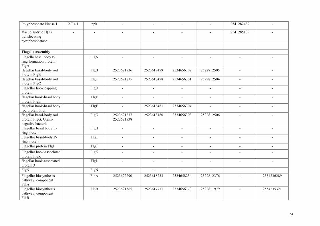
Polyphosphate kinase 1 2.7.4.1 ppk - - - - 2541282432 -
Vacuolar-type H(+) translocating pyrophosphatase
- - - - - - 2541285109 -
Flagella assembly Flagella basal body P-ring formation protein FlgA
FlgA - - - - - -
flagellar basal-body rod protein FlgB
FlgB 2523621836 2523618479 2534656302 2522812505 - -
flagellar basal-body rod protein FlgC
FlgC 2523621835 2523618478 2534656301 2522812504 - -
Flagellar hook capping protein
FlgD - - - - - -
flagellar hook-basal body protein FlgE
FlgE - - - - - -
flagellar hook-basal body rod protein FlgF
FlgF - 2523618481 2534656304 - - -
flagellar basal-body rod protein FlgG, Gram-negative bacteria
FlgG 2523621837 2523621838
2523618480 2534656303 2522812506 - -
Flagellar basal body L-ring protein
FlgH - - - - - -
Flagellar basal-body P-ring protein
FlgI - - - - - -
Flagellar protein FlgJ FlgJ - - - - - - Flagellar hook-associated protein FlgK
FlgK - - - - - -
flagellar hook-associated protein 3
FlgL - - - - - -
FlgN FlgN - - - - - - Flagellar biosynthesis pathway, component FlhA
FlhA 2523622290 2523618233 2534658234 2522812376 - 2554236289
Flagellar biosynthesis pathway, component FlhB
FlhB 2523621565 2523617711 2534656770 2522811979 - 2554235321
154

Flagellin and related hook-associated proteins
FliC - - - - - -
Flagellar capping protein FliD - - - - - - flagellar hook-basal body complex protein FliE
FliE 2523622294 2523618229 - 2522812380 - -
flagellar basal-body M-ring protein/flagellar hook-basal body protein (fliF)
FliF - - - - - -
Flagellar motor switch protein FliG
FliG - - - - - -
Flagellar biosynthesis/type III secretory pathway protein
FliH - - - - - -
type III secretion system ATPase, FliI/YscN (EC 3.6.3.15)
FliI 2523621472 2523618466 2523619149
2534657369 2522811726 - 2554236374
flagellar export protein FliJ
FliJ 2523621613 2523618709 - 2522811828 - -
Flagellar hook-length control protein FliK
FliK - - - - - -
Flagellar basal body-associated protein
FliL - - - - - -
Flagellar motor switch protein FliM
FliM - - - - - -
Flagellar motor switch protein FliN
FliN/SpoA - - - - - -
Flagellar biosynthesis protein, FliO
FliO - - - - - -
Flagellar biosynthesis pathway, component FliP
FliP 2523622788 2523618755 2534657253 2522811903 - 2554235810
Flagellar biosynthesis pathway, component FliQ
FliQ - - - - - -
Flagellar biosynthesis pathway, component FliR
FliR 2523622786 2523618758 2534657251 2522812209 - 2554235468
flagellar biosynthetic protein FliS
FliS - - - - - -
155

Flagellar protiein FliT FliT - - - - - - Flagella motor component
MotA - - - - - -
Flagella motor protein MotB - - - - - - Flagellar motor protein OmpA - - 2534656717 - - - Hydrogenases [FeFe] hydrogenase H-cluster radical SAM maturase HydE
2.8.1.6 HydE - - 2534657619 2522813318 - 2554235784
iron-only hydrogenase maturation protein HydF
- HydF - 2523618604 2534656733 2522811229 - 2554235601
iron-only hydrogenase maturation protein HydG
- HydG - 2523617045 2534657108 2522813171 - 2554235630
[FeFe] hydrogenase, group B1/B3
- - - - 2534657038 2522811239 - -
Iron only hydrogenase large subunit, C-terminal domain
- - - 2523617266 2534657035 2522811236 - -
NAD(P)-dependent iron-only hydrogenase diaphorase component flavoprotein
1.6.5.3 - - 2523617687 2534657247 2522812755 - 2554235203
NAD(P)-dependent iron-only hydrogenase catalytic subunit
1.6.5.3 - - 2523617686 2534657246 2522812756 - 2554235202
Ni,Fe-hydrogenase I small subunit
1.12.99.6 - - - - - 2541282828 -
Ni,Fe-hydrogenase I large subunit
1.12.99.6 - - - - - 2541282829 -
hydrogenase maturation protease
- HycI - - - - 2541282830 -
Ni,Fe-hydrogenase III small subunit
- - 2523623068 - - - 2541283385 -
Ni,Fe-hydrogenase III large subunit
- - 2523623069 - - - 2541283386 -
Hydrogenase 4 membrane component (E)
1.- - 2523623071 - - - 2541283388 -
156

Coenzyme F420-reducing hydrogenase, alpha subunit
- - - - - - 2541281832 -
Coenzyme F420-reducing hydrogenase, beta subunit
- - - - 2534657715 - - -
Coenzyme F420-reducing hydrogenase, delta subunit
- - - - - - 2541285052 -
Coenzyme F420-reducing hydrogenase, gamma subunit
- - - - - - 2541281831 -
Zn finger protein HypA/HybF (possibly regulating hydrogenase expression)
- HypA/HybF
- - - - 2541282821 -
Hydrogenase nickel incorporation protein HypB
- HypB - - - - 2541282820 -
Hydrogenase maturation protein HypC
- HypC - - - - 2541282822 -
Hydrogenase maturation protein HypD
- HypD - - - - 2541282823 2541285008
-
Hydrogenase maturation protein, carbamoyl dehydratase HypE
- HypE - - - - 2541282825 -
Hydrogenase maturation protein, carbamoyltransferase HypF
- HypF - - - - 2541282826 -
Polyphosphate metabolism
Polyphosphate kinase - - - - - 2522813547 - - polyphosphate kinase 1 2.7.4.1 - - - - - 2541282432 - polyphosphate kinase 2, PA0141 family
- - - - - - 2541282023 -
Polyphosphate:AMP phosphotransferase
2.7.4.- - - - - - 2541284980 -
Guanosine polyphosphate
3.1.7.2 - 2523622599 2523617618 2534656623 - 2541284760 -
157

pyrophosphohydrolases/synthetases Exopolyphosphatase 3.6.1.11 - - - - - 2541282351 - Adenylate kinase 2.7.4.3 - 2523623169 2523616999 2534657048 2522813109 2541282076 2554235454
158

Table S2.6. Genomes used to produce the flagella gene tree Listed are the IMG taxon ID, the organism name and the phylum that the organism belongs to. IMG taxon ID Organism Phylum 643692001 Acidobacterium capsulatum ATCC 51196 Acidobacteria 649633100 Terriglobus saanensis SP1PR4, DSM 23119 Acidobacteria 642555107 Bifidobacterium longum DJO10A Actinobacteria 2508501106 Mycobacterium rhodesiae NBB3 Actinobacteria 2517434006 Brevibacterium casei S18 Actinobacteria 646564582 Thermocrinis albus HI 11/12, DSM 14484 Aquificae 643692050 Sulfurihydrogenibium azorense Az-Fu1 Aquificae 649633104 Thermovibrio ammonificans HB-1, DSM
15698 Aquificae
2511231141
Alicyclobacillus acidocaldarius acidocaldarius Tc-4-1
Bacillus
649633013 Bacteroides salanitronis BL78, DSM 18170 Bacteroides 640753008 Bacteroides vulgatus ATCC 8482 Bacteroides 637000065 Chlamydophila abortus S26/3 Chlamydia 637000067 Chlamydophila felis Fe/C-56 Chlamydia 646564588 Waddlia chondrophila WSU 86-1044 Chlamydia 637000073 Chlorobium tepidum TLS Chlorobi 637000072 Chlorobium chlorochromatii CaD3 Chlorobi 642555122 Chlorobium phaeobacteroides BS1 Chlorobi 2508501111 Herpetosiphon aurantiacus DSM 785 Chloroflexi 649989977 Oscillochloris trichoides DG6 Chloroflexi 649633005 Anaerolinea thermophila UN-1 Chloroflexi 649633038 Desulfurispirillum indicum S5, DSM 22839 Chrysiogenetes 637000076 Clostridium acetobutylicum ATCC 824 Clostridia 2503508009 Mahella australiensis 50-1 BON, DSM
15567 Clostridia
641522632 Heliobacterium modesticaldum Ice1 Clostridia 649633052 Halanaerobium hydrogenoformans Clostridia 640427120 Metallosphaera sedula DSM 5348 Crenarchaeota 641228499 Nitrosopumilus maritimus SCM1 Crenarchaeota 648028062 Vulcanisaeta distributa DSM 14429 Crenarchaeota 2503982047 Anabaena cylindrica PCC 7122 Cyanobacteria 639857037 Nodularia spumigena CCY9414 Cyanobacteria 637000313 Synechococcus sp. JA-3-3Ab Cyanobacteria 637000121 Gloeobacter violaceus PCC 7421 Cyanobacteria 2523533517 Zag_1 Cyanobacteria 2531839741 Zag_111 Cyanobacteria 2523533519 Ca. Gastranaerophilus phascolarctosicola Cyanobacteria 2522572068 MH_37 Cyanobacteria 2541046959 Mel_A1 Cyanobacteria 2541046956 Mel_B1 Cyanobacteria 2541046940 Mel_B2 Cyanobacteria 2541046938 Mel_C1 Cyanobacteria
159

2531839742 Ca. Caenarcanum bioreactoricola Cyanobacteria 2541046960 Ca. Obscuribacter phosphatis Cyanobacteria 2541046958 ACD20 Cyanobacteria 643348543 Dictyoglomus turgidum DSM 6724 Dictyoglomi 643348542
Dictyoglomus thermophilum H-6-12, ATCC 35947
Dictyoglomi
642555127 Elusimicrobium minutum Pei191 Elusimicrobia 642555172 Candidatus Endomicrobium sp. Rs-D17 Elusimicrobia 644736373 Halorhabdus utahensis AX-2 Euryarcheota 648028040 Methanoplanus petrolearius SEBR 4847 Euryarcheota 638154522 Thermoplasma volcanium GSS1 Euryarcheota 650377942 Fibrobacter succinogenes succinogenes S85 Fibrobacter 637000117 Fusobacterium nucleatum nucleatum ATCC
25586 Fusobacteria
646311952 Sebaldella termitidis ATCC 33386 Fusobacteria 644736384 Leptotrichia buccalis C-1013-b, DSM 1135 Fusobacteria 643692024 Gemmatimonas aurantiaca T-27T Gemmatimonadetes 640963040 Lentisphaera araneosa HTCC2155 Lentisphaerae 650633000 Victivallis vadensis ATCC BAA-548 Lentisphaerae 638154511 Nanoarchaeum equitans Kin4-M Nanoarchaeota 2540341086 Leptospirillum ferrooxidans C2-3 Nitrospirae 637000236 Rhodopirellula baltica SH 1 Planctomycetes 649633083 Planctomyces brasiliensis IFAM 1448, DSM
5305 Planctomycetes
641736268 Gemmata obscuriglobus UQM 2246 Planctomycetes 643692004 Azotobacter vinelandii DJ, ATCC BAA-1303 Proteobacteria 649633004 Alicycliphilus denitrificans BC Proteobacteria 650716078 Pusillimonas sp. T7-7 Proteobacteria 637000241 Rhodospirillum rubrum S1, ATCC 11170 Proteobacteria 646311920 Dickeya dadantii Ech586 Proteobacteria 644736355 Dickeya zeae Ech1591 Proteobacteria 637000207 Photorhabdus luminescens laumondii TTO1 Proteobacteria 650377903 Acinetobacter calcoaceticus PHEA-2 Proteobacteria 643348518 Borrelia duttonii Ly Spirochaetes 2506783010 Leptonema illini 3055, DSM 21528 Spirochaetes 2511231215 Treponema pallidum pertenue Gauthier Spirochaetes 2505119043 Thermovirga lienii Cas60314, DSM 17291 Synergistetes 646311961 Thermanaerovibrio acidaminovorans Su883,
DSM 6589 Synergistetes
645951855 Jonquetella anthropi E3_33 E1 Synergistetes 2508501115 Deinococcus pimensis KR-235 Thermi 646564545 Meiothermus ruber 21, DSM 1279 Thermi 2515154172 Thermus igniterrae ATCC 700962 Thermi 2505119042 Thermodesulfatator indicus CIR29812, DSM
15286 Thermodesulfobacteria
640427150 Thermotoga petrophila RKU-1 Thermotoga 2510065086 Mesotoga prima MesG1Ag4.2 Thermotoga
160

2519899531 Thermotoga maritima MSB8, DSM 3109 Thermotoga 2517572100 Opitutaceae sp. TAV2 Verrucomicrobia 641522643 Opitutus terrae PB90-1 Verrucomicrobia 642791618 Chthoniobacter flavus Ellin428 Verrucomicrobia
161

Table S2.7. Table of Pfams used to differentiate cell wall types and flagella assembly, and GI numbers for photosynthesis genes and (bacterio)chlorophyll biosynthesis genes The PFAM numbers that were used to differentiate cell wall were obtained from Albertsen et al., 2013. The flagella assembly genes are outlined in Pallen and Matzke, 2006 and the the (bacterio)chlorophyll biosynthesis genes and cut-offs were obtained from Sousa et al., 2012. Pfam/GI Pfam description PF04413 Glycos_transf_N – (kdotransferase) PF02614 LpxK – Tetraacyldisaccharide-1-P 4’-kinase PF02684 LpxB – Lipid-A-disaccharide synthetase PF03331 LpxC – UDP-3-O-acyl N-acetylglycosamine deacetylase PF04613 LpxD – UDP-3-O-[3-hydroxymyristoyl] glucosamine N-acyltransferase, LpxD PF02472 ExbD – Biopolymer transport protein ExbD/TolR PF07244 Surf_Ag_VNR – Surface antigen variable number repeat PF03739 YjgP_YjgQ – Predicted permease YjgP/YjgQ family PF01103 Bac_surface_Ag – Surface antigen PF00263 Secretin – Bacterial type II and III secretion system protein PF02321 OEP – Outer membrane efflux protein PF03968 OstA – OstA-like protein PF00593 TonB_dep_Rec – TonB dependent receptor PF04166 PdxA – Pyridoxal phosphate biosynthesis protein PdxA PF06835 Lipopolysaccharide-assembly, LptC-related PF03740 PdxJ – Pyridoxal phosphate biosynthesis protein PdxJ PF03548 LolA – Outer membrane lipoprotein carrier protein LolA PF04052 TolB_N – TolB amino-terminal domain PF04453 OstA_C – Organic solvent tolerance protein PF02645 DegV – Uncharacterised protein, DegV family COG1307 PF05103 DivIVA – DivIVA protein PF02650 HTH_WhiA – Sporulation Regulator WhiA C terminal domain PF10298 WhiA_N – Sporulation Regulator WhiA N terminal PF04472 DUF552 – Protein of unknown function (DUF552) PF04203 Sortase – Sortase family PF03816 LytR_cpsA_psr – Cell envelope-related transcriptional attenuator domain PF09269 DUF1967 – Domain of unknown function (DUF1967) PF01424 R3H – R3H domain PF01618 MotA_ExbB – MotA/TolQ/ExbB proton channel family PF13677 MotB_plug – Membrane MotB of proton-channel complex MotA/MotB PF03963 FlgD – Flagellar hook capping protein – N-terminal region PF00460 PF06429
Flg_bb_rod – Flagella basal body rod protein Flg_bbr_C – Flagellar basal body rod FlgEFG protein C-terminal
PF02107 FlgH - Flagellar L-ring protein PF02119 FlgI - Flagellar P-ring protein PF00669 PF00700
Flagellin_N – Bacterial flagellin N-terminal helical region Flagellin_C – Bacterial flagellin C-terminal helical region
PF02465 PF07195
FliD_N - Flagellar hook-associated protein 2 N-terminus FliD_C – Flagellar hook-associated protein 2 C-terminus
PF02049 FliE – Flagellar hook-basal body complex protein FliE PF01514 PF08345
YscJ_FliF – Secretory protein of YscJ/FliF family YscJ_FliF_C – Flagellar M-ring protein C-terminal
PF01706 FliG_C - FliG C-terminal domain
162

PF02108 FliH – Flagellar assembly protein FliH PF02050 FliJ – Flagellar FliJ protein PF02154 FliM - Flagellar motor switch protein FliM PF01052 SpoA - Surface presentation of antigens (SPOA) PF00813 FliP – FliP family PF01313 Bac_export_3 – Bacterial export proteins, family 3 PF01311 Bac_export_1 – Bacterial export proteins, family 1 PF02561 FliS – Flagellar protein FliS PF00771 FHIPEP - FHIPEP protein family PF01312 Bac_export_2 – FlhB HrpN YscU SpaS Family 189347628 21674769 37522897 77463857
bchH
189346994 21674119 37520439 77463844
bchD
21674120 37521283 77463843
bchI
21674770 37523971 77463859
bchM
21674771 77463851
bchE
77463865 acsF 21673889 bciA 16331168 bciB 159462468 LPOR 189347668 21674961 37521938 77463855
bchN
21674960 37519784 77463856
bchB
189347666 21674959 37521939 77463858
bchL
163

164

Appendix B: Supplementary figures and tables for Chapter 3 Supplementary Figures
Figure S3.1. Depth of coverage against GC content for the assembled contigs. Contigs assigned to V. chlorellavorus are represented by red circles. C. vulgaris chloroplast contigs are represented by green circles and C. vulgaris contigs are represented by black circles. The size of the circle corresponds to the length of the contig.
Dep
th o
f cov
erag
e
100
200
300
400
500
30 40 50 60
Length100kb
200kb
300kb
400kb
500kb
600kb
GC content
V. chlorellavorus
C. vulgaris chloroplast
C. vulgaris nuclear
16S/23S rRNA
Transposon
Transposon
16S/23S rRNA18S/28S rRNA
165

Figure S3.2. A maximum likelihood concatenated gene tree showing the Cyanobacteria and select bacterial phyla. The phylogenetic tree was inferred from the concatenation of 109 conserved marker genes (Table S3.1) and consists of 7,732 bacterial and 169 archaeal genomes from the IMG database (Markowitz et al., 2014).
c__Oxyphotobacteria
MH_37MEL_C1Zag_1
100
MEL_A1
100
Zag_111
100
Ca. Gastranaerophilus phascolarctosicola
64
MEL_B1
100
MEL_B2
100
ACD20
100
Vampirovibrio chlorellavorus
100
Ca. Caenarcanum bioreactoricolaCa. Obscuribacter phosphatis
100
99
100
100
p__Tenericutes
p__Fusobacteria
88
100
p__Saccharibacteria
p__Parcubacteria
100
100
p__Microgenomates
86
93
50
96
100
p__Firmicutes
p__Actinobacteria
p__Deinococci
100
100
p__Chloroflexi
p__Armatimonadetes
100
100
68
p__Synergistetes100
p__Proteobacteria
p__Acidobacteria
p__Nitrospirae
100
100
p__Epsilonmicrobia
p__Thermotogae
p__Aquificae
100
100
94
p__Deferribacteres100
p__Bacteroidetes
p__Ignavibacteriae
p__Chlorobi
80
100
100
69
p__Marinimicrobia
100
99
p__Cloacimonetes
p__Fibrobacteres
100
100
100
64
p__Gemmatimonadetes
89
100
p__Chlamydiae
p__Verrucomicrobia
p__Lentisphaerae
100
100
100
100
p__Planctomycetes
100
100
72
79
p__Spirochaetes100
p__Elusimicrobia100
p__TM6
88
100
0.10
c_Melainabacteriap_Cyanobacteria
166

Figure S3.3. Methyl-accepting chemotaxis proteins (MCP) encoded on the V. chlorellavorus genome. Three MCPs are found in V. chlorellavorus. MCP domains were predicted with InterProScan5 (Jones et al., 2014). Directional arrows represent the putative genes and orientation (positive or negative strand) and the rounded rectangles represent domains.
Methyl-accepting chemotaxis protein (MCP) signalling
GAF
Target SNARE coiled-coil
HAMP linker
Globin
98270
Contig 14
114549
Methyl-accepting chemotaxis protein (MCP)
Chemotaxis-related domains and protein
Hypothetical protein
Contig 7
Pili-related protein
111562 124333
DomainsORFs
167

Figure S3.4. Type IV secretion system (T4SS) in the V. chlorellavorus genome and plasmids. The schematic diagram shows the presence of T4SS genes identified by IMG/ER on both plasmids and one contig. The arrows represent annotated genes and their direction. The numbers above the genes are the IMG/ER accession numbers and these have been identified as alien genes by PHX analysis (Table S3.2). T4SS genes have the predicted gene names italicised below.
Plasmid 1
traF traG/virD4 trbI trbH trbG trbF trbL trbE/VirB4trbJ trbD trbC trbB
Plasmid 2
trbI trbH trbG trbF trbL trbJ trbE/virB4 trbD trbC trbB
traC traL 25025
1
1
71965
traM traI traG/virD4 traFtraL traKtraJ traC traD traL
T4SSIntegraseATPaseEfflux transporter/pump/outer memberane proteinCell wall/membrane/envelope biogenesisReplication, recombination and repairTransposaseDomain of unknown functionHypothetical proteinOther
24606
2462150153
traI traJtraL
Legend
25028 50739
50736
Contig 21
traB/trbI trbG trbF trbL trbJ trbE/virB4 trbD trbC trbBtrbHtraDtraM
traFtraG/virD4traI traJtraM traL traK18262
10806435520
26879
2600257499
2600257516 2600257517 2600257527
2600257630 2600257632 2600257646
2600257663 2600257665
2600257677
260025767626002576752600257674260025767326002576722600257671260025766926002576672600257666
26002577112600257699
26002576912600257686260025768526002576802600257678
2600257732
168

Figure S3.5. A maximum likelihood phylogenetic tree of virB4 genes. Aligned sequences and naming conventions were obtained from Christie, 2004. virB4_T is based on the T-DNA conjugation system of Agrobacterium tumefaciens plasmid Ti, virB4_F is based on the plasmid F, virB4_I is based on the Incl plasmid R64 and virB4_G is based on ICEHIN1056. The other T4SS have homologues to VirB4 and include the Cyanobacteria (virB4_C), Bacteroides (virB4_B), Firmicutes (virB4_FA and virB4_FATA), Actinobacteria (virB4_FA and virB4_FATA), Tenericutes (virB4_FATA) and Archaea (virB4_FATA) (Christie, 2004). The V. chlorellavorus genome contains T4SS that belong to the virB4_T. virD4 is used as the outgroup. Black circles represent nodes with ≥ 90% bootstrap support, grey circles represent nodes with ≥ 80% bootstrap support and white circles represent nodes with ≥ 70% bootstrap support. (p) corresponds to virB4 genes found on plasmids and (c) corresponds to virB4 genes found on the chromosome.
�������0/��������0/�
virB4_FATA
virB4_FA
virB4_F
virB4_B
virB4_T
virB4_T
virB4_T
virB4_G
virB4_C
virB4_I
virD4
0.10�������0/�
virB4_T
Nitrosomonas eutropha�&���1&B��������S�Photobacterium profundum 66��1&��������F�
Shewanella baltica�26����1&B��������S�Legionella longbeachae 16:����1&B��������F�
Rhizobium etli &)1����1&B��������S�Agrobacterium vitis�6��1&B��������S�
Desulfovibrio salexigens '60������1&B��������F�Desulfotalea psychrophila /6Y���1&B��������S�Polaromonas naphthalenivorans�&-��1&B��������F�
Thiomonas intermedia .���1&B��������F�Methylovorus VS�6,3����1&B��������S�
Halothiobacillus meapolitanus�F��1&B��������F�Acidithiobacillus ferrooxidans�$7&&�������1&B��������F�
Prosthecochloris aestuarii '60�����1&B��������S�Leptotrichia buccalis�'60������1&B��������F�
Acidobacterium capsulatum $7&&�������1&B��������F�Acidobacterium capsulatum $7&&�������1&B��������F�
Vampirovibrio chlorellavorus �SODVPLG���
Vampirovibrio chlorellavorus �SODVPLG���Vampirovibrio chlorellavorus �FRQWLJ����
virB4_T/trbE
169

Figure S3.6. A maximum likelihood phylogenetic tree of fliI genes. The phylogenetic tree is constructed from 2,256 finished genomes from the IMG database (Markowitz et al., 2009). The tree is unrooted and only the Melainabacteria and its closest neighbours are shown. V. chlorellavorus is in red and the other three Melainabacteria representatives are in blue. Phyla are in bold. Black circles in the tree represents nodes with ≥ 90% bootstrap support and white circles represents nodes with ≥ 80% bootstrap support.
Caldanaerobius polysaccharolyticum KMTHCJ, DSM 13641
Desulfurispirillum indicum S5, DSM 22839
Coprothermobacter proteolyticus DSM 5265
Desulfurella acetivorans A63, DSM 5264
Hippea maritima MH2, DSM 10411
Caldithrix abyssi DSM 13497 Melioribacter roseus P3M
Ignavibacterium album JCM 16511
Chthonomonas calidirosea T49
Halanaerobium praevalens GSL, DSM 2228
Halothermothrix orenii H 168
Vampirovibrio chlorellavorus
ACD20
MEL_B2
MEL_B1
0.10
Proteobacteria
Thermotogae
Firmicutes
Firmicutes
Deferribacteres
� 90% (ML)
� 80% (ML)
170

Figure S3.7. Clusters of orthologous groups (COGs) for the class Melainabacteria. There is an increase in the COGs associated with cell motility (category N) and intracellular trafficking, secretion, and vesicular transport (category U) for V. chlorellavorus. STAMP (Parks et al., 2014) was used to explore the resulting COG profiles and create summary plots.
171

Supplementary Tables Table S3.1. Marker genes used for constructing the concatenated gene tree. A set of 178 single copy genes present exactly once in >90% of the trusted genomes (found in >90% of the genomes) from the Integrated Microbial Genomes (IMG; (Markowitz et al., 2014)) database was identified. From the 178 initial genes, 69 were removed from consideration as they exhibited divergent phylogenetic histories in >1% of the trusted genomes. The remaining 109 genes were used to construct a concatenated gene tree (Figure S3.2).
Gene ID Description Length (aa) Included markers TIGR03723 T6A_YgjD: tRNA threonylcarbamoyl adenosine
modification protein YgjD 314
TIGR03953 rplD_bact: 50S ribosomal protein L4 188 TIGR00755 ksgA: dimethyladenosine transferase 256 TIGR00138 gidB: 16S rRNA (guanine(527)-N(7))-methyltransferase
GidB 183
TIGR01953 NusA: transcription termination factor NusA 340 PF00410 Ribosomal protein S8 129 PF00380 Ribosomal protein S9/S16 121 TIGR03625 L3_bact: 50S ribosomal protein L3 202 TIGR01049 rpsJ_bact: ribosomal protein S10 99 TIGR01044 rplV_bact: ribosomal protein L22 103 TIGR00964 secE_bact: preprotein translocase, SecE subunit 57 TIGR00613 reco: DNA repair protein RecO 239 TIGR00002 S16: ribosomal protein S16 78 TIGR00001 rpmI_bact: ribosomal protein L35 63 PF13507 CobB/CobQ-like glutamine amidotransferase domain 259 TIGR03591 polynuc_phos: polyribonucleotide nucleotidyltransferase 689 TIGR03594 GTPase_EngA: ribosome-associated GTPase EngA 432 TIGR00088 trmD: tRNA (guanine(37)-N(1))-methyltransferase 233 TIGR00086 smpB: SsrA-binding protein 144 TIGR00084 ruvA: Holliday junction DNA helicase RuvA 192 TIGR00082 rbfA: ribosome-binding factor A 115 TIGR00855 L12: ribosomal protein L7/L12 125 TIGR01032 rplT_bact: ribosomal protein L20 114 TIGR00019 prfA: peptide chain release factor 1 361 TIGR00396 leuS_bact: leucine--tRNA ligase 843 TIGR00012 L29: ribosomal protein L29 56 TIGR00017 cmk: cytidylate kinase 217 TIGR00150 T6A_YjeE: tRNA threonylcarbamoyl adenosine
modification protein YjeE 135
TIGR00152 TIGR00152: dephospho-CoA kinase 188 TIGR00158 L9: ribosomal protein L9 148 PF00281 Ribosomal protein L5 56 TIGR00981 rpsL_bact: ribosomal protein S12 124 TIGR00090 iojap_ybeB: iojap-like ribosome-associated protein 99
172

TIGR00810 secG: preprotein translocase, SecG subunit 73 TIGR00092 TIGR00092: GTP-binding protein YchF 368 TIGR00095 TIGR00095: RNA methyltransferase, RsmD family 194 TIGR00250 RNAse_H_YqgF: RNAse H domain protein, YqgF family 130 TIGR01145 ATP_synt_delta: ATP synthase F1, delta subunit 172 TIGR01029 rpsG_bact: ribosomal protein S7 154 TIGR03725 T6A_YeaZ: tRNA threonylcarbamoyl adenosine
modification protein YeaZ 212
TIGR01021 rpsE_bact: ribosomal protein S5 156 TIGR01024 rplS_bact: ribosomal protein L19 114 TIGR00061 L21: ribosomal protein L21 101 TIGR00337 PyrG: CTP synthase 526 TIGR00060 L18_bact: ribosomal protein L18 114 TIGR01394 TypA_BipA: GTP-binding protein TypA/BipA 594 PF10458 Valyl tRNA synthetase tRNA binding arm 66 TIGR01393 lepA: GTP-binding protein LepA 595 TIGR00436 era: GTP-binding protein Era 270 TIGR00631 uvrb: excinuclease ABC subunit B 658 TIGR00062 L27: ribosomal protein L27 83 TIGR00634 recN: DNA repair protein RecN 563 TIGR00635 ruvB: Holliday junction DNA helicase RuvB 305 TIGR00431 TruB: tRNA pseudouridine(55) synthase 210 TIGR00472 pheT_bact: phenylalanine--tRNA ligase, beta subunit 798 TIGR00496 frr: ribosome recycling factor 176 TIGR03635 S17_bact: 30S ribosomal protein S17 72 PF01192 RNA polymerase Rpb6 57 PF02576 Uncharacterised BCR, YhbC family COG0779 141 TIGR01087 murD: UDP-N-acetylmuramoylalanine--D-glutamate ligase 441 TIGR01169 rplA_bact: ribosomal protein L1 227 TIGR00487 IF-2: translation initiation factor IF-2 587 TIGR00922 nusG: transcription termination/antitermination factor NusG 172 TIGR01164 rplP_bact: ribosomal protein L16 126 TIGR01009 rpsC_bact: ribosomal protein S3 212 TIGR00043 TIGR00043: probable rRNA maturation factor YbeY 111 PF00466 Ribosomal protein L10 100 TIGR01128 holA: DNA polymerase III, delta subunit 314 TIGR00166 S6: ribosomal protein S6 95 TIGR00416 sms: DNA repair protein RadA 454 TIGR02386 rpoC_TIGR: DNA-directed RNA polymerase, beta' subunit 1147 TIGR00344 alaS: alanine--tRNA ligase 847 TIGR00615 recR: recombination protein RecR 196 TIGR02273 16S_RimM: 16S rRNA processing protein RimM 166 TIGR00360 ComEC_N-term: ComEC/Rec2-related protein 171 TIGR03654 L6_bact: ribosomal protein L6 175 TIGR01171 rplB_bact: ribosomal protein L2 275 TIGR00959 ffh: signal recognition particle protein 428 TIGR01071 rplO_bact: ribosomal protein L15 144 TIGR00952 S15_bact: ribosomal protein S15 86
173

TIGR01079 rplX_bact: ribosomal protein L24 104 TIGR00116 tsf: translation elongation factor Ts 293 TIGR00059 L17: ribosomal protein L17 112 PF00673 ribosomal L5P family C-terminus 95 TIGR00593 pola: DNA polymerase I 890 TIGR00595 priA: primosomal protein N' 509 TIGR01632 L11_bact: ribosomal protein L11 140 TIGR02432 lysidine_TilS_N: tRNA(Ile)-lysidine synthetase 189 TIGR00460 fmt: methionyl-tRNA formyltransferase 315 TIGR00468 pheS: phenylalanine--tRNA ligase, alpha subunit 324 TIGR01066 rplM_bact: ribosomal protein L13 141 TIGR01067 rplN_bact: ribosomal protein L14 122 PF00276 Ribosomal protein L23 92 TIGR00422 valS: valine--tRNA ligase 863 TIGR00194 uvrC: excinuclease ABC subunit C 574 TIGR02729 Obg_CgtA: Obg family GTPase CgtA 329 TIGR00020 prfB: peptide chain release factor 2 365 PF13742 OB-fold nucleic acid binding domain 99 TIGR03632 bact_S11: 30S ribosomal protein S11 117 TIGR00029 S20: ribosomal protein S20 87 TIGR03263 guanyl_kin: guanylate kinase 180 TIGR01510 coaD_prev_kdtB: pantetheine-phosphate adenylyltransferase 155 TIGR02013 rpoB: DNA-directed RNA polymerase, beta subunit 1238 TIGR03631 bact_S13: 30S ribosomal protein S13 113 TIGR00091 TIGR00091: tRNA (guanine-N(7)-)-methyltransferase 194 TIGR01050 rpsS_bact: ribosomal protein S19 92 TIGR00188 rnpA: ribonuclease P protein component 111 TIGR00877 purD: phosphoribosylamine--glycine ligase 425 TIGR01011 rpsB_bact: ribosomal protein S2 225 Removed markers TIGR01951 nusB: transcription antitermination factor NusB 131 TIGR00033 aroC: chorismate synthase 351 TIGR00234 tyrS: tyrosine--tRNA ligase 406 PF01416 tRNA pseudouridine synthase 105 TIGR00447 pth: peptidyl-tRNA hydrolase 188 TIGR00445 mraY: phospho-N-acetylmuramoyl-pentapeptide-transferase 321 TIGR00539 hemN_rel: putative oxygen-independent
coproporphyrinogen III oxidase 361
TIGR00963 secA: preprotein translocase, SecA subunit 787 TIGR00009 L28: ribosomal protein L28 58 TIGR00459 aspS_bact: aspartate--tRNA ligase 586 TIGR00456 argS: arginine--tRNA ligase 569 TIGR00008 infA: translation initiation factor IF-1 69 TIGR00083 ribF: riboflavin biosynthesis protein RibF 290 TIGR00329 gcp_kae1: metallohydrolase, glycoprotease/Kae1 family 305 TIGR01031 rpmF_bact: ribosomal protein L32 56 TIGR01034 metK: methionine adenosyltransferase 377
174

TIGR00398 metG: methionine--tRNA ligase 530 TIGR00420 trmU: tRNA (5-methylaminomethyl-2-thiouridylate)-
methyltransferase 351
TIGR00179 murB: UDP-N-acetylenolpyruvoylglucosamine reductase 290 PF04127 DNA / pantothenate metabolism flavoprotein 185 TIGR02027 rpoA: DNA-directed RNA polymerase, alpha subunit 298 TIGR00382 clpX: ATP-dependent Clp protease, ATP-binding subunit
ClpX 414
TIGR00065 ftsZ: cell division protein FtsZ 353 TIGR00064 ftsY: signal recognition particle-docking protein FtsY 279 TIGR01391 dnaG: DNA primase 414 TIGR00544 lgt: prolipoprotein diacylglyceryl transferase 280 TIGR00739 yajC: preprotein translocase, YajC subunit 84 TIGR01017 rpsD_bact: ribosomal protein S4 200 TIGR02191 RNaseIII: ribonuclease III 219 PF07479 NAD-dependent glycerol-3-phosphate dehydrogenase C-
terminus 149
TIGR00174 miaA: tRNA dimethylallyltransferase 288 TIGR02397 dnaX_nterm: DNA polymerase III, subunit gamma and tau 355 TIGR01082 murC: UDP-N-acetylmuramate--alanine ligase 449 TIGR01083 nth: endonuclease III 192 TIGR01085 murE: UDP-N-acetylmuramyl-tripeptide synthetase 472 TIGR00575 dnlj: DNA ligase, NAD-dependent 652 TIGR00884 guaA_Cterm: GMP synthase (glutamine-hydrolyzing), C-
terminal domain 310
TIGR01162 purE: phosphoribosylaminoimidazole carboxylase, catalytic subunit
156
PF01025 GrpE 166 TIGR00233 trpS: tryptophan--tRNA ligase 328 TIGR00186 rRNA_methyl_3: RNA methyltransferase, TrmH family,
group 3 240
TIGR00165 S18: ribosomal protein S18 70 TIGR00414 serS: serine--tRNA ligase 418 TIGR02075 pyrH_bact: UMP kinase 233 TIGR00418 thrS: threonine--tRNA ligase 565 TIGR00419 tim: triose-phosphate isomerase 228 TIGR00362 DnaA: chromosomal replication initiator protein DnaA 437 TIGR00888 guaA_Nterm: GMP synthase (glutamine-hydrolyzing), N-
terminal domain 188
TIGR00054 TIGR00054: RIP metalloprotease RseP 421 TIGR00168 infC: translation initiation factor IF-3 165 TIGR00042 TIGR00042: non-canonical purine NTP pyrophosphatase,
RdgB/HAM1 family 184
TIGR00115 tig: trigger factor 410 PF02601 Exonuclease VII, large subunit 319 TIGR00663 dnan: DNA polymerase III, beta subunit 367 PF00162 Phosphoglycerate kinase 384 TIGR01063 gyrA: DNA gyrase, A subunit 800 TIGR01060 eno: phosphopyruvate hydratase 425
175

TIGR02727 MTHFS_bact: 5-formyltetrahydrofolate cyclo-ligase 182 TIGR02350 prok_dnaK: chaperone protein DnaK 596 TIGR00580 mfd: transcription-repair coupling factor 923 TIGR03534 RF_mod_PrmC: protein-(glutamine-N5) methyltransferase,
release factor-specific 253
TIGR02012 tigrfam_recA: protein RecA 321 TIGR00006 TIGR00006: 16S rRNA (cytosine(1402)-N(4))-
methyltransferase 310
TIGR00442 hisS: histidine--tRNA ligase 406 TIGR01051 topA_bact: DNA topoisomerase I 632 TIGR00967 3a0501s007: preprotein translocase, SecY subunit 414 PF00342 Phosphoglucose isomerase 486 TIGR00643 recG: ATP-dependent DNA helicase RecG 629 TIGR00392 ileS: isoleucine--tRNA ligase 861
176

Table S3.2. Predicted highly expressed (PHX) and alien genes. PHX and alien gene prediction was performed with PHX analysis, using ribosomal proteins, chaperones and transcriptional and translational proteins of V. chlorellavous as representatives of recognised highly expressed genes to identify other putatively highly expressed genes in the genome (Karlin and Mrázek, 2000).
Highly expressed and alien gene IMG number IMG annotation Eg number 2600256191 Protein-export membrane protein, SecD/SecF family 1.05 Highly expressed genes IMG number IMG annotation Eg number 2600254915 ATP synthase F1 subcomplex beta subunit 1.50 2600254966 Superfamily II DNA and RNA helicases 1.10 2600254993 Parvulin-like peptidyl-prolyl isomerase 1.17 2600255027 DNA-binding protein, YbaB/EbfC family 1.20 2600255064 Pyruvate:ferredoxin oxidoreductase and related 1.70
2-oxoacid:ferredoxin oxidoreductases, beta subunit 2600255065 Pyruvate:ferredoxin oxidoreductase and related 1.32
2-oxoacid:ferredoxin oxidoreductases, alpha subunit 2600255172 Thioredoxin-like proteins and domains 1.10 2600255235 5'-deoxy-5'-methylthioadenosine phosphorylase 1.45 2600255249 Sugar transferases involved in lipopolysaccharide synthesis 1.05 2600255283 P22 coat protein - gene protein 5 1.12 2600255291 Vacuolar-type H(+)-translocating pyrophosphatase 1.69 2600255297 Aspartyl-tRNA synthetase, bacterial type 1.35 2600255302 Hypothetical protein 1.10 2600255315 S-adenosylmethionine decarboxylase 1.28 2600255327 Predicted P-loop-containing kinase 1.01 2600255352 Chaperone protein DnaK 1.62 2600255416 NADH dehydrogenase subunit A (EC 1.6.5.3) 1.11 2600255450 Malate dehydrogenase (NAD) (EC 1.1.1.37) 1.13 2600255470 Membrane protein insertase, YidC/Oxa1 family, 1.18 C-terminal domain 2600255524 YtxH-like protein 1.02 2600255533 Signal peptide peptidase SppA, 36K type 1.05 2600255566 Outer membrane protein 1.16 2600255576 Glyceraldehyde-3-phosphate dehydrogenase (NAD+) 1.32 (EC 1.2.1.12) 2600255586 RNA polymerase sigma factor, sigma-70 family 1.41 2600255592 Chaperonin GroL 1.86 2600255626 Ribosomal protein L7/L12 1.43 2600255627 DNA-directed RNA polymerase subunit beta (EC 2.7.7.6) 1.41 2600255628 DNA-directed RNA polymerase gamma chain (EC 2.7.7.6) 1.49 2600255637 Two component transcriptional regulator, LuxR family 1.10 2600255664 Ribonucleotide reductase, beta subunit 1.14 2600255681 ATPases with chaperone activity, ATP-binding subunit 1.21 2600255723 Cbb3-type cytochrome oxidase, subunit 1 1.07 2600255734 Hypothetical protein 1.08 2600255835 Flagellar basal body L-ring protein 1.14
177

2600255847 Superoxide dismutase 1.30 2600255852 Ribosomal protein L9 1.41 2600255862 Protein of unknown function (DUF561) 1.38 2600255880 Septum site-determining protein MinD 1.07 2600255892 Ribosomal protein S3, bacterial type 1.52 2600255895 LSU ribosomal protein L2P 1.40 2600255900 Translation elongation factor 1A (EF-1A/EF-Tu) 1.41 2600255911 Translation elongation factor EF-G 2.00 2600255941 Rare lipoprotein A 1.24 2600255966 Ribosomal protein L17 1.31 2600255967 DNA-directed RNA polymerase, alpha subunit, bacterial 1.23 and chloroplast-type 2600255975 LSU ribosomal protein L15P 1.34 2600255977 Ribosomal protein S5, bacterial/organelle type 1.59 2600256085 Polyribonucleotide nucleotidyltransferase 1.71 2600256086 SSU ribosomal protein S15P 1.07 2600256099 Translation elongation factor P 1.21 2600256132 Phosphopentomutase 1.21 2600256134 Uncharacterized protein conserved in bacteria 1.04 2600256170 Uncharacterized conserved protein 1.05 2600256171 6-pyruvoyl-tetrahydropterin synthase 1.11 2600256173 Bacterial regulatory proteins, gntR family 1.23 2600256207 Enoyl-[acyl-carrier-protein] reductase [NADH] 1.30 (EC 1.3.1.9) 2600256221 Hypothetical protein 1.16 2600256238 Ribose-phosphate pyrophosphokinase 1.15 2600256248 Hypothetical protein 1.28 2600256265 Aconitase (EC 4.2.1.3) 1.11 2600256266 Isocitrate dehydrogenase (NADP) (EC 1.1.1.42) 1.34 2600256282 Peroxiredoxin 1.53 2600256283 Ribosomal protein L1, bacterial/chloroplast 1.47 2600256284 LSU ribosomal protein L11P 1.19 2600256314 S1 RNA binding domain 1.67 2600256336 S1 RNA binding domain 1.38 2600256348 Adenosylhomocysteinase (EC 3.3.1.1) 1.38 2600256354 Carbon storage regulator, CsrA 1.18 2600256361 NusA antitermination factor 1.15 2600256362 Translation initiation factor IF-2 1.24 2600256385 Bacterial SH3 domain 1.28 2600256386 Hypothetical protein 1.84 2600256388 Thioredoxin 1.36 2600256401 Hypothetical protein 1.08 2600256411 FKBP-type peptidyl-prolyl cis-trans isomerases 2 1.14 2600256417 Inosine-5'-monophosphate dehydrogenase 1.06 (EC 1.1.1.205) 2600256436 Glycosyl hydrolases family 8 1.10 2600256437 Threonyl-tRNA synthetase (EC 6.1.1.3) 1.02 2600256466 6-phosphofructokinase 1.10 2600256529 LL-diaminopimelate aminotransferase apoenzyme 1.27 (EC 2.6.1.83) 2600256549 Aspartyl/glutamyl-tRNA(Asn/Gln) amidotransferase 1.12 subunit C (EC 6.3.5.-)
178

2600256550 CTP synthase (EC 6.3.4.2) 1.10 2600256560 Fe2+/Zn2+ uptake regulation proteins 1.11 2600256561 Rubrerythrin 1.09 2600256583 Two component transcriptional regulator, LuxR family 1.12 2600256588 Nucleoside diphosphate kinase (EC 2.7.4.6) 1.41 2600256597 Cyanobacterial porin (TC 1.B.23) 1.67 2600256623 2-oxoacid:acceptor oxidoreductase, alpha subunit 1.79 2600256624 2-oxoacid:acceptor oxidoreductase, beta subunit, 1.41 pyruvate/2-ketoisovalerate family 2600256651 Flagellar hook-basal body protein 1.17 2600256660 Biopolymer transport proteins 1.08 2600256663 Hypothetical protein 1.09 2600256675 3-deoxy-D-arabinoheptulosonate-7-phosphate 1.03 synthase (EC 2.5.1.54) 2600256710 GTP-binding protein TypA/BipA 1.46 2600256731 Hypothetical protein 1.13 2600256733 Adenine phosphoribosyltransferase (EC 2.4.2.7) 1.18 2600256757 ATP synthase F1 subcomplex alpha subunit 1.31 2600256779 Hemolysin activation/secretion protein 1.23 2600256821 Phosphate transport regulator 1.05 2600256833 Hypothetical protein 1.06 2600256897 YlqD protein 1.18 2600256918 Exodeoxyribonuclease VII small subunit (EC 3.1.11.6) 1.06 2600256944 S-layer homology domain 1.07 2600256966 RNA polymerase, sigma 28 subunit, SigD/FliA/WhiG 1.38 2600256998 Glutaredoxin-related protein 1.14 2600257077 Cell division protein FtsZ 1.28 2600257160 Alpha-glucan phosphorylases 1.38 2600257195 Peroxiredoxin, OsmC subfamily 1.43 2600257223 Type III secretion system ATPase, FliI/YscN 1.08 2600257249 Hypothetical protein 1.13 2600257292 Protein of unknown function (DUF1292) 1.33 2600257293 Succinyl-CoA synthetase (ADP-forming) beta subun 1.28 (EC 6.2.1.5) 2600257294 Succinyl-CoA synthetase, alpha subunit 1.27 2600257316 Ribosomal protein S2, bacterial type 1.79 2600257317 Translation elongation factor Ts (EF-Ts) 1.15 2600257354 Hypothetical protein 1.12 2600257355 Hypothetical protein 1.10 2600257409 ATP-dependent Clp protease ATP-binding subunit 1.00 ClpX (EC 3.4.21.92) 2600257410 ATP-dependent Clp protease proteolytic subunit 1.10 ClpP (EC 3.4.21.92) 2600257411 Trigger factor 1.48 2600257427 Succinate dehydrogenase/fumarate reductase, flavoprotein 1.30 subunit 2600257445 Flagellar basal body rod protein 1.07 2600257449 Flagellar basal-body rod protein FlgC 1.06
179

Alien genes IMG number IMG annotation Eg number 2600256772 Hypothetical protein 0.99 2600255126 Hypothetical protein 0.98 2600256735 Hypothetical protein 0.92 2600255443 Hypothetical protein 0.88 2600257671 Periplasmic protein involved in polysaccharide export 0.88 2600257031 Hypothetical protein 0.86 2600257567 Imidazoleglycerol-phosphate synthase 0.86 2600256774 Beta-lactamase class C and other penicillin binding 0.85 proteins 2600257020 Hypothetical protein 0.85 2600257499 Hypothetical protein 0.85 2600257685 Hypothetical protein 0.85 2600255194 Hypothetical protein 0.84 2600255922 FOG: CheY-like receiver 0.84 2600256485 prepilin-type N-terminal cleavage/methylation domain 0.84 2600257568 Imidazole glycerol phosphate synthase, glutamine 0.84 amidotransferase subunit 2600256419 Hypothetical protein 0.83 2600256973 Hypothetical protein 0.82 2600257103 Outer membrane protein 0.82 2600257113 Hypothetical protein 0.82 2600257585 Transposase and inactivated derivatives 0.82 2600257699 Conjugal transfer protein TrbH 0.82 2600257732 AAA ATPase domain/AAA domain 0.82 2600255868 Hypothetical protein 0.81 2600257187 Twin arginine targeting (Tat) protein translocase TatC 0.81 2600257497 Diadenosine tetraphosphate (Ap4A) hydrolase and other 0.81 HIT family hydrolases 2600257527 Nucleotidyl transferase of unknown function (DUF1814) 0.81 2600257529 Nucleotidyl transferase of unknown function (DUF1814) 0.81 2600257563 Methyltransferase domain 0.81 2600255144 Hypothetical protein 0.80 2600255224 Hypothetical protein 0.80 2600255777 TIR domain 0.80 2600257632 Uncharacterized protein conserved in bacteria 0.80 2600257676 Glycosyl transferase family 2 0.80 2600254951 Hypothetical protein 0.79 2600255590 Hypothetical protein 0.79 2600255756 Hypothetical protein 0.79 2600255862 Protein of unknown function (DUF561) 0.79 2600256409 Domain of unknown function (DUF4145) 0.79 2600256512 Alginate lyase 0.79 2600257109 RHS repeat-associated core domain 0.79 2600257362 Hypothetical protein 0.79 2600257480 Plasmid encoded RepA protein 0.79 2600257489 TaqI-like C-terminal specificity domain/ 0.79 Methyltransferase domain 2600257491 Hypothetical protein 0.79 2600257570 Hypothetical protein 0.79 2600257594 Plasmid encoded RepA protein 0.79
180
2600257630 Predicted ATP-dependent endonuclease of the OLD family 0.79 2600257665 Hypothetical protein 0.79 2600257691 Trypsin-like peptidase domain/PDZ domain 0.79 2600255719 Hypothetical protein 0.78 2600257105 RND family efflux transporter, MFP subunit 0.78 2600257230 Dehydrogenases (flavoproteins) 0.78 2600257492 Predicted ATPase (AAA+ superfamily) 0.78 2600257669 Teichoic acid biosynthesis proteins 0.78 2600255978 Predicted phosphohydrolases 0.77 2600256525 Hypothetical protein 0.77 2600256642 Hypothetical protein 0.77 2600257225 Protein of unknown function (DUF2971) 0.77 2600257562 Predicted pyridoxal phosphate-dependent enzyme 0.77 apparently involved in regulation of cell wall biogenesis 2600257599 Domain of unknown function (DUF389) 0.77 2600257674 Hypothetical protein 0.77 2600257711 DnaA N-terminal domain 0.77 2600256983 Peptidase M15 0.76 2600257091 Hypothetical protein 0.76 2600257561 Nucleoside-diphosphate-sugar epimerases 0.76 2600257610 Uncharacterized conserved protein (COG2071) 0.76 2600257667 Nucleoside-diphosphate-sugar pyrophosphorylase 0.76 involved in lipopolysaccharide biosynthesis/translation initiation factor 2B 2600257680 Endoglucanase 0.76 2600255374 Hypothetical protein 0.75 2600255506 Predicted phosphohydrolases 0.75 2600257104 The (Largely Gram-negative Bacterial) 0.75 Hydrophobe/Amphiphile Efflux-1 (HAE1) Family 2600257228 Predicted naringenin-chalcone synthase 0.75 2600257508 Hypothetical protein 0.75 2600257517 Hypothetical protein 0.75 2600257663 Hypothetical protein 0.75 2600257672 Uncharacterized protein involved in exopolysaccharide 0.75 biosynthesis 2600257678 Glycosyltransferase 0.75 2600255258 Dihydrofolate reductase 0.74 2600255507 Type II secretion system (T2SS), protein F 0.74 2600256033 Sugar transferases involved in lipopolysaccharide synthesis 0.74 2600256720 Hypothetical protein 0.74 2600257493 Adenine-specific DNA methylase containing a Zn-ribbon 0.74 2600257537 RND family efflux transporter, MFP subunit 0.74 2600257646 P-type conjugative transfer protein TrbL 0.74 2600255269 Hypothetical protein 0.73 2600257541 HipA-like C-terminal domain/HipA N-terminal domain/ 0.73 HipA-like N-terminal domain 2600257572 ABC-type multidrug transport system, ATPase and 0.73 permease components 2600257666 UDP-galactose 4-epimerase (EC 5.1.3.2) 0.73 2600257677 Glycosyltransferase 0.73 2600254929 Ankyrin repeats (3 copies) 0.72 2600255090 Restriction endonuclease S subunits 0.72
181

2600255508 Flp pilus assembly protein TadB 0.72 2600256159 Glycosyltransferase 0.72 2600257232 Ankyrin repeats (3 copies)/Ankyrin repeats 0.72 2600255059 Hypothetical protein 0.71 2600256873 Hypothetical protein 0.71 2600257088 UvrD-like helicase C-terminal domain/UvrD/REP 0.71 helicase N-terminal domain 2600257494 Hypothetical protein 0.71 2600257607 DoxX-like family 0.71 2600257673 Lipid A core - O-antigen ligase and related enzymes 0.71 2600257675 Glycosyltransferases involved in cell wall biogenesis 0.71 2600257686 Predicted ATPase (AAA+ superfamily) 0.71 2600255060 Hypothetical protein 0.70 2600255674 Translation factor SUA5 0.70 2600257601 Trehalose-6-phosphate synthase 0.70 2600255536 Hypothetical protein 0.68 2600256155 Hypothetical protein 0.67 2600256156 Glycosyltransferase 0.69 2600257209 Ammonium transporter (TC 1.A.11) 0.69 2600257275 Hypothetical protein 0.69 2600257516 Type IV secretory pathway, VirB10 components 0.68 2600256157 Coenzyme F390 synthetase 0.68 2600256158 Coenzyme F390 synthetase 0.68 2600256160 Membrane protein involved in the export of O-antigen 0.68 and teichoic acid 2600256524 Hypothetical protein 0.68 2600257575 Predicted dehydrogenase 0.68 2600257231 Predicted membrane protein 0.67 2600257569 Hypothetical protein 0.67 2600257369 Hypothetical protein 0.66 2600257564 Asparagine synthase 0.66 2600257017 Dolichyl-phosphate-mannose-protein mannosyltransferase 0.64
182

Table S3.3. Flagella and type IV pili genes encoded on the V. chlorellavorus genome. Putative gene numbers are assigned using IMG/ER (Markowitz et al., 2009). ORF number Gene Function Flagella 2600255340 fliH Flagellar assembly protein 2600255341 flhA Flagellar biosynthesis pathway 2600255378 flgC Flagella basal body rod protein 2600255418 motB Flagellar motor protein 2600255419 motA Flagellar motor component 2600255528 flgE flagellar hook-basal body protein 2600255530 fliJ flagellar export protein 2600255622 fliS flagellar biosynthetic protein 2600255623 fliT Flagellar protein 2600255743 fliP flagellar biosynthetic protein 2600255744 fliQ flagellar biosynthetic protein 2600255745 fliR flagellar biosynthetic protein 2600255746 flhB flagellar biosynthetic protein 2600255747 flhA flagellar biosynthesis protein 2600255829 flgL flagellar hook-associated protein 3 2600255830 flgK flagellar hook-associated protein 2600255831 flgN protein 2600255833 flgJ Rod binding protein 2600255834 flgI Flagellar basal-body P-ring protein 2600255835 flgH Flagellar basal body L-ring protein 2600255836 flgA flagella basal body P-ring formation protein 2600255837 flgG flagellar basal-body rod protein 2600255838 flgG flagellar hook-basal body protein 2600256111 flgB flagellar basal-body rod protein 2600256112 flgC flagellar basal-body rod protein 2600256113 fliE flagellar hook-basal body complex protein 2600256114 fliF flagellar basal-body M-ring protein/flagellar hook-basal body
protein 2600256115 fliG flagellar motor switch protein 2600256116 fliH Flagellar biosynthesis/type III secretory pathway protein 2600256117 fliI type III secretion system ATPase (EC 3.6.3.15) 2600256210 fliN flagellar motor switch protein 2600256211 fliM flagellar motor switch protein 2600256212 fliL Flagellar basal body-associated protein 2600256213 motB Flagellar motor protein 2600256214 motA Flagellar motor component 2600256257 fliD Flagellar capping protein 2600256427 flhB Flagellar biosynthesis pathway 2600256627 fliR Flagellar biosynthesis pathway 2600256628 fliQ Flagellar biosynthesis pathway 2600256629 fliP flagellar biosynthetic protein 2600256630 fliO Flagellar biosynthesis protein 2600256631 spoA Flagellar motor switch/type III secretory pathway protein 2600256648 fliJ Flagellar protein 2600256649 fliK Flagellar hook-length control protein 2600256650 flgD Flagellar hook capping protein - N-terminal region 2600256651 flgE flagellar hook-basal body protein
183

2600257074 fliO Flagellar biosynthesis protein 2600257445 flgF Flagellar basal body rod protein 2600257446 flgG flagellar hook-basal body protein 2600257447 flgB Flagellar basal body protein 2600257449 flgC flagellar basal-body rod protein 2600257451 fliE flagellar hook-basal body complex protein 2600257452 fliF Flagellar biosynthesis/type III secretory pathway lipoprotein Pili 2600255034 pulG prepilin-type N-terminal cleavage/methylation domain 2600255035 pulG prepilin-type N-terminal cleavage/methylation domain 2600255080 pulG prepilin-type N-terminal cleavage/methylation domain 2600255146 pulG prepilin-type N-terminal cleavage/methylation domain 2600255147 pulG prepilin-type N-terminal cleavage/methylation domain 2600255148 pulG prepilin-type N-terminal cleavage/methylation domain 2600255219 pulG prepilin-type N-terminal cleavage/methylation domain 2600255220 pulG prepilin-type N-terminal cleavage/methylation domain 2600255349 pulF Type II secretory pathway, component PulF 2600255350 pulE/pilB Type II secretory pathway, ATPase PulE/Tfp pilus assembly pathway, ATPase PilB 2600255395 tadD Flp pilus assembly protein TadD, contains TPR repeats 2600255417 tadD Flp pilus assembly protein TadD, contains TPR repeats 2600255465 pulG prepilin-type N-terminal cleavage/methylation domain 2600255474 pulG prepilin-type N-terminal cleavage/methylation domain 2600255501 tadD Flp pilus assembly protein TadD, contains TPR repeats 2600255508 tadB Flp pilus assembly protein TadB 2600255509 cpaF Flp pilus assembly protein, ATPase CpaF 2600255510 cpaE Flp pilus assembly protein, ATPase CpaE 2600255511 cpaC Flp pilus assembly protein, secretin CpaC 2600255512 cpaB Flp pilus assembly protein CpaB 2600255572 pulG prepilin-type N-terminal cleavage/methylation domain 2600255587 pulF/pilC Type II secretory pathway, component PulF 2600255588 pulE/pilB Type II secretory pathway, ATPase PulE/Tfp pilus assembly pathway, ATPase PilB 2600255589 pulD Type II secretory pathway, component PulD 2600255642 pilT pilus retraction protein PilT 2600255666 - Flp pilus assembly protein, pilin Flp 2600255863 pilF Tfp pilus assembly protein PilF 2600255931 pulG prepilin-type N-terminal cleavage/methylation domain 2600255932 pulG prepilin-type N-terminal cleavage/methylation domain 2600255933 pulG prepilin-type N-terminal cleavage/methylation domain 2600255934 pulG prepilin-type N-terminal cleavage/methylation domain 2600256022 pilF Tfp pilus assembly protein PilF 2600256198 pulG prepilin-type N-terminal cleavage/methylation domain 2600256230 pilV Tfp pilus assembly protein PilV 2600256231 pulG prepilin-type N-terminal cleavage/methylation domain 2600256293 pulG prepilin-type N-terminal cleavage/methylation domain 2600256298 tadE TadE-like protein 2600256299 cpaB Flp pilus assembly protein CpaB 2600256300 - BON domain/Bacterial type II and III secretion system protein/Pilus formation protein N terminal region 2600256301 cpaE Flp pilus assembly protein, ATPase CpaE
184

2600256302 cpaF Flp pilus assembly protein, ATPase CpaF 2600256303 tadB Flp pilus assembly protein TadB 2600256304 tadC Flp pilus assembly protein TadC 2600256317 pulO Type II secretory pathway, prepilin signal peptidase PulO and related peptidases 2600256344 pulG prepilin-type N-terminal cleavage/methylation domain 2600256395 pulG prepilin-type N-terminal cleavage/methylation domain 2600256424 pulG prepilin-type N-terminal cleavage/methylation domain 2600256425 pulG prepilin-type N-terminal cleavage/methylation domain 2600256484 pulG prepilin-type N-terminal cleavage/methylation domain 2600256485 pulG prepilin-type N-terminal cleavage/methylation domain 2600256491 pilT pilus retraction protein PilT 2600256635 tadD Flp pilus assembly protein TadD, contains TPR repeats 2600256665 pulG prepilin-type N-terminal cleavage/methylation domain 2600256723 pulG prepilin-type N-terminal cleavage/methylation domain 2600256775 pulG prepilin-type N-terminal cleavage/methylation domain 2600256776 pulG prepilin-type N-terminal cleavage/methylation domain 2600256849 pilF Tfp pilus assembly protein PilF 2600256953 pilN Fimbrial assembly protein (PilN) 2600256956 pilF Tfp pilus assembly protein PilF 2600257134 pulG prepilin-type N-terminal cleavage/methylation domain 2600257156 pulG prepilin-type N-terminal cleavage/methylation domain 2600257173 pulG prepilin-type N-terminal cleavage/methylation domain 2600257271 pulG prepilin-type N-terminal cleavage/methylation domain 2600257291 pulG prepilin-type N-terminal cleavage/methylation domain 2600257309 tadE/G Putative Flp pilus-assembly TadE/G-like 2600257323 - Flp pilus assembly protein, pilin Flp 2600257324 - Flp pilus assembly protein, pilin Flp 2600257352 pulG prepilin-type N-terminal cleavage/methylation domain 2600257353 pulG prepilin-type N-terminal cleavage/methylation domain 2600257403 - Flp pilus assembly protein, pilin Flp
185

Table S3.4. Carbohydrate-active enzymes and proteases encoded on the V. chlorellavous genome. Putative genes were annotated with the dbCAN web server (Yin et al., 2012) to identify glycoside hydrolases and checked against the IMG annotations and BLAST results. Known activity Pfam families and domains Number Glycoside Hydrolases Cellulases GH5 Endoglucanase PF00150 1 GH6 Endoglucanase PF01341 1 GH8 Endoglucanase PF01270 2 GH9 Endoglucanase PF02927/PF00759 1 Murein degradation (T4SS) GH23 Transglycosylase PF01464 7 Oligosaccharide-‐degrading enzymes GH3 Beta-‐galactosidase PF00933 1 GH35 Beta-‐galactosidase PF01301 1 GH38 Alpha-‐mannosidase PF01074/PF09261 1 Polysaccharide-‐degrading enzymes GH13 Mainly α-‐amylase PF00128/PF02638/PF02806/PF02922/PF11941 7 GH77 4-α-glucanotransferase PF02446 1 Other GH4 Glycerol-3-phosphate dehydrogenase PF01210/PF07479 1 GH15 Glucoamylase? PF00723 1 GH19 Chitinase PF01471 1 GH57 α-amylase, 4-α-glucanotransferase PF03065/PF09094 2 GH109 Oxidoreductase, 3-hydroxyisobutyrate PF01408/PF02894/PF03435/PF03446/PF14833 6
dehydrogenase, saccharopine dehydrogenase Glycosyltransferases GT1 - PF13528 1 GT2 - PF00534/PF00535/PF07238/PF13579/PF13641 18 GT4 Kdotransferase PF04413/PF00534/PF00535/PF13439/PF13579/
PF13477 17 GT5 - PF08323/PF00534 3
186

GT9 Heptosyltransferase PF01075 5 GT19 Lipid-A-disaccharide synthetase/ PF02684/PF02350 2
UDP-N-acetylglucosamine 2-epimerase GT20 - PF00982 1 GT26 Glycosyl transferase WecB/TagA/CpsF PF02706/PF03808 5 GT27 - PF13641 1 GT28 Monogalactosyldiacylglycerol synthase PF03033/PF06925/PF13528 3 GT30 - PF00534/PF04413 1 GT35 Phosphorylase PF00343 2 GT39 Dolichyl-phosphate-mannose-protein PF13231 1
Mannosyltransferase GT51 Transglycosylase/Transpeptidase PF00905/PF00912 1 GT83 Dolichyl-phosphate-mannose-protein PF13231 6
Mannosyltransferase Auxillary Activities AA2 Catalase/peroxidase PF00141 3 AA6 Flavoprotein PF03358 1 AA7 FAD/FMN-containing dehydrogenase PF01565/PF02913 2 Carbohydrate-Binding Modules CBM48 1,4-alpha-glucan branching enzyme PF00128/PF02922/PF11941 2 CBM50 LysM domain PF01476 1 CBM57 Melectin PF11721 2 Carbohydrate Esterases CE4 Polysaccharide deacetylase PF01522 1 CE7 Esterase/lipase/hydrolase PF12695 3 CE9 N-acetylglucosamine-6-phosphate PF01979 2 deacetylase/cytosine deaminase CE10 Esterase/lipase PF12697/PF12695 2 CE11 UDP-3-O-acyl-N-acetylglucosamine PF03331 1
deacetylase
187

Polysaccharide Lyases PL7 Alginate lyase PF08787 1
188

Table S3.5. Peptidases encoded on the V. chlorellavorus genome. The MEROPS server (Rawlings et al., 2014) was used to identify putative peptidases in V. chlorellavorus using batch BLAST. Aspartic
Cytoplasmic 2 Cytoplasmic membrane 1 Unknown 1
Cysteine Cytoplasmic 9 Unknown 5
Serine Cytoplasmic 11 Cytoplasmic membrane 5 Periplasmic 5 Extracellular 1 Unknown 15
Metallo Cytoplasmic 21 Cytoplasmic membrane 7 Periplasmic 3 Outer membrane 2 Unknown 12
Inhibitor Cytoplasmic 1 Outer membrane 1
NB Cytoplasmic 2
Unknown Cytoplasmic 1
189

190

Appendix C: Supplementary figures and tables for Chapter 4
Supplementary Figure
Figure S4.1. Sample P3D Collected from the active layer palsa (30-33cm below the surface) in Stordalen Mire, northern Sweden in July 2012.
Palsa%3%
191

Supplementary Tables
Table S4.1. Genes belong to pathways from the Obscuribacterales representatives from this study Numbers in the far right corner correspond to the numbers in Figures 4.2 and 4.3. Colours refer to
expression levels based on metatranscriptomics data. Orange (FPKG <0-20), yellow (FPKG 21-50),
green (FPKG 51-100), blue (FPKG 101-500), purple (501-1000), red (FPKG >1000).
EC # Gene P3DObs1 IMG
accession number
P3DObs2 IMG accession number
Cell wall and shape Outer membrane Nucleoside-diphosphate-sugar pyrophosphorylase 2.7.7.13 - 2606134928 2606140980 2606137375 2606143460 Sugar transferases involved in lipopolysaccharide synthesis
-
wbqP 2606136182
2606140635
2606137759 2606143850 Peptidoglycan/LPS O-acetylase OafA/YrhL, contains acyltransferase and SGNH-hydrolase domains
-
-
2606131800
2606139417
2606134965 2606140417 2606135228 2606141600 Muramoyltetrapeptide carboxypeptidase LdcA (peptidoglycan recycling)
2606136763 2606139344
2606136764 2606139345 ADP-heptose:LPS heptosyltransferase - - 2606132132 - 2606133331 - 2606133399 - UDP-N-acetylglucosamine:LPS N-acetylglucosamine transferase
2.4.1.157 ugtP 2606134349
2606143975
2606136249 - O-antigen ligase - - 2606132673 2606140330 2606133537 2606140558 O-antigen ligase like membrane protein - - 2606137741 Membrane protein involved in the export of O-antigen and teichoic acid
- - 2606133358
2606139121
2606135200 2606142531 2606137780 Lipopolysaccharide biosynthesis protein, LPS:glycosyltransferase
- - - 2606140171
Lipid-A-disaccharide kinase 2.4.1.182 ipxK 2606133332 2606142893 2606136064 2606143678 Rod shape Rod shape-determining protein MreB - mreB 2606133294 2606142856 Rod shape-determining protein MreC - mreC 2606133293 2606142855 Rod shape-determining protein MreD - mreD 2606133292 2606142854 Penicillin-binding protein 2 - mrdA 2606133291 2606142853 Rod shape-determining protein RodA - rodA - 2606139744 Cytoskeletal protein RodZ - rodZ 2606133272 - Energy metabolism TCA cycle
192

Phosphoenolpyruvate carboxykinase (GTP) 4.1.1.32 pckA 2606132530 2606140390 2606133597 2606142622 2606134464 - 2606134991 - Citrate synthase 2.3.3.1 gltA 2606137359 2606140963 1 2606137360 2606140965 2 - 2606138655 3 - 2606141706 4 Aconitase 4.2.1.3 acnA 2606136701 - 5 Isocitrate dehydrogenase (NADP) 1.1.1.42 icd - - 2-oxoacid:acceptor oxidoreductase, alpha subunit 1.2.7.3 korA 2606133448 2606139035 6 - 2606139390 2-oxoacid:acceptor oxidoreductase, beta subunit, pyruvate/2-ketoisovalerate family
1.2.7.3
korB 2606133162
2606139389
7
2606133447 2606139036 7 Dihydrolipoamide dehydrogenase 1.8.1.4 pdhD 2606132243 2606139367 8 2-oxoglutarate dehydrogenase, E1 component 1.2.4.2 sucA 2606137480 - 9 Succinyl-CoA synthetase, alpha subunit 6.2.1.5 sucD 2606131698 2606138825 10 Succinyl-CoA synthetase, beta subunit 6.2.1.5 sucC 2606131699 2606138826 11 Succinate dehydrogenase/fumarate reductase, flavoprotein subunit
1.3.5.1 sdhB 2606136695 - 12
Succinate dehydrogenase and fumarate reductase iron-sulfur protein
1.3.5.1
sdhA 2606136696
- 13
Fumarase, class II 4.2.1.2 fumC 2606131843 2606138495 14 2606133434 2606139048 14 Malate dehydrogenase (NAD) 1.1.1.37 mdh 2606137837 2606141647 15 Pyruvate dehydrogenase E1 component, alpha subunit
1.2.4.1 pdhA 2606132246 2606139364 16
Pyruvate/2-oxoglutarate dehydrogenase complex, dehydrogenase (E1) component, eukaryotic type, beta subunit
1.2.4.1 pdhB 2606132245
2606139365
17
Pyruvate/2-oxoglutarate dehydrogenase complex, dihydrolipoamide acyltransferase (E2) component, and related enzymes
2.3.1.12
pdhC 2606132244
2606139366
18
Dihydrolipoamide dehydrogenase 1.8.1.4 pdhD 2606132243 2606139367 19 Glyoxylate shunt Isocitrate lyase 4.1.3.1 aceA 2606132916 2606140966 20 2606137361 2606140984 20 Malate synthase 2.3.3.9 aceB 2606136724 - 21 Electron transport chain NADH dehydrogenase (Complex I) NADH:ubiquinone oxidoreductase subunit 3 (chain A)
1.6.5.3 nuoA 2606134554 2606142182 22
Respiratory-chain NADH dehydrogenase, 30 Kd subunit
1.6.5.3 nuoC 2606134552 2606142180 23
NADH:ubiquinone oxidoreductase 49 kD subunit (chain D)
1.6.5.3 nuoD 2606134551 2606142179 24
NADH:ubiquinone oxidoreductase subunit 1 (chain H)
1.6.5.3 nuoH 2606134550 2606142178 25
NADH-quinone oxidoreductase, chain I 1.6.5.3 nuoI 2606134549 2606142177 26 Proton-translocating NADH-quinone oxidoreductase, chain M
1.6.5.3 nuoM 2606133212 2606139393 27
NADH dehydrogenase subunit N 1.6.5.3 ndhB 2606132447 2606139598 28 NADH dehydrogenase subunit L 1.6.5.3 ndhF - 2606139392 29 NADH:ubiquinone oxidoreductase subunit 6 (chain J)
1.6.5.3 ndhG 2606134548 2606142176 30
NADH dehydrogenase subunit B 1.6.5.3 ndhK 2606134553 2606142181 31 NADH dehydrogenase, FAD-containing subunit 1.6.99.3 ndh - 2606139632 Succinate dehydrogenase/fumarate reductase (Complex II) Succinate dehydrogenase/fumarate reductase, 1.3.5.1 sdhA 2606136696 - 32
193

flavoprotein subunit Succinate dehydrogenase and fumarate reductase iron-sulfur protein
1.3.5.1 sdhB
2606136695
- 33
Cytochrome bc1 complex (Complex III) Cytochrome b subunit of the bc complex - - 2606134688 2606140315 34 2606140318 34 Rieske Fe-S protein 2606134689 2606140314 35 Cytochrome C oxidase, cbb3-type, subunit III/Cytochrome c
2606134690 2606140313 36
Cytochrome c oxidase (Complex IV) Bacterioferritin (cytochrome b1) - bfr 2606133850 2606139614 2606135504 Cbb3-type cytochrome oxidase, subunit 1 1.9.3.1 ccoN 2606134694 2606140309 37 2606139998 37 2606140790 37 Cytochrome c oxidase cbb3-type subunit II - ccoO 2606134693 2606140310 38 Cytochrome c oxidase, cbb3-type, subunit III ccoP 2606135529 2606138074 39 2606140226 39 Cytochrome c-type biogenesis protein CcmH/NrfG - ccmH
/ nrfG
2606132744 2606140744
- 2606133083 - ABC-type transport system involved in cytochrome c biogenesis, permease component
- ccmB 2606137109
-
Cytochrome c - 2606137281 2606140225 2606137873 2606140808 2606135065 2606140810 2606135349 2606140812 Cytochrome c biogenesis protein ResB - resB 2606135519 2606138064 Cytochrome c-type biogenesis protein CcsB - ccsB 2606135520 2606138065 Cytochrome P450 1.14.-.- - 2606136979 2606143604 Oxidoreductase molybdopterin binding domain/Prokaryotic cytochrome b561
- - - 2606139530
Cytochrome bd complex (Complex IV) Cytochrome bd-type quinol oxidase, subunit 1 1.10.3.- cydA 2606137370 2606140975 40 Cytochrome bd-I ubiquinol oxidase subunit 2 apoprotein
1.10.3.- cydB 2606137371
2606140976 41
F-type ATPase (Complex V) Proton translocating ATP synthase, F1 alpha subunit 3.6.3.14 atpA 2606135994 2606143259 42 ATP synthase F1 subcomplex beta subunit 3.6.3.14 atpD 2606133762 2606143594 43 ATP synthase F1 subcomplex gamma subunit 3.6.3.14 atpG 2606135995 2606143258 44 ATP synthase, F1 delta subunit 3.6.3.14 atpH 2606135993 2606143260 45 ATP synthase F1 subcomplex epsilon subunit 3.6.3.14 atpC 2606133760 2606143596 46 ATP synthase F0 subcomplex A subunit 3.6.3.14 atpB 2606135989 2606143264 47 FoF1-type ATP synthase, membrane subunit b or b' - - 2606135991 2606143261 48 3.6.3.14 atpF 2606135992 2606143262 48 ATP synthase, F0 subunit c - atpE 2606135990 2606143263 49 Plasma-membrane proton-efflux P-type ATPase 3.6.3.6 - 2606132974 - Vacuolar-type H(+)-translocating pyrophosphatase 3.6.1.1 hppA 2606137861 2606142270 Nitrate reductase Nitrate oxidoreductase alpha subunit 1.7.99.4 narG - 2606142666 50 Respiratory nitrate reductase beta subunit 1.7.99.4 narH - 2606142667 51 Respiratory nitrate reductase chaperone NarJ delta subunit
- narJ - 2606142668 52
Respiratory nitrate reductase, gamma subunit - narI - 2606142669 53 Nitrate/nitrite transporter NarK - narK - 2606142673 Ethanol and ethanolamine utilisation Ethanol utilisation Acyl-coenzyme A synthetase/AMP-(fatty) acid ligase
6.2.1.1 acs 2606134001 2606143322 54
194

2606135413 2606143919 54 Aldehyde dehydrogenase 1.2.1.3 - 2606134117 2606143598 55 Alcohol dehydrogenase, class IV 1.1.1.1 adh 2606135499 2606141630 56 2606136099 56 Ethanolamine utilisation Ethanolamine utilization protein EutA, possible chaperonin protecting lyase from inhibition
- eutA 2606134618
2606141992
57
Ethanolamine ammonia-lyase light chain 4.3.1.7 eutC 2606134619 2606141991 58 Ethanolamine ammonia-lyase heavy chain 4.3.1.7 eutB 2606134620 2606141990 59 Carboxysome shell and ethanolamine utilization microcompartment protein CcmL/EutN
- CcmL/EutN
2606137163
-
NiFe hydrogenase Ni,Fe-hydrogenase I small subunit - hyaA,
hybO 2606135125 - 60
Ni,Fe-hydrogenase I large subunit - hyaB/ hybC
2606135124 - 61
hydrogenase maturation protease - hybD/ hycI
2606135122 - 62
Pyruvate formate lyase-activating enzyme 1 2606136894 2606141677 63 Ni,Fe-hydrogenase III small subunit - HydA
/HypF
2606132957 2606139229 64
2606134507 2606141531 64 Ni,Fe-hydrogenase III large subunit - HydB 2606132956 2606139230 65 2606134508 2606141532 65 Formate hydrogenlyase subunit 3/Multisubunit Na+/H+ antiporter, MnhD subunit
- hyfF 2606132955 2606140262 66
2606134509 2606139231 66 2606141533 66 Hydrogenase-4 membrane subunit HyfE - hyfE 2606132954 2606139232 67 2606134510 2606140261 67 2606141534 67 Formate hydrogenlyase subunit 4 - hycD 2606132953 2606139233 68 2606134511 2606140260 68 2606141535 68 Formate hydrogenlyase subunit 3/Multisubunit Na+/H+ antiporter, MnhD subunit
hycC 2606132952 2606139234
69
2606134512 2606140259 69 2606141536 69 Formate dehydrogenase family accessory protein FdhD
fdhD 2606132065 2606139611 70
Formate dehydrogenase H 1.1.99.33 2606132066 2606139612 71 NAD(P)-dependent nickel-iron dehydrogenase diaphorase component subunit HoxE
1.6.5.3 hoxE 2606135076
2606140844
NAD(P)-dependent nickel-iron dehydrogenase flavin-containing subunit
1.6.5.3 hoxF 2606135077
2606140845
NAD(P)-dependent nickel-iron dehydrogenase diaphorase component subunit HoxU
1.6.5.3 hoxU 2606135078
2606140846
NAD(P)-dependent nickel-iron dehydrogenase subunit HoxY
1.12.1.2 hoxY 2606135079 2606140847
NAD(P)-dependent nickel-iron dehydrogenase catalytic subunit
1.12.1.2 hoxH 2606135080 2606140848
Zn finger protein HypA/HybF (possibly regulating hydrogenase expression)
- hypA/ hybF
2606135136
2606140897
Hydrogenase accessory protein HypB - hypB 2606135137 2606140898 Hydrogenase maturation protein HypC - hypC 2606135132 2606140893 Hydrogenase maturation protein HypD - hypD 2606135134 2606140895 Hydrogenase expression/formation protein HypE - hypE 2606135135 2606140896 [NiFe] hydrogenase maturation protein HypF - hypF 2606135131 2606140892
195

Carbohydrate degradation Xylose degradation Xylose isomerase 5.3.1.5 xylA 2606131857 2606138469 72 - 2606141645 72 Xylulokinase 2.7.1.17 xylB 2606131859 2606138467 73 Glucose degradation Glucokinase 2.7.1.2 glk 2606131371 2606138118 74 Fructose degradation Fructokinase 2.7.1.4 scrK 2606136097 2606141632 75 - 2606142772 75 Galactose degradation Galactose-1-epimerase 5.1.3.3 galM 2606131896 2606139954 76 Galactokinase 2.7.1.6 galK 2606134799 2606138011 77 UDP-glucose:alpha-D-galactose-1-phosphate uridylyltransferase
2.7.7.12 galT 2606134798 2606138010 78
Beta-phosphoglucomutase 5.4.2.6/ 5.4.2.2
pgm 2606132060 2606143019 79
- 2606143084 79 Starch degradation Alpha amylase 3.2.1.1 treS 2606131439 2606138189 80 Glycogen/starch/alpha-glucan phosphorylases 2.4.1.1 glgP 2606131959 2606139891 81 2606132980 - 81 Glycogen degradation Glycogen phosphorylase 2.4.1.1 glgP 2606131959 2606139891 82 2606132980 - 82 4-alpha-glucanotransferase 2.4.1.25 malQ 2606131738 2606138379 83 2606133181 2606139370 83 Amylo-alpha-1,6,glucosidase 3.2.1.33 - 2606135852 2606138200 84 2606136218 2606138534 84 2606137046 2606144067 84 Glucoamylase 3.2.1.3 - 2606134790 2606138002 85 Cellulose degradation Cellulase 3.2.1.4 - 2606135865 2606139848 86 Broad-specificity cellobiase 3.2.1.21 - 2606133230 2606139407 87 - 2606143727 87 Beta-galactosidase 3.2.1.21 bglB 2606132618 2606141143 88 2606136402 2606139531 88 2606134581 - 88 2606132438 - 88 2606133612 - 88 Mannose degradation Mannose-6-phosphate isomerase, class I - - - 2606139513 89 Mannose-6-phosphate isomerase, cupin superfamily - - - 2606142419 89 Embden-Mayerhof-Parnas pathway Phosphoglucomutase 5.4.2.2 pgm 2606132060 2606143019 90 2606136458 2606143084 90 Glucose-6-phosphate isomerase 5.3.1.9 pgi 2606131369 2606138117 91 6-phosphofructokinase 2.7.1.11 pfkA 2606131700 2606138827 92 2606137597 2606142930 92 Fructose-bisphosphate aldolase 4.1.2.13 fbaB 2606131825 2606138508 93 2606132529 - 93 Triosephosphate isomerase 5.3.1.1 tpiA 2606132976 2606141638 94 2606137848 - 94 Glyceraldehyde-3-phosphate dehydrogenase (NAD+)
1.2.1.12 gapA - -
Phosphoglycerate kinase 2.7.2.3 pgk - - Phosphoglycerate mutase 5.4.2.12 gpml 2606131661 2606138791 95 Phosphopyruvate hydratase/enolase 4.2.1.11 eno - 2606143943 96 - 2606143769 96
196

Pyruvate kinase 2.7.1.40 pyk 2606132287 2606143438 97 2606135182 - 97 Pentose phosphate pathway Ribose-5-phosphate isomerase 5.3.1.6 rpiB 2606132695 2606139768 98 - 2606140578 98 Ribulose-5-phosphate 3-epimerase 5.1.3.1 rpE 2606138808 2606138808 99 Transketolase 2.2.1.1 tktA 2606131732 2606142740 100 2606131733 2606142741 100 2606136133 2606144004 100 Transaldolase 2.2.1.2 tal 2606131369 2606138117 101 2606133170 2606139382 101 2606136629 2606141553 101 Ribose-phosphate pyrophosphokinase 2.7.6.1 prsA 2606132774 2606140777 102 2606136427 2606142043 102 Amino acid biosynthesis Alanine Cysteine desulfurase 2.8.1.7 sufS 2606134958 2606143650 103 Arginine N-acetylglutamate synthase 2.3.1.1 argA 2606131338 2606138087 104 2606133015 2606141177 104 Acetylglutamate kinase 2.7.2.8 argB 2606134449 2606142602 105 N-acetylglutamylphosphate reductase 1.2.1.38 argC 2606134448 2606142601 106 Acetylornithine aminotransferase 2.6.1.11 argD 2606136933 2606140457 107 Acetylornithine deacetylase 3.5.1.16 - 2606133305 2606142866 108 2606135832 2606139806 108 2606137118 - 108 Ornithine carbamoyltransferase 2.1.3.3 argF 2606131465 2606138217 109 Argininosuccinate synthase 6.3.4.5 argG 2606135730 2606141452 110 Argininosuccinate lyase 4.3.2.1 argH 2606135731 2606141453 111 Asparagine Asparagine synthetase [glutamine-hydrolyzing] 1 6.3.5.4 asnB - 2606139676 112 Aspartyl-tRNA synthetase 6.1.1.- aspS 2606136882 - 113 Glutamyl-tRNA amidotransferase 6.3.5.6 gatA 2606136015 - 114 Aspartate Aspartate transaminase 2.6.1.1 aspC 2606132171 2606139449 115 2606133898 2606142008 115 2606134127 2606142019 115 2606134605 2606142068 115 2606136466 2606143072 115 Cysteine Serine acetyltransferase 2.3.1.30 cysE - 2606143589 116 Cysteine synthase 2.5.1.47 cysK 2606133766 2606143590 117 2606136421 2606142050 117 Cystathionine beta-synthase 4.2.1.22 - 2606133780 2606138348 118 Cystathionine gamma-lyase 4.4.1.1 - 2606133781 2606138347 119 Glutamate Glutamate synthase [NADPH] small chain 1.4.1.13 gltD 2606134596 - 120 Glutamate synthase (ferredoxin) (EC 1.4.7.1) 1.4.7.1 gltS 2606131467 2606138219 121 Glutamate dehydrogenase 1.4.1.3 gdhA 2606133985 2606143935 122 2606134071 2606143953 122 2606136958 2606138568 122 Glutamine Glutamine synthetase 6.3.1.2 glnA 2606133242 2606139419 123 Glycine Glycine dehydrogenase 1.4.4.2 - - 2606140307 124 Aminomethyltransferase 2.1.2.10 gcvT - 2606140201 125 Dihydrolipoyl dehydrogenase 1.8.1.4 - 2606132243 2606139367 126 L-threonine aldolase 4.1.2.5 ItaE 2606136029 2606143233 127
197

Proline Glutamate 5-kinase 2.7.2.11 proB 2606132831 2606138712 128 Glutamate-5-semialdehyde dehydrogenase 1.2.1.41 proA 2606132830 2606138711 129 Pyrroline-5-carboxylate reductase 1.5.1.2 proC 2606132829 2606138710 130 Threonine Aspartokinase 2.7.2.4 lysC 2606132057 2606141659 131 2606134720 2606143017 131 2606136876 2606139318 131 2606137293 2606140280 131 2606137674 - 131 Aspartate semialdehyde dehydrogenase 1.2.1.11 - - 2606142697 132 Homoserine dehydrogenase 1.1.1.3 - 2606132054 2606143014 133 Homoserine kinase 2.7.1.39 thrB 2606137078 2606143016 134 2606131854 2606138481 134 2606132056 - 134 Threonine synthase 4.2.3.1 thrC 2606132055 2606143015 135 Tryptophan Anthranilate synthase 4.1.3.27 trpE 2606137232 - 136 2606137233 - 136 Anthranilate phophoribosyl transferase 2.4.2.18 trpD 2606137234 - 137 Phosphoribosylanthranilate isomerase 5.3.1.24 trpF 2606137236 - 138 Indole-3-glycerol phosphate synthase 4.1.1.48 trpC 2606137235 - 139 Tryptophan synthase 4.2.1.20 trpA 2606137238 - 140 trpB 2606137237 - 140 Amino acid transporters ABC-type amino acid transport system, permease component
2606134576
ABC-type amino acid transport/signal transduction system, periplasmic component/domain
2606134146 2606141471
2606134578 - Amino acid transporter 2606133600 2606139329 2606134988 2606139740 2606137652 2606140394 - 2606143864 - 2606144041 Amino acid/peptide transporter (Peptide:H+ symporter), bacterial
2606137333 2606143702
Amino acid/polyamine/organocation transporter, APC superfamily (TC 2.A.3)
2606133135
2606138627
2606134238 2606140928 2606134393 2606142201 ABC-type polar amino acid transport system, ATPase component
2606134577 -
Sulphate activation pathway Sulfate adenylyltransferase 2.7.7.4 cysD 2606136165 2606143561 141 Adenylylsulfate kinase 2.7.1.25 cysC 2606136164 2606140614 142 2'-phospho-adenylylsulfate reductase 1.8.4.8 cysH 2606137072 - 143 Sulfite reductase 1.8.1.2 cysI 2606137068 - 144 Amino acid degradation Asparagine L-asparaginase 3.5.1.1 - 2606133321 2606142882 145 Aspartate Aspartate transaminase 2.6.1.1 aspC 2606132171 2606139449 146 2606133898 2606142008 146 2606134127 2606142019 146 2606134605 2606142068 146 2606136466 2606143072 146 Malate dehydrogenase 1.1.1.37 mdh 2606137837 2606141647 147
198

Glutamate Glutamate dehydrogenase 1.4.1.3 gdhA 2606133985 2606143935 148 2606134071 2606143953 148 2606136958 2606138568 148 Aspartate transaminase 2.6.1.1 aspC 2606132171 2606139449 149 2606133898 2606142008 149 2606134127 2606142019 149 2606134605 2606142068 149 2606136466 2606143072 149 Aspartate ammonia-lyase 4.3.1.1 aspA - 2606138892 150 Glutamine Glutaminase 3.5.1.2 glsA - 2606142426 151 glutamate synthase (NADPH) small subunit 1.4.1.13 gltD 2606134596 - 152 Histidine Histidine ammonia-lyase 4.3.1.3 hutH 2606135948 2606138223 153 2606137252 - 153 2606131472 - 153 Urocanate hydratase 4.2.1.49 hutU 2606134718 2606140283 154 Imidazolone-5-propionate hydrolase 3.5.2.7 hutI 2606131708 2606142709 155 glutamate formiminotransferase 2.1.2.5 - 2606133986 2606143934 156 Proline Proline dehydrogenase 1.5.5.2 - 2606132255 2606139357 157 1-pyrroline-5-carboxylate dehydrogenase 1.2.1.88 - 2606135526 2606138071 158 Tryptophan Tryptophan 2,3-dioxygenase apoenzyme 1.13.11.11 - 2606132488 2606139874 159 Kynurenine formamidase 3.5.1.9 kynB 2606133322 2606142883 160 2606133542 2606140326 160 Kynureninase 3.7.1.3 kynU 2606133657 2606143048 161 Tryptophanase 4.1.99.1 - - 2606142707 162 Serine Serine deaminase 4.3.1.17 - 2606136673 - 163 Alanine Alanine dehydrogenase 1.4.1.1 - - 2606138887 164 Potential adaptations to a cold climate Sigma factors RNA polymerase, sigma 70 subunit, RpoD rpoD 2606132677 2606140562 2606132936 2606137619 RNA polymerase, sigma-24 subunit, RpoE rpoE 2606134775 Chaperones and stress proteins ATP-dependent chaperone ClpB clpB 2606131490 2606138306 Chaperone protein DnaJ dnaJ 2606135408 2606141726 2606137398 2606141768 2606143327 Molecular chaperone DnaK (HSP70) dnaK 2606135407 2606139710 2606139722 2606143328 Molecular chaperone GrpE (heat shock protein) grpE 2606135406 2606143330 Co-chaperonin GroES (HSP10) GroE
S 2606143456
Chaperonin GroL GroL 2606143455 Nucleotide-binding universal stress protein, UspA family
uspA 2606132021 2606138377
2606132176 2606139334 2606133969 2606139695 2606135056 2606140183 2606135060 2606140189 2606135061 2606140223 2606135062 2606140227
199

2606135070 2606140232 2606135071 2606140799 2606135072 2606140805 2606135073 2606140806 2606135090 2606140823 2606135471 2606140827 2606136491 2606140828 2606136781 2606140832 2606137287 2606140833 2606137863 2606140836 2606137864 2606140839 2606137867 2606140840 2606137876 2606140841 2606140856 2606140857 2606141024 2606141946 2606142074 2606142988 2606143874 Transcription and translation Helicase conserved C-terminal domain/DEAD/DEAH box helicase/DSHCT (NUC185) domain
2606136910 2606141568
2606137316 2606141694 NusA antitermination factor 2606134737 2606140486 NusB antitermination factor 2606133529 2606140338 2606133998 2606143923 Translation initiation factor 2 (bIF-2) 2606133982 2606140488 2606134739 2606143938 Translation initiation factor IF-3 2606132636 2606140526 2606132637 - Translation elongation factor TU 2606137695 2606139187 Carbon and energy reserves Polyphosphate kinase 1 ppk1 2606136248 2606142916 Polyphosphate kinase 2, PPK2 family ppk2 2606133068 2606139751 2606141229 Exopolyphosphatase ppx 2606133411 2606139071 Polyphosphate:nucleotide phosphotransferase, PPK2 family
2606132977 2606138340
2606133789 - Malto-oligosyltrehalose trehalohydrolase 3.2.1.141 treZ 2606131432 2606138182 Malto-oligosyltrahalose synthase 5.4.99.15 treY 2606131433 2606138183 Isoamylase 3.2.1.- treX 2606131434 2606138184 2606131445 2606142598 Pullulanase/glycogen debranching enzyme 3.2.1.- 2606137092 - Cryoprotectants ABC-type proline/glycine betaine transport system, ATPase component
- 2606139681
Periplasmic glycine betaine/choline-binding (lipo)protein of an ABC-type transport system (osmoprotectant binding protein)
- 2606139682
Choline dehydrogenase or related flavoprotein 2606135446 - Oxidative stress Catalase 2606134837 2606141744 Peroxiredoxin 2606133036 2606138709 2606133255 2606139038 2606133445 2606139834 2606134579 2606141197 2606134983 2606142817
200

2606135825 2606142966 2606135851 2606143161 2606136211 2606143828 2606137559 2606137647 Superoxide dismutase 2606136079 2606141687 2606136904
201

Table S4.2. Carbohydrate-active enzymes encoded on the Obscuribacterales genomes
HMM P3DObs1 IMG annotation P3DObs2 IMG annotation AA2 1 Peroxidase 0 AA3 1 Choline dehydrogenase or related
flavoprotein 0
AA4 1 FAD/FMN-containing dehydrogenase
1 FAD/FMN-containing dehydrogenase
AA6 1 NAD(P)H-dependent FMN reductase
2 NAD(P)H-dependent FMN reductase
AA7 0 2 FAD/FMN-containing dehydrogenase
CBM16 2 Hypothetical protein 1 HEAT repeats/Protein of unknown function (DUF642)
CBM32 1 Beta-glucanase, GH16 family 0
CBM34 1 Alpha amylase, catalytic domain/Alpha amylase, N-terminal ig-like domain
1 Glycosidase
CBM37 1 Beta-glucanase, GH16 family 0
CBM40 1 Concanavalin A-like lectin/glucanases superfamily/Chitobiase/beta-hexosaminidase C-terminal domain
0
CBM47 1 Uncharacterized protein, contains caspase domain
0
CBM48 4 Malto-oligosyltrehalose trehalohydrolase; Isoamylase; glycogen branching enzyme (EC 2.4.1.18); Glycogen debranching enzyme GlgX
4 Malto-oligosyltrehalose trehalohydrolase; Isoamylase; Glycogen branching enzyme
CBM50 19 LysM domain; LysM domain/Peptidase_C39 like family; Uncharacterized conserved protein RhaS, contains 28 RHS repeats; Transposase and inactivated derivatives
20 LysM domain; RHS repeat-associated core domain
CBM56 2 Concanavalin A-like lectin/glucanases superfamily/Chitobiase/beta-hexosaminidase C-terminal domain
0
CBM66 1 Choline dehydrogenase or related flavoprotein
0
CE1 19 Lysophospholipase, alpha-beta hydrolase superfamily; Poly(3-hydroxybutyrate) depolymerase; Esterase/lipase superfamily enzyme; Predicted hydrolase of the alpha/beta-hydrolase fold; Prolyl oligopeptidase PreP, S9A serine peptidase family; Pimeloyl-ACP methyl ester carboxylesterase; Glutamyl peptidase. Serine peptidase. MEROPS family S09D
26 Poly(3-hydroxybutyrate) depolymerase; Lysophospholipase; Alpha-beta hydrolase superfamily; Dienelactone hydrolase; Pimeloyl-ACP methyl ester carboxylesterase; Prolyl oligopeptidase (EC:3.4.21.26). Serine peptidase. MEROPS family S09A; Predicted hydrolase of the alpha/beta-hydrolase fold; Esterase/lipase superfamily enzyme; Alpha/beta hydrolase of unknown function (DUF900); Glutamyl peptidase. Serine peptidase. MEROPS family S09D; Platelet-activating factor acetylhydrolase, isoform II
CE10 23 Lysophospholipase, alpha-beta hydrolase superfamily; Dipeptidyl
22 Lysophospholipase; Alpha-beta hydrolase superfamily; Alpha/beta
202
aminopeptidase/acylaminoacyl peptidase; Acetyl esterase/lipase; Fermentation-respiration switch protein FrsA, has esterase activity, DUF1100 family
hydrolase fold; Prolyl oligopeptidase family; Acetyl esterase/lipase; Dipeptidyl aminopeptidase/acylaminoacyl peptidase; Fermentation-respiration switch protein FrsA, has esterase activity, DUF1100 family; Esterase/lipase
CE11 1 UDP-3-O-acyl N-acetylglycosamine deacetylase
1 UDP-3-O-acyl-N-acetylglucosamine deacetylase
CE14 4 N-acetylglucosaminyl deacetylase, LmbE family
3 N-acetylglucosaminyl deacetylase, LmbE family
CE2 1 Phosphatidylserine/phosphatidylglycerophosphate/cardiolipin synthase or related enzyme
1 Lysophospholipase L1 or related esterase
CE3 4 Lysophospholipase L1 or related esterase; GDSL-like Lipase/Acylhydrolase family
2 Lysophospholipase L1 or related esterase
CE4 2 Peptidoglycan/xylan/chitin deacetylase, PgdA/CDA1 family; Polysaccharide deacetylase
2 Polysaccharide deacetylase; Peptidoglycan/xylan/chitin deacetylase, PgdA/CDA1 family
CE5 2 Alpha/beta hydrolase family; PE-PPE domain
0
CE6 1 Domain of unknown function (DUF303)
2 Domain of unknown function (DUF303)
CE7 3 Alpha/beta hydrolase family; Lysophospholipase, alpha-beta hydrolase superfamily
3 Lysophospholipase, alpha-beta hydrolase superfamily
CE9 1 Dihydroorotase, multifunctional complex type
2 N-acetylglucosamine 6-phosphate deacetylase (EC 3.5.1.25); Dihydroorotase, multifunctional complex type
GH1 4 Beta-galactosidase; Broad-specificity cellobiase (EC 3.2.1.21); Beta-glucosidase/6-phospho-beta-glucosidase/beta-galactosidase
3 Broad-specificity cellobiase (EC 3.2.1.21); Beta-galactosidase
GH108 0 1 Predicted Peptidoglycan domain/Glycosyl hydrolase 108
GH109 8 Glyoxylase or a related metal-dependent hydrolase, beta-lactamase superfamily II; Predicted dehydrogenase
10 Predicted dehydrogenase; myo-inositol 2-dehydrogenase (EC 1.1.1.18); Glyoxylase or a related metal-dependent hydrolase, beta-lactamase superfamily II; Glycosyltransferase involved in cell wall biosynthesis;
GH110 1 Right handed beta helix region 0
GH116 0 1 Glycogen debranching enzyme (alpha-1,6-glucosidase)
GH125 2 Meiotically up-regulated gene 157 (Mug157) protein (function unknown)
0
GH13 11 Malto-oligosyltrehalose trehalohydrolase/ malto-oligosyltrehalose synthase; Isoamylase; Glycogen branching enzyme (EC 2.4.1.18); Trehalose synthase; Alpha-1,4-glucan:maltose-1-phosphate maltosyltransferase; Glycogen debranching enzyme GlgX; Alpha amylase, catalytic domain/Alpha
10 Malto-oligosyltrehalose trehalohydrolase; Malto-oligosyltrehalose synthase; Isoamylase; Glycogen branching enzyme (EC 2.4.1.18); Trehalose synthase; Alpha-1,4-glucan:maltose-1-phosphate maltosyltransferase; Glycosidase; Glycogen debranching enzyme GlgX; Isoamylase; Glycosidase
203

amylase, N-terminal ig-like domain; Glycosidase; Pullulanase/glycogen debranching enzyme
GH130 3 Predicted glycosyl hydrolase, GH43/DUF377 family
2 Predicted glycosyl hydrolase, GH43/DUF377 family
GH14 3 Glycosyl hydrolase family 14 2 Glycosyl hydrolase family 14
GH15 2 Glucoamylase (EC:3.2.1.3) 1 Glucoamylase (EC:3.2.1.3)
GH16 1 Beta-glucanase, GH16 family 0
GH18 2 Putative hydrolase, CocE/NonD family; Protein of unknown function (DUF3142)
1 Glycosyl hydrolases family 18
GH23 5 Transglycosylase SLT domain; Soluble lytic murein transglycosylase and related regulatory proteins (some contain LysM/invasin domains)
4 Transglycosylase SLT domain
GH25 2 Lyzozyme M1 (1,4-beta-N-acetylmuramidase), GH25 family
5 Lyzozyme M1 (1,4-beta-N-acetylmuramidase), GH25 family;
GH26 0 1 Glycosyl hydrolase family 26
GH28 0 1 Glycosyl hydrolases family 28
GH3 4 Periplasmic beta-glucosidase and related glycosidases
3 Periplasmic beta-glucosidase and related glycosidases
GH30 1 O-Glycosyl hydrolase 2 O-Glycosyl hydrolase
GH31 1 Alpha-glucosidase, glycosyl hydrolase family GH31
2 Alpha-glucosidase, glycosyl hydrolase family GH31
GH33 0 1 BNR repeat-like domain
GH35 4 Glycosyl hydrolases family 35 2 Glycosyl hydrolases family 35
GH38 1 Alpha-mannosidase 1 Alpha-mannosidase
GH46 0 1 Hypothetical protein
GH5 2 Sugar-binding cellulase-like; Aryl-phospho-beta-D-glucosidase BglC, GH1 family
3 Cellulase (glycosyl hydrolase family 5); Aryl-phospho-beta-D-glucosidase BglC, GH1 family
GH55 1 Hypothetical protein 0
GH57 1 Predicted glycosyl hydrolase, contains GH57 and DUF1957 domains
1 Predicted glycosyl hydrolase, contains GH57 and DUF1957 domains
GH65 0 1 Haloacid dehalogenase superfamily, subfamily IA, variant 3 with third motif having DD or ED/beta-phosphoglucomutase family hydrolase
GH73 1 Mannosyl-glycoprotein endo-beta-N-acetylglucosaminidase
1 Mannosyl-glycoprotein endo-beta-N-acetylglucosaminidase
GH75 2 Fungal chitosanase of glycosyl hydrolase group 75
2 Fungal chitosanase of glycosyl hydrolase group 75
GH76 3 Glycosyl hydrolase family 76; Uncharacterized conserved protein YyaL, SSP411 family, contains thoiredoxin and six-hairpin glycosidase-like domains
2 Glycosyl hydrolase family 76; Uncharacterized conserved protein YyaL, SSP411 family, contains thoiredoxin and six-hairpin glycosidase-like domains
GH77 3 Malto-oligosyltrehalose synthase; 4-alpha-glucanotransferase
3 Malto-oligosyltrehalose synthase; 4-alpha-glucanotransferase
GH78 3 Glycogen debranching enzyme (alpha-1,6-glucosidase); Glycogen debranching enzyme, archaeal type, putative
2 Glycogen debranching enzyme (alpha-1,6-glucosidase); Glycogen debranching enzyme, archaeal type, putative
GH93 1 BNR repeat-like domain 0
204

GT1 0 1 Uncharacterized membrane protein, YccA/Bax inhibitor family
GT19 1 Lipid-A-disaccharide synthase (EC 2.4.1.182)
1 Lipid-A-disaccharide synthase (EC 2.4.1.182)
GT2 33 Glycosyltransferase involved in cell wall biosynthesis; Glycosyltransferase, catalytic subunit of cellulose synthase and poly-beta-1,6-N-acetylglucosamine synthase; Glycosyltransferase, GT2 family
40 Glycosyltransferase involved in cell wall biosynthesis; Glycosyl transferase family 2; Glycosyltransferase, catalytic subunit of cellulose synthase and poly-beta-1,6-N-acetylglucosamine synthase; Nucleoside-diphosphate-sugar epimerase; Methyltransferase, FkbM family
GT20 3 Trehalose 6-phosphate synthase (EC 2.4.1.15)
2 Trehalose 6-phosphate synthase (EC 2.4.1.15)
GT21 1 Glycosyltransferase, catalytic subunit of cellulose synthase and poly-beta-1,6-N-acetylglucosamine synthase
1 Glycosyltransferase, catalytic subunit of cellulose synthase and poly-beta-1,6-N-acetylglucosamine synthase
GT26 1 N-acetylmannosaminyltransferase (EC 2.4.1.187)
1 N-acetylmannosaminyltransferase (EC 2.4.1.187)
GT27 2 Glycosyltransferase, GT2 family
2 Glycosyltransferase involved in cell wall biosynthesis; Glycosyltransferase, GT2 family
GT28 3 UDP-N-acetylglucosamine:LPS N-acetylglucosamine transferase
3 Monogalactosyldiacylglycerol (MGDG) synthase/Glycosyltransferase family 28 C-terminal domain; UDP-N-acetylglucosamine-N-acetylmuramylpentapeptide N-acetylglucosamine transferase
GT30 1 3-deoxy-D-manno-octulosonic-acid transferase
1 3-deoxy-D-manno-octulosonic-acid transferase
GT35 2 Glycogen/starch/alpha-glucan phosphorylases
2 Glycogen/starch/alpha-glucan phosphorylases
GT39 3 Dolichyl-phosphate-mannose-protein mannosyltransferase; hypothetical protein
3 Dolichyl-phosphate-mannose-protein mannosyltransferase
GT4 24 Glycosyltransferase involved in cell wall biosynthesis; Glycosyl transferases group 1; N-acetyl-alpha-D-glucosaminyl L-malate synthase BshA; Glycosyl transferases group 1/Glycosyl transferase 4-like domain; Hypothetical protein
20 Glycosyltransferase involved in cell wall biosynthesis; N-acetyl-alpha-D-glucosaminyl L-malate synthase BshA; Glycosyl transferases group 1; Glycosyl transferases group 1/Glycosyl transferase 4-like domain
GT45 2 Signal recognition particle receptor subunit beta, a GTPase; ADP-ribosylation factor family
1 Signal recognition particle receptor subunit beta, a GTPase
GT51 5 Membrane carboxypeptidase (penicillin-binding protein); Penicillin-binding protein, 1A family; Transglycosylase
4 Penicillin-binding protein, 1A family
GT8 0 2 Lipopolysaccharide biosynthesis protein, LPS:glycosyltransferase
GT83 6 Dolichyl-phosphate-mannose-protein mannosyltransferase; Uncharacterized membrane protein
10 Dolichyl-phosphate-mannose-protein mannosyltransferase; 4-amino-4-deoxy-L-arabinose transferase or related glycosyltransferase of PMT family
GT87 5 Protein of unknown function (DUF2029)
5 Protein of unknown function (DUF2029)
205

GT9 4 ADP-heptose:LPS heptosyltransferase; Glycosyltransferase family 9 (heptosyltransferase)
1 ADP-heptose:LPS heptosyltransferase
GT94 0 1 Glycosyltransferase involved in cell wall bisynthesis
PL12 1 Heparinase II/III-like protein 1 Heparinase II/III-like protein
PL22 1 Dipeptidyl aminopeptidase/acylaminoacyl peptidase
2 Uncharacterized N-terminal domain of tricorn protease; WD40-like Beta Propeller Repeat
SLH 2 S-layer homology domain 4 S-layer homology domain
Total 270 (4.1%)
272 (4.4%)
206

Table S4.3. antiSMASH results for the Obscuribacterales genomes
antiSMASH
result
IMG
accession #
IMG annotation
P3DObs1
Bacteriocin 2606132669 Uncharacterized conserved protein, UPF0276 family
Bacteriocin 2606135539 Uncharacterized conserved protein, UPF0276 family
Bacteriocin 2606135789 Protein of unknown function (DUF692)
Bacteriocin 2606136253 Uncharacterized conserved protein, UPF0276 family
TypeIIIpks 2606132052 Predicted naringenin-chalcone synthase
Other KS 2606132250 Acyl transferase domain in polyketide synthase (PKS) enzymes
Other KS 2606132251 Acyl transferase domain in polyketide synthase (PKS) enzymes
P3DObs2
Bacteriocin 2606140554 Protein of unknown function (DUF692)
Bacteriocin 2606143737 Uncharacterized conserved protein, UPF0276 family
Bacteriocin 2606143979 Uncharacterized conserved protein, UPF0276 family
Hglks 2606139360 Acyl transferase domain in polyketide synthase (PKS) enzymes
Hglks 2606139361 Acyl transferase domain in polyketide synthase (PKS) enzymes
TypeIIIpks 2606143013 Predicted naringenin-chalcone synthase
207

208

Appendix D: Screening and visualising Melainabacteria
Screening for Melainabacteria 16S rRNA genes A range of methods targeting the 16S rRNA gene was developed to identify members of the
Melainabacteria using PCR, real-time PCR and amplicon sequencing. Methods for visualising
members of the Melainabacteria were also used including sub-optimal multi-probe fluorescence in
situ hybridisation (SUMP-FISH) with Transmission Electron Microscopy (TEM) and FISH with
helper oligonucleotide probes.
Small-subunit (SSU) amplicon sequencing Recently a plethora of high-throughput DNA sequencing technologies has become available, such
as the 454 GS FLX and GS Junior (Roche), MiSeq and HiSeq2000 (Illumina), SOLiD (Applied
Biosystems/Life-Technologies), IonTorrent PGM (Life Technologies) and the PacBio RS from
Pacific Biosciences [1-3]. These sequencing platforms provide large amounts of data, are less time
consuming and are more cost effective than traditional Sanger sequencing. A large number of
samples can be run in parallel by multiplexing and samples can be split based on unique sample-
specific barcodes, in a process called SSU amplicon sequencing [4].
Methods Genomic DNA (gDNA) was extracted from horse, chicken, koala and guinea pig faeces using a
PowerSoil DNA Isolation Kit (MoBio, CA, USA) as per instructions but with two additional
ethanol washes. The gDNA from an enhanced biological phosphorous removal (EBPR) reactor was
extracted using a FastDNA Spin Kit (MP Biomedicals, CA, USA) as per instructions. The V6 to V8
regions of the 16S rRNA gene was amplified using fusion primers containing 454 adaptor
sequences ligated to the primers 926F (5’- AAACTYAAAKGAATTGRCGG-3’) and 1392R (5’-
ACGGGCGGTGTGTRC-3’) [5]. Multiplex identifiers consisting of five nucleotides were
incorporated in the 1392R primer to allow for multiplexing. Fifty microlitre PCR reactions were
prepared containing 20 ng of template DNA, 5 µL of 10x buffer (Fisher Biotec, Wembley,
Australia), 1 µL of 10 mM dNTP mix (Fisher Biotec) 1.5 µL BSAI (Fisher Biotech), 4 µL 25 mM
MgCl2 (Fisher Biotec), 1 µL of each 10 µM primer, and 1 unit of Taq polymerase (Fisher Biotec).
Cycling conditions were 95°C for 3 mins, followed by 30 cycles of 95°C for 30 s, 55°C for 30 s and
74°C for 30 s followed by a final extension of 74°C for 10 mins. Following amplification, PCR
products for each sample were purified using the Agencourt AMPure XP PCR purification system
209

(Beckman-Coultier, CA, USA) and quantified using the Qubit Fluorometer (Invitrogen, CA, USA).
Amplicons were sequenced from the reverse primers using the Roche 454 GS-FLX Titanium
platform (Roche, CT, USA) at the Australian Centre for Ecogenomics (University of Queensland,
Australia) (ACE, UQ, Australia). Sequence data generated were demultiplexed and processed using
a modified version of the QIIME pipeline [6], which uses Acacia 1.50 (app v 2.0.0) [7] to correct
homopolymer errors (modified pipeline is available at https://github.com/Ecogenomics/APP).
Sequences were clustered at 97% sequence identity and the taxonomy of the representatives from
each operational taxon unit (OTU) was assigned using BLASTN v. 2.2.26 [8] against the
Greengenes database, version 12_10) [9].
Figure 1. Relative phylum abundances for sequence reads in each sample
All EBPR samples from M9 contained Melainabacteria cells in the order Obscuribacterales and all
koala and guinea pig samples contained Melainabacteria cells in the order Gastranaerophilales. The
horse, chicken1 and chicken2 samples did not contain any Melainabacteria cells.
Chan1
Chan2
Chan3
Zagget1
Zagget2
Zagget3
Chicken2
Chicken1
Guinea pig
Horse
M92511
M91801
M93108
M92311
M91210
EBPR
Koala
Euryarchaeota
Acidobacteria
Armatimonadetes
Bacteroidetes
ChlorobiChloroflexi
Cyanobacteria
Firmicutes
Nitrospirae
Planctomycetes
Proteobacteria
Spirochaetes
Synergistetes
Tenericutes
Other
0% 10% 20% 30% 40% 50% 60% 70% 80% 90% 100%
210

Conclusion The EBPR samples from M9 was used as a positive control for primers and probes designed to
target members from the order Obscuribacterales and the koala and guinea pig samples were used
as a positive control for primers and probes used to target members from the order
Gastranaerophilales. The horse and chicken samples were used as a negative control.
Polymerase chain reaction (PCR) The use of the 16S rRNA as a marker gene revolutionised molecular phylogenetics resulting in the
identification of bacteria that had not previously been seen before with cultivation methods. PCR
primers were designed as a way to screen multiple samples for Melainabacteria in a range of
environments.
Methods
Primer design The Greengenes 13_05 database [10] was loaded into ARB [11] and the PROBE_DESIGN option
was used to design probes that would target conserved regions of the 16S rRNA gene at both the
class and order-level of Melainabacteria.
Table 1. Probes designed for PCR
Probe E.coli
no.
Probe sequence Target sequence Target group
DarCy492F 492-
509
CAGARGAAGCATC
GGCTA
CAGARGAAGCAU
CGGCUA
Class
Melainabacteria
DarCy304F 304-
321
TGATCAGCCACAAT
GGGA
UGAUCAGCCACA
AUGGGA
Order
Gastranaerophilales
Obs874F 874-
891
GTATCCCGCCTGAG
UAGT
GUAUCCCGCCUGA
GUAGU
Order
Obscuribacterales
Forward primer Reverse primer Size of product (bp)
DarCy492F 907R [12] 415
DarCy304F 907R [12] 603
Obs874F 1392R [13] 518
211

Different optomisation conditions used
1. Temperature gradient (50-65°C)
2. MgCl2 gradient (0.5mM-4mM)
3. Cycle number (32 and 35)
4. Taq concentration (0.1µl, 0.2µl at 5U/µL)
5. Primer concentration (2.5µM and 5µM)
Samples used
1. Koala faeces (+ve for Melainabacteria, order Gastranaerophilales)
2. Enhanced biological phosphorous removal reactor (Sequencing batch reactor) (SBR) (+ve for
Melainabacteria, order Obscuribacterales)
3. Escherichia coli (–ve)
4. Horse faeces (–ve)
5. Blank
Fifty microliters PCR reactions were prepared containing 20 ng of template DNA, 5 µL of 10x
buffer (Fisher Biotech), 1 µL of 10 mM each dNTP mix (Fisher Biotec), 0.5 mM-4 mM MgCl2
(Fisher Biotec), 0.1 µl-0.2 µl of 5U/µL taq (Fisher Biotec), 0.25-0.5 µL of each 10 µM primer
(Fisher Biotec), 1.5 ul of BSA (Fisher Biotec) and the remaining was MQ water. The PCR was run
with cycling conditions of 95°C for 10 mins, followed by 32 or 35 cycles of 95°C for 3 mins, 95°C
for 30 s, 50-65°C for 30 s and 72°C for 30 s followed by 95°C for 2 mins and 60°C for 15 s.
Results Figure 2. PCR results using primers designed to target Melainabacteria 16S rRNA genes
Primers are labelled above the lanes. Samples are indicated by abbreviations: Sequencing batch reactor (S), koala
(Zagget) (Z), horse (H), E. coli (E) and negative (N).
The DarCy492F primer was designed to target all Melainabacteria and was successful at amplifying
the 16S rRNA genes from SBR and koala faeces at the lowest temperature tested (50°C). It was also
100bp ladder
100bp ladder
DarCy492F/907R 50°C 53°C 56°C 59°C 62°C 65°C
S S S S S SZ ZZZZZ
50°C 53°C 56°C 59°C 62°C 65°CH HHHHH EEEEEE N
DarCy492F/907R
Obs874F/1392R50°C 53°C 56°C 59°C 62°C 65°C 50°C 53°C 56°C 59°C 62°C 65°CS S S S S SZ ZZZZZ H HHHHH EEEEEE N
N
DarCy304F/907R50°C 53°C 56°C 59°C 62°C 65°C 50°C 53°C 56°C 59°C 62°C 65°C
100bp ladder
100bp ladder S S S S S SZ ZZZZZ H HHHHH EEEEEE
212

negative for horse faeces and E. coli. The Obs874F primer designed to target members of the order
Obscuribacterales successfully amplified the SBR sample. However, the negative samples (koala
and horse faeces) were also amplified. By increasing the temperature to 59°C, there was no
amplification of the negative samples but only a weak amplification of the SBR sample. The
DarCy304F primer, designed to target members of the order Gastranaerophilales successfully
amplified koala faeces but there was also amplification of the negative samples, SBR and horse
faeces.
Conclusion There was some success using the DarCy492F primer for amplification of all Melaianbacteria.
However in later experiments the E. coli (negative control) was also amplified. The primers,
Obs874F and DarCy304F were non-specific for orders Obscuribacterales and Gastranaerophilales.
Real-time PCR Real-time PCR can be used to measure the DNA concentration continuously during amplification.
This enables the initial template concentration to be determined and cell numbers to be accurately
counted [14]. This method is more accurate than end point detection PCR and 16S rRNA gene-
targeted group-specific primers can be designed to target a group of interest [5].
Methods
DNA extraction, primer design and real-time PCR Genomic DNA was extracted from koala, possum and human faeces using a PowerSoil DNA
Isolation Kit (MoBio, Carlsbad, CA, USA) as per instructions but with an additional two ethanol
washes. The Greengenes 13_05 database [10] was loaded into ARB [11] and the PROBE_DESIGN
option was used to design probes that would target the 16S rRNA gene from the order
Gastranaerophilales. The V7-V8 regions of the 16S rRNA gene was amplified using Gast1261F (5’-
TGGAACAGCGAGCAGCGA-3’)/Gphas1151 (5’-CTGTGGCTATGGCTCTCT-3’) and 1392R
(5’-ACGGGCGGTGTGTRC-3’) [5]. One hundred microlitre PCR reactions were prepared
containing different dilutions of DNA, 1 µL of each 10 µM primer and 5 µl of 2x SYBR
Green/AmpliTaq Gold DNA polymerase mix (Life Technologies, CA, USA). Primers targeting the
E. coli rpsL gene (forward primer: 5′-GTAAAGTATGCCGTGTTCGT-3′, and reverse primer: 5′-
AGCCTGCTTACGGTCTTTA-3′) [15] was used as an inhibition control, amplifying E. coli
DH10B genome DNA only. Three dilutions 1/100, 1/500 and 1/1000 (microbial template DNA,
Gast1261F/1392wR) were run in triplicate for each sample. The PCR was run on the ViiA7 Real-
213

Time PCR System (Life Technologies) with cycling conditions of 95°C for 10 mins, followed by 40
cycles of 95°C for 15 s, 52°C for 20 s and 72°C for 30 s followed by 95°C for 2 mins and 60°C for
15 s. A melt curve was produced by running a cycle of 2 mins at 95°C and a last cycle of 15 s at
60°C. The cycle threshold (Ct) values were recorded and analysed using ViiA7 v1.2 software (Life
Technologies).
Results Figure 3. Amplification plot of real-time PCR for Melainabacteria
Amplicon sequencing data indicated that the possum faeces contained the highest abundance of
Gastranaerophilales, followed by the koala faeces. However, the koala faeces amplified in an earlier
cycle and the possum faeces had a similar profile to human faeces, which was negative for
Gastranaerophilales.
Figure 4. Melt curve plot of the real-time PCR for Melainabacteria
The melt curve results showed that koala faeces had the highest amplification, whereas the possum
and human faeces had a similar profile.
Koala (red)
Possum (blue)Human (orange)
Blank (blue)
23/06 Ab (orange)
04/07 Ab (red)
23/06 MH2 (orange)
23/06 Blank (red)
Koala (orange)
Human (olive)
Possum (red)
04/07 Blank (blue)
23/06 Ab (yellow)
23/06 Blank (blue)
23/06 MH2 (yellow)
04/07 MH2 (pink)
04/07 Ab (pink)
214

Conclusion It would be expected that the possum faeces would amplify first and show the highest peak as it was
indicated by amplicon sequencing data that it contains the highest abundance of
Gastranaerophilales. However, the primers may be more specific towards the populations found in
the koala faeces as the primers had been designed to target these populations.
Visualising Melainabacteria using the 16S rRNA gene as a target with
Fluorescent in situ hybridisation (FISH)
Fluorescent in situ hybridisation (FISH) works by detecting nucleic acid sequences with a
fluorescently labelled probe that hybridises specifically to its complementary target sequence with
the intact cell. The most common target for FISH is the SSU rRNA gene because of its genetic
stability, it has both conserved and variable regions and it is naturally amplified in the cell [16].
FISH is a technique that can provide information about phylogenetic identity, number, morphology
and spatial distribution of microorganisms without the need for cultivation [17]. FISH can be used
as a way to detect organisms of interest, to help in understanding complex microbial communities
and to a lesser degree to describe the physiological state of a cell. FISH can also be used to look at
organisms and how they interact with other organisms.
Sub-optimal multi-probe fluorescent in situ hybridisation (SUMP-FISH), flow
sorting and Transmission Electron Microscopy (TEM) SUMP-FISH was designed to target multiple orders within the class Melainabacteria. The idea was
to use multiple FISH probes that would attach to different regions of the 16S rRNA gene. It was
hoped that by using multiple probes there would be more fluorescence than a single probe. The
samples would then be sorted using flow cytometry and TEM would be performed to visualise the
cells.
Methods
SUMP-FISH Probes for SUMP-FISH were designed using the design probe module in ARB with the target group
180 Melainabacteria in the Greengenes database [10]. The settings for the SUMP-FISH probe
design was 75% target group with < 5,000 non-target group hits. Seven probes were chosen as in
215

Table 2 with a Cy3 fluorophore attached to the 3’ end. Multiple probes in close proximity were
chosen to assist in opening the hairpin turns in the 16S rRNA gene.
Table 2. Probes designed for SUMP-FISH
Probe E.coli no. Probe sequence Target sequence
DarCy312 312-329 CACAATGGGACTGAGACA TGTCTCAGTCCCATTGTG
DarCy330 330-347 CCGTAGGAGTATGGGCCG CGGCCCATACTCCTACGG
DarCy360 360-377 CCATTGCGCAAAATTCCC GGGAATTTTGCGCAATGG
DarCy1219 1219-1236 TAACACGTGTGTAGCCCA TGGGCTACACACGTGTTA
DarCy1336 1336-1352 GTTTACTAGCGATTCCG CGGAATCGCTAGTAAAC
DarCy1353 1353-1369 GCAGCGTGCTGATCTGC GCAGATCAGCACGCTGC
DarCy1370 1370-1387 CCCGGGAACGTATTCAAC GTTGAATACGTTCCCGGG
Fixation-free FISH was used for enrichment of microbial populations without the use of
paraformaldehyde [18]. Koala faeces from Zagget the koala and EBPR samples were used for
SUMP-FISH. Samples were pelleted by centrifugation and washed twice with phosphate buffered
saline (PBS) before being stored in 10% glycerol at -20°C.
Fifty microlitres of prepared fixed cells were washed twice with 500 µl PBS and centrifuged at
10,000g for 3 mins. Samples were washed with 500 µl of 50%, 80% and 100% ethanol and placed
in a heating block at 4°C to dry. Thirty percent hybridisation buffer was made from 360 µl of 5M
NaCl, 40 µl Tris, 600 µl formamide, 1 ml of MQ and 2 µl of 10% SDS. Forty five microliters of
hybridisation buffer was added to each pellet, which was resuspended. Five microliters of each of
the probes at 25 ng/µl was added to each sample and 5ul of buffer was added to the control (no
probe). The samples were placed in aluminium foil and hybridized at 46°C for 2.5 hours. Washing
buffer was made by adding 112 µl of 5M NaCl, 1 ml of Tris/HCl, 500 µl of 0.5M EDTA, 50 µl of
10% SDS and up to 50 ml of MQ. Five hundred microliters of 50°C washing buffer was added to
each tube. The samples were centrifuged at 10,000 rpm for 3 mins, the supernatant was removed
and another 500 µl of washing buffer was added. The samples were vortexed and placed in a 48°C
waterbath for 10-15 mins. The samples were then centrifuged for 3 mins at 10,000 rpm and the
supernatant was removed. The pellets were washed with 500 µl of ice cold PBS, vortexed and
216

centrifuged for 3 mins at 10,000 rpm. The supernatant was removed and the pellet was resuspended
in 500 µl of cold PBS. Samples were sonicated in a waterbath for 30 secs. Samples were
centrifuged for 3 mins at 10,000 rpm and resuspended in 500 µl of MQ water.
Flow sorting was performed with a BD FACS ARIA III (BD Biosciences, CA, USA) in two stages
to reduce final sort volume. A 561-nm laser was used as the excitation source for light scattering
and fluorescence. Homogenised unfixed samples were diluted to 106-107 cells ml-1 to prevent
nozzle clogging and sorted first at high speed (25,000 cells-1) into 50 ml centrifuge tubes on ice.
Followed by resorting at a lower speed (10,000 cells s-1) into an eppendorf tube.
PCR of sorted fixed cells Twenty five thousand sorted cells were centrifuged at 10,000 rpm for 3 mins and resuspended in 10
µl of MQ water. Three cycles of freeze thaw was used to break open the cells for PCR. One to four
microliters of sorted cells was used as input for PCR and the reaction was run as above with primers
803F (5’-TTAGAKACCCBNGTAGTC-3’) and 1392R (5’-ACGGGCGGTGTGTRC-3’) [5].
Transmission Electron Microscopy Sapphire discs were carbon coated and then coated with poly L-lysine. The cells from the flow
sorter were centrifuged for 3 mins at 10,000 rpm. The supernatant was removed, leaving 2 µl of
liquid at the bottom of the tube. Cells were scraped from the sides and resuspended before placing
the cells on a sapphire disc. The discs were dipped in 20x bovine serum albumin and the cells were
left to settle on the disc for 9 mins. The discs were placed in a hat that had been coated in
hexadicine. The sample was placed into a high-pressure freezer and samples were collected with
liquid nitrogen into a cryovial which had holes punched in them to release the pressure. Samples
were then stored at -80°C until further processing. Samples were cut into thin sections and viewed
with a JEOL JEM2100 LaB6 stem transmission electron microscope at the Centre for Microscopy
and Microanalysis (UQ, Australia). A Gatan Orius 100 slow scan CCD camera was used to capture
images.
217

Results Figure 5. FACS ARIA results of sorted cells from SBR9
The black circles were identified as non-Melainabacteria cells and the orange circles were identified
as potential Melainabacteria cells and were collected for TEM.
Figure 6. TEM images of the cells that were sorted using the SUMP-FISH probes
218

Figure 7. Bar plot of amplicon sequencing data before and after sorting for Melainabacteria
The amplicon sequencing results showed that no Melainabacteria representatives were sorted by the
flow sorter.
Conclusion We decided that SUMP-FISH was not the best method to isolate melainabacterial cells because it
was not specific enough. Single FISH probes with helper oligonucleotide probes were then
designed to be more specific to the 16S rRNA genes of the Melainabacteria.
Fluorescent in situ Hybridisation (FISH) with helper oligonucleotide
probe
Methods Samples were vortexed with paraformaldehyde and fixed for 2 hours. Samples were then
centrifuged and the paraformaldehyde was removed. PBS was added to the samples, which were
then centrifuged and the PBS was removed. Samples were stored in 1:1 Ethanol:PBS in a -20°C
freezer until needed.
The EBPR sample washed off the slides during the washing stage so gelatin was added to the slides
before adding the samples. Gelatin was prepared by adding 0.1% gelatin with 0.01% chromium
potassium sulphate. Approximately 2 µl of sample and ~2 µl of gelatin was added to the slides and
dried overnight in the dark. The slides were dried in an ethanol series (3 mins in each) of 50%, 80%
and 98% ethanol. Following, slides were airdried. Thirty percent hybridisation buffer was prepared
0% 10% 20% 30% 40% 50% 60% 70% 80% 90% 100%
After sorting
Before sorting
Acidobacteria
Actinobacteria
Armatimonadetes
Bacteroidetes
Chlorobi
Chloroflexi
Cyanobacteria
Firmicutes
Nitrospirae
Planctomycetes
Proteobacteria
Spirochaetes
Synergistes
Tenericutes
TM7
Verrucomicrobia
219

in a 2ml eppendorf tube and consisted of 360 µls of 5 M NaCl (autoclaved), 40 µl of 1M Tris/HCl
(autoclaved), 600 µl of formamide, 998 µl of autoclaved MQ water and 2 µl of 10% SDS.
Hybridisation buffer was added to each well on the slides and the remainder was used to moisten a
kimwipe, which was placed in a 50 ml falcon tube. One microlitre of probe from the stock
concentration of 25ng/µl was added and mixed carefully. The slides were placed in the 50 ml falcon
tube containing the moistened kimwipe. The tubes were placed in a hybridisation oven at 46°C for
2 hours. Wash buffer was prepared in a 50 ml falcon tube and consisted of 1.02 mls of 5M NaCl, 1
ml of 1M Tris/HCl, 500 µl of 0.5M ethylenediaminetetraacetic acid (EDTA), up to 50 mls of MQ
water and 50 µl of 10% SDS. After hybridisation, the slides were carefully removed from the falcon
tube and rinsed immediately with wash buffer by pipetting a small amount of wash buffer over the
slide. The slides were then placed into the tube containing the wash buffer and left to incubate in a
water bath at 48°C for 10 mins. The slides were removed and gently immersed in a beaker
containing ice-cold water for ~ 2-3 secs to remove the salt. The slide was vacuum dried. An anti-
fade mounting media, 1,4-Diazabicyclo [2.2.2] octane (DABCO) (Sigma) was added to the slides
and a coverslip was applied. The slides were viewed with a Zeiss LSM510 confocal microscope.
220

Table 3. Probes designed for FISH
Probe E.coli no. Probe sequence Target sequence Specificity
DarCy304 304-321 TCCCATTGTGGCTGATCA
UGAUCAGCCACAAUGGGA
most f__Gastranaerophilaceae
DarCy304H1 283-302 CCTCTCARACCARCTACYGA
UCRGUAGYUGGUYUGAGAGG
DarCy304H2 323-342 GGAGTATGGGCCGTRTCTCA
UGAGAYACGGCCCAUACUCC
DarCy492 492-509 TAGCCGATGCTTCYTCTG
CAGARGAAGCAUCGGCUA
most c__Melainabacteria and c__ML635J-21
DarCy492H1 447-490 GGGTACCGTCANNNTTCGTC
GACGAANNNUGACGGUACCC
DarCy492H2 511-529 CGCGGCTGCTGGCACGKAG
CUMCGUGCCAGCAGCCGCG
VVIB1147 1147-1162 GGCAGTCTGGCCTGAGGG
CCCUCAGGCCAGACUGCC
most of o__Vampirovibrionales
VVIB1147H1 1112-1129 GTAWCAACAGACMACRAGGG
CCCUYGUKGUCUGUUGWUAC
VVIB1147H2 1164-1183 ACCTTCCTCCGGTTTRTCAC
GUGAYAAACCGGAGGAAGGU
Obs874 874-891 ACTACTCAGGCGGGATAC
GUAUCCCGCCUGAGUAGU
o__Obscuribacteriales
Obs874H1 853-872 TABCGCGTTWRCTRCGGCAC
GUGCCGYAGYWAACGCGVUA
Obs874H2 893-912 GAGTTTCAACCTTGCGGCCG
CGGCCGCAAGGUUGAAACUC
H1 and H2 refer to helper 1 and helper 2 probes
221

Figure 8. FISH images of SBR and koala faeces
A. SBR using probes Obs874 (Cy3) and EUB338 (FITC) at 10x magnification. B. SBR using probes DarCy492 (Cy3)
and EUB338 (FITC) at 10x magnification. C. SBR with no probes at 10x magnification. D. Koala using DarCy304
(Cy3) and EUB338 (FITC) at 40x magnification. E. Koala using Darcy492 (Cy3) and EUB338 (FITC) at 40x
magnification. F. Koala with no probes at 40x magnification.
222

Issues with autofluorescence The koala faecal samples contained plant material resulting in autofluorescence in all channels,
making the visualisation of the FISH probes difficult. Two methods were used to try and remove
the autofluorescence: H2O2 treatment [19] and toluidine blue O [20].
H2O2 treatment Alcoholic 6% H2O2 solution was prepared by combining one volume of 30% H2O2 and four
volumes of 100% ethanol. The fixed koala cells were thoroughly washed with 100% ethanol and
incubated in alcoholic H2O2 solution until they were decolourised (5 days). The cells were washed
with 100% ethanol and subjected to microscopic observation.
Toluidine blue O The fixed koala sample was hybridised following the FISH method above, followed by staining
with 400 µl of toluidine blue O (0.05% [wt/vol]) in sterilized distilled water with 0.9M NaCl). The
samples were dyed with toluidine blue O for 15 mins at room temperature and rinsed in distilled
water until the water became clear. After being air dried, the samples were incubated in 99.5%
ethanol for 1.5 mins to remove the dye from the bacterial cells but not the plant material. Then the
samples were immediately washed with distilled water.
Results Figure 9. FISH images of koala faecal samples without H2O2 treatment (left) and with H2O2
treatment (right)
FISH slides were viewed with a Nikon H600L microscope in the Cy3 channel. Both samples were taken at 60x
magnification.
223

Figure 10. FISH images of koala faecal samples without toluidine blue O treatment (left) and with
toluidine blue O treatment (right)
FISH slides were viewed with a Nikon H600L microscope in the Cy3 channel. The sample with treatment is taken at
40x magnification and the sample without treatment is taken at 60x magnification.
Conclusion The two methods to remove autofluorescence did not work. It would be preferable to use samples
collected from mammals in which their main diet does not consist of plant material.
Visualising Vampirovibrio chlorellavorus
Methods Vials of mixed co-cultures of Vampirovibrio chlorellavorus and Chlorella vulgaris were obtained
from NIMB as NIB11383. A small amount of the lyophilised cells were re-hydrated in MQ water
and placed on a microscope slide. The sample was placed under a Nikon H600L microscope under
100x magnification.
Figure 11. Vials of NIB11383 that was deposited to NIB on the 16th Feburary 1978
224

Figure 12. Microscope images of NIB11383
An attempt to culture the lyophilised cells was also performed as per the ATCC product sheet.
Chlorella vulgaris was grown on peptone/glucose (PG) agar (1% peptone difco, 1% glucose, 0.5%
yeast extract and 1.5% Bacto agar for 7 days at room temperature.
ATCC medium:1024 MED IIa was made as below:
Tris Buffer Stock Solution (see below) 1 ml
3.33% MgSO4.7H2O solution 100 µl
5% CaCl2 solution 1 ml
Distilled water to 100 mls
Autoclave at 121°C for 20 mins
Tris buffer stock solution:
Trizma HCl 3.5 g
Trizma base 0.335 g
Distilled water 50 mls
A loop of the V. chlorellavorus co-culture was resuspended in 300 µl of 1024 MedIIa and then
placed in a 50 ml falcon tube with 10 mls of 1024 MedIIa and a loopful of the 7-day old C.
Chlorella vulgaris
Vampirovibrio chlorellavorus
225

vulgaris. Cells were shaken at 100 rpm at 26°C and samples were visualised under the microscope
every 2 days to identify if there was any signs of clumping of the algal cells.
Conclusion Our attempts to culture V. chlorellavorus failed. This may be due to the samples not being stored
correctly, as it is difficult to grow cyanobacterial cells from lyophilised material (see Chapter 3).
References 1. Caporaso, J.G., et al., Ultra-high-throughput microbial community analysis on the Illumina
HiSeq and MiSeq platforms. ISME J, 2012. 6(8): p. 1621-1624.
2. Carneiro, M., et al., Pacific biosciences sequencing technology for genotyping and variation
discovery in human data. BMC Genomics, 2012. 13(1): p. 375.
3. Pilloni, G., et al., Testing the Limits of 454 Pyrotag Sequencing: Reproducibility,
Quantitative Assessment and Comparison to T-RFLP Fingerprinting of Aquifer Microbes.
PLoS ONE, 2012. 7(7): p. e40467.
4. Sogin, M.L., et al., Microbial diversity in the deep sea and the underexplored “rare
biosphere”. Proceedings of the National Academy of Sciences of the United States of
America, 2006. 103(32): p. 12115-12120.
5. Matsuki, T., et al., Use of 16S rRNA Gene-Targeted Group-Specific Primers for Real-Time
PCR Analysis of Predominant Bacteria in Human Feces. Applied and Environmental
Microbiology, 2004. 70(12): p. 7220-7228.
6. Caporaso, J.G., et al., QIIME allows analysis of high-throughput community sequencing
data. Nature Methods, 2010. 7(5): p. 335-6.
7. Bragg, L., et al., Fast, accurate error-correction of amplicon pyrosequences using Acacia.
Nature Meth, 2012. 9(5): p. 425-426.
8. Johnson, M., et al., NCBI BLAST: a better web interface. Nucleic Acids Res, 2008. 36: p.
W5-9.
9. DeSantis, T.Z., et al., Greengenes, a chimera-checked 16S rRNA gene database and
workbench compatible with ARB. Applied and Environmental Microbiology, 2006. 72(7):
p. 5069-72.
10. McDonald, D., et al., An improved Greengenes taxonomy with explicit ranks for ecological
and evolutionary analyses of bacteria and archaea. ISME J, 2012. 6(3): p. 610-8.
226

11. Ludwig, W., et al., ARB: a software environment for sequence data. Nucleic Acids Res,
2004. 32(4): p. 1363-71.
12. Lane, D.J., 16S/23S rRNA seqeuncing, in Nucleic acid techniques in bacterial systematics,
E. Stackebrandt and M. Goodfellow, Editors. 1991, John Wiley and Sons: New York. p.
115-175.
13. Stahl, D.A., et al., Use of phylogenetically based hybridization probes for studies of ruminal
microbial ecology. Applied and Environmental Microbiology, 1988. 54(5): p. 1079-1084.
14. Heid, C.A., et al., Real time quantitative PCR. Genome Res, 1996. 6(10): p. 986-94.
15. Dove, S.G., et al., Future reef decalcification under a business-as-usual CO(2) emission
scenario. Proceedings of the National Academy of Sciences of the United States of America,
2013. 110(38): p. 15342-15347.
16. Moter, A. and U.B. Gobel, Fluorescence in situ hybridization (FISH) for direct visualization
of microorganisms. Journal of Microbiological Methods, 2000. 41(2): p. 85-112.
17. Hugenholtz, P., Exploring prokaryotic diversity in the genomic era. Genome Biology, 2002.
3(2): p. reviews0003.1 - reviews0003.8.
18. Yilmaz, S., et al., Fixation-free fluorescence in situ hybridization for targeted enrichment of
microbial populations. ISME J, 2010. 4(10): p. 1352-6.
19. Koga, R., T. Tsuchida, and T. Fukatsu, Quenching autofluorescence of insect tissues for in
situ detection of endosymbionts. Applied Entomology and Zoology, 2009. 44(2): p. 281-
291.
20. Shinkai, T. and Y. Kobayashi, Localization of Ruminal Cellulolytic Bacteria on Plant
Fibrous Materials as Determined by Fluorescence In Situ Hybridization and Real-Time
PCR. Applied and Environmental Microbiology, 2007. 73(5): p. 1646-1652.
227

228