The Role of Thin Filament Cooperativity in Cardiac Length-Dependent Calcium Activation
9
The Role of Thin Filament Cooperativity in Cardiac Length-Dependent Calcium Activation Gerrie P. Farman, † Edward J. Allen, † Kelly Q. Schoenfelt, † Peter H. Backx, ‡§ and Pieter P. de Tombe † * † Center for Cardiovascular Research, Department of Physiology and Biophysics, University of Illinois at Chicago, Chicago, Illinois; ‡ Department of Physiology, University of Toronto, Toronto, Ontario, Canada; and § Division of Cardiology, University Health Network, Toronto, Ontario, Canada ABSTRACT Length-dependent activation (LDA) is a prominent feature of cardiac muscle characterized by decreases in the Ca 2þ levels required to generate force (i.e., increases in Ca 2þ sensitivity) when muscle is stretched. Previous studies have concluded that LDA originates from the increased ability of (strong) cross-bridges to attach when muscle is lengthened, which in turn enhances Ca 2þ binding to the troponin C (TnC) subunit of the troponin complex. However, our results demonstrate that inhibition of strong cross-bridge attachment with blebbistatin had no effect on the length-dependent modulation of Ca 2þ sensi- tivity (i.e., EC 50 ) or Ca 2þ cooperativity, suggesting that LDA originates upstream of cross-bridge attachment. To test whether LDA arises from length dependence of thin-filament activation, we replaced native cTnC with a mutant cTnC (DM-TnC) that is incapable of binding Ca 2þ . Although progressive replacement of native cTnC with DM-TnC caused an expected monotonic decrease in the maximal force (F max ), DM-TnC incorporation induced much larger increases in EC 50 and decreases in Ca 2þ cooperativity at short lengths than at long lengths. These findings support the conclusion that LDA arises primarily from the influ- ence of length on the modulation of the Ca 2þ cooperativity arising from interaction between adjacent troponin-tropomyosin complexes on the thin filament. INTRODUCTION Myofilament activation is a highly cooperative phenomenon where force development increases steeply as a function of the Ca 2þ concentration (i.e., [Ca 2þ ]) (1–3). Furthermore, as sarcomere length (SL) is increased, the amount of Ca 2þ required to produce 50% of the maximal force (EC 50 ) decreases (2,4,5). Since the ATPase rates per myosin cross-bridge do not depend on SL (6,7), it appears that the length dependence of force production originates from alterations in the number of cross-bridges attached to the thin filaments, a phenomenon known as length-dependent activation (LDA). Current models of the thin filament (8) postulate that troponin/tropomyosin (Tn/Tm) complexes exist in three states: 1), a blocked state, wherein myosin cannot effectively bind actin and no Ca 2þ is bound to troponin C (TnC); 2), a closed state, wherein Ca 2þ is bound to TnC, leading to a shift in Tn/Tm to expose the myosin binding sites, but myosin is still not bound to actin; and 3), an open state, wherein myosin has bound to actin, thereby causing a further shift in the Tn/Tm complex. Although early studies concluded that LDA arises from the intrinsic Ca 2þ binding properties of TnC (9,10), subsequent studies concluded that TnC alone was not responsible for LDA (11), suggesting that other factors are responsible for LDA. The observation that the addition of exogenous myosin fragments (NEM-S1) increased the number of strongly attached cross-bridges while reducing the level of Ca 2þ required to generate force (12–15) supports the possibility that LDA arises from intrinsic differences in binding properties of strongly attached cross-bridges (16) with length. It has been postulated that this length-dependent binding of strongly attached cross- bridges arises from reductions in myofilament lattice spacing that occur with increases in sarcomere length, thus making cross-bridge attachment easier at longer lengths (14,17). In this model, it is predicted that the length-dependent changes in the amount of Ca 2þ required for myofilament activation are secondary to increased cross-bridge attachment. Yet it is also conceivable that LDA involves the influence of length on the Ca 2þ -binding properties of the Tn/Tm complexes along the thin filaments. This Ca 2þ binding is highly cooperative as a result of the influence of end-to-end nearest-neighbor interactions between adjacent TnC-Tn/Tm complexes on the energetics of Ca 2þ binding (18). Cross-bridge attachment to actin within a given TnC-Tn/Tm complex can also participate in the cooperativity of Ca 2þ binding between adjacent TnC-Tn/Tm complexes (19,20). To dissect the mechanism for LDA, we examined the force-Ca 2þ relationship as a function of length in the pres- ence of blebbistatin, an agent that inhibits myosin II ATPase activity by preventing strong cross-bridge attach- ment (21–23), and after replacing native cTnC with mutant cTnC (DM-cTnC) that cannot bind Ca 2þ . We found that when force was inhibited with blebbistatin, estimates of the Ca 2þ sensitivity (i.e., EC 50 ) and Hill coefficient of the force-Ca 2þ relationship were shifted by similar amounts at short and long SLs. By contrast, increased incorporation of DM-cTnC caused larger reductions in the Ca 2þ sensitivity Submitted April 27, 2010, and accepted for publication September 1, 2010. *Correspondence: [email protected] Editor: K. W. Ranatunga. Ó 2010 by the Biophysical Society 0006-3495/10/11/2978/9 $2.00 doi: 10.1016/j.bpj.2010.09.003 2978 Biophysical Journal Volume 99 November 2010 2978–2986
-
Upload
independent -
Category
Documents
-
view
1 -
download
0
Transcript of The Role of Thin Filament Cooperativity in Cardiac Length-Dependent Calcium Activation

2978 Biophysical Journal Volume 99 November 2010 2978–2986
The Role of Thin Filament Cooperativity in Cardiac Length-DependentCalcium Activation
Gerrie P. Farman,† Edward J. Allen,† Kelly Q. Schoenfelt,† Peter H. Backx,‡§ and Pieter P. de Tombe†*†Center for Cardiovascular Research, Department of Physiology and Biophysics, University of Illinois at Chicago, Chicago, Illinois;‡Department of Physiology, University of Toronto, Toronto, Ontario, Canada; and §Division of Cardiology, University Health Network, Toronto,Ontario, Canada
ABSTRACT Length-dependent activation (LDA) is a prominent feature of cardiac muscle characterized by decreases in theCa2þ levels required to generate force (i.e., increases in Ca2þ sensitivity) when muscle is stretched. Previous studies haveconcluded that LDA originates from the increased ability of (strong) cross-bridges to attach when muscle is lengthened, whichin turn enhances Ca2þ binding to the troponin C (TnC) subunit of the troponin complex. However, our results demonstrate thatinhibition of strong cross-bridge attachment with blebbistatin had no effect on the length-dependent modulation of Ca2þ sensi-tivity (i.e., EC50) or Ca
2þ cooperativity, suggesting that LDA originates upstream of cross-bridge attachment. To test whetherLDA arises from length dependence of thin-filament activation, we replaced native cTnC with a mutant cTnC (DM-TnC) thatis incapable of binding Ca2þ. Although progressive replacement of native cTnC with DM-TnC caused an expected monotonicdecrease in the maximal force (Fmax), DM-TnC incorporation induced much larger increases in EC50 and decreases in Ca2þ
cooperativity at short lengths than at long lengths. These findings support the conclusion that LDA arises primarily from the influ-ence of length on the modulation of the Ca2þ cooperativity arising from interaction between adjacent troponin-tropomyosincomplexes on the thin filament.
INTRODUCTION
Myofilament activation is a highly cooperative phenomenonwhere force development increases steeply as a function ofthe Ca2þ concentration (i.e., [Ca2þ]) (1–3). Furthermore, assarcomere length (SL) is increased, the amount of Ca2þ
required to produce 50% of the maximal force (EC50)decreases (2,4,5). Since the ATPase rates per myosincross-bridge do not depend on SL (6,7), it appears that thelength dependence of force production originates fromalterations in the number of cross-bridges attached to thethin filaments, a phenomenon known as length-dependentactivation (LDA). Current models of the thin filament (8)postulate that troponin/tropomyosin (Tn/Tm) complexesexist in three states: 1), a blocked state, wherein myosincannot effectively bind actin and no Ca2þ is bound totroponin C (TnC); 2), a closed state, wherein Ca2þ is boundto TnC, leading to a shift in Tn/Tm to expose themyosin binding sites, but myosin is still not bound to actin;and 3), an open state, wherein myosin has bound to actin,thereby causing a further shift in the Tn/Tm complex.
Although early studies concluded that LDA arises fromthe intrinsic Ca2þ binding properties of TnC (9,10),subsequent studies concluded that TnC alone was notresponsible for LDA (11), suggesting that other factors areresponsible for LDA. The observation that the addition ofexogenous myosin fragments (NEM-S1) increased thenumber of strongly attached cross-bridges while reducingthe level of Ca2þ required to generate force (12–15)
Submitted April 27, 2010, and accepted for publication September 1, 2010.
*Correspondence: [email protected]
Editor: K. W. Ranatunga.
� 2010 by the Biophysical Society
0006-3495/10/11/2978/9 $2.00
supports the possibility that LDA arises from intrinsicdifferences in binding properties of strongly attachedcross-bridges (16) with length. It has been postulated thatthis length-dependent binding of strongly attached cross-bridges arises from reductions in myofilament latticespacing that occur with increases in sarcomere length,thus making cross-bridge attachment easier at longerlengths (14,17). In this model, it is predicted that thelength-dependent changes in the amount of Ca2þ requiredfor myofilament activation are secondary to increasedcross-bridge attachment. Yet it is also conceivable thatLDA involves the influence of length on the Ca2þ-bindingproperties of the Tn/Tm complexes along the thin filaments.This Ca2þ binding is highly cooperative as a result of theinfluence of end-to-end nearest-neighbor interactionsbetween adjacent TnC-Tn/Tm complexes on the energeticsof Ca2þ binding (18). Cross-bridge attachment to actinwithin a given TnC-Tn/Tm complex can also participatein the cooperativity of Ca2þ binding between adjacentTnC-Tn/Tm complexes (19,20).
To dissect the mechanism for LDA, we examined theforce-Ca2þ relationship as a function of length in the pres-ence of blebbistatin, an agent that inhibits myosin IIATPase activity by preventing strong cross-bridge attach-ment (21–23), and after replacing native cTnC with mutantcTnC (DM-cTnC) that cannot bind Ca2þ. We found thatwhen force was inhibited with blebbistatin, estimates of theCa2þ sensitivity (i.e., EC50) and Hill coefficient of theforce-Ca2þ relationship were shifted by similar amounts atshort and long SLs. By contrast, increased incorporation ofDM-cTnC caused larger reductions in the Ca2þ sensitivity
doi: 10.1016/j.bpj.2010.09.003

Length-Dependent Cooperative Interaction 2979
(i.e., EC50) and Hill coefficients at short SL than at long SL.Our results establish that the length-dependent change inCa2þ-dependent myofilament activation is independent ofthe number of strong-binding cross-bridges. Furthermore,reductions in the number of TnC binding sites disrupted thecooperativity ofmyofilament Ca2þ activationmore at shortersarcomere lengths, suggesting a greater dependence onCa2þ-dependent end-to-end (nearest-neighbor) interactionsbetween neighboring TnC-Tn/Tm at short versus long sarco-mere lengths.
MATERIALS AND METHODS
Skinned rat cardiac trabeculae
All experiments were performed according to institutional guidelines
concerning the care and use of experimental animals. Male rats (Sprague-
Dawley, n ¼ 40) weighing ~275–350 g received intraperitoneal injections
of 50 mg/kg sodium pentobarbital and 1.5 ml heparin (24). Under deep
anesthesia, the heart was excised, transferred to a dissection dish, and
perfused retrograde with a modified Krebs-Henseleit solution containing,
in mM, 118.5 NaCl, 5 KCl, 2 NaH2PO4, 1.2 MgSO4, 10 glucose,
26.4 NaHCO3, and 0.2 CaCl2, as well as 20 2,3-butanedione monoxime
(BDM) to inhibit spontaneous contractions. This solution, when aerated
at room temperature with a 95% O2/5% CO2 mixture, had a pH of 7.4.
Only single, isolated, unbranched trabeculae located along the exterior
wall connecting the myocardial valve to the right ventricular wall were
removed for further experiments. The muscles were placed in a dish
containing standard relaxing solution with 1% (v/v) Triton X-100 added
to chemically permeabilize all membranes to allow solutes to flow freely
into and out of the myofilament lattice.
Solutions
The compositions of all solutions have been reported previously (24).
Activating solution and relaxing solution were mixed to obtain activating
solutions containing between 0.64 and 46.8 mM [Ca2þ] (pCa 6.2–4.3).
cTnC double-mutant construction
For these experiments, human cDNA for cardiac TnC was used as the
starting material. The amino acid sequences for human and rat cTnC are
identical except for amino acid 119, which is isoleucine (I) in the human
isoform and methionine (M) in the rat isoform. Accordingly, the sequence
was altered by inserting a methionine in place of the isoleucine. In addition,
glutamates at positions 40 and 76 were mutated to alanines via site-directed
mutagenesis. Glu76, the last amino acid in the binding cleft of site II, was
chosen as the likely candidate for mutation, since structural studies of
TnC have demonstrated that the Glu76 position plays an important role
(25) in the N-terminal rearrangement of TnC. Glu40, which is in the defunct
site I of cardiac TnC, has been shown to be involved in the rearrangement of
the N-terminal portion of cTnC (26–28) in the presence of TnI, and was
chosen to inhibit any structural rearrangement of TnC that might be induced
by cTnI during cooperative activation of neighboring active cTn units.
Primer design
E76A.
50 GGCAGCGGCACGGTGGACTTTGATGAGTTCCTGGTCATGAT
GGTTCGGTGCA 30 (Parent)50 GGCAGCGGCACGGTCGACTTTGATGCGTTCCTGGTCATGAT
GGTTCGGTGCA 30 (Primer)
E40A.
50 GAGGATGGCTGCATCAGCACCAAGGAGCTGGGCAAGGTG
ATGAGGATGCTG 30 (Parent)50 GAGGATGGCTGCATCAGCACCAAAGCTTTGGGCAAGGTGA
TGAGGATGCTG 30 (Primer)
Silent mutations were introduced into both primers to insert enzymatic
cut sites in the new DNA that would make identification of successful
mutagenesis quicker and easier, eliminating unnecessary sequencing
delays. For the E76A primer, the codon for glutamate (GAG) had a point
mutation inserted to convert A to C, thus converting the glutamate to an
alanine. The second point mutation converted GTG to GTC, allowing for
the introduction of a Sal I cleavage site. For the E40A primer, a little
more work was done to insert the new cut site and convert the glutamate
to an alanine. A HindIII cleavage site (AAGCTT) was inserted encompass-
ing the codon sequence for lysine-glutamate-leucine. The insertion of the
HindIII cleavage site allowed us to convert the glutamate to alanine by
switching the GAG codon to GCT, with the silent mutations in the lysine
(AAA to AAG) and leucine (CTG to TTG) codons.
cTnC exchange protocol
A stock solution of TnC (either wild-type (WT) or double-mutant (DM))
was made by resuspension in relaxing solution with protease inhibitors
(protease inhibitor cocktail P8340, Sigma, St. Louis, MO) to an initial
concentration of ~2 mg/mL, and from this stock, working solutions of
200 mg/mL were prepared. To make the final working exchange solutions
used in these experiments, appropriate amounts of the DM and WT TnC
were mixed to yield double mutant/wild type mixtures of 0/100, 10/90,
25/75, 50/50, and 75/25. Endogenous cTnC was passively exchanged
(100 mL working solution) in fibers via mass action at room temperature
for 2 h. Preliminary experiments using 100% DM-cTnC were run to
measure the approximate amount of time needed. No force-Ca measure-
ments were performed in these studies.
Blebbistatin studies
To further study the effects of strong-binding cross-bridges on LDA, we
used various amounts of a myosin II ATPase inhibitor, blebbistatin.
Blebbistatin, recently discovered by means of high-throughput drug
screening, is an ATPase-inhibiting agent specific for myosin (21,29–31).
The drug is stereospecific: the (�) stereo isoform is the active compound,
as determined by in vitro motility assay and by its ability to prevent cell
division (32). Blebbistatin has a high affinity only for the myosin II isoform,
with no appreciable reactivity for the other members of the myosin super-
family (I, V, and X) (29). Recently, we reported results from experiments on
both intact and skinned myocardial tissue, which demonstrated that blebbis-
tatin acts to inhibit force production without altering cross-bridge kinetics
or excitation-contraction coupling (22). For these experiments, the fibers
were chemically skinned for 6 h, incubated as described in Farman et al.
(22) in either 0.1, 0.4, or 1.0 mM blebbistatin overnight in dark conditions,
and mounted and viewed with red light in a light box with red filter (Kodak,
Rochester, NY).
Mechanical experiments
For mechanical studies (except in the case of the blebbistatin experiments),
the muscles were incubated in the skinning solution for 14–18 h at 4�C, thentransferred to fresh relaxing solution. Trabeculae 1–2 mm long and 150–
250 mm thick were attached to aluminum T-clips and then mounted on
hooks between a silicon strain gauge (AE801; SenSonor, Horten Norway)
and a servomotor (308, Cambridge Technology, Cambridge, MA; ~1 ms,
90% step response). SL was adjusted and maintained first at 2.0 5
0.02mm, then at 2.2 5 0.02 mm, during activation, using the first-order
diffraction band from a He-Ne laser (4). If the final maximal calcium
Biophysical Journal 99(9) 2978–2986

2980 Farman et al.
activation run generated <90% of the force of the initial full activation
(for each SL), the data for that fiber were discarded; all experiments
performed at 15�C5 0.5�C, a temperature known to dampen force produc-
tion (33) but used here to lower the amount of rundown in each fiber. As
previously described (24), force-[Ca2þ] relationships were fit individually
to a modified Hill equation,
Frel ¼�Ca2þ
�n=�ECn
50 þ �Ca2þ
�n�; (1)
where Frel is the force as a fraction of maximum force at saturating [Ca2þ](Fmax), EC50 is the [Ca
2þ] at which the Frel is half of Fmax, and n is the Hill
coefficient.
A B
Confocal microscopy
To examine whether both our mutant and wild-type cTnC were incorpo-
rating into the skinned trabeculae uniformly, ~1 mg of each expressed
protein was labeled with an excess of maleimide fluorophores (Alexa Fluor
546, Molecular Probes, Eugene, OR) via disulfide bonds with one or both of
the cysteines on TnC, according to the protocol provided by Molecular
Probes. Adding an excess of BME to the reaction solution quenched
remaining fluorophores with the unreacted fluorophore removed via
dialysis. Fibers were incubated in either pure wild-type or pure double
mutant spiked with a fraction of the labeled protein, according to the initial
protein-exchange protocol. After incubation in the cTnC, incubated fibers
were fixed in a 3.7% formaldehyde/phosphate-buffered saline solution,
then labeled with rhodamine phalloidin (Molecular Probes) to stain for
actin; to prevent photobleaching, the fibers were then mounted in prolong
gold antifade solution (Molecular Probes). Images were obtained using
a Radiance 2100 system with a krypton laser (Bio-Rad, Hercules, CA).
DC
FIGURE 1 Impact of blebbistatin on strong binding cross-bridges and the
Force-Ca relationship. Panels A and B illustrate the impact of increasing
blebbistatin concentration (0 mM circle/solid line, 1 mM squares/dashed
line) on the force calcium relationship as a functionof themaximally calcium
activated force. Blebbistatin induced a similar rightward shift in the Force-
Gel analysis
To determine the amount of incorporation of DM cTnC into the
myofilament lattice, we labeled both WT cTnC and DM cTnC with Alexa
Fluor maleimide dyes (546 forWTand 488 for DM) from Invitrogen (Carls-
bad, CA). After the labeling reaction, each protein mixture was diluted to
a concentration of ~200 mg/mL, the same as the experimental protocol,
with the appropriate mixtures prepared. Fibers were incubated with
the labeled protein(s) for 2 h, at the end of which time the sample was
removed and placed in fresh relaxing solution treated with 8 M urea to
give a final urea concentration of 6 M to ensure complete dissociation of
the proteins from the myofilament lattice. To ensure complete gel separa-
tion of WT and DM cTnC, calcium was added to induce, even in the
presence of urea, a conformational change in cTnC. Samples were then
run on a 15% SDS-PAGE apparatus and imaged on a Bio-Rad molecular
imager FX. After imaging for cTnC, the gel was treated with Blue Silver
stain overnight, for total protein content, and the resulting gel was imaged
on the same system. The ratio of WT and DM cTnC in each fiber was
calculated by first normalizing the cTnC protein content in each lane.
The relative amounts of WT or DM cTnC were calculated by dividing
the relative intensity of the full DM or WT into the relative intensity
of the other lanes. The relative amount of DM to WT for each labeled
protein for each gel was averaged to give a representative amount of
exchange for the fibers.
Ca relationship at both SL’s indicating no SL dependent shift, in contrast
to the results observed in DM-cTnC exchanged fibers (c.f. Fig. 3). Blebbis-
tatin induced an equal decline in the maximally activated force production
(Fmax) at both short (circles) and long (squares) lengths (Panel C) that
smoothly declined up to the maximum amount used (1 mM). As with force,
the impact of blebbistatin on EC50 (PanelD) was to decrease calcium sensi-
tivity with increasing concentration yet demonstrating no SL dependent
shifts in the EC50 as opposed to that observed inDM-cTnC exchanged fibers.
Error bars represent the mean5SE.
RESULTS
Impact of reduced cross-bridge attachmenton the force-Ca2þ relationship
It is conceivable that the length-dependent differencesobserved with DM-cTnC incorporation are mediated by
Biophysical Journal 99(9) 2978–2986
the influence of length on cross-bridge attachment. Toexamine this possibility, skinned fibers were treated withblebbistatin, a potent inhibitor of myosin II ATPase andstrong cross-bridge attachment. Fig. 1 shows the impact of1.0 mM blebbistatin on the (normalized) force-Ca2þ
relationship and Fmax (Fig. 1, A and B, insets) on fibers atsarcomere lengths of 2.0 mm (Fig. 1 A) and 2.2 mm(Fig. 1 B). The relative reductions in Fmax were similar atthe two sarcomere lengths (Fig. 1 C), indicating similaractions of blebbistatin at the two lengths. Moreover, myofil-ament Ca2þ sensitivity, as indexed by the EC50 parameter,was shifted by similar amounts at the two lengths(Fig. 1 D). For example, at a concentration of 0.4 mMblebbistatin, Fmax was reduced by ~61% at an SL of2.0 mm and ~64% at 2.2 mm, whereas the EC50 wasincreased by ~49% and ~39%, respectively. These resultssupport the conclusion that interference with cross-bridgeattachment has minor effects, if any, on length modulationof Ca2þ activation properties of the myofilaments.
Based on the findings above, we speculated that LDAmight originate from length-dependence properties of the

DMcTnC WTcTnC
0/10
0
50/5
0
100/
0
A Mixture
iv) v) vi)
i) ii) iii) C
0
20
40
60
80
100
0 20 40 60 80 100
Per
cent
DM
Con
tent
In F
iber
s
Percent DM In Solution
B
FIGURE 2 Incorporation of DM-cTnC into the myofilament lattice.
Panel A illustrates a typical example of SDS-PAGE gel analysis using
Blue Silver staining to visualize TnC protein. Due to the fluorescent label,
DM-TnC migrated slower allowing separation from WT-TnC. Note that
exchange with 100% DM-TnC revealed a small (~5%) amount endogenous
TnC remaining in the fiber (consistent with the virtual complete elimination
of active force development in 100% DM-TnC exchanged fibers,
c.f. Fig. 3). Fig. 2 B illustrates the percent of DM-cTnC incorporated into
the myofilament (y axis) as a function of the percent DM-cTnC used in
the exchange solution (x axis). Error bars represent the mean 5SE.
As shown, the percent of DM-cTnC incorporated into the myofilament
was linearly related to the amount of DM-cTnC in the exchange solution.
Fig. 2 C illustrates confocal images of two fibers labeled with fluorescent
WT-cTnC or fluorescent DM-cTnC (i and iv) or rhodamine phalloidin
(ii and v), as well as the merge images (iii and vi). Note the sarcomeric
pattern of the TnC labeling in comparison to the rhodamine phalloidin
staining. Merging the images demonstrates that both bacterially expressed
proteins localize to the thin filament when they are allowed to incubate with
skinned fibers.
Length-Dependent Cooperative Interaction 2981
thin filament. To test this, we replaced native wild-type TnCin isolated skinned cardiac trabeculae with varying ratios ofwild-type TnC (WT-TnC) and mutant TnC (DM-TnC)that cannot bind Ca2þ (25). In these studies, DM-TnC andWT-TnC were covalently labeled with Alexa-Fluor 488and Alexa-Fluor 546, respectively, to allow imaging of thefibers. The total concentration of bacterially expressedWT-cTnC plus DM-cTnC was fixed at 200 mg/mL.Fig. 2 A shows typical western blot results for isolatedskinned trabeculae that were incubated for 2 h in solutionscontaining various ratios of bacterially expressed andfluorescently labeled DM-cTnC and WT-cTnC. Note thatthe estimated cTnC levels in the fibers include both theendogenous cTnC (remaining in the fiber) and exogenousWT-cTnC. Fig. 2 B establishes that the amount of exoge-nous DM-TnC relative to exogenous WT-TnC incorporatedinto the fiber correlated tightly with the relative amount ofDM-TnC in the solution. Moreover, the results usingsolutions with 100%DM-cTnC revealed that the 2-h incuba-tion period resulted in replacement of ~95% of the endoge-nous cTnC by exogenous cTnC protein. Furthermore, asillustrated in Fig. 2 C, 2-h exchange with either WT- orDM-cTnC (200 mg/mL), covalently labeled with Alexa-Fluor 546, resulted in a uniform distribution along the fiber(Fig. 2 C, i and iv) and colocalized with rhodamine phalloi-din (Fig. 2 C, iii and vi) within the myofilament lattice.
Having established that we can tightly control theamount of DM-cTnC incorporated into the myofilamentlattice, we next examined the effects of DM-TnC onLDA. Fig. 3 (A and B) summarizes the force-Ca2þ relation-ships measured in fibers at SLs of 2.0 mm and 2.2mm afterincubation (for 2 h) with solutions containing several DM-TnC/WT-TnC ratios (0/100, 10/90, and 50/50). The forcein these figures is normalized to the maximal force (i.e.,Fmax) which is the force generated at saturating Ca2þ levels(i.e., ~43 mM). Consistent with previous reports on LDA(2,34), the Ca2þ concentration required for generatinga force that is 50% of Fmax (i.e., the EC50) is lower(P < 0.01) and the Fmax is greater (P < 0.01) at the longerSL compared to the shorter SL when fibers are bathed insolutions without DM-TnC. Fig. 3 shows Fmax and EC50
as a function of the DM-cTnC/cTnC ratio as measured infibers. It should be noted that the estimates of cTnC includeboth the exogenous cTnC and the residual endogenouscTnC remaining after the incubation period (i.e., ~5%).Fig. 3 C demonstrates that increasing DM-cTnC incorpora-tion causes progressive and similar reductions in Fmax atboth lengths, as expected from the loss of TnC binding siteson the thin filament. Moreover, exchange with 100%Dm-TnC virtually eliminated Ca2þ-activated force develop-ment (c.f. Fig. 3). Despite similar relative reductions in Fmax
at the two lengths, DM-cTnC incorporation caused fargreater (P < 0.01) increases in EC50 (i.e., greater rightwardshifts in the force-Ca2þ relationship) at short SLs thanat longer SLs (Fig. 3 D). For example, when fibers were
Biophysical Journal 99(9) 2978–2986

0
0.2
0.4
0.6
0.8
1Fo
rce (R
elat
ive)
0
25
50
75
0 20 40 60 80 100
Forc
e (m
N/m
m2 )
4
8
12
16
0 10 20 30 40 50 60
EC50
(µM
Ca+2
)
SL=2.0 µm SL=2.2 µm
1 10 50 1 10 50Calcium (µM) Calcium (µM)
A B
C D
Percent Double Mutant (Incorporated)
FIGURE 3 Impact of DM-cTnC incorporation on the force-Ca relation-
ship. (A and B) Impact of increasing DM-cTnC (circles, 0%; squares,
10%; diamonds, 50%) on the force-Ca relationship as a function of the
maximally calcium-activated force. The impact of DM-cTnC is greater at
the short SL (2.0 mm) (A) than at the long SL (2.2 mm) (B), with a larger
shift in the relationship to the right (lower Ca2þ sensitivity) at the short
SL with a concomitant shift in the slope (Hill coefficient). (C) The impact
of DM-cTnC incorporation into the myofilament caused an equal shift in
the maximally activated force production (Fmax) at both short (circles)
and long (squares) SLs that reached an approximate maximum at 25%
DM-cTnC incorporation. (D) The impact of DM-cTnC incorporation on
the EC50 illustrates that the reduction of force had a greater impact at short
SLs (circles) than at long SLs, indicating a large calcium dependence for
activation at short SL. Error bars represent the mean 5SE.
2982 Farman et al.
bathed in solutions with a DM-TnC/WT-TnC of 50/50,reduction of Fmax at short length (~45%) was similar tothat at long length (~38%) and the EC50 increased(P < 0.001) from 7.40 5 0.58 to 14.94 5 1.81 mM atSL ¼ 2.0 mm, compared with a relatively small shift (P ¼0.17) from 5.70 5 0.48 to 7.25 5 1.86 mM at SL ¼2.2 mm. It should be noted that when the DM-cTnC/cTnCratios exceeded ~0.5, we observed a large variability inEC50, as well as in Fmax, which prevented reliable estimationof these parameters. Yet even with these data points wecan see the impact of SL changes on the activation levelsof the myofilament lattice. Comparing the impacts ofdifferent amounts of DM-cTnC, it can be seen that whenFMax levels (e.g., with ~10% DM-cTnC at short SL and25% DM- cTnC at long SL in Fig. 3 C) were similar, theEC50 values (Fig. 3 D) were still different (p < 0.05),providing further evidence for the importance of SL changeson the submaximal activation of the thin filament. As dis-cussed further below, these observations have importantimplications on the length dependence of Ca2þ cooperativesarcomere activation.
Biophysical Journal 99(9) 2978–2986
Interplay between thick- and thin-filamentregulation on LDA
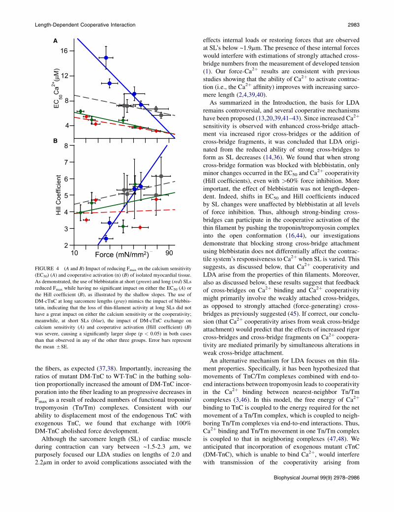
The results above establish that force inhibition byDM-cTnCincorporation has more distinct effects on the length depen-dence of myofilament activation than does that by blebbista-tin. To further illustrate these differences, we plotted therelationships between EC50 and the Hill coefficient (cooper-ative activation) with Fmax for both blebbistatin-treated andDM-cTnC-exchanged fibers. The rationale for this analysisis that Fmax provides an estimate of the extent of interferencewith myofilament activation, thereby allowing comparisonsbetween the DM-TnC interventions at the level of the thinfilament and the blebbistatin interventions at the level ofthe thick filament. Fig. 4 shows that the EC50 correlatedinversely with Fmax at both sarcomere lengths for eitherDM-TnC or blebbistatin treatment. This inverse relationshipbetween EC50 and Fmax is predicted from current models ofCa2þ-dependent myofilament activation that arises fromthe interaction between Ca2þ binding and cross-bridgeattachment (13,35). It is clear that the slope of the EC50-Fmax relationship in the DM-cTnC-exchanged fibers at shortlength (Fig. 4, blue) was much greater (P< 0.05) than for thelong sarcomere length or for the blebbistatin-treated fibers ateither length. On the other hand, the slopes of the Fmax-EC50
relationship did not differ (P¼ 0.20) between the two lengthsin the blebbistatin experiments, nor did these slopes differ(P¼ 0.24) from the DM-TnC-exchange group at long length.The absence of difference between the long and short lengthsin the blebbistatin group is somewhat surprising, sinceprevious studies had concluded that LDA results fromlength-dependent difference in strong cross-bridge attach-ment (36). It is interesting to note that the same relationshipis observed in the plot of Fmax with the Hill parameter, sug-gesting a possible relationship between cooperativity andcalcium sensitivity. These results support the notion thatlength-dependent differences in Ca2þ sensitivity arise fromthe effects of length on thin-filament properties.
DISCUSSION
We compared the contribution of thin-filament activation viathe troponin-tropomyosin complex to that of strongcross-bridge attachment on the LDA in cardiac muscle.Strong cross-bridge attachment was altered in our studiesusing blebbistatin, an agent that has previously been shownto block myosin attachment to actin and myosin ATPaseactivity (21,22,29). To alter thin filament activation, weexchanged native TnC in the skinned fibers with variousmixtures of wild-type TnC and a mutant form of TnC,which is incapable of binding Ca2þ but which is otherwisestructurally normal (25). Under the experimental conditionsused in our replacement studies, we found a ~95% replace-ment of endogenous TnC. Moreover, the exogenous TnC(WT-TnC and DM-TnC) was uniformly incorporated into
4
8
12
16EC
50C
a2+(µ
M)
2
3
4
5
6
7
8
Hill
Coe
ffici
ent
2
A
B
10 90 Force (mN/mm2)
FIGURE 4 (A and B) Impact of reducing Fmax on the calcium sensitivity
(EC50) (A) and cooperative activation (n) (B) of isolated myocardial tissue.
As demonstrated, the use of blebbistatin at short (green) and long (red) SLs
reduced Fmax while having no significant impact on either the EC50 (A) or
the Hill coefficient (B), as illustrated by the shallow slopes. The use of
DM-cTnC at long sarcomere lengths (gray) mimics the impact of blebbis-
tatin, indicating that the loss of thin-filament activity at long SLs did not
have a great impact on either the calcium sensitivity or the cooperativity;
meanwhile, at short SLs (blue), the impact of DM-cTnC exchange on
calcium sensitivity (A) and cooperative activation (Hill coefficient) (B)
was severe, causing a significantly larger slope (p < 0.05) in both cases
than that observed in any of the other three groups. Error bars represent
the mean 5SE.
Length-Dependent Cooperative Interaction 2983
the fibers, as expected (37,38). Importantly, increasing theratios of mutant DM-TnC to WT-TnC in the bathing solu-tion proportionally increased the amount of DM-TnC incor-poration into the fiber leading to an progressive decreases inFmax as a result of reduced numbers of functional troponin/tropomyosin (Tn/Tm) complexes. Consistent with ourability to displacement most of the endogenous TnC withexogenous TnC, we found that exchange with 100%DM-TnC abolished force development.
Although the sarcomere length (SL) of cardiac muscleduring contraction can vary between ~1.5-2.3 mm, wepurposely focused our LDA studies on lengths of 2.0 and2.2mm in order to avoid complications associated with the
effects internal loads or restoring forces that are observedat SL’s below ~1.9mm. The presence of these internal forceswould interfere with estimations of strongly attached cross-bridge numbers from the measurement of developed tension(1). Our force-Ca2þ results are consistent with previousstudies showing that the ability of Ca2þ to activate contrac-tion (i.e., the Ca2þ affinity) improves with increasing sarco-mere length (2,4,39,40).
As summarized in the Introduction, the basis for LDAremains controversial, and several cooperative mechanismshave been proposed (13,20,39,41–43). Since increased Ca2þ
sensitivity is observed with enhanced cross-bridge attach-ment via increased rigor cross-bridges or the addition ofcross-bridge fragments, it was concluded that LDA origi-nated from the reduced ability of strong cross-bridges toform as SL decreases (14,36). We found that when strongcross-bridge formation was blocked with blebbistatin, onlyminor changes occurred in the EC50 and Ca
2þ cooperativity(Hill coefficients), even with >60% force inhibition. Moreimportant, the effect of blebbistatin was not length-depen-dent. Indeed, shifts in EC50 and Hill coefficients inducedby SL changes were unaffected by blebbistatin at all levelsof force inhibition. Thus, although strong-binding cross-bridges can participate in the cooperative activation of thethin filament by pushing the troponin/tropomyosin complexinto the open conformation (16,44), our investigationsdemonstrate that blocking strong cross-bridge attachmentusing blebbistatin does not differentially affect the contrac-tile system’s responsiveness to Ca2þwhen SL is varied. Thissuggests, as discussed below, that Ca2þ cooperativity andLDA arise from the properties of thin filaments. Moreover,also as discussed below, these results suggest that feedbackof cross-bridges on Ca2þ binding and Ca2þ cooperativitymight primarily involve the weakly attached cross-bridges,as opposed to strongly attached (force-generating) cross-bridges as previously suggested (45). If correct, our conclu-sion (that Ca2þ cooperativity arises from weak cross-bridgeattachment) would predict that the effects of increased rigorcross-bridges and cross-bridge fragments on Ca2þ coopera-tivity are mediated primarily by simultaneous alterations inweak cross-bridge attachment.
An alternative mechanism for LDA focuses on thin fila-ment properties. Specifically, it has been hypothesized thatmovements of TnC/Tm complexes combined with end-to-end interactions between tropomyosin leads to cooperativityin the Ca2þ binding between nearest-neighbor Tn/Tmcomplexes (3,46). In this model, the free energy of Ca2þ
binding to TnC is coupled to the energy required for the netmovement of a Tn/Tm complex, which is coupled to neigh-boring Tn/Tm complexes via end-to-end interactions. Thus,Ca2þ binding and Tn/Tm movement in one Tn/Tm complexis coupled to that in neighboring complexes (47,48). Weanticipated that incorporation of exogenous mutant cTnC(DM-TnC), which is unable to bind Ca2þ, would interferewith transmission of the cooperativity arising from
Biophysical Journal 99(9) 2978–2986

FIGURE 5 Schematic drawing of the proposed mechanism of LDA,
showing tropomyosin (gray lines), actin (ovals), and cardiac troponin units
(tricolored rectangles). In our version of the three-state model, the impact
of calcium on the activation of the neighboring units (dashed black line) at
short SL works to activate the neighboring cTn unit, and thus, calcium
needs to bind to more Tn units (arrows), whereas at longer SL, the activa-
tion of one cTn unit, due to the proposed stiffening of the tropomyosin, is
able to activate more cTn units, thus requiring less calcium to activate the
same number of cTn units (arrows). In other words, stretching the SL theo-
retically shifts the requirement for calcium binding from every 1–2 tropo-
nins to approximately every 3–4 troponins, allowing for more binding
sites to be activated with the same levels of calcium activation.
2984 Farman et al.
end-to-end interactions between Tn/Tm and thereby disruptCa2þ cooperativity. Consistent with this prediction,increased DM-TnC incorporation caused progressiveincreases in EC50 and simultaneous reductions in the Hillcoefficient and Fmax at both lengths. However, the impactof DM-TnC incorporation on the EC50 and the Hill coeffi-cient were far more pronounced at the short sarcomerelength. This is best appreciated by plots of the EC50 or Hillcoefficients as a function of the maximal force (Fmax) gener-ated, where Fmax accounts for the differences in force gener-ation between the fibers (c.f. Fig. 4). It is clear that for theranges of DM-TnC incorporation studied, which werelimited to solutions containing >~50% DM-TnC becauseof fiber instability (i.e., large variability between fibers),EC50 and Hill coefficient depended much more steeply onFmax for the short than for the long sarcomere length. Regard-less, for the range of DM-TnC incorporation used in ourstudies, we observed a twofold increase in the EC50 (i.e.,Ca2þ sensitivity) at the short sarcomere length comparedwith a 40% change in EC50 at the long sarcomere lengthwhen Fmax was reduced by ~40%. In a similar way, the Hillcoefficient (i.e., Ca2þ cooperativity) decreased far more atthe short length than at the long length. It is interesting thatthe dependence of EC50 and Hill coefficient on Fmax in theblebbistatin-treated fibers at either length was similar tothat observed at the long length in the DM-TnC group.
How canwe explain our findings that it is interferencewiththe number of TnC/Tm complexes able to bind Ca2þ, and notstrong cross-bridge attachment, that most affects Ca2þ coop-erativity and LDA? In principle, Ca2þ cooperativity of forcegeneration must ultimately arise from interactions betweenneighboring Tn/Tm complexes (44,49). Previous two-statebiochemical models for Tn/Tm (50,51) have linked this co-operativity to feedback of cross-bridge attachment on Ca2þ
binding in adjacent Tn/Tm complexes wherein strongcross-bridge attachment drives Tn/Tm into the open state,which has a high Ca2þ-binding affinity. However, the modesteffect of blebbistatin on the Ca2þ activation properties (i.e.,sensitivity and cooperativity) and LDA, though initiallysurprising given findings in previous studies (13,14,52),cannot be easily explained using a two-state model. On theother hand, our results are consistent with a three-statebiochemical model wherein the states have distinct Ca2þ
binding properties and are topologically connected in a linearmanner (blocked $ closed $ open) (44,47,48). Specifi-cally, because strong cross-bridge attachment did not affectcooperativity, and because strong cross-bridge attachmentin this model is associated with closed-to-open-state transi-tions, this model predicts that there are larger differences inCa2þ affinity between the blocked and closed states thanbetween the closed and open states. Thus, since closed statesare stabilized by weak cross-bridge attachment, weak-binding cross-bridges, which are involved in blocked-to-closed-state transitions, are predicted to be the primarymediators of Ca2þ cooperativity, as proposed previously
Biophysical Journal 99(9) 2978–2986
(53,54). By contrast, closed-to-open-state transitions arenot expected to enhance the Ca2þ-binding affinity and coop-erativity. This conclusion is consistent with those of studiesshowing that Ca2þ cooperativity for myofilament activationis mediated by increased probabilities of closed-state occu-pancy of a Tn/Tm complex after Ca2þ binding to the adjacentTn/Tm complex (55,56). This mechanism also readilyexplains the effects of exogenous myosin and rigor bridgeson Ca2þ sensitivity without invoking the need for strongcross-bridge binding. Specifically, it is predicted that bothexogenous myosin and low ATP conditions to promote rigorbridge formation force Tn/Tm complexes in the process ofbinding to force transitions initially through the closed stateeither by stoichiometric mass action or by distortion of thecross-bridge binding equilibria with actin (due to reducedoff rates of rigor bridges when ATP is low). Consistent withthis suggestion, it has been shown that the structural impactof rigor cross-bridges on Tn/Tm complexes is differentfrom that induced by cycling cross-bridges (57).
The remarkable ability of DM-TnC incorporation tostrongly influence Ca2þ sensitivity and Ca2þ cooperativityat short, compared to long, lengths suggests that SL affectsinteractions between Tn/Tm complexes. In other words,Ca2þ cooperativity appears to depend more strongly on thenumber of TnC Ca2þ binding sites at shorter SLs than atlonger SLs. These findings suggest that coupling betweenadjacent Tn/Tm complexes increases with length, therebymaking activation less dependent on the number of TnC siteswith Ca2þ bound. Thus, when Ca2þ binds to a given Tn/Tmcomplex, the probability of the neighboring complexes (andbeyond) to transition to the closed state is enhanced at longerlengths due to stronger coupling (Fig. 5). In an equivalent

Length-Dependent Cooperative Interaction 2985
manner, within the framework of the flexible Tn/Tm model(49), increases of SL increase the persistence length byreducing Tn/Tm flexibility, thereby allowing the influenceof Ca2þ binding to propagate further along the thin filament.Regardless of the model, the propagation of activation is, ofcourse, aided by weak cross-bridge attachment, whichstabilizes the closed state, leading ultimately to open-statetransitions of Tn/Tm and strong cross-bridge binding. If prop-agation of activation is indeed reduced at short SLs, wepredict, incorporation of DM-TnC molecules will causegreater disruptions of activation at short lengths than at longerlengths, with the effects being more pronounced at submax-imal Ca2þ levels (i.e., affecting EC50 and Hill coefficients)than at maximal Ca2þ levels (i.e., affecting Fmax), as shownby our results. The mechanism for the length dependence ofthe propagation of Ca2þ activation is unclear but couldinvolve the interactions observed between cTnT and regula-tory light chains (58,59) or titin (60) binding to the thin fila-ments at the Z-disc (61) and other proteins (62,63). Clearly,further studies will be needed to assess the molecular basisfor the enhanced propagation of Ca2þ with increased SL.
We thank Dr. Kate Sheehan for her assistance in gathering and analysis of
the confocal images.
These studies were supported in part by the National Institutes of Health
(grant HL-75494 to P.P.d.T.) and the Canadian Institutes for Health
Research (grant MOP-79460 to P.H.B.).
REFERENCES
1. Allen, D. G., and J. C. Kentish. 1985. The cellular basis of the length-tension relation in cardiac muscle. J. Mol. Cell. Cardiol. 17:821–840.
2. Dobesh, D. P., J. P. Konhilas, and P. P. de Tombe. 2002. Cooperativeactivation in cardiac muscle: impact of sarcomere length. Am. J. Phys-iol. Heart Circ. Physiol. 282:H1055–H1062.
3. Gordon, A. M., E. Homsher, and M. Regnier. 2000. Regulation ofcontraction in striated muscle. Physiol. Rev. 80:853–924.
4. Kentish, J. C., H. E. ter Keurs, ., M. I. Noble. 1986. Comparisonbetween the sarcomere length-force relations of intact and skinnedtrabeculae from rat right ventricle. Influence of calcium concentrationson these relations. Circ. Res. 58:755–768.
5. de Beer, E. L., R. L. Grundeman, ., P. Schiereck. 1988. Effect ofsarcomere length and filament lattice spacing on force developmentin skinned cardiac and skeletal muscle preparations from the rabbit.Basic Res. Cardiol. 83:410–423.
6. Palmer, S., and J. C. Kentish. 1994. The role of troponin C in modu-lating the Ca2þ sensitivity of mammalian skinned cardiac and skeletalmuscle fibres. J. Physiol. 480:45–60.
7. Wannenburg, T., P. M. Janssen, ., P. P. de Tombe. 1997. The Frank-Starling mechanism is not mediated by changes in rate of cross-bridgedetachment. Am. J. Physiol. 273:H2428–H2435.
8. Maytum, R., S. S. Lehrer, and M. A. Geeves. 1999. Cooperativity andswitching within the three-state model of muscle regulation. Biochem-istry. 38:1102–1110.
9. Babu, A., S. P. Scordilis,., J. Gulati. 1987. The control of myocardialcontraction with skeletal fast muscle troponin C. J. Biol. Chem.262:5815–5822.
10. Gulati, J., E. Sonnenblick, and A. Babu. 1991. The role of troponin C inthe length dependence of Ca2þ-sensitive force of mammalian skeletaland cardiac muscles. J. Physiol. 441:305–324.
11. Moss, R. L., L. O. Nwoye, and M. L. Greaser. 1991. Substitution ofcardiac troponin C into rabbit muscle does not alter the length depen-dence of Ca2þ sensitivity of tension. J. Physiol. 440:273–289.
12. Fitzsimons, D. P., J. R. Patel,., R. L. Moss. 2001. Cooperative mech-anisms in the activation dependence of the rate of force development inrabbit skinned skeletal muscle fibers. J. Gen. Physiol. 117:133–148.
13. Fitzsimons, D. P., J. R. Patel, and R. L. Moss. 2001. Cross-bridge inter-action kinetics in rat myocardium are accelerated by strong binding ofmyosin to the thin filament. J. Physiol. 530:263–272.
14. Fitzsimons, D. P., and R. L. Moss. 1998. Strong binding of myosinmodulates length-dependent Ca2þ activation of rat ventricular myo-cytes. Circ. Res. 83:602–607.
15. McDonald, K. S., and R. L. Moss. 2000. Strongly binding myosincrossbridges regulate loaded shortening and power output in cardiacmyocytes. Circ. Res. 87:768–773.
16. Vibert, P., R. Craig, and W. Lehman. 1997. Steric-model for activationof muscle thin filaments. J. Mol. Biol. 266:8–14.
17. Zhao, Y., and M. Kawai. 1993. The effect of the lattice spacing changeon cross-bridge kinetics in chemically skinned rabbit psoas musclefibers. II. Elementary steps affected by the spacing change. Biophys.J. 64:197–210.
18. Butters, C. A., J. B. Tobacman, and L. S. Tobacman. 1997. Cooperativeeffect of calcium binding to adjacent troponin molecules on the thinfilament-myosin subfragment 1 MgATPase rate. J. Biol. Chem.272:13196–13202.
19. Campbell, K. B., M. V. Razumova, ., B. K. Slinker. 2001. Nonlinearmyofilament regulatory processes affect frequency-dependent musclefiber stiffness. Biophys. J. 81:2278–2296.
20. Razumova, M. V., A. E. Bukatina, and K. B. Campbell. 2000. Differentmyofilament nearest-neighbor interactions have distinctive effects oncontractile behavior. Biophys. J. 78:3120–3137.
21. Allingham, J. S., R. Smith, and I. Rayment. 2005. The structural basisof blebbistatin inhibition and specificity for myosin II. Nat. Struct. Mol.Biol. 12:378–379.
22. Farman, G. P., K. Tachampa,., P. P. de Tombe. 2008. Blebbistatin: useas inhibitor of muscle contraction. Pflugers Arch. 455:995–1005.
23. Dou, Y., P. Arlock, and A. Arner. 2007. Blebbistatin specificallyinhibits actin-myosin interaction in mouse cardiac muscle. Am. J. Phys-iol. Cell. Physiol. 293:C1148–C1153.
24. Farman, G. P., J. S. Walker, ., T. C. Irving. 2006. Impact of osmoticcompression on sarcomere structure and myofilament calcium sensi-tivity of isolated rat myocardium. Am. J. Physiol. Heart Circ. Physiol.291:H1847–H1855.
25. Strynadka, N. C., M. Cherney,., M. N. James. 1997. Structural detailsof a calcium-induced molecular switch: x-ray crystallographic analysisof the calcium-saturated N-terminal domain of troponin C at 1.75 Aresolution. J. Mol. Biol. 273:238–255.
26. Li, M. X., S. M. Gagne, ., B. D. Sykes. 1997. NMR studies of Ca2þ
binding to the regulatory domains of cardiac and E41A skeletal muscletroponin C reveal the importance of site I to energetics of the inducedstructural changes. Biochemistry. 36:12519–12525.
27. Li, M. X., L. Spyracopoulos, and B. D. Sykes. 1999. Binding of cardiactroponin-I147–163 induces a structural opening in human cardiactroponin-C. Biochemistry. 38:8289–8298.
28. Spyracopoulos, L., M. X. Li, ., B. D. Sykes. 1997. Calcium-inducedstructural transition in the regulatory domain of human cardiactroponin C. Biochemistry. 36:12138–12146.
29. Limouze, J., A. F. Straight, ., J. R. Sellers. 2004. Specificity of bleb-bistatin, an inhibitor of myosin II. J. Muscle Res. Cell Motil. 25:337–341.
30. Kovacs, M., J. Toth,., J. R. Sellers. 2004. Mechanism of blebbistatininhibition of myosin II. J. Biol. Chem. 279:35557–35563.
31. Straight, A. F., A. Cheung, ., T. J. Mitchison. 2003. Dissectingtemporal and spatial control of cytokinesis with a myosin II inhibitor.Science. 299:1743–1747.
Biophysical Journal 99(9) 2978–2986

2986 Farman et al.
32. Sakamoto, T., J. Limouze,., J. R. Sellers. 2005. Blebbistatin, a myosinII inhibitor, is photoinactivated by blue light. Biochemistry. 44:584–588.
33. Ranatunga, K. W. 1999. Endothermic force generation in skinnedcardiac muscle from rat. J. Muscle Res. Cell Motil. 20:489–496.
34. Konhilas, J. P., T. C. Irving, and P. P. de Tombe. 2002. Myofilamentcalcium sensitivity in skinned rat cardiac trabeculae: role of interfila-ment spacing. Circ. Res. 90:59–65.
35. Lehrer, S. S., and M. A. Geeves. 1998. The muscle thin filament asa classical cooperative/allosteric regulatory system. J. Mol. Biol.277:1081–1089.
36. Swartz, D. R., and R. L. Moss. 1992. Influence of a strong-bindingmyosin analogue on calcium-sensitive mechanical properties ofskinned skeletal muscle fibers. J. Biol. Chem. 267:20497–20506.
37. Bell, M. G., E. B. Lankford,., R. J. Barsotti. 2006. Kinetics of cardiacthin-filament activation probed by fluorescence polarization of rhoda-mine-labeled troponin C in skinned guinea pig trabeculae. Biophys.J. 90:531–543.
38. Brenner, B., T. Kraft, ., J. M. Chalovich. 1999. Thin filament activa-tion probed by fluorescence of N-((2-(iodoacetoxy)ethyl)-N-methyl)amino-7-nitrobenz-2-oxa-1,3-diazole-labeled troponin I incorporatedinto skinned fibers of rabbit psoas muscle. Biophys. J. 77:2677–2691.
39. Smith, S. H., and F. Fuchs. 2000. Length-dependence of cross-bridgemediated activation of the cardiac thin filament. J. Mol. Cell. Cardiol.32:831–838.
40. Konhilas, J. P., T. C. Irving, and P. P. de Tombe. 2002. Length-depen-dent activation in three striated muscle types of the rat. J. Physiol.544:225–236.
41. Martyn, D. A., P. B. Chase, ., A. M. Gordon. 2002. A simple modelwith myofilament compliance predicts activation-dependent cross-bridge kinetics in skinned skeletal fibers. Biophys. J. 83:3425–3434.
42. McDonald, K. S., M. R. Wolff, and R. L. Moss. 1997. Sarcomere lengthdependence of the rate of tension redevelopment and submaximaltension in rat and rabbit skinned skeletal muscle fibres. J. Physiol.501:607–621.
43. de Tombe, P. P., R. D. Mateja, ., T. C. Irving. 2010. Myofilamentlength dependent activation. J. Mol. Cell. Cardiol. 48:851–858.
44. McKillop, D. F., and M. A. Geeves. 1993. Regulation of the interactionbetween actin and myosin subfragment 1: evidence for three states ofthe thin filament. Biophys. J. 65:693–701.
45. Smith, L., C. Tainter,., D. A. Martyn. 2009. Cooperative cross-bridgeactivation of thin filaments contributes to the Frank-Starling mecha-nism in cardiac muscle. Biophys. J. 96:3692–3702.
46. Gillis, T. E., D. A. Martyn, A. J. Rivera, and M. Regnier. 2007. Inves-tigation of thin filament near-neighbor regulatory unit interactionsduring skinned rat cardiac muscle force development. J. Physiol.580:561–576. comment 580:358.
47. Eisenberg, E., and T. L. Hill. 1978. A cross-bridge model of musclecontraction. Prog. Biophys. Mol. Biol. 33:55–82.
Biophysical Journal 99(9) 2978–2986
48. Hill, T. L., E. Eisenberg,., R. J. Podolsky. 1975. Some self-consistent
two-state sliding filament models of muscle contraction. Biophys. J.
15:335–372.
49. Boussouf, S. E., and M. A. Geeves. 2007. Tropomyosin and troponin
cooperativity on the thin filament. Adv. Exp. Med. Biol. 592:99–109.
50. Geeves, M. A., and D. J. Halsall. 1987. Two-step ligand binding and
cooperativity. A model to describe the cooperative binding of myosin
subfragment 1 to regulated actin. Biophys. J. 52:215–220.
51. McKillop, D. F., and M. A. Geeves. 1991. Regulation of the acto.my-
osin subfragment 1 interaction by troponin/tropomyosin. Evidence for
control of a specific isomerization between two acto.myosin subfrag-
ment 1 states. Biochem. J. 279:711–718.
52. Fukuda, N., H. Kajiwara, ., S. Kurihara. 2000. Effects of MgADP on
length dependence of tension generation in skinned rat cardiac muscle.
Circ. Res. 86:E1–E6.
53. Rice, J. J., F. Wang, ., P. P. de Tombe. 2008. Approximate model of
cooperative activation and crossbridge cycling in cardiac muscle using
ordinary differential equations. Biophys. J. 95:2368–2390.
54. Rice, J. J., Y. Tu, ., P. P. De Tombe. 2008. Spatially-compressed
cardiac myofilament models generate hysteresis that is not found in
real muscle. Pac. Symp. Biocomput. 2008:366–377.
55. Gong, H., V. Hatch, ., L. S. Tobacman. 2005. Mini-thin filaments
regulated by troponin-tropomyosin. Proc. Natl. Acad. Sci. USA.
102:656–661.
56. Gafurov, B., Y. D. Chen, and J. M. Chalovich. 2004. Ca2þ and ionic
strength dependencies of S1-ADP binding to actin-tropomyosin-troponin: regulatory implications. Biophys. J. 87:1825–1835.
57. Sun, Y. B., F. Lou, and M. Irving. 2009. Calcium- and myosin-depen-
dent changes in troponin structure during activation of heart muscle. J.
Physiol. 587:155–163.
58. Farman, G. P., M. S. Miller,., T. C. Irving. 2009. Phosphorylation and
the N-terminal extension of the regulatory light chain help orient and
align the myosin heads in Drosophila flight muscle. J. Struct. Biol.
168:240–249.
59. Cazorla, O., S. Szilagyi,., A. Lacampagne. 2005. Transmural stretch-
dependent regulation of contractile properties in rat heart and its alter-
ation after myocardial infarction. FASEB J. 19:88–90.
60. Granzier, H., and S. Labeit. 2002. Cardiac titin: an adjustable multi-
functional spring. J. Physiol. 541:335–342.
61. Trombitas, K., and H. Granzier. 1997. Actin removal from cardiac my-
ocytes shows that near Z line titin attaches to actin while under tension.
Am. J. Physiol. 273:C662–C670.
62. Winegrad, S. 1999. Cardiac myosin binding protein C. Circ. Res.
84:1117–1126.
63. Trombitas, K., J. P. Jin, and H. Granzier. 1995. The mechanically active
domain of titin in cardiac muscle. Circ. Res. 77:856–861.