Heterogeneity of a Campylobacter jejuni Protein That Is Secreted through the Flagellar Filament
10
Published Ahead of Print 21 May 2007. 2007, 75(8):3859. DOI: 10.1128/IAI.00159-07. Infect. Immun. Phan, Nicholas J. Savarino and Patricia Guerry Hickey, David Rockabrand, Gary Majam, Lanfong Lee, Julie Frédéric Poly, Cheryl Ewing, Scarlett Goon, Thomas E. Flagellar Filament Protein That Is Secreted through the Campylobacter jejuni Heterogeneity of a http://iai.asm.org/content/75/8/3859 Updated information and services can be found at: These include: REFERENCES http://iai.asm.org/content/75/8/3859#ref-list-1 at: This article cites 50 articles, 29 of which can be accessed free CONTENT ALERTS more» articles cite this article), Receive: RSS Feeds, eTOCs, free email alerts (when new http://journals.asm.org/site/misc/reprints.xhtml Information about commercial reprint orders: http://journals.asm.org/site/subscriptions/ To subscribe to to another ASM Journal go to: on October 2, 2014 by guest http://iai.asm.org/ Downloaded from on October 2, 2014 by guest http://iai.asm.org/ Downloaded from
Transcript of Heterogeneity of a Campylobacter jejuni Protein That Is Secreted through the Flagellar Filament

Published Ahead of Print 21 May 2007. 2007, 75(8):3859. DOI: 10.1128/IAI.00159-07. Infect. Immun.
Phan, Nicholas J. Savarino and Patricia GuerryHickey, David Rockabrand, Gary Majam, Lanfong Lee, Julie Frédéric Poly, Cheryl Ewing, Scarlett Goon, Thomas E. Flagellar Filament Protein That Is Secreted through the
Campylobacter jejuniHeterogeneity of a
http://iai.asm.org/content/75/8/3859Updated information and services can be found at:
These include:
REFERENCEShttp://iai.asm.org/content/75/8/3859#ref-list-1at:
This article cites 50 articles, 29 of which can be accessed free
CONTENT ALERTS more»articles cite this article),
Receive: RSS Feeds, eTOCs, free email alerts (when new
http://journals.asm.org/site/misc/reprints.xhtmlInformation about commercial reprint orders: http://journals.asm.org/site/subscriptions/To subscribe to to another ASM Journal go to:
on October 2, 2014 by guest
http://iai.asm.org/
Dow
nloaded from
on October 2, 2014 by guest
http://iai.asm.org/
Dow
nloaded from

INFECTION AND IMMUNITY, Aug. 2007, p. 3859–3867 Vol. 75, No. 80019-9567/07/$08.00�0 doi:10.1128/IAI.00159-07
Heterogeneity of a Campylobacter jejuni Protein That Is Secretedthrough the Flagellar Filament�
Frederic Poly, Cheryl Ewing, Scarlett Goon, Thomas E. Hickey, David Rockabrand, Gary Majam,Lanfong Lee, Julie Phan, Nicholas J. Savarino, and Patricia Guerry*
Enteric Diseases Department, Naval Medical Research Center, Silver Spring, Maryland 20910
Received 30 January 2007/Returned for modification 12 March 2007/Accepted 13 May 2007
Cj0859c, or FspA, is a small, acidic protein of Campylobacter jejuni that is expressed by a �28 promoter.Analysis of the fspA gene in 41 isolates of C. jejuni revealed two overall variants of the predicted protein,FspA1 and FspA2. Secretion of FspA occurs in broth-grown bacteria and requires a minimum flagellarstructure. The addition of recombinant FspA2, but not FspA1, to INT407 cells in vitro resulted in a rapidinduction of apoptosis. These data define a novel C. jejuni virulence factor, and the observed heterogeneityamong fspA alleles suggests alternate virulence potential among different strains.
Campylobacter jejuni is among the leading causes of bacterialdiarrhea worldwide (12, 40). Campylobacter enteritis canpresent with a range of clinical symptoms, and this variabilityhas been attributed to either differences in the host or differ-ences in virulence among strains. The molecular pathogenesisof C. jejuni has proven particularly refractory to analysis. Thismay be due, in part, to the absence of virulence factors in thegenome that are analogous to those of better-characterizedpathogens. Thus, the only virulence factor in C. jejuni that isshared with other pathogens is cytolethal distending toxin(CDT). There are no genes encoding other known toxins orspecialized secretion systems common to all C. jejuni strains.
Flagella are considered to be virulence factors for C. jejunifor numerous reasons. The motility imparted by the locomo-tory organelle is critical to intestinal colonization of both ani-mals and humans (3, 31). Motility and chemotaxis also modu-late the invasion of intestinal epithelial cells in vitro (7, 20,47–49). The flagellin subunits in the Campylobacter flagellarfilament are heavily glycosylated with pseudaminic acid andlegionaminic acid (29, 44). This glycosylation is required forfilament assembly (15), and changes in glycan composition canaffect both the immunogenicity of flagellin and microcolonyformation (17, 28). Campylobacter flagella also appear to func-tion as a type III secretion system (T3SS) in the absence of aspecialized T3SS in this pathogen. Thus, strains of C. jejunisecrete the Cia (Campylobacter invasion antigen) proteins (24,25, 39) and FlaC (41) through the filament.
There is limited information about the flagellar regulon ofC. jejuni. However, it appears that most genes encoding thestructural components of the basal body, hook, and minorflagellin, FlaB, are regulated by �54 promoters, and the majorflagellin, FlaA, is regulated by a �28 promoter (16) and possiblya �70 promoter as well (21). Carrillo et al. previously per-formed expression profiling studies on two variants of NCTC11168 with different virulence levels (6). Those workers ob-
served that a number of nonflagellar genes that were con-trolled by �54 or �28 promoters displayed distinct levels ofexpression, suggesting that some of these flagellum-coregu-lated genes may contribute to virulence (6). We have recentlyshown that mutation of one of these �28-regulated genes,Cj0977, affected the virulence of C. jejuni 81-176 in vitro and invivo (14). Another of these putative �28-regulated genes,Cj0859c, which encodes a protein with no homology to knownproteins, maps in a region of the chromosome that has beenreported to be variable among strains in a recent microarraystudy (33). Analysis of this region in the C. jejuni genomesdeposited in GenBank indicated that the putative �28 pro-moter was conserved but that there was variation in theCj0859c gene. Here, we demonstrate that the predicted pro-teins encoded by the Cj0859c genes in isolates of C. jejuni fromdifferent geographical areas show considerable allelic varia-tion. We have characterized two variants of the protein anddemonstrate that both forms of the protein are secreted intothe supernatant in a process that requires a minimum flagellarstructure. Moreover, we demonstrate significant biological dif-ferences in two variants of the protein.
MATERIALS AND METHODS
Bacterial strains and growth conditions. C. jejuni strain 81-176 and Campy-lobacter coli strain VC167 have been described previously (3, 19). Table 1 sum-marizes mutants of 81-176 used in this study. All of the flagellar mutants, exceptPG2157 (flgI) and PG2158 (flgK), were previously described (14). PG2157 andPG2158 were generated by transposon mutagenesis of clones of the flgI and flgKgenes in Escherichia coli, respectively, from 81-176 using Tn5-based in vitrotransposition (15, 18), followed by electroporation into 81-176. The insertion intoflgI occurred at bp 363 of the 1,047-bp open reading frame; the insertion into flgKoccurred at bp 113 of the 1,827-bp open reading frame. Both mutants werenonmotile and lacked all flagellar structure by negative-stain electron micros-copy. Other strains of wild-type C. jejuni are shown in Table 2. Campylobacterstrains were grown routinely on Mueller-Hinton (MH) agar supplemented with50 �g/ml kanamycin and/or 15 �g/ml chloramphenicol as needed.
RT-PCR analysis. To confirm the regulation of fspA by �28, the relativeexpression of this gene was determined by real-time PCR (RT-PCR) in wild-type81-176 and DRH311, which is a �28 deletion of 81-176 (�fliA) (21). RNAs wereextracted from mid-log-phase cultures of C. jejuni 81-176 and the �fliA straingrown in biphasic MH medium. Synthesis of cDNA was performed using aniScript cDNA synthesis kit (Bio-Rad, La Jolla, CA). RT-PCR was performedwith the ABI Prism 7000 DNA analyzer (Applied Biosystems, Foster City, CA)using a QuantiTect SYBR green RT-PCR kit (QIAGEN, Valencia, CA) accord-
* Corresponding author. Mailing address: Enteric Diseases Depart-ment, Naval Medical Research Center, 503 Robert Grant Ave., SilverSpring, MD 20910. Phone: (301) 319-7662. Fax: (301) 319-7679. E-mail:[email protected].
� Published ahead of print on 21 May 2007.
3859
on October 2, 2014 by guest
http://iai.asm.org/
Dow
nloaded from

ing to the manufacturer’s protocol. Primer sequences are given in Table 3.RT-PCR was followed by a melting curve analysis in order to confirm amplifi-cation of specific PCR products. The expression of fspA in each strain wasnormalized to 16S and 23S RNA. The relative fspA expression level was obtainedusing the ��CT method as recommended in the Applied Biosystems manual.Results are based on at least three independent experiments.
PCR amplification and sequence analysis of different Cj0859c alleles. TheCj0859c-Cj0860 region of the chromosome of C. jejuni strains was PCR ampli-fied using primers pg06.14 (5�-CCTATTTATGGATTGCAATTTCACCCCG-3�) and pg06.15 (5�-CTTGAAACGATCAAGGGTAGGGCAGC-3�). Primer
TABLE 1. C. jejuni 81-176 mutants used in this study
Strain Genemutated
Parentstrain Genotype Reference or
source
PG2572 Cj0859c 81-176 fspA81-176::cat This studyDRH311 Cj0061 81-176 �fliA 19PG2662 None 81-176 astA::fspA8486 aphA3 This studyPG2573 Cj0859c PG2572 fspA81-176::cat (pCPE2575) This studyPG2663 Cj0859c PG2572 fspA81-176::cat
astA::fspA8486 aphA3This study
PG2453 Cj1729c 81-176 flgE::cat 13PG2139 Cj0548 81-176 fliD::cat 13PG2157 Cj1462 81-176 flgI::cat This studyPG2158 Cj1466 81-176 flgK::cat This study
TABLE 2. Strains used in the characterization of Cj0859c variant frequency in C. jejuni
C. jejuni strain Alternative strainname Origin Penner serotype GenBank accession no.
NCTC 11168a United Kingdom 2 NC_002163.184-25b United States ZP_01100726RM1221b United States 53 NC_003912.781-176b United States 23/36 ZP_01087888HB93-13b China 19 ZP_01071420CF93-6b Japan ZP_01068377260.94b South Africa 41 ZP_01070269MK19 Kuwait EF058200MK50 Kuwait EF058201MK52 Kuwait EF058202MK53 Kuwait EF058203MK59 Kuwait EF058204ATCC 43429 MSC 57360 Canada 1 EF058205ATCC 43430 PC 72 Canada 2 EF058206ATCC 43432 MK 7 Canada 4 EF058207ATCC 43438 Canada 10 EF058208ATCC 43446 MK 104 Canada 19 EF058209ATCC 43449 MK 198 Canada 23 EF058210ATCC 43456 MK 290 Canada 36 EF058212ATCC 43431 TGH 9011 Canada 3 EF058211LAN749 Egypt EF058213HS000128 Egypt EF058214HS001578 Egypt EF058215HS008516 Egypt EF058216OH 4384 Japan 19 EF058217PG836 Puerto Rico 10 EF058218CG-99-8013 Thailand EF058219CG-99-8023 Thailand EF058220CG-99-8071 Thailand EF058221CG-99-8087 Thailand EF058222CG-99-8109 Thailand 4/13/64/66 EF058223CG-99-8131 Thailand EF058224CG-99-8153 Thailand 4/10/13/16/64/66 EF058225CG-99-8245 Thailand EF058226CG-99-8261 Thailand 13 EF058227CG-99-8265 Thailand 13 EF058228CG-99-8289 Thailand EF058229CG-99-8431 Thailand EF058230CG-99-8437 Thailand EF058231CG-99-8486 Thailand 4/13/64/66 EF058232HC37 United States 27 EF058233
a See reference 34.b TIGR Campylobacter Genome Projects (D. E. Fouts and K. E. Nelson, unpublished data).
TABLE 3. Primer pairs used in RT-PCR studies
Locus Primer Primer sequence (5�–3�)
fspA1 RT-01 AAAACGCTTTAGCGCAAGATRT-02 TTGTTGCGTGTTGTTTTGTG
fspA2 RT-03 TGTTCATAGCGACAATGTGGTRT-04 AAGCCTCTAATGCGGGATTT
flaA RT-05 TCGTATTAACACCAATGTTGCAGRT-06 GCTTGAGATCTTAAACTATCTGCTATC
ilvC RT-07 AAAAAGTAGCAATTATAGGCTTTGGRT-08 TTGCTTTAACAGCACTTACACTACC
Cj0977 RT-09 ACCACAAGCGAAATGGTAGCRT-10 TCGTCAAAAAGTGCATGAGC
23S RNA RT-11 GGTAGGAGAGCGTTCTATTTGCRT-12 CGACTTAGGACCCGACTAACC
16S RNA RT-13 GGGAAAGTTTTTCGGTGTAGGRT-14 GTTGCTGCGTCAGGGTTT
3860 POLY ET AL. INFECT. IMMUN.
on October 2, 2014 by guest
http://iai.asm.org/
Dow
nloaded from

pg06.14 bound to the pabA gene (Cj0861), and pg06.15 bound to murA(Cj0858c). Amplicons were subjected to DNA sequence analysis using BigDyechemistry on an Applied Biosystems model 3100 DNA sequencer, and thesequences were assembled using Sequencher 4.5 (Gene Codes). Each consensussequence was manually inspected for quality assessment of the assembly. TheCj0859c gene sequence was identified for each consensus using Aretemis soft-ware (http://www.sanger.ac.uk/Software/Artemis/). Biological sequences werecompared using the SIM alignment tool (http://ca.expasy.org/tools/sim-prot.html). The phylogenetic tree was made using CLC Free Workbench 3 software(http://www.clcbio.com/index.php?id�28).
Expression of Cj0859c in E. coli. The fspA81-176 gene was amplified from81-176 by PCR using Easy-A High Fidelity PCR master mix (Stratagene, LaJolla, CA). The template was DNA from strain 81-176, and the primers usedwere pg05.53 (5�-GACGACGACAAGATGCAAATTAACAATTCCTTAAATAGC-3�) and pg05.54 (5�-GAGGAGAAGCCCGGTTCAAGCTTGTTGGCTTTGGAGTTC-3�). These primers included sequences for cloning into pET41-EKLIC, a vector that allows fusion to both a glutathione S-transferase (GST) tagand a hexahistidine tag (EMD Bioscience, Madison, WI). The 429-bp ampliconwas purified using QIAquick PCR purification columns (QIAGEN, Valencia,CA), ligated into pET41-EK LIC as directed by the manufacturer, and trans-formed into NovaBlue GigaSingles cells (EMD Biosciences). Several resultingclones were sequenced, and one clone, which showed the predicted sequence,was selected for protein purification. The GST-His-Cj0859c protein was purifiedby nickel chromatography (QIAGEN).
The FspA protein from 81-176 was also expressed as a histidine-tagged proteinin E. coli for biological experiments as follows. The gene was PCR amplified from81-176 using primers DR101 (5�-CCATATGCAAATTAACAATTCCTTAAATAGC-3�) and DR102 (5�-GGGATCCTCAAGCTTGTTGGCTTTGGAGTTC-3�) and HF2 DNA polymerase (Clontech). The resulting amplicon was digestedwith NdeI and BamHI and cloned into NdeI-BamHI-digested pET-19b (Nova-gen, San Diego, CA) in E. coli DH5�. Following confirmation of the correctconstruction by DNA sequencing, one resulting clone was transformed intoBL21(DE3), and the protein was overexpressed and purified on Ni-nitrilotriace-tic acid resin as recommended by the supplier (QIAGEN).
FspA from CG8486 was expressed as a histidine-tagged protein in pET-19busing primers DR103 (5�-CCATATGAAAATAGATACTTTGACAAAAAATTTTAGC-3�) and DR102 (shown above), as described above for 81-176.
Antibodies against different forms of FspA. Rabbit polyclonal antiserumagainst the GST-His-FspA from 81-176 and from the His-tagged FspA proteinfrom CG8486 described above were generated by Harlan Biosciences (Madi-son, WI).
SDS-PAGE and immunoblotting. Proteins were separated on 12.5% sodiumdodecyl sulfate (SDS)-polyacrylamide gel electrophoresis (PAGE) gels anddetected with Gel Code Blue (Pierce, Rockford, IL) or transferred onto nitro-cellulose and immunoblotted using the indicated rabbit polyclonal antisera. Pro-teins were immunodetected with either anti-GST-His-FspA81-176 or anti-His-FspA8486 polyclonal rabbit antiserum at a final dilution of 1:5,000. For assays tomeasure the binding of both recombinant proteins to INT407 cells, a mousemonoclonal antibody (Novagen) that recognizes the histidine tag was used at afinal dilution of 1:5,000. The secondary antibody was horseradish peroxidase-labeled goat anti-rabbit immunoglobulin G or goat anti-mouse antibody (Immu-nopure). The reactions were detected using Supersignal West Pico detection kits(Pierce, Rockford, IL). Chemiluminescence was detected using a Kodak (Roch-ester, NY) Image Station 2000R.
Purification of supernatants from C. jejuni. Bacteria were grown in MHbiphasic flasks (50 ml of MH broth over 20 ml MH agar in T75 tissue cultureflasks) for 18 h. Growth from two flasks (or 100 ml of MH broth) was centrifuged,and the supernatants were filtered through 0.2-�m polyvinylidene difluoridemembrane filters (Millipore, Billerica, CA). Trichloroacetic acid was added to afinal concentration of 10%, and the sample was iced for 45 min. The sampleswere centrifuged in a Sorvall RC5C refrigerated centrifuge at 10,000 � g for 15min, and the pellets were washed twice in 20 ml acetone. The pellets were airdried and resuspended in 64 �l of phosphate-buffered saline per 56 ml of culture.An equal volume of 2� solubilization buffer (1 M Tris [pH 6.8], 0.001% bromo-phenol blue, 5% glycerol, 3% SDS, 5% -mercaptoethanol) was added, and thesamples were boiled for 10 min prior to electrophoresis. Equal loading of sam-ples was confirmed by silver staining of SDS-PAGE gels.
Generation of an fspA mutant of 81-176. A mutant in fspA81-176 was con-structed using Tn5-based in vitro transposition (Epicenter, Madison, WI) with acat cassette as previously described (18). The in vitro reaction was performedaccording to the manufacturer’s instructions with a clone from a partiallySau3AI-digested ordered genomic library of 81-176 (L. Holder and P. Guerry,unpublished data), called pLCH8-63, as the target DNA. This clone contained
the region of the 81-176 chromosome that corresponded to genes Cj0856c toCj0860. The reaction products were transformed into E. coli DH5�, and plasmidDNAs from individual transformants were sequenced using primers that read outfrom within the cat cassette to determine the insertion point and the orientationof the transposon within the gene. A plasmid in which the cat cassette hadinserted 259 bp from the translational start of fspA in the same orientation asfspA was selected. This plasmid was used to electroporate C. jejuni 81-176 withselection on MH agar supplemented with chloramphenicol (15 �g/ml). Thesuccessful mutation of fspA was verified by PCR with primers bracketing the Cmr
insertion point to confirm that the DNA had undergone a double crossover. Thismutant was called PG2572.
Complementation of the Cj0859c mutant in trans. The fspA gene and its �28
promoter were PCR amplified from 81-176 using the following primers:pg05.133 (5�-CGGGATCCCACCGCTAATAGCCCAAAAAATACCTCCC-3�)and pg05.134 (5�-GGAATTCCGCAAGTATACTTGAAACGATCAAGGGTAGGG-3�). These primers added BamHI and EcoRI sites, respectively. The re-sulting 744-bp amplicon was digested with BamHI and EcoRI (New EnglandBiolabs, Beverly, MA) and cloned into similarly digested pRY107 (50). Afterconfirmation that the clone was correct by DNA sequence analysis, the plasmidwas transformed into DH5� cells carrying the conjugative plasmid RK212.2. Theresulting cells were used as donors to conjugatively transfer the complementingplasmid, pCE2575, into PG2166, the Cj0859c mutant, with selection on kanamy-cin and chloramphenicol, as previously described (17, 18).
Insertion of fspA8486 into the astA gene of 81-176 and selected mutants. ThefspA8486 gene and its �28 promoter were PCR amplified from CG8486 usingprimers C360.07 F (5�-TGCGGATCCCCGAAGCGGTTTTAACTCAA-3�) andC360.07 R (5�-ATGCGAATTCAAGGGTAGGGCAGCATTTTT-3�), which in-cluded BamHI and EcoRI restriction sites. The resulting amplicon was digestedwith these restriction enzymes and cloned into pBluescript. A SmaI-ended aphA3cassette was cloned into the EcoRV site of the resulting clone. DNA sequenceanalysis indicated that the aphA3 gene was inserted 3� to fspA8486 and was in thesame orientation as fspA8486. This plasmid was digested with BamHI and XhoI,which released fspA8486 and the adjacent aphA3 gene. This fragment was bluntedwith Klenow polymerase (New England Biolabs, Beverly, MA) and cloned intothe EcoRV site of plasmid pYG660 containing the astA (arylsulfatase) gene of81-176 (51). This plasmid was used to electroporate 81-176 and selected flagellarmutants to kanamycin resistance, as shown in Table 1. The resulting Kmr colo-nies were screened for a loss of arylsulfatase activity using MH agar supple-mented with a chromogenic substrate (X-S; Sigma, St. Louis, MO), as previouslydescribed (17, 51).
Adherence and invasion assays. Adherence and invasion assays were done aspreviously described (2, 32, 49). Briefly, about 2 � 106 bacteria were added to amonolayer of about 3 � 105 INT407 cells. After centrifugation at 200 � g for 5min, the assay mixtures were incubated at 37°C for 2 h. For the determination ofadherence, the cells were washed four times with Hanks’ balanced salt solution(HBSS) for 1 min before lysing the monolayer with 0.01% Triton X-100 andenumerating the total bacteria by plate counting on MH agar. For the determi-nation of invasion, the monolayer was washed twice with HBSS, and freshprewarmed modified Eagle’s medium (MEM) supplemented with 100 �g/mlgentamicin was added to wells for an additional 2 h to kill extracellular bacteria.The monolayer was washed four times in HBSS and lysed with 0.01% TritonX-100 for 30 min. Released intracellular bacteria were enumerated by platecount. Invasion was expressed as the percentage of the inoculum surviving thegentamicin treatment, and adherent bacteria were expressed as the total numberof bacteria enumerated without antibiotic treatment.
Binding of recombinant histidine-tagged C. jejuni proteins to INT407 cells.INT407 cells were seeded into 24-well tissue culture plates at about 5 � 105 cellsper well in MEM plus 10% fetal bovine serum. Following incubation at 37°C forabout 19 h, culture medium was removed and replaced with fresh MEM plus10% fetal bovine serum prewarmed at 37°C. Aliquots of 5, 10, 25, and 50 �g ofhistidine-tagged FspA8486 and 50 �g of histidine-tagged FspA81-176 were added,and the cells were incubated for 2 h at 37°C. The monolayer was washed fivetimes with phosphate-buffered saline and lysed in 200 �l of gel loading buffer(10% glycerol, 3% SDS, 0.01% bromophenol blue, 5% -mercaptoethanol, 1 MTris, and Halt protease inhibitor cocktail, EDTA free, from Pierce [Rockford,IL]). The samples were boiled for 10 min, and 10-�l aliquots were loaded ontoa 12.5% SDS-PAGE gel for each sample. The gel was transferred onto nitrocel-lulose and immunodetected with a mouse anti-His-tag monoclonal antibody, asdescribed above.
Apoptosis assays. Monolayers of INT407 cells in 24-well flat-bottom tissueculture dishes were treated with increasing amounts of recombinant FspA8486
or 50 �g of FspA81-176 and incubated for 4 h. Control wells were treated with10 �g of membrane proteins from 81-176 containing CDT (8, 13, 26) or 10 �g
VOL. 75, 2007 FLAGELLUM-SECRETED C. JEJUNI-SPECIFIC VIRULENCE FACTOR 3861
on October 2, 2014 by guest
http://iai.asm.org/
Dow
nloaded from

of membrane proteins from DS104, an isogenic cdtA mutant of 81-176 (22,23). Cells were harvested and stained using the Guava Nexin assay kit ac-cording to the manufacturer’s instructions (Guava Technologies, Hayward,CA). Basically, the cells were stained with annexin V-phycoerythrin andNexin 7-amino-actinomycin D (7-AAD) in cold 1� Nexin buffer in a 50-�lreaction mixture and analyzed in a Guava Technologies personal cytometer.Cells that stained positive with annexin V-phycoerythrin but negative withNexin 7-AAD were scored as being early apoptotic.
Statistical analyses. Statistical analyses of the RT-PCR results were calculatedwith a Wilcoxon two-group test. Statistical analyses of adherence, invasion, andapoptosis assays were done with two-tailed t tests.
RESULTS
Cj0859c is expressed by a �28 promoter in 81-176. Carrilloet al. previously demonstrated reduced expression of Cj0859cin a fliA mutant of NCTC 11168 by microarray, suggesting thatthis gene is �28 regulated, and the putative promoter sequenceis consistent with a �28 recognition sequence (6). Table 4 showsthe results of quantitative RT-PCR analysis of Cj0859c expres-sion in 81-176 and DRH311 (�fliA) (Table 1) (21). The dataindicate that there is a 30-fold reduction in the expression ofCj0859c in DRH311 compared to that of wild-type 81-176. Thisreduction is comparable to that seen for two other �28-regu-lated genes, flaA and Cj0977. In contrast, there was no signif-icant change in the expression of the �70-regulated ilvC(Cj0632) in DRH311 (�fliA) compared to that of 81-176.
Characterization of the Cj0859c locus in multiple strains ofC. jejuni. Analysis of the predicted proteins encoded by thehomologs of Cj0859c in three C. jejuni genomes deposited inGenBank, 260.94, CF93-6, and 84-25, revealed that the pre-dicted proteins, which are highly homologous to each other,show considerable divergence with the Cj0859c protein en-coded by both NCTC 11168 and 81-176. The gene correspond-ing to Cj0859c has been called fspA (flagellar secreted protein)for reasons explained below. We studied the variation of fspAby DNA sequence analysis of PCR products generated from acollection of isolates from diverse geographical locations, asshown in Table 2. The results, summarized in Fig. 1, revealedthat there were two overall clusters of Cj0859c alleles. TheFspA1 cluster, which included 81-176, NCTC 11168, andRM1221, comprised 32% of the total strains (13/41). TheFspA2 cluster comprised 68% of the total strains (28/41). TheFspA2 cluster includes TGH9011, which was originally re-ported to lack Cj0859c based on a microarray analysis using theNCTC 11168 allele as a probe (36). The allele found inTGH9011, however, is truncated and is not detected by immu-noblot (see below). The FspA1 proteins share a minimum of81.7% identity within the group, and the FspA2 proteins sharea minimum of 95.1% identity. There appeared to be no asso-ciation of allele type and geographical isolation, although all ofthe Thai isolates examined contained the FspA2 allele.
The PCR primers used to amplify Cj0859c from these strainsmapped in homologs of Cj0858 and Cj0861 (see Materials andMethods). Curiously, it appeared that the FspA2 allele is al-ways associated with the absence of the Cj0860 gene, whichencodes a putative integral membrane protein. Thus, the PCRproduct obtained from all strains in the FspA1 cluster was1,626 bp, and that obtained from all the strains in FspA2cluster was 799 bp. Although adjacent on the chromosome,Cj0859c and Cj0860 are on opposite strands and are controlledby distinct promoters.
We further characterized two of the variant forms ofCj0859c. The Cj0859c gene from 81-176 (GenBank accessionno. NZAANY01000003.1; locus tag CJJ81176_0875) encodes apredicted soluble, cytoplasmic protein of 15.5 kDa (pI 4.4).The Cj0859c gene from CG8486, a clinical isolate from Thai-land (38), encodes a predicted soluble, cytoplasmic protein of16.0 kDa (pI 5.62). These two variants are 41.6% identical atthe protein level (Fig. 2) and 58% identical at the DNA level.Both share identical �28 promoters (data not shown). TheCj0859c gene from 81-176 (from the FspA1 group) will becalled fspA81-176, and that from GC8486 (from the FspA2group) will be called fspA8486.
FspA81-176 is secreted into the supernatant. Whole cells ofC. jejuni 81-176 and an isogenic insertional mutant of fspA81-176
were immunoblotted with rabbit polyclonal antiserum againsta recombinant form of the protein (GST-His-FspA81-176). Theresults, shown in Fig. 3A, revealed the presence of a band inwild-type 81-176 with an apparent molecular mass of approx-imately 18 kDa, slightly larger than the predicted molecularmass of FspA81-176. A histidine-tagged version of this proteinexpressed in E. coli also migrated aberrantly in SDS-PAGE(data not shown). This band was missing in the fspA81-176
mutant and appeared to be overexpressed when the mutationwas complemented in trans. Since C. jejuni is known to secretemultiple proteins through flagella, we examined supernatantsfor the presence of Cj0859c. FspA81-176 was found in superna-tants of MH agar-grown 81-176 cells and the complement ofthe mutant but not in supernatants of the fspA mutant (Fig.3A). Additional cross-reacting bands were also observed insome supernatant preparations and may represent processingevents, degradation products, or cross-reacting proteins. Tocontrol for possible cell leakage, whole-cell and supernatantpreparations of 81-176 were immunoblotted with antibodies torecombinant forms of three other proteins, as shown in Fig.3B. These were Cj0977, another �28-regulated cytoplasmicprotein that is not secreted (14); Omp18, a lipoprotein (5); andOmp50, an outer membrane protein (4). These proteins werereadily detected in whole cells but not in the supernatantpreparations.
Campylobacter strains that lack fspA are capable of secretionof the protein when the gene is supplied in trans. The FspA8486
protein was also found in the supernatant of cultures ofCG8486, as shown in Fig. 3C, but did not show the aberrantmigration observed with FspA81-176. CG8486 is difficult to ma-nipulate genetically (38). The fspA gene and its �28 promoterfrom CG8486 were inserted into the astA gene of 81-176 togenerate PG2662. The two forms of FspA can be visualized inthe supernatant of this strain by immunodetection with anti-
TABLE 4. Confirmation of �28 regulation of Cj0859c by RT-PCRa
Gene Avg fold down-regulationin �fliA � SD P value
Cj0859c 31.00 � 17.90 �0.028Cj0977 28.64 � 8.62 �0.028flaA 32.20 � 14.73 �0.028ilvC 0.77 � 1.40 �0.88
a The average down-regulation (n-fold) of each gene in DRH311 (�fliA)compared to 81-176 is shown. Statistical significance (P value) was calculatedusing a Wilcoxon two-group test.
3862 POLY ET AL. INFECT. IMMUN.
on October 2, 2014 by guest
http://iai.asm.org/
Dow
nloaded from

FspA8486 antiserum (Fig. 3C). Only FspA8486 can be visualizedin the supernatant of strain PG2663, the fspA81-176 mutant of81-176 containing fspA8486 inserted into astA.
Cj0859c, or fspA, has been reported to be missing in severalstrains of C. jejuni by microarray analysis, including ATCC43431 (TGH9011) (see below) (33, 36, 37). The gene was alsoabsent in sequenced strains of Campylobacter coli, Campy-lobacter upsaliensis, Campylobacter lari, and Helicobacter spp.(1, 11, 42, 46). Cj0859c was not detectable in whole-cell immu-noblots (data not shown) or supernatants of either C. jejuniTGH9011 or a C. coli strain, VC167 T2 (Fig. 3D). However,when pCE2575 carrying fspA81-176 was transferred conjuga-tively into these strains, the FspA81-176 protein could readily be
FIG. 2. ClustalW alignment (45) of FspA81-176 (FspA1) and FspA8486(FspA2). *, identical amino acids; :, high-similarity amino acids; ., low-similarity amino acids.
FIG. 1. Phylogenetic tree based on protein sequences encoded by alleles of the fspA gene. The tree is based on 1,000 bootstraps; scoresare shown for all nodes. The tree clearly separates the two versions of Cj0859c, FspA1 and FspA2. The letters following the strain name referto the country of isolation. C, Canada; CH, China; E, Egypt; J, Japan; K, Kuwait; PR, Puerto Rico; SA, South Africa; T, Thailand; UK,United Kingdom; US, United States. All the strains, with the exception of C. jejuni RM1221, which was isolated from a chicken carcass (30),were clinical isolates.
VOL. 75, 2007 FLAGELLUM-SECRETED C. JEJUNI-SPECIFIC VIRULENCE FACTOR 3863
on October 2, 2014 by guest
http://iai.asm.org/
Dow
nloaded from
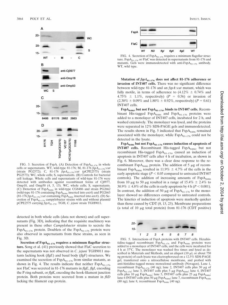
detected in both whole cells (data not shown) and cell super-natants (Fig. 3D), indicating that the requisite machinery waspresent in these other Campylobacter strains to secrete theFspA81-176 protein. Doublets of the FspA81-176 protein werealso observed in supernatants from these strains, as seen inFig. 3D.
Secretion of FspA81-176 requires a minimum flagellar struc-ture. Song et al. (41) previously showed that FlaC secretion tothe supernatants was not observed in C. jejuni TGH9011 mu-tants lacking hook (flgE) and basal body (flgF) structures. Weexamined the secretion of FspA81-176 from similar mutants, asshown in Fig. 4. The results indicate that neither FspA81-176
nor FlaC was secreted in 81-176 mutants in flgE, flgI, encodingthe P ring subunit, or flgK, encoding the hook-filament junctionprotein. Both proteins were secreted from a mutant in fliDlacking the filament cap protein.
Mutation of fspA81-176 does not affect 81-176 adherence orinvasion of INT407 cells. There was no significant differencebetween wild-type 81-176 and an fspA::cat mutant, which wasfully motile, in terms of adherence to (4.12% � 0.74% and4.75% � 1.1%, respectively) (P � 0.56) or invasion of(2.30% � 0.09% and 1.80% � 0.92%, respectively) (P � 0.61)INT407 cells.
FspA8486, but not FspA81-176, binds to INT407 cells. Recom-binant His-tagged FspA8486 and FspA81-176 proteins wereadded to a monolayer of INT407 cells, incubated for 2 h, andwashed extensively. The monolayer was lysed, and the proteinswere separated in 12% SDS-PAGE gels and immunodetected.The results shown in Fig. 5 indicated that FspA8486 remainedassociated with the monolayer, while FspA81-176 could not bedetected in the lysate.
FspA8486 but not FspA81-176 causes induction of apoptosis ofINT407 cells. Recombinant His-tagged FspA8486, but notrecombinant His-tagged FspA81-176, caused an induction ofapoptosis in INT407 cells after 4 h of incubation, as shown inFig. 6. Moreover, there was a clear dose response to the re-combinant FspA8486 protein. The addition of 5 �g of recom-binant FspA8486 resulted in 11.9% � 4.7% of the cells in theearly apoptotic stage (P � 0.05 compared to untreated INT407controls). The addition of increasing amounts of FspA8486
from 10 �g to 50 �g resulted in a range of 15.4% � 2.4% to30.9% � 4.8% of the cells in early apoptosis by 4 h (P � 0.001).In contrast, the addition of 50 �g of FspA81-176 to the mono-layer showed no differences compared to untreated controls.The kinetics of induction of apoptosis were markedly quickerthan those caused by CDT (8, 13, 23). Membrane preparations(a total of 10 �g total protein) from 81-176 (CDT positive)
FIG. 3. Secretion of FspA. (A) Detection of FspA81-176 in wholecells or supernatants. WT, wild-type 81-176; M, 81-176 fspA81-176::cat(strain PG2572); C, 81-176 fspA81-176::cat (pCPE2575) (strainPG2573); WC, whole cells; S, supernatants. (B) Controls for bacterialcell leakage. Whole cells and supernatants of wild-type 81-176 weredetected with antibodies against recombinant forms of Cj0977,Omp18, and Omp50 (4, 5, 13). WC, whole cells; S, supernatants.(C) Detection of FspA8486 in wild-type CG8486 and strain PG2662(wild-type 81-176 containing FspA8486 inserted into astA) and PG2663(81-176 fspA81-176::cat containing FspA8486 inserted into astA). (D) Se-cretion of FspA81-176 campylobacter strains with and without plasmidpCPE2575 carrying fspA81-176. TGH, C. jejuni strain TGH9011.
FIG. 4. Secretion of FspA81-176 requires a minimum flagellar struc-ture. FspA81-176 or FlaC was detected in supernatants from 81-176 andmutants. Gels were immunodetected with anti-FspA81-176 antibody.WT, wild type.
FIG. 5. Interactions of FspA proteins with INT407 cells. Hexahis-tidine-tagged recombinant FspA81-176 and FspA8486 proteins wereadded to a monolayer of INT407 cells, and the cells were incubated for4 h at 37°C. The monolayer was washed five times and lysed as de-scribed in Materials and Methods, and an aliquot (10 �l, or about 750ng protein) of each lysate was electrophoresed on a 12.5% SDS-PAGEgel, transferred onto a nitrocellulose membrane, and probed withanti-histidine-tagged mouse monoclonal antibody (Novagen). Lane 1,recombinant FspA81-176 (40 ng); lane 2, INT407 cells plus 50 �g ofFspA81-176; lane 3, INT407 cells plus 5 �g FspA8486; lane 4, INT407cells plus 10 �g FspA8486; lane 5, INT407 cells plus 25 �g FspA8486;lane 6 INT407 cells plus 50 �g FspA8486; lane 7, recombinant FspA8486(80 ng); lane 8, recombinant FspA8486 (40 ng).
3864 POLY ET AL. INFECT. IMMUN.
on October 2, 2014 by guest
http://iai.asm.org/
Dow
nloaded from
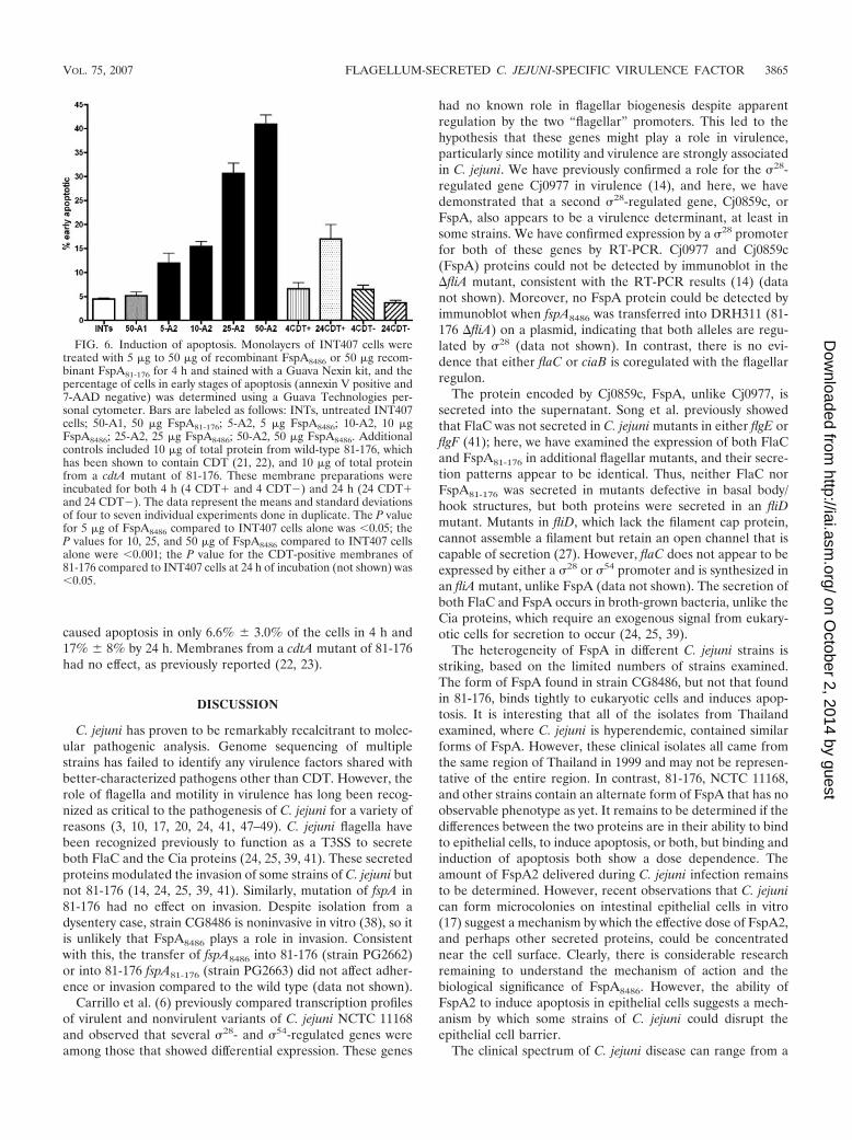
caused apoptosis in only 6.6% � 3.0% of the cells in 4 h and17% � 8% by 24 h. Membranes from a cdtA mutant of 81-176had no effect, as previously reported (22, 23).
DISCUSSION
C. jejuni has proven to be remarkably recalcitrant to molec-ular pathogenic analysis. Genome sequencing of multiplestrains has failed to identify any virulence factors shared withbetter-characterized pathogens other than CDT. However, therole of flagella and motility in virulence has long been recog-nized as critical to the pathogenesis of C. jejuni for a variety ofreasons (3, 10, 17, 20, 24, 41, 47–49). C. jejuni flagella havebeen recognized previously to function as a T3SS to secreteboth FlaC and the Cia proteins (24, 25, 39, 41). These secretedproteins modulated the invasion of some strains of C. jejuni butnot 81-176 (14, 24, 25, 39, 41). Similarly, mutation of fspA in81-176 had no effect on invasion. Despite isolation from adysentery case, strain CG8486 is noninvasive in vitro (38), so itis unlikely that FspA8486 plays a role in invasion. Consistentwith this, the transfer of fspA8486 into 81-176 (strain PG2662)or into 81-176 fspA81-176 (strain PG2663) did not affect adher-ence or invasion compared to the wild type (data not shown).
Carrillo et al. (6) previously compared transcription profilesof virulent and nonvirulent variants of C. jejuni NCTC 11168and observed that several �28- and �54-regulated genes wereamong those that showed differential expression. These genes
had no known role in flagellar biogenesis despite apparentregulation by the two “flagellar” promoters. This led to thehypothesis that these genes might play a role in virulence,particularly since motility and virulence are strongly associatedin C. jejuni. We have previously confirmed a role for the �28-regulated gene Cj0977 in virulence (14), and here, we havedemonstrated that a second �28-regulated gene, Cj0859c, orFspA, also appears to be a virulence determinant, at least insome strains. We have confirmed expression by a �28 promoterfor both of these genes by RT-PCR. Cj0977 and Cj0859c(FspA) proteins could not be detected by immunoblot in the�fliA mutant, consistent with the RT-PCR results (14) (datanot shown). Moreover, no FspA protein could be detected byimmunoblot when fspA8486 was transferred into DRH311 (81-176 �fliA) on a plasmid, indicating that both alleles are regu-lated by �28 (data not shown). In contrast, there is no evi-dence that either flaC or ciaB is coregulated with the flagellarregulon.
The protein encoded by Cj0859c, FspA, unlike Cj0977, issecreted into the supernatant. Song et al. previously showedthat FlaC was not secreted in C. jejuni mutants in either flgE orflgF (41); here, we have examined the expression of both FlaCand FspA81-176 in additional flagellar mutants, and their secre-tion patterns appear to be identical. Thus, neither FlaC norFspA81-176 was secreted in mutants defective in basal body/hook structures, but both proteins were secreted in an fliDmutant. Mutants in fliD, which lack the filament cap protein,cannot assemble a filament but retain an open channel that iscapable of secretion (27). However, flaC does not appear to beexpressed by either a �28 or �54 promoter and is synthesized inan fliA mutant, unlike FspA (data not shown). The secretion ofboth FlaC and FspA occurs in broth-grown bacteria, unlike theCia proteins, which require an exogenous signal from eukary-otic cells for secretion to occur (24, 25, 39).
The heterogeneity of FspA in different C. jejuni strains isstriking, based on the limited numbers of strains examined.The form of FspA found in strain CG8486, but not that foundin 81-176, binds tightly to eukaryotic cells and induces apop-tosis. It is interesting that all of the isolates from Thailandexamined, where C. jejuni is hyperendemic, contained similarforms of FspA. However, these clinical isolates all came fromthe same region of Thailand in 1999 and may not be represen-tative of the entire region. In contrast, 81-176, NCTC 11168,and other strains contain an alternate form of FspA that has noobservable phenotype as yet. It remains to be determined if thedifferences between the two proteins are in their ability to bindto epithelial cells, to induce apoptosis, or both, but binding andinduction of apoptosis both show a dose dependence. Theamount of FspA2 delivered during C. jejuni infection remainsto be determined. However, recent observations that C. jejunican form microcolonies on intestinal epithelial cells in vitro(17) suggest a mechanism by which the effective dose of FspA2,and perhaps other secreted proteins, could be concentratednear the cell surface. Clearly, there is considerable researchremaining to understand the mechanism of action and thebiological significance of FspA8486. However, the ability ofFspA2 to induce apoptosis in epithelial cells suggests a mech-anism by which some strains of C. jejuni could disrupt theepithelial cell barrier.
The clinical spectrum of C. jejuni disease can range from a
FIG. 6. Induction of apoptosis. Monolayers of INT407 cells weretreated with 5 �g to 50 �g of recombinant FspA8486 or 50 �g recom-binant FspA81-176 for 4 h and stained with a Guava Nexin kit, and thepercentage of cells in early stages of apoptosis (annexin V positive and7-AAD negative) was determined using a Guava Technologies per-sonal cytometer. Bars are labeled as follows: INTs, untreated INT407cells; 50-A1, 50 �g FspA81-176; 5-A2, 5 �g FspA8486; 10-A2, 10 �gFspA8486; 25-A2, 25 �g FspA8486; 50-A2, 50 �g FspA8486. Additionalcontrols included 10 �g of total protein from wild-type 81-176, whichhas been shown to contain CDT (21, 22), and 10 �g of total proteinfrom a cdtA mutant of 81-176. These membrane preparations wereincubated for both 4 h (4 CDT� and 4 CDT ) and 24 h (24 CDT�and 24 CDT ). The data represent the means and standard deviationsof four to seven individual experiments done in duplicate. The P valuefor 5 �g of FspA8486 compared to INT407 cells alone was �0.05; theP values for 10, 25, and 50 �g of FspA8486 compared to INT407 cellsalone were �0.001; the P value for the CDT-positive membranes of81-176 compared to INT407 cells at 24 h of incubation (not shown) was�0.05.
VOL. 75, 2007 FLAGELLUM-SECRETED C. JEJUNI-SPECIFIC VIRULENCE FACTOR 3865
on October 2, 2014 by guest
http://iai.asm.org/
Dow
nloaded from

mild, watery diarrhea to a dysentery-like disease. This is likelydue, in part, to the immune status of the host, although it hasbeen speculated that genomic differences among strains mayalso contribute. Complete genome analysis and comparativemicroarrays have identified gross differences among strains,largely in surface carbohydrates (9, 33, 34–37, 43). A recentmicroarray study has also reported heterogeneity in theCj0859c-Cj0860 region, consistent with our data (33). The datareported here indicate that changes among FspA proteins havemajor effects on toxicity for epithelial cells in vitro. Clearly,there are virulent strains of C. jejuni that contain both fspA1and fspA2 alleles, reflecting the multifactorial nature of patho-genicity. However, the observed heterogeneity among fspA al-leles may modulate C. jejuni virulence. The in vitro and in vivoroles of the different forms of FspA in pathogenesis are underinvestigation.
ACKNOWLEDGMENTS
We thank Daniel Scott, Shahida Baqar, David Tribble, and ourcolleagues at AFRIMS, Bangkok, Thailand, and NAMRU3, Cairo,Egypt, for strains; David Hendrixson and Vic DiRita for DRH311; EvaNielsen of the Danish Veterinary Laboratory and Helen Tabor andLawrence Price of the Canadian Science Centre for Human and An-imal Health for Penner serotyping; Rob Williams for electron micros-copy; and Chad Porter for statistical advice.
This work was funded by NIAID grant R01 AI043559 and theMilitary Infectious Disease Research Program (work unit 6000.RAD1.DA3.A0308).
REFERENCES
1. Alm, R. A., L.-S. L. Ling, D. Moir, B. L. King, E. D. Brown, P. Doig, D. R.Smith, B. Noonan, B. C. Guild, B. L. de Jonge, G. Carmel, P. J. Tummino,A. Caruso, M. Uria-Nickelsen, D. M. Mills, C. Ives, R. Gibson, D. Merberg,S. D. Mills, Q. Jian, D. E. Taylor, G. F. Vovis, and T. J. Trust. 1999. Genomicsequence comparison of two unrelated isolates of the human gastric patho-gen Helicobacter pylori. Nature 397:160–189.
2. Bacon, D. J., C. M. Szymanski, D. H. Burr, R. P. Silver, R. A. Alm, and P.Guerry. 2001. A phase variable capsule is involved in virulence of Campy-lobacter jejuni 81-176. Mol. Microbiol. 40:769–777.
3. Black, R. E., M. M. Levine, M. L. Clements, T. P. Hughes, and M. J. Blaser.1988. Experimental Campylobacter jejuni infection in humans. J. Infect. Dis.157:472–479.
4. Bolla, J. M., E. De, A. Dorez, and J. M. Pages. 2000. Purification, charac-terization and sequence analysis of Omp50, a new porin isolated fromCampylobacter jejuni. Biochem. J. 352:637–643.
5. Burnens, A., U. Stucki, J. Nicolet, and J. Frey. 1995. Identification andcharacterization of an immunogenic outer membrane protein of Campy-lobacter jejuni. J. Clin. Microbiol. 33:2826–2832.
6. Carrillo, C. D., E. Taboada, J. H. Nash, P. H. Lanthier, J. Kelly, P. C. Lau,R. Verhulp, O. Mykytczuk, J. Sy, W. A. Findlay, K. Amoako, S. Gomis, P.Willson, J. W. Austin, A. Potters, L. Babiuk, B. Allan, and C. M. Szymanski.2004. Genome-wide expression analyses of Campylobacter jejuni reveals co-ordinate regulation of motility and virulence by flhA. J. Biol. Chem. 279:20327–23008.
7. Chang, C., and J. F. Miller. 2006. Campylobacter jejuni colonization of micewith limited enteric flora. Infect. Immun. 74:5261–5271.
8. Cortes-Bratti, X., C. Karlsson, T. Lagergard, M. Thelestam, and T. Frisan.2001. The Haemophilus ducreyi cytolethal distending toxin induces cell cyclearrest and apoptosis via DNA damage checkpoint pathways. Infect. Immun.276:5296–5302.
9. Dorrell, N., J. A. Mangan, K. G. Laing, J. Hinds, D. Linton, H. Al-Ghusein,B. G. Barrell, J. Parkhill, N. G. Stoker, A. V. Karylshev, P. D. Butcher, andB. W. Wren. 2001. Whole genome comparison of Campylobacter jejuni iso-lates using a low-cost microarray reveals extensive genetic diversity. GenomeRes. 11:1706–1715.
10. Ferrero, R. L., and A. Lee. 1988. Motility of Campylobacter jejuni in a viscousenvironment: comparison with conventional rod shaped bacteria. J. Gen.Microbiol. 134:53–59.
11. Fouts, D. E., E. F. Mongodin, R. E. Mandrell, W. G. Miller, D. A. Rasko, J.Ravel, L. M. Brinkac, R. T. DeBoy, C. T. Parker, S. C. Dougherty, R. J.Dodson, A. S. Durkin, R. Madupu, S. A. Sullivan, J. U. Shetty, M. A. Ayodeji,A. Shvartsbeyn, M. C. Schatz, J. H. Badger, C. M. Fraser, and K. E. Nelson.2005. Major structural differences and novel potential virulence mechanismsfrom the genomes of multiple Campylobacter species. PLOS Biol. 3:e15.
12. Friedman, C. R., J. Neiman, H. C. Wegener, and R. V. Tauxe. 2000. Epide-miology of Campylobacter jejuni infections in the United States and otherindustrialized nations, p. 121–138. In I. Nachamkin and M. J. Blaser (ed.),Campylobacter, 2nd ed. ASM Press, Washington, DC.
13. Gelfanova, V., E. J. Hansen, and S. M. Spinola. 1999. Cytolethal distendingtoxin of Haemophilus ducreyi induces apoptotic death of Jurkat T cells.Infect. Immun. 67:6394–6402.
14. Goon, S., C. P. Ewing, M. Lorenzo, D. Pattarini, G. Majam, and P. Guerry.2006. A �28-regulated nonflagella gene contributes to virulence of Campy-lobacter jejuni 81-176. Infect. Immun. 74:769–772.
15. Goon, S., J. F. Kelly, S. M. Logan, C. P. Ewing, and P. Guerry. 2003.Pseudaminic acid, the major modification on Campylobacter flagellin, issynthesized via the Cj1293 gene. Mol. Microbiol. 50:659–671.
16. Guerry, P., R. A. Alm, M. E. Power, S. M. Logan, and T. J. Trust. 1991. Roleof two genes in Campylobacter motility. J. Bacteriol. 173:4757–4764.
17. Guerry, P., C. P. Ewing, M. Schirm, M. Lorenzo, J. Kelly, D. Pattarini, G.Majam, P. Thibault, and S. M. Logan. 2006. Changes in flagellin glycosyla-tion affect Campylobacter autoagglutination and virulence. Mol. Microbiol.60:299–311.
18. Guerry, P., C. P. Ewing, T. E. Hickey, M. M. Prendergast, and A. P. Moran.2000. Sialylation of lipooligosaccharide cores affects immunogenicity andserum resistance of Campylobacter jejuni. Infect. Immun. 68:6656–6662.
19. Harris, L. A., S. M. Logan, P. Guerry, and T. J. Trust. 1987. Antigenicvariation of Campylobacter flagellin. J. Bacteriol. 169:5066–5071.
20. Hendrixson, D. R., and V. J. DiRita. 2004. Identification of Campylobacterjejuni genes involved in commensal colonization of the chick gastrointestinaltract. Mol. Microbiol. 52:471–484.
21. Hendrixson, D. R., and V. J. DiRita. 2003. Transcription of �54 dependentbut not �28 dependent flagellar genes in Campylobacter jejuni is associatedwith formation of the flagella secretory apparatus. Mol. Microbiol. 50:687–702.
22. Hickey, T. E., A. L. McVeigh, D. A. Scott, R. E. Michielutti, A. Bixby, S. A.Carroll, A. L. Bourgeois, and P. Guerry. 2000. Campylobacter jejuni cytole-thal distending toxin mediates release of interleukin 8 from intestinal epi-thelial cells. Infect. Immun. 68:6535–6541.
23. Hickey, T. E., G. Majam, and P. Guerry. 2005. Intracellular survival ofCampylobacter jejuni in human monocytic cells and induction of apoptoticdeath by cytolethal distending toxin. Infect. Immun. 73:5194–5197.
24. Konkel, M. E., J. D. Klena, V. Rivera-Amill, M. R. Monteville, D. Biswas, B.Raphael, and J. Mickelson. 2004. Secretion of virulence proteins fromCampylobacter jejuni is dependent on a functional flagellar export apparatus.J. Bacteriol. 186:3296–3303.
25. Konkel, M. E., B. J. Kim, V. Rivera-Amill, and S. G. Garvis. 199l. Bacterialsecreted proteins are required for the internalization of Campylobacter jejuniinto cultured mammalian cells. Mol. Microbiol. 32:691–701.
26. Korostoff, J., J. F. Wang, I. Kieba, M. Miller, B. J. Shenker, and E. T. Lally.1998. Actinobacillus actinomycetemcomitans leukotoxin induces apoptosis inHL-60 cells. Infect. Immun. 66:4474–4483.
27. Kutsukake, K. 1994. Excretion of the anti-sigma factor through a flagellastructure couples flagellar gene expression with flagellar assembly in Salmo-nella typhimurium. Mol. Gen. Genet. 243:605–612.
28. Logan, S. M., J. F. Kelly, P. Thibault, C. P. Ewing, and P. Guerry. 2002.Structural heterogeneity of carbohydrate modifications affects serospecificityof Campylobacter flagellins. Mol. Microbiol. 46:587–597.
29. McNally, D. J., A. J. Aubry, J. P. Hui, N. H. Khieu, D. Whitfield, C. P. Ewing,P. Guerry, J.-R. Brisson, S. M. Logan, and E. C. Soo. 2007. Targetedmetabolomics analysis of Campylobacter coli VC167 reveals legionaminicacid derivatives as novel flagellar glycans. J. Biol. Chem. 282:14463–14475.
30. Miller, W. G., A. H. Bates, S. T. Horn, M. T. Brandl, M. R. Wachtel, andR. E. Mandrell. 2000. Detection on surfaces and in Caco-2 cells of Campy-lobacter jejuni transformed with new gfp, yfp, and cfp marker plasmids. Appl.Environ. Microbiol. 66:5426–5436.
31. Morooka, T., A. Umeda, and K. Amako. 1985. Motility as an intestinalcolonization factor for Campylobacter jejuni. J. Gen. Microbiol. 131:1973–1980.
32. Oelschlaeger, T. A., P. Guerry, and D. J. Kopecko. 1993. Unusual microtu-bule-dependent endocytosis mechanisms triggered by Campylobacter jejuniand Citrobacter freundii. Proc. Natl. Acad. Sci. USA 90:6884–6888.
33. Parker, C. T., B. Quinones, W. G. Miller, S. T. Horn, and R. E. Mandrell.2006. Comparative genomic analysis of Campylobacter jejuni strains revealsdiversity due to genomic elements similar to those present in Campylobacterjejuni strain RM1221. J. Clin. Microbiol. 44:4125–4135.
34. Parkhill, J., B. W. Wren, K. Mungall, J. M. Ketley, C. Churcher, D. Basham,T. Chillingworth, R. M. Davies, T. Feltwell, S. Holroyd, K. Jagels, A. V.Karlyshev, S. Moule, M. J. Pallen, C. W. Penn, M. A. Quail, M. A. Rajan-dream, K. M. Rutherford, A. H. M. van Vliet, S. Whitehead, and B. G.Barrell. 2000. The genome sequence of the food-borne pathogen Campy-lobacter jejuni reveals hypervariable tracts. Nature 403:665–668.
35. Pearson, B. M., C. Pin, J. Wright, K. I’Anson, T. Humphrey, and J. M. Wells.2004. Comparative genome analysis of Campylobacter jejuni using wholegenome DNA microarrays. FEBS Lett. 554:224–230.
36. Poly, F., D. Threadgill, and A. Stintzi. 2005. Genomic diversity in Campy-
3866 POLY ET AL. INFECT. IMMUN.
on October 2, 2014 by guest
http://iai.asm.org/
Dow
nloaded from

lobacter jejuni: identification of C. jejuni 81-176-specific genes. J. Clin. Mi-crobiol. 43:2330–2338.
37. Poly, F., D. Threadgill, and A. Stintzi. 2004. Identification of Campylobacterjejuni ATCC 43431-specific genes by whole microbial genome comparisons.J. Bacteriol. 186:4781–4795.
38. Poly, F., T. Read, D. R. Tribble, S. Baqar, M. Lorenzo, and P. Guerry. 16April 2007. Genome sequence of a clinical isolate of Campylobacter jejunifrom Thailand. Infect. Immun. doi:10.1128/IAI.00050-07.
39. Rivera-Amill, V., B. J. Kim, J. Keshu, and M. E. Konkel. 2001. Secretion ofthe virulence-associated Campylobacter invasion antigens from Campy-lobacter jejuni requires a stimulatory signal. J. Infect. Dis. 183:1607–1616.
40. Samuel, M. C., D. J. Vugia, S. Shallow, R. Marcus, S. Segler, T. McGivern,H. Kassenborg, K. Reilly, M. Kennedy, F. Angulo, and R. V. Tauxe. 2004.Epidemiology of sporadic Campylobacter infection in the United States anddeclining trend in incidence, FoodNet 1996–1999. Clin. Infect. Dis. 38(Suppl.3):S165–S174.
41. Song, Y. C., S. Jin, H. Louie, D. Ng, R. Lau, Y. Zhang, R. Weerasekera, S. A.Rashid, L. A. Ward, S. D. Der, and V. L. Chan. 2004. FlaC, a protein ofCampylobacter jejuni TGH9011 (ATCC43431) secreted through the flagellarapparatus, binds epithelial cells and influences cell invasion. Mol. Microbiol.53:541–553.
42. Suerbaum, S., C. Josenhans, T. Sterzenbach, B. Drescher, P. Brandt, M.Bell, M. Droge, B. Fartmann, H. P. Fischer, Z. Ge, A. Horster, R. Holland,K. Kleion, J. Koning, L. Macko, G. L. Mendz, G. Nyakatura, D. B. Schauer,Z. Shen, J. Weber, M. Frosch, and J. G. Fox. 2003. The complete genomesequence of the carcinogenic bacterium Helicobacter hepaticus. Proc. Natl.Acad. Sci. USA 100:7901–7906.
43. Taboada, E. N., R. R. Acedillo, C. D. Carrillo, W. A. Findlay, D. T. Medeiros,O. L. Mykytczuk, M. J. Roberts, C. A. Valencia, J. M. Farber, and J. H. E.Nash. 2004. Large scale comparative genomics meta-analysis of Campy-lobacter jejuni isolates reveals low level of genome plasticity. J. Clin. Micro-biol. 42:4566–4576.
44. Thibault, P., S. M. Logan, J. F. Kelly, J.-R. Brisson, C. P. Ewing, T. J. Trust,
and P. Guerry. 2001. Identification of the carbohydrate moieties and glyco-sylation motifs in Campylobacter jejuni flagellin. J. Biol. Chem. 276:34862–34870.
45. Thompson, J. D., D. G. Higgins, and T. J. Gibson. 1994. CLUSTAL W:improving the sensitivity of progressive multiple sequence alignment throughsequence weighting, position-specific gap penalties and weight matrix choice.Nucleic Acids Res. 22:4673–4680.
46. Tomb, J. F., O. White, A. R. Kerlavage, R. A. Clayton, G. G. Sutton, R. D.Fleischmann, K. A. Ketchum, H. P. Klenk, S. Gill, B. A. Dougherty, K.Nelson, J. Quackenbush, L. Zhou, E. F. Kirkness, S. Peterson, B. Loftus, D.Richardson, R. Dodson, H. G. Khalak, A. Glodek, K. McKenney, L. M.Fitzgerald, N. Lee, M. D. Adams, E. K. Hickey, D. E. Berg, J. D. Gocayne,T. R. Utterback, J. D. Peterson, J. M. Kelley, M. D. Cotton, J. M. Weidman,C. Fujii, C. Bowman, L. Watthey, E. Wallin, W. S. Hayes, M. Borodovsky,P. D. Karp, H. O. Smith, C. M. Fraser, and J. C. Venter. 1997. The completegenome of the gastric pathogen Helicobacter pylori. Nature 388:539–547.
47. Wassenaar, T. M., N. M. C. Bleumink-Pluym, and B. A. M. van der Zeijst.1991. Inactivation of Campylobacter jejuni flagellin genes by homologousrecombination demonstrates that flaA but not flaB is required for invasion.EMBO J. 10:2055–2061.
48. Yao, R., D. H. Burr, and P. Guerry. 1997. CheY mediated modulation ofCampylobacter jejuni virulence. Mol. Microbiol. 23:1021–1032.
49. Yao, R., D. H. Burr, P. Dogi, T. J. Trust, H. Niu, and P. Guerry. 1994.Isolation of motile and non-motile insertional mutants of Campylobacterjejuni: the role of motility in adherence and invasion of eukaryotic cells. Mol.Microbiol. 14:883–893.
50. Yao, R., R. A. Alm, T. J. Trust, and P. Guerry. 1993. Construction of newCampylobacter cloning vectors and a new mutational cat cassette. Gene130:127–130.
51. Yao, R., and P. Guerry. 1996. Molecular cloning and site-directed mutagen-esis of a gene involved in arylsulfatase production in Campylobacter jejuni. J.Bacteriol. 178:3335–3338.
Editor: A. Camilli
VOL. 75, 2007 FLAGELLUM-SECRETED C. JEJUNI-SPECIFIC VIRULENCE FACTOR 3867
on October 2, 2014 by guest
http://iai.asm.org/
Dow
nloaded from