Campylobacter Monitoring in German Broiler Flocks: An Explorative Time Series Analysis
Upload
independentCategory
view
0download
0
JOURNAL OF CLINICAL MICROBIOLOGY, Sept. 2007, p. 3015–3021 Vol. 45, No. 90095-1137/07/$08.00�0 doi:10.1128/JCM.00256-07Copyright © 2007, American Society for Microbiology. All Rights Reserved.
Development of a Real-Time Fluorescence Resonance EnergyTransfer PCR To Detect Arcobacter Species�
Khalil Abdelbaqi,1 Alice Buissonniere,1 Valerie Prouzet-Mauleon,1 Jessica Gresser,1 Irene Wesley,3Francis Megraud,1,2* and Armelle Menard1,2
Universite Victor Segalen Bordeaux 2, Centre National de Reference des Helicobacters et Campylobacters, F33076 Bordeaux, France1;INSERM U853, F33076 Bordeaux, France2; and National Animal Disease Center, ARS, USDA, 2300 Dayton Road, Ames, Iowa3
Received 1 February 2007/Returned for modification 26 March 2007/Accepted 17 May 2007
A real-time PCR targeting the gyrase A subunit gene outside the quinolone resistance-determining regionhas been developed to detect Arcobacter species. The species identification was done by probe hybridization andmelting curve analysis, using fluorescence resonance energy transfer technology. Discrimination betweenArcobacter species was straightforward, as the corresponding melting points showed significant differences withthe characteristic melting temperatures of 63.5°C, 58.4°C, 60.6°C, and 51.8°C for the Arcobacter butzleri,Arcobacter cryaerophilus, Arcobacter cibarius, and Arcobacter nitrofigilis type strains, respectively. The specificityof this assay was confirmed with pure cultures of 106 Arcobacter isolates from human clinical and veterinaryspecimens identified by phenotypic methods and 16S rRNA gene sequencing. The assay was then used to screen345 clinical stool samples obtained from patients with diarrhea. The assay detected A. butzleri in four of theseclinical samples (1.2%). These results were confirmed by a conventional PCR method targeting the 16S rRNAgene with subsequent sequencing of the PCR product. In conclusion, this real-time assay detects and differ-entiates Arcobacter species in pure culture as well as in the competing microbiota of the stool matrix. The assayis economical since only one biprobe is used and multiple Arcobacter species are identified in a single test.
Aerotolerant spirillum/vibrio-like organisms from abortedbovine fetuses were first described by Ellis et al. in 1977 (9) anddesignated Campylobacter cryaerophila on the basis of theirCampylobacter-like morphology, aerotolerance, and growth at25°C (9). After examining human and veterinary isolates ofthese aerotolerant campylobacters, Kiehlbauch et al. identifiedtwo species, Campylobacter butzleri, corresponding to the ma-jority of human clinical isolates, and Campylobacter cryaerophi-lus, consisting of two distinct groups (20). Vandamme et al.proposed the genus Arcobacter to encompass aerotolerantcampylobacters (36). This genus comprises human and veteri-nary species, including Arcobacter butzleri, Arcobacter cryaerophi-lus, Arcobacter cibarius, and Arcobacter skirrowii, and also thetype strain from the root of a Spartina plant, Arcobacter nitro-figilis. Two new species, Arcobacter sulfidicus, which inhabitscoastal marine water (42), and Arcobacter halophilus, isolatedfrom a hypersaline lagoon (8), represent the diversity of Arco-bacter and its capacity to survive in the environment. The genusArcobacter has been included with the genus Campylobacter inthe Campylobacteraceae family, which is part of the Epsilon-proteobacteria (11).
Despite the prevalence of Arcobacter species in food speci-mens, few studies have reported human Arcobacter infections(23). The bacterium was first identified in 2.4% of clinicalisolates from 631 Thai children with diarrhea (34). An out-break of recurrent abdominal cramps in 10 young children inItaly was attributed to A. butzleri (37). Arcobacter butzleri has
been linked to sporadic cases of diarrhea and abdominalcramps in patients with chronic diseases (24), such as neonatalbacteremia (27), liver cirrhosis (44), and acute appendicitis(22). More recently, a Belgian study revealed that 3.5% ofcampylobacters and related organisms isolated from humanstool samples over an 8-year period were A. butzleri (38). Inone of our previous studies, A. butzleri ranked fourth amongthe 2,855 strains of Campylobacter-like organisms received dur-ing a 17-month surveillance of Campylobacter infections inFrance (28). These infections were associated with intestinaldisorders resembling those caused by Campylobacter jejuni.These observations suggest that the prevalence of this emerg-ing pathogen may be underestimated due to inadequate cul-ture conditions and/or false identification (14, 38). Traditionalisolation methods for Arcobacter are similar to those forCampylobacter, although the majority do not grow at 42°C. Theproblems associated with culture failure or with the standardphenotypic methods of identification have led to the investi-gation of new approaches for assistance in the preliminarydiagnosis of Arcobacter infections. Conventional PCR-basedtechniques targeting ribosomal genes, such as the 16S RNAgene (13, 15, 32, 41) and the 23S rRNA gene (15, 16, 18, 26),have been widely used for the detection and identification ofArcobacter species. The increasing relevance of nucleic acidamplification tests for the detection and identification ofArcobacter species is now obvious, particularly in light of culturefailure and misidentification. With respect to nucleic acid am-plification tests, different real-time PCR chemistries are avail-able, with the most frequently used formats being SYBR green,hybridization probes, TaqMan probes, molecular beacons, andScorpion probes. These PCR formats are usually very sensitive,can be performed in less than 3 hours, and minimize the risk ofcross-contamination with other PCR products since all of the
* Corresponding author. Mailing address: INSERM U853, Univer-site Victor Segalen Bordeaux 2, Laboratoire de Bacteriologie, Bat. 2BRDC—Zone Nord, 146 rue Leo Saignat, 33076 Bordeaux cedex,France. Phone: 33 (0)556795910. Fax: 33 (0)556796018. E-mail: [email protected].
� Published ahead of print on 25 July 2007.
3015
on February 12, 2016 by guest
http://jcm.asm
.org/D
ownloaded from
steps occur in the same tube and no postamplification handlingis necessary. Among these methods, two specific TaqMan as-says were recently described for detection of A. butzleri and A.cryaerophilus (4).
Real-time PCR using the fluorescence resonance energytransfer (FRET) hybridization probe method is very promisingsince it can detect single nucleotide polymorphisms (SNP) withonly one biprobe. We have recently described a FRET real-time PCR assay targeting the gyrA gene for rapid and sensitiveidentification and differentiation of C. jejuni and Campy-lobacter coli (25). The present study reports the developmentof a real-time PCR assay using FRET chemistry and targetingthe gyrase A gene (gyrA) outside the quinolone resistance-determining region (QRDR) in Arcobacter species. Subse-quent identification of the species was based on melting curveanalysis (MCA) of the amplified products by detection of thenucleotide changes between the species. The advantages of thisnew test are the relatively low cost (since only one biprobe isused) and the possible detection and identification of multipleArcobacter species in a single test.
MATERIALS AND METHODS
Bacterial strains and growth conditions. The following five type strainswere used as positive controls: the A. butzleri D2686 type strain (CIP 103493T, a human isolate), the A. cryaerophilus A169/B type strain (CIP 104014 T, abovine fetus isolate), the A. cibarius CCUG 48482 type strain (LMG 21996, achicken skin isolate), the A. skirrowii 449/80 type strain (ATCC 51132, an ovinefetus isolate), and the A. nitrofigilis C1 type strain (CIP 103745, a plant isolate).An additional 106 Arcobacter isolates obtained from human (n � 22) or veteri-nary (n � 84) sources were employed in this study. Human strains included A.butzleri strain WRI 996/79 (CCUG 10373) and 21 strains (19 A. butzleri and 2 A.cryaerophilus strains) isolated from human clinical specimens, essentially fecesfrom patients with gastroenteritis, from all over France in 2003 and 2004, andsent to the French National Reference Center for Campylobacter and Helico-bacter (28). Among the 84 Arcobacter strains isolated from animal sources, 3strains (A. butzleri Cipolla 4 and A. cryaerophilus strains PC367 and PC249) wereobtained from the culture collection of the University of Goteborg, Sweden(CCUG 34397 B, CCUG 12020, and CCUG 12018, respectively), and 81 Arco-bacter strains (78 A. butzleri and 3 A. cryaerophilus strains) were obtained fromthe National Animal Disease Centre (USDA, Agricultural Research Service,Ames, IA).
All arcobacters were grown as previously reported (28). Some of these strainswere initially identified as members of the Campylobacteraceae family by pheno-typic methods, i.e., morphology, motility, growth in a microaerobic atmosphere,and positive oxidase activity. Identification at the species level was performed by16S rRNA gene sequencing as previously reported (28).
A panel of bacterial strains was used to evaluate the specificities of thedifferent primers designed during this study. These strains included a large panelof Campylobacter strains, such as C. jejuni CCUG 11284, Campylobacter coli
CCUG 145401, Campylobacter fetus UA60, Campylobacter hyointestinalis CCUG14169, Campylobacter mucosalis CIP 103750, Campylobacter sputorum CCUG 9728,Campylobacter upsaliensis CCUG 14913, and Campylobacter lari CCUG23947, and Helicobacter strains, such as Helicobacter pullorum CCUG 33839,Helicobacter canadensis NCTC 13221, Helicobacter pylori J99, Helicobacterhepaticus ATCC 51448, Helicobacter bilis ATCC 51630, Helicobacter muridarumATCC 49282, Helicobacter felis CCUG 28539 T, “Flexispira rappini” (proposedname) CCUG 29176, and Wolinella succinogenes DSMZ 1740. Other entericbacteria (clinical isolates) commonly isolated from patients (Escherichia coli,Salmonella enterica serovar Enteritidis, and Salmonella enterica serovar Typhi-murium) were also tested.
Genomic DNA was isolated by using a QIAamp DNA mini kit (QIAGEN SA,Courtaboeuf, France) and stored at �20°C until required for analysis.
Stool samples. In total, 345 stool clinical samples were screened with thisassay. They were obtained from patients (male/female ratio, 0.56; mean age, 41.4 �28.9 years) suffering from diarrhea and presenting themselves at Pellegrin Hos-pital (Bordeaux, France) between July and September 2005. Genomic DNA wasextracted from the stool samples by using the QIAamp DNA stool mini kit(QIAGEN SA) and stored at �20°C.
All stool specimens were also cultured for Arcobacter species by using twomethods. The first was inoculation of an enrichment broth (Arco broth) incu-bated for 24 h at 37°C and subcultured on an Arco agar according to a methodpreviously published (6). The plates were incubated at 37°C in a microaerobicatmosphere for 7 days. The second method was a filtration method using a0.65-�m filter (Millipore, Billerica, MA) on a blood agar plate without antibioticincubated for 7 days. Colonies were identified according to standard methods.
PCR and DNA sequencing of the gyrA gene. The available gyrA sequences (1)from the five A. butzleri strains, D2686, 520H-2004, 1137-2003, 1340-2003, andCCUG 34397 B (GenBank accession numbers DQ464331 to DQ464335, respec-tively), were aligned with those of the A. cryaerophilus A169/B type strain, the A.cibarius CCUG 48482 type strain, the A. skirrowii 449/80 type strain (GenBankaccession numbers DQ464336 to DQ464338, respectively), and the A. nitrofigilisA169/B type strain (GenBank accession number DQ464339) by using multiplesequence alignment with hierarchical clustering (5) (http://prodes.toulouse.inra.fr/multalin/multalin.html). Primers were designed to target a conserved regionoutside the QRDR. The resulting primer set (F-Arco-FRET5 and R-Arco-FRET5) was designed using web Primer3 software (30) (http://www.broad.mit.edu/cgi-bin/primer/primer3_www.cgi). Use of these primers resulted in the am-plification of a 905-bp PCR product. PCR was performed with PWO super yieldTaq polymerase (Roche Diagnostics, Meylan, France). The amplification param-eters consisted of 1 cycle at 95°C for 5 min, followed by 35 cycles at 95°C for 30 s,56°C for 30 s, and 72°C for 2 min, and finally 1 cycle at 72°C for 5 min. The 905-bpsequences of the gyrA genes of other clinical Arcobacter isolates (A. butzleristrains 235-2004, 1285-2003, 1188-2003, 1477-2003, 1172-2003, and 1426-2003and A. cryaerophilus strains 322H-2004, 622H-2004, 492-2004, PC367, andPC249) were amplified using this PCR assay and sequenced on both strands withPCR primers using an Applied Biosystems 3130xl genetic analyzer (AppliedBiosystems, Foster City, CA) with a fluorescence BigDye Terminator V1.1 cyclesequencing kit (Applied Biosystems) according to the manufacturer’s instruc-tions.
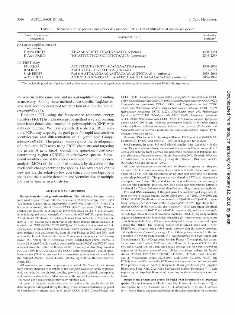
Design of the primers and probes for FRET-PCR identification of Arcobacterspecies. The gyrA sequences (Table 1 and Fig. 1) from A. butzleri (n � 11), A.cryaerophilus (n � 6), A. cibarius (n � 1), A. nitrofigilis (n � 1), and A. skirrowii(n � 1) were aligned and analyzed to identify (i) conserved regions for the primer
TABLE 1. Sequences of the primers and probes designed for FRET-PCR identification of Arcobacter species
Primer function anddesignation Sequence (5� to 3�) Nucleotide
positionsa
gyrA gene amplification andsequencing
F-Arco-FRET5 TTGAAGATTCTTATGATGAAATTGA (sense) 1460–1484R-Arco-FRET5 TGTATTTCTTCCTGCTTTTCTAATTG (antisense) 2364–2339
LC-FRET assayF1-FRET5 ATCTTTAGTATTCTTTACAAGAAATGG (sense) 1830–1856R2-FRET5 AACTGTTGTTCGTTTTCCA (antisense) 2181–2163S-Ab-FRET5 Red 640-ATCAAGGAAGAAGTACAAGAGGTGTAAG-p (antisense) 2039–2066A-Ab-FRET5 AGTCTTGGTCAATGTATTAGATTTGAACTTGAAAAAACAAG-F (antisense) 2036–1996
a The nucleotide positions of primers and probes were compared to the gyrA gene numbering of Arcobacter butzleri D2682, the type strain.
3016 ABDELBAQI ET AL. J. CLIN. MICROBIOL.
on February 12, 2016 by guest
http://jcm.asm
.org/D
ownloaded from
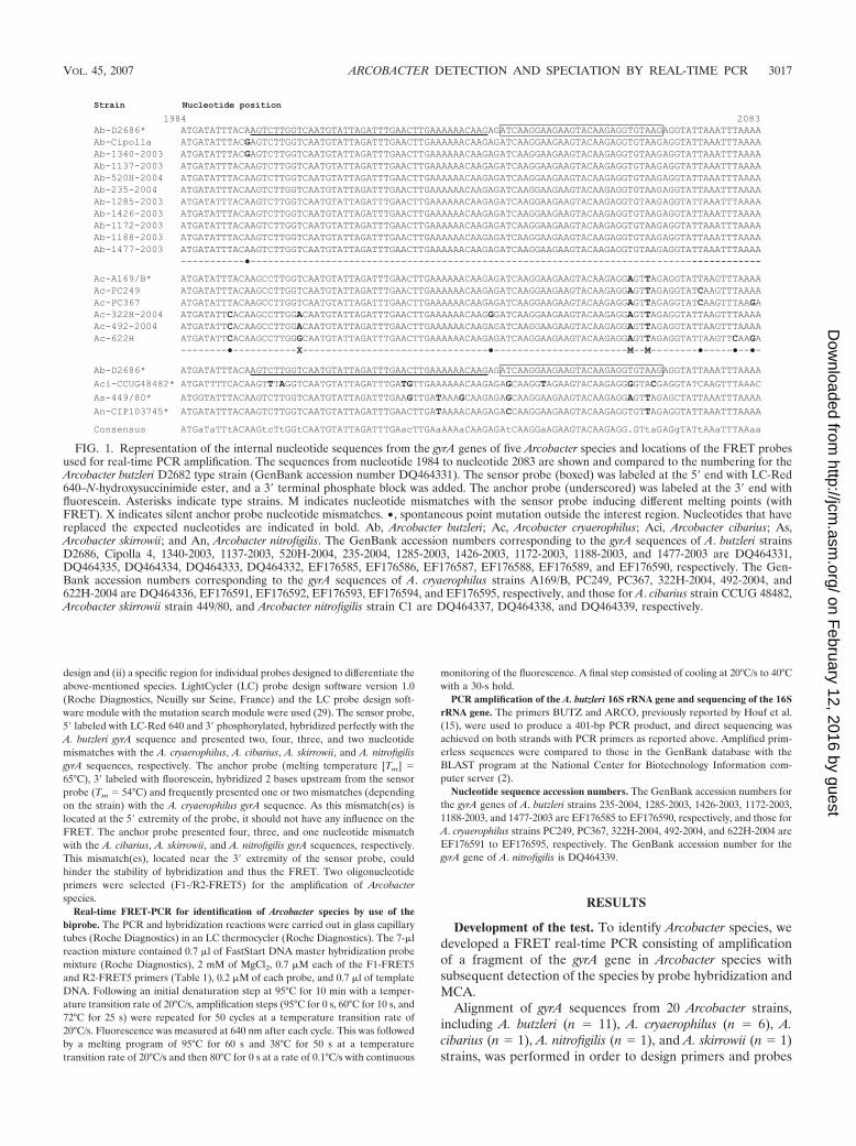
design and (ii) a specific region for individual probes designed to differentiate theabove-mentioned species. LightCycler (LC) probe design software version 1.0(Roche Diagnostics, Neuilly sur Seine, France) and the LC probe design soft-ware module with the mutation search module were used (29). The sensor probe,5� labeled with LC-Red 640 and 3� phosphorylated, hybridized perfectly with theA. butzleri gyrA sequence and presented two, four, three, and two nucleotidemismatches with the A. cryaerophilus, A. cibarius, A. skirrowii, and A. nitrofigilisgyrA sequences, respectively. The anchor probe (melting temperature [Tm] �65°C), 3� labeled with fluorescein, hybridized 2 bases upstream from the sensorprobe (Tm � 54°C) and frequently presented one or two mismatches (dependingon the strain) with the A. cryaerophilus gyrA sequence. As this mismatch(es) islocated at the 5� extremity of the probe, it should not have any influence on theFRET. The anchor probe presented four, three, and one nucleotide mismatchwith the A. cibarius, A. skirrowii, and A. nitrofigilis gyrA sequences, respectively.This mismatch(es), located near the 3� extremity of the sensor probe, couldhinder the stability of hybridization and thus the FRET. Two oligonucleotideprimers were selected (F1-/R2-FRET5) for the amplification of Arcobacterspecies.
Real-time FRET-PCR for identification of Arcobacter species by use of thebiprobe. The PCR and hybridization reactions were carried out in glass capillarytubes (Roche Diagnostics) in an LC thermocycler (Roche Diagnostics). The 7-�lreaction mixture contained 0.7 �l of FastStart DNA master hybridization probemixture (Roche Diagnostics), 2 mM of MgCl2, 0.7 �M each of the F1-FRET5and R2-FRET5 primers (Table 1), 0.2 �M of each probe, and 0.7 �l of templateDNA. Following an initial denaturation step at 95°C for 10 min with a temper-ature transition rate of 20°C/s, amplification steps (95°C for 0 s, 60°C for 10 s, and72°C for 25 s) were repeated for 50 cycles at a temperature transition rate of20°C/s. Fluorescence was measured at 640 nm after each cycle. This was followedby a melting program of 95°C for 60 s and 38°C for 50 s at a temperaturetransition rate of 20°C/s and then 80°C for 0 s at a rate of 0.1°C/s with continuous
monitoring of the fluorescence. A final step consisted of cooling at 20°C/s to 40°Cwith a 30-s hold.
PCR amplification of the A. butzleri 16S rRNA gene and sequencing of the 16SrRNA gene. The primers BUTZ and ARCO, previously reported by Houf et al.(15), were used to produce a 401-bp PCR product, and direct sequencing wasachieved on both strands with PCR primers as reported above. Amplified prim-erless sequences were compared to those in the GenBank database with theBLAST program at the National Center for Biotechnology Information com-puter server (2).
Nucleotide sequence accession numbers. The GenBank accession numbers forthe gyrA genes of A. butzleri strains 235-2004, 1285-2003, 1426-2003, 1172-2003,1188-2003, and 1477-2003 are EF176585 to EF176590, respectively, and those forA. cryaerophilus strains PC249, PC367, 322H-2004, 492-2004, and 622H-2004 areEF176591 to EF176595, respectively. The GenBank accession number for thegyrA gene of A. nitrofigilis is DQ464339.
RESULTS
Development of the test. To identify Arcobacter species, wedeveloped a FRET real-time PCR consisting of amplificationof a fragment of the gyrA gene in Arcobacter species withsubsequent detection of the species by probe hybridization andMCA.
Alignment of gyrA sequences from 20 Arcobacter strains,including A. butzleri (n � 11), A. cryaerophilus (n � 6), A.cibarius (n � 1), A. nitrofigilis (n � 1), and A. skirrowii (n � 1)strains, was performed in order to design primers and probes
FIG. 1. Representation of the internal nucleotide sequences from the gyrA genes of five Arcobacter species and locations of the FRET probesused for real-time PCR amplification. The sequences from nucleotide 1984 to nucleotide 2083 are shown and compared to the numbering for theArcobacter butzleri D2682 type strain (GenBank accession number DQ464331). The sensor probe (boxed) was labeled at the 5� end with LC-Red640–N-hydroxysuccinimide ester, and a 3� terminal phosphate block was added. The anchor probe (underscored) was labeled at the 3� end withfluorescein. Asterisks indicate type strains. M indicates nucleotide mismatches with the sensor probe inducing different melting points (withFRET). X indicates silent anchor probe nucleotide mismatches. •, spontaneous point mutation outside the interest region. Nucleotides that havereplaced the expected nucleotides are indicated in bold. Ab, Arcobacter butzleri; Ac, Arcobacter cryaerophilus; Aci, Arcobacter cibarius; As,Arcobacter skirrowii; and An, Arcobacter nitrofigilis. The GenBank accession numbers corresponding to the gyrA sequences of A. butzleri strainsD2686, Cipolla 4, 1340-2003, 1137-2003, 520H-2004, 235-2004, 1285-2003, 1426-2003, 1172-2003, 1188-2003, and 1477-2003 are DQ464331,DQ464335, DQ464334, DQ464333, DQ464332, EF176585, EF176586, EF176587, EF176588, EF176589, and EF176590, respectively. The Gen-Bank accession numbers corresponding to the gyrA sequences of A. cryaerophilus strains A169/B, PC249, PC367, 322H-2004, 492-2004, and622H-2004 are DQ464336, EF176591, EF176592, EF176593, EF176594, and EF176595, respectively, and those for A. cibarius strain CCUG 48482,Arcobacter skirrowii strain 449/80, and Arcobacter nitrofigilis strain C1 are DQ464337, DQ464338, and DQ464339, respectively.
VOL. 45, 2007 ARCOBACTER DETECTION AND SPECIATION BY REAL-TIME PCR 3017
on February 12, 2016 by guest
http://jcm.asm
.org/D
ownloaded from
for the assay. Numerous nucleotide sequence variations in thegyrA gene were observed among these five Arcobacter species.Because of the constraints imposed by the LC software for theprobe design (the highly conserved anchor probe and mis-matches located on the sensor probe, a maximum gap of 5bases between the probes, and restricted Tms for the probes),only one region of the gyrA gene outside the QRDR (nucleo-tides 1830 to 2364) could be used for the primer and probedesign. Because A. butzleri is the most frequently encounteredArcobacter species, the probes used in this study were designedon the A. butzleri gyrA gene.
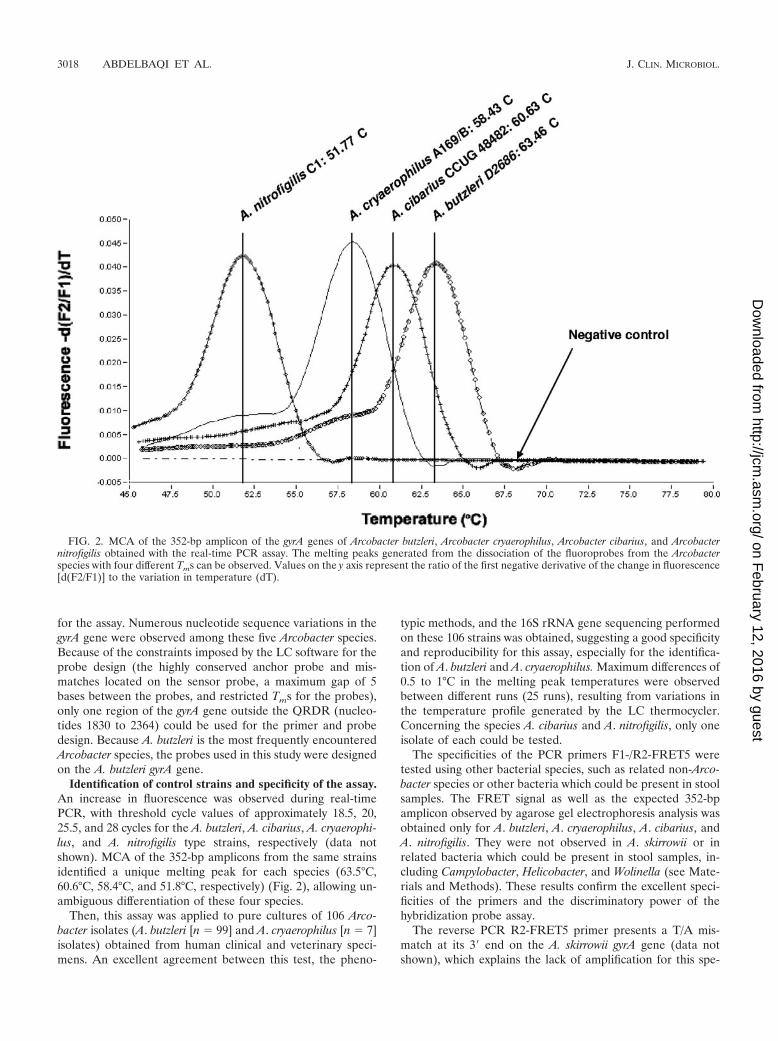
Identification of control strains and specificity of the assay.An increase in fluorescence was observed during real-timePCR, with threshold cycle values of approximately 18.5, 20,25.5, and 28 cycles for the A. butzleri, A. cibarius, A. cryaerophi-lus, and A. nitrofigilis type strains, respectively (data notshown). MCA of the 352-bp amplicons from the same strainsidentified a unique melting peak for each species (63.5°C,60.6°C, 58.4°C, and 51.8°C, respectively) (Fig. 2), allowing un-ambiguous differentiation of these four species.
Then, this assay was applied to pure cultures of 106 Arco-bacter isolates (A. butzleri [n � 99] and A. cryaerophilus [n � 7]isolates) obtained from human clinical and veterinary speci-mens. An excellent agreement between this test, the pheno-
typic methods, and the 16S rRNA gene sequencing performedon these 106 strains was obtained, suggesting a good specificityand reproducibility for this assay, especially for the identifica-tion of A. butzleri and A. cryaerophilus. Maximum differences of0.5 to 1°C in the melting peak temperatures were observedbetween different runs (25 runs), resulting from variations inthe temperature profile generated by the LC thermocycler.Concerning the species A. cibarius and A. nitrofigilis, only oneisolate of each could be tested.
The specificities of the PCR primers F1-/R2-FRET5 weretested using other bacterial species, such as related non-Arco-bacter species or other bacteria which could be present in stoolsamples. The FRET signal as well as the expected 352-bpamplicon observed by agarose gel electrophoresis analysis wasobtained only for A. butzleri, A. cryaerophilus, A. cibarius, andA. nitrofigilis. They were not observed in A. skirrowii or inrelated bacteria which could be present in stool samples, in-cluding Campylobacter, Helicobacter, and Wolinella (see Mate-rials and Methods). These results confirm the excellent speci-ficities of the primers and the discriminatory power of thehybridization probe assay.
The reverse PCR R2-FRET5 primer presents a T/A mis-match at its 3� end on the A. skirrowii gyrA gene (data notshown), which explains the lack of amplification for this spe-
FIG. 2. MCA of the 352-bp amplicon of the gyrA genes of Arcobacter butzleri, Arcobacter cryaerophilus, Arcobacter cibarius, and Arcobacternitrofigilis obtained with the real-time PCR assay. The melting peaks generated from the dissociation of the fluoroprobes from the Arcobacterspecies with four different Tms can be observed. Values on the y axis represent the ratio of the first negative derivative of the change in fluorescence[d(F2/F1)] to the variation in temperature (dT).
3018 ABDELBAQI ET AL. J. CLIN. MICROBIOL.
on February 12, 2016 by guest
http://jcm.asm
.org/D
ownloaded from
cies. Consequently, a new set of primers was designed for A.skirrowii amplification, using the same probes and identicalreaction conditions so that the assay could be run concurrently.MCA of the 394-bp amplicon from A. skirrowii revealed thepresence of a double curve, which was difficult to interpret (notshown). This bimodal curve may result from an unstable hy-bridization of both the anchor and the sensor probes on thegyrA gene of A. skirrowii since nucleotide mismatches are lo-cated at the 3� and 5� extremities of the anchor and sensorprobes, respectively (Fig. 1), thus preventing a good FRET.Considering this result, only A. butzleri, A. cryaerophilus, A.cibarius, and A. nitrofigilis can be identified by this assay.
Comparison with standard nucleic acid amplification tests.The standard PCR described by Houf et al. (15) for speciesidentification of A. butzleri and A. cryaerophilus strains as wellas identification at the species level by sequencing of the 16SrRNA gene (28) was performed on all of the strains. A com-plete agreement was found (data not shown).
Evaluation of the sensitivity of the test. The sensitivity of thePCR was evaluated using 10-fold serial dilutions of a bacterialsuspension of A. butzleri in brucella broth, which was quantifiedby culture on specific media and subsequent colony counts.PCRs with the DNA extracted from the bacterial suspensionsyielded regression curves with slopes between �2.946 and�3.512, spanning the slope value of �3.33, which correspondsto the maximum efficiency. Although this assay was able todetect fewer than 30 CFU, the regression curve was linear onlyfrom 3 � 107 to 3 � 102 CFU with reaction volumes of 20 �lor 7 �l.
Moreover, this assay always led to a positive result with theuse of the conventional boiling method, in contrast to standardPCR (data not shown).
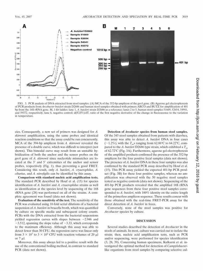
Detection of Arcobacter species from human stool samples.Of the 345 stool samples obtained from patients with diarrhea,this assay was able to detect A. butzleri DNA in four cases(�1.2%), with the Tms ranging from 62.86°C to 64.22°C, com-pared to the A. butzleri D2686 type strain, which exhibited a Tm
of 62.72°C (Fig. 3A). Furthermore, agarose gel electrophoresisof the amplified products confirmed the presence of the 352-bpamplicon for the four positive fecal samples (data not shown).The presence of A. butzleri DNA in these four samples was alsoconfirmed by the standard PCR assay described by Houf et al.(15). This PCR assay yielded the expected 401-bp PCR prod-uct (Fig. 3B) for these four positive samples, whereas no am-plification was observed with the 50 negative stool samplestested as negative controls (data not shown). Sequencing of the401-bp PCR products revealed that the amplified 16S rRNAgene sequences from these four positive stool samples corre-sponded to A. butzleri, with 100% identity on the entire lengthof the primerless amplicon sequence. These results concur withthose obtained with the real-time FRET-PCR assay for thedirect detection of A. butzleri in feces.
Conversely, none of the stool samples was positive forArcobacter species by culture.
DISCUSSION
Several studies described the detection of Arcobacter in thestools of animals. In most, culture was carried out to isolate thestrain; then, nucleic acid amplification tests, such as PCRand/or sequencing, were performed for species identification(3, 28, 39). Concerning human specimens, Kulkarni et al. in-vestigated the optimal method for detection of Campylobacter-like organisms from stool samples by comparing selective cul-
FIG. 3. PCR analysis of DNA extracted from stool samples. (A) MCA of the 352-bp amplicon of the gyrA gene. (B) Agarose gel electrophoresisof PCR products from Arcobacter butzleri strain D2686 and human stool samples obtained with primers ARCO and BUTZ for amplification of 401bp from the 16S rRNA gene. M, 1-kb ladder; lane 1, A. butzleri strain D2686 as a reference; lanes 2 to 5, human stool samples 51669, 52654, 54941,and 59372, respectively; lane 6, negative control; d(F2/F1)/dT, ratio of the first negative derivative of the change in fluorescence to the variationin temperature.
VOL. 45, 2007 ARCOBACTER DETECTION AND SPECIATION BY REAL-TIME PCR 3019
on February 12, 2016 by guest
http://jcm.asm
.org/D
ownloaded from
ture with membrane filtration and PCR (21). They concludedthat membrane filtration was the best method for detection ofArcobacter species in stool samples, but this conclusion wasbased on the detection of a single positive sample by culture. Incontrast, the results obtained by Engberg et al. showed thedifficulty in screening human stool samples for the presence ofArcobacter species, since no single method can successfullyidentify isolates from all taxa (10). A more recent study showedthat among 255 patients hospitalized with gastrointestinal com-plaints in South Africa, A. butzleri, A. cryaerophilus, and A.skirrowii were detected by PCR in 7.5, 3.5, and 2% of the stoolspecimens, respectively (31). The authors emphasized the use-fulness of molecular techniques in the identification of Arco-bacter species compared to that of the culture method. Molec-ular techniques constitute an important advance in the rapiddiagnosis of gastrointestinal infections when the organisms arefastidious. Among the numerous molecular techniques, real-time PCR is an important tool for the rapid and unequivocaldetection of bacterial species in complex human clinical sam-ples, such as stool samples.
The PCR format described herein has shown its ability todetect specific Arcobacter species, with the exception of A.skirrowii, in a specific and reliable way. This method is rapidand convenient and, moreover, in comparison to the real-timePCR method previously described (4) is economical since ithas the advantage of using only one biprobe in a low finalvolume. Such a method should facilitate the detection of theemerging pathogenic Arcobacter species. Of the most popularreal-time detection formats currently used (SYBR green, hy-bridization probes, TaqMan probes, molecular beacons, andScorpion probes), the SYBR green format was excluded since(i) the specificity is not optimal, as no probe is used, (ii) theidentification of several species in a single test remains difficultas several primer sets are necessary, and (iii) distinct Tms arealso necessary to identify each species without ambiguity. Forthe identification of the two most frequently encounteredArcobacter species, two specific real-time PCRs using TaqManchemistry were recently reported (4). These TaqMan assaysrequire a mixture of probes (each labeled twice, as is the casefor molecular beacons and Scorpion probes), producing a costtwice as high as that of FRET hybridization probe chemistrywith the SNP detection format, for which only one biprobe(each probe labeled once) has to be used. For this reason, aFRET hybridization probe PCR with the SNP detection for-mat was chosen. Consequently, the primer set and biprobewere designed to amplify a gene common to the Arcobacterspecies but presenting ample sequence divergences for speciesdifferentiation by MCA. Concerning the target gene, the 16SrRNA gene speciation of Arcobacter species seems satisfactorysince several techniques were developed successfully (13, 15,32, 41). However, 16S rRNA gene-based taxonomy is nothighly discriminant for Epsilonproteobacteria. Indeed, discor-dant 16S and 23S rRNA gene phylogenies among Helicobacterspecies (7) as well as a lack of discrimination among Campy-lobacter species (12) were reported. Therefore, in this studyribosomal genes were not chosen as a possible target for thePCR. The gyrA gene encoding a subunit of DNA gyrase, anessential bacterial enzyme (33, 35, 40), was reported to be animportant tool for bacterial phylogeny (17, 19, 43). Further-more, a FRET real-time PCR targeting the gyrA gene was
previously used as a rapid and sensitive method for identifica-tion and differentiation of C. jejuni and C. coli (25), whichmotivated our choice for this approach.
The results obtained with the 345 stool specimens testedwith this PCR assay showed a higher sensitivity than those forculture since no positive culture could be obtained from thesespecimens. However, slightly higher Tms were observed for thethree positive stool samples than for the control isolates. In-deed, 10% of the extracted stool sample was directly used forthe assay whereas DNA extracted from the strains was highlydiluted before use. These Tm differences observed for the stoolsamples may be due to the stool composition modifying thefinal salt concentration and/or the pH of the PCR mix.
With regard to the previously reported PCR assays (4, 13,15, 16, 18, 26, 32, 41), this FRET real-time PCR assay is ofinterest for detection and identification of A. butzleri, A.cryaerophilus, A. cibarius, and A. nitrofigilis in a single test. Thisassay detected Arcobacter species in 1.2% of the stool samplesfrom 345 patients with diarrhea, which is higher than previousestimates for France and Belgium obtained by culture (28, 38).This demonstrates the utility of molecular techniques in thediagnosis of human Arcobacter infections and highlights thefact that human Arcobacter infection is underestimated. Inter-estingly, another study performed by PCR supports our resultssince Arcobacter species were detected in 12.9% of the stools ofdiarrheic patients from South Africa (31). The latest study alsorevealed the presence of Arcobacter species in 3% of the stoolsof asymptomatic children from South Africa. Moreover, anArcobacter-selective isolation procedure revealed the presenceof A. cryaerophilus in 1.4% of the fecal samples of asymptom-atic humans in Switzerland (14). It would be interesting to usethe FRET real-time PCR developed in this study to determinethe prevalence of Arcobacter species in healthy subjects indifferent countries of the world.
ACKNOWLEDGMENTS
We are grateful to Leila Labadi, Christine Camou, and BrigitteTauzin for their technical assistance. Khalil Abdelbaqi was the recip-ient of a doctoral fellowship of the French government program for theCentre Regional des œuvres Universitaires et Scolaires.
REFERENCES
1. Abdelbaqi, K., A. Menard, V. Prouzet-Mauleon, F. Bringaud, P. Lehours,and F. Megraud. 2007. Nucleotide sequence of the gyrA gene of Arcobacterspecies and characterization of human ciprofloxacin resistant clinical iso-lates. FEMS Immunol. Med. Microbiol. 49:337–345.
2. Altschul, S. F., T. L. Madden, A. A. Schaffer, J. Zhang, Z. Zhang, W. Miller,and D. J. Lipman. 1997. Gapped BLAST and PSI-BLAST: a new generationof protein database search programs. Nucleic Acids Res. 25:3389–3402.
3. Atabay, H. I., M. Waino, and M. Madsen. 2006. Detection and diversity ofvarious Arcobacter species in Danish poultry. Int. J. Food Microbiol. 109:139–145.
4. Brightwell, G., E. Mowat, R. Clemens, J. Boerema, D. J. Pulford, and S. L.On. 2006. Development of a multiplex and real time PCR assay for thespecific detection of Arcobacter butzleri and Arcobacter cryaerophilus. J. Mi-crobiol. Methods 17:17.
5. Corpet, F. 1988. Multiple sequence alignment with hierarchical clustering.Nucleic Acids Res. 16:10881–10890.
6. de Boer, E., J. J. Tilburg, D. L. Woodward, H. Lior, and W. M. Johnson.1996. A selective medium for the isolation of Arcobacter from meats. Lett.Appl. Microbiol. 23:64–66.
7. Dewhirst, F. E., Z. L. Shen, M. S. Scimeca, L. N. Stokes, T. Boumenna, T. T.Chen, B. J. Paster, and J. G. Fox. 2005. Discordant 16S and 23S rRNA genephylogenies for the genus Helicobacter: implications for phylogenetic infer-ence and systematics. J. Bacteriol. 187:6106–6118.
8. Donachie, S. P., J. P. Bowman, S. L. On, and M. Alam. 2005. Arcobacterhalophilus sp. nov., the first obligate halophile in the genus Arcobacter. Int. J.Syst. Evol. Microbiol. 55:1271–1277.
3020 ABDELBAQI ET AL. J. CLIN. MICROBIOL.
on February 12, 2016 by guest
http://jcm.asm
.org/D
ownloaded from
9. Ellis, W. A., S. D. Neill, J. J. O’Brien, H. W. Ferguson, and J. Hanna. 1977.Isolation of Spirillum/Vibrio-like organisms from bovine fetuses. Vet. Rec.100:451–452.
10. Engberg, J., S. L. On, C. S. Harrington, and P. Gerner-Smidt. 2000. Prev-alence of Campylobacter, Arcobacter, Helicobacter, and Sutterella spp. in hu-man fecal samples as estimated by a reevaluation of isolation methods forcampylobacters. J. Clin. Microbiol. 38:286–291.
11. Garrity, G. M., J. A. Bell, and T. Lilburn. 2005. Family II. Helicobacteraceaefam. nov., 2nd ed., vol. 2. Springer, New York, NY.
12. Gorkiewicz, G., G. Feierl, C. Schober, F. Dieber, J. Kofer, R. Zechner, andE. L. Zechner. 2003. Species-specific identification of campylobacters bypartial 16S rRNA gene sequencing. J. Clin. Microbiol. 41:2537–2546.
13. Harmon, K. M., and I. V. Wesley. 1996. Identification of Arcobacter isolatesby PCR. Lett. Appl. Microbiol. 23:241–244.
14. Houf, K., and R. Stephan. 2007. Isolation and characterization of the emerg-ing foodborn pathogen Arcobacter from human stool. J. Microbiol. Methods68:408–413.
15. Houf, K., A. Tutenel, L. De Zutter, J. Van Hoof, and P. Vandamme. 2000.Development of a multiplex PCR assay for the simultaneous detection andidentification of Arcobacter butzleri, Arcobacter cryaerophilus, and Arcobacterskirrowii. FEMS Microbiol. Lett. 193:89–94.
16. Hurtado, A., and R. J. Owen. 1997. A molecular scheme based on 23S rRNAgene polymorphisms for rapid identification of Campylobacter and Arco-bacter species. J. Clin. Microbiol. 35:2401–2404.
17. Hurtle, W., E. Bode, D. A. Kulesh, R. S. Kaplan, J. Garrison, D. Bridge, M.House, M. S. Frye, B. Loveless, and D. Norwood. 2004. Detection of theBacillus anthracis gyrA gene by using a minor groove binder probe. J. Clin.Microbiol. 42:179–185.
18. Kabeya, H., Y. Kobayashi, S. Maruyama, and T. Mikami. 2003. One-steppolymerase chain reaction-based typing of Arcobacter species. Int. J. FoodMicrobiol. 81:163–168.
19. Kasai, H., T. Tamura, and S. Harayama. 2000. Intrageneric relationshipsamong Micromonospora species deduced from gyrB-based phylogeny andDNA relatedness. Int. J. Syst. Evol. Microbiol. 50:127–134.
20. Kiehlbauch, J. A., D. J. Brenner, M. A. Nicholson, C. N. Baker, C. M. Patton,A. G. Steigerwalt, and I. K. Wachsmuth. 1991. Campylobacter butzleri sp. nov.isolated from humans and animals with diarrheal illness. J. Clin. Microbiol.29:376–385.
21. Kulkarni, S. P., S. Lever, J. M. Logan, A. J. Lawson, J. Stanley, and M. S.Shafi. 2002. Detection of Campylobacter species: a comparison of culture andpolymerase chain reaction based methods. J. Clin. Pathol. 55:749–753.
22. Lau, S. K., P. C. Woo, J. L. Teng, K. W. Leung, and K. Y. Yuen. 2002.Identification by 16S ribosomal RNA gene sequencing of Arcobacter butzleribacteraemia in a patient with acute gangrenous appendicitis. Mol. Pathol.55:182–185.
23. Lehner, A., T. Tasara, and R. Stephan. 2005. Relevant aspects of Arcobacterspp. as potential foodborne pathogen. Int. J. Food Microbiol. 102:127–135.
24. Lerner, J., V. Brumberger, and V. Preac-Mursic. 1994. Severe diarrheaassociated with Arcobacter butzleri. Eur. J. Clin. Microbiol. Infect. Dis. 13:660–662.
25. Menard, A., F. Dachet, V. Prouzet-Mauleon, M. Oleastro, and F. Megraud.2005. Development of a real-time fluorescence resonance energy transferPCR to identify the main pathogenic Campylobacter spp. Clin. Microbiol.Infect. 11:281–287.
26. Moreno, Y., S. Botella, J. L. Alonso, M. A. Ferrus, M. Hernandez, andJ. Hernandez. 2003. Specific detection of Arcobacter and Campylobacterstrains in water and sewage by PCR and fluorescent in situ hybridization.Appl. Environ. Microbiol. 69:1181–1186.
27. On, S. L., A. Stacey, and J. Smyth. 1995. Isolation of Arcobacter butzleri froma neonate with bacteraemia. J. Infect. 31:225–227.
28. Prouzet-Mauleon, V., L. Labadi, N. Bouges, A. Menard, and F. Megraud.2006. Arcobacter butzleri: underestimated enteropathogen. Emerg. Infect.Dis. 12:307–309.
29. Roche Applied Science. 2001. LightCycler probe design software, version 1.0.Roche Applied Science, Mannheim, Germany.
30. Rozen, S., and H. Skaletsky. 2000. Primer3 on the WWW for general usersand for biologist programmers. Methods Mol. Biol. 132:365–386.
31. Samie, A., C. L. Obi, L. J. Barrett, S. M. Powell, and R. L. Guerrant. 2007.Prevalence of Campylobacter species, Helicobacter pylori and Arcobacter spe-cies in stool samples from the Venda region, Limpopo, South Africa: studiesusing molecular diagnostic methods. J. Infect. 54:558–566.
32. Snaidr, J., R. Amann, I. Huber, W. Ludwig, and K. H. Schleifer. 1997.Phylogenetic analysis and in situ identification of bacteria in activated sludge.Appl. Environ. Microbiol. 63:2884–2896.
33. Sternglanz, R., S. DiNardo, K. A. Voelkel, Y. Nishimura, Y. Hirota, K.Becherer, L. Zumstein, and J. C. Wang. 1981. Mutations in the gene codingfor Escherichia coli DNA topoisomerase I affect transcription and transpo-sition. Proc. Natl. Acad. Sci. USA 78:2747–2751.
34. Taylor, D. N., J. A. Kiehlbauch, W. Tee, C. Pitarangsi, and P. Echeverria.1991. Isolation of group 2 aerotolerant Campylobacter species from Thaichildren with diarrhea. J. Infect. Dis. 163:1062–1067.
35. Trucksis, M., and R. E. Depew. 1981. Identification and localization of a genethat specifies production of Escherichia coli DNA topoisomerase I. Proc.Natl. Acad. Sci. USA 78:2164–2168.
36. Vandamme, P., E. Falsen, R. Rossau, B. Hoste, P. Segers, R. Tytgat, and J.De Ley. 1991. Revision of Campylobacter, Helicobacter, and Wolinella taxon-omy: emendation of generic descriptions and proposal of Arcobacter gen.nov. Int. J. Syst. Bacteriol. 41:88–103.
37. Vandamme, P., P. Pugina, G. Benzi, R. Van Etterijck, L. Vlaes, K. Kersters,J. P. Butzler, H. Lior, and S. Lauwers. 1992. Outbreak of recurrent abdom-inal cramps associated with Arcobacter butzleri in an Italian school. J. Clin.Microbiol. 30:2335–2337.
38. Vandenberg, O., A. Dediste, K. Houf, S. Ibekwem, H. Souayah, S. Cadranel,N. Douat, G. Zissis, J. P. Butzler, and P. Vandamme. 2004. Arcobacterspecies in humans. Emerg. Infect. Dis. 10:1863–1867.
39. Van Driessche, E., K. Houf, F. Vangroenweghe, L. De Zutter, and J. VanHoof. 2005. Prevalence, enumeration and strain variation of Arcobacter spe-cies in the faeces of healthy cattle in Belgium. Vet. Microbiol. 105:149–154.
40. Wang, B., and H. K. Kuramitsu. 2003. Assessment of the utilization of theantisense RNA strategy to identify essential genes in heterologous bacteria.FEMS Microbiol. Lett. 220:171–176.
41. Wesley, I. V., L. Schroeder-Tucker, A. L. Baetz, F. E. Dewhirst, and B. J.Paster. 1995. Arcobacter-specific and Arcobacter butzleri-specific 16S rRNA-based DNA probes. J. Clin. Microbiol. 33:1691–1698.
42. Wirsen, C. O., S. M. Sievert, C. M. Cavanaugh, S. J. Molyneaux, A. Ahmad,L. T. Taylor, E. F. DeLong, and C. D. Taylor. 2002. Characterization of anautotrophic sulfide-oxidizing marine Arcobacter sp. that produces filamen-tous sulfur. Appl. Environ. Microbiol. 68:316–325.
43. Yamamoto, S., H. Kasai, D. L. Arnold, R. W. Jackson, A. Vivian, and S.Harayama. 2000. Phylogeny of the genus Pseudomonas: intrageneric struc-ture reconstructed from the nucleotide sequences of gyrB and rpoD genes.Microbiology 146:2385–2394.
44. Yan, J. J., W. C. Ko, A. H. Huang, H. M. Chen, Y. T. Jin, and J. J. Wu. 2000.Arcobacter butzleri bacteremia in a patient with liver cirrhosis. J. Formos.Med. Assoc. 99:166–169.
VOL. 45, 2007 ARCOBACTER DETECTION AND SPECIATION BY REAL-TIME PCR 3021
on February 12, 2016 by guest
http://jcm.asm
.org/D
ownloaded from
Copyright © 2022 FDOKUMEN