β-arrestin1 phosphorylation by GRK5 regulates G protein-independent 5HT4 receptor signalling
13
b-arrestin1 phosphorylation by GRK5 regulates G protein-independent 5-HT 4 receptor signalling Gae ¨ l Barthet 1,2,8 , Gae ¨ lle Carrat 1,2,8 , Elizabeth Cassier 1,2 , Breann Barker 3 , Florence Gaven 1,2 , Marion Pillot 1,2 , Be ´ re ´ nice Framery 1,2 , Lucie P Pellissier 1,2 , Julie Augier 1,2 , Dong Soo Kang 3 , Sylvie Claeysen 1,2 , Eric Reiter 4,5,6 , Jean-Louis Bane ` res 7 , Jeffrey L Benovic 3 , Philippe Marin 1,2 , Joe ¨ l Bockaert 1,2, * and Aline Dumuis 1,2, * 1 Institut de Ge ´nomique Fonctionnelle, Universite ´s de Montpellier, CNRS, Montpellier, France, 2 INSERM, Montpellier, France, 3 Department of Biochemistry and Molecular Biology, Thomas Jefferson University, Philadelphia, PA, USA, 4 INRA, GMR6175 Physiologie de la Reproduction et des Comportements, Nouzilly, France, 5 CNRS, Nouzilly, France, 6 University of Tours, Nouzilly, France and 7 Institut des Biomole ´cules Max Mousseron, CNRS UMR5247, Montpellier, France G protein-coupled receptors (GPCRs) have been found to trigger G protein-independent signalling. However, the regulation of G protein-independent pathways, especially their desensitization, is poorly characterized. Here, we show that the G protein-independent 5-HT 4 receptor (5-HT 4 R)-operated Src/ERK (extracellular signal-regulated kinase) pathway, but not the G s pathway, is inhibited by GPCR kinase 5 (GRK5), physically associated with the proximal region of receptor’ C-terminus in both human embryonic kidney (HEK)-293 cells and colliculi neurons. This inhibition required two sequences of events: the association of b–arrestin1 to a phosphorylated serine/ threonine cluster located within the receptor C-t domain and the phosphorylation, by GRK5, of b–arrestin1 (at Ser 412 ) bound to the receptor. Phosphorylated b-arrest- in1 in turn prevented activation of Src constitutively bound to 5-HT 4 Rs, a necessary step in receptor-stimulated ERK signalling. This is the first demonstration that b-arrestin1 phosphorylation by GRK5 regulates G protein-independent signalling. The EMBO Journal (2009) 28, 2706–2718. doi:10.1038/ emboj.2009.215; Published online 6 August 2009 Subject Categories: signal transduction Keywords: ERK signalling; GRK5; p-S 412 -b-arrestin1; Src family kinase; 5-HT 4 R Introduction G protein-coupled receptor (GPCRs) have been first charac- terized for their ability to activate G proteins and to engage G protein-dependent signalling pathways. The desensitization process, which controls the duration and the intensity of these G protein-mediated signals, has been extensively char- acterized during the last two decades. It requires GPCR kinase (GRK)-dependent phosphorylation of GPCRs, their uncou- pling from the G protein, and subsequent recruitment of b-arrestins (b-arrs). This latter event promotes receptor endo- cytosis, precluding further G protein activation. Recently, additional signalling pathways have been discovered in addi- tion to the classical G protein-dependent signalling respon- sible for second messenger generation. For instance, activation of the extracellular signal-regulated kinase (ERK) (Shenoy et al, 2006), Src (Heuss et al, 1999; Barthet et al, 2007; Sun et al, 2007) and phospholipase D pathways (Cao et al, 1996) by ligands of certain GPCRs occurs independently of G protein activation and the generation of second messen- gers (see, for reviews, Heuss and Gerber, 2000; Bockaert et al, 2004a; Kim et al, 2005; Lefkowitz and Shenoy, 2005; Premont and Gainetdinov, 2007). The regulation and desensitization of G protein-independent signalling pathways are poorly char- acterized. In particular, a fundamental issue is to determine whether the desensitization of G protein-dependent and -independent signals are governed by common or distinct molecular mechanisms, and whether key molecules involved in the desensitization of G protein-dependent signalling such as GRKs and b-arrs are also important for G protein-indepen- dent signalling desensitization. A few reports have described a role of GRKs in the regulation of G protein-independent ERK signalling. GRK2, the major GRK involved in recruitment of b-arrs by GPCRs, is also able to inhibit G protein-independent, b-arr-dependent ERK activation by angiotensin II type 1A (AT 1A ), V 2 vasopres- sin (V 2 ) and follicle-stimulating hormone (FSH) receptors (Hunton et al, 2005; Kim et al, 2005; Ren et al, 2005; Kara et al, 2006). In contrast, GRK5/6 promote rather than inhibit G protein-independent, b-arr-mediated ERK activation by AT 1A ,V 2 , b 2 -adrenergic (b 2 -AR) and FSH receptors (Kim et al, 2005; Ren et al, 2005; Kara et al, 2006; Shenoy et al, 2006). In addition to their role in desensitization of G protein- dependent signals, both b-arr1 and b-arr2 can serve as plat- forms for the recruitment of specific signalling proteins. For instance, they have been identified as essential components involved in the activation of the ERK signalling pathway mediated by several GPCRs (DeWire et al, 2007). However, for certain GPCRs such as AT 1A receptor, b-arr1 can act as a dominant-negative inhibitor of b-arr2-dependent, receptor- operated ERK activation (Ahn et al, 2004a, 2004b). The 5–HT 4 R is a GPCR, which signals through both G protein-dependent and -independent pathways. The major G protein-dependent pathway engaged by 5–HT 4 R is a G s /cAMP/PKA pathway, which is desensitized by GRK2 Received: 10 February 2009; accepted: 6 July 2009; published online: 6 August 2009 *Corresponding authors. J Bockaert or A Dumuis, Institut de Ge ´nomique Fonctionnelle, 141 Rue de la Cardonille, Montpellier Cedex 5, F-34094, France. Tel.: þ 33 467 14 29 30; Fax: þ 33 467 54 24 32; E-mail: [email protected] or Tel.: þ 33 467 14 29 34; Fax: þ 33 467 54 24 32; E-mail: [email protected] 8 These authors contributed equally to this work The EMBO Journal (2009) 28, 2706–2718 | & 2009 European Molecular Biology Organization | All Rights Reserved 0261-4189/09 www.embojournal.org The EMBO Journal VOL 28 | NO 18 | 2009 & 2009 European Molecular Biology Organization EMBO THE EMBO JOURNAL THE EMBO JOURNAL 2706
Transcript of β-arrestin1 phosphorylation by GRK5 regulates G protein-independent 5HT4 receptor signalling
b-arrestin1 phosphorylation by GRK5 regulatesG protein-independent 5-HT4 receptor signalling
Gael Barthet1,2,8, Gaelle Carrat1,2,8,Elizabeth Cassier1,2, Breann Barker3,Florence Gaven1,2, Marion Pillot1,2,Berenice Framery1,2, Lucie P Pellissier1,2,Julie Augier1,2, Dong Soo Kang3,Sylvie Claeysen1,2, Eric Reiter4,5,6,Jean-Louis Baneres7, Jeffrey L Benovic3,Philippe Marin1,2, Joel Bockaert1,2,*and Aline Dumuis1,2,*1Institut de Genomique Fonctionnelle, Universites de Montpellier,CNRS, Montpellier, France, 2INSERM, Montpellier, France, 3Departmentof Biochemistry and Molecular Biology, Thomas Jefferson University,Philadelphia, PA, USA, 4INRA, GMR6175 Physiologie de la Reproductionet des Comportements, Nouzilly, France, 5CNRS, Nouzilly, France,6University of Tours, Nouzilly, France and 7Institut des BiomoleculesMax Mousseron, CNRS UMR5247, Montpellier, France
G protein-coupled receptors (GPCRs) have been found to
trigger G protein-independent signalling. However, the
regulation of G protein-independent pathways, especially
their desensitization, is poorly characterized. Here, we
show that the G protein-independent 5-HT4 receptor
(5-HT4R)-operated Src/ERK (extracellular signal-regulated
kinase) pathway, but not the Gs pathway, is inhibited by
GPCR kinase 5 (GRK5), physically associated with the
proximal region of receptor’ C-terminus in both human
embryonic kidney (HEK)-293 cells and colliculi neurons.
This inhibition required two sequences of events: the
association of b–arrestin1 to a phosphorylated serine/
threonine cluster located within the receptor C-t domain
and the phosphorylation, by GRK5, of b–arrestin1
(at Ser412) bound to the receptor. Phosphorylated b-arrest-
in1 in turn prevented activation of Src constitutively
bound to 5-HT4Rs, a necessary step in receptor-stimulated
ERK signalling. This is the first demonstration that
b-arrestin1 phosphorylation by GRK5 regulates G
protein-independent signalling.
The EMBO Journal (2009) 28, 2706–2718. doi:10.1038/
emboj.2009.215; Published online 6 August 2009
Subject Categories: signal transduction
Keywords: ERK signalling; GRK5; p-S412-b-arrestin1; Src
family kinase; 5-HT4R
Introduction
G protein-coupled receptor (GPCRs) have been first charac-
terized for their ability to activate G proteins and to engage G
protein-dependent signalling pathways. The desensitization
process, which controls the duration and the intensity of
these G protein-mediated signals, has been extensively char-
acterized during the last two decades. It requires GPCR kinase
(GRK)-dependent phosphorylation of GPCRs, their uncou-
pling from the G protein, and subsequent recruitment of
b-arrestins (b-arrs). This latter event promotes receptor endo-
cytosis, precluding further G protein activation. Recently,
additional signalling pathways have been discovered in addi-
tion to the classical G protein-dependent signalling respon-
sible for second messenger generation. For instance,
activation of the extracellular signal-regulated kinase (ERK)
(Shenoy et al, 2006), Src (Heuss et al, 1999; Barthet et al,
2007; Sun et al, 2007) and phospholipase D pathways (Cao
et al, 1996) by ligands of certain GPCRs occurs independently
of G protein activation and the generation of second messen-
gers (see, for reviews, Heuss and Gerber, 2000; Bockaert et al,
2004a; Kim et al, 2005; Lefkowitz and Shenoy, 2005; Premont
and Gainetdinov, 2007). The regulation and desensitization of
G protein-independent signalling pathways are poorly char-
acterized. In particular, a fundamental issue is to determine
whether the desensitization of G protein-dependent and
-independent signals are governed by common or distinct
molecular mechanisms, and whether key molecules involved
in the desensitization of G protein-dependent signalling such
as GRKs and b-arrs are also important for G protein-indepen-
dent signalling desensitization.
A few reports have described a role of GRKs in the
regulation of G protein-independent ERK signalling. GRK2,
the major GRK involved in recruitment of b-arrs by GPCRs, is
also able to inhibit G protein-independent, b-arr-dependent
ERK activation by angiotensin II type 1A (AT1A), V2 vasopres-
sin (V2) and follicle-stimulating hormone (FSH) receptors
(Hunton et al, 2005; Kim et al, 2005; Ren et al, 2005; Kara
et al, 2006). In contrast, GRK5/6 promote rather than inhibit
G protein-independent, b-arr-mediated ERK activation by
AT1A, V2, b2-adrenergic (b2-AR) and FSH receptors (Kim
et al, 2005; Ren et al, 2005; Kara et al, 2006; Shenoy et al,
2006). In addition to their role in desensitization of G protein-
dependent signals, both b-arr1 and b-arr2 can serve as plat-
forms for the recruitment of specific signalling proteins. For
instance, they have been identified as essential components
involved in the activation of the ERK signalling pathway
mediated by several GPCRs (DeWire et al, 2007). However,
for certain GPCRs such as AT1A receptor, b-arr1 can act as a
dominant-negative inhibitor of b-arr2-dependent, receptor-
operated ERK activation (Ahn et al, 2004a, 2004b).
The 5–HT4R is a GPCR, which signals through both G
protein-dependent and -independent pathways. The major G
protein-dependent pathway engaged by 5–HT4R is a
Gs/cAMP/PKA pathway, which is desensitized by GRK2Received: 10 February 2009; accepted: 6 July 2009; published online:6 August 2009
*Corresponding authors. J Bockaert or A Dumuis, Institut deGenomique Fonctionnelle, 141 Rue de la Cardonille, MontpellierCedex 5, F-34094, France. Tel.: þ 33 467 14 29 30; Fax: þ 33 467 54 24 32;E-mail: [email protected] or Tel.: þ 33 467 14 29 34;Fax: þ 33 467 54 24 32; E-mail: [email protected] authors contributed equally to this work
The EMBO Journal (2009) 28, 2706–2718 | & 2009 European Molecular Biology Organization | All Rights Reserved 0261-4189/09
www.embojournal.org
The EMBO Journal VOL 28 | NO 18 | 2009 &2009 European Molecular Biology Organization
EMBO
THE
EMBOJOURNAL
THE
EMBOJOURNAL
2706
(Barthet et al, 2005). We have recently described a G protein-
independent pathway activated by this receptor (the Src/ERK
pathway) whose desensitization mechanism is unknown
(Barthet et al, 2007).
Here, we investigated the regulation of the G protein-
independent signalling of 5–HT4R by GRKs. We showed
that the G protein-independent Src/ERK activation by
5–HT4R was inhibited by GRK5. This negative regulation
was specific of the G protein-independent Src/ERK pathway,
as GRK5 only played a marginal role in 5–HT4Rs/Gs uncou-
pling (Barthet et al, 2005). Moreover, we demonstrated that
GRK5 inhibited 5–HT4R-mediated Src/ERK activation through
direct phosphorylation of b-arr1 bound to a S/T cluster
located within its C-terminal domain (C-t).
Results
Inhibition of the 5-HT4R-stimulated
G protein-independent Src/ERK pathway by GRK5
We earlier identified GRK2 as the major GRK responsible for
the desensitization of the 5–HT4R-mediated Gs pathway
(Barthet et al, 2005). Here, we have searched for a GRK
able to inhibit the 5-HT4R-mediated G protein-independent
Src/ERK pathway without affecting the 5-HT4R-mediated Gs
pathway.
We focused on GRK5 because its membrane recruitment at
the plasma membrane does not depend on G proteins. Co-
expression of GRK5 with 5–HT4R in human embryonic
kidney (HEK)-293 cells did not significantly reduce cAMP
formation evoked by 5-HT (Supplementary Figure S1A). In
contrast, GRK5 expression strongly attenuated phosphoryla-
tion of ERK induced by 5- and 30-min exposures to 10 mM 5-
HT (Figure 1; Supplementary Figure S1B). Moreover, GRK5
expression prevented 5-HT-induced activation of Src, as as-
sessed by its phosphorylation on Tyr416 (p-Y416-Src, Figure 1).
These inhibitory effects depended on GRK5 activity as ex-
pression of a kinase-dead GRK5 mutant (K215R) did not
reduce receptor-mediated phosphorylation of Src and ERK
(Figure 1). In contrast, expression of GRK5 (K215R) enhanced
ERK phosphorylation (þ 30±11%, n¼ 6, Figure 1B). This
observation likely reflects a dominant negative effect of
overexpressed GRK5 (K215R) on the inhibition of the ERK
pathway elicited by endogenous GRK5.
Role of a S/T cluster within the 5-HT4R C-terminus on
GRK5-mediated inhibition of the receptor-operated
ERK pathway
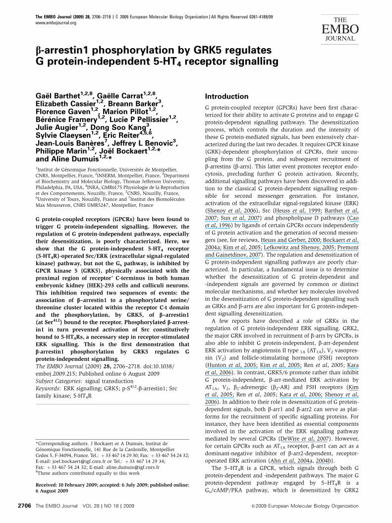
The 5-HT4R C-t encompasses several S/T residues, which are
potential GRK5 phosphorylation sites. We first examined the
possible role of these residues in GRK5-mediated inhibition of
ERK by generating several 5-HT4R truncated mutants
(Figure 2A). GRK5 failed to inhibit ERK phosphorylation
induced by 5-HT in cells expressing either the D329 mutant
lacking the entire C-terminus or the D346 mutant lacking a S/
T cluster (residues 347–355, Figure 2A). Deletion of the S/T
cluster likewise prevented inhibition, by GRK5, of Src phos-
phorylation elicited by 5-HT (Figure 2B). GRK5 still inhibited
phosphorylation of ERK induced by 5-HT in cells expressing
either the truncated receptor lacking its PDZ ligand (DSCF,
Figure 2A) with a putative phosphorylated site (S385) or the
D358 mutant comprising the S/T cluster and lacking the last
four scattered S/T residues, but not in cells expressing the
corresponding truncated receptor in which all S/T residues of
the cluster have been mutated into alanine (D358-Ala,
Figure 2A).
Collectively, these results indicate that the S/T cluster
(residues 347–355), which was previously shown to bind to
b-arr (Barthet et al, 2005), constitutes a key molecular
determinant implicated in the regulation of ERK activation
by GRK5. Further supporting its essential role in the regula-
tion by GRK5 of the ERK pathway, progressive deletion of the
S/T residues within the cluster concomitantly reduced the
GRK5-mediated inhibition of ERK phosphorylation
(Supplementary Figure S2).
p-ERK
p-Src
K215RGRK5
p-E
RK
(%
of m
axim
al 5
-HT
stim
ulat
ion)
*
5HT4
5HT4 + GRK5
5HT4 + GRK5 K215R
0
20
40
60
80
100
120
140
160
* *
5-HT (10 μM) – 5′ 30′ – 5′ 30′ – 5′ 30′
5-HT4RA
B
5-HT (10 μM) – 5′ – 5′ – 5′
p-S
rc (
% o
f max
imal
5-H
T s
timul
atio
n)
0
20
40
60
80
100
120
5-HT (10 μM) – 5′ – 5′ – 5′
Figure 1 GRK5 suppresses the 5-HT4R-mediated activation of theSrc/ERK pathway. (A) HEK-293 cells transiently expressing 5-HT4Rswere co-transfected or not with GRK5 or the kinase-dead mutantGRK5 (K215R) and serum starved before 5-HT (10mM) exposure forthe indicated time. HEK-293 cells were lysed in SDS sample buffer,subjected to SDS–PAGE. ERK and Src activation were analysed bywestern blotting using antibodies against phospho-Thr202/Tyr204-ERK1/2 (p-ERK) and phospho-Tyr416-Src (p-Y416-Src). Total ERK1/2and total Src were revealed on the same blot with polyclonalantibody recognizing ERK and Src independently of their phosphor-ylation sites (not shown). Note that total ERK and Src were notaffected in all experiments. The data are representative of a series ofblots performed in the same conditions. (B) P-ERK and p-Y416-Srcexpressed as percentage of maximal 5-HT stimulation±s.e.m.,represented by densitometric quantification of western blot per-formed from four different experiments. *Po0.05 or **Po0.01versus corresponding values measured in cells transfected withWT 5-HT4R alone.
GRK5 inhibits 5-HT4R-mediated ERK activationG Barthet et al
&2009 European Molecular Biology Organization The EMBO Journal VOL 28 | NO 18 | 2009 2707
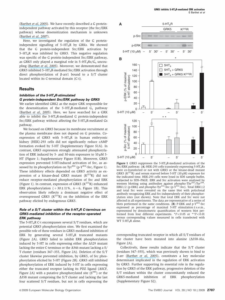
We then analysed agonist-dependent phosphorylation sites
on the receptor by tandem mass spectrometry (MS/MS).
HEK-293 cells expressing HA-tagged 5-HT4R and GRK5 con-
structs were treated or not with 5-HT for 10 min. Receptors
were then immunoprecipitated on anti-HA agarose beads.
Immunoprecipitated receptors were resolved by SDS–PAGE
and digested in-gel with trypsin. In control cells (not treated
with 5-HT), analysis of duplicate samples revealed the pre-
sence of a non-phosphorylated form of the peptide compris-
ing the S/Tcluster (R336-R359). In contrast, several versions of
the peptide were identified in cells exposed to 5-HT: the non-
phosphorylated one and three phosphorylated forms with
one, two and three phosphates attached, respectively. Loss of
phosphate on fragmentation indicated phosphorylation of
S354 in the mono-phosphorylated peptide (Figure 3B) and
the presence of an additional phosphorylated residue within
the S347TTT350 motif in the peptide with two phosphates
attached (Figure 3C). The tri-phosphorylated peptide incor-
porated an additional phosphate within the S347TTT350 motif
(not illustrated). These results indicated sequential phosphor-
ylation of the peptide, first on S354 and then in the S347TTT350
motif.
Physical association of GRK5 with 5-HT4Rs, a necessary
step in GRK5-mediated inhibition of 5-HT4R-operated
ERK signal
We next determined whether GRK5 physically interacted with
5-HT4R. GRK5/5-HT4R interaction was first investigated in an
in vitro binding assay using purified GRK5 and recombinant
5-HT(10 μM)
– 5′ – 5′
p-ERK
+ GRK5
p-ERK
p-ERK
p-ERK
p-ERK
p-ERK
5-HT4Rs
ΔSCF
ΔD358Ala
Δ358
Δ346
Δ329
WT
– 5′ – 5′ – 5′ – 5′
p-Src
p-ERK
+ GRK5
WT Δ346
+ GRK5
5-HT (10 μM)
B
A
Figure 2 A Serine/Threonine cluster within the 5-HT4R C-t isessential for inhibition, by GRK5, of receptor operated ERK signal-ling. (A) Topology of 5-HT4R C-t domain. The successive points oftruncation are illustrated represented on the left with an arrow.HEK-293 cells were transiently transfected with WT 5-HT4R or thecorresponding truncated or mutated receptors (D329, D346, D358,D358Ala) in combination with or without GRK5. Identical expres-sion levels of the transfected constructs were controlled by ELISA.Cells expressing the indicated receptors were treated or not with10mM 5-HT for 5 min. ERK activation was analysed by immuno-blotting with p-ERK1/2 antibody. (B) HEK-293 cells wereeither transfected with a plasmid encoding Myc-tagged WT 5-HT4R or Myc-tagged D346 alone or co-transfected with GRK5.They were challenged with 10 mM 5-HT for 5 min. Total lysateswere analysed by sequential immunoblotting, using p-Y416-Src,p-ERK1/2 antibodies.
m/z
m/z
m/z
A
Rel
ativ
e in
tens
ity
20
40
60
80
100
R P P I L G Q T V P C S T T T I N G S T H V L R3121 3 4 5 6 7 8 9 10 1611 12 1514 17 20 21 2218 19 23
yn
bn
12331 4567891016 111215 14172021 1923
B
Rel
ativ
e in
tens
ity
20
40
60
80
100
R P P I L G Q T V P C S T T T I N G pS T H V L R3121 3 4 5 6 7 8 9 10 1611 12 1514 17 20 21 2218 19 23
yn
bn
12331 4567891016 111215 1417202122 181923
*
CR
elat
ive
inte
nsity
20
40
60
80
100
R P P I L G Q T V P C S T T T
*
I N G pS T H V L R3121 3 4 5 6 7 8 9 10 1611 12 1514 17 20 21 22 23
yn
bn
12331 47891016 111215 1417202122 181923
18 19
56
11001000900800700600500400
18001600140012001000800600400200
1600140012001000800600400200
111213141822
Figure 3 Analysis of 5-HT-dependent phosphorylation of 5-HT4Rby tandem mass spectrometry. HEK-293 cells co-transfected withHA-tagged 5-HT4R and GRK5 constructs were treated with 5-HT(10 mM, 10 min). Immunoprecipitated receptors were digested withtrypsin and peptides were analysed by nano-LC FT MS/MS. MS/MSspectra resulting from higher energy collisional dissociation (HCD)fragmentation of the non-phosphosphorylated, monophosphory-lated and bi-phosphorylated versions of the R336-R359 peptide aredepicted in (A), (B) and (C) respectively. The three peptides haverespective mascot scores of 66, 65 and 59. (C) The bracket indicatesthat one residue of the S347TTT350 motif is phosphorylated. Its exactposition could not be determined by MS/MS.
GRK5 inhibits 5-HT4R-mediated ERK activationG Barthet et al
The EMBO Journal VOL 28 | NO 18 | 2009 &2009 European Molecular Biology Organization2708

S-tagged receptors immobilized on a S-protein agarose col-
umn (Baneres et al, 2005). 5-HT induced association of GRK5
with 5-HT4R, as assessed by the lack of GRK5 detection in the
flow-through fraction when 5-HTwas present (FT, Figure 4A).
Moreover, a greater amount of GRK5 was co-eluted with the
receptor compared with sample not treated with 5-HT
(E, Figure 4A). In contrast, leukotriene B4 failed to induce
association of GRK5 with immobilized, recombinant BLT1
receptors (Figure 4A), indicating that GRK5 does not interact
with every GPCR on activation by its cognate agonist.
These results indicate that 5-HT promotes direct interac-
tion of GRK5 with 5-HT4Rs. We then explored the molecular
determinants involved in the interaction of GRK5 with 5-
HT4Rs in HEK-293 cells by co-immunoprecipitation. GRK5 co-
immunoprecipitated with 5-HT4R and the amount of GRK5
was enhanced on 5-HT exposure (Figure 4B). GRK5 did not
co-immunoprecipitate with the D329 truncation mutant lack-
ing the entire C-terminus in the presence of 5-HT
(Supplementary Figure S3), whereas the D346 mutant that
lacks the S/T (347–355) cluster phosphorylated by GRK5
(Supplementary Figure S2) still interacted with GRK5
(Figure 4B). Thus, GRK5 can bind to 5-HT4Rs without the
presence of the S/T cluster substrates. However, note that
5-HT did not stimulate GRK5 binding to the D346 mutant.
A
Receptors
GRK5
FTFT EE
Agonist – + + +– +
B
WT WTDDE-3A
**
GR
K5
boun
d(%
of G
RK
5 bo
und
to W
T 5
-HT
4R)
Basal
5-HT (5′)
GRK5
GRK5
5-HT4Rs
GRK5
5-HT4Rs
WT
DDE-3AWT DDE-3AWT
5-HT4Rs
– 5′ – 5′ – 5′ – 5′
Δ346DDE-3AWT
Δ346DDE-3AWT
0
20
40
60
80
100
120
C
GR
K5
boun
d (%
of C
trl)
40
60
80
100**
IP 5-HT4R
D
p-E
RK
(%
of C
trl)
Ctrl-Pep
**
0
50
100
150
200
250
p-ERK
+ +
Inh-PepCtrl-Pep
Inh-PepCtrl-Pep
BLT1R5-HT4R
IP 5-HT4RInput
5-HT(10 μM)
Δ346 Δ346DDE-3A
Inh-Pep
5-HT (10 μM)
GRK5
+ +
Inh-PepCtrl-Pep
5-HT (10 μM)
Δ346WT Δ346
Figure 4 See over for legend.
GRK5 inhibits 5-HT4R-mediated ERK activationG Barthet et al
&2009 European Molecular Biology Organization The EMBO Journal VOL 28 | NO 18 | 2009 2709
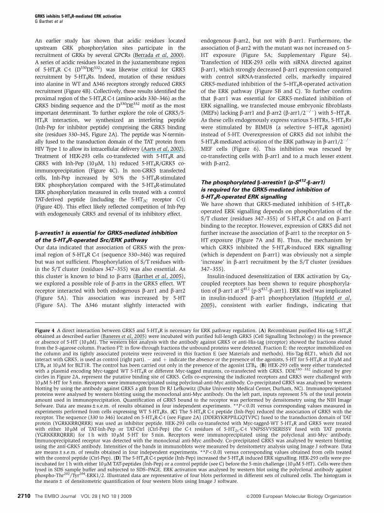
An earlier study has shown that acidic residues located
upstream GRK phosphorylation sites participate in the
recruitment of GRKs by several GPCRs (Berrada et al, 2000).
A series of acidic residues located in the juxtamembrane region
of 5-HT4R C-t (D330DE332) was likewise critical for GRK5
recruitment by 5-HT4Rs. Indeed, mutation of these residues
into alanine in WT and D346 receptors strongly reduced GRK5
recruitment (Figure 4B). Collectively, these results identified the
proximal region of the 5-HT4R C-t (amino-acids 330–346) as the
GRK5 binding sequence and the D330DE332 motif as the most
important determinant. To further explore the role of GRK5/5-
HT4R interaction, we synthesized an interfering peptide
(Inh-Pep for inhibitor peptide) comprising the GRK5 binding
site (residues 330–345, Figure 2A). The peptide was N-termin-
ally fused to the transduction domain of the TAT protein from
HIV Type 1 to allow its intracellular delivery (Aarts et al, 2002).
Treatment of HEK-293 cells co-transfected with 5-HT4R and
GRK5 with Inh-Pep (10mM, 1 h) reduced 5-HT4R/GRK5 co-
immunoprecipitation (Figure 4C). In non-GRK5 transfected
cells, Inh-Pep increased by 50% the 5-HT4R-stimulated
ERK phosphorylation compared with the 5-HT4R-stimulated
ERK phosphorylation measured in cells treated with a control
TAT-derived peptide (including the 5-HT2C receptor C-t)
(Figure 4D). This effect likely reflected competition of Inh-Pep
with endogenously GRK5 and reversal of its inhibitory effect.
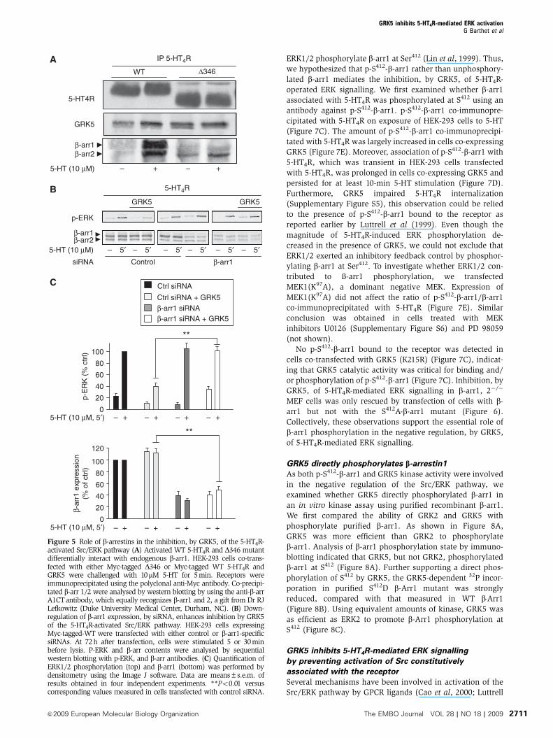
b-arrestin1 is essential for GRK5-mediated inhibition
of the 5-HT4R-operated Src/ERK pathway
Our data indicated that association of GRK5 with the prox-
imal region of 5-HT4R C-t (sequence 330–346) was required
but was not sufficient. Phosphorylation of S/T residues with-
in the S/T cluster (residues 347–355) was also essential. As
this cluster is known to bind to b-arrs (Barthet et al, 2005),
we explored a possible role of b-arrs in the GRK5 effect. WT
receptor interacted with both endogenous b-arr1 and b-arr2
(Figure 5A). This association was increased by 5-HT
(Figure 5A). The D346 mutant slightly interacted with
endogenous b-arr2, but not with b-arr1. Furthermore, the
association of b-arr2 with the mutant was not increased on 5-
HT exposure (Figure 5A; Supplementary Figure S4).
Transfection of HEK-293 cells with siRNA directed against
b-arr1, which strongly decreased b-arr1 expression compared
with control siRNA-transfected cells, markedly impaired
GRK5-mediated inhibition of the 5–HT4R-operated activation
of the ERK pathway (Figure 5B and C). To further confirm
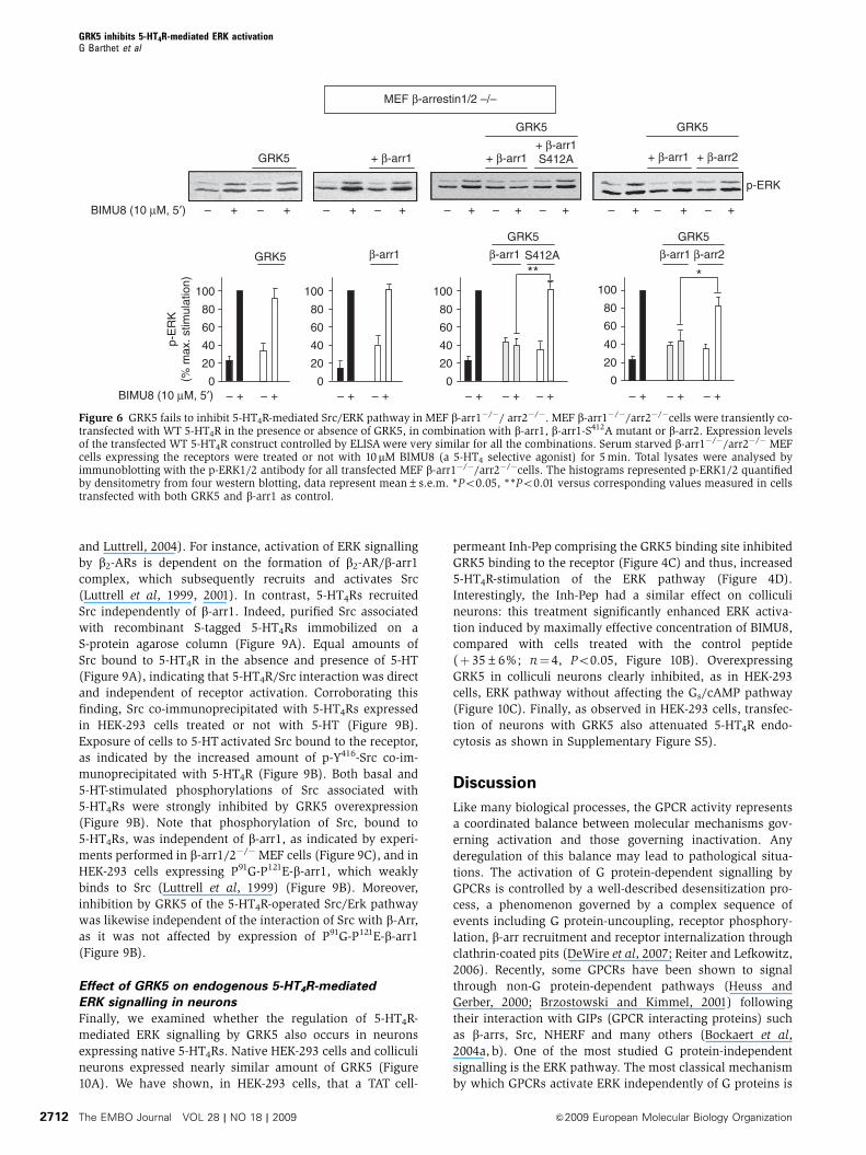
that b-arr1 was essential for GRK5-mediated inhibition of
ERK signalling, we transfected mouse embryonic fibroblasts
(MEFs) lacking b-arr1 and b-arr2 (b-arr1/2�/�) with 5–HT4R.
As these cells endogenously express various 5-HTRs, 5-HT4Rs
were stimulated by BIMU8 (a selective 5–HT4R agonist)
instead of 5-HT. Overexpression of GRK5 did not inhibit the
5-HT4R-mediated activation of the ERK pathway in b-arr1/2�/�
MEF cells (Figure 6). This inhibition was rescued by
co-transfecting cells with b-arr1 and to a much lesser extent
with b-arr2.
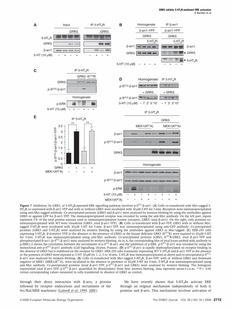
The phosphorylated b-arrestin1 (p-S412-b-arr1)
is required for the GRK5-mediated inhibition of
5-HT4R-operated ERK signalling
We have shown that GRK5-mediated inhibition of 5-HT4R-
operated ERK signalling depends on phosphorylation of the
S/T cluster (residues 347–355) of 5-HT4R C-t and on b-arr1
binding to the receptor. However, expression of GRK5 did not
further increase the association of b-arr1 to the receptor on 5-
HT exposure (Figure 7A and B). Thus, the mechanism by
which GRK5 inhibited the 5-HT4R-induced ERK signalling
(which is dependent on b-arr1) was obviously not a simple
‘increase’ in b-arr1 recruitment by the S/T cluster (residues
347–355).
Insulin-induced desensitization of ERK activation by Gai-
coupled receptors has been shown to require phosphoryla-
tion of b-arr1 at S412 (p-S412-b-arr1). ERK itself was implicated
in insulin-induced b-arr1 phosphorylation (Hupfeld et al,
2005), consistent with earlier findings, indicating that
Figure 4 A direct interaction between GRK5 and 5-HT4R is necessary for ERK pathway regulation. (A) Recombinant purified His-tag 5-HT4Robtained as described earlier (Baneres et al, 2005) were incubated with purified full-length GRK5 (Cell Signalling Technology) in the presenceor absence of 5-HT (10 mM). The western blot analysis with the antibody against GRK5 or anti-His-tag (receptor) showed the fractions elutedfrom the S-agarose column. Fraction FT: in flow-through fractions the unbound proteins were detected. Fraction E: the receptor immobilized onthe column and its tightly associated proteins were recovered in this fraction E (see Materials and methods). His-Tag-BLT1, which did notinteract with GRK5, is used as control (right part). � and þ indicate the absence or the presence of the agonists, 5-HT for 5-HT4R at 10mM andLTB4 at 10mM for BLT1R. The control has been carried out only in the presence of the agonist LTB4. (B) HEK-293 cells were either transfectedwith a plasmid encoding Myc-tagged WT 5-HT4R or different Myc-tagged mutants, co-transfected with GRK5. DDE330�332 indicated by greycircles in Figure 2A, represent the putative binding site of GRK5. Cells co-expressing the indicated receptors and GRK5 were challenged with10mM 5-HT for 5 min. Receptors were immunoprecipitated using polyclonal-anti-Myc antibody. Co-precipitated GRK5 was analysed by westernblotting by using the antibody against GRK5 a gift from Dr RJ Lefkowitz (Duke University Medical Center, Durham, NC). Immunoprecipitatedproteins were analysed by western blotting using the monoclonal anti-Myc antibody. On the left part, inputs represent 5% of the total proteinamount used in immunoprecipitation. Quantification of GRK5 bound to the receptor was performed by densitometry using the NIH ImageSofware. Data are means±s.e.m. of results obtained in four independent experiments. **Po0.01 versus corresponding values measured inexperiments performed from cells expressing WT 5-HT4Rs. (C) The 5-HT4R C-t peptide (Inh-Pep) reduced the association of GRK5 with thereceptor. The sequence (330 to 346) located on 5-HT4R-C-t (see Figure 2A) (DDERYKRPPILGQTVPC) fused to the transduction domain of TATprotein (YGRKKRRQRRR) was used as inhibitor peptide. HEK-293 cells co-transfected with Myc-tagged-WT 5-HT4R and GRK5 were treatedwith either 10mM of TAT-Inh-Pep or TAT-Ctrl (Ctrl-Pep) (the C-t residues of 5-HT2c-C-t VNPSSVVSERISSV fused with TAT protein(YGRKKRRQRRR) for 1 h with 10mM 5-HT for 5 min. Receptors were immunoprecipitated using the polyclonal anti-Myc antibody.Immunoprecipitated receptor was detected with the monoclonal anti-Myc antibody. Co-precipitated GRK5 was analysed by western blottingusing the anti-GRK5 antibody. Intensities of the bands in immunoblots were measured by densitometry analysis using Image J software. Dataare means±s.e.m. of results obtained in four independent experiments. **Po0.01 versus corresponding values obtained from cells treatedwith the control peptide (Ctrl-Pep). (D) The 5-HT4R C-t peptide (Inh-Pep) increased the 5-HT4R induced ERK signalling. HEK-293 cells were pre-incubated for 1 h with either 10mM TAT-peptides (Inh-Pep) or a control peptide (see C) before the 5-min challenge (10mM 5-HT). Cells were thenlysed in SDS sample buffer and subjected to SDS–PAGE. ERK activation was analysed by western blot using the polyclonal antibody againstphospho-Thr202/Tyr204-ERK1/2. Illustrated data are representative of four blots performed in different sets of cultured cells. The histogram isthe means± of densitometric quantification of four western blots using Image J software.
GRK5 inhibits 5-HT4R-mediated ERK activationG Barthet et al
The EMBO Journal VOL 28 | NO 18 | 2009 &2009 European Molecular Biology Organization2710
ERK1/2 phosphorylate b-arr1 at Ser412 (Lin et al, 1999). Thus,
we hypothesized that p-S412-b-arr1 rather than unphosphory-
lated b-arr1 mediates the inhibition, by GRK5, of 5-HT4R-
operated ERK signalling. We first examined whether b-arr1
associated with 5-HT4R was phosphorylated at S412 using an
antibody against p-S412-b-arr1. p-S412-b-arr1 co-immunopre-
cipitated with 5-HT4R on exposure of HEK-293 cells to 5-HT
(Figure 7C). The amount of p-S412-b-arr1 co-immunoprecipi-
tated with 5-HT4R was largely increased in cells co-expressing
GRK5 (Figure 7E). Moreover, association of p-S412-b-arr1 with
5-HT4R, which was transient in HEK-293 cells transfected
with 5-HT4R, was prolonged in cells co-expressing GRK5 and
persisted for at least 10-min 5-HT stimulation (Figure 7D).
Furthermore, GRK5 impaired 5-HT4R internalization
(Supplementary Figure S5), this observation could be relied
to the presence of p-S412-b-arr1 bound to the receptor as
reported earlier by Luttrell et al (1999). Even though the
magnitude of 5-HT4R-induced ERK phosphorylation de-
creased in the presence of GRK5, we could not exclude that
ERK1/2 exerted an inhibitory feedback control by phosphor-
ylating b-arr1 at Ser412. To investigate whether ERK1/2 con-
tributed to �-arr1 phosphorylation, we transfected
MEK1(K97A), a dominant negative MEK. Expression of
MEK1(K97A) did not affect the ratio of p-S412-b-arr1/b-arr1
co-immunoprecipitated with 5-HT4R (Figure 7E). Similar
conclusion was obtained in cells treated with MEK
inhibitors U0126 (Supplementary Figure S6) and PD 98059
(not shown).
No p-S412-b-arr1 bound to the receptor was detected in
cells co-transfected with GRK5 (K215R) (Figure 7C), indicat-
ing that GRK5 catalytic activity was critical for binding and/
or phosphorylation of p-S412-b-arr1 (Figure 7C). Inhibition, by
GRK5, of 5-HT4R-mediated ERK signalling in b-arr1, 2�/�
MEF cells was only rescued by transfection of cells with b-
arr1 but not with the S412A-b-arr1 mutant (Figure 6).
Collectively, these observations support the essential role of
b-arr1 phosphorylation in the negative regulation, by GRK5,
of 5-HT4R-mediated ERK signalling.
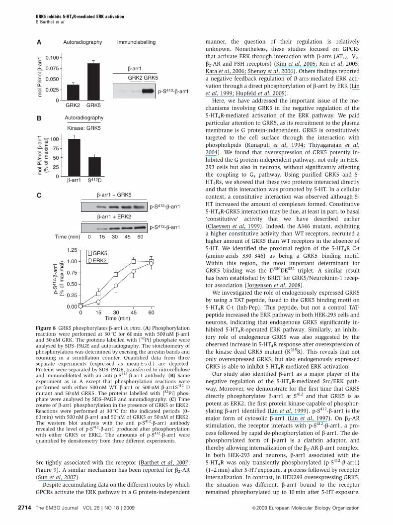
GRK5 directly phosphorylates b-arrestin1
As both p-S412-b-arr1 and GRK5 kinase activity were involved
in the negative regulation of the Src/ERK pathway, we
examined whether GRK5 directly phosphorylated b-arr1 in
an in vitro kinase assay using purified recombinant b-arr1.
We first compared the ability of GRK2 and GRK5 with
phosphorylate purified b-arr1. As shown in Figure 8A,
GRK5 was more efficient than GRK2 to phosphorylate
b-arr1. Analysis of b-arr1 phosphorylation state by immuno-
blotting indicated that GRK5, but not GRK2, phosphorylated
b-arr1 at S412 (Figure 8A). Further supporting a direct phos-
phorylation of S412 by GRK5, the GRK5-dependent 32P incor-
poration in purified S412D b-Arr1 mutant was strongly
reduced, compared with that measured in WT b-Arr1
(Figure 8B). Using equivalent amounts of kinase, GRK5 was
as efficient as ERK2 to promote b-Arr1 phosphorylation at
S412 (Figure 8C).
GRK5 inhibits 5-HT4R-mediated ERK signalling
by preventing activation of Src constitutively
associated with the receptor
Several mechanisms have been involved in activation of the
Src/ERK pathway by GPCR ligands (Cao et al, 2000; Luttrell
ControlsiRNA
β-arr1β-arr2
5-HT (10 μM) – 5′ – 5′ – 5′ – 5′ – 5′ – 5′
p-ERK
5-HT4R
GRK5 GRK5
β-arr1
Ctrl siRNA
Ctrl siRNA + GRK5β-arr1 siRNAβ-arr1 siRNA + GRK5
**
0
20
40
60
80
100
**
p-E
RK
(%
ctr
l)
5-HT (10 μM, 5′)
0
20
40
60
80
100
120
β-ar
r1 e
xpre
ssio
n(%
of c
trl)
5-HT (10 μM, 5′)
– + – + – + – +
– + – + – + – +
C
B
5-HT (10 μM)
WT Δ346
β-arr2
GRK5
5-HT4R
+ +– –
β-arr1
IP 5-HT4RA
Figure 5 Role of b-arrestins in the inhibition, by GRK5, of the 5-HT4R-activated Src/ERK pathway (A) Activated WT 5-HT4R and D346 mutantdifferentially interact with endogenous b-arr1. HEK-293 cells co-trans-fected with either Myc-tagged D346 or Myc-tagged WT 5-HT4R andGRK5 were challenged with 10mM 5-HT for 5min. Receptors wereimmunoprecipitated using the polyclonal anti-Myc antibody. Co-precipi-tated b-arr 1/2 were analysed by western blotting by using the anti-b-arrA1CTantibody, which equally recognizes b-arr1 and 2, a gift from Dr RJLefkowitz (Duke University Medical Center, Durham, NC). (B) Down-regulation of b-arr1 expression, by siRNA, enhances inhibition by GRK5of the 5-HT4R-activated Src/ERK pathway. HEK-293 cells expressingMyc-tagged-WT were transfected with either control or b-arr1-specificsiRNAs. At 72h after transfection, cells were stimulated 5 or 30minbefore lysis. P-ERK and b-arr contents were analysed by sequentialwestern blotting with p-ERK, and b-arr antibodies. (C) Quantification ofERK1/2 phosphorylation (top) and b-arr1 (bottom) was performed bydensitometry using the Image J software. Data are means±s.e.m. ofresults obtained in four independent experiments. **Po0.01 versuscorresponding values measured in cells transfected with control siRNA.
GRK5 inhibits 5-HT4R-mediated ERK activationG Barthet et al
&2009 European Molecular Biology Organization The EMBO Journal VOL 28 | NO 18 | 2009 2711
and Luttrell, 2004). For instance, activation of ERK signalling
by b2-ARs is dependent on the formation of b2-AR/b-arr1
complex, which subsequently recruits and activates Src
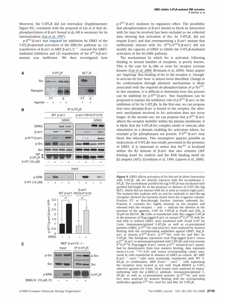
(Luttrell et al, 1999, 2001). In contrast, 5-HT4Rs recruited
Src independently of b-arr1. Indeed, purified Src associated
with recombinant S-tagged 5-HT4Rs immobilized on a
S-protein agarose column (Figure 9A). Equal amounts of
Src bound to 5-HT4R in the absence and presence of 5-HT
(Figure 9A), indicating that 5-HT4R/Src interaction was direct
and independent of receptor activation. Corroborating this
finding, Src co-immunoprecipitated with 5-HT4Rs expressed
in HEK-293 cells treated or not with 5-HT (Figure 9B).
Exposure of cells to 5-HT activated Src bound to the receptor,
as indicated by the increased amount of p-Y416-Src co-im-
munoprecipitated with 5-HT4R (Figure 9B). Both basal and
5-HT-stimulated phosphorylations of Src associated with
5-HT4Rs were strongly inhibited by GRK5 overexpression
(Figure 9B). Note that phosphorylation of Src, bound to
5-HT4Rs, was independent of b-arr1, as indicated by experi-
ments performed in b-arr1/2�/�MEF cells (Figure 9C), and in
HEK-293 cells expressing P91G-P121E-b-arr1, which weakly
binds to Src (Luttrell et al, 1999) (Figure 9B). Moreover,
inhibition by GRK5 of the 5-HT4R-operated Src/Erk pathway
was likewise independent of the interaction of Src with b-Arr,
as it was not affected by expression of P91G-P121E-b-arr1
(Figure 9B).
Effect of GRK5 on endogenous 5-HT4R-mediated
ERK signalling in neurons
Finally, we examined whether the regulation of 5-HT4R-
mediated ERK signalling by GRK5 also occurs in neurons
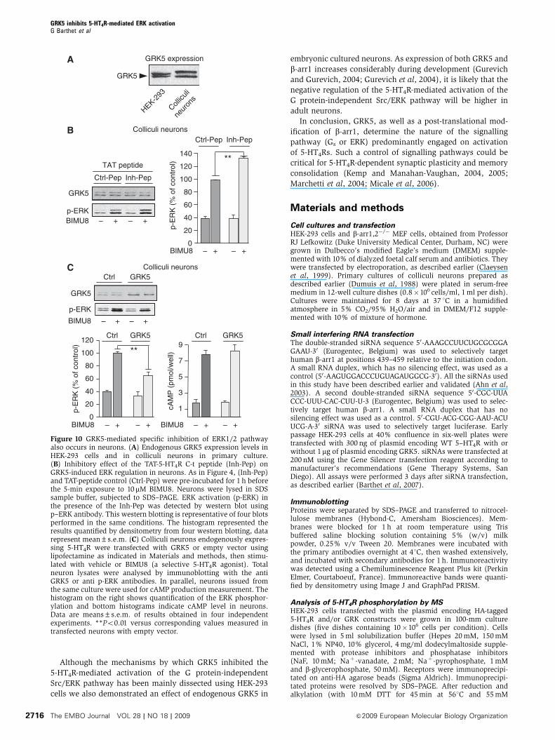
expressing native 5-HT4Rs. Native HEK-293 cells and colliculi
neurons expressed nearly similar amount of GRK5 (Figure
10A). We have shown, in HEK-293 cells, that a TAT cell-
permeant Inh-Pep comprising the GRK5 binding site inhibited
GRK5 binding to the receptor (Figure 4C) and thus, increased
5-HT4R-stimulation of the ERK pathway (Figure 4D).
Interestingly, the Inh-Pep had a similar effect on colliculi
neurons: this treatment significantly enhanced ERK activa-
tion induced by maximally effective concentration of BIMU8,
compared with cells treated with the control peptide
(þ 35±6%; n¼ 4, Po0.05, Figure 10B). Overexpressing
GRK5 in colliculi neurons clearly inhibited, as in HEK-293
cells, ERK pathway without affecting the Gs/cAMP pathway
(Figure 10C). Finally, as observed in HEK-293 cells, transfec-
tion of neurons with GRK5 also attenuated 5-HT4R endo-
cytosis as shown in Supplementary Figure S5).
Discussion
Like many biological processes, the GPCR activity represents
a coordinated balance between molecular mechanisms gov-
erning activation and those governing inactivation. Any
deregulation of this balance may lead to pathological situa-
tions. The activation of G protein-dependent signalling by
GPCRs is controlled by a well-described desensitization pro-
cess, a phenomenon governed by a complex sequence of
events including G protein-uncoupling, receptor phosphory-
lation, b-arr recruitment and receptor internalization through
clathrin-coated pits (DeWire et al, 2007; Reiter and Lefkowitz,
2006). Recently, some GPCRs have been shown to signal
through non-G protein-dependent pathways (Heuss and
Gerber, 2000; Brzostowski and Kimmel, 2001) following
their interaction with GIPs (GPCR interacting proteins) such
as b-arrs, Src, NHERF and many others (Bockaert et al,
2004a, b). One of the most studied G protein-independent
signalling is the ERK pathway. The most classical mechanism
by which GPCRs activate ERK independently of G proteins is
GRK5 GRK5
p-ERK
– + – + – + – + – + – + – + – + – + – +
MEF β-arrestin1/2 –/–
+ β-arr1 + β-arr1 + β-arr2+ β-arr1GRK5
GRK5
*
β-arr1
p-E
RK
(% m
ax. s
timul
atio
n)
GRK5
0– + – +
20
40
60
80
100
0– + – +
20
40
60
80
100
0– + – + – + – + – + – +
20
40
60
80
100
S412A
GRK5
β-arr1 β-arr1 β-arr2
**
0
20
40
60
80
100
BIMU8 (10 μM, 5′)
+ β-arr1S412A
BIMU8 (10 μM, 5′)
Figure 6 GRK5 fails to inhibit 5-HT4R-mediated Src/ERK pathway in MEF b-arr1�/�/ arr2�/�. MEF b-arr1�/�/arr2�/�cells were transiently co-transfected with WT 5-HT4R in the presence or absence of GRK5, in combination with b-arr1, b-arr1-S412A mutant or b-arr2. Expression levelsof the transfected WT 5-HT4R construct controlled by ELISA were very similar for all the combinations. Serum starved b-arr1�/�/arr2�/� MEFcells expressing the receptors were treated or not with 10 mM BIMU8 (a 5-HT4 selective agonist) for 5 min. Total lysates were analysed byimmunoblotting with the p-ERK1/2 antibody for all transfected MEF b-arr1�/�/arr2�/�cells. The histograms represented p-ERK1/2 quantifiedby densitometry from four western blotting, data represent mean±s.e.m. *Po0.05, **Po0.01 versus corresponding values measured in cellstransfected with both GRK5 and b-arr1 as control.
GRK5 inhibits 5-HT4R-mediated ERK activationG Barthet et al
The EMBO Journal VOL 28 | NO 18 | 2009 &2009 European Molecular Biology Organization2712
through their direct interaction with b-arrs, a process
followed by receptor endocytosis and recruitment of the
Src/Raf/MEK machinery (Luttrell et al, 1999, 2001).
We have recently shown that 5-HT4Rs activate ERK
through an original mechanism independently of both G
proteins and b-arrs. This mechanism involves activation of
β-arr1
GRK5
5-HT4R
IP β-arr1
β-arr1-YFP
Homogenate
β-arr1-YFP
GRK5 GRK5
B
GRK5
β-arr1
5-HT4R 5-HT4R
– + – +– + – +5-HT (10 μM)
A
β-arr1
GRK5 GRK5
GRK5
Input
– + – + – + – +
IP 5-HT4R
5-HT (10 μM)
5-HT4R
D
+ GRK5+ GRK5
IP 5-HT4RHomogenate
p-S412-β-arr1
p-S412-β-arr1
5-HT (10 μM) – 1′ 2′ 5′10′ – 1′ 2′ 5′10′
C
GRK5
p-ERK
GRK5
5-HT (10 μM)
Homogenate
p-S412-β-arr1
– + – + – +
K215R
IP 5-HT4R
E
GRK5MEK1(K97A)
0
0.5
1
1.5
2
2.5
**
3
3.5**
p-S
412 -
β-ar
r1/β
-arr
1-Y
FP
– + – +– – + +
IP 5-HT4R
GRK5
β-arr1
p-S412-β-arr1
p-ERK
β-arr1
p-S412-β-arr1
5-HT (10 μM)
GRK5
Homogenate
– + – +
– – + +
– + – +
– – + +
IP 5-HT4R
MEK1(K97A) MEK1(K97A)
MEK1(K97A)
Figure 7 Inhibition, by GRK5, of 5-HT4R-operated ERK signalling pathway involves p-S412-b-arr1. (A) Cells co-transfected with Myc-tagged 5-HT4R co-expressed with b-arr1-YFP and with or without GRK5 were incubated with 10mM 5-HT for 5 min. Receptors were immunoprecipitatedusing anti-Myc-tagged antibody. Co-precipitated proteins (GRK5 and b-arr1) were analysed by western blotting by using the antibodies againstGRK5 or against GFP for b-arr1-YFP. The immunoprecipitated receptor was revealed by using the anti-Myc antibody. On the left part, inputsrepresent 5% of the total protein amount used in immunoprecipitation lysates (receptor; GRK5; total b-arr1). On the right, only proteins co-immunoprecipitated with WT were visualized (GRK5, total b-arr1-YFP). (B) Cells co-transfected with b-arr-YFP, GRK5 with or without Myc-tagged 5-HT4R were incubated with 10mM 5-HT for 5 min. b-arr1-YFP was immunoprecipitated using anti-GFP antibody. Co-precipitatedproteins (GRK5 and 5-HT4R) were analysed by western blotting by using the antibodies against GRK5 or Myc-tagged. (C) HEK-293 cellsexpressing 5-HT4R, b-arrestin1-YFP in the absence or the presence of GRK5 or the kinase deficient GRK5 (K215R) were exposed to 10mM 5-HTfor 5 min. 5-HT4R was immunoprecipitated using anti-Myc antibody. Co-precipitated proteins (GRK5, K215R-GRK5, total b-arr1-YFP andphosphorylated b-arr1 (p-S412-b-arr1) were analysed by western blotting. As in A, the corresponding blot of total lysate probed with antibody top-ERK1/2 shows the correlation between the recruitment of p-S412-b-arr1 and the inhibition of p-ERK. p-S412-b-arr1 was revealed by using themonoclonal anti-p-S412-b-arr1 antibody (Cell Signalling, Ozyme, France). (D) p-S412-b-arr1 is rapidly dephosphorylated on receptor binding inthe absence of GRK5 but is stabilized on the receptor by GRK5. HEK-293 cells transiently expressing WT 5–HT4R and b-arr1-YFP in the absenceor the presence of GRK5 were exposed to 5-HT 10 mM for 1, 2, 5 or 10 min. 5-HT4R was immunoprecipitated as above and co-precipitated p-S412-b-arr1 was analysed by western blotting. (E) Cells co-transfected with Myc-tagged 5-HT4R, b-arr-YFP, with or without GRK5 and dominantnegative of MEK1 (MEK1(K97A), were incubated in the absence or presence of 10mM 5-HT for 5 min. 5-HT4R was immunoprecipitated usinganti-Myc antibody. Co-precipitated proteins (total b-arr1-YFP, p-S412-b-arr1 and GRK5) were analysed by western blotting. The histogramrepresented total b-arr1-YFP, p-S412-b-arr1 quantified by densitometry from four western blotting, data represent mean±s.e.m. **Po 0.01versus corresponding values measured in cells transfected in absence of GRK5 as control.
GRK5 inhibits 5-HT4R-mediated ERK activationG Barthet et al
&2009 European Molecular Biology Organization The EMBO Journal VOL 28 | NO 18 | 2009 2713
Src tightly associated with the receptor (Barthet et al, 2007;
Figure 9). A similar mechanism has been reported for b2-AR
(Sun et al, 2007).
Despite accumulating data on the different routes by which
GPCRs activate the ERK pathway in a G protein-independent
manner, the question of their regulation is relatively
unknown. Nonetheless, these studies focused on GPCRs
that activate ERK through interaction with b-arrs (AT1A, V2,
b2-AR and FSH receptors) (Kim et al, 2005; Ren et al, 2005;
Kara et al, 2006; Shenoy et al, 2006). Others findings reported
a negative feedback regulation of �-arrs-mediated ERK acti-
vation through a direct phosphorylation of b-arr1 by ERK (Lin
et al, 1999; Hupfeld et al, 2005).
Here, we have addressed the important issue of the me-
chanisms involving GRK5 in the negative regulation of the
5-HT4R-mediated activation of the ERK pathway. We paid
particular attention to GRK5, as its recruitment to the plasma
membrane is G protein-independent. GRK5 is constitutively
targeted to the cell surface through the interaction with
phospholipids (Kunapuli et al, 1994; Thiyagarajan et al,
2004). We found that overexpression of GRK5 potently in-
hibited the G protein-independent pathway, not only in HEK-
293 cells but also in neurons, without significantly affecting
the coupling to Gs pathway. Using purified GRK5 and 5-
HT4Rs, we showed that these two proteins interacted directly
and that this interaction was promoted by 5-HT. In a cellular
context, a constitutive interaction was observed although 5-
HT increased the amount of complexes formed. Constitutive
5-HT4R-GRK5 interaction may be due, at least in part, to basal
‘constitutive’ activity that we have described earlier
(Claeysen et al, 1999). Indeed, the D346 mutant, exhibiting
a higher constitutive activity than WT receptors, recruited a
higher amount of GRK5 than WT receptors in the absence of
5-HT. We identified the proximal region of the 5-HT4R C-t
(amino-acids 330–346) as being a GRK5 binding motif.
Within this region, the most important determinant for
GRK5 binding was the D330DE332 triplet. A similar result
has been established by BRET for GRK5/Neurokinin-1 recep-
tor association (Jorgensen et al, 2008).
We investigated the role of endogenously expressed GRK5
by using a TAT peptide, fused to the GRK5 binding motif on
5-HT4R C-t (Inh-Pep). This peptide, but not a control TAT-
peptide increased the ERK pathway in both HEK-293 cells and
neurons, indicating that endogenous GRK5 significantly in-
hibited 5-HT4R-operated ERK pathway. Similarly, an inhibi-
tory role of endogenous GRK5 was also suggested by the
observed increase in 5-HT4R response after overexpression of
the kinase dead GRK5 mutant (K215R). This reveals that not
only overexpressed GRK5, but also endogenously expressed
GRK5 is able to inhibit 5-HT4R-mediated ERK activation.
Our study also identified b-arr1 as a major player of the
negative regulation of the 5-HT4R-mediated Src/ERK path-
way. Moreover, we demonstrate for the first time that GRK5
directly phosphorylates b-arr1 at S412 and that GRK5 is as
potent as ERK2, the first protein kinase capable of phosphor-
ylating b-arr1 identified (Lin et al, 1999). p-S412-b-arr1 is the
major form of cytosolic b-arr1 (Lin et al, 1997). On b2-AR
stimulation, the receptor interacts with p-S412-b-arr1, a pro-
cess followed by rapid de-phosphorylation of b-arr1. The de-
phosphorylated form of b-arr1 is a clathrin adaptor, and
thereby allowing internalization of the b2-AR-b-arr1 complex.
In both HEK-293 and neurons, b-arr1 associated with the
5-HT4R was only transiently phosphorylated (p-S412-b-arr1)
(1–2 min) after 5-HTexposure, a process followed by receptor
internalization. In contrast, in HEK293 overexpressing GRK5,
the situation was different. b-arr1 bound to the receptor
remained phosphorylated up to 10 min after 5-HT exposure.
p-S412-β-arr1
GRK2
β-arr1
GRK5
AutoradiographyA Immunolabelling
GRK20
0.050
0.100
0.025
0.075
GRK5
mol
Pi/m
ol β
-arr
1
BKinase: GRK5
Autoradiography
β-arr10
50
100
25
75
mol
Pi/m
ol β
-arr
1(%
of m
axim
al)
S412D
p-S412-β-arr1
β-arr1 + ERK2
β-arr1 + GRK5
p-S412-β-arr1
C
Time (min)
Time (min)
1.25
0.00
0.25
0.50
0.75
1.00
GRK5ERK2
0 15 30 45 60
0 15 30 45 60
p-S
412 -
β-ar
r1(%
of m
axim
al)
Figure 8 GRK5 phosphorylates b-arr1 in vitro. (A) Phosphorylationreactions were performed at 30 1C for 60 min with 500 nM b-arr1and 50 nM GRK. The proteins labelled with [32Pi] phosphate wereanalysed by SDS–PAGE and autoradiography. The stoichiometry ofphosphorylation was determined by excising the arrestin bands andcounting in a scintillation counter. Quantified data from threeseparate experiments (expressed as mean±s.d.) are depicted.Proteins were separated by SDS–PAGE, transferred to nitrocelluloseand immunoblotted with an anti p-S412-b-arr1 antibody. (B) Sameexperiment as in A except that phosphorylation reactions wereperformed with either 500 nM WT b-arr1 or 500 nM b-arr1S412 Dmutant and 50 nM GRK5. The proteins labelled with [32Pi] phos-phate were analysed by SDS–PAGE and autoradiography. (C) Timecourse of b-arr1 phosphorylation in the presence of GRK5 or ERK2.Reactions were performed at 30 1C for the indicated periods (0–60 min) with 500 nM b-arr1 and 50 nM of GRK5 or 50 nM of ERK2.The western blot analysis with the anti p-S412-b-arr1 antibodyrevealed the level of p-S412-b-arr1 produced after phosphorylationwith either GRK5 or ERK2. The amounts of p-S412-b-arr1 werequantified by densitometry from three different experiments.
GRK5 inhibits 5-HT4R-mediated ERK activationG Barthet et al
The EMBO Journal VOL 28 | NO 18 | 2009 &2009 European Molecular Biology Organization2714
Moreover, the 5-HT4R did not internalize (Supplementary
Figure S5), consistent with the proposal of Lin et al that de-
phosphorylation of b-arr1 bound to b2-AR is necessary for its
internalization (Lin et al, 1997).
p-S412-b-arr1 was required for inhibition by GRK5 of the
5-HT4R-operated activation of the ERK/Src pathway as: (1)
transfection of b-arr1 in MEF b-arr1/2�/� rescued the GRK5-
mediated inhibition and (2) transfection of the S412A-b-arr1
mutant was inefficient. We then investigated how
p-S412-b-arr1 mediates its regulatory effect. The possibility
that phosphorylation of b-arr1 known to block its interaction
with Src may be involved has been excluded as we collected
data showing that activation of Src, by 5-HT4R, did not
require b-arr1 and that overexpressing a b-arr1 mutant that
inefficiently interact with Src (P91G-P121E-b-arr1) did not
modify the capacity of GRK5 to inhibit the 5-HT4R-mediated
activation of the Src/ERK pathway.
The mechanisms by which Src is activated, following
binding to several families of receptors, is poorly known.
This is the case for b3-ARs or even for receptor tyrosine
kinases (Cao et al, 2000; Bromann et al, 2004). Many papers
are ‘implying’ that binding of Src to the receptor is ‘enough’
to activate Src but ‘how’ is almost never described. Change in
Src conformation through allosteric mechanisms is likely
associated with the required de-phosphorylation of p-Tyr527.
In this situation, it is difficult to determine how this process
can be inhibited by p-S412-b-arr1. Two hypotheses can be
proposed to explain the inhibitory role of p-S412-b-arr1 on the
inhibition of Src by 5-HT4Rs. In the first one, we can propose
that once phospho-b-arr is bound to the receptor, the allos-
teric mechanism involved in Src activation does not occur
longer. In the second one, we can propose that p-S412-b-arr1
affects the receptor mobility within the plasma membrane. It
is likely that the 5-HT4R-Src complex needs to relocate after
stimulation in a domain enabling Src activation where, for
example p-Tyr phosphatases are present. P-S412-b-arr1 may
block this relocation. This assumption appears possible as
endocytosis of 5-HT4Rs was totally prevented in the presence
of GRK5. It is important to notice that Ser412 is localized
within the R2 domain of b-arr1 that also contains LIEF
binding motif for clathrin and the RXR binding motif for
b2-adaptin (AP2) (Goodman et al, 1996; Laporte et al, 2000).
A
Receptor
Src
+– + – + +AgonistFT E FT E
5-HT4R BLT1R
– + – + – + – +
– + –
– + – +
+ – + – +
B
5-HT (10 μM)
Input
p-ERK
GRK5
GRK5
5-HT4RNP
p-Src
Src
p-S412-β-arr1
β-arr1
GRK5
GRK50
0.5
1
1.5
2
2.5
****3
3.5
WT β-arr1β-arr1
P91G-P121E
IP 5-HT4R
β-arr1-P91G-P121EWT β-arr1
5-HT (10 μM)
p-S
412 -
β-ar
r1/β
-arr
1-Y
FP
– + – +
CInput
p-ERK
p-Src
Src
p-Src
Src
MEF β-arr1/2 –/–
IP 5-HT4R
5-HT4R
BIMU 8 (10 μM, 5′)
Figure 9 GRK5 affects activation of Src but not its direct interactionwith 5-HT4R. (A) Src directly interacts with the recombinant 5-HT4R. The recombinant purified His-tag 5-HT4R was incubated withpurified full-length Src in the presence or absence of 5-HT; His-TagBLT1, which did not interact with Src is used as control (right part).The western blot analysis with an anti-Src antibody or anti-His-tag(receptor) showed the fractions eluted from the S-agarose column.Fraction FT: or flow-through fraction contains unbound Src.Fraction E: contains Src, highly retained on the receptor andreleased with the receptor. � and þ indicate the absence or thepresence of the agonists, 5-HT for 5-HT4R at 10mM and LTB4 at10mM for BLT1R. (B) Cells co-transfected with Myc-tagged 5-HT4Rin the presence of Flag-tagged �-arr1 or mutant P91G-P121E with Srcand with or without GRK5, were incubated with 10 mM 5-HT for5 min. Immunoprecipitated 5-HT4Rs as well as co-precipitatedproteins (GRK5, p-Y416-Src and total Src) were analysed by westernblotting with the corresponding antibodies against GRK5, flag b-arr1 or mutant p-S412-b-arr1, p-Y416-Src, total Src and Myc for5-HT4R. The histogram represents total Flag-tagged b-arr1 versusp-S412-b-arr1 co-immunoprecipitated with 5-HT4Rs and total mutantP91G-P121E Flag-tagged b-arr1 versus p-S412 mutant-b-arr1 quanti-fied by densitometry from four western blotting, data representmean±s.e.m. **Po0.01 and versus corresponding values mea-sured in cells transfected in absence of GRK5 as control. (C) MEFb-arr1�/�/arr2�/�cells were transiently transfected with WT 5-HT4R, in combination. MEF b-arr1�/�/arr2�/� cells expressingthe receptors were treated or not with 10mM BIMU8 (a 5-HT4
selective agonist) for 5 min. Total lysates were analysed by immu-noblotting with the p-ERK1/2 antibody. Immunoprecipitated 5-HT4Rs as well as co-precipitated proteins (p-Y416-Src and totalSrc) were analysed by western blotting with the correspondingantibodies againstp-Y416-Src, total Src and Myc for 5-HT4R.
GRK5 inhibits 5-HT4R-mediated ERK activationG Barthet et al
&2009 European Molecular Biology Organization The EMBO Journal VOL 28 | NO 18 | 2009 2715
Although the mechanisms by which GRK5 inhibited the
5-HT4R-mediated activation of the G protein-independent
Src/ERK pathway has been mainly dissected using HEK-293
cells we also demonstrated an effect of endogenous GRK5 in
embryonic cultured neurons. As expression of both GRK5 and
b-arr1 increases considerably during development (Gurevich
and Gurevich, 2004; Gurevich et al, 2004), it is likely that the
negative regulation of the 5-HT4R-mediated activation of the
G protein-independent Src/ERK pathway will be higher in
adult neurons.
In conclusion, GRK5, as well as a post-translational mod-
ification of b-arr1, determine the nature of the signalling
pathway (Gs or ERK) predominantly engaged on activation
of 5-HT4Rs. Such a control of signalling pathways could be
critical for 5-HT4R-dependent synaptic plasticity and memory
consolidation (Kemp and Manahan-Vaughan, 2004, 2005;
Marchetti et al, 2004; Micale et al, 2006).
Materials and methods
Cell cultures and transfectionHEK-293 cells and b-arr1,2�/� MEF cells, obtained from ProfessorRJ Lefkowitz (Duke University Medical Center, Durham, NC) weregrown in Dulbecco’s modified Eagle’s medium (DMEM) supple-mented with 10% of dialyzed foetal calf serum and antibiotics. Theywere transfected by electroporation, as described earlier (Claeysenet al, 1999). Primary cultures of colliculi neurons prepared asdescribed earlier (Dumuis et al, 1988) were plated in serum-freemedium in 12-well culture dishes (0.8�106 cells/ml, 1 ml per dish).Cultures were maintained for 8 days at 37 1C in a humidifiedatmosphere in 5% CO2/95% H2O/air and in DMEM/F12 supple-mented with 10% of mixture of hormone.
Small interfering RNA transfectionThe double-stranded siRNA sequence 50-AAAGCCUUCUGCGCGGAGAAU-30 (Eurogentec, Belgium) was used to selectively targethuman b-arr1 at positions 439–459 relative to the initiation codon.A small RNA duplex, which has no silencing effect, was used as acontrol (50-AAGUGGACCCUGUAGAUGGCG-30). All the siRNAs usedin this study have been described earlier and validated (Ahn et al,2003). A second double-stranded siRNA sequence 50-CGC-UUACCC-UUU-CAC-CUU-U-3 (Eurogentec, Belgium) was used to selec-tively target human b-arr1. A small RNA duplex that has nosilencing effect was used as a control. 50-CGU-ACG-CGG-AAU-ACUUCG-A-30 siRNA was used to selectively target luciferase. Earlypassage HEK-293 cells at 40% confluence in six-well plates weretransfected with 300 ng of plasmid encoding WT 5–HT4R with orwithout 1mg of plasmid encoding GRK5. siRNAs were transfected at200 nM using the Gene Silencer transfection reagent according tomanufacturer’s recommendations (Gene Therapy Systems, SanDiego). All assays were performed 3 days after siRNA transfection,as described earlier (Barthet et al, 2007).
ImmunoblottingProteins were separated by SDS–PAGE and transferred to nitrocel-lulose membranes (Hybond-C, Amersham Biosciences). Mem-branes were blocked for 1 h at room temperature using Trisbuffered saline blocking solution containing 5% (w/v) milkpowder, 0.25% v/v Tween 20. Membranes were incubated withthe primary antibodies overnight at 41C, then washed extensively,and incubated with secondary antibodies for 1 h. Immunoreactivitywas detected using a Chemiluminescence Reagent Plus kit (PerkinElmer, Courtaboeuf, France). Immunoreactive bands were quanti-fied by densitometry using Image J and GraphPad PRISM.
Analysis of 5-HT4R phosphorylation by MSHEK-293 cells transfected with the plasmid encoding HA-tagged5-HT4R and/or GRK constructs were grown in 100-mm culturedishes (five dishes containing 10�106 cells per condition). Cellswere lysed in 5 ml solubilization buffer (Hepes 20 mM, 150 mMNaCl, 1% NP40, 10% glycerol, 4 mg/ml dodecylmaltoside supple-mented with protease inhibitors and phosphatase inhibitors(NaF, 10 mM; Naþ -vanadate, 2 mM; Naþ -pyrophosphate, 1 mMand b-glycerophosphate, 50 mM). Receptors were immunoprecipi-tated on anti-HA agarose beads (Sigma Aldrich). Immunoprecipi-tated proteins were resolved by SDS–PAGE. After reduction andalkylation (with 10 mM DTT for 45 min at 561C and 55 mM
Colliculi neuronsC
– + – +
GRK5
p-ERK
BIMU8
GRK5Ctrl
BIMU8
p-E
RK
(%
of c
ontr
ol)
120
20
40
60
80
100
0– + – + – + – +
**
GRK5Ctrl
1
3
5
7
9
cAM
P (
pmol
/wel
l)
GRK5Ctrl
BIMU8
– + – +BIMU8
120
20
40
60
80
100
0
140
Ctrl-Pep Inh-Pep
**
Colliculi neurons
GRK5
TAT peptide
Ctrl-Pep
p-ERKBIMU8 – +– +
B
A
GRK5
GRK5 expression
Collicu
li
neur
ons
HEK-293
Inh-Pepp-
ER
K (
% o
f con
trol
)
Figure 10 GRK5-mediated specific inhibition of ERK1/2 pathwayalso occurs in neurons. (A) Endogenous GRK5 expression levels inHEK-293 cells and in colliculi neurons in primary culture.(B) Inhibitory effect of the TAT-5-HT4R C-t peptide (Inh-Pep) onGRK5-induced ERK regulation in neurons. As in Figure 4, (Inh-Pep)and TAT-peptide control (Ctrl-Pep) were pre-incubated for 1 h beforethe 5-min exposure to 10 mM BIMU8. Neurons were lysed in SDSsample buffer, subjected to SDS–PAGE. ERK activation (p-ERK) inthe presence of the Inh-Pep was detected by western blot usingp–ERK antibody. This western blotting is representative of four blotsperformed in the same conditions. The histogram represented theresults quantified by densitometry from four western blotting, datarepresent mean±s.e.m. (C) Colliculi neurons endogenously expres-sing 5-HT4R were transfected with GRK5 or empty vector usinglipofectamine as indicated in Materials and methods, then stimu-lated with vehicle or BIMU8 (a selective 5-HT4R agonist). Totalneuron lysates were analysed by immunoblotting with the antiGRK5 or anti p-ERK antibodies. In parallel, neurons issued fromthe same culture were used for cAMP production measurement. Thehistogram on the right shows quantification of the ERK phosphor-ylation and bottom histograms indicate cAMP level in neurons.Data are means±s.e.m. of results obtained in four independentexperiments. **Po0.01 versus corresponding values measured intransfected neurons with empty vector.
GRK5 inhibits 5-HT4R-mediated ERK activationG Barthet et al
The EMBO Journal VOL 28 | NO 18 | 2009 &2009 European Molecular Biology Organization2716
iodoacetamide for 30 min at room temperature), receptors weredigested in-gel by trypsin (600 ng, Gold, Promega) and trypsicpeptides were extracted from the polyacrylamide, as describedearlier (Shevchenko et al, 1996). Trypsic digests were enriched inphosphopeptides using the PHOS-Select Iron Affinity Gel (SigmaAldrich), as described by the manufacturer, dehydrated in a vacuumcentrifuge, and solubilized in 3 ml of 0.1% formic acid, 2%acetonitrile in water. Samples were analysed online by nano-flowHPLC-nanoelectrospray ionization using a LTQ Orbitrap massspectrometer (LTQ Orbitrap XL, Thermofisher Scientific) coupledwith an Ultimate 3000 HPLC (Dionex). Desalting and pre-concentration of samples were performed on-line on a Pepmaps
precolumn (0.3�10 mm). A gradient consisting of 0–40% A in30 min, 40–80% B in 15 min (A¼ 0.1% formic acid, 2% acetonitrilein water; B¼ 0.1% formic acid in acetonitrile) at 300 nl/min wasused to elute peptides from the capillary (0.075�150 mm) reverse-phase column (Pepmaps, Dionex). LC-MS/MS experiments com-prised cycles of six events: a MS1 scan with Orbitrap mass analysisat 15 000 resolution followed by HCD (higher energy collisionaldissociation) of the five most abundant precursors. Fragment ionsgenerated by HCD were detected in the Orbitrap analyzer at 7500resolution. Normalized collision energy of 45 and activation time of30 ms were used for HCD. All Spectra were recorded under positiveion mode using the Xcalibur 2.0.7 software (Thermofisher Scientific).The mass scanning range was m/z 400–2000 and the capillarytemperature was 2001C. Source parameters were adjusted as follows:Ion spray voltage, 2.45 kV; capillary voltage, 20 V and tube lens,140 V. Spectra were acquired with the instrument operating in theinformation-dependent acquisition mode throughout the HPLCgradient. Spectra were analysed using Proteome Discover softwarev 1.0 (Thermofisher Scientific). MS/MS spectra were searchedagainst the sequence of the HA-tagged 5-HT4aR by using the Mascotv 2.2 algorithm (http://www.matrixscience.com). All spectra forphosphorylated peptides were validated by visual inspection.
b-arrestin phosphorylation assayPhosphorylation of b-arr were performed using 500 nM purifiedb-arr1 or b-arr2 (Milano et al, 2002) and 50 nM purified GRK2 orGRK5 (Kim et al, 1993; Kunapuli et al, 1994), in the presence orabsence of 0.1 mg/ml crude soybean phosphatidylcholine. Thephosphorylation medium (20 ml) including 100mM [g-32P]ATP (2.0–7.5 cpm/fmol), 20 mM Tris–HCl, pH 7.5, 2 mM EDTA, and 7.5 mMMgCl2 for 60 min at 30 1C. SDS sample buffer was added toterminate the reaction and proteins were resolved by SDS–PAGE(10%). After autoradiography, the 32P incorporation into b-arr wasdetermined by Cerenkov counting.
To determine the site of phosphorylation, the above reaction wasperformed in the absence of radiolabelled ATP. Reaction mixtureswere also subjected to western blotting to determine b-arr1phosphorylation on S412 by using the p-S412-b-arr1 antibody.
Co-immunoprecipitationCells were transfected with WT or truncated and mutated Myc-tagged-5-HT4R constructs generated as described earlier (Claeysenet al, 1999). Cells were seeded in 150 mm plate (106 cells by plate)48 h before the experiment. They were exposed for 5-min to 10mM
5-HT stimulation with 10mM at 371C in DMEM without serum.Then, the cross-linking reaction was realized during 30 min in PBScompleted with 1.25 mM of dithiobissuccinimidyl propionate(Pierce, Perbio-Brebieres, France) a membrane–permeable, hydro-lysable, covalent cross-linker. The cross-linking reaction wasstopped with Tris 50 mM pH 7,4. After two washes in PBS, cellswere incubated solubilization buffer (see above). For 30 min at 41C,samples were centrifuged at 15 000 r.p.m. for 15 min. Solubilizedproteins were incubated overnight at 4 1C with 20ml of protein A/G-sepharose beads pre-coupled with 8mg of antibody. Immunopreci-pitated proteins were eluted in Laemmli sample buffer, resolved bySDS–PAGE and detected by western blotting.
In vitro protein interaction assayRecombinant 5-HT4R was produced and purified as describedearlier (Baneres et al, 2005), which includes an S-tag at itsN-terminus and an His-Tag (His)6 sequence at its C-terminus.Purified receptors were incubated with either purified GRK5 (Cellsignalling Technology) or purified human Src (Sigma) in theabsence or presence 10mM 5-HT. Protein concentrations in the5 mM range were used. After 1 h incubation at 15 1C, the complexwas loaded on a S-protein agarose column (100ml resin) and theflow-through fraction recovered (FT fractions). The column waswashed with a first buffer containing 12.5 mM sodium borate pH7.8, 25 mM KCl, 1% DMPC, 1% CHAPS, and 0.02% cholesterylhemisuccinate. Finally, the receptor immobilized on the columnand associated proteins were recovered with a 12.5 mM sodiumborate, 25 mM KCl, pH 7.8, 1% DMPC, 1% CHAPS, 0.02%cholesteryl hemisuccinate, 3 M MgCl2 buffer (E fractions).
Data analysisAll data represented correspond to the means±s.e.m. of at leastthree independent experiments performed in triplicate. Statisticalanalysis was carried out with the t-test using GraphPadPrism 3.0 software. P-values o0.05 were considered as statisticallysignificant.
Supplementary dataSupplementary data are available at The EMBO Journal Online(http://www.embojournal.org).
Acknowledgements
We are grateful to Federica Bertaso for constructive discussion andcritical reading of the manuscript and AL Turner-Madeuf for help inlanguage revision. This study was supported by grants from CNRS,INSERM, the French Minister of Research (ANR Blanc 2006) and theFondation pour la Recherche Medicale (FRM). Gael Barthet was arecipient of fellowships from the French Minister of Research andFRM. Elizabeth Cassier was supported by ANR Blanc 2006 (GPCRdimers). cAMP measurements were carried out using facilities ofthe Pharmacological Screening platform and identification of 5-HT4 receptor phosphorylated residues was performed using massspectrometry facilities of the Functional Proteomic platform of theMontpellier Languedoc Roussillon.
References
Aarts M, Liu Y, Liu L, Besshoh S, Arundine M, Gurd JW, Wang YT,Salter MW, Tymianski M (2002) Treatment of ischemic braindamage by perturbing NMDA receptor- PSD-95 protein interac-tions. Science 298: 846–850
Ahn S, Nelson CD, Garrison TR, Miller WE, Lefkowitz RJ (2003)Desensitization, internalization, and signaling functions of beta-arrestins demonstrated by RNA interference. Proc Natl Acad SciUSA 100: 1740–1744
Ahn S, Shenoy SK, Wei H, Lefkowitz RJ (2004a) Differential kineticand spatial patterns of beta-arrestin and G protein-mediatedERK activation by the angiotensin II receptor. J Biol Chem 279:35518–35525
Ahn S, Wei H, Garrison TR, Lefkowitz RJ (2004b) Reciprocalregulation of angiotensin receptor-activated extracellular signal-regulated kinases by beta-arrestins 1 and 2. J Biol Chem 279:7807–7811
Baneres JL, Mesnier D, Martin A, Joubert L, Dumuis A, Bockaert J(2005) Molecular characterization of a purified 5-HT4 receptor: astructural basis for drug efficacy. J Biol Chem 280: 20253–20260
Barthet G, Framery B, Gaven F, Pellissier L, Reiter E, Claeysen S,Bockaert J, Dumuis A (2007) 5-Hydroxytryptamine4 receptoractivation of the extracellular signal-regulated kinase pathwaydepends on Src activation but not on G protein or beta-arrestinsignaling. Mol Biol Cell 18: 1979–1991
Barthet G, Gaven F, Framery B, Shinjo K, Nakamura T, Claeysen S,Bockaert J, Dumuis A (2005) Uncoupling and endocytosis of 5-hydroxytryptamine 4 receptors. Distinct molecular events withdifferent GRK2 requirements. J Biol Chem 280: 27924–27934
Berrada K, Plesnicher CL, Luo X, Thibonnier M (2000) Dynamicinteraction of human vasopressin/oxytocin receptor subtypeswith G protein-coupled receptor kinases and protein kinase Cafter agonist stimulation. J Biol Chem 275: 27229–27237
GRK5 inhibits 5-HT4R-mediated ERK activationG Barthet et al
&2009 European Molecular Biology Organization The EMBO Journal VOL 28 | NO 18 | 2009 2717

Bockaert J, Fagni L, Dumuis A, Marin P (2004a) GPCR interactingproteins (GIP). Pharmacol Ther 103: 203–221
Bockaert J, Roussignol G, Becamel C, Gavarini S, Joubert L, DumuisA, Fagni L, Marin P (2004b) GPCR-interacting proteins (GIPs):nature and functions. Biochem Soc Trans 32: 851–855
Bromann PA, Korkaya H, Courtneidge SA (2004) The interplaybetween Src family kinases and receptor tyrosine kinases.Oncogene 23: 7957–7968
Brzostowski JA, Kimmel AR (2001) Signaling at zero G: G-protein-independent functions for 7-TM receptors. Trends Biochem Sci 26:291–297
Cao W, Luttrell LM, Medvedev AV, Pierce KL, Daniel KW, Dixon TM,Lefkowitz RJ, Collins S (2000) Direct binding of activated c-Src tothe beta 3-adrenergic receptor is required for MAP kinase activa-tion. J Biol Chem 275: 38131–38134
Cao YZ, Reddy PV, Sordillo LM, Hildenbrandt GR, Reddy CC (1996)Evidence for G-protein-dependent and G-protein-independentactivation of phospholipase D in lymphocytes. Biochem BiophysRes Commun 229: 630–634
Claeysen S, Sebben M, Becamel C, Bockaert J, Dumuis A (1999)Novel brain-specific 5-HT4 receptor splice variants show markedconstitutive activity: role of the C-terminal intracellular domain.Mol Pharmacol 55: 910–920
DeWire SM, Ahn S, Lefkowitz RJ, Shenoy SK (2007) Beta-arrestinsand cell signaling. Annu Rev Physiol 69: 483–510
Dumuis A, Bouhelal R, Sebben M, Cory R, Bockaert J (1988) A nonclassical 5-hydroxytryptamine receptor positively coupled withadenylate cyclase in the central nervous system. Mol Pharmacol34: 880–887
Goodman Jr OB, Krupnick JG, Santini F, Gurevich VV, Penn RB,Gagnon AW, Keen JH, Benovic JL (1996) Beta-arrestin acts as aclathrin adaptor in endocytosis of the beta2-adrenergic receptor.Nature 383: 447–450
Gurevich EV, Benovic JL, Gurevich VV (2004) Arrestin2 expressionselectively increases during neural differentiation. J Neurochem91: 1404–1416
Gurevich VV, Gurevich EV (2004) The molecular acrobatics ofarrestin activation. Trends Pharmacol Sci 25: 105–111
Heuss C, Gerber U (2000) G-protein-independent signaling byG-protein-coupled receptors. Trends Neurosci 23: 469–475
Heuss C, Scanziani M, Gahwiler BH, Gerber U (1999) G-protein-independent signaling mediated by metabotropic glutamatereceptors. Nat Neurosci 2: 1070–1077
Hunton DL, Barnes WG, Kim J, Ren XR, Violin JD, Reiter E, MilliganG, Patel DD, Lefkowitz RJ (2005) Beta-arrestin 2-dependentangiotensin II type 1A receptor-mediated pathway of chemotaxis.Mol Pharmacol 67: 1229–1236
Hupfeld CJ, Resnik JL, Ugi S, Olefsky JM (2005) Insulin-inducedbeta-arrestin1 Ser-412 phosphorylation is a mechanism for de-sensitization of ERK activation by Galphai-coupled receptors.J Biol Chem 280: 1016–1023
Jorgensen R, Holliday ND, Hansen JL, Vrecl M, Heding A, SchwartzTW, Elling CE (2008) Characterization of G-protein coupledreceptor kinase interaction with the neurokinin-1 receptor usingbioluminescence resonance energy transfer. Mol Pharmacol 73:349–358
Kara E, Crepieux P, Gauthier C, Martinat N, Piketty V, Guillou F,Reiter E (2006) A phosphorylation cluster of five serine andthreonine residues in the C-terminus of the follicle-stimulatinghormone receptor is important for desensitization but notfor beta-arrestin-mediated ERK activation. Mol Endocrinol 20:3014–3026
Kemp A, Manahan-Vaughan D (2004) Hippocampal long-termdepression and long-term potentiation encode differentaspects of novelty acquisition. Proc Natl Acad Sci USA 101:8192–8197
Kemp A, Manahan-Vaughan D (2005) The 5-hydroxytryptamine4receptor exhibits frequency-dependent properties in synapticplasticity and behavioural metaplasticity in the hippocampalCA1 region in vivo. Cereb Cortex 15: 1037–1043
Kim CM, Dion SB, Onorato JJ, Benovic JL (1993) Expression andcharacterization of two beta-adrenergic receptor kinase isoformsusing the baculovirus expression system. Receptor 3: 39–55
Kim J, Ahn S, Ren XR, Whalen EJ, Reiter E, Wei H, Lefkowitz RJ(2005) Functional antagonism of different G protein-coupledreceptor kinases for beta-arrestin-mediated angiotensin II recep-tor signaling. Proc Natl Acad Sci USA 102: 1442–1447
Kunapuli P, Gurevich VV, Benovic JL (1994) Phospholipid-stimu-lated autophosphorylation activates the G protein-coupled recep-tor kinase GRK5. J Biol Chem 269: 10209–10212
Laporte SA, Oakley RH, Holt JA, Barak LS, Caron MG (2000) Theinteraction of beta-arrestin with the AP-2 adaptor is required forthe clustering of beta 2-adrenergic receptor into clathrin-coatedpits. J Biol Chem 275: 23120–23126
Lefkowitz RJ, Shenoy SK (2005) Transduction of receptor signals bybeta-arrestins. Science 308: 512–517
Lin FT, Krueger KM, Kendall HE, Daaka Y, Fredericks ZL, Pitcher JA,Lefkowitz RJ (1997) Clathrin-mediated endocytosis of the beta-adrenergic receptor is regulated by phosphorylation/dephosphor-ylation of beta-arrestin1. J Biol Chem 272: 31051–31057
Lin FT, Miller WE, Luttrell LM, Lefkowitz RJ (1999) Feedbackregulation of beta-arrestin1 function by extracellular signal-regu-lated kinases. J Biol Chem 274: 15971–15974
Luttrell DK, Luttrell LM (2004) Not so strange bedfellows: G-protein-coupled receptors and Src family kinases. Oncogene 23:7969–7978
Luttrell LM, Ferguson SS, Daaka Y, Miller WE, Maudsley S, Della RoccaGJ, Lin F, Kawakatsu H, Owada K, Luttrell DK, Caron MG, LefkowitzRJ (1999) Beta-arrestin-dependent formation of beta2 adrenergicreceptor-Src protein kinase complexes. Science 283: 655–661
Luttrell LM, Roudabush FL, Choy EW, Miller WE, Field ME, PierceKL, Lefkowitz RJ (2001) Activation and targeting of extracellularsignal-regulated kinases by beta-arrestin scaffolds. Proc Natl AcadSci USA 98: 2449–2454
Marchetti E, Chaillan FA, Dumuis A, Bockaert J, Soumireu-MouratB, Roman FS (2004) Modulation of memory processes andcellular excitability in the dentate gyrus of freely moving ratsby a 5-HT4 receptors partial agonist, and an antagonist.Neuropharmacology 47: 1021–1035
Micale V, Marco Leggio G, Mazzola C, Drago F (2006) Cognitiveeffects of SL65.0155, a serotonin 5-HT(4) receptor partial agonist,in animal models of amnesia. Brain Res 1121: 207–215
Milano SK, Pace HC, Kim YM, Brenner C, Benovic JL (2002)Scaffolding functions of arrestin-2 revealed by crystal structureand mutagenesis. Biochemistry 41: 3321–3328
Premont RT, Gainetdinov RR (2007) Physiological roles of Gprotein-coupled receptor kinases and arrestins. Annu RevPhysiol 69: 511–534
Reiter E, Lefkowitz RJ (2006) GRKs and beta-arrestins: roles inreceptor silencing, trafficking and signaling. Trends EndocrinolMetab 17: 159–165
Ren XR, Reiter E, Ahn S, Kim J, Chen W, Lefkowitz RJ (2005)Different G protein-coupled receptor kinases govern G proteinand beta-arrestin-mediated signaling of V2 vasopressin receptor.Proc Natl Acad Sci USA 102: 1448–1453
Shenoy SK, Drake MT, Nelson CD, Houtz DA, Xiao K, Madabushi S,Reiter E, Premont RT, Lichtarge O, Lefkowitz RJ (2006) Beta-arrestin-dependent, G protein-independent ERK1/2 activation bythe beta2 adrenergic receptor. J Biol Chem 281: 1261–1273
Shevchenko A, Wilm M, Vorm O, Mann M (1996) Mass spectro-metric sequencing of proteins silver-stained polyacrylamide gels.Anal Chem 68: 850–858
Sun Y, Huang J, Xiang Y, Bastepe M, Juppner H, Kobilka BK, ZhangJJ, Huang XY (2007) Dosage-dependent switch from G protein-coupled to G protein-independent signaling by a GPCR. EMBO J26: 53–64
Thiyagarajan MM, Stracquatanio RP, Pronin AN, Evanko DS,Benovic JL, Wedegaertner PB (2004) A predicted amphipathichelix mediates plasma membrane localization of GRK5. J BiolChem 279: 17989–17995
GRK5 inhibits 5-HT4R-mediated ERK activationG Barthet et al
The EMBO Journal VOL 28 | NO 18 | 2009 &2009 European Molecular Biology Organization2718