PHF20 regulates NF-kB signalling by disrupting recruitment of PP2A to p65
13
ARTICLE Received 13 Dec 2012 | Accepted 28 May 2013 | Published 25 Jun 2013 PHF20 regulates NF-kB signalling by disrupting recruitment of PP2A to p65 Tiejun Zhang 1,2,3 , Kyeong Ah Park 1,2 , Yuwen Li 1,2 , Hee Sun Byun 1,2 , Juhee Jeon 1,2 , Yoonjung Lee 1,2 , Jang Hee Hong 1,2 , Jin Man Kim 2,4 , Song-Mei Huang 4 , Seung-Won Choi 3 , Seon-Hwan Kim 3 , Kyung-Cheol Sohn 5 , Hyunju Ro 6 , Ji Hoon Lee 7 , Tao Lu 8 , George R. Stark 9 , Han-Ming Shen 10 , Zheng-gang Liu 11 , Jongsun Park 1,2 & Gang Min Hur 1,2 Constitutive NF-kB activation in cancer cells is caused by defects in the signalling network responsible for terminating the NF-kB response. Here we report that plant homeodomain finger protein 20 (PHF20) maintains NF-kB in an active state in the nucleus by inhibiting the interaction between PP2A and p65. We show that PHF20 induces canonical NF-kB signalling by increasing the DNA-binding activity of NF-kB subunit p65. In PHF20 overexpressing cells, the termination of tumour necrosis factor-induced p65 phosphorylation is impaired whereas upstream signalling events triggered by tumour necrosis factor are unaffected. This effect strictly depends on the interaction between PHF20 and methylated lysine residues of p65, which hinders recruitment of PP2A to p65, thereby maintaining p65 in a phosphorylated state. We further show that PHF20 levels correlate with p65 phosphorylation levels in human glioma specimens. Our work identifies PHF20 as a novel regulator of NF-kB activation and suggests that elevated expression of PHF20 may drive constitutive NF-kB activation in some cancers. DOI: 10.1038/ncomms3062 1 Department of Pharmacology, Infection Signaling Network Research Center, College of Medicine, Chungnam National University, Daejeon 301 747, South Korea. 2 Research Institute for Medical Science, Infection Signaling Network Research Center, College of Medicine, Chungnam National University, Daejeon 301 747, South Korea. 3 Department of Neurosurgery, College of Medicine, Chungnam National University, Daejeon 301 747, South Korea. 4 Department of Pathology, College of Medicine, Chungnam National University, Daejeon 301 747, South Korea. 5 Department of Dermatology, College of Medicine, Chungnam National University, Daejeon 301 747, South Korea. 6 Department of Biological Sciences, College of Biosciences and Biotechnology, Chungnam National University, Daejeon 306 764, South Korea. 7 Department of Physics, Amherst College, Amherst, Massachusetts 01002 5000, USA. 8 Department of Pharmacology and Toxicology, School of Medicine, Indiana University, Indianapolis, Indiana 46202, USA. 9 Department of Molecular Genetics, Lerner Research Instituite, Cleveland Clinic Foundation, Cleveland, Ohio 44195, USA. 10 Department of Physiology, Yong Loo Lin School of Medicine, National University of Singapore, Singapore 117597, Singapore. 11 Cell and Cellular Biology Branch, Center for Cancer Research, National Cancer Institute, National Institute of Health, Bethesda, Maryland 20892, USA. Correspondence and requests for materials should be addressed to J.P. (email: [email protected]) or to G.M.H. (email: [email protected]). NATURE COMMUNICATIONS | 4:2062 | DOI: 10.1038/ncomms3062 | www.nature.com/naturecommunications 1 & 2013 Macmillan Publishers Limited. All rights reserved.
Transcript of PHF20 regulates NF-kB signalling by disrupting recruitment of PP2A to p65
ARTICLE
Received 13 Dec 2012 | Accepted 28 May 2013 | Published 25 Jun 2013
PHF20 regulates NF-kB signalling by disruptingrecruitment of PP2A to p65Tiejun Zhang1,2,3, Kyeong Ah Park1,2, Yuwen Li1,2, Hee Sun Byun1,2, Juhee Jeon1,2, Yoonjung Lee1,2,
Jang Hee Hong1,2, Jin Man Kim2,4, Song-Mei Huang4, Seung-Won Choi3, Seon-Hwan Kim3,
Kyung-Cheol Sohn5, Hyunju Ro6, Ji Hoon Lee7, Tao Lu8, George R. Stark9, Han-Ming Shen10,
Zheng-gang Liu11, Jongsun Park1,2 & Gang Min Hur1,2
Constitutive NF-kB activation in cancer cells is caused by defects in the signalling network
responsible for terminating the NF-kB response. Here we report that plant homeodomain
finger protein 20 (PHF20) maintains NF-kB in an active state in the nucleus by inhibiting the
interaction between PP2A and p65. We show that PHF20 induces canonical NF-kB signalling
by increasing the DNA-binding activity of NF-kB subunit p65. In PHF20 overexpressing cells,
the termination of tumour necrosis factor-induced p65 phosphorylation is impaired whereas
upstream signalling events triggered by tumour necrosis factor are unaffected. This effect
strictly depends on the interaction between PHF20 and methylated lysine residues of p65,
which hinders recruitment of PP2A to p65, thereby maintaining p65 in a phosphorylated
state. We further show that PHF20 levels correlate with p65 phosphorylation levels in human
glioma specimens. Our work identifies PHF20 as a novel regulator of NF-kB activation and
suggests that elevated expression of PHF20 may drive constitutive NF-kB activation in some
cancers.
DOI: 10.1038/ncomms3062
1 Department of Pharmacology, Infection Signaling Network Research Center, College of Medicine, Chungnam National University, Daejeon 301 747,South Korea. 2 Research Institute for Medical Science, Infection Signaling Network Research Center, College of Medicine, Chungnam National University,Daejeon 301 747, South Korea. 3 Department of Neurosurgery, College of Medicine, Chungnam National University, Daejeon 301 747, South Korea.4 Department of Pathology, College of Medicine, Chungnam National University, Daejeon 301 747, South Korea. 5 Department of Dermatology, College ofMedicine, Chungnam National University, Daejeon 301 747, South Korea. 6 Department of Biological Sciences, College of Biosciences and Biotechnology,Chungnam National University, Daejeon 306 764, South Korea. 7 Department of Physics, Amherst College, Amherst, Massachusetts 01002 5000, USA.8 Department of Pharmacology and Toxicology, School of Medicine, Indiana University, Indianapolis, Indiana 46202, USA. 9 Department of MolecularGenetics, Lerner Research Instituite, Cleveland Clinic Foundation, Cleveland, Ohio 44195, USA. 10 Department of Physiology, Yong Loo Lin School of Medicine,National University of Singapore, Singapore 117597, Singapore. 11 Cell and Cellular Biology Branch, Center for Cancer Research, National Cancer Institute,National Institute of Health, Bethesda, Maryland 20892, USA. Correspondence and requests for materials should be addressed to J.P.(email: [email protected]) or to G.M.H. (email: [email protected]).
NATURE COMMUNICATIONS | 4:2062 | DOI: 10.1038/ncomms3062 | www.nature.com/naturecommunications 1
& 2013 Macmillan Publishers Limited. All rights reserved.
NF-kB is a member of a family of transcription factors thatcontrol the expression of a multitude of critical genes thatregulate cell survival, proliferation, apoptosis and immune
responses1–3. Given the pivotal role of NF-kB signallingdownstream of a multitude of receptors for a variety of ligands,such as tumour necrosis factor (TNF), interleukin 1 (IL-1) andtoll-like receptor (TLR) ligands, NF-kB signalling must beengaged temporally and spatially in check by well orchestratednegative feedback loops to prevent excessive activation4,5. On theother hand, defects in the regulation of NF-kB pathwayscontribute to a variety of pathological diseases, includingautoimmune disease and cancer6–11, implicating the existenceof a strong association between loss of normal regulation of NF-kB and cancer. Discovered over a series of monumental steps,well-established negative regulators include the ubiquitin-editingproteins A20 and CYLD as well as the quintessential NF-kBinhibitor IkBs12–16. Despite the progress, the negative regulationand/or aberrant dysregulation of active NF-kB in the nucleus islargely unexplored.
Plant homeodomain finger protein 20 (PHF20, also termedglioma-expressed antigen 2) was initially discovered as anautoantibody in patients suffering from glioblastoma17.Subsequently, it was found that PHF20 was abundantlyexpressed in various cancers18-20 suggesting that PHF20 couldhave a role in cancer development. Although little is known aboutits cellular function, recent studies have revealed that PHF20 is acomponent of H4K16 histone acetyltransferase ‘male absent onthe first’ (MOF) complex, which can bind to methylated Lysresidue on the histone tail21,22. Moreover, PHF20-deficient micedemonstrated defective transcriptional activation of H4K16 targetgenes23, suggesting that PHF20 is a potent transcriptionalactivator by a epigenetic-based mechanism. Interestingly, arecent report has shown that a Tudor domain in PHF20 canalso associate with p53 through dimethylated Lys residues,leading to stabilization of p53 (ref. 24). Therefore, it is assumedthat, in addition to histone methylation, PHF20 may also targetmethylated non-histone proteins such as transcription factor p53or NF-kB for transcriptional activation.
Herein, we describe a novel role of PHF20 in NF-kB signalling:PHF20 promotes NF-kB transcriptional activity by interactingwith p65 in a methylation-dependent manner. We found that theinteraction of PHF20 with methylated p65 contributes topersistent p65 phosphorylation by disrupting the recruitment ofphosphatase PP2A. Moreover, we observe significant correlationsbetween PHF20 and p65 phosphorylation in sets of clinicalglioma tissues. These results establish a novel function of PHF20as a key protein in the positive feedback mechanism toconstitutively maintain NF-kB in a default active state in cancerdevelopment.
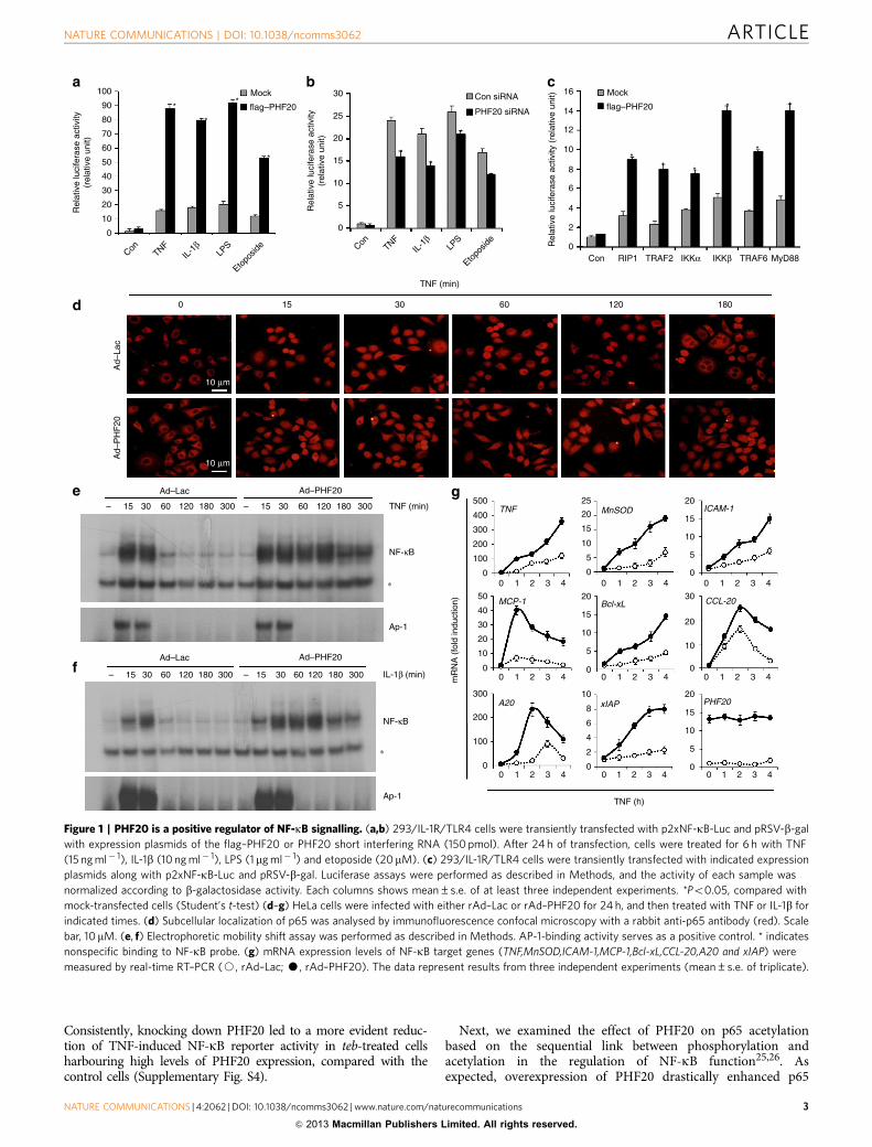
ResultsPHF20 acts as a positive regulator of NF-kB signalling. Basedon its domain structure, PHF20 is predicted to function as aregulator of transcription factor. In an initial screening using anactivation profiling array, we found that NF-kB was one of thepositive transcription factors identified in cells with ectopicexpression of PHF20 (data not shown). To systematically assessthe involvement of PHF20 in NF-kB signalling, we first examinedthe transcriptional activity of NF-kB in 293/IL-1R/TLR4 cellsstably overexpressing IL-1R and TLR4. Overexpression of PHF20resulted in a remarkable increase of NF-kB luciferase activity inresponse to various stimuli, including TNF, IL-1b, lipopoly-saccharide (LPS) and a DNA-damaging agent (etoposide)(Fig. 1a). Consistently, PHF20 knockdown led to a significantlyreduction of NF-kB reporter activity induced by each of these
stimuli (Fig. 1b). Such observations thus indicate that PHF20 isable to upregulate NF-kB activation in a canonical pathway.Confirming this suspicion, NF-kB activation mediated by theIKKs and adaptor proteins RIP1, TRAF2, TRAF6 and MyD88also was significantly enhanced by PHF20 (Fig. 1c).
Furthermore, in HeLa cells with overexpression of PHF20,prolonged nuclear retention of p65 and p50 occurred aftertreatment with TNF in comparison with the untreated controlcells (Fig. 1d and Supplementary Fig. S1). Notably, in cells withexpression of PHF20, we observed prolonged NF-kB-DNAbinding induced by both TNF (Fig. 1e) and IL-1b (Fig. 1f).Importantly, AP-1-binding activities in response to TNF or IL-1bwere not affected by PHF20. Consistently, expression of theNF-kB target genes induced by TNF was substantially enhancedin cells overexpressed with PHF20 (Fig. 1g). Furthermore, suchenhanced expression of the NF-kB target genes by PHF uponTNF treatment was drastically abolished in p65 null (p65� /� )mouse embryonic fibroblasts (MEFs), in contrast to that in wild-type MEFs (Supplementary Fig. S2), confirming that the effect ofPHF20 on NF-kB activation is p65-dependent. Together, datafrom this part demonstrate that PHF20 is able to upregulateNF-kB activity by promoting its nuclear retention and NF-kB–DNA binding.
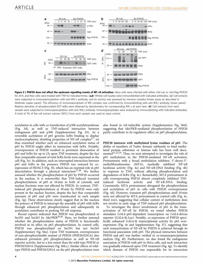
PHF20 does not affect upstream events of NF-kB signalling. Togain further insight into the molecular mechanisms underlyingPHF20-mediated NF-kB activation, we next examined thephosphorylation and degradation of IkBa. Upon TNF treatment,IkBa was phosphorylated and degraded with the similar degreeand kinetics in rAd–PHF20-infected cells, as compared with rAd–Lac-infected cells (Fig. 2a,b). Consistently, TNF-induced IKKactivity was not affected by PHF20 overexpression (Fig. 2c),indicating that upregulation of TNF-induced NF-kB signalling byPHF20 is independent of IKK. This conjecture also seems to beconsistent with the above observation that PHF20 does notinterfere with p65 nuclear translocation (Fig. 1d). Moreover,treatment of cells with TNF led to immediate interaction ofIKK-g with hyper-ubiquitinated RIP1, and consistently the extentand duration of TNF-induced ubiquitinated RIP1–IKK interac-tion was not affected by PHF20 overexpression (Fig. 2d), con-firming that PHF20 likely functions downstream of IKKactivation and IkBa degradation in simulating NF-kB activation.
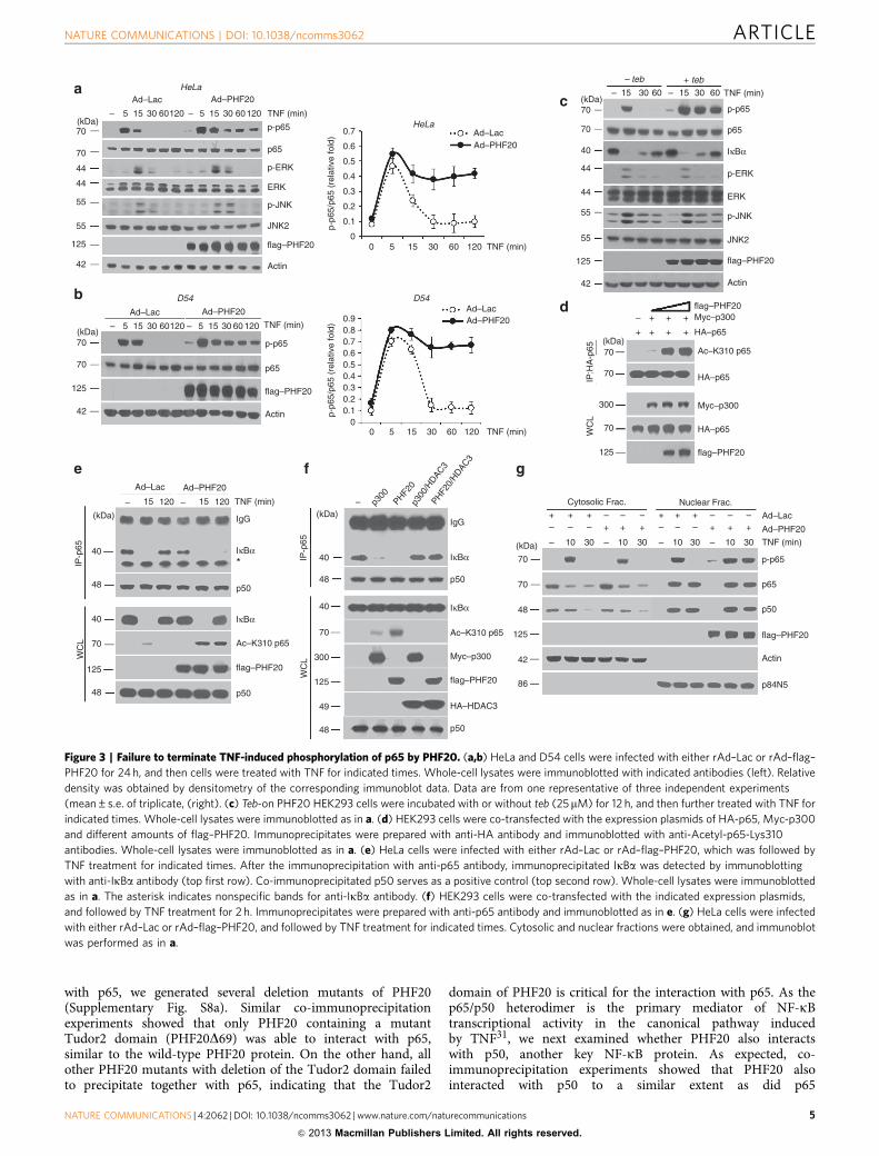
PHF20 induces sustained phosphorylation of p65. To furtherprobe the role of PHF20 in the nuclear step of NF-kB signalling,we next examined whether PHF20 affects the phosphorylationstatus of the p65 subunit of NF-kB, an essential event in nuclearNF-kB signalling. Notably, phosphorylation of p65 was markedlyprolonged in rAd–PHF20-infected cells in response to TNF,whereas only transient p65 phosphorylation was found in controlcells (Fig. 3a,b, top). Moreover, we found that such effect byPHF20 was specific to p65, based on the results that TNF-inducedphosphorylation of ERK and JNK was not affected by PHF20(Fig. 3a, third and fifth rows). To further confirm the generality ofour observation, we generated a single vector-inducible system bytebufenozide (teb). This system displayed tight regulation of teb-dependent PHF20 expression in a dose- and time-dependentmanner without any detectable basal level (SupplementaryFig. S3a,b). Important to note, the overexpression of PHF20using this inducible system also caused persistent phosphoryla-tion of p65, but not of ERK and JNK (Fig. 3c). Furthermore, wealso observed that different expression levels of PHF20 withdifferent dosage of teb well correlated with the extent of the TNF-induced persistent p65 phosphorylation (Supplementary Fig. S3b)as well as NF-kB reporter activity (Supplementary Fig. S3c).
ARTICLE NATURE COMMUNICATIONS | DOI: 10.1038/ncomms3062
2 NATURE COMMUNICATIONS | 4:2062 | DOI: 10.1038/ncomms3062 | www.nature.com/naturecommunications
& 2013 Macmillan Publishers Limited. All rights reserved.
Consistently, knocking down PHF20 led to a more evident reduc-tion of TNF-induced NF-kB reporter activity in teb-treated cellsharbouring high levels of PHF20 expression, compared with thecontrol cells (Supplementary Fig. S4).
Next, we examined the effect of PHF20 on p65 acetylationbased on the sequential link between phosphorylation andacetylation in the regulation of NF-kB function25,26. Asexpected, overexpression of PHF20 drastically enhanced p65
100
90
80
70
60
50
40
30
20
10
0
Rel
ativ
e lu
cife
rase
act
ivity
(rel
ativ
e un
it)
Rel
ativ
e lu
cife
rase
act
ivity
(rel
ativ
e un
it)
Rel
ativ
e lu
cife
rase
act
ivity
(re
lativ
e un
it)
ConTNF
IL-1
βLP
S
Etopo
side Con
TNFIL
-1β
LPS
Etopo
side
TNF (min)
30
25
20
15
10
5
0
16
14
12
10
8
6
4
2
0
Mock
flag–PHF20
Mock
flag–PHF20
Con RIP1 TRAF2 IKKα IKKβ TRAF6 MyD88
Con siRNA
PHF20 siRNA
a b c
0 15 30 60 120 180
Ad–
Lac
Ad–
PH
F20
10 μm
10 μm
d
e
f
gAd–Lac Ad–PHF20
– 15 30 60 120 180 300 – 15 30 60 120 180 300
Ad–Lac Ad–PHF20
– 15 30 60 120 180 300 – 15 30 60 120 180 300
∗
∗
TNF (min)
NF-κB
Ap-1
NF-κB
Ap-1
IL-1β (min)
TNF (h)
0 1 2 3 4 0 1 2 3 4 0 1 2 3 4
0 1 2 3 4 0 1 2 3 4 0 1 2 3 4
0 1 2 3 4 0 1 2 3 4 0 1 2 3 4
500
400
300
200
100
0
300
200
100
0
TNF
MCP-1
A20 xIAP
Bcl-xL
MnSOD ICAM-1
CCL-20
PHF20
50
40
30
20
10
0
20
15
10
5
0
10
8
6
4
2
0
20
15
10
5
0
30
20
10
0
25
20
15
10
5
0
20
15
10
5
0
mR
NA
(fo
ld in
duct
ion)
*
*
*
**
*
*
*
** *
*
*
*
Figure 1 | PHF20 is a positive regulator of NF-kB signalling. (a,b) 293/IL-1R/TLR4 cells were transiently transfected with p2xNF-kB-Luc and pRSV-b-gal
with expression plasmids of the flag–PHF20 or PHF20 short interfering RNA (150 pmol). After 24 h of transfection, cells were treated for 6 h with TNF
(15 ng ml� 1), IL-1b (10 ng ml� 1), LPS (1mg ml� 1) and etoposide (20mM). (c) 293/IL-1R/TLR4 cells were transiently transfected with indicated expression
plasmids along with p2xNF-kB-Luc and pRSV-b-gal. Luciferase assays were performed as described in Methods, and the activity of each sample was
normalized according to b-galactosidase activity. Each columns shows mean±s.e. of at least three independent experiments. *Po0.05, compared with
mock-transfected cells (Student’s t-test) (d–g) HeLa cells were infected with either rAd–Lac or rAd–PHF20 for 24 h, and then treated with TNF or IL-1b for
indicated times. (d) Subcellular localization of p65 was analysed by immunofluorescence confocal microscopy with a rabbit anti-p65 antibody (red). Scale
bar, 10mM. (e, f) Electrophoretic mobility shift assay was performed as described in Methods. AP-1-binding activity serves as a positive control. * indicates
nonspecific binding to NF-kB probe. (g) mRNA expression levels of NF-kB target genes (TNF,MnSOD,ICAM-1,MCP-1,Bcl-xL,CCL-20,A20 and xIAP) were
measured by real-time RT–PCR (J, rAd–Lac; K, rAd–PHF20). The data represent results from three independent experiments (mean±s.e. of triplicate).
NATURE COMMUNICATIONS | DOI: 10.1038/ncomms3062 ARTICLE
NATURE COMMUNICATIONS | 4:2062 | DOI: 10.1038/ncomms3062 | www.nature.com/naturecommunications 3
& 2013 Macmillan Publishers Limited. All rights reserved.
acetylation in cells with co-transfection of p300 acetylatransferase(Fig. 3d), as well as TNF-induced interaction betweenendogenous p65 and p300 (Supplementary Fig. S5). As areversible acetylation of p65 governs IkBa binding to displaynucleocytoplasmic shuttling properties of NF-kB complex27, wethus examined whether such an enhanced acetylation status ofp65 by PHF20 might affect its interaction with IkBa. Notably,overexpression of PHF20 resulted in persistent dissociation ofp65 and IkBa for up to 2 h upon TNF treatment, despite the factthat comparable amount of total IkBa levels were expressed in thecell (Fig. 3e). In addition, such an interrupted interaction betweenp65 and IkBa in the presence PHF20 was restored by co-expression of HDAC3 (Fig. 3f), which has an essential role in p65deacetylation through a physical interaction27,28. We furtherassessed whether the phosphorylation of p65 by PHF20 occurredin the nucleus. It is noteworthy that TNF-induced transientphosphorylations of p65 at 10 min in both of cytosolic andnuclear fractions were not affected by PHF20. In contrast, TNF-induced p65 phosphorylations at 30 min by PHF20 were onlypresent in the nuclear fraction, despite the fact that comparableamounts of p65 and p50 were translocated in each sample(Fig. 3g). These observations clearly suggest that in the nucleusthe potency of PHF20 to interrupt the assembly of p65 with IkBathrough enhanced p65 phosphorylation and subsequently itsacetylation contributes to prolong NF-kB response.
Recent reports indicated that PHF20 was phosphorylated atSer291 and Ser265 by Akt/PKB29,30. Here, we further assessedwhether the phosphorylation status of PHF20 is functionallyinvolved in its effect on p65 phosphorylation. In control cells,PHF20 was phosphorylated on Ser291 but not Ser265(Supplementary Fig. S6a). Upon TNF treatment, overexpressionof non-phosphorylatable mutant of PHF20 (PHF20/S291A) alsoenhanced persistent p65 phosphorylation as well as NF-kBreporter activity, but to a less extent than the wild-type PHF20 orPHF20/S265A (Supplementary Fig. S6b,c). Similar effects of wild-type PHF20 and PHF20/S291A on the p65 phosphorylation were
also found in teb-inducible system (Supplementary Fig. S6d),suggesting that Akt/PKB-mediated phosphorylation of PHF20partly contribute to its regulatory effect on p65 phosphorylation.
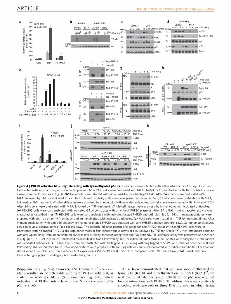
PHF20 interacts with methylated lysine residues of p65. Theability of members of Tudor domain subfamily to bind methy-lated protein substrates or histone tails has been well docu-mented21,22,24. Thus, we next attempted to investigate the role ofp65 methylation in the PHF20-mediated NF-kB activation.Pretreatment with a broad methylation inhibitor, 50-deoxy-50-methylthioadenosine (MTA), markedly inhibited NF-kBluciferase activity (Fig. 4a) and NF-kB-DNA binding (Fig. 4b)in response to TNF, without affecting phosphorylation anddegradation of IkBa (Fig. 4c). Remarkably, MTA pretreatment incells overexpressing PHF20 almost completely inhibited TNF-induced luciferase activity and NF-kB-DNA binding.Consistently, MTA pretreatment abrogated the phosphorylationand acetylation of p65 in cells with PHF20 overexpression(Fig. 4d). However, transient p65 phosphorylations until 10 minwere not affected by MTA pretreatment in control cells (Fig. 4c,third row), suggesting that cellular context of methylation doesnot involve in early stage of TNF-induced p65 phosphorylation.
To investigate the direct involvement of p65 in the trans-activation of NF-kB by PHF20, we tested whether PHF20stimulates GAL4–p65-dependent transcription on GAL4-drivenreporter (GAL4-tk-Luc). Notably, co-expression of PHF20 speci-fically enhanced GAL4-tk transcriptional activity by p65 over-expression (Fig. 4e and Supplementary Fig. S7), suggesting thatsuch transactivation of NF-kB by PHF20 is achieved through itsfunctional association with p65. The physical interaction betweenPHF20 and p65 was further verified in co-immunoprecipitationanalysis (Fig. 4f). Furthermore, we also found the endogenousassociation of PHF20 with p65 in HeLa cells, and such interactionwas gradually enhanced upon TNF treatment (Fig. 4g). To identifywhich domain of PHF20 was responsible for its interaction
(kDa)40
40
42
125
p-IκBα
IκBα
flag–PHF20
flag–PHF20
flag–PHF20
Actin
Ad–PHF20Ad–Lac– 2 5 10 15 – 2 5 10 15 TNF (min)
(kDa)40
87
42
125
IκBα
IKKβ
flag–PHF20
Actin
Ad–PHF20Ad–Lac
– 5 15 30 60 120 – 5 15 30 60 120 TNF (min)
Ad–PHF20Ad–Lac– 10 30 – 10 30
Ad–PHF20Ad–Lac– 10 30 60 – 10 30 60
TNF (min)
Ub–RIP1
IKKβ
IKKβ
RIP1
(kDa)
(kDa)114
87
IP :
IKK
γ
IP : IKKγ
WC
LWC
L 87
78
125
IKKγ
GST–IκBα
TNF (min)
40
48
125
Substrate: GST–IκBα
IVK
c d
a b
Figure 2 | PHF20 does not affect the upstream signalling events of NF-kB activation. HeLa cells were infected with either rAd–Lac or rAd–flag–PHF20
for 24 h, and then cells were treated with TNF for indicated times. (a,b) Whole-cell lysates were immunoblotted with indicated antibodies. (c) Cell extracts
were subjected to immunoprecipitation with anti-IKK-g antibody, and its activity was assessed by immune complex kinase assay as described in
Methods (upper panel). The efficiency of immunoprecipitant of IKK complex was confirmed by immunoblotting with anti-IKKg antibody (lower panel).
Relative densities of phosphorylated GST–IkBa were obtained by densitometry for corresponding IKK-g at each lane. (d) Cell extracts from each
sample were subjected to immunoprecipitation with anti-IKKg antibody. Immunoprecipitates were analysed by immunoblotting with indicated antibodies.
A total of 1% of the cell extract volume (WCL) from each sample was used as input control.
ARTICLE NATURE COMMUNICATIONS | DOI: 10.1038/ncomms3062
4 NATURE COMMUNICATIONS | 4:2062 | DOI: 10.1038/ncomms3062 | www.nature.com/naturecommunications
& 2013 Macmillan Publishers Limited. All rights reserved.
with p65, we generated several deletion mutants of PHF20(Supplementary Fig. S8a). Similar co-immunoprecipitationexperiments showed that only PHF20 containing a mutantTudor2 domain (PHF20D69) was able to interact with p65,similar to the wild-type PHF20 protein. On the other hand, allother PHF20 mutants with deletion of the Tudor2 domain failedto precipitate together with p65, indicating that the Tudor2
domain of PHF20 is critical for the interaction with p65. As thep65/p50 heterodimer is the primary mediator of NF-kBtranscriptional activity in the canonical pathway inducedby TNF31, we next examined whether PHF20 also interactswith p50, another key NF-kB protein. As expected, co-immunoprecipitation experiments showed that PHF20 alsointeracted with p50 to a similar extent as did p65
HeLa
HeLa
Ad–Lac
Ad–Lac(kDa)70
70
44
44
55
55
125
42
– 5 15 30 60120
Ad–PHF20
Ad–PHF20
Ad–Lac
Ad–Lac
Ad–PHF20
– 5 15 30 60120 TNF (min)
TNF (min)
p-p65
p65
p-ERK
ERK
p-JNK
JNK2
flag–PHF20
Actin
Actin
D54 D54
Ad–Lac
(kDa)70
70
42
125
e f
– 5 15 30 60120
Ad–PHF20
– 5 15 30 60 120 TNF (min)
p-p65
p65
flag–PHF20
Actin
(kDa)
(kDa)
70
70
40
44
44
55
55
42
125
–
– teb + teb
15 30 60 – 15 30 60 TNF (min)
p-p65
p65
p-ERK
ERK
p-JNK
JNK2
flag–PHF20
flag–PHF20
flag–PHF20
Actin
0.7
0.6
0.5
0.4
0.3
0.2
0.1
0
p-p6
5/p6
5 (r
elat
ive
fold
)p-
p65/
p65
(rel
ativ
e fo
ld)
0 5 15 30 60 120
TNF (min)
TNF (min)
0 5 15 30 60 120
a
b
g
c
d
Ad–Lac
(kDa)
40
48
40
48
70
125
– 15 120 – 15 120
Ad–PHF20
TNF (min)
p50
p50
flag–PHF20
Ac–K310 p65
Ac–K310 p65
IgG
IκBα
IκBα
IκBα
*
WC
LIP
-p65
(kDa)
40
48
40
70
49
48
300
125
(kDa)
70
70
48
86
125
42
p50
p50
flag–PHF20
Ac–K310 p65
IgG
IκBα
IκBα
WC
LIP
-p65
WC
LIP
:HA
-p65
Myc–p300
Myc–p300
Myc–p300
HA–p65
HA–p65
HA–p65
– + + +
++ + +
70
70
70
300
125
0.90.80.70.60.50.40.30.20.1
0
HA–HDAC3
p50
p65
p-p65
p84N5
flag–PHF20
– p300
PHF20
p300
/HDAC3
PHF20/
HDAC3
Ad–PHF20
Cytosolic Frac. Nuclear Frac.+ + +
+ + +–
––
– –– – –
10 1030 30
+ + ++ + +–
––
– –– – –
10 1030 30
Figure 3 | Failure to terminate TNF-induced phosphorylation of p65 by PHF20. (a,b) HeLa and D54 cells were infected with either rAd–Lac or rAd–flag–
PHF20 for 24 h, and then cells were treated with TNF for indicated times. Whole-cell lysates were immunoblotted with indicated antibodies (left). Relative
density was obtained by densitometry of the corresponding immunoblot data. Data are from one representative of three independent experiments
(mean±s.e. of triplicate, (right). (c) Teb-on PHF20 HEK293 cells were incubated with or without teb (25mM) for 12 h, and then further treated with TNF for
indicated times. Whole-cell lysates were immunoblotted as in a. (d) HEK293 cells were co-transfected with the expression plasmids of HA-p65, Myc-p300
and different amounts of flag–PHF20. Immunoprecipitates were prepared with anti-HA antibody and immunoblotted with anti-Acetyl-p65-Lys310
antibodies. Whole-cell lysates were immunoblotted as in a. (e) HeLa cells were infected with either rAd–Lac or rAd–flag–PHF20, which was followed by
TNF treatment for indicated times. After the immunoprecipitation with anti-p65 antibody, immunoprecipitated IkBa was detected by immunoblotting
with anti-IkBa antibody (top first row). Co-immunoprecipitated p50 serves as a positive control (top second row). Whole-cell lysates were immunoblotted
as in a. The asterisk indicates nonspecific bands for anti-IkBa antibody. (f) HEK293 cells were co-transfected with the indicated expression plasmids,
and followed by TNF treatment for 2 h. Immunoprecipitates were prepared with anti-p65 antibody and immunoblotted as in e. (g) HeLa cells were infected
with either rAd–Lac or rAd–flag–PHF20, and followed by TNF treatment for indicated times. Cytosolic and nuclear fractions were obtained, and immunoblot
was performed as in a.
NATURE COMMUNICATIONS | DOI: 10.1038/ncomms3062 ARTICLE
NATURE COMMUNICATIONS | 4:2062 | DOI: 10.1038/ncomms3062 | www.nature.com/naturecommunications 5
& 2013 Macmillan Publishers Limited. All rights reserved.
(Supplementary Fig. S9a). However, TNF treatment of p65� /�MEFs resulted in no detectable binding of PHF20 with p50, asevident in wild-type MEFs (Supplementary Fig. S9b), whichindicates that PHF20 interacts with the NF-kB complex (p65/p50) via p65.
It has been demonstrated that p65 was monomethylated onlysine 218 (K218) and dimethylated on lysine221 (K221)32; wenext examined whether lysine methylation of p65 was requiredfor the interaction with PHF20. To address this issue, constructsencoding wild-type p65 or three K-A mutants, in which lysine
Ad-Lac
Ad-PHF20
90
80
70
60
50
40
30
20
10
0Con TNF TNF+MTA
*Rel
ativ
e lu
cife
rase
act
ivity
(rel
ativ
e un
it)R
elat
ive
luci
fera
se a
ctiv
ity(r
elat
ive
unit)
Rel
ativ
e lu
cife
rase
act
ivity
(rel
ativ
e un
it)a b c d
e
f
i kj
g h
(kDa)90 SP-1
NF-κB
TNF (min)
TNF (min)
Ad–Lac Ad–PHF20
DMSO MTA
– 30 180
– 30 180
DMSO MTADMSO MTA
– 30 180
– 30 180
(kDa)40
40
70
70
42
– 2 5 10 20 – 2 5 10 20
p-p65
p65
Actin
IκBα
p-IκBα
TNF (min)DMSO MTA
(kDa)70
70
70
40
42
– 15 30 60 – 15 30 60
p-p65
p65
Actin
IκBα
Ac–K310 p65
Ad–PHF20
GAL4-tk-Luc*
*
*
1009080706050403020100
GAL4GAL4–p65
flag–PHF20 (μg)
– –
––
– 0.5 1.0 2.0 0.5 1.0 2.0
– – –––
– – –+ + + +
+ + + +
IP-H
AW
CL
+ +++
––
+ +++
––
flag–PHF20
(kDa)
(kDa)
40
125
40
125
IP-H
AW
CL
(kDa)125
40
120
40W
CL
WC
L
HA–p65
HA–p65
flag–PHF20
flag–PHF20
flag–PHF20
flag–PHF20
HA–p65
HA–p65
HA–p65
IP-p
65
IP:X
p-P
HF
20
flag–PHF20
HA–p65
flag–p65
flag–p65
125
48
70
48
70
125
– 5 10 15 30 TNF (min)
PHF20
IgG
IgG
*
p50
p65
p50
p65
PHF20
(kDa)
70
125
70
125
Xp–PHF20
Xp–PHF20
flag–p65
Xp–PHF20
––
–+ + + + +
WT K21
8A
K221A
K218
/K22
1A
WT
Moc
k
K218A
K221A
K218/K
221A
40
35
30
25
20
15
10
5
0
flag–p65
p65-
/-M
EF
Xp-Vec
Xp–Vec
Xp-PHF20
Xp-PHF20
Xp–PHF20
Xp–PHF20
Xp–PHF20
flag–p65
flag–p65
p-p65
p65
p65
p-p65
TNF (min)
TNF (min)
Actin flag-p65
flag–p65
p-p65
TNF (min)
Actin
Mock WT K218A K218/221AK221A
Mock WT K218A K218/221AK221A
– 15 30 – 15 30 – 15 30 – 15 30 – 15 30
– 15 30 – 15 30 – 15 30 – 15 30
WT K221A– 15 30– 15 30
WT K221A– 15 30– 15 30
– 15 30
(kDa)70
70
42
70
70
42
125
(kDa)IP
-fla
g-p6
529
3T
70
70
125
Mock*
*
Figure 4 | PHF20 activates NF-kB by interacting with Lys-methylated p65 (a) HeLa cells were infected with either rAd–Lac or rAd–flag–PHF20 and
transfected with an NF-kB-responsive reporter plasmid. After 24 h, cells were pretreated with MTA (1 mM) for 1 h, and treated with TNF for 6 h. Luciferase
assays were performed as in Fig. 1a. (b) HeLa cells were infected with either rAd–Lac or rAd–flag–PHF20. After 24 h, cells were pretreated with
MTA, followed by TNF for indicated times. Electrophoretic mobility shift assay was performed as in Fig. 1e. (c) HeLa cells were pretreated with MTA,
followed by TNF treatment. Whole-cell lysates were analysed by immunoblot with indicated antibodies. (d) HeLa cells were infected with rAd–flag–PHF20.
After 24 h, cells were pretreated with MTA, followed by TNF treatment. Whole-cell lysates were analysed by immunoblot with indicated antibodies.
(e) HEK293 cells were co-transfected with indicated GAL4 constructs with or without PHF20 plasmids. After 24 h, GAL4-tk-Luc reporter activity was
measured as described in a. (f) HEK293 cells were co-transfected with indicated tagged PHF20 and p65 plasmids for 24 h. Immunoprecipitates were
prepared with anti-flag or anti-HA antibody, and immunoblotted with indicated antibodies. (g) HeLa cells were treated with TNF for indicated times. After
immunoprecipitation with anti-p65 antibody, immunoprecipitated PHF20 was detected with anti-PHF20 antibody (top first row). Co-immunoprecipitated
p50 serves as a positive control (top second row). The asterisk indicates nonspecific bands for anti-PHF20 antibody. (h,i) HEK293 cells were co-
transfected with Xp-tagged PHF20 along with either mock or flag-tagged various forms of p65, followed by TNF for 10 min. (h) After immunoprecipitation
with anti-Xp antibody, immunoprecipitated p65 was measured by immunoblotting with anti-flag antibody. (i) Luciferase assay was performed as described
in a. (j) p65� /� MEFs were co-transfected as described in h and followed by TNF for indicated times. Whole-cell lysates were analysed by immunoblot
with indicated antibodies. (k) HEK293 cells were co-transfected with Xp-tagged PHF20 along with flag-tagged p65 (WT or K221A) as described in h and,
followed by TNF for indicated times. Immunoprecipitates were prepared with anti-flag antibody and immunoblotted with indicated antibodies. Each column
shows mean±s.e. of at least three independent experiments (Student’s t-test). *Po0.05, compared with TNF-treated group (a), GAL4–p65 only-
transfected group (e) or wild-type p65-transfected group (i).
ARTICLE NATURE COMMUNICATIONS | DOI: 10.1038/ncomms3062
6 NATURE COMMUNICATIONS | 4:2062 | DOI: 10.1038/ncomms3062 | www.nature.com/naturecommunications
& 2013 Macmillan Publishers Limited. All rights reserved.
was substituted by alanine, were transiently transfected withPHF20 into HEK293 cells. In the co-immunoprecipitation assay,K221A or K218/221A mutant of p65 completely abolished theinteraction with PHF20, whereas K218A mutant only partiallyaffected the interaction in comparison with WT–p65 (Fig. 4h). Ofnote, the ability of PHF20 to NF-kB activation was drasticallyimpaired in cells with the K221A or K218/221A mutant of p65,compared with that with wild-type p65 (Fig. 4i). These resultssuggest that K221 methylation of p65, which is required for itsinteraction with PHF20, has a critical role in PHF20-mediatedNF-kB activation. To confirm the methylation-dependentinteraction between PHF20 and p65, the full-length PHF20with point mutations (W97A and/or Y103A) of the methyl-lysine-binding cage within the Tudor2 domain were generated, asreported earlier24. In the context of full-length proteins, theW97A– and/or Y103A–PHF20 markedly decreased the ability ofthe interaction with p65, compared with that of wild-type PHF20(Supplementary Fig. S10a). Consistent with these results, theW97A– and/or Y103A–PHF20 failed to upregulate TNF-inducedNF-kB activation, whereas PHF20 mutant containing a Tudor2domain (PHF20D69) enhanced the activation of NF-kB by TNF,to a degree similar to that obtained with wild-type PHF20(Supplementary Fig. S10b).
To further scrutinize whether K221 methylation is directlyinvolved in p65 phosphorylation mediated by PHF20, were-expressed three mutants of p65 or wild-type p65 in p65� /� MEF. Interestingly, no difference was found for TNF-inducedp65 phosphorylation among in p65� /� MEF reconstitutedwith either WT or three methylation-deficient p65, without theco-expression of PHF20 (Fig. 4j, top). In contrast, a strikinglydifferent result was obtained in cells with PHF20 overexpression:reconstitution with mutant p65 (K221A� or K218/221A) thatwas unable to bind PHF20 abolished the persistent p65phosphorylation in response to TNF (Fig. 4j, bottom). To furtherconfirm the above observations, we compared the ability ofPHF20 to regulate p65 phosphorylation in the immunopreci-pitants of WT and K221A mutant of p65. Consistently, thephosphorylation of K221A–p65 at a later time point was onlyfaintly detectable compared with that of WT–p65, whereas thetransient phosphorylation at 15 min was unaffected by PHF20overexpression (Fig. 4k). Taken together, these results clearlyindicate that Lys221 site of p65 is indeed essential for the functionof PHF20 in amplifying TNF-induced NF-kB activation viapersistent phosphorylation of p65.
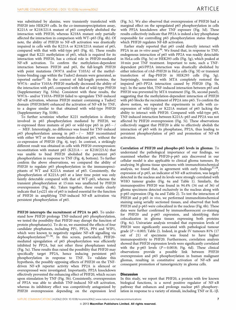
PHF20 interrupts the recruitment of PP2A to p65. To under-stand how PHF20 prolongs TNF-induced p65 phosphorylation,we tested the possibility that PHF20 may disrupt the function ofprotein phosphatase(s). To do so, we examined the effects of fourcandidate phophatases, including PP1, PP2A, PP4 and WIP1,which were known to negatively regulate NF-kB signalling viadephosphorylation33–36. In this screen, particularly, PHF20-mediated upregulation of p65 phosphorylation was efficientlyinhibited by PP2A, but not other three phosphatases tested(Fig. 5a). These results thus raised the possibility that PHF20 mayspecifically target PP2A, hence inducing persistent p65phosphorylation in response to TNF. To validate thishypothesis, the possibly opposing effects of PHF20 on the TNF-driven NF-kB reporter activity when PP2A is silenced oroverexpressed were investigated. Importantly, PP2A knockdowneffectively prevented the enhancing effect of PHF20, which occursupon stimulation by TNF (Fig. 5b). Consistently, overexpressionof PP2A was able to abolish TNF-induced NF-kB activation,whereas its inhibitory effect was competitively antagonized byPHF20 overexpression depending on the expression level
(Fig. 5c). We also observed that overexpression of PHF20 had amarginal effect on the upregulated p65 phosphorylation in cellswith PP2A knockdown after TNF exposure (Fig. 5d). Theseresults collectively indicate that PP2A is indeed a key phosphataseresponsible for controlling p65 phosphorylation status throughwhich PHF20 regulates NF-kB activation.
Earlier study reported that p65 could directly interact withPP2A in an in-vitro assay36. We found that, in response to TNF,endogenous interaction of p65 with PP2A was readily detectablein HeLa cells (Fig. 5e) or HEK293 cells (Fig. 5g), which peaked at10 min post TNF treatment. Important to note, such a TNF-dependent p65/PP2A interaction was drastically abolished byeither infection of rAd–PHF20 in HeLa cells (Fig. 5f) or transienttransfection of flag–PHF20 in HEK293 cells (Fig. 5g).Surprisingly, treatment with MTA completely restored theimpaired p65–PP2A interaction caused by PHF20 (Fig. 5h,top). In the same blot, TNF-induced interaction between p65 andPHF20 was prevented by MTA treatment (Fig. 5h, second panel),suggesting that the methylation-dependent interaction of PHF20with p65 blocks the recruitment of PP2A into p65. To confirm theabove notion, we repeated the experiments in cells with co-expression of wild-type or K221A mutant of p65 lacking theability to interact with PHF20. Compared with wild-type p65,TNF-induced interaction between K221A–p65 and PP2A was notaffected by PHF20 overexpression (Fig. 5i). These observationscollectively suggest that PHF20 is able to effectively abolish theinteraction of p65 with its phosphatase, PP2A, thus leading topersistent phosphorylation of p65 and promotion of NF-kBactivation.
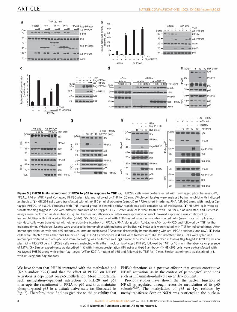
Correlation of PHF20 and phospho-p65 levels in gliomas. Tounderstand the pathological importance of our findings, weexamined whether the PHF20-p-p65 axis discovered in ourcellular model is also applicable to clinical glioma tumours. Byanalysing 36 glioma tissue specimens with immunohistochemicalanalysis, we found that, in agreement with early findings37,38,expression of p-p65, an indicator of NF-kB activation, was largelydetected in the nucleus and its levels were strongly correlated withWHO tumour grades (Fig. 6a and Table 1). Similarly, theimmunopositive PHF20 was found in 94.4% (34 out of 36) ofglioma specimens detected exclusively in the nucleus along withp-p65 expression (Fig. 6a and Table 2). To test the interaction ofPHF20 and p-p65 in vivo, we performed immunohistochemicalstaining using serially sectioned tissues, and observed that bothPHF20 and p-p65 were colocalized in the nucleus (Fig. 6b). Thesedata were further confirmed by immunofluorescent co-stainingfor PHF20 and p-p65 expression, and identifying theircolocalization in glioma tissues expressing both proteins(Fig. 6c). Univariate analysis revealed that expression levels ofPHF20 were significantly associated with pathological tumourgrade (P¼ 0.003; Table 2). Indeed, in grade IV tumours 81% (17out of 21) of specimens was found to have higherimmunopositivity to PHF20. Furthermore, correlation analysisshowed that PHF20 expression levels were significantly correlatedwith the p-p65 levels (P¼ 0.0018; Fig. 6d). These clinicalobservations provide a possible link between PHF20overexpression and p65 phosphorylation in human malignantgliomas, resulting in constitutive activation of NF-kB andconsequent promotion of tumorigenicity in glioma cells.
DiscussionIn this study, we report that PHF20, a protein with few knownbiological functions, is a novel positive regulator of NF-kBpathway that enhances and prolongs nuclear p65 phosphory-lation in response to a variety of stimuli, including TNF or IL-1b.
NATURE COMMUNICATIONS | DOI: 10.1038/ncomms3062 ARTICLE
NATURE COMMUNICATIONS | 4:2062 | DOI: 10.1038/ncomms3062 | www.nature.com/naturecommunications 7
& 2013 Macmillan Publishers Limited. All rights reserved.
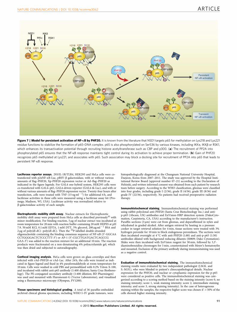
We have shown that PHF20 interacted with the methylated p65(K218 and/or K221) and that the effect of PHF20 on NF-kBactivation is dependent on p65 methylation. More importantly,such methylation-dependent interaction of PHF20 and p65interrupts the recruitment of PP2A to p65 and thus maintainsphosphorylated p65 in a default active state (as illustrated inFig. 7). Therefore, these findings give rise to the possibility that
PHF20 functions as a positive effector that causes constitutiveNF-kB activation, as in the context of pathological conditionssuch as inflammation-linked cancer development.
Previous studies have shown that the nuclear function ofNF-kB is regulated through reversible methylation of its p65subunit32,39. The methylation of p65 at Lys residues bymethyltransferase Set9 or NSD1 was restricted to the nucleus,
TNF (20 min)
Vector PP4 WIP1
(kDa)70
70
79
36
125
70
PP1 PP2Ac flag–PPases
flag–PPases
Xp–PHF20
Xp–PHF20
Xp–PHF20
p-p65
p65
Actin
70
60
50
40
30
20
10
0
Rel
ativ
e lu
cife
rase
act
ivity
(rel
ativ
e un
it)
Rel
ativ
e lu
cife
rase
act
ivity
(rel
ativ
e un
it)
siCon siPP2Ac
siCon siPP2Ac
PP2Ac
TNF
Xp–PHF20
Xp–PHF20
TNF
–– –
+ – ++ +
–– –
+ – ++ +
–– –
+ – ++ +
–– –
+ – ++ +
Actin
p-p65
p65
(kDa)36
125
42
70
70
siCon siPP2Ac
PP2Ac
flag–PHF20
TNF (min)
Actin
p-p65
p65
(kDa)70
70
36
125
42
98
7
65
43210
PP2Ac
–– – –
+ + + + TNFXp–PHF20
(kDa)36
125
40
70
––– –
–+
–++
++
++ TNF
flag–PP2Ac
flag–PP2AcXp–PHF20
Xp–PHF20
p65
IκBα
Ad-Lac– 10 30 60120
Ad-Lac Ad–PHF20– 10 30 60120 – 10 30 60120
WC
LIP
:p65
(kDa) – 5 10 30 TNF (min)IgG
36
36
48
48
70
70
PP2Ac
p50
p65
PP2Ac
p50
p65
WC
LIP
-p65
(kDa)– 10 3060 – 10 3060 TNF (min)
IgG
36
36
48
48
70
70
125
PP2Ac
p50
p65
PP2Ac
p50
p65
a b
c d e
f g h i
flag–PHF20
Ad–Lac Ad–PHF20
WC
LIP
-p65
(kDa)–– – – + +
10 30 10 30 TNF (min)
IgG
36
36
48
48
70
70125
PP2Ac
p50
p65
PP2Ac
p50
p65
flag–PHF20
flag–PHF20
WC
LIP
-p65
(kDa)–
– – +–– – +
+
10 10 10 TNF (min)
IgG
125
36
36
48
125
70
48
70
PP2Ac
p50
p65
PP2Ac
p50
p65
flag–PHF20
flag–PHF20
flag–PHF20
WC
LIP
–fla
g–p6
5
(kDa)–
– – +
++ +
–
–––––
–
+
+
10 10 10 10 TNF (min)
IgG
36
36
125
48
48
70
70
125
PP2Ac
PP2Ac
p50
p50
Xp–PHF20
Xp–PHF20
flag–p65
flag–p65
MTAWT–p65
K221A
Xp–PHF20
*
*
*
Figure 5 | PHF20 limits recruitment of PP2A to p65 in response to TNF. (a) HEK293 cells were co-transfected with flag-tagged phosphatases (PP1,
PP2Ac, PP4 or WIP1) and Xp-tagged PHF20 plasmids, and followed by TNF for 20 min. Whole-cell lysates were analysed by immunoblot with indicated
antibodies. (b) HEK293 cells were transfected with either 150 pmol of scramble (control) or PP2Ac short interfering RNA (siRNA) along with mock or Xp-
tagged PHF20. *Po0.05, compared with TNF-treated group in scramble siRNA-transfected cells (mean±s.e. of triplicates). (c) HEK293 cells were co-
transfected flag-tagged PP2Ac with different amounts of Xp-tagged PHF20. After 48 h, cells were treated with TNF for 6 h as indicated, and luciferase
assays were performed as described in Fig. 1a. Transfection efficiency of either overexpression or knock downed expression was confirmed by
immunoblotting with indicated antibodies (right). *Po0.05, compared with TNF-treated group in mock-transfected cells (mean±s.e. of triplicates).
(d) HeLa cells were transfected with either scramble (control) or PP2Ac siRNA along with rAd–Lac or rAd–flag–PHF20 and followed by TNF for the
indicated times. Whole-cell lysates were analysed by immunoblot with indicated antibodies. (e) HeLa cells were treated with TNF for indicated times. After
immunoprecipitation with anti-p65 antibody, co-immunoprecipitated PP2Ac was detected by immunoblotting with anti-PP2Ac antibody (top row). (f) HeLa
cells were infected with either rAd–Lac or rAd–flag–PHF20 as described in d and were treated with TNF for indicated times. Cells were lysed and
immunoprecipitated with anti-p65 and immunoblotting was performed in e. (g) Similar experiments as described in f using flag-tagged PHF20 expression
plasmid in HEK293 cells. HEK293 cells were transfected with either mock or flag-tagged PHF20, followed by TNF for 10 min in the absence or presence
of MTA. (h) Similar experiments as described in f, with immunoprecipitation (IP) using anti-p65 antibody. (i) HEK293 cells were co-transfected with
Xp-tagged PHF20 along with either flag-tagged WT or K221A mutant of p65 and followed by TNF for 10 min. Similar experiments as described in f,
with IP using anti-flag antibody.
ARTICLE NATURE COMMUNICATIONS | DOI: 10.1038/ncomms3062
8 NATURE COMMUNICATIONS | 4:2062 | DOI: 10.1038/ncomms3062 | www.nature.com/naturecommunications
& 2013 Macmillan Publishers Limited. All rights reserved.

IHCgrade
IHCgrade
IHCgrade
Negativecontrol
20 μm
20 μm
p-p65
PHF20
+ ++ +++
p-p65b d
c
a
PHF20
p-p65 PHF20 Merge
p-p65 PHF20 Merge
50.0 μm 50.0 μm
20.0 μm20.0 μm20.0 μm
+++
++
+
–
– + ++ +++
�=0.5031P=0.0018
PHF20 expression
p-p6
5 ex
pres
sion
100
80
60
40
20
0
Rel
ativ
e no
. pos
itive
cel
ls(%
of t
otal
cel
ls)
Figure 6 | p-p65 levels are associated with PHF20 expression in primary human glioma specimens. (a) Immunohistochemical staining of p-p65 and
PHF20 in paraffin-embedded tissue specimens from 36 glioma patients. Negative control with exclusion of primary antibody. The intensity of histological
staining grade was scored using a four-point scale: � , no staining, þ , weak staining, þ þ intermediate staining and þ þ þ strong staining. Each
representative case was shown (original magnification, �400). Scale bar, 20mm). (b,c) Representative image of colocalization between p-p65 and PHF20
expression in the nuclei of the glioma tumour cells. (b) Serial sections of tissues were prepared and stained simultaneously with anti-p-p65 and anti-PHF20
antibodies. Scale bars, 50mm. (c) Immunofluorescence images of colocalization of p-p65 and PHF20 in glioma tissue specimen (grade IV, original
magnification, �600). Merge: the merge images represent the merging of p-p65 (green) and PHF20 (red) immunofluorescent staining (Scale bars:
20.0mm). The percentage of cells with immunopositive staining was quantified by counting the number of positive cells by ImageJ Colocalization Plugin
software at original magnification � 20 (mean±s.e. of triplicate, (right)). (d) Correlation between the expression levels of p-p65 and PHF20 were
determined by the w2 test or linear-by-linear association in 36 collected human glioma samples (P¼0.0018).
Table 1 | Correlation between the histopathological tumourgrading and expression of p-p65 in tissues of patients withglioma.
p-p65 expression level (IHC grade)
Nonen¼ 2(%)
þn¼ 7(%)
þ þn¼ 11(%)
þ þ þn¼ 16(%) P-value
Histological GradeI 0 (0) 2 (28.6) 0 (0) 0 (0) 0.032*
II 2 (100) 0 (0) 2 (18.2) 0 (0)III 0 (0) 3 (42.8) 1 (9.1) 4 (25.0)IV 0 (0) 2 (28.6) 8 (72.7) 12 (75.0)
*w2 test or linear-by-linear association.
Table 2 | Correlation between the histopathological tumourgrading and expression of PHF20 in tissues of patients withglioma.
PHF20 expression level (IHC grade)
Nonen¼2(%)
þn¼6(%)
þ þn¼ 7(%)
þ þ þn¼ 21(%) P-value
Histological gradeI 0 (0) 1 (16.7) 1 (14.3) 0 (0) 0.003*
II 2 (100) 0 (0) 1 (14.3) 1 (4.8)III 0 (0) 2 (33.3) 3 (42.8) 3 (14.2)IV 0 (0) 3 (50.0) 2 (28.6) 17 (81.0)
*w2 test or linear-by-linear association.
NATURE COMMUNICATIONS | DOI: 10.1038/ncomms3062 ARTICLE
NATURE COMMUNICATIONS | 4:2062 | DOI: 10.1038/ncomms3062 | www.nature.com/naturecommunications 9
& 2013 Macmillan Publishers Limited. All rights reserved.
and is required for the expression of a subset of NF-kB targetgenes. Nevertheless, how this methylation, especially this thataffects nuclear NF-kB proteins such as p65, is controlled andregulated is a question that remains yet largely unresolved. Givenseveral protein domains, such as Tudor, Chromo, MBT and PHDdomain, that have been identified as methylated lysine bindingdomains40,41, it has been hypothesized that these proteins maycontribute to p65 methylation-dependent NF-kB activation. Inthis study, we showed that overexpression of a Tudor domain-containing protein, PHF20, results in a persistent p65phosphorylation in the nucleus as well as in NF-kB-DNAbinding. Such findings thus indicate that PHF20 functions as atranscriptional activator of NF-kB in the nucleus. Thisinterpretation was further supported by the observations that(i) PHF20 expression was exclusively localized within the nucleusin cells and glioma tissues, and (ii) PHF20 induces persistent p65phosphorylation only in nuclear fractions of TNF-treated cells.Most importantly, we found that PHF20 specifically interactswith p65 in a methylation (at the site of K221)-dependent mannervia the Tudor2 domain, and such interaction is clearly requiredfor persistent p65 phosphorylation induced by PHF20. Suchobservations indicate that PHF20 functions through therecognition of K221 dimethylation of p65, thus revealing apreviously unknown mechanism for p65 methylation-mediatedNF-kB hyperactivation in the nucleus. In regard to the functionalconsequence of p65 methylation, it has been reported thatdemethylation of p65 by knocking down methyltransferase Set9did not affect TNF-induced IkBa expression, while the expressionof other TNF-induced NF-kB target genes (IP-10 and TNF)were almost completely impaired39. Similarly, TNF-inducedtranscription of MnSOD, TNF and CCL-20 was markedlyreduced in cells pretreated with MTA, while induction of IkBawas not affected (data not shown). These discrepancies oftranscriptional regulation between IkBa and other NF-kB targetgenes may have resulted from the recruitment of different co-activators that depends on the status of p65 methylation. Furtherexperiments will be needed to fully understand the regulatorymechanism of PHF20 on the transcription of NF-kB target genes.
On the other hand, PHF20 was found to be an immunogenicantigen in the serum of patients with some malignanciesincluding glioblastoma17–20. In this sense, how PHF20translocates from the nucleus to extracellular matrix is also aquestion that remains to be further studied. Given that PHF20was overexpressed in gliomas, it is possible that PHF20-mediatedconstitutive NF-kB activation caused by proinflammatorycytokines in tumour microenvironment may result in tissueinjury and/or cell death42, leading to the release of PHF20 fromthe nucleus to serum in these patients. Therefore, further in vivoexperiments will be needed to elucidate the dynamic cellulardistribution of PHF20. In addition, future prospective clinicalstudies with a large scale of patients is required to establish theassociation between the NF-kB activation status and the serumlevels of PHF20, providing evidence for developing PHF20 as abiomarker for NF-kB activation in certain types of malignancies.
A critical finding from this study demonstrates that PHF20induces persistent p65 phosphorylation by interacting with Lys-methylated p65. An earlier study reported that purifiedrecombinant PP2A dephosphorylate p65 through physicalinteraction with p65 in in vitro36. In this study, we establishedthat interactions between p65 and PP2A occur in response toTNF treatment. As the kinetics of such interaction correlated wellwith that of p65 phosphorylation, it is highly likely that PP2A,when recruited to p65, is responsible for the termination of p65phosphorylation. More importantly, we show that dimethylationof p65 at Lys221 is required for interaction with PHF20, which inturn prevents the recruitment of PP2A into p65 and the
subsequent dephosphorylation. Such observations thus reveal anovel mechanism through which PHF20 promotes NF-kBactivation. The implications of this study raise multiple queriesthat remain yet to be explored. It is currently unclear how thismethylation-dependent interaction of PHF20 and p65 functionsto block the recruitment of PP2A. One of the possibilities is thatinteraction of PHF20 with methylated Lys221 within the loopstructure of p65 may change the conformation of the loop and inturn block PP2A from accessing p65, as illustrated in Fig. 7.Therefore, it is an open issue to clarify the structural basis for thedynamic interactions between PHF20 and methylated p65, and tofully understand the precise mechanisms through which PHF20controls p65 phosphorylation.
MethodsAntibodies and reagents. All commercial antibodies and reagents were pur-chased from the following resources: anti-phospho-Ser/Thr PKB substrate (#9611,1:1,000 dilution), anti-PKB (#9272, 1:1,000 dilution), anti-PHF20 (#39340, 1:1,000dilution), anti-phospho-IkBa (#9246, 1:1,000 dilution), anti-IkBa (#9242, 1:1,000dilution), anti-phospho-p65 (#3033, 1:1,000 dilution), anti-phospho-ERK1/2Thr202/Tyr204 (#9101, 1:1,000 dilution), anti-phospho-JNK (#9251, 1:1,000 dilu-tion), anti-myc (#2272, 1:2,000 dilution) and anti-acetyl-p65-Lys310 (#3045, 1:500dilution) antibodies were from Cell Signaling Technology; anti-RIP1 (sc-7881,1:1,000 dilution), anti-IKK-b (sc-7330, 1:1,000 dilution), anti-IKKg (sc-8330,1:1,000 dilution), anti-ERK (sc-94, 1:1,000 dilution), anti-JNK (sc-7345, 1:1,000dilution), anti-p65 (sc-8008, 1:1,000 dilution), anti-p50 (sc-114, 1:1,000 dilution),anti-Xpress (sc-499, 1:2,000 dilution), anti-HA (sc-805, 1:2,000 dilution) and anti-SP1 (sc-59, 1:1,000 dilution) were from Santa Cruz Biotechnology Inc.; anti-actin(A2066, 1:2,000 dilution) and anti-flag (F3165, 1:1,000 dilution) antibodies, bac-terial LPS, teb, etoposide, 50-deoxy-50-methylthioadenosine (MTA) were fromSigma-Aldrich; anti-p84N5 was from GeneTex; anti-PP2A catalytic subunit wasfrom BD Biosciences; recombinant TNF and IL-1b were purchased from R&DSystems. Protein A and G-sepharose were purchased from Amersham PharmaciaBiotech; GST-IkBa (residues 1–54) were expressed and purified from Escherichiacoli as described previously43.
teb-inducible expression plasmid construction. An ecdysone agonist, teb-dependent gene induction system, has become widely used in generating transgenicanimals44. To develop easier and more versatile usage of the system in themammalian cell, we combined the discrete driver and effector vectors into a singleplasmid with several modifications, and designated the modified vector, termed aspEUI. After pEUI was digested with Eco RI, PCR product of PHF20 (WT orS291A) was introduced by EZ cloning core kit (Enzynomics, Korea). The detailedmanuscript about the pEUI vector is under preparation.
Immnunoblot analysis and immunoprecipitation. After treatment as describedin the figure legends, cells were collected and lysed in M2 buffer (20 mM Tris, pH7.6, 0.5% NP-40, 250 mM NaCl, 3 mM EDTA, 3 mM EGTA, 2 mM dithiothreitol(DTT), 0.5 mM phenylmethyl sulphonyl fluoride, 20 mM b-glycerol phosphate,1 mM sodium vanadate and 1 mg ml� 1 leupeptin). Cell lysates were fractionated bySDS–polyacrylamide gel electrophoresis (SDS–PAGE) and visualized by enhancedchemiluminescence, according to the manufacturer’s instruction (Amersham). Forimmunopreciptation assays, the lysates were mixed and precipitated with therelevant antibody and protein G-A agarose beads by overnight incubation at 4 �C.The beads were washed three times with M2 buffer, and the bound proteins wereresolved in 10% SDS–PAGE for immunoblot analysis.
IKK kinase assay. After treatments as described in the figure legends, cells werecollected and lysed in M2 buffer. Whole-cell extracts were immunoprecipitatedwith an anti-IKKg antibody, and protein A agarose beads for overnight. The beadswere washed with lysis buffer, and kinase assay was then performed in completekinase assay buffer (20 mM HEPES at pH 7.5, 20 mM b-glycerol phosphate, 10 mMMgCl2, 1 mM DTT, 10 mM PNPP, 50mM sodium vanadate and 20mM ATP) withthe addition of [g-32P]-ATP and 1 mg of GST–IkBa (1–54), as a substrate. After20 min at 30 �C, sample buffer was added and proteins were resolved in 12%SDS–PAGE, and phosphorylated substrates were visualized by autoradiography.
Quantitative real-time PCR analysis. After the total RNA was extracted fromcells using a TRIZOL reagent (Life Technologies Inc.), the oligo(dT)-primedcomplementary DNA was used for the reverse transcription of the purified RNA.The amount of transcript of the genes of interest was measured by real-timequantitative RT–PCR assay using the SYBR Green detection (Applied Biosystems,Carlsbad, CA). All reactions were independently repeated at least three times toensure the reproducibility of the results. Primer sequences are listed inSupplementary Table S1).
ARTICLE NATURE COMMUNICATIONS | DOI: 10.1038/ncomms3062
10 NATURE COMMUNICATIONS | 4:2062 | DOI: 10.1038/ncomms3062 | www.nature.com/naturecommunications
& 2013 Macmillan Publishers Limited. All rights reserved.
Luciferase reporter assays. 293/IL-1R/TLR4, HEK293 and HeLa cells were co-transfected with p2xNF-kB-Luc, pRSV-b galactosidase, with or without variousamounts of flag–PHF20, Xp-PHF20 expression vector or Ad–flag–PHF20 asindicated in the figure legends. For GAL4 one-hybrid system, HEK293 cells wereco-transfected with GAL4–p65, GAL4-driven reporter (GAL4-tk-Luc), and with orwithout various amounts of flag–PHF20 expression vector. Twenty-four hours aftertransfection, cells were treated with TNF (15 ng ml� 1) for additional 6 h, andluciferase activities in these cells were measured using a luciferase assay kit (Pro-mega; Madison, WI, USA). Luciferase activity was normalized relative tob-galactosidase activity of each sample.
Electrophoretic mobility shift assay. Nuclear extracts for Electrophoreticmobility shift assay were prepared from HeLa cells as described previously45 withminor modification. For binding reaction, 5 mg of nuclear extract was incubated atroom temperature for 20 min with reaction buffer containing 20 mM HEPES at pH7.9, 50 mM KCl, 0.1 mM EDTA, 1 mM DTT, 5% glycerol, 200mg ml� 1 BSA and2 mg of poly(dI-dC) � poly(dI-dC). Then the 32P-labelled double-strandedoligonucleotide containing the binding consensus sequence of NF-kB (50-GGCAACCTGGGGACTCTCCCTTT-30) or AP-1 (50-CGCTTGATGACTCAGCCGGAA-30) was added to the reaction mixture for an additional 10 min. The reactionproducts were fractionated on a non-denaturating 6% polyacrylamide gel, whichwas then dried and subjected to autoradiography.
Confocal imaging analysis. HeLa cells were grown on glass coverslips and theninfected with rAd–PHF20 or rAd–Lac. After 24 h, the cells were treated as indi-cated in figure legend and fixed in 4% paraformaldehyde at room temperature for10 min. Cells were washed in cold PBS and permeablized with 0.2% Triton X-100,and incubated with rabbit anti-p65 antibody (1:400 dilution; Santa Cruz Biotheno-logy). The PE-conjugated secondary antibody (1:400 dilution; BD Pharmingen)was used and mounted with Fluromount-G (Vector Laboratories), and visualizedusing a fluorescence microscopy (Olympus, FV1200).
Tissue specimens and histological grading. A total of 36 paraffin-embeddedarchived clinical glioma specimens, including WHO I–IV grade tumours, were
histopathologically diagnosed at the Chungnam National University Hospital,Daejeon, Korea from 2007–2011. The study was approved by the Hospital Insti-tutional Review Board (approval number 07–21) according to the Declaration ofHelsinki, and written informed consent was obtained from each patient by researchteam before surgery. According to the WHO classification, gliomas were classifiedinto four grades, including grade I (2/36), grade II (4/36), grade III (8/36) andgrade IV (22/36), respectively. No patients had received preoperative radiationtherapy.
Immunohistochemcal staining. Immunohistochemical staining was performedusing rabbit polyclonal anti-PHF20 (Santa Cruz Biotechnology Inc.) and anti-p-p65 (Abcam, UK) antibodies and EnVision-HRP detection system (DakoCyto-mation, Carpinteria, CA, USA) according to the manufacturer’s instruction.Paraffin sections (3mm) were cut from gliomas, and deparaffinized in xylen andrehydrated in graded alcohol. After antigen retrieval by heating in a pressurecooker in target retrieval solution for 4 min, tissue sections were treated with 3%hydrogen peroxide for 10 min to block endogenous peroxidases. The sections werethen incubated overnight at 4 �C with anti-PHF20 (1:80) and anti-p-p65 (1:50)antibodies diluted with background reducing diluents (S0809; Dako Cytomation).Slides were then incubated with EnVision reagent for 30 min, followed by 3,30-diaminobenzidine chromogen for 5 min, counterstained with Meyer’s hematoxylinand mounted. Exclusion of the primary antibody during immunostaining was usedas a negative control.
Evaluation of immunohistochemical staining. The immunohistochemicalstaining results were evaluated by two independent pathologist (J.M.K. andS.-M.H.), who were blinded to patient’s clinocopathological details. Nuclearexpression for the PHF20, and nuclear or cytoplasmic expression for the p-p65were considered as positive cells. The immunohistochemical staining was cate-gorized according to a scoring method based on the staining intensity (score 0, nostaining intensity; score 1, weak staining intensity; score 2, intermediate stainingintensity; and score 3, strong staining intensity). In the case of heterogenousstaining within the samples, the respective higher score was chosen if 450% of thecells showed higher staining intensity.
NSD1 IKKβ
IKKα RSK1
p50 p65 p50 p65
K218 K221 S536K218 K221 S536
M1 M2 PCBP/p300
K310
p50 p65
K218 K221 S536
M1 M2 P
CBP/p300
p50 p65
M1 M2 P
CBP/p300
AC
p50 p65
M1 M2 PCBP/p300
AC
p50 p65
M1 M2 PCBP/p300
AC
PP2A
p50 p65
M1 M2
P
CBP/p300
PP2A
b
a
PHF20
PHF20
PP2A
PersistentNF-κB activation
TransientNF-κB activation
Figure 7 | Model for persistent activation of NF-kB by PHF20. It is known from the literature that NSD1 targets p65 for methylation on Lys218 and Lys221
residue functions to stabilize the formation of p65–DNA complex. p65 is also phosphorylated on Ser536 by various kinases, including IKKa, IKKb or RSK1,
which enhances its transactivation potential through recruiting histone acetyltransferase such as CBP and p300. (a) The recruitment of PP2A into
phosphorylated p65 ensures that the NF-kB response maintains tight control during its activation to achieve proper termination. (b) Gain of PHF20
recognizes p65 methylated at Lys221, and associates with p65. Such association may block a docking site for recruitment of PP2A into p65 that leads to
persistent NF-kB response.
NATURE COMMUNICATIONS | DOI: 10.1038/ncomms3062 ARTICLE
NATURE COMMUNICATIONS | 4:2062 | DOI: 10.1038/ncomms3062 | www.nature.com/naturecommunications 11
& 2013 Macmillan Publishers Limited. All rights reserved.

Immunofluorescent staining. After having deparaffinized and hydrated with thesame protocol used in immunohistochemistry, tissue specimens were incubatedwith the primary rabbit polyclonal anti-PHF20 (1:80) and mouse monoclonal anti-p-p65 (1:50) antibodies overnight at 4 �C. The sections were further incubated in amixture of FITC- and Cys3-conjugated secondary antibodies, donkey anti-rabbitand goat anti-mouse IgG, for 1 h at room temperature, respectively. Each incu-bation step was followed by four 5 min rinses in PBS, and final tissue sections weremounted in PBS and analysed by fluorescence microscopy (Olympus, FV1200).Quantification of immunoreactive cells were performed by counting the number ofpositive cells at least 15 fields from each sample using ImageJ Colocalization Pluginsoftware (version 1.43).
Statistical analysis. Data are expressed as the mean±s.e. from at least threeseparate experiments performed in triplicate. The differences between groups wereanalysed using Student’s t-test, and a P-value o0.05 was considered statisticallysignificant. Statistical analyses were carried out using SPSS software ver. 13.0 (SPSSInc., Chicago, IL). For statistical analysis of immunohistochemical staining inten-sity, group comparisons of categorical variables were evaluated using the w2 test orlinear-by-linear association.
Additional methods. Information on the construction of plasmid and adenovirus,cell culture, adenovirus infection and transfection is available in the SupplementaryMethods.
References1. Ghosh, S. & Hayden, M. S. New regulators of NF-kappaB in inflammation. Nat.
Rev. Immunol. 8, 837–848 (2008).2. Pasparakis, M. Regulation of tissue homeostasis by NF-kappaB signaling:
implications for inflammatory diseases. Nat. Rev. Immunol. 9, 778–788 (2009).3. Vallabhapurapu, S. & Karin, M. Regulation and function of NF-kappaB
transcription factors in the immune system. Annu. Rev. Immunol. 27, 693–733(2009).
4. Renner, F. & Schmitz, M. L. Autoregulatory feedback loops terminating theNF-kappaB response. Trends. Biochem. Sci. 34, 128–135 (2009).
5. Ruland, J. Return to homeostasis: downregulation of NF-kB responses. Nat.Immunol. 12, 709–714 (2011).
6. Dong, J., Jimi, E., Zeiss, C., Hayden, M. S. & Ghosh, S. Constitutively active NF-kappaB triggers systemic TNFalpha-dependent inflammation and localizedTNFalpha-independent inflammatory disease. Gen. Dev. 24, 1709–1717 (2010).
7. Karin, M. & Greten, F. R. NF-kappaB: linking inflammation and immunity tocancer development and progression. Nat. Rev. Immunol. 5, 749–759 (2005).
8. Pham, L. V., Tamayo, A. T., Yoshimura, L. C., Lin-Lee, Y. C. & Ford, R. J.Constitutive NF-kappaB and NFAT activation in aggressive B-cell lymphomassynergistically activates the CD154 gene and maintains lymphoma cell survival.Blood 106, 3940–3947 (2005).
9. Sakamoto, K. et al. Constitutive NF-kappaB activation in colorectal carcinomaplays a key role in angiogenesis, promoting tumor growth. Clin. Cancer Res. 15,2248–2258 (2009).
10. Vlantis, K. et al. Constitutive IKK2 activation in intestinal epithelial cellsinduces intestinal tumors in mice. J. Clin. Invest. 121, 2781–2793 (2011).
11. Ling, J. et al. KrasG12D-induced IKK2/b/NF-kB activation by IL-1a and p62feedforward loops is required for development of pancreatic ductaladenocarcinoma. Cancer Cell 21, 105–120 (2012).
12. Lee, E. G. et al. Failure to regulate TNF-induced NF-kappaB and cell deathresponses in A20-deficient mice. Science 289, 2350–2354 (2000).
13. Kovalenko, A. et al. The tumour suppressor CYLD negatively regulatesNF-kappaB signalling by deubiquitination. Nature 424, 801–805 (2003).
14. Trompouki, E. et al. CYLD is a deubiquitinating enzyme that negativelyregulates NF-kappaB activation by TNFR family members. Nature 424,793–796 (2003).
15. Wertz, I. E. & Dixit, V. M. Signaling to NF-kappaB: regulation byubiquitination. Cold Spring Harb. Perspect. Biol. 2, a003350 (2010).
16. Liu, Y. C., Penninger, J. & Karin, M. Immunity by ubiquitylation: a reversibleprocess of modification. Nat. Rev. Immunol. 5, 941–952 (2005).
17. Fischer, U. et al. Glioma-expressed antigen 2 (GLEA2): a novel protein that canelicit immune responses in glioblastoma patients and some controls. Clin. Exp.Immunol. 126, 206–213 (2001).
18. Wang, Y. et al. Large scale identification of human hepatocellular carcinoma-associated antigens by autoantibodies. J. Immunol. 169, 1102–1109 (2002).
19. Bankovic, J. et al. Identification of genes associated with non-small-cell lungcancer promotion and progression. Lung Cancer 67, 151–159 (2010).
20. Zataar, A. M. et al. Whole blood transcriptome correlates with treatmentresponse in nasopharyngeal carcinoma. Exp. Clin. Cancer Res. 31, 76 (2012).
21. Dou, Y. et al. Physical association and coordinate function of the H3 K4methyltransferase MLL1 and the H4 K16 acetyltransferase MOF. Cell 121,873–885 (2005).
22. Li, X., Wu, L., Corsa, C. A., Kunkel, S. & Dou, Y. Two mammalian MOFcomplexes regulate transcription activation by distinct mechanisms. Mol. Cell36, 290–301 (2009).
23. Badeaux, A. I. et al. Loss of the methyl lysine effector protein PHF20 impactsthe expression of genes regulated by the lysine acetyltransferase MOF. J. Biol.Chem. 287, 429–437 (2012).
24. Cui, G. et al. PHF20 is an effector protein of p53 double lysine methylation thatstabilizes and activates p53. Nat. Struct. Mol. Biol. 19, 916–924 (2012).
25. Zhong, H., Voll, R. E. & Ghosh, S. Phosphorylation of NF-kappa B p65 by PKAstimulates transcriptional activity by promoting a novel bivalent interactionwith the coactivator CBP/p300. Mol. Cell. 1, 661–671 (1998).
26. Chen, L. F. et al. NF-kappaB RelA phosphorylation regulates RelA acetylation.Mol. Cell. Biol. 25, 7966–7975 (2005).
27. Chen, L. f., Fischle, W., Verdin, E. & Greene, W. C. Duration of nuclear NF-kappaB action regulated by reversible acetylation. Science 293, 1653–1657(2001).
28. Kiernan, R., Bres, V., Ng, R. W. & Benkirane, M. Post-activation turn-off of NF-kappa B-dependent transcription is regulated by acetylation of p65. J. Biol.Chem. 278, 2758–2766 (2003).
29. Li, Y. et al. PKB-mediated PHF20 phosphorylation on Ser291 is required forp53 function in DNA damage. Cell. Signal. 25, 74–84 (2012).
30. Park, S. et al. Identification of Akt interaction protein PHF20/TZP thattranscriptionally regulates p53. J. Biol. Chem. 287, 11151–11163 (2012).
31. Ghosh, S., May, M. J. & Kopp, E. B. NF-kappa B and Rel proteins:evolutionarily conserved mediators of immune responses. Annu. Rev. Immunol.16, 225–260 (1998).
32. Lu, T. et al. Regulation of NF-kappaB by NSD1/FBXL11-dependent reversiblelysine methylation of p65. Proc. Natl Acad. Sci. USA 107, 46–51 (2010).
33. Li, S., Wang, L., Berman, M. A., Zhang, Y. & Dorf, M. E. RNAi screen in mouseastrocytes identifies phosphatases that regulate NF-kappaB signaling. Mol. Cell24, 497–509 (2006).
34. Li, H. Y. et al. Deactivation of the kinase IKK by CUEDC2 through recruitmentof the phosphatase PP1. Nat. Immunol. 9, 533–541 (2008).
35. Chew, J. et al. WIP1 phosphatase is a negative regulator of NF-kappaBsignalling. Nat. Cell. Biol. 11, 659–666 (2009).
36. Yang, J., Fan, G. H., Wadzinski, B. E., Sakurai, H. & Richmond, A. Proteinphosphatase 2A interacts with and directly dephosphorylates RelA. J. Biol.Chem. 276, 47828–47833 (2001).
37. Biswas, D. K. et al. NF-kappa B activation in human breast cancer specimensand its role in cell proliferation and apoptosis. Proc. Natl Acad. Sci. USA 101,10137–10142 (2004).
38. Viatour, P., Merville, M. P., Bours, V. & Chariot, A. Phosphorylation of NF-kappaB and IkappaB proteins: implications in cancer and inflammation. TrendsBiochem. Sci. 30, 43–52 (2005).
39. Ea, C. K. & Baltimore, D. Regulation of NF-kappaB activity through lysinemonomethylation of p65. Proc. Natl Acad. Sci. USA 106, 18972–18977 (2009).
40. Ruthenburg, A. J., Li, H., Patel, D. J. & Allis, C. D. Multivalent engagement ofchromatin modifications by linked binding modules. Nat. Rev. Mol. Cell. Biol.8, 983–994 (2007).
41. Lasko, P. Tudor domain. Curr. Biol. 20, R666–R667 (2010).42. Li, Q. & Verma, I. M. NF-kappaB regulation in the immune system. Nat. Rev.
Immunol. 2, 725–734 (2002).43. Yang, J. et al. The essential role of MEKK3 in TNF-induced NF-kappaB
activation. Nat. Immunol. 2, 620–624 (2001).44. Esengil, H., Chang, V., Mich, J. K. & Chen, J. K. Small-molecule regulation of
zebrafish gene expression. Nat. Chem. Biol. 3, 154–155 (2007).45. Devary, Y., Rosette, C., DiDonato, J. A. & Karin, M. NF-kappa B activation by
ultraviolet light not dependent on a nuclear signal. Science 261, 1442–1445(1993).
AcknowledgementsThis work was supported by the National Research Foundation of Korea (NRF) grantfunded by the Korea government (MEST) (No. 20070054932), and a grant from theNational R&D Program for Cancer Control Ministry of Health and Welfare, Republic ofKorea (No: 0720560). This work was also supported by a grant of the Korean HealthTechnology R&D Project, Ministry of Health and Welfare, Republic of Korea (A121183).
Author contributionsT.Z. participated in the design of the study, carried out bench experiments and analyseddata. K.A.P., Y.L., H.S.B., J.J., Y.L. and J.H.H. helped carrying out bench experimentsrelated to this study. J.M.K. and S.-M.H. carried out immunohistochemical experimentsand helped statistical analysis. S.-W.C. and S.-H.K. provided clinical glioma tissues andreviewed drafts of the manuscript. K.-C.S. and H.R. helped the experiments for pur-ification of adenovirus and teb-inducible expression plasmid construction. J.H.L., T.L.,G.R.S., H.-M.S. and Z.-g.L. provided material for this study and helped drafting themanuscript by providing critical intellectual input. J.P. and G.M.H. designed this study
ARTICLE NATURE COMMUNICATIONS | DOI: 10.1038/ncomms3062
12 NATURE COMMUNICATIONS | 4:2062 | DOI: 10.1038/ncomms3062 | www.nature.com/naturecommunications
& 2013 Macmillan Publishers Limited. All rights reserved.

and wrote the manuscript with comments from the coauthors, and all authorscollaborated on the work.
Additional informationSupplementary Information accompanies this paper at http://www.nature.com/naturecommunications
Competing financial interests: The authors declare no competing financial interests.
Reprints and permission information is available online at http://npg.nature.com/reprintsandpermissions/
How to cite this article: Zhang, T. et al. PHF20 regulates NF-kB signalling by disruptingrecruitment of PP2A to p65. Nat. Commun. 4:2062 doi: 10.1038/ncomms3062 (2013).
NATURE COMMUNICATIONS | DOI: 10.1038/ncomms3062 ARTICLE
NATURE COMMUNICATIONS | 4:2062 | DOI: 10.1038/ncomms3062 | www.nature.com/naturecommunications 13
& 2013 Macmillan Publishers Limited. All rights reserved.