Regulation of Pol I-Transcribed 45S rDNA and Pol III-Transcribed 5S rDNA in Arabidopsis
Upload
independentCategory
view
4download
0
Charalambos Spilianakis1,2,Androniki Kretsovali2, Theodora Agalioti3,Takis Makatounakis2, Dimitris Thanos3 andJoseph Papamatheakis1,2,4
1Department of Biology, University of Crete, 2Institute of MolecularBiology and Biotechnology, Foundation of Research and Technology,Heraklion 71110, Crete and 3Institute of Molecular Biology andGenetics, Biomedical Sciences Research Center `Al. Fleming',34 Al. Fleming Street, Vari-Athens 16602, Greece
4Corresponding authore-mail: [email protected]
C.Spilianakis, A.Kretsovali and T.Agalioti contributed equally to thiswork
We describe the temporal order of recruitment oftranscription factors, cofactors and basal transcrip-tional components and the consequent biochemicalevents that lead to activation of the major histocom-patibility class II (MHCII) DRA gene transcription byIFN-g. We found that the gene is `poised' for acti-vation since both the activators and a fraction of thebasal transcriptional machinery are pre-assembled atthe enhancer and promoter prior to IFN-g treatment.The class II transactivator is synthesized followingIFN-g treatment and it is recruited to the enhanceo-some leading to the subsequent recruitment of theCBP and GCN5 coactivators. This is followed by his-tone acetylation and recruitment of the SWI/SNFchromatin remodeling complex. CIITA also recruitsthe CDK7 and CDK9 kinases and enhances the abilityof CDK7 to phosphorylate Pol II at Ser5 leading toinitiation of mRNA synthesis. Thus, the gene-speci®cclass II transactivator selects the target genes forexpression by coordinating a multiple set of biochem-ical activities ranging from chromatin alterationsand pre-initiation complex assembly to promoterclearance.Keywords: chromatin immunoprecipitation/CIITA/enhanceosome/RNA Pol II phosphorylation/transcriptional regulation
Introduction
Gene transcription is regulated at multiple steps includingbinding of transcription factors to the gene's regulatoryelements, recruitment and assembly of the basal transcrip-tional apparatus at the core promoter, and elongation ofmRNA synthesis (Lee and Young, 2000; Lemon and Tjan,2000). In some genes, localized chromatin alterations suchas histone modi®cations and nucleosome remodeling(Narlikar et al., 2002) are required in order to allowthese processes to take place in the context of an otherwisenon-permissive chromatin environment. In signal-depend-
ent transcription, the cascade of events that follow receptortriggering results in the formation of activatory circuits inthe nucleus, which ensure a highly cell-speci®c transcrip-tional response. A classical example of signal-dependenttranscriptional mechanisms is the IFN-g-induced acti-vation of the major histocompatibility complex class II(MHCII) genes.
The MHCII gene products are surface glycoproteins thatdetermine antigen presentation to the T-cell receptor(TCR) of CD4+ T lymphocytes and play a critical role inthe development of the thymus TCR repertoire and theperipheral immune response (Cresswell, 1994; Viret andJaneway, 1999). MHCII gene expression is regulated bydevelopmental cues during differentiation of B-lympho-cytes and by cytokines, mainly by IFN-g, in several celltypes (Reith and Mach, 2001; Ting and Trowsdale, 2002).Regulated MHCII gene transcription depends on thecombined presence of promoter proximal conservedDNA regulatory elements known as S (also known as W,Z or H in the mouse), X, X2 and Y boxes thatcombinatorially form the class II enhancer (Glimcherand Kara, 1992). Studies on the bare lymphocyte syn-drome (BLS), a hereditary immunode®ciency disease dueto de®cient MHCII gene expression, have led to theidenti®cation of the regulatory factors that are essential forMHCII expression. These genes encode the X box bindingRFX complex and the MHCII transactivator CIITA(Waldburger et al., 2000). Other ubiquitously expressedproteins such as CREB and the NFY complex also bind tothe well-conserved motifs S, X, Y of the MHCII enhancer(Reith and Mach, 2001). RFX, CREB and NFY interactand bind cooperatively to the enhancer DNA to assemblethe MHCII enhanceosome (Masternak et al., 2000; Reithand Mach, 2001). Although essential, these transcriptionfactors cannot support class II transcription in the absenceof an additional factor, the MHCII-speci®c transcriptionalco-activator CIITA. CIITA is a non-DNA binding factor,which is expressed under the same conditions as MHCIIgenes and which is constitutively present in professionalantigen presenting cells (B cells, dendritic cells) or it iscytokine-inducible in most other cells (Harton and Ting,2000; Waldburger et al., 2000; Reith and Mach, 2001).CIITA is recruited to its target genes (Masternak et al.,2000) via multiple protein±protein interactions withseveral components of the enhanceosome (DeSandroet al., 2000; Hake et al., 2000; Zhu et al., 2000). Inaddition, CIITA can interact with several components ofthe basal transcription machinery such as TBP, TAFII32,TAFII70, TFIIH and P-TEFb (Fontes et al., 1997b;Mahanta et al., 1997; Kanazawa et al., 2000) and thus itcould affect pre-initiation complex assembly and/or func-tion. Furthermore CIITA also binds to members of thehistone acetyltransferase-type (HAT) general coactivators
CIITA regulates transcription onset viaSer5-phosphorylation of RNA Pol II
The EMBO Journal Vol. 22 No. 19 pp. 5125±5136, 2003
ã European Molecular Biology Organization 5125
CBP/p300 (Kretsovali et al., 1998; Fontes et al., 1999) andpCAF (Spilianakis et al., 2000).
The in vivo relevance of the above protein±proteininteractions and the way their functions are integrated andcoordinated during induction by IFN-g is not known. To
this end, we have examined here the functional interplaybetween the enhanceosome and CIITA that leads to theassembly of cofactors required for IFN-g-induced tran-scription of the human MHCII gene DRA. We show thatseven proteins that constitute a minimal class II enhanceo-
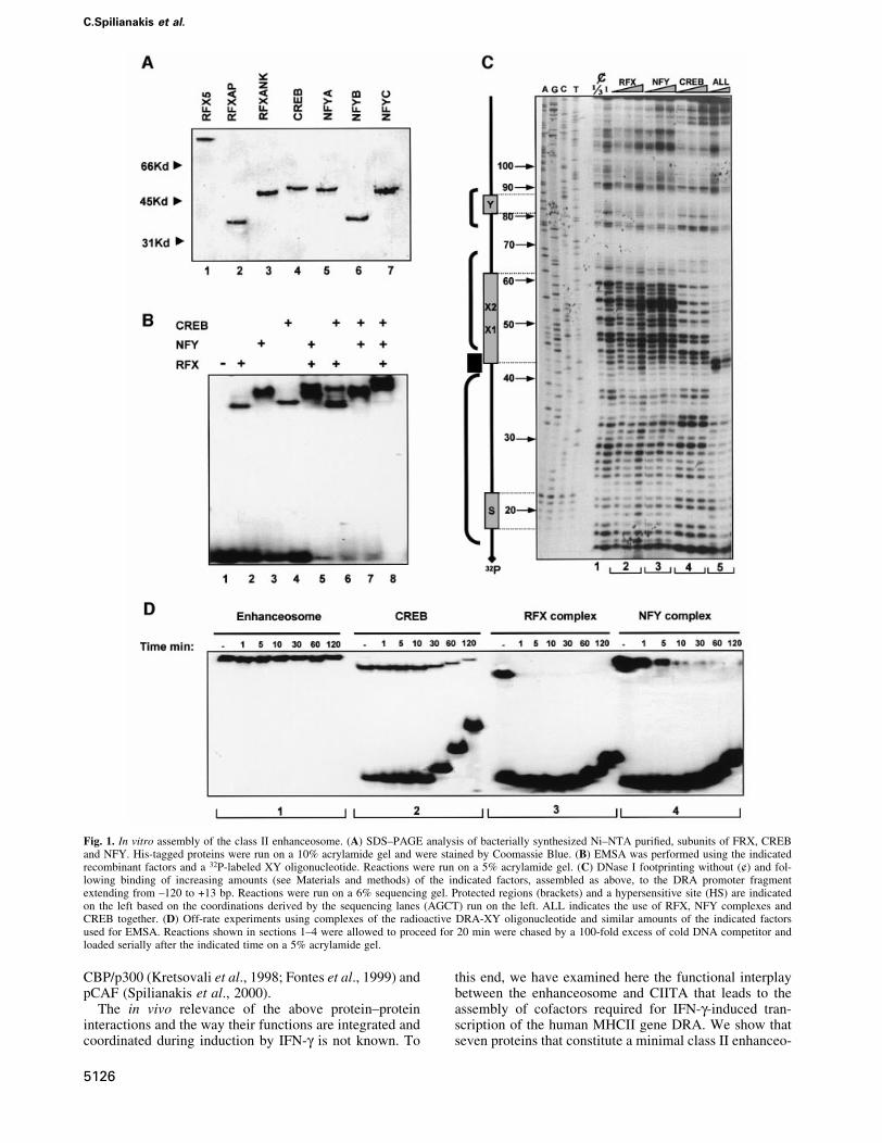
Fig. 1. In vitro assembly of the class II enhanceosome. (A) SDS±PAGE analysis of bacterially synthesized Ni±NTA puri®ed, subunits of FRX, CREBand NFY. His-tagged proteins were run on a 10% acrylamide gel and were stained by Coomassie Blue. (B) EMSA was performed using the indicatedrecombinant factors and a 32P-labeled XY oligonucleotide. Reactions were run on a 5% acrylamide gel. (C) DNase I footprinting without (¢) and fol-lowing binding of increasing amounts (see Materials and methods) of the indicated factors, assembled as above, to the DRA promoter fragmentextending from ±120 to +13 bp. Reactions were run on a 6% sequencing gel. Protected regions (brackets) and a hypersensitive site (HS) are indicatedon the left based on the coordinations derived by the sequencing lanes (AGCT) run on the left. ALL indicates the use of RFX, NFY complexes andCREB together. (D) Off-rate experiments using complexes of the radioactive DRA-XY oligonucleotide and similar amounts of the indicated factorsused for EMSA. Reactions shown in sections 1±4 were allowed to proceed for 20 min were chased by a 100-fold excess of cold DNA competitor andloaded serially after the indicated time on a 5% acrylamide gel.
C.Spilianakis et al.
5126
some are necessary and suf®cient for recruiting to thepromoter not only CIITA, but also a subset of the generaltranscription factors and RNA Pol II. CIITA in turnfunctions by stabilizing the enhanceosome±cofactorassembly, inducing promoter chromatin acetylation and®nally by facilitating Ser5 phosphorylation and promoterclearance of RNA Pol II, which determines the exacttiming of IFN-g-induced transcription.
Results
In vitro assembly and characterization of the MHCclass II enhanceosomePrevious studies performed using crude nuclear extracts asa source of transcription factors have suggested that CIITAis recruited to the class II enhanceosome by interactingwith multiple enhanceosome components such as RFX,NFY and CREB (Masternak et al., 2000). To investigatewhether the class II enhanceosome can recruit directlyCIITA to the promoter in the absence of any other factor,we reconstituted the enhanceosome in vitro usingrecombinant factors and tested its ability to recruitCIITA. The three RFX subunits (RFX5, RFXAP andRFXANK), together with the three NFY subunits (NFYA,NFYB and NFYC) and CREB were expressed and puri®edto near homogeneity from Escherichia coli (Figure 1A).Equimolar amounts of each subunit of the RFX and NFYcomplex were co-renatured and the assembly and identityof the trimeric complexes was monitored by electrophore-tic mobility shift assays (EMSAs) coupled with antibodysupershift experiments (data not shown). Class II en-hanceosome assembly was monitored by EMSAs and byDNase I footprinting experiments. Figure 1B shows thatthe MHCII DRA promoter (run alone in lane 1) formsdistinct complexes with RFX, NFY and CREB (lanes 2±4).
Pairwise combination of the factors revealed the assemblyof class II promoter complexes containing both of thefactors as well as individual DNA-bound proteins (lanes5±7). Remarkably, when all factors were allowed tointeract with the promoter a new complex was formedcontaining all three proteins (Figure 1B, lane 8). Thecooperative nature of the MHCII enhanceosome assemblywas monitored by quantitative DNase I footprintingexperiments. Figure 1C shows that increasing amountsof recombinant RFX or NFY only slightly protected thepromoter from DNase I cleavage at the X and Y boxesrespectively (Figure 1C, lane sections 2 and 3, numberedat the base of the gel). In contrast, CREB alone boundstably to the X2 subregion (Figure 1C, section 4).Remarkably, when the two lower concentrations of RFX,NFY and CREB were allowed to interact with thepromoter simultaneously, a strong footprint that extendedover the X-Y-S region was observed, indicative ofenhanceosome assembly (Figure 1C, section 5).Importantly, enhanceosome assembly is highly coopera-tive since these activators are incapable of strong protec-tion of DNA when tested alone (compare sections 2, 3, 4with 5). Figure 1D compares the DNA binding stability ofthe enhanceosome factors by off-rate competition experi-ments. In agreement with earlier observations usingnuclear extracts (Reith et al., 1994a,b) or recombinantRFX and NFY (Caretti et al., 2000) individual enhanceo-some subunits bind very unstably to DNA (Figure 1D,sections 3 and 4 with apparent half life <1 min for RFXand 1±5 min for NFY). CREB showed higher stability(Figure 1D, section 2 with half life ~40 min). Strikingly,the enhanceosome showed very high stability (Figure 1D,section 1, half life >2 h) as compared with individualCREB, RFX or NFY subcomplexes. Since CREB bindsrelatively stably on its own, we assume that it contributes
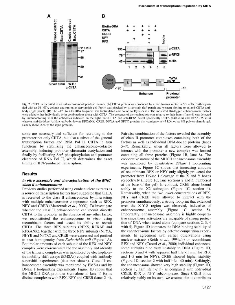
Fig. 2. CIITA is recruited in an enhanceosome-dependent manner. (A) CIITA protein was produced by a baculovirus vector in Sf9 cells, further puri-®ed with an Ni±NTA column and run on an acrylamide gel. Purity was checked by silver stain (left panel) and western blotting to an anti-CIITA anti-body (right panel). (B) The ±120 to +13 DRA fragment was biotinylated and bound to Dyna-beads. The indicated His-tagged enhanceosome factorswere added either individually or in combinations along with CIITA. The presence of the retained proteins relative to their inputs (lane 6) was detectedby immunoblotting with the antibodies indicated on the right: anti-CIITA and anti-RFX5 detect speci®cally CIITA (140 kDa) and RFX5 (75 kDa)whereas anti-histidine (a-His) antibody detects RFXANK, CREB, NFYA and NFYC proteins that comigrate at 45 kDa on an 8% polyacrylamide gel.Lane 6 shows 20% of the input proteins.
Mechanism of transcriptional regulation by CIITA
5127

signi®cantly to the overall stability of the enhanceosome.A similar effect was reported for the X2BP±RFXsubcomplex using nuclear extracts (Reith et al., 1994a).
To investigate the ability of the enhanceosome to recruitdirectly CIITA we carried out in vitro recruitmentexperiments using immobilized promoter templates and
C.Spilianakis et al.
5128
recombinant factors. A biotinylated fragment extendingfrom ±120 to +13 bp of the DRA promoter (Figure 2B,bottom) was attached to paramagnetic streptavidin beads(Dynal) followed by enhanceosome assembly. Next, thetemplates were incubated with recombinant and puri®edCIITA protein (Figure 2A) and after washing the boundproteins were detected by western blotting. Figure 2Bdepicts the immunodetection of CIITA and RFX5 usingspeci®c antibodies as well as detection of the comigratingRFX/Ank, NFY/A, NFY/C and CREB at 45 kDa usinganti-His antisera. Figure 2B shows that CIITA alone (lane5 top) cannot bind to naked DNA. Similarly RFX, NFY orCREB cannot recruit CIITA when bound individually tothe DRA promoter (lanes 1±3). However, the class IIenhanceosome assembled from all three factors (RFX,NFY and CREB) recruited CIITA ef®ciently (lane 4).Thus seven proteins (three subunits of RFX, three of NFYand CREB) constitute the minimal enhanceosome that isnecessary and suf®cient for CIITA recruitment to DRApromoter.
In vivo recruitment of coactivators and chromatinmodi®ers to the promoterTo investigate recruitment of CIITA to the endogenousclass II promoters we carried out chromatin immunopre-cipitation experiments using chromatin prepared fromHeLa cells induced with IFN-g for different amounts oftime. Figure 3A shows that in uninduced cells the DRApromoter is occupied by various enhanceosomecomponents such as CREB and RFX5 (top two rows),although the DRA gene is not expressed (Figure 3B, lane1). This observation is in agreement with previous in vivofootprinting data indicating partial protection of class IIpromoters in uninduced HeLa cells (Kara and Glimcher,1993). Surprisingly, we found that a subset of the basaltranscriptional machinery including TBP, TAFII 250,RNA Pol II and the p89 subunit of TFIIH were also boundto the DRA promoter in the uninduced and non-expressingcells (Figure 3A). These results suggested that the class IIenhanceosome could recruit these basal factors even in theabsence of CIITA. To investigate this possibility, wecarried out in vitro recruitment experiments using theimmobilized DRA promoter with or without the enhan-ceosome and HeLa nuclear extracts (HNEs). Figure 3Cshows that when HNEs that contain low levels ofendogenous enhanceosome factors were allowed to reactwith the DNA very low levels of these basal factors wererecruited to the promoter (lane 1). However, templatesbearing the recombinant enhanceosome ef®ciently re-cruited TBP, Pol II and TFIIH (p89) but not TFIIE (lane 2).Thus, the class II enhanceosome can direct recruitment ofkey components of the basal machinery to the promoter in
the absence of CIITA, a result consistent with thechromatin immunoprecipitation experiments describedabove.
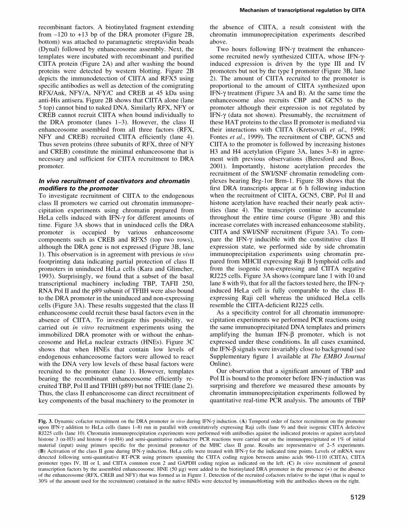
Two hours following IFN-g treatment the enhanceo-some recruited newly synthesized CIITA, whose IFN-g-induced expression is driven by the type III and IVpromoters but not by the type I promoter (Figure 3B, lane2). The amount of CIITA recruited to the promoter isproportional to the amount of CIITA synthesized uponIFN-g treatment (Figure 3A and B). At the same time theenhanceosome also recruits CBP and GCN5 to thepromoter although their expression is not regulated byIFN-g (data not shown). Presumably, the recruitment ofthese HAT proteins to the class II promoter is mediated viatheir interactions with CIITA (Kretsovali et al., 1998;Fontes et al., 1999). The recruitment of CBP, GCN5 andCIITA to the promoter is followed by increasing histonesH3 and H4 acetylation (Figure 3A, lanes 3±8) in agree-ment with previous observations (Beresford and Boss,2001). Importantly, histone acetylation precedes therecruitment of the SWI/SNF chromatin remodeling com-plexes bearing Brg-1or Brm-1. Figure 3B shows that the®rst DRA transcripts appear at 6 h following inductionwhen the recruitment of CIITA, GCN5, CBP, Pol II andhistone acetylation have reached their nearly peak activ-ities (lane 4). The transcripts continue to accumulatethroughout the entire time course (Figure 3B) and thisincrease correlates with increased enhanceosome stability,CIITA and SWI/SNF recruitment (Figure 3A). To com-pare the IFN-g inducible with the constitutive class IIexpression state, we performed side by side chromatinimmunoprecipitation experiments using chromatin pre-pared from MHCII expressing Raji B lymphoid cells andfrom the isogenic non-expressing and CIITA negativeRJ225 cells. Figure 3A shows (compare lane 1 with 10 andlane 8 with 9), that for all the factors tested here, the IFN-g-induced HeLa cell is fully comparable to the class II-expressing Raji cell whereas the uniduced HeLa cellsresemble the CIITA-de®cient RJ225 cells.
As a speci®city control for all chromatin immunopre-cipitation experiments we performed PCR reactions usingthe same immunoprecipitated DNA templates and primersamplifying the human IFN-b promoter, which is notexpressed under these conditions. In all cases examined,the IFN-b signals were invariably close to background (seeSupplementary ®gure 1 available at The EMBO JournalOnline).
Our observation that a signi®cant amount of TBP andPol II is bound to the promoter before IFN-g induction wassurprising and therefore we measured these amounts bychromatin immunoprecipitation experiments followed byquantitative real-time PCR analysis. The amounts of TBP
Fig. 3. Dynamic cofactor recruitment on the DRA promoter in vivo during IFN-g induction. (A) Temporal order of factor recruitment on the promoterupon IFN-g addition to HeLa cells (lanes 1±8) run in parallel with constitutively expressing Raji cells (lane 9) and their isogenic CIITA defectiveRJ225 cells (lane 10). Chromatin immunoprecipitation experiments were performed with antibodies against the indicated proteins or against acetylatedhistone 3 (a-H3) and histone 4 (a-H4) and semi-quantitative radioactive PCR reactions were carried out on the immunoprecipitated or 1% of initialmaterial (input) using primers speci®c for the proximal promoter of the MHC class II gene. Results are representative of 2±5 experiments.(B) Activation of the class II gene during IFN-g induction. HeLa cells were treated with IFN-g for the indicated time points. Levels of mRNA weredetected following semi-quantitative RT-PCR using primers spanning the CIITA coding region between amino acids 960±1110 (CIITA), CIITApromoter types IV, III or I, and CIITA common exon 2 and GAPDH coding region as indicated on the left. (C) In vitro recruitment of generaltranscription factors by the assembled enhanceosome. HNE (50 mg) were added to the biotinylated DRA promoter in the presence (+) or the absenceof the enhanceosome (RFX, CREB and NFY) that was formed as in Figure 1. Detection of the recruited cofactors relative to the input (that is equal to30% of the amount used for the recruitment) contained in the native HNEs were detected by immunoblotting with the antibodies shown on the right.
Mechanism of transcriptional regulation by CIITA
5129
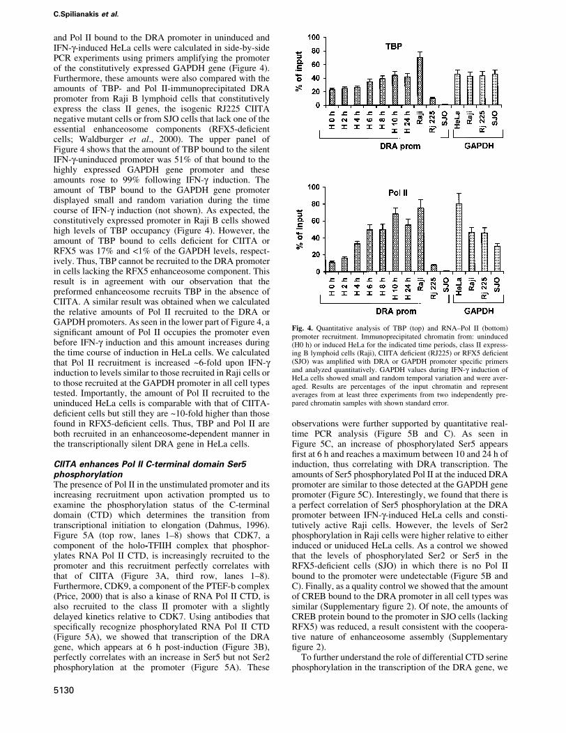
and Pol II bound to the DRA promoter in uninduced andIFN-g-induced HeLa cells were calculated in side-by-sidePCR experiments using primers amplifying the promoterof the constitutively expressed GAPDH gene (Figure 4).Furthermore, these amounts were also compared with theamounts of TBP- and Pol II-immunoprecipitated DRApromoter from Raji B lymphoid cells that constitutivelyexpress the class II genes, the isogenic RJ225 CIITAnegative mutant cells or from SJO cells that lack one of theessential enhanceosome components (RFX5-de®cientcells; Waldburger et al., 2000). The upper panel ofFigure 4 shows that the amount of TBP bound to the silentIFN-g-uninduced promoter was 51% of that bound to thehighly expressed GAPDH gene promoter and theseamounts rose to 99% following IFN-g induction. Theamount of TBP bound to the GAPDH gene promoterdisplayed small and random variation during the timecourse of IFN-g induction (not shown). As expected, theconstitutively expressed promoter in Raji B cells showedhigh levels of TBP occupancy (Figure 4). However, theamount of TBP bound to cells de®cient for CIITA orRFX5 was 17% and <1% of the GAPDH levels, respect-ively. Thus, TBP cannot be recruited to the DRA promoterin cells lacking the RFX5 enhanceosome component. Thisresult is in agreement with our observation that thepreformed enhanceosome recruits TBP in the absence ofCIITA. A similar result was obtained when we calculatedthe relative amounts of Pol II recruited to the DRA orGAPDH promoters. As seen in the lower part of Figure 4, asigni®cant amount of Pol II occupies the promoter evenbefore IFN-g induction and this amount increases duringthe time course of induction in HeLa cells. We calculatedthat Pol II recruitment is increased ~6-fold upon IFN-ginduction to levels similar to those recruited in Raji cells orto those recruited at the GAPDH promoter in all cell typestested. Importantly, the amount of Pol II recruited to theuninduced HeLa cells is comparable with that of CIITA-de®cient cells but still they are ~10-fold higher than thosefound in RFX5-de®cient cells. Thus, TBP and Pol II areboth recruited in an enhanceosome-dependent manner inthe transcriptionally silent DRA gene in HeLa cells.
CIITA enhances Pol II C-terminal domain Ser5phosphorylationThe presence of Pol II in the unstimulated promoter and itsincreasing recruitment upon activation prompted us toexamine the phosphorylation status of the C-terminaldomain (CTD) which determines the transition fromtranscriptional initiation to elongation (Dahmus, 1996).Figure 5A (top row, lanes 1±8) shows that CDK7, acomponent of the holo-TFIIH complex that phosphor-ylates RNA Pol II CTD, is increasingly recruited to thepromoter and this recruitment perfectly correlates withthat of CIITA (Figure 3A, third row, lanes 1±8).Furthermore, CDK9, a component of the PTEF-b complex(Price, 2000) that is also a kinase of RNA Pol II CTD, isalso recruited to the class II promoter with a slightlydelayed kinetics relative to CDK7. Using antibodies thatspeci®cally recognize phosphorylated RNA Pol II CTD(Figure 5A), we showed that transcription of the DRAgene, which appears at 6 h post-induction (Figure 3B),perfectly correlates with an increase in Ser5 but not Ser2phosphorylation at the promoter (Figure 5A). These
observations were further supported by quantitative real-time PCR analysis (Figure 5B and C). As seen inFigure 5C, an increase of phosphorylated Ser5 appears®rst at 6 h and reaches a maximum between 10 and 24 h ofinduction, thus correlating with DRA transcription. Theamounts of Ser5 phosphorylated Pol II at the induced DRApromoter are similar to those detected at the GAPDH genepromoter (Figure 5C). Interestingly, we found that there isa perfect correlation of Ser5 phosphorylation at the DRApromoter between IFN-g-induced HeLa cells and consti-tutively active Raji cells. However, the levels of Ser2phosphorylation in Raji cells were higher relative to eitherinduced or uniduced HeLa cells. As a control we showedthat the levels of phosphorylated Ser2 or Ser5 in theRFX5-de®cient cells (SJO) in which there is no Pol IIbound to the promoter were undetectable (Figure 5B andC). Finally, as a quality control we showed that the amountof CREB bound to the DRA promoter in all cell types wassimilar (Supplementary ®gure 2). Of note, the amounts ofCREB protein bound to the promoter in SJO cells (lackingRFX5) was reduced, a result consistent with the coopera-tive nature of enhanceosome assembly (Supplementary®gure 2).
To further understand the role of differential CTD serinephosphorylation in the transcription of the DRA gene, we
Fig. 4. Quantitative analysis of TBP (top) and RNA±Pol II (bottom)promoter recruitment. Immunoprecipitated chromatin from: uninduced(H0 h) or induced HeLa for the indicated time periods, class II express-ing B lymphoid cells (Raji), CIITA de®cient (RJ225) or RFX5 de®cient(SJO) was ampli®ed with DRA or GAPDH promoter speci®c primersand analyzed quantitatively. GAPDH values during IFN-g induction ofHeLa cells showed small and random temporal variation and were aver-aged. Results are percentages of the input chromatin and representaverages from at least three experiments from two independently pre-pared chromatin samples with shown standard error.
C.Spilianakis et al.
5130
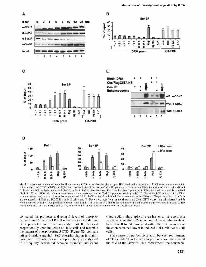
compared the promoter and exon 5 levels of phospho-serine 2 and 5 recruited Pol II under various conditions.Both promoter and exon associated Pol II increasedproportionally upon induction of HeLa cells and resemblethe pattern of phosphoserine 5 CTD (Figure 5D, compareleft and middle graphs). Ser5 phosphorylation is mainlypromoter linked whereas serine 2 phosphorylation showedto be equally distributed between promoter and exons
(Figure 5D, right graph) or even higher at the exons at alate time point after IFN induction. However, the levels ofSer2P Pol II found associated with either the promoter orthe exon remained lower in induced HeLa relative to Rajicells.
Since there is a perfect correlation between recruitmentof CDKs and CIITA to the DRA promoter, we investigatedthe role of the latter in CDK recruitment: the enhanceo-
Fig. 5. Dynamic recruitment of RNA Pol II kinases and CTD serine phosphorylation upon IFN-g-induced transcription. (A) Chromatin immunoprecipi-tation analysis of CDK7, CDK9 and RNA Pol II-serine2 (Ser2P) or -serine5 (Ser5P) phosphorylation during IFN-g induction of HeLa cells. (B andC) Real-time PCR analysis of the Ser2 (Ser2P) or Ser5 (Ser5P) phosphorylated Pol II on the class II promoter in IFN-g-induced HeLa and B lymphoid(Raji, RJ225 and SJO) cells. Control experiments were performed on the GAPDH promoter (right panels). (D) Real-time PCR analysis of the DRApromoter (gray bars) or exon 5 (open bars) associated Pol II, Ser2P or Ser5P as labeled. HeLa were uninduced (H0h) or IFN-g-induced for 10 or 24 hand compared with Raji and RJ225 B lymphoid cell types. (E) Nuclear extracts from control (lanes 1 and 2) or CIITA expressing cells (lanes 4 and 5)were incubated with the DRA promoter without (lanes 1 and 4) or with (lanes 2 and 5) the addition of the enhanceosome factors used in Figure 2. Therecruitment of CDK7 and CDK9 and CIITA relative to their input (20%) was monitored by speci®c antibodies.
Mechanism of transcriptional regulation by CIITA
5131

some was assembled in vitro and the amount of kinasesrecruited in the presence of control or CIITA containingnuclear extracts was examined. As shown in Figure 5E,both the endogenous and the recombinant enhanceosomedid not recruit either CDK7 or CDK9 (lanes 1 and 2) in theabsence of CIITA. However, in the presence of CIITA theenhanceosome recruited CDK7 and CDK9 ef®ciently(lanes 4 and 5).
The CIITA-dependent recruitment of kinases led us toinvestigate whether CIITA interacts with CDKs. Figure 6A
shows that antibodies against the endogenous CDK7 orCDK9, can precipitate myc-tagged CIITA (lanes 3 and 4)that was expressed into COS cells following transfection.In contrast, an antibody against Pol II could not precipitateCIITA (lane 2), although it did precipitate the p89 subunitof TFIIH (lanes 5 and 6). The western blots of Figure 6A(lanes 7, 8 and 9) depict the inputs and speci®city of theantibodies used for immunoprecipitation.
The experiments described above suggested that CIITAcould affect the phosphorylation status of Pol II by
Fig. 6. CIITA promotes serine 5 phosphorylation of Pol II. (A) CIITA interacts in vivo with CDK7 and CDK9. Whole cell extracts containing Myc-tagged CIITA were separated on a 8% SDS±PAGE and immunoblotted with an antibody against the myc epitope (a-Myc) or the p89 subunit of TFIIH(a-TFIIH) either before (Input) or following immunoprecipitation (IP) with antibodies against RNA Pol II, CDK7 and CDK9 as indicated. Input of theextracts (10%) were separated on 6 and 10% gels and immunoblotted with anti-Pol II and anti-CDK7, -CDK9 antibodies respectively. (B) RNA Pol IIwas immunoprecipitated from HNEs (lanes 2, 3 and 5±10) and equal amounts were phosphorylated in vitro with [g-32P] ATP in the presence (lanes 3,6, 8 and 10) or the absence (lanes 2, 5, 7 and 9) of CIITA. Reactions were separated on 6% SDS±PAGE together with input HNEs (lanes 11±13). Theradioactive part of the gel was subject to ®xation drying and autoradiography. The non-radioactive part was transferred onto nitrocellulose for subse-quent immunoblotting with antibodies against Pol II and the Ser2 (Ser2P) and the Ser5 (Ser5P) phosphorylated forms of the CTD of Pol II. IIo and IIacorrespond to the phosphorylated and non-phosphorylated forms of Pol II. (C) In vitro phosphorylation reactions of Pol II were performed in the pres-ence of CIITA and/or apyrase as indicated. Phosphorylated forms of RNA Pol II were detected by immunoblotting using antibodies H5 and H14 spe-ci®c for the phosphorylated Ser2P and Ser5P of Pol II. (D) A GST fusion of the CTD of Pol II (GST±CTD) was incubated with immunoprecipitatedCDK7 (lanes 1 and 2) in the presence (lane 2) or the absence (lane 1) of 100 ng of baculovirus CIITA. Ser 5 phosphorylated RNA Pol II CTD wasdetected by H14 antibody and CDK7 was detected by speci®c antibody.
C.Spilianakis et al.
5132
recruiting the CDK7 and CDK9 kinases. To investigatethis possibility directly we examined the effect of CIITAon RNA Pol II phosphorylation in vitro. The Pol IIholoenzyme complex was immunoprecipitated using aspeci®c Pol II antibody and Pol II phosphorylation wasdetermined by adding [g-32P]ATP and recombinant CIITAprotein in the reaction. Figure 6B shows that the additionof baculovirus-expressed CIITA increased the in vitrophosphorylation of Pol II (compare lanes 2 and 3) by theassociated kinases. To examine the relative contribution ofCDK7 and CDK9 to this effect, we used extractsimmunodepleted for these kinases (Supplementary®gure 3). Depletion of CDK7 rendered CIITA unable toenhance Pol II phosphorylation (compare lanes 7 and 8with 5 and 6) whereas CDK9 depletion had no effect (lanes9 and 10). These results strongly suggest that CDK7 or anassociated unknown kinase is the main Pol II kinasepotentiated by CIITA. The western blots of Figure 6B(lanes 11±13) depict the inputs and identify the phos-phorylated Pol II species as RNA Pol II form IIo.
To determine the Ser residue of the RNA Pol II CTDthat was preferentially phosphorylated we performed coldin vitro phosphorylation assays followed by western blotanalysis using antibodies against phosphorylated Ser2 orSer5 CTD. Figure 6C shows that CIITA speci®callyincreased phosphorylation of Ser5 but not Ser2 in theabsence (lane 3) but not in the presence of apyrase (lane 4).Importantly, bacterially expressed CTD was used as aphosphorylation substrate for CDK7 since previous deple-tion experiments indicated that it is the kinase that isaffected by CIITA. Immunoprecipitated CDK7 phos-phorylated Ser5 of CTD (Figure 6D, lane 1). In thepresence of CIITA Ser5 phosphorylation by CDK7 wasstrongly increased (Figure 6D, compare lanes 1 and 2).Thus, one of the actions of CIITA is to recruit both Pol IIkinases and increase the Ser5 phosphorylation by CDK7.
Discussion
Previous studies have shown that assembly of thetranscriptional apparatus in response to environmentalcues is governed by distinct mechanisms in differentgenes. In general, the transcriptional activators bindsequentially to their regulatory elements and this leads toa de®ned order of recruitment of both chromatin-modify-ing activities and general transcription factors (Cosma,2002). These observations led to the hypothesis that thesedifferences are associated with different biological needsfor each gene product. In this manuscript we describe theorder of events taking place during activation of an MHCIIgene in response to IFN-g. We found that in contrast to thepreviously described cases of signal-regulated gene tran-scription by virus (Agalioti et al., 2000), estrogens (Shang,et al., 2000), phosphate deprivation (Gregory et al., 1999)or by differentiating signals (Soutoglou and Talianidis,2002) the activators responsible for the expression of theDRA gene are prebound to the enhancer forming anenhanceosome. The enhanceosome then serves as adocking surface for the subsequent recruitment of a subsetof the basal transcription machinery that remains associ-ated to the core promoter even under conditions where thegene is transcriptionally silent. In other words the class IIgenes are `poised' for activation. Transcriptional acti-
vation in response to IFN-g requires the synthesis ofCIITA, which is rapidly recruited to class II promoters bythe enhanceosome. We showed that this recruitmentcorrelates with the subsequent recruitment of chromatin-modifying activities such as HATs and ATP-dependentremodeling machines. Surprisingly, CIITA also recruitsCDK7 and CDK9 thus causing RNA Pol II CTDphosphorylation and transcriptional elongation.Collectively, our data describe a novel strategy by whicha gene-speci®c transcriptional coactivator orchestrates theMHCII gene expression program.
The class II enhanceosome is assembled via multiplecooperative interactions between its protein constituentsand the DNA (Masternak et al., 2000). Previous reportsemploying nuclear extracts (Reith et al., 1994a,b; Morenoet al., 1995; Fontes et al., 1997a; Louis-Plence et al., 1997)or a subset of recombinant enhanceosome components(RFX and NFY) (Caretti et al., 2000) have showncooperative DNA binding between RFX, NFY andX2BP. We have reconstituted the enhanceosome in vitrousing all seven subunits of the class II binding factors(RFX, CREB and NFY) and found that it can recruitCIITA. This is the ®rst report of CIITA recruitment by apurely recombinant class II gene enhanceosome.
Our chromatin immunoprecipitation experiments re-vealed that the enhanceosome and a subset of the basaltranscriptional machinery that is already bound to thepromoter before induction is `energized' by the presenceof newly synthesized CIITA recruited upon IFN-g treat-ment. CIITA recruitment leads to a further stabilization ofthe transcription complex by integrating and coordinatinga new set of protein±protein interactions. Thus, CIITAcoordinates several alterations of cofactor recruitment tothe promoter that precede or are concomitant with theonset of transcription.
An extensive cofactor recruitment analysis by chroma-tin immunoprecipitations indicated that the moleculararchitecture of the transcription complexes of IFN-g-induced HeLa cells is qualitatively similar to theconstitutively expressing Raji cells. On the other hand,uninduced HeLa cells resemble the CIITA negative Blymphoblastoid cells RJ225 with the exception of TBP thatshows high levels in the former cell type.
CIITA orchestrates recruitment of the HATs CBP andGCN5 correlating with chromatin acetylation that pre-cedes initiation of transcription. Therefore, the synergybetween CIITA and CBP and/or PCAF/GCN5 that wasshown in transfection systems (Kretsovali et al., 1998;Fontes et al., 1999; Spilianakis et al., 2000) re¯ects aphysiological role of HAT-type coactivators in class IIgene transcription. Although the HAT activity of variouscoactivatorsÐas assayed by transient experimentsÐseems to be partly dispensable for MHCII gene expression,the present data in combination with results that correlatetranscriptional inactivation after IFN-g removal (Beresfordand Boss, 2001) or in regulatory mutant cell lines withreduced acetylation (Masternak and Reith, 2002), supporta causal relationship between promoter localized histoneacetylation and transcription. Our experiments alsoshowed that both Brg-1 and Brm-1 containing chromatinremodeling machines are recruited to the promoter,subsequently to the wave of acetylation and their presencetemporally correlates with a role in transcriptional elonga-
Mechanism of transcriptional regulation by CIITA
5133
tion. Thus, the need for SWI±SNF-mediated remodeling isnot only restricted to the expression of CIITA itself(Mudhasani and Fontes, 2002) but it is also directlyrequired for MHCII gene transcription.
Although promoter occupancy by factors of the basalmachinery did not change signi®cantly over time afterIFN-g induction, occupancy by Pol II showed a strongincrease that temporally parallels both CIITA recruitmentand gene transcription. Phosphorylation of the CTD of PolII is a critical process that controls promoter clearance andelongation (Dahmus, 1996). The cyclin H±CDK7 complex(which belongs to TFIIH) is responsible for Ser5 phos-phorylation whereas cyclin T±CDK9 (P-TEF) is respon-sible for Ser2 phosphorylation. CIITA interacts with andrecruits both RNA Pol II kinases CDK7 and CDK9.Interestingly, CDK7 recruitment is directly proportional tothe recruitment of CIITA. As a consequence, CTDphosphorylation at Ser5 is the event that closely correlateswith DRA gene transcription onset that occurs 6 h postinduction. In another system of signal-induced transcrip-tion, the IL-8 gene, induction by TNF leads to both Ser5and Ser2 phosphorylation of promoter-bound Pol II(Nissen and Yamamoto, 2000; Barboric et al., 2001).Apparently the kinase speci®city for distinct CTD serinesis subject to gene-, cell- and regulatory-type speci®c rules.Previous work with Saccharomyces cerevisiae has dem-onstrated that RNA Pol II is Ser5 phosphorylated at thepromoters and Ser2 phosphorylated within transcribedregions (Komarnitsky et al., 2000). On the DRA gene,RNA Pol II and its Ser5-phosphorylated form are moreconcentrated at the promoter as compared with the codingregions. In contrast, Ser2-phosphorylated RNA Pol II isevenly distributed between promoter and coding regions oreven more concentrated at the coding regions. These dataare in agreement with reports from studies on the DHFRand g-actin genes in mammalian cells (Cheng and Sharp,2003). Ser2 phosphorylation in IFN-g-induced epithelial-like cells is low relative to Raji B lymphoid cells thatconstitutively transcribe the DRA gene. Induced HeLaexpress ~53 less DRA mRNA than Raji cells (not shown)yet their Pol II levels do not differ signi®cantly. Therefore,we conclude that Raji cells have a higher rate or density ofelongating polymerase relative to HeLa. Alternatively,paused Pol II at the DRA promoter, similarly to the heatshock (Lis, 1998), c-myc (Krumm et al., 1992) and c-fos(Pinaud and Mirkovitch, 1998) genes, might occur inhigher proportions in HeLa cells relative to the Raji.
Strikingly, CIITA augments serine 5 phosphorylation ofPol II in vitro. Such an effect may be due to its ability toincrease enzyme activity or stabilize enzyme±substrateinteraction. We provide evidence that CIITA potentiates theCDK7-driven phosphorylation of RNA Pol II CTD employ-ing either depleted extracts or immunoprecipitated CDK7and recombinant CTD which was phosphorylated at Ser5.
In summary, our data describe a novel mechanism ofcontrolling gene transcription in which a mammalian geneis poised for activation and awaits to be `energized' by theincoming gene-speci®c class II transactivator. The latter isable to promote Ser5 phosphorylation of RNA Pol II whichis the critical event that determines the timing oftranscription onset through promoter clearance and tran-scription elongation.
Materials and methodsCell culture and reagentsHeLa and COS-1 cells were grown in DMEM supplemented with 10%bovine fetal serum. B lymphoid cell lines were maintained in RPMI.IFN-g (R&D) was used at 100±200 U/ml. Antibodies against acetylatedH3 and H4 were from Upstate Biotechnology. Monoclonal antibodies H5and H14 were from Covance. All other antibodies were from Santa CruzBiotechnology.
Recombinant proteinsHis-tagged recombinant proteins were expressed in E.coli HB101 cellsfrom the pRSET vector and were puri®ed on an Ni±NTA column. Adetailed protocol is included in Supplementary data section. Baculo-CIITA was produced using the Bac-to-Bac Baculovirus expressionsystem (Gibco-BRL) according to manufacturers instructions. Theprotein was then puri®ed on Ni±NTA agarose beads under non-denaturing conditions.
Electrophoretic mobility shift assaysBinding reactions contained radiolabeled (0.3 3 106 c.p.m./reaction) ofthe XY fragment XY sense 5¢-GATCCCCTAGCAACAGATGCGT-CATCTCAAAATATTTTT.
For the off-rate EMSA experiments 3 3 106 c.p.m. radiolabeled DRAenhancer DNA fragment and the appropriate amounts of each proteincomplex were used: RFX5, 2 ng; RFXAP, 1 ng; RFXANK, 1 ng; NFYA,1.25 ng; NFYB, 1.25 ng; NFYC, 1.25 ng; CREB, 1.25 ng. Bindingreactions were left to set on ice for 20 min and then 1003 excess coldcompetitor of the same DRA DNA fragment was added to the reaction forthe indicated time periods and run in 6% polyacrylamide gel. The ®rstlane of each off-rate EMSA experiment for the enhanceosome, CREB,RFX complex and NFY complex are binding reactions using no coldcompetitor. The abnormal kinetics in some lanes is due to loading afterincreasing time periods.
In vitro DNase I footprintingIn vitro DNase I footprinting used C-terminally radiolabeled DNAfragments of the human DRA enhancer. A detailed protocol is included inthe Supplementary data section.
Recruitment on immobilized templateThe biotinylated DRA promoter was synthesized by PCR using speci®cDRA primers. Blocking was performed in BC-100 buffer (100 mM KCl,20 mM HEPES pH 7.9, 20% glycerol, 0.2 mM EDTA, 0.5 mM DTT,0.5 mM PMSF) supplemented with 10% BSA for 1 h at 4°C in thepresence of 2 mg of salmon sperm DNA. Then recombinant RFX, NFYand CREB were added at concentrations established on DNasefootprinting (RFX5, 80 ng; RFXAP, 40 ng; RFXANK, 40 ng; NFYA,50 ng; NFYB, 50 ng; NFYC, 50 ng; CREB, 100 ng) and incubated for anadditional hour. Finally baculoviral CIITA (~100 ng as determined bysilver staining) was added for 1 h. Washing was performed three times inBC-100 supplemented with 0.01% Triton.
RT±PCRRNA was prepared with the Trizol reagent (Gibco-BRL). Reversetranscription±PCR reactions were made using MMLV Promega enzyme.PCR conditions for all the reactions included an initial 5 min step at 94°C,followed by 24 cycles of 30 s at 94°C, 30 s at 64°C, 30 s at 72°C, and a®nal step of 5 min at 72°C. PCR products were analyzed in a 2% agarosegel.
The sequences of RT±PCR primers for CIITA are the following: CIITAtype III.sense, 5¢-ATGCGTTGCCTGGCTCCACGCC-3¢; CIITA typeIV.sense, 5¢-CCAGAGCTGGCGGGAGGGAG-3¢; CIITA type I.sense,5¢-GCCATCAGCCCAGCCTGGTGC-3¢.
The above primers were used along with the following commonreverse primer: CIITA.antisense, 5¢-CAGGCAGCTCAACGAGGAAC-TGGAG-3¢.
Sequences of RT±PCR primers for the DRA gene are: DRA.exIIIsense,5¢-GAGTTTGATGCTCCAAGCCCTCTCCCA-3¢; DRA.exIIIantisense,5¢-CAGAGGCCCCCTGCGTTCTGCTGCATT-3¢.
Sequences for RT±PCR primers for GAPDH gene are: GAPDH.sense,5¢-CCCCCTCTGCTGATGCCC-3¢; GAPDH.antisense, 5¢-CCCCGCG-GCCATCACG-3¢.
Chromatin immunoprecipitationsA detailed protocol for chromatin immunoprecipitation assays is providedin the Supplementary data section. For radioactive PCR analysis, DNA
C.Spilianakis et al.
5134
was resuspended in 100 ml TE buffer pH 7.5 and 10 ml were used in aradioactive PCR reaction using [32P]dATP and [32P]dCTP supplementedwith 0.1 mM cold dNTPs, 3 units of Taq polymerase, 0.25 mM MgCl2,13 PCR reaction buffer and 400 ng of each primer. PCR conditionsincluded an initial step of 5 min at 94°C, 24 cycles of 30 s at 94°C, 30 s at64°C, 30 s at 72°C and a ®nal step of 2 min at 72°C.
Real-time PCR analysis was performed using the Opticon monitor (MJResearch) and the intercalating ¯uorescent dye SYBR Green I. Thefraction of immunoprecipitated promoter DNA was calculated from astandard curve/relative to standard input chromatin. The immunopreci-pitated values obtained with different antibodies using the class IIpromoter were compared with the values of the reference gene GAPDHwhich displays high levels of expression and is not regulated by IFN-g.The IFN-b promoter was used as a negative control. Chromatinimmunoprecipitations were repeated at least three times from differentchromatin material and radioactive or real-time PCR analysis wasrepeated three to ®ve times.
The primers used were the following:DRA promoter: DRAsense, 5¢-GTTGTCCTGTTTGTTTAAGAAC-3¢;
DRAantisense, 5¢-GCTCTTTTGGGAGTCAG-3¢.DRAexon 5: DRAsense, 5¢-GAAAGCAGTCATCTTCAGCGTT-3¢;
DRAantisense 5¢-ATTATCACCATGCAATGCCTCT-3¢.IFN-b promoter: IFN-bsense, 5¢-GCTTTCCTTTGCTTTCTCCCAA-
GTC-3¢; IFN-bantisense, 5¢-CCTTTCTCCATGGGTATGGCC-3¢.GAPDH-promoter: GAPDHsense, 5¢-TGAGCAGACCGGTGTCAC-
TA-3¢; GAPDHantisense, 5¢-AGGACTTTGGGAACGACTGA-3¢.
In vitro phosphorylation assaysHNEs (~600 mg) were immunoprecipitated with 30 ml (200 ng/ml, SantaCruz) of anti-Pol II antibody overnight at 4°C, supplemented with 20 ml ofProtein A slurry preblocked with 500 ml of BC100, 5% BSA, 0.01%Triton X-100, for 1 h at 4°C. The immune complexes were pelleted,resuspended in 10 ml of BC-100 and used for phosphorylationexperiments. Phosphorylation reaction was set up in a buffer containing5 mM HEPES pH 7.8, 20 mM Tris±HCl pH 7.9, 7 mM MgCl2, 60 mMKCl, 12% (v/v) glycerol, 2% (w/v) polyethylene glycol 8000, 2 mM2-mercaptoethanol, 0.1 mM EDTA, 6 mg of BSA, 3 mCi [g-32P]ATP forradioactive reactions and 15 mM cold ATP. Reactions were performed for1 h at 25°C, in a total volume of 25 ml. For detection of phosphorylatedforms of Pol II by western blotting we used 0.2 mM ATP and themonoclonal antibodies H14 and H5 that recognize the phosphoserine 5and phosphoserine 2 version of RNA Pol II±CTD respectively (Covance-Babco). For the experiments employing CDK7 and CDK9-depletedextracts, two rounds of treatment with 30 ml of anti-CDK7 or 30 ml of anti-CDK9 antibodies preceded the immunoprecipitation of RNA Pol II. Forthe experiments using RNA Pol II CTD as a template, 500 ng of a GST±CTD fusion protein (kindly provided by Dr D.Reinberg) was phos-phorylated by CDK7 immunoprecipitated from 100 mg of HNEs in abuffer containing 50 mM Tris pH 7.5, 4 mM MgCl2, 5 mM DTT, 5 mMMnCl2, 0.1 mM ZnSO4 and 0.2 mM ATP.
Supplementary dataSupplementary data are available at The EMBO Journal Online.
Acknowledgements
We thank G.Vretzos for expert assistance with cell culture andE.Tzortzakaki and S.Lomvardas for help with some experiments. Wealso thank D.Reinberg for GST±CTD and I.Talianidis for antibodies. Thiswork was supported by grants from the Greek General Secretariat forScience and Technology (PENED 2284).
References
Agalioti,T., Lomvardas,S., Parekh,B., Yie,J., Maniatis,T. and Thanos,D.(2000) Ordered recruitment of chromatin modifying and generaltranscription factors to the IFN-a promoter. Cell, 103, 667±678.
Barboric,M., Nissen,R.M., Kanazawa,S., Jabrane-Ferrat,N. andPeterlin,B.M. (2001) NF-kappaB binds P-TEFb to stimulatetranscriptional elongation by RNA polymerase II. Mol. Cell, 8, 327±337.
Beresford,G.W. and Boss,J.M. (2001) CIITA coordinates multiplehistone acetylation modi®cations at the HLA±DRA promoter. Nat.Immunol., 2, 652±657.
Caretti,G., Cocchiarella,F., Sidoli,C., Villard,J., Peretti,M., Reith,W. and
Mantovani,R. (2000) Dissection of functional NF-Y-RFX cooperativeinteractions on the MHC class II Ea promoter. J. Mol. Biol., 302, 539±552.
Cheng,C. and Sharp,P. (2003) RNA polymerase II accumulation in thepromoter-proximal region of the dihydrofolate reductase and gamma-actin genes. Mol. Cell. Biol., 23, 1961±1967.
Cosma,M.P. (2002) Ordered recruitment: gene-speci®c mechanism oftranscription activation. Mol. Cell, 10, 227±236.
Cresswell,P. (1994) Antigen presentation. Getting peptides into MHCclass II molecules. Curr. Biol., 4, 541±543.
Dahmus,M.E. (1996) Reversible phosphorylation of the C-terminaldomain of RNA polymerase II. J. Biol. Chem., 271, 19009±19012.
DeSandro,A.M., Nagarajan,U.M. and Boss,J.M. (2000) Associations andinteractions between bare lymphocyte syndrome factors. Mol. Cell.Biol., 20, 6587±6599.
Fontes,J., Jabrane-Ferrat,N. and Peterlin,B. (1997a) Assembly offunctional regulatory complexes on MHC class II promoters in vivo.J. Mol. Biol., 270, 336±345.
Fontes,J.D., Jiang,B. and Peterlin,B.M. (1997b) The class II trans-activator CIITA interacts with the TBP-associated factor TAFII32.Nucleic Acids Res., 25, 2522±2528.
Fontes,J.D., Kanazawa,S., Jean,D. and Peterlin,B.M. (1999) Interactionsbetween the Class II transactivator and CREB binding protein increasetranscription of major histocompatibility complex Class II genes. Mol.Cell. Biol., 19, 941±947.
Glimcher,L.H. and Kara,C.J. (1992) Sequences and factors: a guide toMHC class-II transcription. Annu. Rev. Immunol., 10, 13±49.
Gregory,P.D., Schmid,A., Zavari,M., Munsterkotter,M. and Horz,W.(1999) Chromatin remodelling at the PHO8 promoter requires SWI±SNF and SAGA at a step subsequent to activator binding. EMBO J.,18, 6407±6414.
Hake,S., Masternak,K., Kammerbauer,C., Jansen,C., Reith,W. andSteimle,V. (2000) CIITA leucine-rich repeats control nuclearlocalization in vivo recruitment to the major histocompatibilitycomplex (MHC) class II enhanceosome and MHC class II genetranscription. Mol. Cell. Biol., 20, 7716±7725.
Harton,J. and Ting,J.P. (2000) Class II transactivator: mastering the artof major histocompatibility complex expression. Mol. Cell. Biol., 20,6185±6194.
Kanazawa,S., Okamoto,T. and Peterlin,B.M. (2000) Tat competes withCIITA for the binding to P-TEFb and blocks the expression of MHCclass II genes in HIV infection. Immunity, 12, 61±70.
Kara,C.J. and Glimcher,L.H. (1993) Developmental and cytokine-mediated regulation of MHC class II gene promoter occupancyin vivo. J. Immunol., 150, 4934±4942.
Komarnitsky,P., Cho,E. and Buratowski,S. (2000) Differentphosphorylated forms of RNA polymerase II and associated mRNAprocessing factors during transcription. Genes Dev., 14, 2452±2460.
Kretsovali,A., Agalioti,T., Spilianakis,C., Tzortzakaki,E., Merika,M. andPapamatheakis,J. (1998) Involvement of CREB binding protein inexpression of major histocompatibility complex class II genes viainteraction with the class II transactivator. Mol. Cell. Biol., 18, 6777±6783.
Krumm,A., Meulia,T., Brunvand,M. and Groudine,M. (1992) The blockto transcriptional elongation within the human c-myc gene isdetermined in the promoter-proximal region. Genes Dev., 6, 2201±2213.
Lee,T.I. and Young,R.A. (2000) Transcription of eukaryotic protein-coding genes. Annu. Rev. Genet., 34, 77±137.
Lemon,B. and Tjan,R. (2000) Orchestrated response: a symphony oftranscription factors for gene control. Genes Dev., 14, 2551±2569.
Lis,J. (1998) Promoter-associated pausing in promoter architecture andpostinitiation transcriptional regulation. Cold Spring Harb. Symp.Quant. Biol., 63, 347±356.
Louis-Plence,P., Moreno,C.S. and Boss,J.M. (1997) Formation of aregulatory factor X/X2 box-binding protein/nuclear factor-Ymultiprotein complex on the conserved regulatory regions of HLAclass II genes. J. Immunol., 159, 3899±3909.
Mahanta,S.K., Scholl,T., Yang,F.C. and Strominger,J.L. (1997)Transactivation by CIITA, the type II bare lymphocyte syndrome-associated factor, requires participation of multiple regions of theTATA box binding protein. Proc. Natl Acad. Sci. USA, 94, 6324±6329.
Masternak,K. and Reith,W. (2002) Promoter-speci®c functions of CIITAand the MHC class II enhanceosome in transcriptional activation.EMBO J., 21, 1379±1388.
Masternak,K., Muhlethaler-Mottet,A., Villard,J., Zufferey,M.,
Mechanism of transcriptional regulation by CIITA
5135
Steimle,V. and Reith,W. (2000) CIITA is a transcriptional coactivatorthat is recruited to the MHC class II promoters by multiple synergisticinteractions with an enhanceosome complex. Genes Dev., 14, 1156±1166.
Moreno,C.S., Emery,P., West,J.E., Durand,B., Reith,W., Mach,B. andBoss,J.M. (1995) Puri®ed X2 binding protein (X2BP) cooperativelybinds the class II MHC X box region in the presence of puri®ed RFX,the X box factor de®cient in the bare lymphocyte syndrome. J.Immunol., 155, 4313±4321.
Mudhasani,R. and Fontes,J.D. (2002) The class II transactivator requiresbrahma-related gene 1 to activate transcription of majorhistocompatibility complex class II genes. Mol. Cell. Biol., 22,5019±5026.
Narlikar,G.J., Fan,H.-Y. and Kingston,R.E. (2002) Cooperation betweencomplexes that regulate chromatin structure and transcription. Cell,108, 475±487.
Nissen,R. and Yamamoto,K. (2000) The glucocorticoid receptor inhibitsNFkB by interfering with serine-2 phosphorylation of the RNApolymerase II carboxy-terminal domain. Genes Dev., 14, 2314±2329.
Pinaud,S. and Mirkovitch,J. (1998) Regulation of c-fos expression byRNA polymerase elongation competence. J. Mol. Biol., 280, 785±798.
Price,D. (2000) P-TEFb, a cyclin-dependent kinase controllingelongation by RNA polymerase II. Mol. Cell. Biol., 20, 2629±2634.
Reith,W. and Mach,B. (2001) The bare lymphocyte syndrome and theregulation of MHC expresion. Annu. Rev. Immunol., 19, 331±373.
Reith,W., Kobr,M., Emery,P., Durand,B., Siegrist,C.A. and Mach,B.(1994a) Cooperative binding between factors RFX and X2bp to the Xand X2 boxes of MHC class II promoters. J. Biol. Chem., 269, 20020±20025.
Reith,W., Siegrist,C., Durand,B., Barras,E. and Mach,B. (1994b)Function of major histocompatibility complex class II promotersrequires cooperative binding between factors RFX and NF-Y. Proc.Natl Acad. Sci. USA, 91, 554±558.
Shang,Y., Hu,X., DiRenzo,J., Lazar,M.A. and Brown,M. (2000) Co-factor dynamics and suf®ciency in estrogen receptor-regulatedtranscription. Cell, 103, 843±852.
Soutoglou,E. and Talianidis,I. (2002) Coordination of PIC assembly andchromatin remodeling during differentiation-induced gene activation.Science, 295, 1901±1904.
Spilianakis,C., Papamatheakis,J. and Kretsovali,A. (2000) Acetylationby PCAF enhances CIITA nuclear accumulation and transactivation ofmajor histocompatibility complex class II genes. Mol. Cell. Biol., 20,8489±8498.
Ting,J.P. and Trowsdale,J. (2002) Genetic control of MHC class IIexpression. Cell, 109, S21±S33.
Viret,C. and Janeway,C.J. (1999) MHC and T cell development. Rev.Immunogenet., 1, 91±104.
Waldburger,J.M., Masternak,K., Muhlethaler-Mottet,A., Villard,J.,Perreti,M., Landman,S. and Reith,W. (2000) Lessons from the barelymphocyte syndrome: molecular mechanisms regulating MHC classII expression. Immunol. Rev., 178, 148±165.
Zhu,X.-S., Linhof,M.W., Li,G., Chin,K.-C., Maity,S.N. and Ting,J.P.(2000) Transcriptional scaffold: CIITA interacts with NF-Y, RFX andCREB to cause stereospeci®c regulation of the class II majorhistocompatibility complex promoter. Mol. Cell. Biol., 20, 6051±6061.
Received January 1, 2003; revised July 22, 2003;accepted August 11, 2003
C.Spilianakis et al.
5136
Copyright © 2022 FDOKUMEN