Ultrastructural changes of sparkling wine lees during long-term aging in real enological conditions
11
RESEARCH ARTICLE Ultrastructural changes of sparkling wine lees during long-term aging in real enological conditions Rebeca Tudela 1 , Joan J. Gallardo-Chaco ´n 2 , Nu ´ ria Rius 3 , Elvira Lo ´ pez-Tamames 1 & Susana Buxaderas 1 1 Departament de Nutricio ´ i Bromatologia, Xarxa de Refere ` ncia en Tecnologia dels Aliments (XaRTA), Facultat de Farma ` cia, Universitat de Barcelona, Barcelona, Spain; 2 Institut Universitaire de la Vigne et du Vin ‘Jules Guyot’, EA 581 EMMA, Universite ´ de Bourgogne, Dijon, France; and 3 Departament de Microbiologia i Parasitologia Sanita ` ries, Facultat de Farma ` cia, Universitat de Barcelona, Barcelona, Spain Correspondence: Nu ´ ria Rius, Departament de Microbiologia i Parasitologia Sanita ` ries, Facultat de Farma ` cia, Universitat de Barcelona, Avda. Joan XXIII s/n, 08028 Barcelona, Spain. Tel.: +34 93 402 4497; fax: +34 93 402 4498; e-mail: [email protected] Received 16 August 2011; revised 20 December 2011; accepted 5 March 2012. Final version published online 2 April 2012. DOI: 10.1111/j.1567-1364.2012.00800.x Editor: Isak Pretorius Keywords Saccharomyces cerevisiae; sparkling wine; yeast lees; ultrastructure; autolysis; autophagy. Abstract Ultrastructural changes of lees of three series of sparkling wines produced using the traditional method during long-term aging (4 years) were assessed by high- pressure freezing in combination with transmission electron microscopy. The stratified structure of the cell wall disappeared throughout aging. After 18 months, the microfibrous material of the cell wall appeared more diffuse and the amorphous midzone of the inner wall layer was progressively degraded. From 30 months onward, the cell wall consisted of a tangled structure of fibers. In spite of these changes, the cell wall of yeasts remained unbroken at 48 months of wine aging. Cell membrane breakage was observed for the first time in lees of Saccharomyces cerevisiae. An increase in the thickness of the periplasmic space owing to plasmolysis and of the number of cells with less cytoplasmic content was observed during aging. Morphological evidence of microautophagy was detected for the first time in S. cerevisiae in enological conditions. Introduction Sparkling wines are distinguished from other wines because they undergo two fermentation processes: grapes must fer- ment to produce wine and then, after the addition of the ti- rage liquor, the wine itself is fermented. They also undergo biological aging which consists of leaving the lees of the sec- ond fermentation in contact with the sparkling wine in the same bottle or tank where the fermentation is carried out (Official Journal of the European Union L 148/47). Biologi- cal aging duration varies, but for quality sparkling wines of European origin (e.g. Champagne in France, Cava in Spain, Sekt in Germany, and Franciacorta, Asti, and Prosecco in Italy) it usually takes anywhere from a few months to several years. In the case of Cava, the minimum aging period is 9 months, with 15 months for the Reserve and 30 months for the Great Reserve. Biological aging gives to these wines particular organoleptic characteristics owing to their contact with the lees. It is assumed that the sensorial profile acquired by these wines is because of the autolytic process of cells dur- ing aging (Torrens et al., 2010). Many researchers have tried to connect the changes observed in the sparkling wine dur- ing aging with autolysis by analyzing the evolution of many compounds in the wine over time. When an increase was observed in the content of cell compounds, such as proteins, peptides, amino acids, fatty acids, polysaccharides, etc., dur- ing aging, it was suggested that the cells had released these compounds into the wine as a result of cellular self-destruc- tion (Feuillat & Charpentier, 1982; Moreno-Arribas et al., 1996; Fornairon-Bonnefond et al., 2002; Gallart et al., 2002; Charpentier et al., 2005; Pe ´rez-Serradilla & Luque de Castro, 2008; Martı ´nez-Rodriguez & Pueyo, 2009). Other composi- tional changes were postulated as a result of the activity of enzymes released by the dead cells (Leroy et al., 1990; Alex- andre et al., 2001). Although induced autolysis is widely used (Fornairon- Bonnefond et al., 2002; Zhao & Fleet, 2003, 2005; Alexan- dre & Guilloux-Benatier, 2006), few studies focusing on autolysis have been made during the evolution of sparkling wine fermented and aged in the bottle, and under real bio- ª 2012 Federation of European Microbiological Societies FEMS Yeast Res 12 (2012) 466–476 Published by Blackwell Publishing Ltd. All rights reserved YEAST RESEARCH
-
Upload
independent -
Category
Documents
-
view
1 -
download
0
Transcript of Ultrastructural changes of sparkling wine lees during long-term aging in real enological conditions

R E S EA RCH AR T I C L E
Ultrastructural changes of sparkling wine lees during long-termaging in real enological conditions
Rebeca Tudela1, Joan J. Gallardo-Chacon2, Nuria Rius3, Elvira Lopez-Tamames1 &Susana Buxaderas1
1Departament de Nutricio i Bromatologia, Xarxa de Referencia en Tecnologia dels Aliments (XaRTA), Facultat de Farmacia, Universitat de
Barcelona, Barcelona, Spain; 2Institut Universitaire de la Vigne et du Vin ‘Jules Guyot’, EA 581 EMMA, Universite de Bourgogne, Dijon, France;
and 3Departament de Microbiologia i Parasitologia Sanitaries, Facultat de Farmacia, Universitat de Barcelona, Barcelona, Spain
Correspondence: Nuria Rius, Departament
de Microbiologia i Parasitologia Sanitaries,
Facultat de Farmacia, Universitat de
Barcelona, Avda. Joan XXIII s/n, 08028
Barcelona, Spain. Tel.: +34 93 402 4497;
fax: +34 93 402 4498; e-mail: [email protected]
Received 16 August 2011; revised 20
December 2011; accepted 5 March 2012.
Final version published online 2 April 2012.
DOI: 10.1111/j.1567-1364.2012.00800.x
Editor: Isak Pretorius
Keywords
Saccharomyces cerevisiae; sparkling wine;
yeast lees; ultrastructure; autolysis;
autophagy.
Abstract
Ultrastructural changes of lees of three series of sparkling wines produced using
the traditional method during long-term aging (4 years) were assessed by high-
pressure freezing in combination with transmission electron microscopy. The
stratified structure of the cell wall disappeared throughout aging. After
18 months, the microfibrous material of the cell wall appeared more diffuse
and the amorphous midzone of the inner wall layer was progressively degraded.
From 30 months onward, the cell wall consisted of a tangled structure of
fibers. In spite of these changes, the cell wall of yeasts remained unbroken at
48 months of wine aging. Cell membrane breakage was observed for the first
time in lees of Saccharomyces cerevisiae. An increase in the thickness of the
periplasmic space owing to plasmolysis and of the number of cells with less
cytoplasmic content was observed during aging. Morphological evidence of
microautophagy was detected for the first time in S. cerevisiae in enological
conditions.
Introduction
Sparkling wines are distinguished from other wines because
they undergo two fermentation processes: grapes must fer-
ment to produce wine and then, after the addition of the ti-
rage liquor, the wine itself is fermented. They also undergo
biological aging which consists of leaving the lees of the sec-
ond fermentation in contact with the sparkling wine in the
same bottle or tank where the fermentation is carried out
(Official Journal of the European Union L 148/47). Biologi-
cal aging duration varies, but for quality sparkling wines of
European origin (e.g. Champagne in France, Cava in Spain,
Sekt in Germany, and Franciacorta, Asti, and Prosecco in
Italy) it usually takes anywhere from a few months to several
years. In the case of Cava, the minimum aging period is
9 months, with 15 months for the Reserve and 30 months
for the Great Reserve. Biological aging gives to these wines
particular organoleptic characteristics owing to their contact
with the lees. It is assumed that the sensorial profile acquired
by these wines is because of the autolytic process of cells dur-
ing aging (Torrens et al., 2010). Many researchers have tried
to connect the changes observed in the sparkling wine dur-
ing aging with autolysis by analyzing the evolution of many
compounds in the wine over time. When an increase was
observed in the content of cell compounds, such as proteins,
peptides, amino acids, fatty acids, polysaccharides, etc., dur-
ing aging, it was suggested that the cells had released these
compounds into the wine as a result of cellular self-destruc-
tion (Feuillat & Charpentier, 1982; Moreno-Arribas et al.,
1996; Fornairon-Bonnefond et al., 2002; Gallart et al., 2002;
Charpentier et al., 2005; Perez-Serradilla & Luque de Castro,
2008; Martınez-Rodriguez & Pueyo, 2009). Other composi-
tional changes were postulated as a result of the activity of
enzymes released by the dead cells (Leroy et al., 1990; Alex-
andre et al., 2001).
Although induced autolysis is widely used (Fornairon-
Bonnefond et al., 2002; Zhao & Fleet, 2003, 2005; Alexan-
dre & Guilloux-Benatier, 2006), few studies focusing on
autolysis have been made during the evolution of sparkling
wine fermented and aged in the bottle, and under real bio-
ª 2012 Federation of European Microbiological Societies FEMS Yeast Res 12 (2012) 466–476Published by Blackwell Publishing Ltd. All rights reserved
YEA
ST R
ESEA
RC
H
logical aging conditions, temperature often below 15 °Cand under a carbon dioxide pressure of 6 atmospheres
(Piton et al., 1988; Leroy et al., 1990; Martınez-Rodrıguez
et al., 2001). These biological aging conditions of sparkling
wines are far from the optimal conditions of yeast autolysis
(pH 5.0, and temperature 45 °C) (Charpentier, 2010).In the last 20 years, enological additives have been
developed based on inactive dry yeast with technological
and sensorial purposes (Pozo-Bayon et al., 2009a, b). The
development of these products has advanced the knowl-
edge of the structural composition of the wall and other
cellular organelles, as well as many of the structural
changes suffered by the dead yeast cells when autolysis
was induced. However, most of these studies were carried
out in model wines where lees were subjected to acceler-
ated autolysis. Such induced autolysis was not comparable
with the natural autolysis that occurs during the biologi-
cal aging of sparkling wines, as has been reported by
Martınez-Rodrıguez et al. (2001).
There is a single published study on the structural
changes of the second fermentation lees of a bottle-
fermented sparkling wine during long-term aging. Piton
et al. (1988) used thin-section electron microscopy to
observe the structural changes of lees from some cham-
pagne bottles with different aging. These authors observed
that most aged lees have a thinner wall than younger
dead cells and that even in one 7-year-old sample the cell
wall of the lees remained unbroken.
The aim of this study was to demonstrate the evolution
of structural changes and cellular degradation of lees dur-
ing long-term aging in real enological conditions. For this
purpose, high-pressure freezing (HPF) in combination
with transmission electron microscopy (TEM) was used
to monitoring the ultrastructural changes of lees of three
series of sparkling wines produced with the same Saccha-
romyces cerevisiae strain and aged for 4 years in bottles.
Materials and methods
Sampling
Three sparkling wines series from three different blends
were produced on an industrial scale at Freixenet S.A. win-
ery using the traditional method. The three base wines were
bottled after the addition of the same ‘liqueur de tirage’.
This liqueur is composed of 1–2 9 106 yeast cells mL�1
(yeast starter), 500 g L�1 of sucrose, 0.1–0.2 g L�1 of ben-
tonite, and ammonium phosphate (50 mL hL�1). Taking
into account that approximately 40–42 mL of ‘liqueur de
tirage’/750 mL bottle is added, each bottle receives between
20 and 21 g of sucrose (Flanzy et al., 1999). The S. cerevisi-
ae F11 strain belongs to the private collection of Freixenet
S.A. winery. Cava series (500 bottles of 750 mL 9 3 series)
were stored in the cellar for 48 months during the second
fermentation ‘prise de mousse’ and aging. Three bottles of
each series were taken at 1, 9, 13, 18, 21, 30, and 48 months
after bottling. These sampling points were selected accord-
ing to representative aging periods of Cava categories: (1)
Cava (� 9 months), (2) Reserve (� 15 months), and (3)
Great Reserve (� 30 months). In addition, a yeast starter
sample for the second fermentation was collected as a refer-
ence to recognize the ultrastructures of living cells (time 0).
Isolation of lees
The lees were prepared as follows: the content of three
bottles (750 mL each) was centrifuged at 8350 g for
15 min at 4 °C using a Centrikon T-124 centrifuge (Kon-
tron Instruments, Cumbernauld, UK); the pellet was
washed three times with 30 mL of NaCl 0.9% (Panreac,
Barcelona, Spain) (Leroy et al., 1990).
Transmission electron microscopy
Lees pellets were selected under a stereomicroscope and
transferred to 1.5 mm diameter and 200-lm deep planch-
ettes and immediately cryoimmobilized using a Leica EM-
Pact high-pressure freezer (Leica, Vienna, Austria), and
then stored in liquid nitrogen until further use (Nevot
et al., 2006). After rapid freezing, specimens were freeze-
substituted in anhydrous acetone containing 2% osmium
tetroxide and 0.1% uranyl acetate at �90 °C for 3 days.
They were gradually warmed to room temperature at a
temperature progression rate of 5 °C h�1. After several
acetone rinses, the samples were embedded in epon resin.
The epon was then polymerized for 48 h at 60 °C(Walther & Ziegler, 2002). Ultrathin sections were cut
using a Leica Ultracut UCT ultra microtome, mounted
on formvar-coated copper grids and poststained with 2%
uranyl acetate in water and lead citrate (Bozzola & Russell,
1999). The ultrathin sections were observed with a Tecnai
Spirit electron microscope (FEI Co, Hillsboro, OR) at an
accelerating voltage of 120 kV. On average, 10 overview
and approximately 55–65 detailed electron micrographs
for each sample point were taken.
Identification of yeast organelles and structure has been
carried out on the basis of the TEM recognition studies
on living cells reported by Wright (2000), Walther &
Ziegler (2002), Giddings (2003), Osumi et al. (2006), and
Yamaguchi et al. (2011).
Results and discussion
HPF in combination with TEM was used to study the
ultrastructural changes in yeast cells along with sparkling
wine aging on lees. Cryofixation by HPF preserves cell
FEMS Yeast Res 12 (2012) 466–476 ª 2012 Federation of European Microbiological SocietiesPublished by Blackwell Publishing Ltd. All rights reserved
Ultrastructural changes of sparkling wine lees during aging 467
ultrastructure close to the normal living state because the
intrinsic cell water turns into vitreous ice and the cell
content is rapidly and efficiently immobilized (Studer
et al., 2008).
No differences were observed among the lees of the
three series of cava (data not shown), although the base
wines were different.
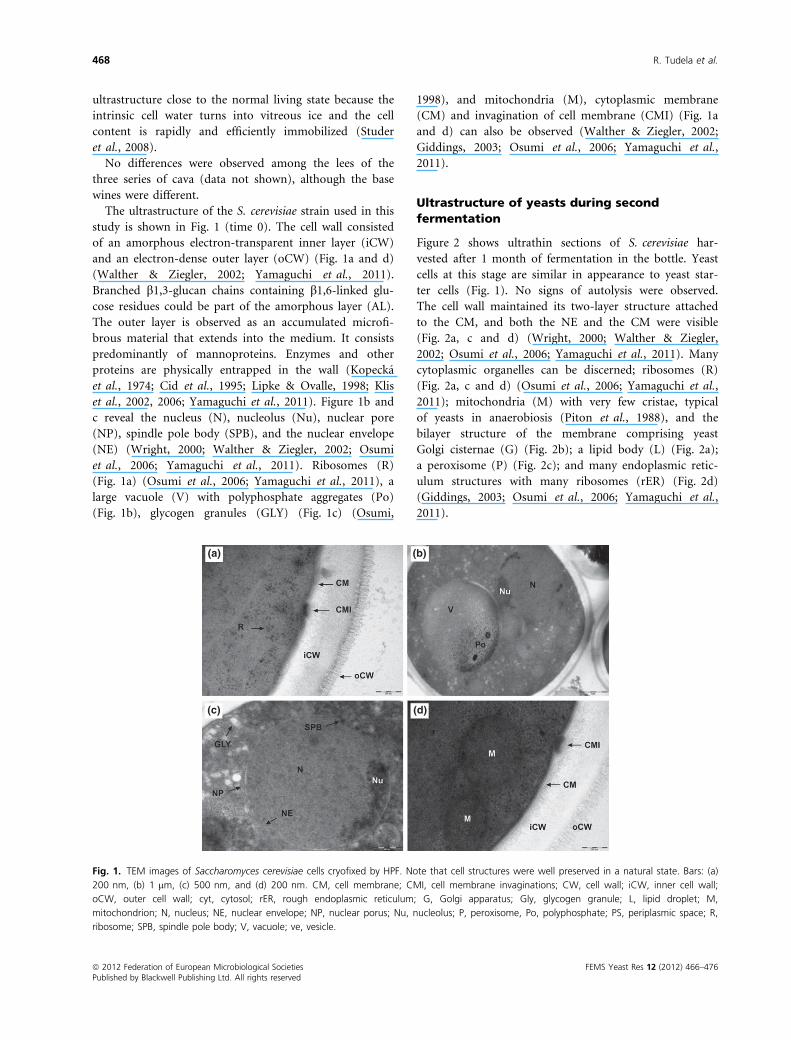
The ultrastructure of the S. cerevisiae strain used in this
study is shown in Fig. 1 (time 0). The cell wall consisted
of an amorphous electron-transparent inner layer (iCW)
and an electron-dense outer layer (oCW) (Fig. 1a and d)
(Walther & Ziegler, 2002; Yamaguchi et al., 2011).
Branched b1,3-glucan chains containing b1,6-linked glu-
cose residues could be part of the amorphous layer (AL).
The outer layer is observed as an accumulated microfi-
brous material that extends into the medium. It consists
predominantly of mannoproteins. Enzymes and other
proteins are physically entrapped in the wall (Kopecka
et al., 1974; Cid et al., 1995; Lipke & Ovalle, 1998; Klis
et al., 2002, 2006; Yamaguchi et al., 2011). Figure 1b and
c reveal the nucleus (N), nucleolus (Nu), nuclear pore
(NP), spindle pole body (SPB), and the nuclear envelope
(NE) (Wright, 2000; Walther & Ziegler, 2002; Osumi
et al., 2006; Yamaguchi et al., 2011). Ribosomes (R)
(Fig. 1a) (Osumi et al., 2006; Yamaguchi et al., 2011), a
large vacuole (V) with polyphosphate aggregates (Po)
(Fig. 1b), glycogen granules (GLY) (Fig. 1c) (Osumi,
1998), and mitochondria (M), cytoplasmic membrane
(CM) and invagination of cell membrane (CMI) (Fig. 1a
and d) can also be observed (Walther & Ziegler, 2002;
Giddings, 2003; Osumi et al., 2006; Yamaguchi et al.,
2011).
Ultrastructure of yeasts during second
fermentation
Figure 2 shows ultrathin sections of S. cerevisiae har-
vested after 1 month of fermentation in the bottle. Yeast
cells at this stage are similar in appearance to yeast star-
ter cells (Fig. 1). No signs of autolysis were observed.
The cell wall maintained its two-layer structure attached
to the CM, and both the NE and the CM were visible
(Fig. 2a, c and d) (Wright, 2000; Walther & Ziegler,
2002; Osumi et al., 2006; Yamaguchi et al., 2011). Many
cytoplasmic organelles can be discerned; ribosomes (R)
(Fig. 2a, c and d) (Osumi et al., 2006; Yamaguchi et al.,
2011); mitochondria (M) with very few cristae, typical
of yeasts in anaerobiosis (Piton et al., 1988), and the
bilayer structure of the membrane comprising yeast
Golgi cisternae (G) (Fig. 2b); a lipid body (L) (Fig. 2a);
a peroxisome (P) (Fig. 2c); and many endoplasmic retic-
ulum structures with many ribosomes (rER) (Fig. 2d)
(Giddings, 2003; Osumi et al., 2006; Yamaguchi et al.,
2011).
(a)
(c) (d)
(b)
Fig. 1. TEM images of Saccharomyces cerevisiae cells cryofixed by HPF. Note that cell structures were well preserved in a natural state. Bars: (a)
200 nm, (b) 1 lm, (c) 500 nm, and (d) 200 nm. CM, cell membrane; CMI, cell membrane invaginations; CW, cell wall; iCW, inner cell wall;
oCW, outer cell wall; cyt, cytosol; rER, rough endoplasmic reticulum; G, Golgi apparatus; Gly, glycogen granule; L, lipid droplet; M,
mitochondrion; N, nucleus; NE, nuclear envelope; NP, nuclear porus; Nu, nucleolus; P, peroxisome, Po, polyphosphate; PS, periplasmic space; R,
ribosome; SPB, spindle pole body; V, vacuole; ve, vesicle.
ª 2012 Federation of European Microbiological Societies FEMS Yeast Res 12 (2012) 466–476Published by Blackwell Publishing Ltd. All rights reserved
468 R. Tudela et al.

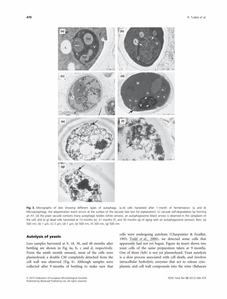
Autophagy of yeasts
Evidence of microautophagy and macroautophagy was
detected in sparkling wine lees (Fig. 3). In cells harvested
at 1 month of bottling, we were able to observe a large
vacuole (V) that formed arm-like extensions of its mem-
brane engulfing a vesicle (ves) (Fig. 3a) and lipid bodies
(L) (Fig. 3c). Fusion and concomitant sealing of this
growing membrane will result in the incorporation of the
vesicle and the lipid body into the vacuole. This micro-
autophagic process has been described for peroxisome
degradation in Pichia pastoris (Mijaljica et al., 2007; Kraft
et al., 2009). Figure 3a also showed a nonselective auto-
phagy process by which bulk cytosol (cyt) was taken up
by tubular invaginations of the vacuole. Lipid bodies (L)
were also assimilated by arm-like protrusions (Fig. 3b,
arrows) of the vacuole surface. A similar microautophagic
process was postulated by Kraft et al. (2009) to take up
large cytosol components in starved S. cerevisiae cells in
laboratory conditions. Figure 3c shows a yeast cell whose
vacuole has formed an autophagic tube (AT). Vacuolar
invaginations in nitrogen-starved S. cerevisiae cells were
visualized with the viability dye FM 4-64 under a confocal
laser scanning microscope by Mijaljica et al. (2007).
These authors observed that membrane vacuolar invagin-
ations formed AT that disappeared within one hour of
labeling with FM 4-64, leaving a diffuse stain in the vacu-
ole. They suggested that an ‘inverse-budding’ reaction of
the AT maybe related to the regulation of vacuolar
membrane homeostasis. We propose that the vacuole of
the cell shown in Fig. 3c was undergoing a similar micro-
autophagic process to control its own homeostasis. To
our knowledge we showed here, for the first time, mor-
phological evidence of industrial S. cerevisiae undergoing
microautophagy during real sparkling wine aging.
An autophagosome near the vacuole (Fig. 3d, black
arrow) and autophagic bodies were observed inside the
vacuoles of S. cerevisiae cells (Fig. 3d, white arrows), con-
firming the findings of Cebollero & Gonzalez (2006) that
macroautophagy was also induced during the second fer-
mentation of wine in a closed bottle. Samples taken after
1 month of bottling were filled with living cells that were
undergoing autophagy but not yet autolysis. Our results
agree well with those reported by Cebollero & Gonzalez
(2006) that autophagy takes place before autolysis. The
detection of a single and reminiscent autophagosome in
the cytoplasm of cells at 13, 21, and 30 months of aging
(Fig. 3e, f and g, respectively) could be owing to the
resistance of bilayered membranes to degradation (see
Evolution of cell cytoplasm lees).
After 1 month of bottled fermentation, S. cerevisiae
had reached the stationary phase (data provided by Fre-
ixenet S.A.), and the growth arrest is the consequence of
depletion of nutrients and high levels of ethanol (Bauer &
Pretorius, 2000). At this stage, different autophagy
processes might be induced to control cellular homeosta-
sis and to contribute to the outcome of autolysis in
enological conditions.
(a) (b)
(c) (d)
Fig. 2. Ultrathin sections at high magnification of Saccharomyces cerevisiae lees after 1 month of bottling. Bilayered structure of cytoplasmic
(CM), nuclear envelope (NE), mitochondrion (M), and Golgi (G) membranes is well distinguished. Bars: (a and b) 500 nm, (c and d) 200 nm.
FEMS Yeast Res 12 (2012) 466–476 ª 2012 Federation of European Microbiological SocietiesPublished by Blackwell Publishing Ltd. All rights reserved
Ultrastructural changes of sparkling wine lees during aging 469
Autolysis of yeasts
Lees samples harvested at 9, 18, 30, and 48 months after
bottling are shown in Fig. 4a, b, c and d, respectively.
From the ninth month onward, most of the cells were
plasmolysed; a double CM completely detached from the
cell wall was observed (Fig. 4). Although samples were
collected after 9 months of bottling to make sure that
cells were undergoing autolysis (Charpentier & Feuillat,
1993; Todd et al., 2000), we detected some cells that
apparently had not yet begun. Figure 4a insert shows two
yeast cells of the same preparation taken at 9 months.
One of them (left) is not yet plasmolysed. Yeast autolysis
is a slow process associated with cell death, and involves
intracellular hydrolytic enzymes that act to release cyto-
plasmic and cell wall compounds into the wine (Babayan
(a)
(c)
(e)
(g)
(f)
(d)
(b)
Fig. 3. Micrographs of lees showing different types of autophagy. (a–d) cells harvested after 1 month of fermentation. (a and b)
Microautophagy: the sequestration event occurs at the surface of the vacuole (see text for explanation); (c) vacuole self-degradation by forming
an AT; (d) the yeast vacuole contains many autophagic bodies (white arrows), an autophagosome (black arrow) is observed in the cytoplasm of
the cell; and (e–g) dead cells harvested at 13 months (e), 21 months (f), and 30 months (g) of aging with an autophagosome (arrows). Bars: (a)
500 nm, (b) 1 lm, (c) 2 lm, (d) 1 lm, (e) 500 nm, (f) 200 nm, (g) 500 nm.
ª 2012 Federation of European Microbiological Societies FEMS Yeast Res 12 (2012) 466–476Published by Blackwell Publishing Ltd. All rights reserved
470 R. Tudela et al.
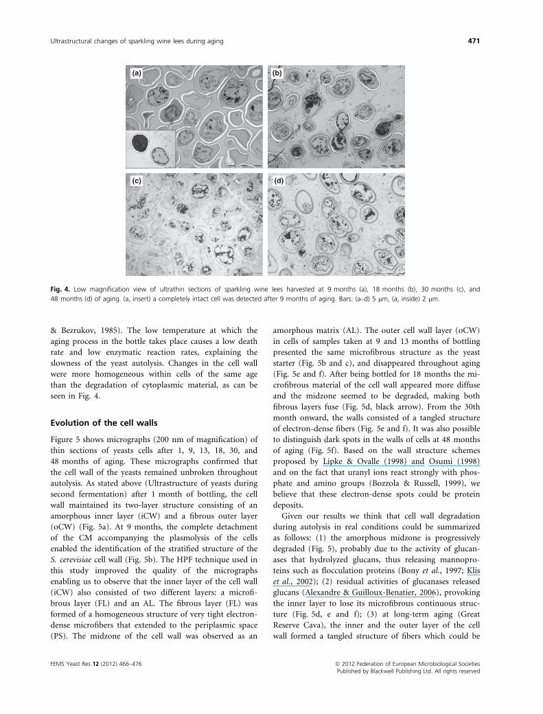
& Bezrukov, 1985). The low temperature at which the
aging process in the bottle takes place causes a low death
rate and low enzymatic reaction rates, explaining the
slowness of the yeast autolysis. Changes in the cell wall
were more homogeneous within cells of the same age
than the degradation of cytoplasmic material, as can be
seen in Fig. 4.
Evolution of the cell walls
Figure 5 shows micrographs (200 nm of magnification) of
thin sections of yeasts cells after 1, 9, 13, 18, 30, and
48 months of aging. These micrographs confirmed that
the cell wall of the yeasts remained unbroken throughout
autolysis. As stated above (Ultrastructure of yeasts during
second fermentation) after 1 month of bottling, the cell
wall maintained its two-layer structure consisting of an
amorphous inner layer (iCW) and a fibrous outer layer
(oCW) (Fig. 5a). At 9 months, the complete detachment
of the CM accompanying the plasmolysis of the cells
enabled the identification of the stratified structure of the
S. cerevisiae cell wall (Fig. 5b). The HPF technique used in
this study improved the quality of the micrographs
enabling us to observe that the inner layer of the cell wall
(iCW) also consisted of two different layers: a microfi-
brous layer (FL) and an AL. The fibrous layer (FL) was
formed of a homogeneous structure of very tight electron-
dense microfibers that extended to the periplasmic space
(PS). The midzone of the cell wall was observed as an
amorphous matrix (AL). The outer cell wall layer (oCW)
in cells of samples taken at 9 and 13 months of bottling
presented the same microfibrous structure as the yeast
starter (Fig. 5b and c), and disappeared throughout aging
(Fig. 5e and f). After being bottled for 18 months the mi-
crofibrous material of the cell wall appeared more diffuse
and the midzone seemed to be degraded, making both
fibrous layers fuse (Fig. 5d, black arrow). From the 30th
month onward, the walls consisted of a tangled structure
of electron-dense fibers (Fig. 5e and f). It was also possible
to distinguish dark spots in the walls of cells at 48 months
of aging (Fig. 5f). Based on the wall structure schemes
proposed by Lipke & Ovalle (1998) and Osumi (1998)
and on the fact that uranyl ions react strongly with phos-
phate and amino groups (Bozzola & Russell, 1999), we
believe that these electron-dense spots could be protein
deposits.
Given our results we think that cell wall degradation
during autolysis in real conditions could be summarized
as follows: (1) the amorphous midzone is progressively
degraded (Fig. 5), probably due to the activity of glucan-
ases that hydrolyzed glucans, thus releasing mannopro-
teins such as flocculation proteins (Bony et al., 1997; Klis
et al., 2002); (2) residual activities of glucanases released
glucans (Alexandre & Guilloux-Benatier, 2006), provoking
the inner layer to lose its microfibrous continuous struc-
ture (Fig. 5d, e and f); (3) at long-term aging (Great
Reserve Cava), the inner and the outer layer of the cell
wall formed a tangled structure of fibers which could be
(a)
(c) (d)
(b)
Fig. 4. Low magnification view of ultrathin sections of sparkling wine lees harvested at 9 months (a), 18 months (b), 30 months (c), and
48 months (d) of aging. (a, insert) a completely intact cell was detected after 9 months of aging. Bars: (a–d) 5 lm, (a, inside) 2 lm.
FEMS Yeast Res 12 (2012) 466–476 ª 2012 Federation of European Microbiological SocietiesPublished by Blackwell Publishing Ltd. All rights reserved
Ultrastructural changes of sparkling wine lees during aging 471
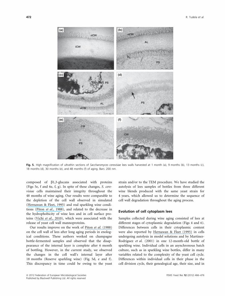
composed of b1,3-glucans associated with proteins
(Figs 5e, f and 6e, f, g). In spite of these changes, S. cere-
visiae cells maintained their integrity throughout the
48 months of wine aging. Our results were comparable to
the depletion of the cell wall observed in simulated
(Hernawan & Fleet, 1995) and real sparkling wine condi-
tions (Piton et al., 1988), and related to the decrease in
the hydrophobicity of wine lees and in cell surface pro-
teins (Vichi et al., 2010), which were associated with the
release of yeast cell wall mannoproteins.
Our results improve on the work of Piton et al. (1988)
on the cell wall of lees after long aging periods in enolog-
ical conditions. These authors worked on champagne
bottle-fermented samples and observed that the disap-
pearance of the internal layer is complete after 6 month
of bottling. However, in the current study, we observed
the changes in the cell wall’s internal layer after
18 months (Reserve sparkling wine) (Fig. 5d, e and f).
This discrepancy in time could be owing to the yeast
strain and/or to the TEM procedure. We have studied the
autolysis of lees samples of bottles from three different
wine blends produced with the same yeast strain for
4 years, which allowed us to determine the sequence of
cell wall degradation throughout the aging process.
Evolution of cell cytoplasm lees
Samples collected during wine aging consisted of lees at
different stages of cytoplasmic degradation (Figs 4 and 6).
Differences between cells in their cytoplasmic content
were also reported by Hernawan & Fleet (1995) in cells
undergoing autolysis in model solutions and by Martınez-
Rodrıguez et al. (2001) in one 12-month-old bottle of
sparkling wine. Individual cells in an asynchronous batch
culture, such as in sparkling wine bottles, differ in many
variables related to the complexity of the yeast cell cycle.
Differences within individual cells in their phase in the
cell division cycle, their genealogical age, their size, and in
(a)
(c)
(e) (f)
(d)
(b)
Fig. 5. High magnification of ultrathin sections of Saccharomyces cerevisiae lees walls harvested at 1 month (a), 9 months (b), 13 months (c),
18 months (d), 30 months (e), and 48 months (f) of aging. Bars: 200 nm.
ª 2012 Federation of European Microbiological Societies FEMS Yeast Res 12 (2012) 466–476Published by Blackwell Publishing Ltd. All rights reserved
472 R. Tudela et al.

clonal variability (Porro et al., 2009; Portell et al., 2011)
may yield the cell morphology distribution of the aging
lees. In addition, stationary-phase cultures of S. cerevisiae
include both quiescent and nonquiescent cells (Allen
et al., 2006), increasing the complexity of the yeast popu-
lation of lees. Thus, we were not able to establish a time
sequence of cytoplasmic autolysis during aging as we did
for cell wall degradation.
In general, owing to plasmolysis, the thickness of the
periplasmic space (PS) increased with time. Furthermore,
although the CM formed deep invaginations and some
breakages of the membrane were also observed, the dou-
ble-layered ultrastructure was preserved throughout aging
period (Fig. 6a and e). Although S. cerevisiae responds to
ethanol stress by increasing fatty acyl chain length and
the proportion of unsaturated fatty acids and sterols in
(a) (e)
(f)
(g)
(h)
(b)
(c)
(d)
Fig. 6. Ultrathin sections of Saccharomyces cerevisiae lees showing different stages of autolysis after 9 months (a–d) and 48 months (e–f) of
aging. Cells from the same sample showed different degrees of autolysis (see text for explanation). Bars: (a) 200 nm, (b) 1 lm, (c) 500 nm, (d)
200 nm, (e) 200 nm, (f) 1 lm, (g) 1 lm, (h) 100 nm.
FEMS Yeast Res 12 (2012) 466–476 ª 2012 Federation of European Microbiological SocietiesPublished by Blackwell Publishing Ltd. All rights reserved
Ultrastructural changes of sparkling wine lees during aging 473

the plasma membrane (Bauer & Pretorius, 2000; Walker
& Van Dijck, 2006), one of the major consequences of
yeast cell exposure to high levels of ethanol is the
disruption of membrane structural integrity (Walker &
Van Dijck, 2006). This is in agreement with our observa-
tion of plasma membrane (CM) invaginations and break-
ages after 9 months of aging (Fig. 6a and e dashed
arrows). It has been reported that plasma membrane deg-
radation starts after 3 to 6 months, with lipids being
released into the medium (Alexandre & Guilloux-Bena-
tier, 2006), but little is known about the fate of the
plasma membrane during autolysis in sparkling wine
aging on lees. Experiments in a model wine system
showed that no phospholipids were released into the
medium (Hernawan & Fleet, 1995; Pueyo et al., 2000),
which could suggest that the plasma membrane is
degraded inside the cell. Fragments of double-layered
membranes from the plasma membrane or from organ-
elles such as mitochondria, the nucleus, or autophago-
somes were observed in the cytoplasm of the cell (Fig. 6c,
dashed arrows). Our micrographs may indicate that the
first step of plasma membrane degradation during aging
on lees is the production of small fragments that remain
inside the cell (Fig. 6a). These fragments of bilayered
membranes would then be digested.
Autolysis was accompanied by extensive loss and disor-
ganization of the intracellular contents (Figs 4 and 6)
(Hernawan & Fleet, 1995; Martınez-Rodrıguez et al.,
2001). Nonelectron-dense lipid droplets could be observed
in lees from 9 months (Fig. 6b) to 48 months (Fig. 6f) of
wine aging. Degradation of the cytoplasm content was
already observed at 9 months, as vesicles with different
material could be detected (Fig. 6c, solid arrows) and
ribosomes (R) were present in the periplasmic space
(Fig. 6d). However, the nucleus and mitochondria were
observed in some cells 48 months old (Fig. 6g). Figure 6h
(arrow) shows some double-membrane organelles, but
owing to the degradation process it was not possible to
discern if they belonged to the Golgi system or the endo-
plasmic reticulum (Giddings, 2003; Osumi et al., 2006;
Yamaguchi et al., 2011). Our results showed that cytoplas-
mic degradation was a process that extended for at least
4 years. Cell depletion, nevertheless, was not complete
after 48 months in the bottle (Figs 4d and 6f, g). These is
in agreement with the results reported by Leroy et al.
(1990) and Moreno-Arribas et al. (1998) that there is still
autolytic activity in yeasts after 60 and 31 months of aging
in wine, respectively. These authors based their conclu-
sions on the analysis of the products released into the
wine. In addition, a great increase of intracellular proteo-
lytic activity was reported in sparkling wine (Feuillat &
Charpentier, 1982) and in champagne (Leroy et al., 1990)
during the aging process. Moreover, an increase in the
concentration of nucleic acid materials (Leroy et al., 1990)
and in the concentration of the most representative nucle-
otides (Zhao & Fleet, 2003, 2005; Charpentier et al., 2005)
was observed in champagnes, which could indicate the
degradation of organelles such as the nucleus, mitochon-
dria, and ribosomes, and subsequent degradation to nucle-
otides (Fig. 6). Our TEM images may agree with
analytical data obtained by several authors, among others
Leroy et al. (1990), Charpentier et al. (2005), and Moreno-
Arribas et al. (1996), which described compositional
changes in sparkling wine during aging.
This is the first time that high-resolution TEM images
have been obtained, which provides evidence of several
morphological changes in the ultrastructure of S. cerevisi-
ae lees throughout real long-term aging. Most of the
studies on yeast autolysis have focused on the chemical
analysis of wine, but few of them have studied the cyto-
logical changes of yeasts during aging. Many of the
changes observed in our work could explain the evolution
of the compositional and sensorial characteristics of spar-
kling wines reported by previous authors. Finally, our
results suggest that autophagy is involved in the autolytic
process. Moreover, different types of autophagy were
observed in lees in real enological conditions. Further
research should be conducted to understand the role of
microautophagy and macroautophagy in the autolysis of
lees in sparkling wine during aging.
Acknowledgements
This study was supported by the Spanish Ministerio de
Ciencia y Tecnologıa, project AGL2008-03392, by the
Generalitat de Catalunya, project SGR2009-606, and by
XaRTA (Xarxa de Referencia en Tecnologia dels Aliments;
ajuts de valoritzacio de la recerca), and through a MEC
grant to the PhD student J.J. G-C. We are grateful to
Freixenet S.A. wineries for sampling. We appreciate
Carmen Lopez-Iglesias laboratory assistance in TEM
preparations at the Scientific Technical Center at the
University of Barcelona.
References
Alexandre H & Guilloux-Benatier M (2006) Yeast autolysis in
sparkling wine – a review. Aust J Grape Wine Res 12: 119–127.Alexandre H, Heintz D, Chassagne D, Guilloux-Benatier M,
Charpentier C & Feuillat M (2001) Protease A activity and
nitrogen fractions during alcoholic fermentation and
autolysis in enological conditions. J Ind Microbiol Biotechnol.
26: 235–240.Allen C, Buttner S, Aragon AD et al. (2006) Isolation of
quiescent and nonquiescent cells from yeast stationary-phase
cultures. J Cell Biol 174: 89–100.
ª 2012 Federation of European Microbiological Societies FEMS Yeast Res 12 (2012) 466–476Published by Blackwell Publishing Ltd. All rights reserved
474 R. Tudela et al.
Babayan TL & Bezrukov MG (1985) Autolysis in yeast. Acta
Biotechnol 5: 129–136.Bauer FF & Pretorius IS (2000) Yeast stress response and
fermentation efficiency: how to survive the making of wine
– a review. S Afr J Enol Vitic 21: 27–51.Bony M, Thines-Sempoux D, Barre P & Blondin B (1997)
Localization and cell surface anchoring of the Saccharomyces
cerevisiae flocculation protein Flo1p. J Bacteriol 179: 4929–4936.
Bozzola JJ & Russell LD (1999) Specimen staining and contrast
methods for transmission electron microscopy. Electron
Microscopy: principles and techniques for biologists. The Jones
and Bartlett series in biology, pp. 120–147. Jones & Bartlett
Publishers, Boston, MA.
Cebollero E & Gonzalez R (2006) Induction of autophagy by
second-fermentation yeasts during elaboration of sparkling
wines. Appl Environ Microbiol 72: 4121–4127.Charpentier C (2010) Ageing on lees (sur lies) and the use of
speciality inactive yeasts during wine fermentation.
Managing Wine Quality: Oenology and Wine Quality, Vol. 2
(Reynolds AG, ed), pp. 164–187. Whoodhead Publishing
Limited, Cambridge, UK.
Charpentier C & Feuillat M (1993) Yeast autolysis. Wine
Microbiology and Biotechnology (Fleet GH, ed), pp. 225–242.Harwood Academic Publishers, Chur, Switzerland.
Charpentier C, Aussenac J, Charpentier M, Prome J-C,
Duteurtre B & Feuillat M (2005) Release of nucleotides and
nucleosides during yeast autolysis: kinetics and potential
impact on flavor. J Agric Food Chem 53: 3000–3007.Cid VJ, Duran A, Del Rey F, Snyder MP, Nombela C &
Sanchez M (1995) Molecular basis of cell integrity and
morphogenesis in Saccharomyces cerevisiae. Microbiol Rev 59:
345–386.Feuillat M & Charpentier C (1982) Autolysis of yeasts in
champagne. Am J Enol Viticult 33: 6–13.Flanzy C, Salgues M, Bidan P, Dubois C, Moulin JP &
Sablayrolles JM (1999) Sparkling wine vinification.
Oenology: Fondements scientifiques et technologiques (Flanzy
C, ed), pp. 497–516. Technique et Documantation, Paris,
France.
Fornairon-Bonnefond C, Camarasa C, Moutounet M &
Salmon JM (2002) New trends on yeast autolysis and wine
ageing on lees: a bibliographic review. J Int Sci Vigne Vin
36: 49–69.Gallart M, Lopez-Tamames E, Suberbiola G & Buxaderas S
(2002) Influence of fatty acids on wine foaming. J Agric
Food Chem 50: 7042–7045.Giddings TH (2003) Freeze-substitution protocols for
improved visualization of membranes in high-pressure
frozen samples. J Microsc 212: 53–61.Hernawan T & Fleet G (1995) Chemical and cytological
changes during the autolysis of yeasts. J Ind Microbiol 14:
440–450.Klis FM, Mol P, Hellingwerf K & Brul S (2002) Dynamics of
cell wall structure in Saccharomyces cerevisiae. FEMS
Microbiol Rev 26: 239–256.
Klis FM, Boorsma A & De Groot PWJ (2006) Cell wall
construction in Saccharomyces cerevisiae. Yeast 2: 185–202.Kopecka M, Phaff HJ & Fleet GH (1974) Demonstration of a
fibrillar component in the cell wall of the yeast
Saccharomyces cerevisiae and its chemical nature. J Cell Biol
62: 66–76.Kraft C, Reggiori F & Peter M (2009) Selective types of
autophagy in yeasts. Biochim Biophys Acta 1793: 1404–1412.Leroy MJ, Charpentier M, Duteurtre B, Feuillat M &
Charpentier C (1990) Yeast autolysis during champagne
aging. Am J Enol Viticult 41: 21–28.Lipke PN & Ovalle R (1998) Cell wall architecture in yeast:
new structure and new challenges. J Bacteriol 180: 3735–3740.
Martınez-Rodriguez AJ & Pueyo E (2009) Sparkling wines and
yeast autolysis. Wine Chemistry and Biochemistry (Moreno-
Arribas MV & Polo MC, eds), pp. 61–80. Springer,Heidelberg.
Martınez-Rodrıguez AJ, Polo MC & Carrascosa AV (2001)
Structural and ultrastructural changes in yeast cells during
autolysis in a model wine system and in sparkling wines. Int
J Food Microbiol 71: 45–51.Mijaljica D, Prescot M, Klionsky DJ & Devenish RJ (2007)
Autophagy and vacuole homeostasis: a case for self-
degradation? Autophagy 3: 417–421.Moreno-Arribas V, Pueyo E & Polo MC (1996) Peptides in
musts and wines. Changes during the manufacture of cavas
(sparkling wines). J Agric Food Chem 44: 3783–3788.Moreno-Arribas V, Pueyo E, Polo MC & Martınez-Alvarez PJ
(1998) Changes in the amino acid composition of the
different nitrogenous fractions during the aging of wine
with yeasts. J Agric Food Chem 46: 4042–4051.Nevot M, Deroncele V, Lopez-Iglesias C, Bozal N, Guinea J &
Mercade E (2006) Ultrastructural analisis of the extracellular
matter secreted by the psychrotolerant bacterium
Pseudoalteromonas antarctica NF3. Microb Ecol 51: 501–507.Osumi M (1998) The ultrastructure of yeast: cell wall structure
and formation. Micron 29: 207–233.Osumi M, Konomi M, Sugawara T, Takagi T & Baba M
(2006) High-pressure freezing is a powerful tool for
visualization of Schizosaccharomyces pombe cells: ultra-low
temperature and low-voltage scanning electron microscopy
and immunoelectron microscopy. J Electron Microsc 55: 75–88.
Perez-Serradilla J & Luque de Castro MD (2008) Role of lees
in wine production: a review. Food Chem 111: 447–456.Piton F, Charpentier M & Troton D (1988) Cell wall and lipid
changes in Saccharomyces cerevisiae during aging of
champagne wine. Am J Enol Viticult 39: 221–226.Porro D, Vai M, Vanoni M, Alberghina L & Hatzis C (2009)
Analysis and modeling of growing budding yeast
populations at the single cell level. Cytometry A 75A: 114–120.
Portell X, Ginovart M, Carvo R, Gras A & Vives-Rego J (2011)
Population analysis of a commercial Saccharomyces cerevisiae
wine yeast in a batch culture by electric particle analysis,
FEMS Yeast Res 12 (2012) 466–476 ª 2012 Federation of European Microbiological SocietiesPublished by Blackwell Publishing Ltd. All rights reserved
Ultrastructural changes of sparkling wine lees during aging 475

light diffraction and flow cytometry. FEMS Yeast Res 11:
18–28.Pozo-Bayon MA, Andujar-Ortiz I & Moreno-Arribas MV
(2009a) Scientific evidences beyond the application of
inactive dry yeast preparations in winemaking. Food Res Int
42: 754–761.Pozo-Bayon MA, Andujar-Ortiz I & Moreno-Arribas MV
(2009b) Volatile profile and potential of inactive dry yeast-
based winemaking additives to modify the volatile
composition of wines. J Sci Food Agric 89: 1665–1673.Pueyo E, Martınez-Rodrıguez A, Polo MC, Santa-Marıa G &
Bartolome B (2000) Release of lipids during yeast autolysis
in a model wine system. J Agric Food Chem 48: 116–122.Studer D, Humbel BM & Chiquet M (2008) Electron
microscopy of high pressure frozen samples: bridging the
gap between cellular ultrastructure and atomic resolution.
Histochem Cell Biol 130: 877–889.Todd BEN, Fleet GH & Henschke PA (2000) Promotion of
autolysis through the interaction of killer and sensitive
yeasts: potential application in sparkling wine production.
Am J Enol Viticult 51: 65–72.Torrens J, Riu-Aumatell M, Vichi S, Lopez-Tamames E &
Buxaderas S (2010) Assessment of volatile and sensory
profiles between base and sparkling wines. J Agric Food
Chem 58: 2455–2461.Vichi S, Gallardo-Chacon JJ, Pradelles R, Chassagne D, Lopez-
Tamames E & Buxaderas S (2010) Surface properties of
Saccharomyces cerevisiae lees during sparkling wine ageing
and their effect on flocculation. Int J Food Microbiol 140:
125–130.Walker GM & Van Dijck P (2006) Physiological and molecular
responses of yeasts to the environment. Yeasts in Food and
Beverages. The Yeast Handbook, vol. 2 (Querol A & Fleet
GH, eds), pp. 111–152. Springer-Verlag, Heidelberg.
Walther P & Ziegler A (2002) Freeze substitution of high-
pressure frozen samples: the visibility of biological
membranes is improved when substitution medium
contains water. J Microsc 208: 3–10.Wright R (2000) Transmission electron microscopy of yeast.
Microsc ResTech 51: 496–510.Yamaguchi M, Namiki Y, Okada H, Mori Y, Furukawa H,
Wang J, Ohkusu M & Kawamoto S (2011) Structome of
Saccharomyces cerevisiae determined by freeze-substitution
and serial ultrathin-sectioning electron microscopy.
J Electron Microsc 60: 337–351.Zhao J & Fleet GH (2003) Degradation of DNA during the
autolysis of Saccharomyces cerevisiae. J Ind Microbiol
Biotechnol 30: 175–182.Zhao J & Fleet GH (2005) Degradation of RNA during the
autolysis of Saccharomyces cerevisiae produces
predominantly ribonucleotides. J Ind Microbiol Biotechnol
32: 415–423.
ª 2012 Federation of European Microbiological Societies FEMS Yeast Res 12 (2012) 466–476Published by Blackwell Publishing Ltd. All rights reserved
476 R. Tudela et al.