Calcium dysregulation in heart diseases: Targeting calcium ...

Tumour necrosis factor alpha inhibits purinergic calcium signalling
in blood–brain barrier endothelial cells
Wouter Vandamme, Katleen Braet, Liesbet Cabooter and Luc Leybaert
Department of Physiology and Pathophysiology, Ghent University, Ghent, Belgium
Abstract
The breaching of the blood–brain barrier is an essential aspect
in the pathogenesis of neuroinflammatory diseases, in which
tumour necrosis factor alpha (TNF-a) as well as endothelial
calcium ions play a key role. We investigated whether TNF-a
could influence the communication of calcium signals between
brain endothelial cells (GP8 and RBE4). Intercellular calcium
waves triggered by mechanical stimulation or photoliberation
of InsP3 in single cells were significantly reduced in size after
TNF-a exposure (1000 U/mL, 2 and 24 h). Calcium signals
are communicated between cells by means of gap junctional
and paracrine purinergic signalling. TNF-a significantly inhib-
ited gap junctional coupling, stimulated the basal release of
ATP, and dose-dependently blocked the triggered component
of ATP release. The cytokine displayed similar effects on the
uptake of a fluorescent reporter dye into the cells. Previous
work with connexin mimetic peptides demonstrated that the
triggered ATP release in these cells is connexin-related; these
peptides did, however, not influence the elevated basal ATP
release caused by TNF-a. We conclude that TNF-a depresses
calcium signal communication in blood–brain barrier endot-
helial cells, by reducing gap junctional coupling and by inhib-
iting triggered ATP release. The cytokine thus inhibits
connexin-related communication pathways like gap junctions
and connexin hemichannels.
Keywords: ATP release, blood–brain barrier, calcium waves,
connexins, cytokines.
J. Neurochem. (2004) 88, 411–421.
The brain is composed of neurones and glial cells and, while
neurones are electrically excitable cells, glial cells like
astrocytes form a calcium excitable cell population, i.e. they
respond to certain stimuli with a calcium response that is
communicated to surrounding cells as a calcium wave
(Scemes 2000). Brain endothelial cells are in close contact
with astrocytes at the blood–brain barrier, and these cells
communicate calcium signals between each other in a
homotypic and heterotypic manner (Leybaert et al. 1998;
Braet et al. 2001). Calcium signals between astrocytes and
endothelial cells form a bi-directional communication path-
way, transferring blood-borne signals to the neuropil, thereby
possibly affecting synaptic function (Haydon 2001), and
carrying astrocytic calcium signals to the blood–brain barrier,
possibly modulating its transport and barrier functions (Braet
and Leybaert 2000).
The breaching of the blood–brain barrier, i.e. the loss of
interendothelial tight junctions, is an essential aspect in the
pathogenesis of neuroinflammatory diseases like multiple
sclerosis and AIDS dementia (Petito and Cash 1992; Poser
1993). There is now ample evidence that cytokines like
tumour necrosis factor alpha (TNF-a), interleukin-1-beta
(IL1-b) and interferon gamma (IFN-c) released from
lymphocytes, macrophages and many other cell types, play
a key role in the opening of the barrier (de Vries et al. 1996;
Anthony et al. 1997; Munoz-Fernandez and Fresno 1998).
These cytokines stimulate the expression of the cell adhesion
molecules intercellular adhesion molecule-1 (ICAM-1) and
vascular cell adhesion molecule-1 (VCAM-1) on brain
endothelium, thereby facilitating lymphocyte adherence.
The subsequent lymphocyte–endothelial interaction activates
diverse signalling cascades: a protein kinase C (PKC) and
tyrosine kinase pathway, acting at occludins and the zonula
occludens-1 (ZO-1) accessory protein to disrupt tight junc-
tions, and a Rho based pathway activating the actin
cytoskeleton and tearing the tight junction proteins away
from the cell–cell interface (Pfau et al. 1995; Bolton et al.
Received June 17, 2003; revised manuscript received August 21, 2003;
accepted September 25, 2003.
Address correspondence and reprint requests to Luc Leybaert,
Department Physiology and Pathophysiology, Ghent University, De
Pintelaan 185 (Block B, Rm 306), B-9000 Ghent, Belgium.
E-mail: [email protected]
Abbreviations used: InsP3, inositol trisphosphate; TNF-a, tumour
necrosis factor alpha; IL1-b, interleukin-1-beta; IFN-c, interferon gam-
ma; PKC, protein kinase C; PMA, phorbol 12-myristate 13-acetate.
Journal of Neurochemistry, 2004, 88, 411–421 doi:10.1046/j.1471-4159.2003.02163.x
� 2003 International Society for Neurochemistry, J. Neurochem. (2004) 88, 411–421 411

1998; Friedrich et al. 2002). An increase in endothelial
cytoplasmic calcium concentration is, in addition to this,
central to the opening of the blood–brain barrier (Abbott
2000; Etienne-Manneville et al. 2000; Brown and Davis
2002; Wolburg and Lippoldt 2002). We hypothesize that
calcium signals communicated between endothelial cells may
act to spatially spread and thus amplify the calcium-induced
opening of the blood–brain barrier. In the present study we
asked the question whether the cytokine TNF-a influences
the communication of calcium signals between endothelial
cells, thereby affecting the postulated spatial spread of
blood–brain barrier opening.
Our work shows that the cytokine depresses endothelial
calcium signal communication by inhibiting gap junctional
coupling and by blocking the triggered component of ATP
release. TNF-a thus seems to limit the spread of calcium
signals at the blood–brain barrier by various mechanisms.
Because the cytokine affects the first and most essential step
of the paracrine ATP signalling pathway, i.e. the phase of
ATP release, the TNF-a actions are expected not to be
limited to endothelial cell communication but may also
influence paracrine communication to other cell types in the
neighbourhood such as blood cells, astrocytes and smooth
muscle cells. Finally, TNF-a appears to affect two connexin-
related pathways in a parallel manner: it reduces gap
junctional communication (connexin channels) and blocks
triggered ATP release, which in previous work was demon-
strated to be related to the opening of connexin hemichannels
(Braet et al. 2003a; Braet et al. 2003b) (connexin hemichan-
nels are reviewed in Goodenough and Paul 2003).
Materials and methods
Materials
Fluo-3 acetoxymethyl ester (fluo-3 AM), D-myo-inositol 1,4,5-
trisphosphate, P4(5)-1-(2-nitrophenyl)ethyl ester trisodium salt
(NPE-caged InsP3), DMNB-caged fluorescein dextran (MW
3000), propidium iodide (MW 668), 6-carboxyfluorescein (MW
376), dextran tetramethylrhodamine (MW 10 000) and dextran
fluorescein (MW 70 000) were obtained from Molecular Probes
(Leiden, the Netherlands). Tumour necrosis factor alpha (TNF-a),
the ATP assay kit, phorbol 12-myristate 13-acetate (PMA) and
adenosine 5¢-trisphosphate disodium salt (ATP) were from Sigma
(Bornem, Belgium). The peptide GAP 26 (VCYDKSFPISHVR)
was a kind gift of Professor W.H. Evans (Department of Medical
Biochemistry, University of Wales College of Medicine, Cardiff,
UK). It was synthesized by solid phase chemistry and purified by
HPLC and purity (95%) assessed by HPLC.
Cell cultures
In this study we used two rat brain endothelial cell lines: RBE4
(a kind gift of Dr F. Roux, Neurotech SA, Evry, France) (Roux et al.
1994) and GP8 (GP8/3.9 (Greenwood et al. 1996)). Culture media
(all from Gibco, Merelbeke, Belgium) were alpha-MEM/Ham’s F10
(1 : 1) with 10% fetal bovine serum, 1 lg/mL bFGF (Boehringer
Mannheim, Brussels, Belgium) and 0.3 mg/mL geneticin (Gibco,
Merelbeke, Belgium) for RBE4 and GP8. Cells were grown on
glass-bottomed Petri dishes (MatTek Corporation, Ashwood, MA,
USA) coated with collagen type I (Boehringer Mannheim, Brussels,
Belgium) and used for experiments upon confluency. TNF-a was
added to the culture medium and applied to the cells incubated under
culture conditions (humidified 5% CO2) for the time periods used
(2 h or 24 h). Matched control cultures were treated with culture
medium not containing TNF-a in the same manner as the ones that
received the cytokine.
Calcium imaging
Cytoplasmic free calcium was measured using the calcium-sensitive
dye fluo-3 in combination with epifluorescence video microscopy
and digital imaging. Cell cultures were loaded with fluo-3 for 1 h at
room temperature in Hanks’ balanced salt solution buffered with
25 mM HEPES (HBSS-HEPES, pH 7.4) containing 10 lM fluo-3
AM (Molecular Probes, Leiden, the Netherlands) and 1 mM
probenecid. Cultures were then washed with HBSS-HEPES and
left at room temperature for 30 min for de-esterification. HBSS-
HEPES was the bathing solution for all calcium imaging experi-
ments. Cells were viewed with an inverted epifluorescence
microscope (Nikon Eclipse TE300, Analis, Ghent, Belgium) using
a · 40 oil immersion lens (CFI Plan Fluor, Nikon). Fluo-3
fluorescence images were obtained by excitation at 485 nm,
reflection off a dichroic mirror with cut-off at 510 nm, and emission
bandpass filtering at 535 nm (485DF22, 505DRLPXR and 535DF35
filters, respectively, from Omega Optical, Brattleboro, VT, USA).
Images were captured using an intensified CCD (Extended Isis
camera, Photonic Science, East Sussex, UK) and stored on an S-
VHS video recorder (Panasonic, Avicom, De Pinte, Belgium) or
directly to a PC equipped with an image acquisition and processing
board (DT3155, Data Translation, Marlboro, MA, USA). Each
experiment was concluded by recording background images in a
Petri dish containing only medium and no cells; background images
were subtracted from the fluo-3 fluorescence image sequences.
Loading of caged InsP3 by electroporation and UV spot
illumination
The cells were loaded with caged InsP3 by electroporation. Cultures
were briefly rinsed with electroporation buffer (300 mM sorbitol,
4.2 mM KH2PO4, 10.8 mM K2HPO4, 1 mM MgCl2 and 2 mM
HEPES, pH 7.20) and thereafter a small volume (5 lL) of caged
InsP3 (200 lM) in electroporation buffer was added. This solution
contained in addition dextran rhodamine (100 lM) to visualize the
electroporation zone. Electroporation was done on the stage of an
inverted microscope with a parallel wire electrode positioned close
to the cell surface, as described in detail in Braet et al. (2003a). The
zone of caged InsP3 loaded cells was estimated to be approximately
1.3 mm wide and 10 mm long. UV spot illumination for photore-
lease in calcium imaging experiments was performed with a Hg-arc
lamp coupled to the microscope epifluorescence input, as described
before (Braet et al. 2003a) (Leybaert and Sanderson 2001). The UV
spot had a half-energy diameter of 10 lm, as determined by flashing
a thin layer of DMNB-caged fluorescein dextran (MW 3000) at
3 mM mixed (1 : 1) with Dako Glycergel (Dako Corporation,
Carpinteria, CA, USA).
412 W. Vandamme et al.
� 2003 International Society for Neurochemistry, J. Neurochem. (2004) 88, 411–421

Mechanical cell stimulation
Mechanical stimulation of a single cell was performed by gentle
stimulation with a glass microneedle (2 lm tip size) mounted on a
piezo-electric device and driven by a single pulse of 100 ms
duration to produce a tip displacement of approximately 5 lm.
Apoptotic index
The apoptotic index was determined in cell cultures that were fixed
for 20 min in 4% formaldehyde and stained with DAPI (1/10 000)
for 5 min. The cells were washed three times with phosphate-
buffered saline after each step. The apoptotic nuclei were counted in
six frames each containing approximately 170 cells.
Scrape loading and dye-transfer
Gap junctional coupling was investigated with the scrape loading
and dye-transfer technique (Blomstrand et al. 1999; Opsahl and
Rivedal 2000). Confluent cultures of GP8 and RBE4 were placed in
a nominally calcium-free saline consisting of 137 mM NaCl,
5.36 mM KCl, 0.81 mM MgCl2, 5.55 mM D-glucose and 25 mM
HEPES (pH 7.4) containing 0.4 mM 6-carboxyfluorescein for
10 min before applying a linear scratch across the culture with a
syringe needle and left for another 10 min in the same solution. The
cultures were then washed three times with HBSS-HEPES and left
for 30 min to recover. The cells were viewed as described above and
images were processed with custom-made software to acquire a
fluorescence diffusion profile. This profile was fitted to a mono-
exponential function using a non-linear least square fitting in order
to obtain a spatial constant of lateral intercellular diffusion.
Extracellular ATP measurements
ATP release in response to photoliberation of InsP3 was determined
in GP8 and RBE4 cultures using an ATP bioluminescent luciferin/
luciferase assay kit (product no. FL-AA, Sigma, Bornem, Belgium).
The photolytic UV light in these experiments was applied as a field
illumination (not spot illumination) exposing multiple cells. To that
purpose, light from a Hg-arc lamp with quartz collector lens was
directed through a shutter, a 330-nm bandpass filter and a mirror
(10D510AL.2, Newport, Leuven, Belgium) to form an image of the
arc at the level of the cell culture. The total energy dose per unit of
surface area applied in UV field illumination experiments was
adjusted so that it was equal to the energy dose per unit of surface in
the spot illumination experiments, as described in detail in Braet
et al. (2003a). The UV exposure time for field illumination was 2 s
when determined in this way. The photolytic efficiency with this
exposure time was in the order of the quantum efficiency of caged
InsP3 (0.65), indicating that the entire amount of caged probe
present in the cultures was effectively photocleaved upon a single
field illumination exposure. All experimental groups received the
photolytic light. Immediately after UV exposure, 100 lL of the
supernatant was collected from the cell cultures and transferred to
100 lL ATP assay mix solution (used at fivefold dilution). Light
emission from the 200 lL mix was then measured with a custom
build luminometer consisting of a photomultiplier tube (9924B,
Thorn-Emi Electron Tubes, Middlesex, UK). Luminescence was
determined from the photomultiplier current that was registered with
custom-made computer software. ATP release was also measured in
response to short (2 min) exposure of the cells to zero extracellular
calcium conditions (calcium- and magnesium-free HBSS-HEPES
with 1 mM EGTA added). The cells received 200 lL of this solution
and after 2 min 100 lL of the supernatant was collected and
transferred to 100 lL ATP assay mix solution (used at fivefold
dilution).
Propidium iodide uptake
The zero extracellular calcium condition used for ATP release was
also employed to trigger propidium iodide uptake into the cells
(Arcuino et al. 2002; Braet et al. 2003b). Cell cultures received the
zero calcium solution containing 2 mM propidium iodide for 10 min
at room temperature, were washed three times with HBSS-HEPES,
and pictures were then acquired with a · 10 objective, TRITC
epifluorescence settings and a cooled CCD camera (SensiCam,
PCO, Kelheim, Germany). In each culture, 10 images were taken in
which average dye uptake was determined. Propidium iodide
positivity was quantified by calculating the number of pixels with
a fluorescence intensity above a threshold value. Comparison
between manually counted propidium iodide positive cells and
supra-threshold pixels showed a linear relation between both
parameters. The number of cells demonstrating zero calcium-
triggered propidium iodide uptake amounted to 13.0 ± 0.9% of the
cells (n ¼ 10). Separate control experiments showed that zero
calcium exposure caused, in addition to propidium iodide uptake,
also uptake of small dyes like 6-carboxyfluorescein (MW 376) while
excluding larger dyes like dextran tetramethylrhodamine 10 kDa or
dextran fluorescein 70 kDa.
Data analysis and statistics
Calcium changes were determined as relative fluo-3 fluorescence
changes, i.e. DF/F0 ¼ [F – F0]/F0, where F0 is the fluorescence
before stimulation and F the time-dependent fluorescence signal
after stimulation. The extent of cell-to-cell propagation of calcium
changes was determined from the size of the intercellular calcium
wave at its maximal state of extension and was quantified by
determining the surface area where DF/F0 was above a threshold of
50%. We determined the surface area rather than the wave radius
because it is a much more sensitive parameter to detect changes in
the extent of calcium signal propagation (changes are amplified
quadratically as compared with the radius). Average DF/F0 changes
for the whole calcium waves were determined as the average
calculated from all pixels that showed an above threshold change.
Curve fittings for dose–response relations were performed with non-
linear least square procedures available in the program Inplot. The
data are expressed as mean ± SEM with n denoting the number of
experiments. Statistical significance was tested using a t-test for
unpaired observations and using a p-value of less than 0.05.
Multiple groups were compared using variance analysis followed by
the Dunnett test for multiple comparisons to the control group or the
Student–Newman–Keuls’ test for comparison of all groups among
each other.
Results
TNF-a reduces the size of intercellular calcium waves
Cell-to-cell calcium signal communication was investigated
by triggering intercellular calcium waves in confluent
TNF-a and endothelial calcium signalling 413
� 2003 International Society for Neurochemistry, J. Neurochem. (2004) 88, 411–421

cultures of RBE4 and GP8 brain capillary endothelial cell
lines. Intercellular calcium waves were triggered either by
mechanical cell stimulation or by photoliberating InsP3 in a
single cell. Gentle mechanical stimulation of a single cell
triggered an intercellular propagating calcium wave in the
two cell lines. The calcium wave size, determined as the
surface area of cells showing an above threshold calcium
change (DF/F0 > 50%) at maximal wave extension, averaged
17 448 ± 1162 lm2 (n ¼ 23) in RBE4 cells and
30 774 ± 1254 lm2 (n ¼ 55) in GP8 (corresponding to a
wave radius of approximately 75 and 99 lm, respectively).
Treating the cultures with TNF-a for 2 h (1000 U/mL)
significantly reduced the size of intercellular calcium waves to
approximately 80–90% of their control size (Figs 1 and 2a).
Extending the TNF-a exposure to 24 h further reduced the
calcium wave size to approximately 60–75% of control.
Photoliberating InsP3 in a single cell triggered intercellular
calcium waves that were significantly smaller than waves
triggered by mechanical stimulation [5688 ± 159 lm2
(n ¼ 35) for RBE4 and 8944 ± 302 lm2 (n ¼ 57) for
GP8, p < 0.0001; wave radii corresponding to these areas are,
respectively, 42 and 53 lm]. Two-hour exposure to TNF-asignificantly reduced the calcium wave size to an extent
comparable with the observations in the mechanical
stimulation experiments, and 24 h TNF-a slightly reduced
it further (Fig. 2b). Pooling the data for the two cell lines and
for the two stimulation protocols showed that the calcium
wave following 24 h TNF-a was significantly smaller as
compared with the 2 h condition (relative wave size of
100.0 ± 1.8% under control conditions, 80.8 ± 3.0% with
2 h TNF-a and 70.0 ± 2.1% with 24 h TNF-a; all values
significantly different from each other with p at least smaller
than 0.01; n ¼ 124). TNF-a exposure had no significant
effect on the amplitude of the calcium changes associated
with the calcium wave (DF/F0 attained 93.4 ± 3.1% of a
control amplitude of 100.0 ± 3.0% following 2 h TNF-a and
97.1 ± 3.6% for 24-h treatment; pooled data for the two cell
lines and for the two stimulation protocols; n ¼ 124). TNF-ais known to trigger apoptosis in many cell types when
applied at a high dose (Peiretti et al. 1997), and to determine
whether the observed reduction in wave calcium wave size
was perhaps due to the induction of apoptosis, we counted
apoptotic nuclei in RBE4 cells. TNF-a did not affect the
apoptotic index in RBE4 cells (0.2 ± 0.1% under control
conditions and 0.3 ± 0.1% after 24 h TNF-a, n ¼ 4).
TNF-a reduces gap junctional coupling
Intercellular calcium waves in endothelial cells can be
communicated by an intracellular/gap junction dependent
pathway and an extracellular/purinergic pathway, and TNF-a
(a)
(b)
Fig. 1 Effect of TNF-a on intercellular calcium waves in representative
experiments performed on confluent GP8 cultures. (a) Calcium waves
triggered by mechanical stimulation of a single cell. (b) Calcium waves
triggered by photoliberation of InsP3 in a single cell. TNF-a (1000 U/ml)
reduced the extent of intercellular calcium wave propagation for both
stimulation types and for both short (2 h) and longer (24 h) exposure
times. The images depict relative fluo-3 fluorescence changes (DF/F0)
colour coded as indicated in the colour bar; sub-threshold pixels (DF/
F0 < 50 %) are given in black. The calcium waves are each shown at
their maximal state of extension. Note that the InsP3-triggered calcium
waves are smaller than the mechanically triggered waves. All calcium
waves shown were recorded in separate cell cultures. The white
calibration strip measures 100 lm.
(a)
(b)
Fig. 2 Averaged data summarizing the effect of TNF-a on the extent
of intercellular calcium wave propagation. (a) Effect on mechanically
triggered calcium waves. (b) Effect on InsP3-triggered calcium waves.
Two hour TNF-a treatment (1000 U/ml) significantly reduced the
extent of intercellular calcium wave propagation (calcium wave size) in
both RBE4 and GP8 cultures and this effect was more pronounced
after 24-h treatment. The calcium wave size was determined from the
surface area of cells showing an above threshold calcium change
(DF/F0 > 50 %) at maximal wave extension and is expressed as the
wave size relative to the control wave size. *p < 0.05; **p < 0.01 as
compared with control (Con); n ¼ 23–57.
414 W. Vandamme et al.
� 2003 International Society for Neurochemistry, J. Neurochem. (2004) 88, 411–421
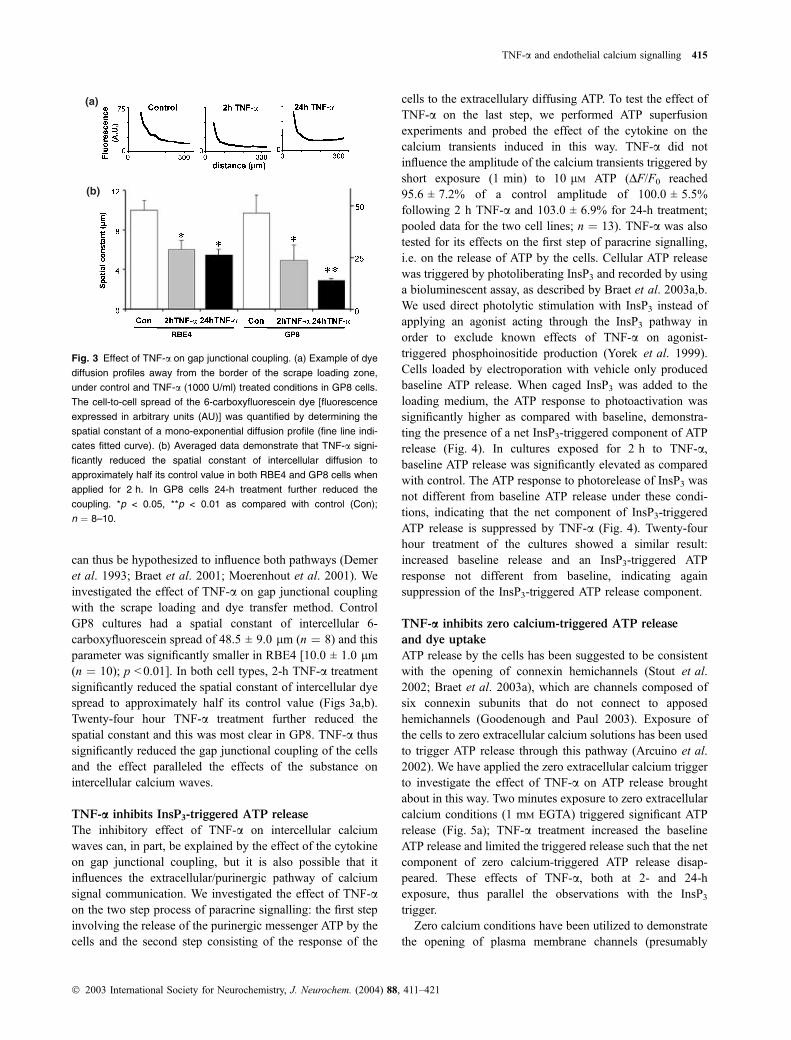
can thus be hypothesized to influence both pathways (Demer
et al. 1993; Braet et al. 2001; Moerenhout et al. 2001). We
investigated the effect of TNF-a on gap junctional coupling
with the scrape loading and dye transfer method. Control
GP8 cultures had a spatial constant of intercellular 6-
carboxyfluorescein spread of 48.5 ± 9.0 lm (n ¼ 8) and this
parameter was significantly smaller in RBE4 [10.0 ± 1.0 lm
(n ¼ 10); p < 0.01]. In both cell types, 2-h TNF-a treatment
significantly reduced the spatial constant of intercellular dye
spread to approximately half its control value (Figs 3a,b).
Twenty-four hour TNF-a treatment further reduced the
spatial constant and this was most clear in GP8. TNF-a thus
significantly reduced the gap junctional coupling of the cells
and the effect paralleled the effects of the substance on
intercellular calcium waves.
TNF-a inhibits InsP3-triggered ATP release
The inhibitory effect of TNF-a on intercellular calcium
waves can, in part, be explained by the effect of the cytokine
on gap junctional coupling, but it is also possible that it
influences the extracellular/purinergic pathway of calcium
signal communication. We investigated the effect of TNF-aon the two step process of paracrine signalling: the first step
involving the release of the purinergic messenger ATP by the
cells and the second step consisting of the response of the
cells to the extracellulary diffusing ATP. To test the effect of
TNF-a on the last step, we performed ATP superfusion
experiments and probed the effect of the cytokine on the
calcium transients induced in this way. TNF-a did not
influence the amplitude of the calcium transients triggered by
short exposure (1 min) to 10 lM ATP (DF/F0 reached
95.6 ± 7.2% of a control amplitude of 100.0 ± 5.5%
following 2 h TNF-a and 103.0 ± 6.9% for 24-h treatment;
pooled data for the two cell lines; n ¼ 13). TNF-a was also
tested for its effects on the first step of paracrine signalling,
i.e. on the release of ATP by the cells. Cellular ATP release
was triggered by photoliberating InsP3 and recorded by using
a bioluminescent assay, as described by Braet et al. 2003a,b.
We used direct photolytic stimulation with InsP3 instead of
applying an agonist acting through the InsP3 pathway in
order to exclude known effects of TNF-a on agonist-
triggered phosphoinositide production (Yorek et al. 1999).
Cells loaded by electroporation with vehicle only produced
baseline ATP release. When caged InsP3 was added to the
loading medium, the ATP response to photoactivation was
significantly higher as compared with baseline, demonstra-
ting the presence of a net InsP3-triggered component of ATP
release (Fig. 4). In cultures exposed for 2 h to TNF-a,
baseline ATP release was significantly elevated as compared
with control. The ATP response to photorelease of InsP3 was
not different from baseline ATP release under these condi-
tions, indicating that the net component of InsP3-triggered
ATP release is suppressed by TNF-a (Fig. 4). Twenty-four
hour treatment of the cultures showed a similar result:
increased baseline release and an InsP3-triggered ATP
response not different from baseline, indicating again
suppression of the InsP3-triggered ATP release component.
TNF-a inhibits zero calcium-triggered ATP release
and dye uptake
ATP release by the cells has been suggested to be consistent
with the opening of connexin hemichannels (Stout et al.
2002; Braet et al. 2003a), which are channels composed of
six connexin subunits that do not connect to apposed
hemichannels (Goodenough and Paul 2003). Exposure of
the cells to zero extracellular calcium solutions has been used
to trigger ATP release through this pathway (Arcuino et al.
2002). We have applied the zero extracellular calcium trigger
to investigate the effect of TNF-a on ATP release brought
about in this way. Two minutes exposure to zero extracellular
calcium conditions (1 mM EGTA) triggered significant ATP
release (Fig. 5a); TNF-a treatment increased the baseline
ATP release and limited the triggered release such that the net
component of zero calcium-triggered ATP release disap-
peared. These effects of TNF-a, both at 2- and 24-h
exposure, thus parallel the observations with the InsP3
trigger.
Zero calcium conditions have been utilized to demonstrate
the opening of plasma membrane channels (presumably
(a)
(b)
Fig. 3 Effect of TNF-a on gap junctional coupling. (a) Example of dye
diffusion profiles away from the border of the scrape loading zone,
under control and TNF-a (1000 U/ml) treated conditions in GP8 cells.
The cell-to-cell spread of the 6-carboxyfluorescein dye [fluorescence
expressed in arbitrary units (AU)] was quantified by determining the
spatial constant of a mono-exponential diffusion profile (fine line indi-
cates fitted curve). (b) Averaged data demonstrate that TNF-a signi-
ficantly reduced the spatial constant of intercellular diffusion to
approximately half its control value in both RBE4 and GP8 cells when
applied for 2 h. In GP8 cells 24-h treatment further reduced the
coupling. *p < 0.05, **p < 0.01 as compared with control (Con);
n ¼ 8–10.
TNF-a and endothelial calcium signalling 415
� 2003 International Society for Neurochemistry, J. Neurochem. (2004) 88, 411–421

connexin hemichannels) and the entry of fluorescent dyes
into the cells (de Vries et al. 1996; Li et al. 1996; Braet et al.
2003b). We have employed a similar protocol to test whether
TNF-a affects dye uptake. Ten minutes exposure to zero
extracellular calcium triggered significant uptake of propi-
dium iodide (MW 562) into the cells while excluding larger
dextran-coupled dyes. TNF-a increased the baseline dye
uptake and reduced the triggered uptake to the baseline level,
indicating suppression of the net component of zero calcium-
triggered dye uptake (Fig. 5b). These effects of TNF-a are
thus similar to the observations made with InsP3- or zero
calcium-triggered ATP release.
We have previously demonstrated that the triggered
component of cellular ATP release and dye uptake in
endothelial cells are connexin-dependent phenomena, based
on the drastic blocking effect of connexin mimetic peptides
like gap 26 or gap 27 (Braet et al. 2003a, b). We tested gap
26 (0.25 mg/mL, 15 min) in the present work to determine
whether the TNF-a induced increase in baseline ATP release
is also connexin-related. The elevated basal ATP release
following TNF-a exposure was not affected by gap 26,
suggesting the involvement of a connexin-independent
release mechanism (Fig. 6a).
TNF-a is known to act through activation of kinases like
PKC (van Rijen et al. 1998; Ferro et al. 2000; Brosnan et al.
2001). We tested the effect of PKC stimulation with PMA on
basal and zero calcium-triggered ATP release. PMA (10 nM,
2 h) did not affect basal ATP release but significantly
reduced the triggered part of ATP release (Fig. 6b). PKC is
known to phosphorylate connexins thereby inhibiting gap
junctions (Lampe et al. 2000) and also connexin hemichan-
nels (Ngezahayo et al. 1998) involved in triggered ATP
release. The activation of baseline ATP release by TNF-a is
Fig. 4 Effect of TNF-a on ATP release triggered by photoliberation of
InsP3. The first open bar shows baseline ATP release in cells loaded
with vehicle only and kept under control conditions. The second open
bar (InsP3) depicts photoactivation-triggered ATP release from cells
loaded with caged InsP3 and kept under control conditions. The net
component of InsP3-triggered ATP release can be estimated from the
difference between the two bars (vertically hatched portion). The grey
bars depict ATP release following 2 h TNF-a (1000 U/ml) and dem-
onstrate increased baseline ATP release (as compared with control)
and a reduced InsP3-triggered component as can be inferred from the
equal height of the baseline and InsP3 bars. The black bars depict ATP
release following 24 h TNF-a, again demonstrating stimulated base-
line release and disappearance of the net component of
InsP3-triggered release. These results were very comparable in the
two cell lines used (RBE4 and GP8). All bars are expressed relative to
the first bar (baseline under control conditions). *p < 0.05; **p < 0.01 as
compared with control vehicle; the vehicle and InsP3 bars under TNF-a
conditions are not significantly different from each other; n ¼ 9 for
RBE4 and 13 for GP8.
Fig. 5 Effect of TNF-a on ATP release and dye uptake triggered by
zero extracellular calcium in GP8 cells. (a) Zero calcium-triggered ATP
release. Open bars show baseline and zero calcium-triggered ATP
release (O Ca2+). Grey bars depict the effect of 2 h TNF-a (1000 U/ml)
and black bars the effect of 24-h exposure. TNF-a increased the
baseline ATP release and inhibited the triggered component of ATP
release (difference between baseline and Ø Ca2+ bars, both in the
2- and 24-h exposures). (b) Zero calcium-triggered dye uptake (pro-
pidium iodide). The bars represent the same conditions as described
under (a). TNF-a increased the baseline dye uptake and inhibited the
triggered component of dye uptake (baseline and Ø Ca2+ difference).
*p < 0.05; **p < 0.01 as compared with control vehicle; n ¼ 10–20 in (a)
and six in (b).
416 W. Vandamme et al.
� 2003 International Society for Neurochemistry, J. Neurochem. (2004) 88, 411–421

not connexin-related and the absence of any effect of PKC
stimulation on baseline ATP release is thus in line with the
expectations.
To determine whether the inhibitory effect of TNF-a on
ATP release occurs in a graded manner, we performed
dose–response experiments in which the cells were exposed
to various TNF-a concentrations and where the inhibitory
effect on stimulated ATP release was assessed. Both InsP3-
and zero calcium-triggered ATP release were tested, and both
triggers demonstrated a dose-dependent inhibitory effect of
TNF-a with a half-maximal effect concentration in the order
of 1 U/mL (Fig. 7). This concentration corresponds to
picomolar concentrations, a value that is in good agreement
with the half-maximal effect concentrations that have been
reported for interaction of the cytokine with the type-1
receptor (1.5–20 pM range; Coffman et al. 1988; Grell et al.
1998).
Discussion
This study was undertaken to investigate the effect of the
proinflammatory cytokine TNF-a on the pathways of
calcium signal communication operating between brain
endothelial cells. Our results show that TNF-a reduces the
extent of intercellular calcium wave propagation, reduces the
degree of gap junctional coupling, stimulates basal ATP
release, inhibits the release of ATP triggered by InsP3 or zero
extracellular calcium, and impedes zero calcium-triggered
dye uptake.
Cell-to-cell propagating calcium signals, appearing in
monolayer cell cultures as so called intercellular calcium
waves, can in general be communicated through two
different pathways: an intracellular pathway involving the
diffusion of a messenger like InsP3 through gap junctions,
and an extracellular pathway implicating the release of a
purinergic or glutamatergic messenger, acting in a paracrine
way (Osipchuk and Cahalan 1992; Blomstrand et al. 1999).
Mechanical cell stimulation and photoliberation of InsP3 in a
single cell invokes these two pathways in brain endothelial
cells (Braet et al. 2001). TNF-a reduced the size of
intercellular calcium waves, both in RBE4 and GP8 brain
endothelial cells, to a degree that was comparable for the two
stimulation modes and that was more prominent following
24-h as compared with 2-h exposure. This reduced calcium
wave size can be the result from diverse causes that will be
further examined.
First, we considered that the cytokine reduces the mag-
nitude of the calcium changes associated with the calcium
wave. This possibility could result in a smaller wave size
because of a smaller number of suprathreshold pixels. TNF-ahas been shown to influence agonist-induced calcium
transients, either stimulating transients triggered by potas-
sium chloride, carbachol or AMPA (Amrani et al. 1996; De
et al. 2000), inhibiting transients triggered by endothelin
(Yorek et al. 1999), or buffering transients due to increased
expression of the calcium-binding protein calbindin (Cheng
et al. 1994). In our experiments, TNF-a did not affect the
amplitude of the calcium changes associated with the
intercellular calcium wave, making this possibility unlikely.
Fig. 6 (a) The connexin mimetic peptide gap 26 blocks the triggered
ATP release but does not influence the TNF-a-induced elevation of
basal ATP release in GP8 cells. Zero calcium-triggered ATP release
was reduced to baseline level by gap 26 (0.25 mg/ml, 15 min) (open
bars). Two-hour treatment with TNF-a (1000 U/ml) significantly
increased the baseline ATP signal but this kind of stimulated release
was not affected by gap 26. (b) PKC stimulation mimics the TNF-a
effects on triggered ATP release. PKC stimulation with PMA (10 nM,
2 h) inhibited the triggered component of ATP release but did not
influence baseline ATP release. *p < 0.05; **p < 0.01 as compared
with the corresponding baseline; n ¼ 4–10 in (a) and 6–10 in (b).
Fig. 7 Dose-response curve for the inhibitory effect of TNF-a on ATP
release in GP8 cells (24-h TNF-a exposures). InsP3-triggered and zero
calcium-triggered ATP release showed comparable sensitivity to
the inhibitory effect of TNF-a, with half-maximal effects in the order of
0.5–2.6 U/mL (n ¼ 2 for each condition).
TNF-a and endothelial calcium signalling 417
� 2003 International Society for Neurochemistry, J. Neurochem. (2004) 88, 411–421

A second possibility is that TNF-a triggers apoptosis,
thereby reducing the number of cells contributing to the
calcium wave or the cell volume (Okada et al. 2001). The
apoptotic index was low in our cultures and TNF-a did not
affect it, in line with observations in other types of
endothelial cells (van Rijen et al. 1998), making this option
invalid. A third possibility is that the cytokine reduces the
level of gap junctional coupling, as has been described in
other cell types (van Rijen et al. 1998; Fernandez-Cobo et al.
1999; Chanson et al. 2001) and also with other cytokines
like IL1-b (John et al. 1999). This would hamper the
intercellular spread of InsP3 and consequently reduce the size
of the calcium wave. Our experiments indeed confirm
depressed dye coupling after 2 h TNF-a treatment that is
further reduced following 24-h exposure. A last possibility,
clearly endorsed by the present results, is that TNF-a affects
the purinergic calcium-signalling pathway. Other cytokines
like IL1-b have been demonstrated to induce a shift in the
P2Y receptor subtype expression thereby promoting the
propagation of calcium signals (John et al. 1999). Our
experiments with TNF-a did not reveal any effects of the
cytokine on ATP-triggered calcium transients. This does,
however, not exclude subtle changes in receptor subtype
expression, for which more elaborate experiments with
specific agonists and antagonists are needed. TNF-a did,
however, drastically alter the pattern of cellular ATP release
as evidenced by the ATP assay experiments, shifting the
focus to the first step of the paracrine-signalling cascade.
TNF-a had a differential effect on ATP release: it strongly
stimulated baseline release and limited the stimulated release
to the (increased) baseline level, i.e. it thus removed the net
component of InsP3-triggered release (Fig. 4). A similar
potentiating effect on baseline ATP release has been
demonstrated for the cytokine IFN-c (Verderio and Matteoli
2001). It could be advocated that the largely elevated
baseline level observed in the experiments presented in
Fig. 4, attained a ceiling level impeding any further triggered
release. However, the baseline elevation was much less
pronounced in GP8 cells receiving 24-h TNF-a treatment
(Fig. 4) and also in the zero calcium trigger experiments
(Fig. 5a). Under these less elevated baseline conditions, the
net component of InsP3-triggered release was still suppressed
making the reaching of a ceiling, above which no further
release is possible, an improbable explanation. At present it
is not clear why the TNF-a-induced elevation of baseline
ATP release was more prominent in the photolysis experi-
ments as compared with the experiment with the zero
calcium challenge. It might be related to different experi-
mental conditions as the cells in the former case received
electroporation treatment while those in the latter case did
not. TNF-a also potentiated the baseline dye uptake into the
cells and limited the triggered dye uptake to the baseline
level (Fig. 5b). These results parallel the observations with
ATP release and thus demonstrate similar TNF-a effects for
the movement of a substance in the outside-in direction.
Under these conditions, the reaching of a ceiling is very
unlikely given the presence of the large extracellular dye
pool available for moving into the cells. The experiments
with gap 26, a blocker of InsP3- and zero calcium-triggered
connexin-dependent ATP release (Braet et al. 2003a; Braet
et al. 2003b), demonstrate that ATP release with these
triggers involves a mechanism that is distinct from the one
involved in the TNF-a induced elevation of basal ATP
release. TNF-a thus inhibits the net component of triggered
ATP release and stimulates the baseline release by acting on
two different ATP release mechanisms. In line with this
notion, PKC activation with PMA, a known downstream
signal of the TNF-a signalling cascade acting on connexins
(Lampe et al. 2000), did mimic the inhibitory effect of
TNF-a on triggered ATP release but was without effects on
baseline release. Taken over all, TNF-a appears to inhibit
connexin-related pathways like gap junctional coupling and
connexin hemichannel opening, while having opposite
effects on non-connexin related basal ATP release.
TNF-a thus seems to influence intercellular calcium signal
communication in brain endothelial cells at several key
points within the signalling cascade: first, it reduces gap
junctional communication and, second, it inhibits paracrine
purinergic communication by reducing the stimulated ATP
release and possibly also by desensitizing purinergic recep-
tors because of the increased basal release. Despite these
actions, intercellular calcium wave propagation was only
moderately inhibited (to 70–80% of the control size). There
are several possibilities to explain this apparent discrepancy:
First, it is possible that other messengers such as ADP, UTP,
glutamate or nitric oxide contribute to calcium wave
propagation (Lazarowski et al. 1997; Homolya et al. 2000;
Innocenti et al. 2000; Willmott et al. 2000; Moerenhout
et al. 2001). Previous work indeed showed that a substantial
part of the calcium wave remains after the combined
inhibition of gap junctional and purinergic communication
(Braet et al. 2003a). Second, the presently used cell lines rely
relatively more on gap junctions than on paracrine ATP
signalling to communicate calcium signals (Braet et al.
2003a), so complete blockage of triggered ATP release can
translate into limited effects on calcium wave propagation.
Finally, the size of calcium waves propagated through gap
junctions does not lineary relate to the degree of cell-to-cell
coupling (dye spread) as demonstrated by modelling studies
(Sneyd et al. 1995).
The combined effects of TNF-a on gap junctions and
paracrine ATP signalling differ from the inverse relationship
between gap junctional expression and purinergic calcium
signalling that has been reported in astrocytes (Scemes et al.
2000), where a reduction of the former is associated with a
stimulation of the latter (purinoceptor subtype shift). In brain
endothelial cells, the actions of TNF-a seem to be targeted at
depressing the two calcium-signalling pathways. Depressed
418 W. Vandamme et al.
� 2003 International Society for Neurochemistry, J. Neurochem. (2004) 88, 411–421

calcium signal communication can be hypothesized to reduce
the spatial spread of blood–brain barrier opening, as outlined in
the introduction. A possible interpretation could thus be that
the cytokine limits the opening of the blood–brain barrier to the
site where the lymphocytes, which initiate barrier opening, are
transmigrating to leave the blood and enter the neural tissue.
Because TNF-a blocked the release step of paracrine
purinergic signalling, it follows that all types of purinergic
signalling, not only purinergic calcium signalling, will be
affected. Reducing purinergic signalling at the blood–brain
barrier might have profound effects on the function of the
cells located in the vicinity of the barrier, i.e. the glial cells
and the blood cells. Glial cells like astrocytes and microglial
cells are indeed endowed with various purinoceptors that
have been implicated in cell-to-cell calcium signal commu-
nication (Verderio and Matteoli 2001), in glutamate release
and trophic effects in astrocytes (Fields and Stevens 2000),
and in the activation of microglial cells (Inoue 2002).
Potentially interesting is the fact that ATP can trigger TNF-arelease from microglial cells (Inoue 2002). If the inhibitory
effect of TNF-a on triggered ATP release is also operative in
microglial cells, ATP-triggered TNF-a release could feed-
back on these cells and thereby limit ATP release. Also of
potential interest is the fact that leukocytes and lymphocytes
are endowed with purinergic receptors that mediate the
proinflammatory actions of ATP on these cells (Di Virgilio
et al. 2001a; Di Virgilio et al. 2001b). TNF-a is increased in
neuroinflammatory diseases like multiple sclerosis and AIDS
dementia (Navikas and Link 1996), to concentrations
compatible with the inhibitory effects observed in the present
study (several tens of U/mL; Sharief and Hentges 1991).
Decreased ATP signalling might thus be hypothesized to
influence the complex immunological interactions of these
blood cells with the blood–brain barrier endothelial cells.
Further work will be needed to identify the mechanisms by
which TNF-a exerts its effects on ATP release and to
determine the functional impact of these actions.
Acknowledgements
We are very grateful for the unfailing assistance of E. Steenhoudt
and D. De Gruytere. Research supported by the Fund for Scientific
Research Flanders, Belgium (FWO; grant numbers 3G023599,
3G001201 and G.0335.03 to LL), the Belgian Society for Scientific
Research in Multiple Sclerosis (WOMS) (grant numbers 51F06700
to LL), the Queen Elisabeth Medical Foundation and Ghent
University (BOF) (grant numbers 01115099, 01107101 and
01113403 to LL).
References
Abbott N. J. (2000) Inflammatory mediators and modulation of blood–
brain barrier permeability. Cell. Mol. Neurobiol. 20, 131–147.
Amrani Y., Panettieri R. A. Jr, Frossard N. and Bronner C. (1996)
Activation of the TNF alpha-p55 receptor induces myocyte
proliferation and modulates agonist-evoked calcium transients in
cultured human tracheal smooth muscle cells. Am. J. Respir Cell
Mol Biol. 15, 55–63.
Anthony D. C., Bolton S. J., Fearn S. and Perry V. H. (1997) Age-related
effects of interleukin-1 beta on polymorphonuclear neutrophil-
dependent increases in blood–brain barrier permeability in rats.
Brain 120, 435–444.
Arcuino G., Lin J. H., Takano T., Liu C., Jiang L., Gao Q., Kang J. and
Nedergaard M. (2002) Intercellular calcium signaling mediated by
point-source burst release of ATP. Proc. Natl Acad. Sci. USA 99,
9840–9845.
Blomstrand F., Aberg N. D., Eriksson P. S., Hansson E. and Ronnback L.
(1999) Extent of intercellular calcium wave propagation is related
to gap junction permeability and level of connexin-43 expression
in astrocytes in primary cultures from four brain regions. Neuro-
science 92, 255–265.
Bolton S. J., Anthony D. C. and Perry V. H. (1998) Loss of the tight
junction proteins occludin and zonula occludens-1 from cerebral
vascular endothelium during neutrophil-induced blood–brain bar-
rier breakdown in vivo. Neuroscience 86, 1245–1257.
Braet K. and Leybaert L. (2000) Endothelial GLUT-1 mediated glucose
uptake is acutely stimulated by ATP and histamine. Pflugers Arch.
Eur J. Physiol. 440, R6 (Abstract).
Braet K., Paemeleire K., D’Herde K., Sanderson M. J. and Leybaert L.
(2001) Astrocyte-endothelial cell calcium signals conveyed by two
signalling pathways. Eur. J. Neurosci. 13, 79–91.
Braet K., Vandamme W., Martin P. E., Evans W. H. and Leybaert L.
(2003a) Photoliberating inositol-1,4,5-trisphosphate triggers ATP
release that is blocked by the connexin mimetic peptide gap 26.
Cell Calcium 33, 37–48.
Braet K., Vandamme W., Martin P. E. M., Evans W. H. and Leybaert L.
(2003b) Pharmacological sensitivity of ATP release triggered by
photoliberating InsP3 or by reduced extracellular calcium in brain
endothelial cells. J. Cell Physiol. 197, 205–213.
Brosnan C. F., Scemes E. and Spray D. C. (2001) Cytokine regulation of
gap junction connectivity: an open-and-shut case or changing
partners at the nexus? Am. J. Pathol. 197, 205–213.
Brown R. C. and Davis T. P. (2002) Calcium modulation of adherens and
tight junction function: a potential mechanism for blood–brain
barrier disruption after stroke. Stroke 33, 1706–1711.
Chanson M., Berclaz P. Y., Scerri I., Dudez T., Wernke-Dollries K.,
Pizurki L., Pavirani A., Fiedler M. A. and Suter S. (2001) Regu-
lation of gap junctional communication by a pro-inflammatory
cytokine in cystic fibrosis transmembrane conductance regulator-
expressing but not cystic fibrosis airway cells. Am. J. Pathol. 158,
1775–1784.
Cheng B., Christakos S. and Mattson M. P. (1994) Tumor necrosis
factors protect neurons against metabolic-excitotoxic insults and
promote maintenance of calcium homeostasis. Neuron 12, 139–
153.
Coffman F. D., Green L. M. and Ware C. F. (1988) The relationship of
receptor occupancy to the kinetics of cell death mediated by tumor
necrosis factor. Lymphokine Res. 7, 371–383.
De A., Simasko S. M. and Krueger J. M. (2000) TNFalpha increases
intracellular calcium responses to KCl and AMPA in primary
cultures of fetal hippocampal neurons. Soc. Neurosci. Abstr. 26,
1363.
Demer L. L., Wortham C. M., Dirksen E. R. and Sanderson M. J. (1993)
Mechanical stimulation induces intercellular calcium signaling in
bovine aortic endothelial cells. Am. J. Physiol. 264, H2094–H2102.
Di Virgilio F., Borea P. A. and Illes P. (2001a) P2 receptors meet the
immune system. Trends Pharmacol. Sci. 22, 5–7.
Di Virgilio F., Chiozzi P., Ferrari D., Falzoni S., Sanz J. M., Morelli A.,
Torboli M., Bolognesi G. and Baricordi O. R. (2001b) Nucleotide
TNF-a and endothelial calcium signalling 419
� 2003 International Society for Neurochemistry, J. Neurochem. (2004) 88, 411–421

receptors: an emerging family of regulatory molecules in blood
cells. Blood 97, 587–600.
Etienne-Manneville S., Manneville J. B., Adamson P., Wilbourn B.,
Greenwood J. and Couraud P. O. (2000) ICAM-1-coupled cyto-
skeletal rearrangements and transendothelial lymphocyte migration
involve intracellular calcium signaling in brain endothelial cell
lines. J. Immunol. 165, 3375–3383.
Fernandez-Cobo M., Gingalewski C., Drujan D. and De Maio A. (1999)
Downregulation of connexin 43 gene expression in rat heart during
inflammation. The role of tumour necrosis factor. Cytokine 11,
216–224.
Ferro T., Neumann P., Gertzberg N., Clements R. and Johnson A. (2000)
Protein kinase C-alpha mediates endothelial barrier dysfunction
induced by TNF-alpha. Am. J. Physiol. Lung Cell Mol. Physiol.
278, L1107–L1117.
Fields R. D. and Stevens B. (2000) ATP: an extracellular signaling
molecule between neurons and glia. Trends Neurosci. 23, 625–633.
Friedrich E. B., Sinha S., Li L., Dedhar S., Force T., Rosenzweig A. and
Gerszten R. E. (2002) Role of integrin-linked kinase in leukocyte
recruitment. J. Biol. Chem. 277, 16371–16375.
Goodenough D. A. and Paul D. L. (2003) Beyond the gap: functions of
unpaired connexon channels. Nat. Rev. Mol. Cell Biol. 4, 285–
294.
Greenwood J., Pryce G., Devine L., Male D. K., dos S. W., Calder V. L.
and Adamson P. (1996) SV40 large T immortalised cell lines of the
rat blood–brain and blood–retinal barriers retain their phenotypic
and immunological characteristics. J. Neuroimmunol. 71, 51–63.
Grell M., Wajant H., Zimmermann G. and Scheurich P. (1998) The type
1 receptor (CD120a) is the high-affinity receptor for soluble tumor
necrosis factor. Proc. Natl Acad. Sci. USA 95, 570–575.
Haydon P. G. (2001) GLIA: listening and talking to the synapse. Nat.
Rev. Neurosci. 2, 185–193.
Homolya L., Steinberg T. H. and Boucher R. C. (2000) Cell to cell
communication in response to mechanical stress via bilateral
release of ATP and UTP in polarized epithelia. J. Cell Biol. 150,
1349–1360.
Innocenti B., Parpura V. and Haydon P. G. (2000) Imaging extracellular
waves of glutamate during calcium signaling in cultured astrocytes.
J. Neurosci. 20, 1800–1808.
Inoue K. (2002) Microglial activation by purines and pyrimidines. Glia
40, 156–163.
John G. R., Scemes E., Suadicani S. O., Liu J. S., Charles P. C., Lee S.
C., Spray D. C. and Brosnan C. F. (1999) IL-1beta differentially
regulates calcium wave propagation between primary human fetal
astrocytes via pathways involving P2 receptors and gap junction
channels. Proc. Natl Acad. Sci. USA 96, 11613–11618.
Lampe P. D., TenBroek E. M., Burt J. M., Kurata W. E., Johnson R. G.
and Lau A. F. (2000) Phosphorylation of connexin43 on serine368
by protein kinase C regulates gap junctional communication.
J. Cell Biol. 149, 1503–1512.
Lazarowski E. R., Homolya L., Boucher R. C. and Harden T. K. (1997)
Direct demonstration of mechanically induced release of cellular
UTP and its implication for uridine nucleotide receptor activation.
J. Biol. Chem. 272, 24348–24354.
Leybaert L. and Sanderson M. J. (2001) Intercellular calcium signaling
and flash photolysis of caged compounds: a sensitive method to
evaluate gap junctional coupling.Methods Mol Biol. 154, 407–430.
Leybaert L., Paemeleire K., Strahonja A. and Sanderson M. J. (1998)
Inositol-trisphosphate-dependent intercellular calcium signaling in
and between astrocytes and endothelial cells. Glia 24, 398–407.
Li H., Liu T. F., Lazrak A., Peracchia C., Goldberg G. S., Lampe P. D.
and Johnson R. G. (1996) Properties and regulation of gap junc-
tional hemichannels in the plasma membranes of cultured cells.
J. Cell Biol. 134, 1019–1030.
Moerenhout M., Himpens B. and Vereecke J. (2001) Intercellular com-
munication upon mechanical stimulation of CPAE-endothelial cells
is mediated by nucleotides. Cell Calcium 29, 125–136.
Munoz-Fernandez M. A. and Fresno M. (1998) The role of tumour
necrosis factor, interleukin 6, interferon-gamma and inducible
nitric oxide synthase in the development and pathology of the
nervous system. Prog. Neurobiol. 56, 307–340.
Navikas V. and Link H. (1996) Review: cytokines and the pathogenesis
of multiple sclerosis. J. Neurosci. Res. 45, 322–333.
Ngezahayo A., Zeilinger C., Todt I. I., Marten I. I. and Kolb H. (1998)
Inactivation of expressed and conducting rCx46 hemichannels by
phosphorylation. Pflugers Arch. 436, 627–629.
Okada Y., Maeno E., Shimizu T., Dezaki K., Wang J. and Morishima S.
(2001) Receptor-mediated control of regulatory volume decrease
(RVD) and apoptotic volume decrease (AVD). J. Physiol. 532,
3–16.
Opsahl H. and Rivedal E. (2000) Quantitative determination of gap
junction intercellular communication by scrape loading and image
analysis. Cell Adhes. Commun. 7, 367–375.
Osipchuk Y. and Cahalan M. (1992) Cell-to-cell spread of calcium sig-
nals mediated by ATP receptors in mast cells. Nature 359, 241–
244.
Peiretti F., Alessi M. C., Henry M., Anfosso F., Juhan-Vague I. and
Nalbone G. (1997) Intracellular calcium mobilization suppresses
the TNF-alpha-stimulated synthesis of PAI-1 in human endothelial
cells. Indications that calcium acts at a translational level. Arteri-
oscler. Thromb. Vasc. Biol. 17, 1550–1560.
Petito C. K. and Cash K. S. (1992) Blood–brain barrier abnormalities in
the acquired immunodeficiency syndrome: immunohistochemical
localization of serum proteins in postmortem brain. Ann. Neurol.
32, 658–666.
Pfau S., Leitenberg D., Rinder H., Smith B. R., Pardi R. and Bender J. R.
(1995) Lymphocyte adhesion-dependent calcium signaling in
human endothelial cells. J. Cell Biol. 128, 969–978.
Poser C. M. (1993) The pathogenesis of multiple sclerosis: additional
considerations. J. Neurol. Sci. 115, S3–S15.
van Rijen H. V., van Kempen M. J., Postma S. and Jongsma H. J. (1998)
Tumour necrosis factor alpha alters the expression of connexin43,
connexin40, and connexin37 in human umbilical vein endothelial
cells. Cytokine 10, 258–264.
Roux F., Durieu-Trautmann O., Chaverot N., Claire M., Mailly P.,
Bourre J. M., Strosberg A. D. and Couraud P. O. (1994) Regulation
of gamma-glutamyl transpeptidase and alkaline phosphatase
activities in immortalized rat brain microvessel endothelial cells.
J. Cell Physiol. 159, 101–113.
Scemes E. (2000) Components of astrocytic intercellular calcium sign-
aling. Mol. Neurobiol. 22, 167–179.
Scemes E., Suadicani S. O. and Spray D. C. (2000) Intercellular com-
munication in spinal cord astrocytes: fine tuning between gap
junctions and P2 nucleotide receptors in calcium wave propaga-
tion. J. Neurosci. 20, 1435–1445.
Sharief M. K. and Hentges R. (1991) Association between tumor nec-
rosis factor-alpha and disease progression in patients with multiple
sclerosis. N. Engl. J. Med. 325, 467–472.
Sneyd J., Wetton B. T., Charles A. C. and Sanderson M. J. (1995)
Intercellular calcium waves mediated by diffusion of inositol
trisphosphate: a two-dimensional model. Am. J. Physiol. 268,
C1537–C1545.
Stout C. E., Costantin J. L., Naus C. C. and Charles A. C. (2002) Inter-
cellular calcium signaling in astrocytes via ATP release through
connexin hemichannels. J. Biol. Chem. 277, 10482–10488.
Verderio C. and Matteoli M. (2001) ATP mediates calcium signaling
between astrocytes and microglial cells: modulation by IFN-
gamma. J. Immunol. 166, 6383–6391.
420 W. Vandamme et al.
� 2003 International Society for Neurochemistry, J. Neurochem. (2004) 88, 411–421

de Vries H. E., Blom-Roosemalen M. C., van Oosten M., de Boer A. G.,
van Berkel T. J., Breimer D. D. and Kuiper J. (1996) The influence
of cytokines on the integrity of the blood–brain barrier in vitro.
J. Neuroimmunol. 64, 37–43.
Willmott N. J., Wong K. and Strong A. J. (2000) A fundamental role for
the nitric oxide-G-kinase signaling pathway in mediating inter-
cellular Ca(2+) waves in glia. J. Neurosci. 20, 1767–1779.
Wolburg H. and Lippoldt A. (2002) Tight junctions of the blood–brain
barrier: development, composition and regulation. Vascul. Phar-
macol. 38, 323–337.
Yorek M., Jaipaul N., Dunlap J. and Bielefeldt K. (1999) Endothelin-
stimulated Ca2+ mobilization by 3T3-L1 adipocytes is suppressed
by tumor necrosis factor-alpha. Arch. Biochem. Biophys. 361, 241–
251.
TNF-a and endothelial calcium signalling 421
� 2003 International Society for Neurochemistry, J. Neurochem. (2004) 88, 411–421
Copyright © 2022 FDOKUMEN