ORMDL3 modulates store-operated calcium entry and lymphocyte activation
Upload
independentCategory
view
3download
0
A RT I C L E S
STIM1 signalling controls store-operated calcium entry required for development and contractile function in skeletal muscleJonathan Stiber1,3, April Hawkins1,3, Zhu-Shan Zhang1,3, Sunny Wang1, Jarrett Burch1, Victoria Graham1, Cary C. Ward1, Malini Seth1, Elizabeth Finch1, Nadia Malouf2, R. Sanders Williams1, Jerry P. Eu1 and Paul Rosenberg1,4
It is now well established that stromal interaction molecule 1 (STIM1) is the calcium sensor of endoplasmic reticulum stores required to activate store-operated calcium entry (SOC) channels at the surface of non-excitable cells. However, little is known about STIM1 in excitable cells, such as striated muscle, where the complement of calcium regulatory molecules is rather disparate from that of non-excitable cells. Here, we show that STIM1 is expressed in both myotubes and adult skeletal muscle. Myotubes lacking functional STIM1 fail to show SOC and fatigue rapidly. Moreover, mice lacking functional STIM1 die perinatally from a skeletal myopathy. In addition, STIM1 haploinsufficiency confers a contractile defect only under conditions where rapid refilling of stores would be needed. These findings provide insight into the role of STIM1 in skeletal muscle and suggest that STIM1 has a universal role as an ER/SR calcium sensor in both excitable and non-excitable cells.
Store-operated calcium entry (SOC) is a well established mechanism for refilling internal calcium stores in many cell types. Replenishing internal calcium stores depends on an endoplasmic reticulum (ER) calcium sensor that was recently identified as STIM1 (refs 1, 2). STIM1 is a single-pass transmembrane phosphoprotein located in the membrane of the ER, where it interacts with SOC channels in the plasma membrane3. After release of stored calcium, STIM1 molecules sense internal store depletion and aggregate at sites within the ER (called punctae) in close proximity (10–15 nm) to SOC channels located in the plasma membrane4–6. STIM1 punctae thus function as crucial links between internal calcium stores and SOC channels on the plasma membrane7. STIM1-dependent SOC is important for many cell processes, including calcium-dependent gene expression.
STIM1 influences the activity of several different types of calcium channels, including store-operated (Orai1 and TRPC1/4) and recep-tor-operated (TRPC3/6 and ARC) channels8. Orai1 was shown to be required for store-operated calcium entry by genome-wide screens, and STIM1 is needed to activate Orai channels9,10 Recent work has also identified several transient receptor potential (TRP) channels and ara-chidonate-regulated (ARC) channels as STIM1-regulated channels that are activated following agonist stimulation in non-excitable cells11,12. Orai (CRACM) family members (Orai1-3) form highly selective cal-cium channels with the characteristics of calcium-release-activated
calcium currents (Icrac) when co-expressed with STIM1. In contrast, Orai channels co-expressed with STIM2 show both store-operated and store-independent gating13. Orai1 may also exist in a complex with TRP channels (TRPC1) where, together, STIM1, Orai1 and TRPC1 form store-operated channels, as has been described in epithelial cells of salivary glands14. STIM1 has also been shown to influence TRPC1-channel gating through a direct interaction that requires the carboxy-terminal ERM (exrin/radixin/moesin) or Lys-rich region12,15. Finally, STIM1 can also influence other TRPC channels through an indirect mechanism that requires TRPC1, as silencing of STIM1 reduced the TRPC3 currents12. Thus, it seems clear that STIM1 is required as part of a more general mechanism to activate plasma membrane calcium entry channels of several types.
Although substantial evidence supports the crucial role of STIM1 signalling in regulating SOC in non-excitable cells, far less is known about SOC in excitable cells, such as striated muscle, where large fluctuations in cellular calcium are required for muscle contrac-tion16. This requires excitation-contraction (EC) coupling, a process by which changes in the membrane potential evoke calcium release from sarcoplasmic reticulum (SR) stores in response to stimulation of the ryanodine receptor (Ryr) channel Ryr1. Refilling of internal stores for EC coupling was previously thought to occur exclusively through re-sequestration of calcium by the highly efficient calcium pumps (SERCA) located in the SR membrane17. However, SOC was
1Department of Medicine, Duke University School of Medicine, Durham, NC and 2Department of Pathology, University of North Carolina, Chapel Hill, NC,USA.3These authors contributed equally to this work4Correspondence should be addressed to P.R. (e-mail: [email protected])
Received 7 February 2008; accepted 21 April 2008; published online 18 May 2008; DOI: 10.1038/ncb1731
688 nature cell biology volume 10 | number 6 | June 2008
© 2008 Nature Publishing Group

A RT I C L E S
recently shown to exist in myofibres where it can be activated rapidly in response to store depletion18.
It is thought that the fundamental role of SOC in muscle is to refill internal calcium stores and prepare the myofibre for subsequent muscle contraction19. Defects in muscle SOC channels are believed to cause muscle fatigue and exercise intolerance20. However, we recently proposed that SOC is also necessary to maintain NFAT transactivation during both muscle development and the remodel-ling response to exercise19,21. In this context, we hypothesize that NFAT transactivation through sustained SOC confers a form of memory to the trained muscle of recent neurostimulation. The identification of STIM1 as the sensor of calcium-store depletion allowed for the generation of a genetic model to test our hypoth-esis that SOC provides a pool of calcium required not only for skeletal muscle contraction, but also for muscle development and remodelling.
RESULTSSTIM1 regulates NFAT-dependent myogenesisWe pursued studies of STIM1 to assess a previously unrecognized role for this protein in skeletal muscle, where SOC is necessary for NFAT translocation and NFAT signalling is known to be important in myo-genesis and in adaptation to exercise19,22,23 (Supplementary Information, Fig. S1a). We first analysed the spatial and temporal expression pattern for STIM1 during myogenesis. We found that STIM1 is expressed at low levels in myoblasts, but that its expression is increased after differentia-tion into multinucleated myotubes (Fig. 1a). Interestingly, STIM1 redis-tributes from a perinuclear localization in myoblasts to the cell periphery of differentiated myotubes (Fig. 1b, c). This localization of STIM1 near the plasma membrane seems to occur under basal conditions even when stores are fully loaded with calcium, unlike the redistribution of STIM1 to the cell periphery following store depletion in non-excitable cells4,24. STIM1 redistribution seems to be a unique feature of myotubes and
STIM1MB day 1
STIM1MT day 5
b
c
10 µm
20 µm
0 1 2 3 4 5
Differentiation (days):
STIM180
100
55
a
Mr(K)
SERCA1
Tubulin
Thapsigargin 2 µM + verapamil, 10 µM
Ba2+ 2 µm0.60
0.55
0.50
0.45
0.40
0.35
0.30
0.25
0.20
200 s
d
Rat
io 3
40/3
80
0.0035
0.0030
0.0025
0.0020
0.0015
0.0010
0.0000
0.0005
Rat
e of
Ba2+
ent
ry
MB MT
*
e
Figure 1 Muscle differentiation is associated with increased expression of STIM1 and redistribution of STIM1. (a) Differentiating C2C12 cells were collected at the indicated times. Protein lysates were separated by SDS–PAGE and immunoblotted for STIM1 and SERCA1 using specific antibodies. Complete scans of these gels are shown in Supplementary Information, Fig. S6. (b, c) STIM1 expression in C2C12 myoblasts (MB, b) and in C2C12 cells allowed to differentiate into myotubes (MT, c). STIM1 aggregation and redistribution to the cellular periphery occurs during myogenesis. Arrows represent peripherally localized STIM1. (d) SOC was greater in myotubes than in myoblasts. Fura-2-loaded C2C12 myoblasts and myotubes were
placed in zero-calcium medium and treated with thapsigargin to induce store depletion and verapamil to inhibit L-type Ca2+ channels. Once cytoplasmic Ca2+ returned to baseline, Ba2+ was added to the extracellular medium as a surrogate for Ca2+. Representative average tracings of individual myoblasts and myotubes showed a significant increase in store-operated influx in myotubes. (e) The rate of store-operated Ba2+ entry, calculated by the first derivative of the 340/380 nm ratio in the first 100 s of influx, was 5.57 × 10⁻⁴ ± 4 × 10⁻⁵ arbitrary units s–1 (n = 23) in myoblasts and 2.72 × 10⁻³ ± 3 × 10⁻⁴ arbitrary units s–1 (n = 6) in myotubes (*P < 0.001). The data shown represent the mean ± s.e.m.
nature cell biology volume 10 | number 6 | June 2008 689
© 2008 Nature Publishing Group

A RT I C L E S
may reflect the spontaneous calcium release required for myogenesis. Consistent with the increase in STIM1 expression and its peripheral location in myotubes, the rate of Ba2+ entry (a surrogate for Ca2+ entry) in myotubes was 4–5 times faster (2.72 × 10⁻³ arbitrary units s–1), than in myoblasts (5.57 × 10⁻⁴ arbitrary units s–1; P < 0.001, Fig. 1d, e). In addi-tion, myotubes overexpressing a wild-type or a constitutively active form of STIM1 (ref. 1) showed an increase (2.5- and 4.5-fold, respectively) in basal NFAT transactivation when compared with myotubes express-ing endogenous STIM1 or myotubes in which STIM1 expression was silenced using a short hairpin (sh) RNA plasmid directed against mouse Stim1 (Supplementary Information, Fig. S1b–d). These data suggest that differentiation signals upregulate STIM1 in skeletal myotubes which is correlated with greater SOC. Moreover, myotubes overexpressing wild-type STIM1 or a constitutively active form of STIM1 (D76A) showed enhanced NFAT-dependent transcriptional regulation during myogen-esis (Supplementary Information, Fig. S1b).
Calcineurin/NFAT signalling controls morphogenetic events of muscle formation, which occur around embryonic day 15.5 (E15.5; refs 25–27). Stim1 mRNA expression increases in the embryo starting at E7.5 to E15.5; during this period morphogenic events that are control-led by NFAT transactivation also occur (Supplementary Information, Fig. S2a). A STIM1-specific probe detected Stim1 mRNA by in situ hybridization in the embryonic limbs and brain at E16.5 (Supplementary Information, Fig. S2b–d), whereas the sense (control) probe failed to detect any signal (Supplementary Information Fig. S2c–e). Thus, results of these in vitro and in vivo studies indicate that STIM1 may have a relevant role in muscle differentiation.
Gene trap of STIM1 results in a loss-of-function modelWe next established a loss-of-function model for STIM1 using a gene-trap approach that results in expression of a STIM1–LacZ fusion pro-tein under the control of the endogenous Stim1 promoter (ES cell line RRS558; Fig. 2a–c). STIM1–LacZ leaves the amino-terminal sterile α motif (SAM) and EF hand domains of the native STIM1 protein intact, but disrupts the ERM coiled-coiled domains that are required for SOC activation15,28. Localization of STM1–LacZ in Stim1+/gt heterozygous mice can thus be used to determine which cells express endogenous STIM1. We detected STIM1–LacZ in all muscle groups that were obtained from Stim1+/gt heterozygous mice, Purkinje neurons of the cerebellum and in a selected subset of spleen and thymus cells (Supplementary Information, Fig. S3a–e). Intercrossing Stim1+/gt heterozygotes revealed neonatal lethality affecting most Stim1gt/gt animals before weaning (Table 1). Surviving Stim1gt/gt mice showed a significant reduction in body weight, hypotonia of the lower limbs on hindlimb suspension and generalized fatigue (Fig. 2d). Embryos collected between E11.5 and E16.5 showed normal Mendelian ratios for Stimgt/gt mice (25%), but only 10% of neonates examined between postnatal day (P) 0 to P7 were Stim1gt/gt, indicating late fetal or neonatal demise.
To determine whether deleting the C terminus of STIM1 would affect SOC, primary myotubes were isolated from Stim1+/+, Stim1+/gt and Stim1gt/gt mice. SOC, as assessed using the rate of Ba2+ entry, was significantly reduced in Fura-2-loaded myotubes from Stim1gt/gt and Stim1+/gt mice (1.62 × 10⁻⁴ and 2.92 × 10⁻³ arbitrary units s–1, respec-tively), compared with Stim1+/+ myotubes (7.0 × 10⁻³ arbitrary units s–1, Fig. 2e–f). These studies demonstrate that the mutation in Stim1 results in a loss-of-function model.
STIM1 regulates SOC currents in muscle cellsWe analysed the properties of SOC currents (Isoc) in primary myotubes isolated from Stim1+/+, Stim1+/gt and Stim1gt/gt mice using whole-cell patch-clamp recording. For these studies, we used solutions and pro-tocols that are standard for recording Isoc in other cells29,30 and SOC was activated by depleting calcium stores with the SERCA antagonist thap-sigargin (2 µM, Fig. 3a). SOC currents were analysed from 33 Stim1+/+ myotubes that showed a voltage-gated Na+ current. SOC currents recorded from these myotubes showed current-voltage relationships with two distinct patterns: an inwardly rectifying current (19/33 cells) and a linear current (14/33 cells) (Fig. 3a–c, and Supplementary Information, Fig. S4a–c, respectively). The inwardly rectifying cur-rent displayed a current density at –80 mV of –5.05 pA pF–1 for Stim1+/+ and –1.98 pA pF–1 for Stim1+/gt myotubes. In contrast, store depletion with thapsigargin (2 µM) evoked little change in the current density in Stim1gt/gt myotubes. The inwardly rectifying currents recorded from Stim1+/+ and Stim1+/gt myotubes were similar to SOC currents recorded in other systems31 and were therefore studied in greater detail. The myotube SOC current was as permeable to Ba2+ (98%) or Cs+ (90%) as to Ca2+ (Fig. 3d, e), which is consistent with the ion selectivity profile of SOC channels32. Moreover, changing the external solution from one containing 2 mM Ca2+ to a divalent-free solution resulted in a large increase in the current amplitude (278% of control, Fig. 3d–e), indi-cating an increase in the permeability of monovalent cations in the absence of Ca2+, which is another hallmark of SOC currents. Finally, myotube SOC currents were inhibited by the trivalent cation Gd3+ (10 µM, Fig. 3d–e) and the SOC inhibitor SKF96365 (10 µM, data not shown). These findings suggest that SOC currents recorded from Stim1+/+ myotubes are similar to calcium release-activated calcium (CRAC) currents recorded from many non-excitable cells33,34. The linear currents that were recorded from a subset of myotubes suggest that, in addition to (classic) CRAC-like SOC currents, other plasma-membrane currents may also be activated following store depletion in skeletal myotubes. Although the nature of these currents is unknown, future studies to further characterize the currents activated by store depletion in skeletal myotubes will more fully define the mechanism(s) underlying SOC in these excitable cells. It is possible, for example, that the observation of a linear current in some myotubes and an inwardly rectifying current in other myotubes may reflect differences in the
Table 1 Analysis of offspring from intercrossed heterozygotes
Age Stim1+/+ Stim1+/gt Stim1gt/gt Total χ2 P
Embryo 10 (8.25) 15 (16.25) 7 (8.25) 33 0.3 NS
Neonates 34 (26.75) 61(53.5) 12 (26.75) 107 6.97 < 0.003
3 weeks postnatal 36 (33.25) 94 (66.5) 3 (33.25) 133 30.38 < 0.001
Numbers of offspring of each genotype are indicated for observed and expected numbers (parentheses) based on Mendelian ratios of 1:2:1.
690 nature cell biology volume 10 | number 6 | June 2008
© 2008 Nature Publishing Group

A RT I C L E S
complement of channels that are activated by store depletion in these two populations of myotubes and may also reflect differences in SOC currents between excitable cells and non-excitable cells.
STIM1 localizes to the muscle SRWe next examined the localization of STIM1 in skeletal muscle from the hindlimbs of adult mice. Immunostaining for STIM1 using two independent antibodies showed a striated pattern that partially colo-calized with the Ryr channels that are known to be present at the terminal cisternae (Fig. 4a–c). These studies suggest that STIM1 may localize to the skeletal myofibre SR. We next examined STIM1 expres-sion in subcellullar fractions of rabbit skeletal muscle. Isolated micro-somal fractions were obtained using sucrose gradients and showed STIM1 expression in fractions corresponding to the t-tubule, lon-gitudinal SR, and the terminal cisternae (Fig. 4f). We also examined
the expression of STIM1–LacZ by electron microscopy (Fig. 4d–e). Aggregates of the reaction products of β-galactosidase were detected in the longitudinal SR as well as the junction of the t-tubule and ter-minal cisternae. STIM1–LacZ aggregates were detected in most, but not all, foot processes, which is probably because we examined only Stim1+/gt muscles. These results provide insight into the structure of the SOC complex in muscle and may explain differences in SOC kinetics, compared with non-excitable cells that we and others have observed in myofibres35.
Mice lacking a functional STIM1 die of skeletal myopathyWe next carried out a series of studies to investigate how the loss of STIM1 affects skeletal muscle structure and function. Histological sec-tions of muscles from Stim1gt/gt mice revealed increased central nuclea-tion (Supplementary Information, Fig. S5a–c), a hallmark of a congenital
0.150.250.35
0.45
0.550.65
0.75
0.150.250.35
0.450.550.650.75
0.150.25
0.35
0.45
0.550.65
0.75
0.0010.002
0.0030.0040.005
0.0060.007
0.000
Rat
e of
Ba2+
ent
ry
No Ca2+, thapsigargin, 2 µM+ verapamil, 10 µM
Ba2+, 2 mM
No Ca2+, thapsigargin, 2 µM+ verapamil, 10 µM Ba2+, 2 mM
No Ca2+, thapsigargin, 2 µM+ verapamil, 10 µM
Ba2+, 2 mM
Stim1+/+
Stim1+/gt
Stim1gt/gt
50 s
50 s
50 s
+/+ +/gt gt/gt
*
**
+/+ +/gt gt/gt +/+ MStim1–LacZ
Stim1
Actin
220
12011080
b
c d
e
f
a
1 2 3 4 5 6 7 8 9 1011 12
ATG
enSA β-gal SV40pA
Gene Trap
EF SAM ERM coiled coil
TM
EF SAM
TM
β-gal
Rat
io 3
40/3
80R
atio
340
/380
Rat
io 3
40/3
80
Mr(K)
Stimgt/gt
Stimgt/gt
Stim+/+
Stim+/+
Figure 2 Gene trap strategy for STIM1. (a) Mouse Stim1 gene showing the exon structure in boxes (upper panel). Corresponding Stim1 locus with gene-trap vector insertion (lower panel). The gene-trap vector carries the engrailed2 intron (en2) and splice acceptor site (SA), β-galactosidase reporter gene and a SV40 polyadenylation site (SV40pA) inserted between exons 7 and 8. (b) Both wild-type (Stim1+/+) and gene-trapped (Stim1gt/
gt) STIM1 contains the EF hands and SAM domain of the N terminus that localizes to the ER lumen, and the membrane spanning region (TM). The cytosolic loop is depicted at the C terminus only in the wild-type locus as the gene trap product fuses the first 30 amino acids of STIM1 N terminus to β-galactosidase. (c) Lysates prepared from muscles of Stim1+/+, Stim1+/gt, and Stim1gt/gt mice and immunoblotted for STIM1 showed wild-type STIM1 and
STIM1–LacZ fusion protein (M, molecular weight marker. Complete scans of these gels are shown in Supplementary Information, Fig. S6). (d) Two-week-old Stim1gt/gt mice seemed smaller and weaker, compared with wild-type littermates. (e–f) SOC in primary myotubes prepared from Stim1+/+, Stim1+/
gt, and Stim1gt/gt mice. Fura-2-loaded primary myotubes were placed in zero Ca2+ medium, and treated with thapsigargin to induce store depletion and verapamil to inhibit L-type Ca2+ channels. Once cytoplasmic Ca2+ returned to baseline, Ba2+ was added to the extracellular medium as a surrogate for Ca2+. Representative average tracings of individual myotubes showed a significant decrease in store-operated influx in Stim1+/gt, and Stim1gt/gt myotubes, compared with myotubes prepared from wild-type littermate controls, with minimal influx in Stim1gt/gt myotubes.
nature cell biology volume 10 | number 6 | June 2008 691
© 2008 Nature Publishing Group

A RT I C L E S
myopathy. In addition, dystrophin staining of muscles from Stim1gt/gt mice showed a marked reduction in the muscle cross-sectional area, compared with Stim1+/+ mice (Fig. 5a–b). Ultrastructural analysis of the tibialis anterior (TA) muscle of Stim1gt/gt mice using transmission electron microscopy revealed markedly swollen mitochondria in the subsarco-lemmal and intramyofibrillary space, compared with control Stim1+/+ littermates (Fig. 5c, d, Stim1gt/gt, Fig. 5e, Stim1+/+). These ultrastructural abnormalities in muscles of Stim1gt/gt mice were also associated with altered expression of muscle-specific proteins in the SR and sarcomere (Fig. 6a). There was a marked decrease in the expression of SERCA1 and myosin heavy chain in hindlimb muscles from Stim1gt/gt neonatal mice, supporting the histological evidence of muscle damage.
Based on these indications of muscle pathology, we hypothesized that the perinatal lethality in Stim1gt/gt is due to a congenital myopathy. To test our hypothesis, we assessed the physical and functional characteristics of skeletal muscle from Stim1+/gt mice as well. Sarcomeric architecture was basically preserved: foot processes seemed to be intact and the weights, lengths and single twitch contractions of isolated extensor digitorum longus (EDL) muscles evoked with stimulation at 60 and 80 mV were not different between 8-week-old Stim1+/gt and Stim1+/+ mice (data not
shown). However, force-frequency measurements of isolated EDL mus-cles36 showed an inability of Stim1+/gt muscles to generate the same level of tetanic force as Stim1+/+ mice (Fig. 6d–e). Moreover, EDL muscles isolated from Stim1+/gt mice showed a marked reduction in the time to fatigue, compared with those taken from Stim1+/+ mice (24 ± 4.7 s versus 35.6 ± 0.5 s, P < 0.002, Fig. 6f). Taken together, these results suggest that muscles from Stim1+/gt mice have severe defects in force generation and fatigue resistance, and support the notion that neonatal STIM1-mutant mice manifest significant muscle weakness.
Myotubes lacking STIM1 manifest fatigueRefilling of internal calcium stores following membrane depolarization has long been recognized to occur through the rapid action of SERCA located in the longitudinal SR37. However, recent evidence that SOC is required to refill internal stores has implicated it in the regenera-tive calcium oscillations observed in muscle and suggests that SOC is required to prevent muscle fatigue20. Calcium oscillations in myotubes depend in part on calcium entry and may function in regulating gene expression during muscle differentiation21,38,39. We therefore examined KCl-evoked calcium transients from Stim1+/+, Stim1gt/+ and Stim1gt/gt
Membrane potential (mV)
–8
–6
–4
–2
0
2
4
*
*
cba
0
–100 mV
100
200ms
ed
0
100
200
300 *
*DVF Ba2+ Gd3+
0 60 120
–100 100–50
–4
–8
500
–4
–8
4
Ba
Thapsigargin 2 µm
–80mV
80 mV
–80mV
–80 mV
Thapsigargin 2 µm
Gd3+
Time (s)
Time (s)
I (p
A p
F–1)
I (pA pF–1) –80 mV +80 mV
I soc
(pA
pF–1
)
DVF Gd
–10
0
0 60 120 180 240
5
I (p
A p
F–1)
I soc
chan
ge (p
erce
ntag
e)
Figure 3 Store depletion fails to activate SOC current in primary myotubes lacking functional STIM1. SOC currents in response to thapsigargin were recorded from myotubes prepared from Stim1gt/gt, Stim1+/gt and Stim1+/+ mice. (a) Examples of thapsigargin-induced Isoc responses in Stim1gt/gt, Stim1+/gt and Stim1+/+ myotubes. The currents were induced by a 200 ms voltage ramp protocol (1 mV ms–1), from 100 mV to –100mV, from a holding potential of 0 mV (see inset). Sweeps occurred every 2 s. Peak Isoc was leak-subtracted and normalized by membrane capacitance. Isoc current density was measured at –80 mV. Store-depletion resulted in a large Isoc peak in Stim1+/+ myotubes (green trace) and a smaller Isoc response in Stim1+/gt myotubes (red trace), but no significant response in Stim1gt/gt (blue trace) myotubes. Isoc was inhibited rapidly after the addition of gadolinium (Gd3+,
100 µM). (b) I/V plots of the Isoc currents after thapsigargin perfusion at the times indicated in a of Stim1+/+ (green trace), Stim1+/gt (red trace) and Stim1gt/gt (blue trace). Note that the stimulatory effect of thapsigargin was absent in Stim1gt/gt myotubes. (c) Group mean values of peak Isoc at –80 mV and +80 mV in Stim1+/+ (n = 19), Stim1+/gt (n = 8) and Stim1gt/gt (n = 8) myotubes; *P < 0.05, Stim1gt/gt, versus Stim1+/+ and Stim1+/gt myotubes. (d) An example of thapsigargin-induced Isoc response at +80 mV and –80 mV in solutions containing Ca2+ or Ba2+, divalent-free (DVF) and Gd3+-containing solutions. (e) Group mean changes of peak Isoc at –80 mV in the presence of Ba2+ (n = 12), DVF (n = 10), and Gd3+ (n = 9) containing external solutions. Open bars represent control Isoc (100%). *P < 0.05, control versus Ba2+, DVF, or with Gd3+.
692 nature cell biology volume 10 | number 6 | June 2008
© 2008 Nature Publishing Group

A RT I C L E S
myotubes. Whereas a single KCl-evoked calcium transient was not sig-nificantly different between Stim1+/+, Stim1gt/+ and Stim1gt/gt myotubes (indicating comparable levels of internal calcium stores), a train of KCl pulses produced a rapid decrement in the amplitude of subsequent cal-cium transients in Stim1+/gt and Stim1gt/gt myotubes. Control Stim1+/+ myotubes responded to subsequent KCl pulses with a minor decre-ment in calcium transient amplitude (Fig. 7a–d). At the end of each KCl stimulation protocol, we measured the SR calcium store content by depleting stores with thapsigargin (2 µM) and caffeine (10 mM) and found that Stim1+/gt and Stim1gt/gt myotubes showed significant defects in refilling of internal stores, compared with Stim1+/+ myotubes (Fig. 7e). These results indicate that SOC is needed to refill internal calcium stores in a muscle that is subjected to repeated stimulation, to prepare for the next depolarization.
DISCUSSIONIn this study, we provide evidence that mice carrying mutant STIM1 have defects in muscle differentiation and contractile activity. We found that neonatal mice lacking functional STIM1-dependent SOC died from a perinatal myopathy and that haploinsufficiency of STIM1 in adult mice confered increased susceptibility to fatigue. These findings support a model where STIM1 is required to activate SOC and refill internal stores in myotubes in response to signals associated with muscle differentiation
and in myofibres subjected to increased motor-nerve activity. Although the precise signals are not known at present, it is likely that STIM1-mediated SOC is important for both short-term calcium responses, that is, muscle contraction, and long-term responses, such as a remodelling through calcium-dependent gene expression.
We also provide evidence that STIM1 is essential for myotube develop-ment and that loss of STIM1 causes defective SOC, which underlies defec-tive muscle differentiation both in vitro and in vivo. STIM1 expression increased during myotube differentiation and correlated with increased SOC activity in myotubes, compared with myoblasts. STIM1-dependent SOC is important for NFAT-dependent gene expression, as shown by STIM1 gain- and loss-of-function studies in C2C12 cells. It is also likely that STIM1-dependent store-refilling is important to maintain calcium oscillations that are needed for muscle differentiation40. For example, Stim1gt/gt myotubes failed to show SOC in response to thapsigargin-in-duced SR store depletion or in response to repeated KCl pulses. Moreover, Stim1+/+ myotubes exposed to a series of depolarizing signals maintained full calcium stores, whereas Stim1gt/gt myotubes failed to refill their stores. These results suggest that STIM1 is important for store-refilling after calcium release from Ryr1-containing calcium stores by membrane depo-larization, which is important for muscle differentiation41.
In mature muscle, we found that STIM1 haploinsufficiency con-fers a contractile defect only under conditions of increased contractile
STIM1 Ryr1 Merge
5 µM
d
a b c
e f
1 2 3 4 5
11080
Mr(K)
STIM1
SERCA1
*
500 nm
M
Figure 4 STIM1 Localization. (a) Immunostaining for STIM1 in skeletal muscle using a STIM1-specific antibody showed a striated pattern. (b) Immunostaining for Ryr. (c) Merged panel shows partial overlap of STIM1 and Ryr. (d, e) Expression of STIM1–LacZ, observed by electron microscopy. Aggregates of the reaction products of β-galactosidase were detected in the longitudinal SR (white arrowhead) as well as the junction of the t-tubule and terminal SR (black arrowhead). M, mitochondria. Scale bars are 5 µm (a–c)
and 500 nm (d, e) (f) Isolated microsomal fractions were obtained using sucrose gradients from rabbit muscle which revealed the absence of STIM1 expression in the fraction corresponding to the contractile proteins and debris (1), and the presence of STIM1 expression in the terminal cisternae (2), longitudinal SR (2, 3), and t-tubular fractions (4, 5). The asterisk indicates the fraction with greatest 3+H-Ryr binding. Complete scans of these gels are shown in Supplementary Information, Fig. S6).
nature cell biology volume 10 | number 6 | June 2008 693
© 2008 Nature Publishing Group

A RT I C L E S
demand, where calcium store depletion is most likely to occur. Under these conditions of increased muscle usage, STIM1 haploinsufficiency probably results in ineffective sensing of store depletion that occurs with high-frequency stimulation, thus resulting in a defect in force genera-tion and early fatigue. In the present study we were unable to determine whether the increased fatigue results from defective refilling of Ryr1-containing calcium stores or from a developmental defect. We did find, however, that levels of SERCA1 and myosin heavy chain expression were reduced in muscles from Stim1gt/gt mice. Reduced expression of either of these proteins may cause reduced muscle performance. Taken together, these studies of STIM1-mutant mice indicate that SOC is an important calcium signalling pathway in muscle, operating to refill calcium stores needed for muscle contraction.
We found that STIM1 is localized to the muscle SR where calcium is released by Ryr1 and refilled by SERCA1. In fact, STIM1 localizes to both the foot process and longitudinal SR. It is possible, therefore, that SOC is important in muscle to augment EC coupling, through a mechanism in which STIM1 senses the depletion of Ryr stores with augmented con-tractile activity, activates SOC channels in the t-tubule membrane, and thus increases calcium-store refilling. In this way, STIM1 may function as a sensor of contractile stress. According to this model, muscles under ambient conditions would cycle calcium through the Ryr calcium stores in a rapid fashion; however under conditions of increased motor-nerve activ-ity, muscles would activate STIM1-dependent SOC to rapidly refill internal stores. SOC would provide a sustained increase in subsarcolemmal calcium that would set in motion a series of remodelling events aimed at optimizing muscle performance. This muscle remodelling may include upregulation of TRPC3 expression that serves to augment SOC with subsequent bouts of exercise19. Interestingly, recent reports suggest that STIM1 can interact with TRPC1 and indirectly with TRPC3 channels to mediate SOC in diverse cell types12,15,42,43. The findings presented here provide the first genetic evidence for essential physiological functions of SOC in skeletal muscle and validate a conceptual model in which SOC confers cellular memory of recent motor-nerve activity. In this way, STIM1-dependent SOC in muscle provides a mechanism that sustains increases in the cytosolic calcium concentration to preserve contractile function during repeated contractions and to activate calcium-dependent signalling events that underlie remodelling responses associated with neurostimulation.
SOC has been recognized as a mechanism of calcium overload in skel-etal myopathies, such as muscle dystrophy44–46. However, the only physi-ological functions known to date for STIM1 and Orai1 have been shown by mutations in the human Orai1 gene, where a defect in SOC causes severe combined immunodeficiency, and in Caenorhabditis elegans mutants, where mutations in Stim1 and Orai homologues cause abnormal gut func-tion and infertility47,48. However, one patient with a mutation in Orai1 also manifested a skeletal myopathy that may involve impaired calcineurin/NFAT signalling3. Moreover, Orai1 reporter mice show the robust expres-sion of Orai1 in mature skeletal muscle49. Here we provide direct evidence that loss of STIM1 in mice produces a skeletal myopathy. Taken together, these results indicate that STIM1 participates in a conserved calcium signalling network that is active in diverse cell types that use calcineurin signalling to respond to changing environmental stimuli.
METHODSCell culture. C2C12 cells were propagated in Dulbecco’s modified Eagle’s medium (DMEM)-low glucose, supplemented with 10% fetal bovine serum (FBS) and penicillin-streptomycin (100 U ml–1). C2C12 differentiation from myoblasts to myotubes took place over 5 days while incubated in differentiating medium containing DMEM-high glucose containing 2% horse serum, 10 µg ml–1 trans-ferrin, 10 µg ml–1 insulin, 50 mM HEPES buffer pH 7.4, 100 U/ ml–1 penicillin-streptomycin. Experiments involving C2C12 cells were performed after 5 days in differentiating medium except when otherwise noted. Primary myoblasts were isolated from Stim1gt/gt, Stim1+/gt, and Stim1+/+ neonates by collagenase digestion using a previously described protocol50 and subsequently allowed to differentiate into myotubes, as described above for C2C12 cells.
Intracellular Ca2+ imaging. C2C12 cells were plated on 0.1% gelatin-coated glass coverslips at least 12 h before imaging. Imaging of myotubes was performed after transferring cells to differentiating medium for 5 days. Ca2+ imaging was per-formed with a ×40 objective on an automated fluorescence microscope with a Photometrics CoolSnap camera. C2C12 cells were loaded with 10 µM fura-2-ace-toxymethyl ester in extracellular buffer (140 mM NaCl, 2.8 mM KCl, 2 mM CaCl2, 2 mM MgCl2, 10 mM glucose and 10 mM Hepes) for 30 min at room temperature while shielded from light. Fura-2 fluorescence was measured by illuminating the cells with an alternating 340/380 nm light every 1–2 s and fluorescent images were captured at 510 nm. Changes in intracellular Ca2+ concentration were derived from changes in the ratio of fluorescence intensity at 340 and 380 nm. For Ca2+ add-back experiments, C2C12 cells were bathed in Ca2+-free medium (140 mM NaCl, 2.8 mM KCl, 4 mM MgCl2, 10 mM glucose, 10 mM HEPES and 10 mM EGTA) and then treated with thapsigargin (2 µM) and verapamil (10 µM), followed by Ba2+ (2 mM) after Ca2+ store depletion.
Stim1+/+
dystrophin
a c d e
b
Stim1gt/gt
dystrophin
200 µm
200 µm Stim1gt/gt Stim1gt/gt500 nm 500 nm 500 nmStim1+/+
Figure 5 Mice without functional STIM1 show a neonatal skeletal myopathy. (a, b) Dystrophin immunostaining of cross sections taken from neonatal muscle of Stim1+/+ and Stim1gt/gt mice. Nuclei were counterstained with DAPI. (c–e) Transmission electron microscopy
was used to examine muscle ultrastructure from Stim1gt/gt mice (c, d) and Stim1+/+ mice (e). Muscles were taken from 7–10-day-old Stim1+/+ and Stim1gt/gt mice. ×5000 images were obtained from muscles of two mice.
694 nature cell biology volume 10 | number 6 | June 2008
© 2008 Nature Publishing Group

A RT I C L E S
Immunhistochemistry. C2C12 cells were fixed as myoblasts or myotubes with ice-cold methanol at –20 °C for 10 min. Immunostaining with an anti-STIM1 antibody (BD Biosciences) was performed at a dilution of 1:100. Images were obtained with a ×40 objective on Zeiss-LSM 510 META fluorescence microscope and analysed with MetaMorph software (Universal Imaging). For immunofluo-rescence microscopy studies of muscle tissues, muscles were frozen in OCT and then cryosectioned. Images were obtained using a ×40 objective on a Zeiss LSM 510 inverted confocal microscope. Rabbit anti-Ryr1 antibody was obtained from Gerhard Meissner (University of North Carolina, Chapel Hill).
Whole-cell patch-clamp recordings. Patch-clamp experiments were performed to record currents in the whole-cell mode with pipettes filled with solutions con-taining 137 mM caesium aspartate, 2 mM CsCl, 8 mM MgSO4, 15 mM HEPES, 12 mM BAPTA, pH 7.2 (with CsOH), 310 mOsm (with d-Mannitol). The external solution consisted of 150 mM NaCl, 2 mM CaCl2, 1 mM MgCl2, 10 mM HEPES, 10 mM glucose, 20 mM sucrose, pH 7.4 (with NaOH), 320 mOsm(with d-man-nitol); in NMDG solution, Na+ was replaced an with equivalent concentration of NMDG; in Ba2+ solution, Ca2+ replaced by Ba2+; in divalent-free solution, Ca2+ and Mg2+ were omitted and 10 mM EDTA was added. To block the L-type Ca2+ channel, verapamil (10 µM) was added to the external solutions; K+ channels were blocked by adding Cs+ in the internal solution and the voltage-dependent Na+ channel was inactivated by the stimulation protocol. The osmolarity of each solution was verified with a freezing-point osmometer (Advanced Instruments). Voltage across the cell-attached membrane patch was controlled and currents recorded using an Axonpatch-200A amplifier with Digidata 1200 interface and analysed with pCLAMP software. Currents were induced by 200 ms voltage ramp protocols every 2 s (1 mV ms–1, from 100 mV to –100 mV), at a holding potential 0 mV. Experiments were performed at room temperature with a sample rate of 4 kHz (filter 2 kHz). For analysis of Isoc, the first ramps before activation of SOC currents (usually 1–3) were pooled and used for leak-subtraction of all subsequent current recordings.
Antibodies and western blotting. Cell extracts were prepared by washing the cells with PBS and then extracting proteins with lysis buffer: PBS, 5 mM EDTA, 5 mM EGTA, 1 mM sodium vanadate, 10 mM sodium pyrophosphate, 50 mM sodium
fluoride, and 1% Triton X-100. Proteins were resolved on SDS–polyacrylamide gels and electroblotted onto nitrocellulose membranes (Amersham Biosciences, Hybond-C Extra). After transfer, nitrocellulose membranes were blocked for 1 h at room temperature in 5% milk with Tris-buffered saline/Tween 20 (TBST): 10 mM Tris HCl, pH 8.0, 150 mM NaCl, 0.1% Tween 20. Next, the membranes were incu-bated overnight at 4 °C with an anti-STIM1 primary antibody (BD Biosciences) diluted 1:250 with 1% milk in TBST. After washing with TBST, membranes were incubated at room temperature for 1 h with a secondary antibody (1:200; Jackson Laboratories). Peroxidase activity was detected with enhanced chemilumines-cence (Amersham Biosciences, ECL advance western blotting detection kit). SERCA1 and MyoD antibodies were obtained from Affinity Bioreagents. Unless otherwise stated, equal loading was confirmed by immunoblotting with an anti-tubulin antibody (1:200; (Santa Cruz).
Stim1 gene silencing. DNA templates for the synthesis of silencing RNA were cloned into an expression plasmid for subsequent transfection. The coding sequence for targeting Stim1 mRNA was selected using the siRNA Target Finder and Design Tool from Ambion. The potential target sequence was subjected to a BLAST search against mouse EST libraries to ensure specificity of the target. The oligonucle-otide sequences were: si-4 construct, sense, 5´-GACCTCAATTACCATGACC-3´, antisense, 3´-CTGGAGTTAATGGTACTGG-5´; si-6 con-struct, sense, 5´-CCGTTACTCTAAGGAGCAC-3´, antisense 3´-GGCAATGAGATTCCTCGTG-5´. C2C12 myocytes were transfected using Fugene reagent (Roche).
STIM1-targeted mice. The ES cell line RRS558 from BayGenomics was gener-ated by using a gene-trap protocol with pGT0Lxf vector containing the engrailed 2 gene and β-galactosidase–neomycin-resistance fusion protein. A comparison between the BayGenomics database and the NCBI UniGene database suggested that the insertion site for this gene-trap construct is in exon 8 of Stim1, which corresponds to a fusion protein consisting of the extracellular and transmembrane domain of STIM1 with a β-galactosidase protein on the N terminus. A PCR-based strategy was used to map the exact location that the gene-trap construct inserted into the Stim1 gene. Genotyping of littermates using tail-digest genomic DNA first involved amplifying the LacZ gene to identify the mice with a targeted allele
110
200
50
+/+ +/++/gt gt/gt +/gt gt/gt
Stim1 genotype
SERCA1
MHC
Tubulin
0
0.2
0.4
0.6
0.8
1.0
1.21.4
1.2
1.0
0.8
0.6
0.4
0.2
0SE
RC
A1
exp
ress
ion
MF2
0 ex
pre
ssio
n
*
**#
##
40
3530
2520
15105
0+/+ +/gt
Tim
e to
fatig
ue (s
)
0
500
1000
1500
Max
imal
tet
anic
forc
es (g
cm
–2)
+/+ +/gt
**
Stim+/gt
Stim+/+
0
500
1000
1500
Forc
es (g
cm
–2)
0 50 100 150 200 250 300
Frequency (Hz)
a b c
d e f
Mr(K)
+/+ +/gt gt/gt +/+ +/gt gt/gt
Figure 6 Muscle gene expression and functional analysis of STIM1-mutant mice. (a–c) Muscle lysate proteins taken from neonatal mice (Stim1gt/gt, Stim1+/gt and Stim1+/+) showed a reduction in SERCA1 (top panel) and myosin heavy chain (MHC, middle panel) in mutant STIM1 mice as assessed by immunoblotting with specific antibodies for SERCA1 and MHC (MF20). Quantification using densitometry is provided for studies of three mice for each genotype for SERCA1 (b) and MHC (c). Complete scans of these gels can be found in Supplementary Information, Fig. S6). (d) Contractile force measurements after tetanic stimulation of EDL muscles taken from Stim1+/gt (n = 4) and Stim1+/+ mice
(n = 4). (e) Bar graphs represent maximal forces (mean ± s.e.m.) following tetanic stimulation for Stim1+/gt and Stim1+/+. (f) Bar graphs showing time to fatigue after repetitive stimulation of muscles taken from Stim1+/gt and Stim1+/+ mice. Time to fatigue was measured using a protocol of 1 stimulation at 100 Hz s–1, for a duration of 200 ms. Values (mean ± s.e.m.) represent the time required for a decay in force generation to 50% maximal force, following stimulation of 4 muscles from each genotype. *P < 0.005, Stim+/gt versus Stim+/+ (b, e, f); **P < 0.005, Stimgt/gt versus Stim+/+ (b); #P < 0.005, Stim+/gt versus Stim+/+; ##P < 0.005, Stimgt/gt versus Stim+/+ (c).
nature cell biology volume 10 | number 6 | June 2008 695
© 2008 Nature Publishing Group

A RT I C L E S
and then a second round of PCR using primers specific to the insertion site of the gene-trap construct was performed to identify homozygous mice.
β-galactosidase staining. All mouse tissues were dissected in cold PBS and immediately fixed with 4% PFA at 4 °C for 1 h. After rinsing the tissues with rinsing solution (5 mM EGTA, 0.01% deoxycholate, 0.02% NP40, 2 mM MgCl2) 3 times for 15 min at room temperature, the tissues were incubated in the dark with staining solution (5 mM K3Fe(CN)6, 5 mM K4Fe(CN)6, 5 mM EGTA, 0.01% deoxycholate, 0.02% NP40, 2 mM MgCl2, 1 mg ml–1 X-gal solution) at 37 °C until the desired intensity was reached. The specimens were then washed with PBST, post-fixed with 4% PFA overnight and stored in 70% EtOH. Paraffin section-ing of the stained organs was performed using standard methods and subse-quently stained with haematoxylin and eosin (H&E). For electron microscopy of Stim1gt/+ muscle, samples were post-fixed in 2% PFA and 2% glutaraldehyde in 0.1 M cacodylate buffer pH 7.4 for 12 h. Sections were fixed in 1% OSO4 and embedded in an epoxy resin mixture. Ultrathin sections were studied with an EM 410 electron microscope (Phillips). For control experiments Stim1+/+ samples were processed as above.
Skeletal muscle contractility and fatigability. Stim1+/gt and Stim1+/+ control mice were anaesthetized with pentobarbitone (10 mg, intraperitoneally) and intact EDL muscles were removed and placed in Krebs buffer (pH 7.4). The intact whole muscles were placed in a 30-ml chamber between platinum stimulating-electrodes and bathed in Krebs buffer, which was continuously aerated with 95% O2. The force transducer measurements were recorded and analysed on computer using Polyview software (Grass). Muscles were subjected to multiple frequencies of stimulation of 200 ms duration to produce the force-frequency relationship. Muscles were fatigued by stimulation at the frequency which produces maximum tetanic force, as well as submaximal frequencies, at 1-s intervals for 5 min while recording the changes in force production over time. All animal studies were performed under an approved IACUC protocol.
Note: Supplementary Information is available on the Nature Cell Biology website.
ACkNoWlEdGEMENtSSpecial thanks to Mary Hutson for her guidance on whole-mount in situ hybridization and imaging techniques and to Margaret Kirby’s lab for providing imaging equipment. We thank Gerhard Meissner and Qui-An Sun for their help with microsomal preparations. This research was carried out at the Sarah W. Stedman Nutrition and Metabolism Center at Duke University and supported by the following grants: an HHMI Physician-Scientist Early Career Award and NIH award (K08-HL077520) to J.A.S., NIH award (K08-HL-071841-04), Mandel Foundation award and MDA research award to P.B.R., and an HHMI Training Fellowship for Medical Students to S.W.
AutHoR CoNtRiButioNSJ.S. carried out the confocal imaging and contributed to the calcium imaging experiments; A.F. and S.W. generated the Stim1 gene trap mice and isolated primary and cultured myotubes; Z.S.Z. designed and executed all patch-clamp experiments; V.G. prepared all samples for electron microscopy and confocal imaging; J.B. and E.F. performed all calcium imaging experiments and analysed the data; M.S. generated silencing adenoviruses; N.M. interpreted the electron microscopy studies; J.E. performed force-frequency experiments; R.S.W. contributed to the writing of the manuscript; P.R. planned the experiments, analysed the data and wrote the manuscript.
CoMPEtiNG FiNANCiAl iNtEREStSThe authors declare no competing financial interests.
Published online at http://www.nature.com/naturecellbiology/ Reprints and permissions information is available online at http://npg.nature.com/reprintsandpermissions/
1. Liou, J. et al. STIM is a Ca2+ sensor essential for Ca2+-store-depletion-triggered Ca2+ influx. Curr. Biol. 15, 1235–1241 (2005).
2. Roos, J. et al. STIM1, an essential and conserved component of store-operated Ca2+ channel function. J. Cell Biol. 169, 435–445 (2005).
0.2
0.4
0.6
0.8
1.0
1.2
0.2
0.4
0.6
0.8
1.0
1.2
0.2
0.4
0.6
0.8
1.0
1.2
0.4
0.5
0.6
0.8
0.9
1.0
0.7
Stim1+/+
Stim1+/gt
Stim1gt/gt
Rat
io P
x/P
1
0 1 2 3 4 5
KClKCl
KCl
No Ca2+
Thapsigargin 2 µMCaffeine 10 µM
No Ca2+
Thapsigargin 2 µMCaffeine 10 µM
No Ca2+
Thapsigargin 2 µMCaffeine 10 µM
100
9080
70
60
50
4030
20
10
0+/+ +/gt gt/gt
Ca2+
sto
re c
onte
nt (A
UC
)
Rat
io 3
40/3
80
Rat
io 3
40/3
80
Rat
io 3
40/3
80
a b
c d e
Figure 7 STIM1-mediated store refilling is required for fatigue resistance in skeletal myotubes. (a–c) Calcium transients were measured from Stim1+/+ (a), Stim1+/gt (b) and Stim1gt/gt (c) myotubes by a series of KCl pulses (55 mM) in the absence of Ca2+. SR store content was then determined by stimulating myotubes in a zero Ca2+ solution with thapsigargin (2 µM) and caffeine (10 mM). (d) Stim1+/+ (black trace) myotubes responded to a series of KCl stimulations with
little change in the amplitude of the calcium transient. Stim1+/gt (red trace) and Stim1gt/gt (blue trace) myotubes responded to the series of KCl-pulses with a decrement in peak amplitude of the calcium transient as measured by the ratio of the amplitude of subsequent KCl pulses to the initial KCl pulse (PX/P1). (e) Calcium store content after KCl stimulation in Stim1+/+ (n = 21), Stim1+/gt (n = 17 )and Stim1gt/gt (n = 25) myotubes (mean ± s.e.m.).
696 nature cell biology volume 10 | number 6 | June 2008
© 2008 Nature Publishing Group

A RT I C L E S
3. Zhang, S. L. et al. STIM1 is a Ca2+ sensor that activates CRAC channels and migrates from the Ca2+ store to the plasma membrane. Nature 437, 902–905 (2005).
4. Luik, R. M., Wu, M. M., Buchanan, J. & Lewis, R. S. The elementary unit of store-operated Ca2+ entry: local activation of CRAC channels by STIM1 at ER-plasma mem-brane junctions. J. Cell Biol. 174, 815–825 (2006).
5. Wu, M. M., Buchanan, J., Luik, R. M. & Lewis, R. S. Ca2+ store depletion causes STIM1 to accumulate in ER regions closely associated with the plasma membrane. J. Cell Biol. 174, 803–813 (2006).
6. Liou, J., Fivaz, M., Inoue, T. & Meyer, T. Live-cell imaging reveals sequential oligomeri-zation and local plasma membrane targeting of stromal interaction molecule 1 after Ca2+ store depletion. Proc. Natl Acad. Sci. USA 104, 9301–9306 (2007).
7. Wu, M. M., Luik, R. M. & Lewis, R. S. Some assembly required: constructing the elementary units of store-operated Ca2+ entry. Cell Calcium 42, 163–172 (2007).
8. Parekh, A. B. & Putney, Jr, J. W. Store-operated calcium channels. Physiol. Rev. 85, 757–810 (2005).
9. Vig, M. et al. CRACM1 multimers form the ion-selective pore of the CRAC channel. Curr. Biol. 16, 2073–2079 (2006).
10. Feske, S. et al. A mutation in Orai1 causes immune deficiency by abrogating CRAC channel function. Nature 441, 179–185 (2006).
11. Mignen, O., Thompson, J. L. & Shuttleworth, T. J. STIM1 regulates Ca2+ entry via arachidonate-regulated Ca2+-selective (ARC) channels without store depletion or trans-location to the plasma membrane. J. Physiol. 579, 703–715 (2007).
12. Yuan, J. P., Zeng, W., Huang, G. N., Worley, P. F. & Muallem, S. STIM1 heteromultimer-izes TRPC channels to determine their function as store-operated channels. Nature Cell Biol. 9, 636–645 (2007).
13. Parvez, S. et al. STIM2 protein mediates distinct store-dependent and store-independ-ent modes of CRAC channel activation. FASEB J. 22, 752–761 (2007).
14. Ong, H. L. et al. Dynamic assembly of TRPC1–STIM1–Orai1 ternary complex is involved in store-operated calcium influx. Evidence for similarities in store-operated and cal-cium release-activated calcium channel components. J. Biol. Chem. 282, 9105–9116 (2007).
15. Huang, G. N. et al. STIM1 carboxyl-terminus activates native SOC, I(crac) and TRPC1 channels. Nature Cell Biol. 8, 1003–1010 (2006).
16. Hamilton, S. L. Ryanodine receptors. Cell Calcium 38, 253–260 (2005).17. MacLennan, D. H. Ca2+ signaling and muscle disease. Eur. J. Biochem. 267, 5291–
5297 (2000).18. Kurebayashi, N. & Ogawa, Y. Depletion of Ca2+ in the sarcoplasmic reticulum stimulates
Ca2+ entry into mouse skeletal muscle fibres. J. Physiol. 533, 185–199 (2001).19. Rosenberg, P. et al. TRPC3 channels confer cellular memory of recent neuromuscular
activity. Proc. Natl Acad. Sci. USA 101, 9387–9392 (2004).20. Pan, Z. et al. Dysfunction of store-operated calcium channel in muscle cells lacking
mg29. Nature Cell Biol. 4, 379–383 (2002).21. Stiber, J. A. et al. Homer modulates NFAT-dependent signaling during muscle differ-
entiation. Dev. Biol. 287, 213–224 (2005).22. Chin, E. R. et al. A calcineurin-dependent transcriptional pathway controls skeletal
muscle fiber type. Genes Dev. 12, 2499–2509. (1998).23. Williams, R. S. & Rosenberg, P. Calcium-dependent gene regulation in myocyte
hypertrophy and remodeling. Cold Spring Harb. Symp. Quant. Biol. 67, 339–344 (2002).
24. Luik, R. M. & Lewis, R. S. New insights into the molecular mechanisms of store-operated Ca2+ signaling in T cells. Trends Mol. Med. 13, 103–107 (2007).
25. Horsley, V., Jansen, K. M., Mills, S. T. & Pavlath, G. K. IL-4 acts as a myoblast recruit-ment factor during mammalian muscle growth. Cell 113, 483–494 (2003).
26. Kegley, K. M., Gephart, J., Warren, G. L. & Pavlath, G. K. Altered primary myogenesis in NFATC3–/– mice leads to decreased muscle size in the adult. Dev. Biol. 232, 115–126. (2001).
27. Friday, B. B., Horsley, V. & Pavlath, G. K. Calcineurin activity is required for the initia-tion of skeletal muscle differentiation. J. Cell Biol. 149, 657–666. (2000).
28. Dziadek, M. A. & Johnstone, L. S. Biochemical properties and cellular localisation of STIM proteins. Cell Calcium 42, 123–132 (2007).
29. Yeromin, A. V., Roos, J., Stauderman, K. A. & Cahalan, M. D. A store-operated calcium channel in Drosophila S2 cells. J. Gen. Physiol. 123, 167–182 (2004).
30. Prakriya, M. & Lewis, R. S. Separation and characterization of currents through store-operated CRAC channels and Mg2+-inhibited cation (MIC) channels. J. Gen. Physiol. 119, 487–507 (2002).
31. Mercer, J. C. et al. Large store-operated calcium selective currents due to co-expression of Orai1 or Orai2 with the intracellular calcium sensor, Stim1. J. Biol. Chem. 281, 24979–24990 (2006).
32. Prakriya, M. & Lewis, R. S. CRAC channels: activation, permeation, and the search for a molecular identity. Cell Calcium 33, 311–321 (2003).
33. Prakriya, M. et al. Orai1 is an essential pore subunit of the CRAC channel. Nature 443, 230–233 (2006).
34. Smyth, J. T. et al. Emerging perspectives in store-operated Ca2+ entry: roles of Orai, Stim and TRP. Biochim. Biophys. Acta 1763, 1147–1160 (2006).
35. Launikonis, B. S. & Rios, E. Store-operated Ca2+ entry during intracellular Ca2+ release in mammalian skeletal muscle. J. Physiol. 583, 81–97 (2007).
36. Eu, J. P. et al. Concerted regulation of skeletal muscle contractility by oxygen ten-sion and endogenous nitric oxide. Proc. Natl Acad. Sci. USA 100, 15229–15234 (2003).
37. Maclennan, D. H. Interactions of the calcium ATPase with phospholamban and sarcoli-pin: structure, physiology and pathophysiology. .J. Musc. Res. Cell Motil. 25, 600–601 (2004).
38. Lorenzon, P., Giovannelli, A., Ragozzino, D., Eusebi, F. & Ruzzier, F. Spontaneous and repetitive calcium transients in C2C12 mouse myotubes during in vitro myogenesis. Eur. J. Neurosci. 9, 800–808 (1997).
39. Lorenzon, P., Grohovaz, F. & Ruzzier, F. Voltage- and ligand-gated ryanodine receptors are functionally separated in developing C2C12 mouse myotubes. J. Physiol. 525, 499–507. (2000).
40. Chun, L. G., Ward, C. W. & Schneider, M. F. Ca2+ sparks are initiated by Ca2+ entry in embryonic mouse skeletal muscle and decrease in frequency postnatally. Am. J. Physiol. 285, C686–C697 (2003).
41. Konig, S., Beguet, A., Bader, C. R. & Bernheim, L. The calcineurin pathway links hyperpolarization (Kir2.1)-induced Ca2+ signals to human myoblast differentiation and fusion. Development 133, 3107–3114 (2006).
42. Philipp, S. et al. TRPC3 mediates T-cell receptor-dependent calcium entry in human T-lymphocytes. J. Biol. Chem. 278, 26629–26638 (2003).
43. Lopez, J. J., Salido, G. M., Pariente, J. A. & Rosado, J. A. Interaction of STIM1 with endogenously expressed human canonical TRP1 upon depletion of intracellular Ca2+ stores. J. Biol. Chem. 281, 28254–28264 (2006).
44. Mallouk, N., Jacquemond, V. & Allard, B. Elevated subsarcolemmal Ca2+ in mdx mouse skeletal muscle fibers detected with Ca2+-activated K+ channels. Proc. Natl Acad. Sci. USA 97, 4950–4955 (2000).
45. Whitehead, N. P., Streamer, M., Lusambili, L. I., Sachs, F. & Allen, D. G. Streptomycin reduces stretch-induced membrane permeability in muscles from mdx mice. Neuromuscul. Disord. 16, 845–854 (2006).
46. Suchyna, T. M. & Sachs, F. Mechanosensitive channel properties and membrane mechanics in mouse dystrophic myotubes. J. Physiol. 581, 369–387 (2007).
47. Lorin-Nebel, C., Xing, J., Yan, X. & Strange, K. CRAC channel activity in Caenorhabditis elegans is mediated by Orai1 and STIM1 homologues and is essential for ovulation and fertility. J. Physiol. 580, 67–85 (2007).
48. Yan, X. et al. Function of a STIM1 homologue in Caenorhabditis elegans: evidence that store-operated Ca2+ entry is not essential for oscillatory Ca2+ signaling and ER Ca2+ homeostasis. J. Gen. Physiol. 128, 443–459 (2006).
49. Vig, M. et al. Defective mast cell effector functions in mice lacking the CRACM1 pore subunit of store-operated calcium release-activated calcium channels. Nature Immunol. 9, 89–96 (2008).
50. Mohler, P. J., Gramolini, A. O. & Bennett, V. The ankyrin-B C-terminal domain deter-mines activity of ankyrin-B/G chimeras in rescue of abnormal inositol 1,4,5-trisphos-phate and ryanodine receptor distribution in ankyrin-B–/– neonatal cardiomyocytes. J. Biol. Chem. 277, 10599–10607 (2002).
nature cell biology volume 10 | number 6 | June 2008 697
© 2008 Nature Publishing Group

s u p p l e m e n ta ry i n f o r m at i o n
www.nature.com/naturecellbiology 1
Figure S1 NFAT trafficking to the nucleus requires store operative calcium influx. A) Fura-2 calcium transients of C2C12 myotubes using a calcium depletion/re-addition protocol reveal store operative calcium entry. C2C12 cells were treated with 10µM CPA in a zero calcium solution followed by re-addition of 2mM calcium. To assess the effects of SOCE on NFAT translocation to the nucleus, C2C12 myotubes were incubated under the identical conditions to the calcium addition/re-addition protocol for calcium imaging and subsequently protein lysates were separated into cytosolic and nuclear fractions. Immunoblotting for NFATc1 demonstrates the need for SOCE to maintain NFAT in the nucleus. MEF2 immunoblotting of stripped membranes demonstrates the nuclear fraction purity. B) NFAT/MEF2 transactivation in
C2C12 myotubes is influenced by STIM1. Cells were transfected with reporter constructs for NFAT/MEF2 transactivation and either a scrambled control construct, a STIM1 shRNA plasmid, a STIM1 overexpression plasmid, or the STIM1 D76A mutant (1). Basal reporter gene activity was measured for the NFAT/MEF2 reporter (luciferase) and transfection control (LacZ). Data is expressed as fold change compared with control. C) C2C12 myoblasts transfected with shRNA constructs demonstrate silencing of endogenous STIM1. Two separate shRNA constructs were used. D) STIM1 overexpression in C2C12 myotubes increases barium entry after store depletion by treatment with 2µM thapsigargin in a zero calcium solution, consistent with increased SOCE compared to controls.

s u p p l e m e n ta ry i n f o r m at i o n
2 www.nature.com/naturecellbiology
Figure S2 STIM1 is expressed in developing muscle. A) Northern hybridization was performed on a blot of whole embryo mRNA (E5.5-16.5) with a mouse STIM1 cDNA probe. STIM1 is expressed from day E7.5 on during
embryogenesis. GAPDH served as a loading control. B-E) In situ hybridization of embryonic muscle (E16.5) using a STIM1 sense (C and E) and antisense (B and D) probes revealed STIM1 expression in the immature limb and brain.

s u p p l e m e n ta ry i n f o r m at i o n
www.nature.com/naturecellbiology 3
Figure S3 STIM1 expression in the adult mouse. A-E) Tissues from STIM1 +/gt mice were harvested and histochemically stained for LacZ. Images
from whole tissues were acquired for the adult cerebellum (A-B), hindlimb muscles (C-D) and spleen (E).

s u p p l e m e n ta ry i n f o r m at i o n
4 www.nature.com/naturecellbiology
Figure S4 Membrane currents from primary muscle cells. Myotubes were prepared from STIM1+/+, STIM1+/gt and STIM1gt/gt mice and currents recorded using whole cell patch clamp (as described in Methods section). A) Time course for currents from STIM1+/+, STIM1+/gt and STIM1gt/gt myotubes are shown for +80 mV and -80 mV. Myotubes were stimulated
with thapsigargin 2µM followed by gadolinium 100 µM. B) Current-voltage relationships are shown for the current recorded in (A) at the labeled points (a-d respectively). C) Group mean changes of peak Isoc at -80mV and +80mV for myotubes from STIM1+/+, STIM1+/gt and STIM1gt/gt mice. * indicates p<0.05.
s u p p l e m e n ta ry i n f o r m at i o n
www.nature.com/naturecellbiology 5
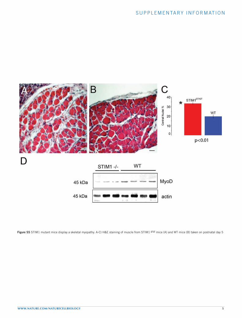
Figure S5 STIM1 mutant mice display a skeletal myopathy. A-C) H&E staining of muscle from STIM1 gt/gt mice (A) and WT mice (B) taken on postnatal day 5
Copyright © 2022 FDOKUMEN