Electrical, contractile and structural remodeling during atrial ...
17
Cardiovascular Research 54 (2002) 230–246 www.elsevier.com / locate / cardiores Review Electrical, contractile and structural remodeling during atrial fibrillation * Maurits Allessie , Jannie Ausma, Ulrich Schotten Department of Physiology, Cardiovascular Research Institute Maastricht, University of Maastricht, P .O. Box 616, 6200 MD Maastricht, The Netherlands Received 29 October 2001; accepted 14 January 2002 Abstract The natural history of atrial fibrillation (AF) is characterized by a gradual worsening with time. The recent finding that AF itself produces changes in atrial function and structure has provided a possible explanation for the progressive nature of this arrhythmia. Electrical remodeling (shortening of atrial refractoriness) develops within the first days of AF and contributes to an increase in stability of AF. However, ‘domestication of AF’ must also depend on a ‘second factor’ since the persistence of AF continues to increase after electrical remodeling has been completed. Atrial contractile remodeling (loss of contractility) leads to a reduced atrial transport function after cardioversion of AF. An important clinical consequence is that during several days after restoration of sinus rhythm, the risk of atrial thrombus formation is still high. In addition, the reduction of atrial contractility during AF may enhance atrial dilatation which may add to the persistence of AF. Tachycardia-induced structural remodeling takes place in a different time domain (weeks to months). Myolysis probably contributes to the loss of atrial contractile force. Although it might explain the loss of efficacy of pharmacological cardioversion and the development of permanent AF, the role of structural remodeling in the progression of AF is still unclear. Atrial structural remodeling also occurs as a result of heart failure and other underlying cardiovascular diseases. The associated atrial fibrosis might explain intra-atrial conduction disturbances and the susceptibility for AF. Thus, both AF itself and the underlying heart disease are responsible for the development of the arrhythmogenic substrate. New strategies for prevention and termination of AF should be build on our knowledge of the mechanisms and time course of AF-induced atrial remodeling. 2002 Elsevier Science B.V. All rights reserved. Keywords: Remodeling; Supraventr. arrhythmia 1. Electrical remodeling period. Given its long-term nature (days to weeks) these tachycardia-induced changes in atrial refractoriness were The concept of tachycardia-induced electrical remodel- thought to be due to alterations in the expression of ion ing of the atria was introduced in 1995 by two independent channels and were referred to as ‘electrical remodeling’ experimental studies [1,2]. In a dog model of prolonged [1]. More importantly, these studies showed that long-term rapid atrial pacing (400 / min) Morillo et al. found that the rapid atrial pacing or maintenance of AF led to a progres- atrial refractory period was reduced by about 15%. In the sive increase in the susceptibility to atrial fibrillation (AF). goat, Wijffels et al. maintained AF by a fibrillation After 6 weeks of rapid atrial pacing, in 82% of the dogs pacemaker automatically delivering bursts of stimuli (1 s, episodes of AF lasting .15 min could be induced [2]. In 50 Hz) as soon as sinus rhythm occurred. This resulted in the goat this effect was even more striking. Whereas during an even more marked shortening in atrial refractoriness control, only short paroxysms of AF were induced by burst from 146619 to 81622 ms ( 245%) and a loss (or even pacing (mean 663 s), after 2 days of AF the paroxysms inversion) of the normal rate adaptation of the refractory lasted more than 4 h (2416459 min) and by that time in two of 12 animals AF had become sustained ( .24 h). After 2–3 weeks in 90% of the goats AF was persistent *Corresponding author. Tel.: 131-43-388-1202 / 00; fax: 131-43-388- 4166. E-mail address: [email protected] (M. Allessie). Time for primary review 22 days 0008-6363 / 02 / $ – see front matter 2002 Elsevier Science B.V. All rights reserved. PII: S0008-6363(02)00258-4 Downloaded from https://academic.oup.com/cardiovascres/article/54/2/230/270994 by guest on 06 March 2022
-
Upload
khangminh22 -
Category
Documents
-
view
1 -
download
0
Transcript of Electrical, contractile and structural remodeling during atrial ...

Cardiovascular Research 54 (2002) 230–246www.elsevier.com/ locate /cardiores
Review
Electrical, contractile and structural remodeling during atrialfibrillation
*Maurits Allessie , Jannie Ausma, Ulrich SchottenDepartment of Physiology, Cardiovascular Research Institute Maastricht, University of Maastricht, P.O. Box 616, 6200 MD Maastricht,
The Netherlands
Received 29 October 2001; accepted 14 January 2002
Abstract
The natural history of atrial fibrillation (AF) is characterized by a gradual worsening with time. The recent finding that AF itselfproduces changes in atrial function and structure has provided a possible explanation for the progressive nature of this arrhythmia.Electrical remodeling (shortening of atrial refractoriness) develops within the first days of AF and contributes to an increase in stability ofAF. However, ‘domestication of AF’ must also depend on a ‘second factor’ since the persistence of AF continues to increase afterelectrical remodeling has been completed. Atrial contractile remodeling (loss of contractility) leads to a reduced atrial transport functionafter cardioversion of AF. An important clinical consequence is that during several days after restoration of sinus rhythm, the risk of atrialthrombus formation is still high. In addition, the reduction of atrial contractility during AF may enhance atrial dilatation which may add tothe persistence of AF. Tachycardia-induced structural remodeling takes place in a different time domain (weeks to months). Myolysisprobably contributes to the loss of atrial contractile force. Although it might explain the loss of efficacy of pharmacological cardioversionand the development of permanent AF, the role of structural remodeling in the progression of AF is still unclear. Atrial structuralremodeling also occurs as a result of heart failure and other underlying cardiovascular diseases. The associated atrial fibrosis mightexplain intra-atrial conduction disturbances and the susceptibility for AF. Thus, both AF itself and the underlying heart disease areresponsible for the development of the arrhythmogenic substrate. New strategies for prevention and termination of AF should be build onour knowledge of the mechanisms and time course of AF-induced atrial remodeling. 2002 Elsevier Science B.V. All rights reserved.
Keywords: Remodeling; Supraventr. arrhythmia
1. Electrical remodeling period. Given its long-term nature (days to weeks) thesetachycardia-induced changes in atrial refractoriness were
The concept of tachycardia-induced electrical remodel- thought to be due to alterations in the expression of ioning of the atria was introduced in 1995 by two independent channels and were referred to as ‘electrical remodeling’experimental studies [1,2]. In a dog model of prolonged [1]. More importantly, these studies showed that long-termrapid atrial pacing (400/min) Morillo et al. found that the rapid atrial pacing or maintenance of AF led to a progres-atrial refractory period was reduced by about 15%. In the sive increase in the susceptibility to atrial fibrillation (AF).goat, Wijffels et al. maintained AF by a fibrillation After 6 weeks of rapid atrial pacing, in 82% of the dogspacemaker automatically delivering bursts of stimuli (1 s, episodes of AF lasting .15 min could be induced [2]. In50 Hz) as soon as sinus rhythm occurred. This resulted in the goat this effect was even more striking. Whereas duringan even more marked shortening in atrial refractoriness control, only short paroxysms of AF were induced by burstfrom 146619 to 81622 ms (245%) and a loss (or even pacing (mean 663 s), after 2 days of AF the paroxysmsinversion) of the normal rate adaptation of the refractory lasted more than 4 h (2416459 min) and by that time in
two of 12 animals AF had become sustained (.24 h).After 2–3 weeks in 90% of the goats AF was persistent*Corresponding author. Tel.: 131-43-388-1202/00; fax: 131-43-388-
4166.E-mail address: [email protected] (M. Allessie). Time for primary review 22 days
0008-6363/02/$ – see front matter 2002 Elsevier Science B.V. All rights reserved.PI I : S0008-6363( 02 )00258-4
Dow
nloaded from https://academ
ic.oup.com/cardiovascres/article/54/2/230/270994 by guest on 06 M
arch 2022

M. Allessie et al. / Cardiovascular Research 54 (2002) 230 –246 231
(Fig. 1). This observation of tachycardia-induced electrical different types of AF were first distinguished by Wells etremodeling creating a substrate for persistent AF, led to the al. [7] based on difference in morphology of bipolarconcept that ‘Atrial Fibrillation Begets Atrial Fibrillation’ fibrillation electrograms and later by Konings et al. by[1]. different degrees of complexities in high density maps of
The higher susceptibility to AF was explained by a AF [8]. Due to the shortening in wavelength, now multipleshortening of the wavelength of the atrial impulse [3,4]. wavelets were wandering under the mapping electrodeWhen the wavelength is short, small regions of intra-atrial (type III AF). This higher degree of spatial dissociationconduction block may already serve as a site for initiation lowers the chance that the fibrillation waves will all dieof reentry, thus increasing the vulnerability for AF. A short out, making it less likely that AF will self-terminate.wavelength is also expected to increase the stability of AF Shortly after the demonstration of tachycardia-inducedbecause it allows more reentering wavelets to coexist in electrical remodeling, the ionic mechanisms underlyingthe available surface area of the atria. This is illustrated in this arrhythmogenic process have been elucidated by athe right part of Fig. 1, showing high density maps number of elegant and convincing studies [9–13]. Action(diameter 4 cm, 240 electrodes) from the free wall of the potential recordings and patch clamp experiments inright atrium during paroxysmal (top) and persistent AF isolated atrial cells from animal models and patients in(bottom) [5]. Whereas during control (no remodeling) the chronic AF showed a consistent pattern. The most im-right atrium was activated by broad fibrillation waves (type portant impact of AF on the ion channels was a marked
21I AF), after electrical remodeling the fibrillation waves reduction in the L-type Ca current. This explains thewere much more disorganized (type III AF) [6]. These shortening of the atrial action potential and the loss of the
Fig. 1. Left: prolongation of the duration of episodes of electrically induced AF in the goat as a result of electrical remodeling (from Wijffels et al. [1]).Right: high density mapping of the free wall of the right atrium of a goat during acutely induced (top) and persistent AF (bottom). The mapping array(diameter 4 cm) contained 240 electrodes with an interelectrode distance of 2.25 mm. Isochrones are drawn every 10 ms. The direction of propagation isindicated by arrows (from Konings et al. [5]).
Dow
nloaded from https://academ
ic.oup.com/cardiovascres/article/54/2/230/270994 by guest on 06 M
arch 2022
232 M. Allessie et al. / Cardiovascular Research 54 (2002) 230 –246
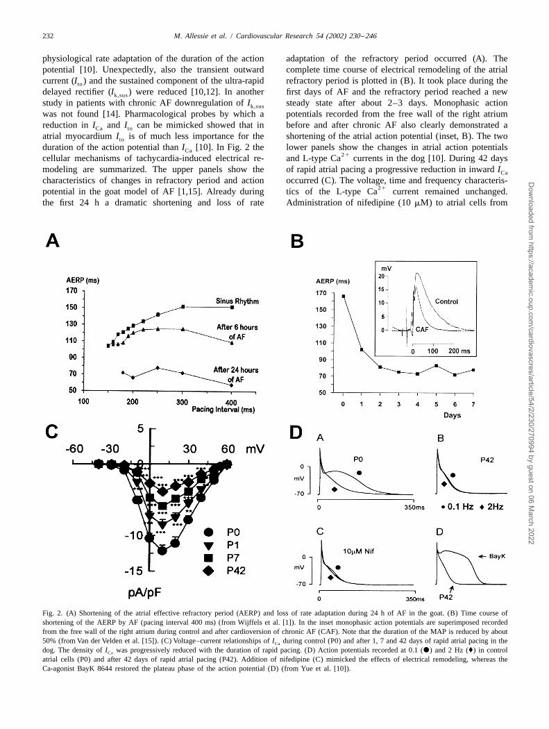
physiological rate adaptation of the duration of the action adaptation of the refractory period occurred (A). Thepotential [10]. Unexpectedly, also the transient outward complete time course of electrical remodeling of the atrialcurrent (I ) and the sustained component of the ultra-rapid refractory period is plotted in (B). It took place during theto
delayed rectifier (I ) were reduced [10,12]. In another first days of AF and the refractory period reached a newk,sus
study in patients with chronic AF downregulation of I steady state after about 2–3 days. Monophasic actionk,sus
was not found [14]. Pharmacological probes by which a potentials recorded from the free wall of the right atriumreduction in I and I can be mimicked showed that in before and after chronic AF also clearly demonstrated aCa to
atrial myocardium I is of much less importance for the shortening of the atrial action potential (inset, B). The twoto
duration of the action potential than I [10]. In Fig. 2 the lower panels show the changes in atrial action potentialsCa21cellular mechanisms of tachycardia-induced electrical re- and L-type Ca currents in the dog [10]. During 42 days
modeling are summarized. The upper panels show the of rapid atrial pacing a progressive reduction in inward ICa
characteristics of changes in refractory period and action occurred (C). The voltage, time and frequency characteris-21potential in the goat model of AF [1,15]. Already during tics of the L-type Ca current remained unchanged.
the first 24 h a dramatic shortening and loss of rate Administration of nifedipine (10 mM) to atrial cells from
Fig. 2. (A) Shortening of the atrial effective refractory period (AERP) and loss of rate adaptation during 24 h of AF in the goat. (B) Time course ofshortening of the AERP by AF (pacing interval 400 ms) (from Wijffels et al. [1]). In the inset monophasic action potentials are superimposed recordedfrom the free wall of the right atrium during control and after cardioversion of chronic AF (CAF). Note that the duration of the MAP is reduced by about50% (from Van der Velden et al. [15]). (C) Voltage–current relationships of I during control (P0) and after 1, 7 and 42 days of rapid atrial pacing in theCa
dog. The density of I was progressively reduced with the duration of rapid pacing. (D) Action potentials recorded at 0.1 (d) and 2 Hz (♦) in controlCa
atrial cells (P0) and after 42 days of rapid atrial pacing (P42). Addition of nifedipine (C) mimicked the effects of electrical remodeling, whereas theCa-agonist BayK 8644 restored the plateau phase of the action potential (D) (from Yue et al. [10]).
Dow
nloaded from https://academ
ic.oup.com/cardiovascres/article/54/2/230/270994 by guest on 06 M
arch 2022

M. Allessie et al. / Cardiovascular Research 54 (2002) 230 –246 233
animals in sinus rhythm mimicked the shortening and loss tuted a clinical entity’ [16]. In 1986, loss of rate adaptationof rate adaptation due to rapid pacing. Vice versa, adding of the refractory period and action potential duration was
21the Ca -channel agonist BayK to a large extent could confirmed in isolated right atrial tissue of patients with‘undo’ the effects of electrical remodeling (D). chronic AF [17]. The first clinical study demonstrating
Some important steps in our knowledge of AF-induced electrical remodeling in human atria after prolongedelectrical remodeling in humans are depicted in Fig. 3. The tachyarrhythmias was done by Franz et al. [18]. In controlearliest clinical observations that abnormalities in rate patients, the APD of the monophasic action potential of90
adaptation of the refractory period were related to AF were the right atrium was compared with the APD in patients90
made by Attuel et al. [16]. In 1982 they measured the atrial with chronic atrial flutter or fibrillation. In patients withrefractory period in 39 patients and noticed that atrial AF or atrial flutter, the APD measured during slow90
tachyarrhythmias preferentially occurred in patients in pacing 15–30 min after electrical cardioversion was 130–whom the atrial refractory period failed to adapt to changes 150 ms shorter than in the control group. The curvein pacing rate (Fig. 3A). They suggested that a poor or describing the relation between the APD and the steady90
absent rate adaptation of the atrial refractory period was a state cycle length was shifted downward and flattened inmarker of some ‘cryptic’ atrial pathology which caused the range between 400 and 800 ms (Fig. 3B). In humansAF. They further suggested that maladaptation of the atrial the adaptation of atrial refractoriness and APD duration torefractory period and a propensity to AF together ‘consti- changes in heart rate is more pronounced than in dog and
Fig. 3. (A) Physiological rate adaptation of the effective refractory period in 11 control patients (left) and non-adaptation of the ERP in 17 patients with ahigh vulnerability for atrial fibrillation (right) (from Attuel et al. [16]). (B) Average APD90 duration6S.D. plotted as a function of steady state cyclelength. Asterisks denote significant differences in average APD90 of patients with atrial fibrillation (Afib) or flutter (Aflut) (from Franz et al. [18]). (C,D)
21Action potentials and L-type Ca current in atrial cells from humans in sinus rhythm and atrial fibrillation (from Bosch et al. [12].
Dow
nloaded from https://academ
ic.oup.com/cardiovascres/article/54/2/230/270994 by guest on 06 M
arch 2022

234 M. Allessie et al. / Cardiovascular Research 54 (2002) 230 –246
goat [1,19]. Also the degree of loss of rate adaptationmight be different in different patient populations [16,18].This might explain why in electrically remodeled humanatria still some rate adaptation exists at high pacing rates[18]. The association between a short monophasic actionpotential and the difficulty to maintain sinus rhythm inpatients had already been noted earlier by Olsson andco-workers [20,21].
Also on a cellular level the changes in repolarization andionic mechanisms have shown to be similar as in animalmodels [11–13,22]. Human AF was associated with amarked shortening in action potential duration and bluntingof its rate adaptation (Fig. 3C). As in animal studies, both
21the transient outward current and the L-type Ca currentwere reduced by about 70% (Fig. 3D). In addition, therecovery from inactivation of the I current was slowerCa,L
in cells from AF patients, which contributes to a decreased21Ca influx at high rates [12]. Van Wagoner et al. showed
that, like in the dog model of rapid atrial pacing, the lossof rate adaptation of the action potential could bemimicked by administration of 10 mM of nifedipine [13].
21The question whether the reduction in Ca influx is solelythe result of a reduction of the ion-channel proteins in thecell membrane is not completely settled. In animal modelsof AF or rapid atrial pacing the mRNA-level of the
21a -subunit of the L-type Ca channel was reduced1C
Fig. 4. Top: an example of reverse remodeling of the atrial effective[15,23]. A reduced mRNA content of the a -subunit in1C refractory period (AERP) after conversion of 7 weeks of AF. Within 3humans with AF was found in some studies [24,25] but days of sinus rhythm the AERP returned to its control value (modifiedwere not confirmed in others [26]. On the protein level, the from Wijffels et al. [1]). Bottom: reversal of atrial electrical remodelingexpression of the a -subunit was found to be reduced in after cardioversion of long-term AF in man. Both in the right atrial1C
appendage (RAA) and the distal coronary sinus (DCS) the refractoryone study [25] but not in another [27].period gradually prolonged and reached control steady state values withinThe time course of reverse electrical remodeling after3 days of sinus rhythm. PCL, pacing cycle length; D, day (from Yue et al.
restoration of sinus rhythm has been studied both in goats [10]).and humans [1,28]. Even after prolonged periods of AF(months to years), the shortening of the atrial refractoryperiod and diminished rate adaptation are still completelyreversible (Fig. 4). The fact that atrial refractoriness positive voltages, which increases rather than decreases thebecomes normal again within only a few days of sinus availability of these channels. It is equally unclear whetherrhythm has important clinical implications. It means that changes in atrial gap junctions may cause slowing of atrialrecurrences of AF occurring more than 1 week after conduction. First of all, the data on remodeling of the atrialcardioversion, cannot be explained on the basis of abnor- connexins are not consistent. Elvan et al. [29] reported anmalities in atrial repolarization due to electrical remodel- increase in expression of connexin43 in dogs, whereas ining. humans a decrease in connexin43 was found [30]. In the
It is not yet clear whether prolonged rapid atrial rates goat model of AF Van der Velden et al. reported no changealso lead to slowing in atrial conduction. Whereas in the in connexin43 but instead a decrease and more heteroge-dog, after 42 days of rapid pacing a decrease in atrial neous distribution of connexin40 [31]. Second, althoughconduction velocity of 25% was reported [19], mapping of the gap junctions play a major role in conduction, thethe right atrium in the goat showed no slowing in atrial speed of propagation of the atrial impulse is only affectedconduction even after several months of AF [1,15]. At all when the connexins are down-regulated by more than 40%voltage ranges I was significantly reduced in the chronic [32]. Spatial heterogeneities in connexins might createNa
dog model of AF and its inactivation kinetics were slowed microscopic obstacles for conduction which not necessarily[9]. In contrast, in isolated cells from fibrillating human disturb the conduction of a broad wavefront, but may serveatria neither the current density nor the voltage dependence as turning points or areas of zig-zag conduction when theof the rapid sodium channels were altered [12]. The wavefront becomes fragmented. It therefore remains avoltage-dependent inactivation of I was shifted to more possibility that gap junctional remodeling is involved inNa
Dow
nloaded from https://academ
ic.oup.com/cardiovascres/article/54/2/230/270994 by guest on 06 M
arch 2022

M. Allessie et al. / Cardiovascular Research 54 (2002) 230 –246 235
the creation of a substrate for persistent AF. Indeed, there increased stability of AF. Such atrial remodeling of aare good reasons to believe that shortening of the atrial ‘different sort’ could explain the development of a sub-action potential is not the only factor involved in the strate for AF in old age, rheumatic valve disease and heartdevelopment of permanent AF. The longer time course of failure.the development of sustained AF and the cumulativeeffects of repetitive 1-month episodes of AF, stronglysuggest that a much slower so-called ‘second factor’ is 2. Contractile remodelinginvolved [1,33]. A good candidate for such a second factoris an increased tissue anisotropy due to changes in local Already more than 30 years ago Logan et al. docu-expression of gap junctional proteins or tissue fibrosis as mented that after cardioversion of AF the a-wave in thedemonstrated in a canine model of heart failure [34]. In atrial pressure curve was lost (Fig. 5A) [35]. Usingthis model of heart failure induced by 5 weeks of rapid echocardiographic techniques, later studies revealed thatventricular pacing, the atrial refractory period and spatial this atrial contractile dysfunction correlated with thedispersion of refractoriness were not altered. Instead, duration of AF and that it could take months before thediscrete regions of slow conduction were the cause of the atrial transport function was fully recovered [36,37].
Fig. 5. (A) Left atrial pressure recordings of a control patient in SR and a patient immediately after cardioversion of chronic AF. The a-wave is completely abolished in the AF patient (modified from Logan et al. [35]). (B) Using a 7.5 MHz intravascular ultrasound probe (AcuNav , Acuson Sequoia ) right
atrial appendage Doppler flow velocities were measured before and after 3 days of lone AF in the goat. The white arrows point to the peak emptying flowduring atrial systole (A). The peak emptying velocity of the right atrial appendage during atrial systole is clearly reduced after 3 days of AF. (C) Atrialpressure–diameter loops during atrial pacing at a cycle length of 400 ms. During the first 48 h of AF, the atrial work index (surface area of thepressure–diameter loop) diminished from 16 to 1 mmHg?mm. The almost completely closed loop after 2 days of AF indicates a virtually complete loss ofatrial contractility. (D) Superimposed recordings of the force of contraction of small isolated right atrial trabeculae from 47 patients undergoing mitralvalve surgery. In AF patients the average force of atrial contraction was reduced by about 75% (from Schotten et al. [48]).
Dow
nloaded from https://academ
ic.oup.com/cardiovascres/article/54/2/230/270994 by guest on 06 M
arch 2022
236 M. Allessie et al. / Cardiovascular Research 54 (2002) 230 –246
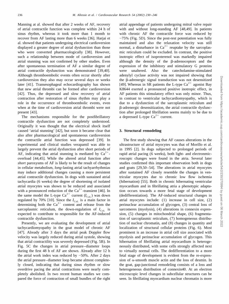
Manning at al. showed that after 2 weeks of AF, recovery atrial appendage of patients undergoing mitral valve repairof atrial contractile function was complete within 24 h of with and without long-standing AF [48,49]. In patientssinus rhythm, whereas it took more than 1 month to with chronic AF the contractile force was reduced byrecover from AF lasting more than 6 weeks [36]. Harjai et |75% (Fig. 5D). Since the post-rest potentiation was fullyal. showed that patients undergoing electrical cardioversion maintained and also the relaxation velocity was still
21displayed a greater degree of atrial dysfunction than those normal, a disturbance in Ca reuptake by the sarcoplas-who were converted pharmacologically [38]. However, mic reticulum could be excluded. In contrast, the positivesuch a relationship between mode of cardioversion and inotropic effect of isoproterenol was markedly impairedatrial stunning was not confirmed by other studies. Even although the density of the b-adrenoceptors and theafter spontaneous termination of AF a similar degree of expression of the inhibitory and stimulatory G proteinsatrial contractile dysfunction was demonstrated [39,40]. were unaltered. Also the catecholamine-stimulatedAlthough thromboembolic events often occur shortly after adenylyl cyclase activity was not impaired showing thatcardioversion they also may occur several days or weeks the b-adrenergic signal transduction was not desensitized
21later [41]. Transesophageal echocardiography has shown [49]. Whereas in SR patients the L-type Ca agonist Baythat new atrial thrombi can be formed after cardioversion K8644 exerted a pronounced positive inotropic effect, in[42]. Thus, the depressed and slow recovery of atrial AF patients this stimulatory effect was only minor. Thus,contraction after restoration of sinus rhythm may play a in contrast to ventricular tachycardiomyopathy, which isrole in the occurrence of thromboembolic events, even due to a dysfunction of the sarcoplasmic reticulum andwhen at the time of cardioversion atrial thrombi were not b-adrenergic desensitization, the atrial contractile dysfunc-present [43]. tion after prolonged fibrillation seems mainly to be due to
21The mechanisms responsible for the postfibrillatory a depressed L-type Ca current.contractile dysfunction are not completely understood.Originally it was thought that the electrical shock itselfcaused ‘atrial stunning’ [42], but soon it became clear that 3. Structural remodelingalso after pharmacological and spontaneous cardioversionthe contractile atrial function was depressed [39]. In The first study showing that AF causes alterations in theexperimental and clinical studies verapamil was able to ultrastructure of atrial myocytes was that of Morillo et al.largely prevent the atrial dysfunction after short periods of in 1995 [2]. In dogs subjected to prolonged periods of
21AF, indicating that atrial stunning is mediated by Ca - rapid atrial pacing (6 weeks), both light- and electronmic-overload [44,45]. While the altered atrial function after roscopic changes were found in the atria. Several latershort paroxysms of AF is likely to be the result of changes studies confirmed this important observation both in dogsin cellular metabolism, long-lasting atrial tachyarrhythmias and goats [29,50–54]. The alterations in atrial myocytesmay induce additional changes causing a more persistent after sustained AF closely resemble the changes in ven-atrial contractile dysfunction. In dogs with sustained atrial tricular myocytes due to chronic low flow ischemiatachycardia (6 weeks) the degree of shortening of isolated (hibernation) [55]. Both in chronic hibernating ventricularatrial myocytes was shown to be reduced and associated myocardium and in fibrillating atria a phenotypic adapta-
21with a pronounced reduction of the Ca -transient [46]. In tion occurs towards a more fetal stage of development21the same model the L-type Ca current (I ) was down (dedifferentiation). The AF-induced structural changes inCaL
regulated by 70% [10]. Since the I is a main factor in atrial myocytes include: (1) increase in cell size, (2)CaL21determining both the Ca content and release from the perinuclear accumulation of glycogen, (3) central loss of
sarcoplasmic reticulum, the down-regulation of I is sarcomeres (myolysis), (4) alterations in connexin expres-CaL
expected to contribute to responsible for the AF-induced sion, (5) changes in mitochondrial shape, (6) fragmenta-contractile dysfunction. tion of sarcoplasmic reticulum, (7) homogeneous distribu-
Presently, we are evaluating the development of atrial tion of nuclear chromatin, and (8) changes in quantity andtachycardiomyopathy in the goat model of chronic AF localization of structural cellular proteins (Fig. 6). Most[47]. Already after 3 days the atrial peak Doppler flow prominent is an increase in atrial cell size associated withvelocity was largely reduced during atrial systole, showing myolysis and perinuclear accumulation of glycogen. Thisthat atrial contractility was severely depressed (Fig. 5B). In hibernation of fibrillating atrial myocardium is heteroge-Fig. 5C the changes in atrial pressure–diameter loops neously distributed, with some cells strongly affected nextduring the first 48 h of AF are shown. Already after 12 h to virtually normal cells. The dedifferentiation to a morethe atrial work index was reduced by |50%. After 2 days fetal stage of development is evident from the re-expres-the atrial pressure–diameter loop became almost complete- sion of a-smooth muscle actin and the loss of desmin. Inly closed, indicating that during sinus rhythm or slow the goat, gap-junctional remodeling consists of a loss andoverdrive pacing the atrial contractions were nearly com- heterogeneous distribution of connexin40. At an electronpletely abolished. In two recent human studies we com- microscopic level changes in subcellular structures can bepared the force of contraction of small bundles of the right seen. In fibrillating myocardium nuclear chromatin is more
Dow
nloaded from https://academ
ic.oup.com/cardiovascres/article/54/2/230/270994 by guest on 06 M
arch 2022

M. Allessie et al. / Cardiovascular Research 54 (2002) 230 –246 237
Fig. 6. Structural remodeling of atrial myocytes after 4 months of AF in the goat. The left pictures are taken from goats in sinus rhythm, the rightphotographs are from goats in chronic AF. Light microscopy (upper left panel) shows cells with severe myolysis (loss of sarcomeres: blue staining) andaccumulation of glycogen (red). Immunostaining of structural proteins (right upper panel) demonstrates the dedifferentiation of the atrial myocardium by aclear increase in fetal a-smooth muscle actin (red staining in upper pictures). In the lower pictures of this panel the myocytes are stained for desmin (red).The nuclei are stained by blue DAPI. During AF desmin looses its cross-striated pattern in the cytoplasm and at the intercalated disks the intensified desminstaining is no longer present. In the lower left panel changes in gap-junctions are shown. Labeling of Cx40 (green) and Cx43 (red) revealed a clearreduction in Cx40 and no change in Cx43 expression. Electron microscopy (lower right) shows changes in the subcellular organization of the atrialmyocytes. During AF the atrial nuclei get a more homogeneous distribution of chromatin For comparison the normal clustering of chromatin at the nuclearmembrane is indicated by arrows in the upper left panel. During AF many small donut shaped mitochondria can be found (arrowheads right lower panel)(from Ausma et al. [50] and Van der Velden et al. [15]).
homogeneously distributed and the mitochondria are small- similar structural changes, some differences exist betweener with longitudinally oriented cristae. different species and different models of atrial tachyar-
Although in general the different animal models show rhythmias. In the dog, a high atrial rate is associated with
Dow
nloaded from https://academ
ic.oup.com/cardiovascres/article/54/2/230/270994 by guest on 06 M
arch 2022

238 M. Allessie et al. / Cardiovascular Research 54 (2002) 230 –246
an increase in size of mitochondria [2], whereas in the goat [57,59]. Compared to animal models, the more extensivemodel of AF numerous small mitochondria with longitudi- structural changes found in patients might be related to thenally oriented cristae were found [50]. Whereas in models older age and/or associated heart diseases [48,61–66].with pure atrial tachyarrhythmias the extracellular matrixwas not changed [2,50], in canine atria subjected to acombination of rapid atrial pacing and mitral regurgitation, 4. Relation between electrical, contractile, andthe volume of the intercellular space was increased [54]. structural remodelingThe effects of structural remodeling on gap junctions alsodiffer in different species [29,51]. To study the relationship between electrical remodeling
These structural changes caused by AF should not be and loss of atrial contractility, goats were instrumentedregarded as degenerative, since signs of irreversible with epicardial electrodes and sonomicrometer crystalschanges leading to cell death (disruption of mitochondrial together with a right atrial pressure catheter [47]. Duringcristae, abnormal secondary lysosomes, cytosolic blebs, the first 5 days of AF, the atrial refractory period and worklipid droplets, discontinuities of the sarcolemma) and index were measured 30 min after spontaneous conversionmarkers of apoptosis (bcl-2, P53, proliferating nuclear of AF during regular atrial pacing. As expected, theantigen, TUNEL reactivity) are all absent in chronic lone refractory period shortened considerably from |130 to |80AF [52]. Instead, the structural changes in response to AF ms (Fig. 7). Also the strength of the atrial contractionsmight be considered as the consequence of a physiological diminished and the atrial work index decreased from 16 to
21adaptation to chronic Ca overload and metabolic stress. less than 2 mmHg?mm. After restoration of sinus rhythmThis is supported by the fact that after longterm AF the this loss of atrial contractility completely recovered follow-expression of heat-shock-proteins (HSP70, GRP94) is ing the same time course as reverse electrical remodeling.upregulated [56]. After 2 days of SR both the atrial work index and the
In patients, data about structural remodeling as a conse- refractory period were back at control values. The fact thatquence of AF are still limited [48,57–59]. Only one study electrical and contractile remodeling go ‘hand in hand’,investigated the structural changes associated with lone AF strongly suggests that they are the result of a common[58]. Similar signs of dedifferentiation of human atrial mechanism. Since electrical remodeling is known to bemyocardium were found as in various animal models. mainly due to a reduction of I , also atrial contractileCaL
However, in patients with AF and atrial dilatation also remodeling is probably directly related to a reduction in21degenerative changes were observed. Some nuclei of atrial Ca inward current. However, so far the time course of
myocytes showed a strong TUNEL reactivity indicative for AF-induced down-regulation of the I channels has notCaL
DNA cleavage and programmed cell death [60]. Further- been directly compared with the time course of shorteningmore, the degree of interstitial fibrosis, both between of the atrial action potential and loss of contractility.individual myocytes (endomysial) and atrial bundles In humans, even after prolonged AF (months to years)(perimysial) is increased in patients with chronic AF electrical remodeling is completely reversible within a few
Fig. 7. Changes in atrial effective refractory period (AERP) and atrial work index (AWI) during 5 days of AF followed by 5 days of sinus rhythm in fivechronically instrumented goats. During electrical remodeling and its reversal after cardioversion of AF, the changes in atrial work index followed the exactsame time course as the changes in AERP (modified from Schotten et al. [47]).
Dow
nloaded from https://academ
ic.oup.com/cardiovascres/article/54/2/230/270994 by guest on 06 M
arch 2022

M. Allessie et al. / Cardiovascular Research 54 (2002) 230 –246 239
days [28,67]. In contrast, depending on the duration of AF other, so far unidentified mechanisms are responsible forthe recovery of the atrial transport function may take the delayed recovery of the atrial contractile function afterseveral months [36,37]. This delayed recovery of contrac- cardioversion of prolonged AF.tile remodeling suggests that, apart from the down-regula- In the 1980s Boyden and co-workers studied the rela-tion of I , in long-term AF additional mechanisms are tionship between atrial enlargement and electrophysiologi-CaL
operative. One possibility is that the slow component of cal properties in dogs and cats with mitral valve diseasethe recovery of atrial contractility reflects the slow re- and ventricular cardiomyopathy [68–70]. In dilated atriasynthesis of sarcomeres which have been lost during AF increased amounts of connective tissue were found be-(myolysis) [48,50]. In a recent study we investigated the tween enlarged myocytes. Also signs of degeneration and acontribution of myolysis to the loss of atrial contraction in loss of myofilaments were observed. These dilated atriapatients with and without chronic AF [48]. In patients with had a high susceptibility for initiation and perpetuation ofAF the contractile force of isolated right atrial trabeculae atrial arrhythmias. Transmembrane action potentials werewas reduced by 75% (Fig. 8). However, after increasing not found to be significantly different from non-dilated
21the Ca concentration the maximal force of contraction atria. In a canine model of heart failure AF could be easilywas reduced by only 15%. Histological quantification of induced and was of long duration [34]. Also in thesethe degree of myolysis revealed a total reduction of animals an increase in atrial size and extensive interstitialsarcomeres of 14% (Fig. 8B). Thus, post-AF atrial stun- fibrosis was found. The main electrophysiological changesning seems to be the result of two different mechanisms. consisted of a marked increase in spatial heterogeneity inThe first and most important component is a functional atrial conduction velocity. The susceptibility to AF in theseloss of contraction due to decreased activation of the models was explained by the increased interstitial fibrosiscontractile apparatus due to the reduction of I . AF- and a higher likelihood of local intra-atrial conductionCaL
induced atrial myolysis causes an additional 15% reduction block leading to smaller and more numerous reentrantin force of contraction. The functional part of atrial circuits. Thus, both electrical and structural remodeling canstunning recovers quickly (a matter of days) [47], whereas either create a substrate for AF. The dimensions of intra-complete restoration of the atrial transport function in atrial circuits can become smaller either by shortening ofpatients with chronic AF may take much longer (up to the action potential (electrical remodeling) or by localseveral months) [36]. Since the contribution of myolysis to conduction delay (enhanced nonuniform anisotropy). Whilethe AF-induced atrial dysfunction is limited, most probably electrical remodeling occurs in a couple of days, structural
21Fig. 8. The effects of extracellular Ca concentration on force of contraction in isolated right atrial bundles from patients in sinus rhythm and chronic AF.21At a physiological Ca concentration of 2.5 mM, the force of contraction was 75% less in AF patients compared to SR patients. However, in both groups
21elevation of the extracellular Ca concentration elicited a strong positive inotropic effect. This resulted in only 15% less contractile force at maximal21activation by high Ca in AF patients. The sarcomere content of the atrial myocytes (blue staining) was reduced to a similar extent (214%). The red
staining in the myolytic cells is due to glycogen accumulation (modified from Schotten et al. [48]).
Dow
nloaded from https://academ
ic.oup.com/cardiovascres/article/54/2/230/270994 by guest on 06 M
arch 2022

240 M. Allessie et al. / Cardiovascular Research 54 (2002) 230 –246
minutes before a new steady state in atrial refractoryperiod, conduction velocity and ion concentrations isreached. Similarly, when AF terminates the action po-tential will only return gradually to its original shape,explaining why the refractory period is still short duringthe first minutes after conversion to sinus rhythm [74]. Thechanges in atrial contractility after termination of short-lasting AF are more complex. The first contractions arestronger than during steady state sinus rhythm due to the
21high intracellular Ca concentration build up during thepreceding AF episode [45]. However, already after acouple of seconds atrial contractility declines indicating
21that the Ca overload disappears rapidly. Actually, theatrial contractions temporarily become 50% weaker thanduring steady state sinus rhythm (undershoot). Thereafter,
Fig. 9. Three proposed positive feedback-loops of atrial remodeling on21 the force of contraction gradually increases to its baselineAF. Down-regulation of the L-type Ca channels is considered to be the
value with a similar time course as the prolongation of theprimary cause for electrical and contractile remodeling. Stretch of theatrial myocardium, which is the result of loss of contractility and increase action potential [45,74]. Also in isolated atrial myocytesin compliance of the fibrillating atria, is hypothesized to act as a stimulus short-term rapid stimulation (3 min) results in a shortfor structural remodeling of the atria. The resulting electro-anatomical period of hypercontractility, followed by a phase ofsubstrate of AF consists of enlarged atria allowing intra-atrial circuits of
hypocontractility before gradual recovery [72]. The majorsmall size, due to a reduction in wavelength (shortening of refractorinessmechanism of the depressed cellular contractile functionand slowing of conduction) and increased non-uniform tissue anisotropy
21(zig-zag conduction). was a lowering of Ca available for release from thesarcoplasmic reticulum.
Thus, after cardioversion of AF the early electrical andremodeling is a much slower process which may continue contractile changes of the atria have short on- and offsetfor several months. In Fig. 9 the three cascades of kinetics. As emphasized by Pandozi and Santini, theseelectrical, contractile and structural remodeling are de- changes should be clearly distinguished from ‘true’ electri-picted. The positive feed back between electrical remodel- cal and contractile remodeling which are based on altera-ing and AF is well established, whereas the proposed tions in gene expression with far slower kinetics. In thiscascades of contractile and structural remodeling are still respect, the recently introduced terms ‘short-term remodel-partly hypothetical. The electro-anatomical substrate of AF ing’ [75] or ‘pseudo-remodeling’ [76] are somewhatmay consist of dilated atria with small local intra-atrial confusing, since they actually have nothing to do withcircuits, both due to shortening of refractoriness and remodeling. The metabolic shortening of the atrial re-increased non-uniform anisotropy. Increased non-uniform fractory period during AF may explain the higher vul-anisotropy may result from alterations in expression of nerability of the atria briefly after conversion to sinusconnexins or atrial architecture (dissociation of atrial rhythm [77]. In electrically remodeled atria, this transientbundles, endo- and perimysial fibrosis). metabolic shortening of the refractory period causes an
additional shortening of the atrial action potential immedi-ately after cardioversion of AF. The resulting temporary
5. Different time domains ultra-short refractory period provides a good explanationfor immediate recurrences of AF (IRAF) frequently seen
5.1. The first minutes after electrical cardioversion [78,79].
Within the first minutes of AF, both the oxygen con- 5.2. The first dayssumption and coronary flow of the atria increases nearly3-fold [71]. Profound changes in atrial metabolism occur, During the first days of AF a progressive reduction inwhich is expressed by a reduction in atrial creatine refractory period and atrial contractility occurs until after
1phosphate [45]. Due to the high rate, the cytosolic Na and 3–5 days a new steady state is reached. Also reversal of21 21Ca concentrations increase, the Ca load of the sarcop- this AF-induced electrical and contractile remodeling takes
lasmic reticulum rises [72] and moderate cellular acidosis a couple of days [1,47]. This slower time course compared21develops. The increase in Ca concentration contributes to the more rapid metabolically mediated changes, sug-
to the rate dependent shortening of the action potential by gests that different mechanisms are involved. At present it21inactivation of the L-type Ca channel. Also changes in is still uncertain whether the reduction in I is due to aCaL
21the intracellular redox potential can inhibit the L-type Ca decrease in the actual number of channels in the atrial cellchannels [73]. After the onset of AF it takes several membrane or to changes in channel properties. Also
Dow
nloaded from https://academ
ic.oup.com/cardiovascres/article/54/2/230/270994 by guest on 06 M
arch 2022

M. Allessie et al. / Cardiovascular Research 54 (2002) 230 –246 241
insufficient knowledge exists about the exact time course reached a new steady state after 3–5 days, it took anin reduction of the I and the related AF-induced additional 1–2 weeks before AF became persistent [1].CaL
electrical and contractile remodeling. A direct correlation This led to the hypothesis that a so-called ‘second factor’21between the density of the L-type Ca channels, the was involved in the development of persistent AF.
calcium inward current and atrial refractoriness was found The time course of AF-induced structural changes inby Gaspo et al. [80]. However, in this study it took several atrial myocytes has been extensively studied (Table 1)weeks of rapid atrial pacing for the atria to remodel, [83]. The first sign of structural remodeling is a morewhereas in the goat model of AF electrical remodeling is homogeneous distribution of nuclear chromatin resemblingcomplete within 3–5 days [1]. nuclei of embryonic myocytes and a decrease in the
During the first days of AF the refractory period myocardial protein cardiotin. Both phenomena occurringshortens considerably (20–40%), whereas after 6 weeks of after 1 week of AF are general signs of dedifferentiationrapid atrial pacing in the dog atrial conduction velocity and are not very likely to play a role in the stabilization ofwas found to be moderately decreased [19]. Thus, as a AF. In the time between 1 and 4 weeks of AF severalresult of electrical remodeling the wavelength of the atrial additional changes occur such as a decrease and heteroge-impulse shortens by a shortening in refractoriness and neous distribution of connexin40 (gap-junctional remodel-possibly also by slowing in atrial conduction. This short- ing) [31], an increase in size of the atrial myocytes, loss ofening of the wavelength during AF allows more wavelets sarcomeres (myolysis) and perinuclear accumulation ofto coexist in the atria which can at least partly explain the glycogen. When AF continues for longer than 1 month aincreased stability of AF with time. Also, recurrences of further increase in cell size, myolysis, glycogen accumula-AF are facilitated by electrical remodeling. In patients with tion and dedifferentiation occur. In addition, the sarcoplas-chronic AF a positive correlation between the shortest mic reticulum became fragmented and the number of smallcoupling interval of premature atrial beats and early mitochondria increase. After 4 months of AF the totalrecurrence of the arrhythmia was found [81]. In humans amount of atrial connective tissue was not changed.with chronic AF it has been shown that the electrical However, because the atrial cells have become larger theremodeling of the atria (shortening of refractoriness) is amount of connective tissue per myocyte was increased.completely reversible within 3 days of sinus rhythm [28]. The question whether the structural changes caused byThis means that recurrences of AF occurring later than 3–5 prolonged AF are reversible or not, was addressed by twodays after cardioversion cannot be due to electrical re- recent studies [54,84]. In the dog, 2 weeks after cardiover-modeling. Recent experiments in goats have shown that sion of 8 weeks of AF combined with mitral regurgitation,also the contractility of the atria largely diminishes during no regression of the structural changes was yet observed.the first days of AF. As a result, the compliance of the This was true despite the fact that by that time thefibrillating atria will increase and the atria will dilate even AF-induced electrical remodeling was completely reversedwhen the mean atrial pressure does not increase [82]. [54]. From this study it is not clear whether absence of
recovery of structural remodeling was due to the short time5.3. The first months window studied or to the still existing mitral regurgitation,
which in itself may cause tissue fibrosis. In the goat modelThere are reasons to believe that, besides the shortening of 16 weeks of lone AF, 8 or 16 weeks after cardioversion
of refractoriness also other factors play a role in the reversion of structural remodeling was still far fromdevelopment of chronic AF. In the first study of Wijffels et complete. Recovery of gap junctions occurred relativelyal. in the goat in which ‘AF begets AF’ was demonstrated, rapid and the expression of connexin 40 was normalizedit was already noted that the time course of the changes in within 8 weeks of sinus rhythm [84]. However, even afteratrial refractoriness did not run parallel with the increase in 16 weeks of sinus rhythm, many atrial myocytes were stillpersistence of AF. Whereas the AF cycle length already myolytic and showed perinuclear glycogen accumulation.
Table 1Time course of AF-induced structural remodeling
1w AF 2w AF 4w AF 8 w AF 16 w AF
Nuclear chromatin 1 1 1 1 1
Downregulation of Cx40 1 11 11 11 11
Cell swelling /myolysis 1 1 11 111 111
a-Smooth muscle actin 1 1 11 111 111
Loss of cardiotin 1 1 11 111 111
Small mitochondriae 1 11 11
Remnants of SR 1 11 11
Loss of titin 1 11 111
Loss of desmin 1 11
Dow
nloaded from https://academ
ic.oup.com/cardiovascres/article/54/2/230/270994 by guest on 06 M
arch 2022

242 M. Allessie et al. / Cardiovascular Research 54 (2002) 230 –246
The hypothesis that a ‘second factor’ is involved in the time required for AF to become sustained during thedevelopment of persistent AF was recently tested by two second or third 5-day episode of AF. In a second study thisstudies (Fig. 10) [33,85]. In the first study, three successive protocol was repeated, but now the duration of the5-day periods of AF were maintained by burst pacing, each consecutive AF episodes was prolonged to 1 month.interrupted by 2 days of sinus rhythm. During these 2 days Following each month, AF was cardioverted electricallythe electrical remodeling was completely reversed and the and the atrial refractory period was allowed to return toatrial refractory period returned to normal. It was hypoth- control before the next episode of AF. Although the timeesized that, in case a ‘second factor’, repetitive AF course of electrical remodeling was the same, the timeepisodes would exert a cumulative effect on the stability of required for development of persistent AF became shorterAF. However, no significant differences were found in the after each AF episode [33]. This evidence suggests that
Fig. 10. Repetitive electrical remodeling by 5 days of AF interrupted by 2 days of sinus rhythm had no cumulative effect in the goat. In contrast, threeconsecutive 1-month episodes of AF resulted in a progressive shortening of the time required for the development of persistent atrial fibrillation. Thisstrongly supports the hypothesis that a ‘second factor’ other than the atrial refractory period is involved in the remodeling process which creates a substratefor self-perpetuation of AF (from Garatt et al. [85] and Todd et al. [33]).
Dow
nloaded from https://academ
ic.oup.com/cardiovascres/article/54/2/230/270994 by guest on 06 M
arch 2022

M. Allessie et al. / Cardiovascular Research 54 (2002) 230 –246 243
indeed a second factor is involved in the transition from even irreversible [84]. Thus the prevention of structuralparoxysmal to persistent AF. More evidence for the remodeling by AF might be an important new element inpresence of a slow second factor was recently obtained by AF management. Recently, the ACE-inhibitor enalaprilserial pharmacological cardioversion of lone AF. In goats was shown to attenuate atrial fibrosis and conductionwithout any underlying heart disease, the efficacy of abnormalities in a canine model of heart failure [90].cardioversion by class Ic drugs progressively reduced from Activation of the renin–angiotensin system causes atrial78% after 1 month to 30% after 4 months of AF [86]. cell growth, proliferation of fibroblasts and atrial fibrosis.Pharmacological cardioversion failed despite the fact that This might explain why ACE-inhibitors are effective tohigher dosages of the drug were administered. Whereas a prevent AF in patients with heart failure [91] and leftreduced efficacy of pharmacological cardioversion during ventricular dysfunction after myocardial infarction [92].the first days of AF is readily explained by electrical Thus, whereas electrical remodeling is ‘forgiving’ and onlyremodeling, failure in the course of several months of AF plays a short-lasting role in the occurrence and perpetua-might be due to the much slower structural remodeling of tion of AF, structural atrial remodeling may be lessthe atria. reversible. Thus, conservation of the normal atrial size and
In patients AF has been related to the extent of structural architecture by preventing structural atrial remodeling duechanges [62] which were found to be a predictor for failure to AF and ventricular dysfunction seems of prime impor-of cardioversion [61]. However, it is not easy to under- tance for the future management of AF.stand how certain changes in cellular structure like in-creased cell size, glycogen accumulation and differentexpression of structural proteins could play a role in
Referencesperpetuation of AF. On the other hand structural changesin gap junctions and interstitial fibrosis might result in
[1] Wijffels MC, Kirchhof CJ, Dorland R, Allessie MA. Atrial fibrilla-inhomogeneities in conduction. The enhanced nonuniformtion begets atrial fibrillation. A study in awake chronically in-
tissue anisotropy might be responsible for slow conduction strumented goats. Circulation 1995;92:1954–1968.and reentry which stabilize AF. The increase in atrial size [2] Morillo CA, Klein GJ, Jones DL, Guiraudon CM. Chronic rapid
atrial pacing. Structural, functional, and electrophysiological charac-due to loss of contractility will also increase the number ofteristics of a new model of sustained atrial fibrillation. Circulationwavelets. Some studies showed that atrial enlargement was1995;91:1588–1595.positively correlated to the recurrence of AF after conver-
[3] Rensma PL, Allessie MA, Lammers WJ, Bonke FI, Schalij MJ.sion to sinus rhythm [87,88] and very recently, a signifi- Length of excitation wave and susceptibility to reentrant atrialcant correlation between atrial dimensions and the stability arrhythmias in normal conscious dogs. Circ Res 1988;62:395–410.
[4] Allessie MA. Atrial electrophysiologic remodeling: another viciousof AF was demonstrated in dogs with heart failure [89].circle? J Cardiovasc Electrophysiol 1998;9:1378–1393.Regional differences in wall thickness resulting in
[5] Konings KT, Wijffels M, Dorland R, Mast F, Allessie M. High-inhomogeneous wall stress will further add to the increaseddensity mapping of the right atrium during acute and chronic atrial
heterogeneity in conduction. However, at the present time fibrillation in the goat. Pacing Clin Electrophysiol 1999;22:727.the exact nature of the ‘second factor’ involved in develop- [6] Konings KT, Kirchhof CJ, Smeets JR, Wellens HJ, Penn OC,
Allessie MA. High-density mapping of electrically induced atrialment of permanent AF is still unknown.fibrillation in humans. Circulation 1994;89:1665–1680.
[7] Wells Jr. JL, Karp RB, Kouchoukos NT, MacLean WA, James TN,Waldo AL. Characterization of atrial fibrillation in man: studies
6. Future perspectives following open heart surgery. Pacing Clin Electrophysiol1978;1:426–438.
New strategies for the management of AF, amongst [8] Konings KT. Mapping of electrically induced atrial fibrillation inhumans. Thesis, Maastricht University, 1999.other things, will depend on a better understanding of the
[9] Gaspo R, Bosch RF, Bou-Abboud E, Nattel S. Tachycardia-inducedmechanisms underlying atrial remodeling. In humans withchanges in Na1 current in a chronic dog model of atrial fibrillation.
chronic AF atrial electrical remodeling has been shown to Circ Res 1997;81:1045–1052.be completely reversible within 3–4 days after cardiover- [10] Yue L, Feng J, Gaspo R, Li GR, Wang Z, Nattel S. Ionic remodelingsion of AF [28]. Recurrences of AF are frequent during the underlying action potential changes in a canine model of atrial
fibrillation. Circ Res 1997;81:512–525.first week after cardioversion and may be related to the[11] Van Wagoner DR, Pond AL, McCarthy PM, Trimmer JS, Nerbonneprocess of reverse electrical remodeling [78,81]. Because
JM. Outward K1 current densities and Kv1.5 expression areof the short time course of AF-induced electrical remodel- reduced in chronic human atrial fibrillation. Circ Res 1997;80:772–ing and its complete and rapid reversibility, AF-recur- 781.
¨rences occurring after 1 week cannot be explained on this [12] Bosch RF, Zeng X, Grammer JB, Popovic K, Mewis C, KuhlkampV. Ionic mechanisms of electrical remodeling in human atrialbasis. A persisting high susceptibility to AF might be duefibrillation. Cardiovasc Res 1999;44:121–131.to structural remodeling of the atria as a result of pro-
[13] Van Wagoner DR, Pond AL, Lamorgese M, Rossie SS, McCarthylonged AF. The reversibility of AF-induced structural 21PM, Nerbonne JM. Atrial L-type Ca currents and human atrialchanges has proven to be a very slow process which takes fibrillation. Circ Res 1999;85:428–436.at least several months. Some structural changes may be [14] Grammer JB, Bosch RF, Kuhlkamp V, Seipel L. Molecular remodel-
Dow
nloaded from https://academ
ic.oup.com/cardiovascres/article/54/2/230/270994 by guest on 06 M
arch 2022

244 M. Allessie et al. / Cardiovascular Research 54 (2002) 230 –246
ing of Kv4.3 potassium channels in human atrial fibrillation. J [32] Jongsma HJ, Wilders R. Gap junctions in cardiovascular disease.Circ Res 2000;86:1193–1197.Cardiovasc Electrophysiol 2000;11:626–633.
[33] Todd DM, Walden AP, Fynn SP, Hobbs WJ, Garratt CJ. Repetitive[15] Van der Velden HMW, van der Zee L, Wijffels MC, van Leuven C,one-month periods of atrial electrical remodeling promote stabilityDorland R, Vos MA, Jongsma HJ, Allessie MA. Atrial fibrillation inof atrial fibrillation. Circulation 2000;102:II154–155.the goat induces changes in monophasic action potential and mRNA
[34] Li D, Fareh S, Leung TK, Nattel S. Promotion of atrial fibrillationexpression of ion channels involved in repolarization. J Cardiovascby heart failure in dogs: atrial remodeling of a different sort.Electrophysiol 2000;11:1262–1269.Circulation 1999;100:87–95.[16] Attuel P, Childers R, Cauchemez B, Poveda J, Mugica J, Coumel P.
[35] Logan W, Rowlands D, Howitt G, Holmes A. Left atrial activityFailure in the rate adaptation of the atrial refractory period: itsfollowing cardioversion. Lancet 1965;ii:471–473.relationship to vulnerability. Int J Cardiol 1982;2:179–197.
[36] Manning WJ, Silverman DI, Katz SE, Riley MF, Come PC, Doherty[17] Boutjdir M, Le Heuzey JY, Lavergne T, Chauvaud S, Guize L,RM, Munson JT, Douglas PS. Impaired left atrial mechanicalCarpentier A, Peronneau P. Inhomogeneity of cellular refractorinessfunction after cardioversion: relation to the duration of atrialin human atrium: factor of arrhythmia? Pacing Clin Electrophysiolfibrillation. J Am Coll Cardiol 1994;23:1535–1540.1986;9:1095–1100.
[37] Manning WJ, Silverman DI, Katz SE, Riley MF, Doherty RM,[18] Franz MR, Karasik PL, Li C, Moubarak J, Chavez M. Electrical
Munson JT, Douglas PS. Temporal dependence of the return ofremodeling of the human atrium: similar effects in patients with
atrial mechanical function on the mode of cardioversion of atrialchronic atrial fibrillation and atrial flutter. J Am Coll Cardiol
fibrillation to sinus rhythm. Am J Cardiol 1995;75:624–626.1997;30:1785–1792.
[38] Harjai KJ, Mobarek SK, Cheirif J, Boulos LM, Murgo JP, Abi-[19] Gaspo R, Bosch RF, Talajic M, Nattel S. Functional mechanisms Samra F. Clinical variables affecting recovery of left atrial me-
underlying tachycardia-induced sustained atrial fibrillation in a chanical function after cardioversion from atrial fibrillation. J Amchronic dog model. Circulation 1997;96:4027–4035. Coll Cardiol 1997;30:481–486.
[20] Olsson SB. Chronic atrial fibrillation—what is wrong with the [39] Grimm RA, Leung DY, Black IW, Stewart WJ, Thomas JD, Kleinatrium? Eur Heart J 1999;20:856–857. AL. Left atrial appendage ‘stunning’ after spontaneous conversion
[21] Olsson SB, Cotoi S, Varnauskas E. Monophasic action potential and of atrial fibrillation demonstrated by transesophageal Doppler ech-sinus rhythm stability after conversion of atrial fibrillation. Acta ocardiography. Am Heart J 1995;130:174–176.Med Scand 1971;190:381–387. [40] Falk RH, Decara J, Abascal V. Is pharmacologic cardioversion of
¨ ¨[22] Skasa M, Jungling E, Picht E, Schondube F, Luckhoff A. L-type atrial fibrillation really preferable to electrical cardioversion? J Amcalcium currents in atrial myocytes from patients with persistent and Coll Cardiol 1998;31:1446–1447.non-persistent atrial fibrillation. Basic Res Cardiol 2001;96:151– [41] Resnekov L, McDonald L. Complications in 220 patients with159. cardiac dysrhythmias treated by phased direct current shock, and
[23] Yue L, Melnyk P, Gaspo R, Wang Z, Nattel S. Molecular mecha- indications for electroconversion. Br Heart J 1967;29:926–936.nisms underlying ionic remodeling in a dog model of atrial [42] Fatkin D, Kuchar DL, Thorburn CW, Feneley MP. Transesophagealfibrillation. Circ Res 1999;84:776–784. echocardiography before and during direct current cardioversion of
[24] Lai LP, Su MJ, Lin JL, Lin FY, Tsai CH, Chen YS, Huang SK, atrial fibrillation: evidence for ‘atrial stunning’ as a mechanism ofTseng YZ, Lien WP. Down-regulation of L-type calcium channel thromboembolic complications. J Am Coll Cardiol 1994;23:307–and sarcoplasmic reticular Ca(21)-ATPase mRNA in human atrial 316.fibrillation without significant change in the mRNA of ryanodine [43] Black IW, Fatkin D, Sagar KB, Khandheria BK, Leung DY,receptor, calsequestrin and phospholamban: an insight into the Galloway JM, Feneley MP, Walsh WF, Grimm RA, Stollberger C.mechanism of atrial electrical remodeling. J Am Coll Cardiol Exclusion of atrial thrombus by transesophageal echocardiography1999;33:1231–1237. does not preclude embolism after cardioversion of atrial fibrillation.
[25] Brundel BJ, Van Gelder IC, Henning RH, Tieleman RG, Tuinenburg A multicenter study. Circulation 1994;89:2509–2513.AE, Wietses M, Grandjean JG,Van Gilst WH, Crijns HJ. Ion channel [44] Daoud EG, Marcovitz P, Knight BP, Goyal R, Man KC, Strickbergerremodeling is related to intraoperative atrial effective refractory SA, Armstrong WF, Morady F. Short-term effect of atrial fibrillationperiods in patients with paroxysmal and persistent atrial fibrillation. on atrial contractile function in humans. Circulation 1999;99:3024–Circulation 2001;103:684–690. 3027.
¨[26] Grammer JB, Zeng X, Bosch RF, Kuhlkamp V. Atrial L-type [45] Leistad E, Aksnes G, Verburg E, Christensen G. Atrial contractileCa21-channel, beta-adrenorecptor, and 5-hydroxytryptamine type 4 dysfunction after short-term atrial fibrillation is reduced by ver-receptor mRNAs in human atrial fibrillation. Basic Res Cardiol apamil but increased by BAY K8644. Circulation 1996;93:1747–2001;96:82–90. 1754.
[27] Schotten U, Haase H, Frechen D, Stellbrink C, Schoendube F, [46] Sun H, Gaspo R, Leblanc N, Nattel S. Cellular mechanisms of atrialHanrath P, Allessie M. Protein expression of L-type Ca channel contractile dysfunction caused by sustained atrial tachycardia.subunits is not reduced in atrial myocardium of patients with atrial Circulation 1998;98:719–727.fibrillation. Pacing Clin Electrophysiol 2000;23:604. [47] Schotten U, Allessie M. Electrical and contractile remodeling during
[28] Yu WC, Lee SH, Tai CT, Tsai CF, Hsieh MH, Chen CC, Ding YA, atrial fibrillation go hand-in-hand. Pacing Clin ElectrophysiolChang MS, Chen SA. Reversal of atrial electrical remodeling 2001;24:572.following cardioversion of long-standing atrial fibrillation in man. [48] Schotten U, Ausma J, Stellbrink C, Sabatschus I, Vogel M, FrechenCardiovasc Res 1999;42:470–476. D, Schoendube F, Hanrath P, Allessie MA. Cellular mechanisms of
[29] Elvan A, Huang XD, Pressler ML, Zipes DP. Radiofrequency depressed atrial contractility in patients with chronic atrial fibrilla-catheter ablation of the atria eliminates pacing-induced sustained tion. Circulation 2001;103:691–698.atrial fibrillation and reduces connexin 43 in dogs. Circulation [49] Schotten U, Greiser M, Benke D, Buerkel K, Ehrenteidt B,1997;96:1675–1685. Stellbrink C, Vazquez-Jimenez JF, Schoendube F, Hanrath P, Alles-
[30] Patel P, Jones D, Dupont E. Remodeling of human connexin 43 sie M. Atrial fibrillation-induced atrial contractile dysfunction: aexpression in human atrial fibrillation. Eur Heart J 2001;19:465. tachycardiomyopathy of a different sort. Cardiovasc Res
[31] van der Velden HM, Ausma J, Rook MB, Hellemons AJ, van Veen 2002;53:192–201.TA, Allessie MA, Jongsma HJ. Gap junctional remodeling in [50] Ausma J, Wijffels M, Thone F, Wouters L, Allessie M, Borgers M.relation to stabilization of atrial fibrillation in the goat. Cardiovasc Structural changes of atrial myocardium due to sustained atrialRes 2000;46:476–486. fibrillation in the goat. Circulation 1997;96:3157–3163.
Dow
nloaded from https://academ
ic.oup.com/cardiovascres/article/54/2/230/270994 by guest on 06 M
arch 2022

M. Allessie et al. / Cardiovascular Research 54 (2002) 230 –246 245
[51] Van der Velden HM, van Kempen MJ, Wijffels MC, van Zijverden [70] Boyden PA, Hoffman BF. The effects on atrial electrophysiologyM, Groenewegen WA, Allessie MA, Jongsma HJ. Altered pattern of and structure of surgically induced right atrial enlargement in dogs.connexin40 distribution in persistent atrial fibrillation in the goat. J Circ Res 1981;49:1319–1331.Cardiovasc Electrophysiol 1998;9:596–607. [71] White CW, Kerber RE, Weiss HR, Marcus ML. The effects of atrial
[52] Dispersyn GD, Ausma J, Thone F, Flameng W, Vanoverschelde JL, fibrillation on atrial pressure-volume and flow relationships. CircAllessie MA, Ramaekers FC, Borgers M. Cardiomyocyte remodel- Res 1982;51:205–215.ling during myocardial hibernation and atrial fibrillation: prelude to [72] Sun H, Chartier D, Leblanc N, Nattel S. Intracellular calciumapoptosis. Cardiovasc Res 1999;43:947–957. changes and tachycardia-induced contractile dysfunction in canine
[53] Ausma J, Dispersyn GD, Duimel H, Thone F, Ver Donk L, Allessie atrial myocytes. Cardiovasc Res 2001;49:751–761.MA, Borgers M. Changes in ultrastructural calcium distribution in [73] Fearon IM, Varadi G, Koch S, Isaacsohn I, Ball SG, Peers C. Splicegoat atria during atrial fibrillation. J Mol Cell Cardiol 2000;32:355– variants reveal the region involved in oxygen sensing by recombi-364. nant human L-type Ca(21) channels. Circ Res 2000;87:537–539.
[54] Everett TH, Li H, Mangrum JM, McRury ID, Mitchell MA, Redick [74] Duytschaever M, Danse P, Eijsbouts S, Allessie M. The presence ofJA, Haines DE. Electrical, morphological, and ultrastructural re- a supervulnerable period immediately after conversion of atrialmodeling and reverse remodeling in a canine model of chronic atrial
fibrillation. Pacing Clin Electrophysiol 1999;22:707.fibrillation. Circulation 2000;102:1454–1460.
[75] Jayachandran JV, Zipes DP, Weksler J, Olgin JE. Role of the[55] Borgers M, Thone F, Wouters L, Ausma J, Shivalkar B, Flameng W.
Na(1) /H(1) exchanger in short-term atrial electrophysiologicalStructural correlates of regional myocardial dysfunction in patients
remodeling. Circulation 2000;101:1861–1866.with critically coronary artery stenosis:Chronic hibernation? Car-[76] Pandozi C, Santini M. Update on atrial remodelling owing to rate;diovasc Pathol 1993;2:237–245.
does atrial fibrillation always ‘beget’ atrial fibrillation? Eur Heart J[56] Vitadello M, Ausma J, Borgers M, Gambino A, Casarotto DC,2001;22:541–553.Gorza L. Increased myocardial GRP94 amounts during sustained
[77] Daoud EG, Bogun F, Goyal R, Harvey M, Man KC, Strickbergeratrial fibrillation: a protective response? Circulation 2001;103:2201–SA, Morady F. Effect of atrial fibrillation on atrial refractoriness in2206.humans. Circulation 1996;94:1600–1606.[57] Thiedemann KU, Ferrans VJ. Left atrial ultrastructure in mitral
[78] Van Noord T, Van Gelder IC, Schoonderwoerd BA, Crijns HJ.valvular disease. Am J Pathol 1977;89:575–594.Immediate reinitiation of atrial fibrillation after electrical cardiover-[58] Frustaci A, Chimenti C, Bellocci F, Morgante E, Russo MA, Maserision predicts subsequent pharmacologic and electrical conversion toA. Histological substrate of atrial biopsies in patients with lone atrialsinus rhythm and amiodarone. Am J Cardiol 2000;86:1384–1385.fibrillation. Circulation 1997;96:1180–1184.
[79] Timmermans C, Rodriguez LM, Smeets JL, Wellens HJ. Immediate[59] Wouters L, Liu GS, Flameng W, Thijssen VL, Thone F, Borgers M.reinitiation of atrial fibrillation following internal atrial defibrilla-Structural remodeling of atrial myocardium in patients with cardiaction. J Cardiovasc Electrophysiol 1998;9:122–128.valve disease and atrial fibrillation. Exp Clin Cardiol 2001;5:158–
163. [80] Gaspo R, Sun H, Fareh S, Levi M, Yue L, Allen BG, Hebert TE,[60] Aime-Sempe C, Folliguet T, Rucker-Martin C, Krajewska M, Nattel S. Dihydropyridine and beta adrenergic receptor binding in
Krajewska S, Heimburger M, Aubier M, Mercadier JJ, Reed JC, dogs with tachycardia-induced atrial fibrillation. Cardiovasc ResHatem SN. Myocardial cell death in fibrillating and dilated human 1999;42:434–442.right atria. J Am Coll Cardiol 1999;34:1577–1586. [81] Tieleman RG, Van Gelder IC, Crijns HJ, de Kam PJ, Van Den Berg
[61] Bailey GW, Braniff BA, Hancock EW, Cohn KE. Relation of left MP, Haaksma J, Van Der Woude HJ, Allessie MA. Early recurrencesatrial pathology to atrial fibrillation in mitral valvular disease. Ann of atrial fibrillation after electrical cardioversion: a result of fibril-Intern Med 1968;69:13–20. lation-induced electrical remodeling of the atria? J Am Coll Cardiol
[62] Fenoglio Jr. JJ, Wagner BM. Studies in rheumatic fever. VI. 1998;31:167–173.Ultrastructure of chronic rheumatic heart disease. Am J Pathol [82] Sanfilippo AJ, Abascal VM, Sheehan M, Oertel LB, Harrigan P,1973;73:623–640. Hughes RA, Weyman AE. Atrial enlargement as a consequence of
[63] Pham TD, Wit AL, Hordof AJ, Malm JR, Fenoglio Jr. JJ. Right atrial fibrillation. A prospective echocardiographic study. Circulationatrial ultrastructure in congenital heart disease. I. Comparison of 1990;82:792–797.ventricular septal defect and endocardial cushion defect. Am J [83] Ausma J, Litjens N, Lenders M-H, Duimel H, Mast F, Wouters L,Cardiol 1978;42:973–982. Ramaekers F, Allessie M, Borgers M. Time course of atrial
[64] Fenoglio Jr. JJ, Pham TD, Hordof A, Edie RN, Wit AL. Right atrial fibrillation-induced cellular structural remodeling in atria of theultrastructure in congenital heart disease. II. Atrial septal defect: goat. J Mol Cell Cardiol 2001;33:2083–2094.effects of volume overload. Am J Cardiol 1979;43:820–827. [84] Ausma J, van der Velden HMW, Lenders M-H, Duimel H, Borgers
[65] Mary Rabine L, Albert A, Pham TD, Hordof A, Fenoglio JJJ, Malm M, Allessie M. Partial recovery from structural atrial remodelingJR, Rosen MR. The relationship of human atrial cellular elec- after prolonged atrial fibrillation. Circulation 2001;104:372.trophysiology to clinical function and ultrastructure. Circ Res [85] Garratt CJ, Duytschaever M, Killian M, Dorland R, Mast F, Allessie1983;52:188–199. MA. Repetitive electrical remodeling by paroxysms of atrial fibrilla-
[66] Kitzman DW, Edwards WD. Age-related changes in the anatomy of tion in the goat: no cumulative effect on inducibility or stability ofthe normal human heart. J Gerontol 1990;45:M33–M39. atrial fibrillation. J Cardiovasc Electrophysiol 1999;10:1101–1108.
[67] Hobbs WJ, Fynn S, Todd DM, Wolfson P, Galloway M, Garratt CJ. [86] Ausma J, Duytschaever M, Wijffels M, Borgers M, Allessie M. LossReversal of atrial electrical remodeling after cardioversion of of efficacy of cardioversion by class Ic drugs after long term atrialpersistent atrial fibrillation in humans. Circulation 2000;101:1145– fibrillation in the goat. Eur Heart J 2001;21:543.1151. [87] Brodsky MA, Allen BJ, Capparelli EV, Luckett CR, Morton R,
[68] Boyden PA, Tilley LP, Pham TD, Liu SK, Fenoglic JJJ, Wit AL. Henry WL. Factors determining maintenance of sinus rhythm afterEffects of left atrial enlargement on atrial transmembrane potentials chronic atrial fibrillation with left atrial dilatation. Am J Cardioland structure in dogs with mitral valve fibrosis. Am J Cardiol 1989;63:1065–1068.1982;49:1896–1908. [88] Verhorst PM, Kamp O, Welling RC, Van Eenige MJ, Visser CA.
[69] Boyden PA, Tilley LP, Albala A, Liu SK, Fenoglio Jr. JJ, Wit AL. Transesophageal echocardiographic predictors for maintenance ofMechanisms for atrial arrhythmias associated with cardiomyopathy: sinus rhythm after electrical cardioversion of atrial fibrillation. Am Ja study of feline hearts with primary myocardial disease. Circulation Cardiol 1997;79:1355–1359.1984;69:1036–1047. [89] Shi Y, Ducharme A, Li D, Gaspo R, Nattel S, Tardif JC. Remodel-
Dow
nloaded from https://academ
ic.oup.com/cardiovascres/article/54/2/230/270994 by guest on 06 M
arch 2022

246 M. Allessie et al. / Cardiovascular Research 54 (2002) 230 –246
ing of atrial dimensions and emptying function in canine models of [91] Gurlek A, Erol C, Basesme E. Antiarrhythmic effect of convertingatrial fibrillation. Cardiovasc Res 2001;52:217–225. enzyme inhibitors in congestive heart failure. Int J Cardiol
[90] Li D, Shinagawa K, Pang L, Leung TK, Cardin S, Wang Z, Nattel S. 1994;43:315–318.Effects of Angiotensin-converting enzyme inhibition on the develop- [92] Pedersen OD, Bagger H, Kober L, Torp-Pedersen C. Trandolaprilment of the atrial fibrillation substrate in dogs with ventricular reduces the incidence of atrial fibrillation after acute myocardialtachypacing-induced congestive heart failure. Circulation infarction in patients with left ventricular dysfunction. Circulation2001;104:2608–2614. 1999;100:376–380.
Dow
nloaded from https://academ
ic.oup.com/cardiovascres/article/54/2/230/270994 by guest on 06 M
arch 2022