Identification, expression, and tissue distribution of the three rat lysyl hydroxylase isoforms
Upload
independentCategory
view
3download
0
Traumatic brain injury reduces striatal tyrosine hydroxylaseactivity and potassium-evoked dopamine release in rats
Samuel S. Shin, Eric R. Bray, Cathy Q. Zhang, and C. Edward Dixon*
Brain Trauma Research Center, Department of Neurosurgery, University of Pittsburgh, 3434 FifthAve, Suite 201, Pittsburgh, PA 15260, USA; Safar Center for Resuscitation Research, Universityof Pittsburgh, Pittsburgh, PA 15260, USA
AbstractThere is increasing evidence that traumatic brain injury (TBI) induces hypofunction of the striataldopaminergic system, the mechanisms of which are unknown. In this study, we analyzed theactivity of striatal tyrosine hydroxylase (TH) in rats at 1 day, 1 week, and 4 weeks after TBI usingthe controlled cortical impact model. There were no changes in the level of TH phosphorylated atserine 40 site (pser40TH) at 1 day or 4 weeks. At 1 week, injured animals showed decreasedpser40TH to 73.9±7.3% (p≤0.05) of sham injured rats. The in vivo TH activity assay showed nosignificant difference between injured and sham rats at 1 day. However, there was a decreasedactivity in injured rats to 62.1±8.2% (p≤0.05) and 68.8±6.2% (p≤0.05) of sham injured rats at 1and 4 weeks, respectively. Also, the activity of protein kinase A, which activates TH, decreased at1 week (injured: 87.8±2.8%, sham: 100.0± 4.2%, p≤0.05). To study the release activity ofdopamine after injury, potassium (80 mM)-evoked dopamine release was measured bymicrodialysis in awake, freely moving rats. Dialysates were collected and analyzed by high-performance liquid chromatography. There were no significant differences in dopamine release at1 day and 4 weeks between sham and injured groups. At 1 week, there was a significant decrease(injured: 0.067±0.015 μM, sham: 0.127 ± 0.027 μM, p ≤ 0.05). These results suggest that TBI-induced dopamine neurotransmission deficits are, at least in part, attributable to deficits in THactivity.
KeywordsDopamine; Traumatic; Microdialysis; Tyrosine; Hydroxylase; Phosphorylation
1. IntroductionThe effects of traumatic brain injury (TBI) on the dopamine system have been shown invarious studies in the past (Bales et al., 2009). Several pharmacological treatments aimed atthe dopamine system have been reported to be beneficial in improving cognitive functions inanimals and humans. Catecholamine agonist therapy shows motor and cognitiveimprovement in both humans and animals (Phillips et al., 2003). Methylphenidate (Kline etal., 1994, 2000) and D-amphetamine (Feeney et al., 1981; Hornstein et al., 1996), whichincrease synaptic dopamine levels by inhibiting dopamine transporter (DAT) function, haveshown to enhance functional outcome after experimental TBI. Also, L-DOPA (Kraus andMaki, 1997; Koeda and Takeshita, 1998; Lal et al., 1988), which increases dopaminesynthesis, and amantadine (Sawyer et al., 2008; Dixon et al., 1999), which increases
*Corresponding author. Department of Neurosurgery, University of Pittsburgh, 3434 Fifth Ave, Suite 201, Pittsburgh, PA 15260,USA. Fax: +1 412 624 0943. [email protected] (C.E. Dixon).
NIH Public AccessAuthor ManuscriptBrain Res. Author manuscript; available in PMC 2012 January 19.
Published in final edited form as:Brain Res. 2011 January 19; 1369: 208–215. doi:10.1016/j.brainres.2010.10.096.
NIH
-PA Author Manuscript
NIH
-PA Author Manuscript
NIH
-PA Author Manuscript
extracellular dopamine concentrations by inhibition of reuptake and facilitation of dopaminesynthesis (Von Voightlander and Moore, 1971; Bak et al., 1972; Gianutsos et al., 1985),both facilitate functional recovery after TBI.
Neurochemical studies also suggest that there are alterations in the levels of striataldopamine and proteins that synthesize and transport dopamine after injury. Dopamine levelsincrease acutely in the striatum (Massucci et al., 2004; McIntosh et al., 1994), andelectrically evoked dopamine release as measured by fast scanning cyclic voltammetry islater decreased in the striatum following TBI (Wagner et al., 2005, 2009). Protein levels oftyrosine hydroxylase (TH) increase chronically (Yan et al., 2007), and DAT decreaseschronically after TBI (Wagner et al., 2005), possibly reflecting compensatory changes forrecovery of the dopaminergic system.
Tyrosine hydroxylase is a rate-limiting synthesis enzyme for dopamine and norepinephrine.However, striatal tissue contains very small level of norepinephrine and TH represents thepresence of dopaminergic fibers (Hokfelt et al., 1977; Anden et al., 1964). The activity levelof striatal TH after TBI has not been previously reported. We provide evidence here for thefirst time that striatal dopamine neurotransmission deficits after TBI may be attributable todeficits in tyrosine hydroxylase activity. Increased activity of TH has previously beenassociated with phosphorylation of TH at serine 40 site (pser40TH), but not serine 19 site(pser19TH) (Lindgren et al., 2000; Harada et al., 1996). Thus, we monitored pser40TH andpser19TH levels by Western blotting at 1 day, 1 week, and 4 weeks after injury. Activitylevels of striatal cAMP-dependent protein kinase A (PKA), a major kinase for TH, were alsomeasured at these time points. In vivo activity level of TH and potassium stimulus-evokeddopamine release, which are parameters that could be affected by dopamine synthesis, werealso monitored at 1 day, 1 week, and 4 weeks following injury.
2. Results2.1. Western blots: tyrosine hydroxylase phosphorylation after TBI
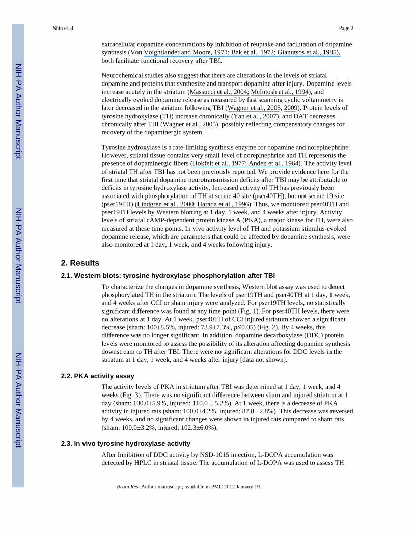
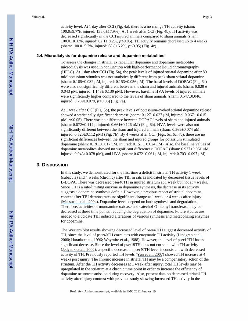
To characterize the changes in dopamine synthesis, Western blot assay was used to detectphosphorylated TH in the striatum. The levels of pser19TH and pser40TH at 1 day, 1 week,and 4 weeks after CCI or sham injury were analyzed. For pser19TH levels, no statisticallysignificant difference was found at any time point (Fig. 1). For pser40TH levels, there wereno alterations at 1 day. At 1 week, pser40TH of CCI injured striatum showed a significantdecrease (sham: 100±8.5%, injured: 73.9±7.3%, p≤0.05) (Fig. 2). By 4 weeks, thisdifference was no longer significant. In addition, dopamine decarboxylase (DDC) proteinlevels were monitored to assess the possibility of its alteration affecting dopamine synthesisdownstream to TH after TBI. There were no significant alterations for DDC levels in thestriatum at 1 day, 1 week, and 4 weeks after injury [data not shown].
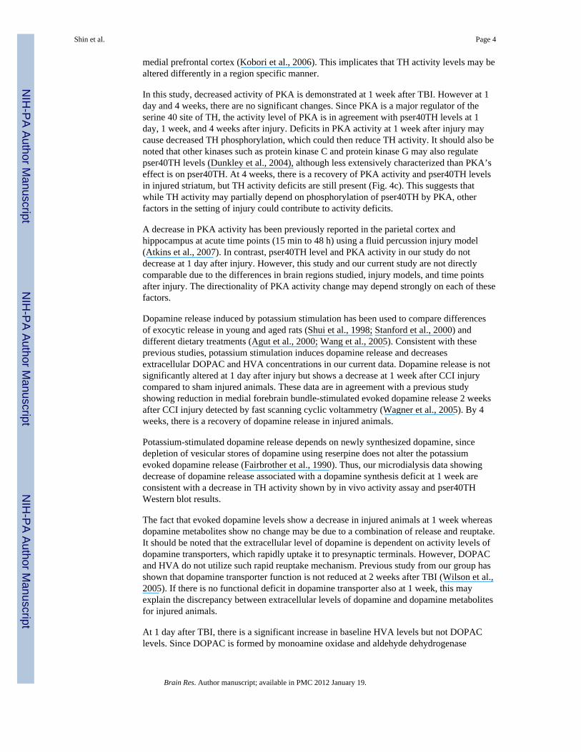
2.2. PKA activity assayThe activity levels of PKA in striatum after TBI was determined at 1 day, 1 week, and 4weeks (Fig. 3). There was no significant difference between sham and injured striatum at 1day (sham: 100.0±5.9%, injured: 110.0 ± 5.2%). At 1 week, there is a decrease of PKAactivity in injured rats (sham: 100.0±4.2%, injured: 87.8± 2.8%). This decrease was reversedby 4 weeks, and no significant changes were shown in injured rats compared to sham rats(sham: 100.0±3.2%, injured: 102.3±6.0%).
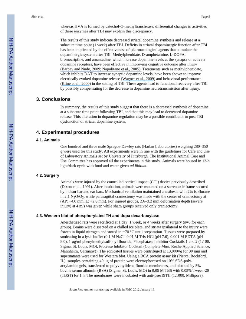
2.3. In vivo tyrosine hydroxylase activityAfter Inhibition of DDC activity by NSD-1015 injection, L-DOPA accumulation wasdetected by HPLC in striatal tissue. The accumulation of L-DOPA was used to assess TH
Shin et al. Page 2
Brain Res. Author manuscript; available in PMC 2012 January 19.
NIH
-PA Author Manuscript
NIH
-PA Author Manuscript
NIH
-PA Author Manuscript
activity level. At 1 day after CCI (Fig. 4a), there is a no change TH activity (sham:100.0±9.7%, injured: 138.0±17.9%). At 1 week after CCI (Fig. 4b), TH activity wasdecreased significantly in the CCI injured animals compared to sham animals (sham:100.0±10.6%, injured: 62.1± 8.2%, p≤0.05). TH activity remains decreased up to 4 weeks(sham: 100.0±5.2%, injured: 68.8±6.2%, p≤0.05) (Fig. 4c).
2.4. Microdialysis for dopamine release and dopamine metabolitesTo assess the changes in striatal extracellular dopamine and dopamine metabolites,microdialysis was used in conjunction with high-performance liquid chromatography(HPLC). At 1 day after CCI (Fig. 5a), the peak levels of injured striatal dopamine after 80mM potassium stimulus was not statistically different from peak sham striatal dopamine(sham: 0.105±0.032 μM, injured: 0.153±0.056 μM). The basal levels of DOPAC (Fig. 6a)were also not significantly different between the sham and injured animals (sham: 0.829 ±0.043 μM, injured: 1.148± 0.130 μM). However, baseline HVA levels of injured animalswere significantly higher compared to the levels of sham animals (sham: 0.547±0.046,injured: 0.789±0.079, p≤0.05) (Fig. 7a).
At 1 week after CCI (Fig. 5b), the peak levels of potassium-evoked striatal dopamine releaseshowed a statistically significant decrease (sham: 0.127±0.027 μM, injured: 0.067± 0.015μM, p≤0.05). There was no difference between DOPAC levels of sham and injured animals(sham: 0.872±0.114 μ injured: 0.661±0.126 μM) (Fig. 6b). HVA levels were also notsignificantly different between the sham and injured animals (sham: 0.569±0.074 μM,injured: 0.520±0.112 μM) (Fig. 7b). By 4 weeks after CCI (Figs. 5c, 6c, 7c), there are nosignificant differences between the sham and injured groups for potassium stimulateddopamine (sham: 0.191±0.017 μM, injured: 0.151 ± 0.024 μM). Also, the baseline values ofdopamine metabolites showed no significant differences: DOPAC (sham: 0.937±0.061 μM,injured: 0.943±0.078 μM), and HVA (sham: 0.672±0.061 μM, injured: 0.703±0.097 μM).
3. DiscussionIn this study, we demonstrated for the first time a deficit in striatal TH activity 1 week(subacute) and 4 weeks (chronic) after TBI in rats as indicated by decreased tissue levels ofL-DOPA. There was decreased pser40TH in injured striatum at 1 week but not at 4 weeks.Since TH is a rate-limiting enzyme in dopamine synthesis, the decrease in its activitysuggests a dopamine synthesis deficit. However, a previous report of striatal dopaminecontent after TBI demonstrates no significant change at 1 week or 4 weeks after injury(Massucci et al., 2004). Dopamine levels depend on both synthesis and degradation.Therefore, activities of monoamine oxidase and catechol-O-methyl transferase may bedecreased at these time points, reducing the degradation of dopamine. Future studies areneeded to elucidate TBI induced alterations of various synthesis and metabolizing enzymesfor dopamine.
The Western blot results showing decreased level of pser40TH suggest decreased activity ofTH, since the level of pser40TH correlates with enzymatic TH activity (Lindgren et al.,2000; Harada et al., 1996; Waymire et al., 1988). However, the level of pser19TH has nosignificant decrease. Since the level of pser19TH does not correlate with TH activity(Jedynak et al., 2002), a specific decrease in pser40TH level is consistent with decreasedactivity of TH. Previously reported TH levels (Yan et al., 2007) showed TH increase at 4weeks post injury. The chronic increase in striatal TH may be a compensatory action of thestriatum. After the TH activity decreases at 1 week after injury, total TH levels may beupregulated in the striatum at a chronic time point in order to increase the efficiency ofdopamine neurotransmission during recovery. Also, present data on decreased striatal THactivity after injury contrast with previous study showing increased TH activity in the
Shin et al. Page 3
Brain Res. Author manuscript; available in PMC 2012 January 19.
NIH
-PA Author Manuscript
NIH
-PA Author Manuscript
NIH
-PA Author Manuscript
medial prefrontal cortex (Kobori et al., 2006). This implicates that TH activity levels may bealtered differently in a region specific manner.
In this study, decreased activity of PKA is demonstrated at 1 week after TBI. However at 1day and 4 weeks, there are no significant changes. Since PKA is a major regulator of theserine 40 site of TH, the activity level of PKA is in agreement with pser40TH levels at 1day, 1 week, and 4 weeks after injury. Deficits in PKA activity at 1 week after injury maycause decreased TH phosphorylation, which could then reduce TH activity. It should also benoted that other kinases such as protein kinase C and protein kinase G may also regulatepser40TH levels (Dunkley et al., 2004), although less extensively characterized than PKA’seffect is on pser40TH. At 4 weeks, there is a recovery of PKA activity and pser40TH levelsin injured striatum, but TH activity deficits are still present (Fig. 4c). This suggests thatwhile TH activity may partially depend on phosphorylation of pser40TH by PKA, otherfactors in the setting of injury could contribute to activity deficits.
A decrease in PKA activity has been previously reported in the parietal cortex andhippocampus at acute time points (15 min to 48 h) using a fluid percussion injury model(Atkins et al., 2007). In contrast, pser40TH level and PKA activity in our study do notdecrease at 1 day after injury. However, this study and our current study are not directlycomparable due to the differences in brain regions studied, injury models, and time pointsafter injury. The directionality of PKA activity change may depend strongly on each of thesefactors.
Dopamine release induced by potassium stimulation has been used to compare differencesof exocytic release in young and aged rats (Shui et al., 1998; Stanford et al., 2000) anddifferent dietary treatments (Agut et al., 2000; Wang et al., 2005). Consistent with theseprevious studies, potassium stimulation induces dopamine release and decreasesextracellular DOPAC and HVA concentrations in our current data. Dopamine release is notsignificantly altered at 1 day after injury but shows a decrease at 1 week after CCI injurycompared to sham injured animals. These data are in agreement with a previous studyshowing reduction in medial forebrain bundle-stimulated evoked dopamine release 2 weeksafter CCI injury detected by fast scanning cyclic voltammetry (Wagner et al., 2005). By 4weeks, there is a recovery of dopamine release in injured animals.
Potassium-stimulated dopamine release depends on newly synthesized dopamine, sincedepletion of vesicular stores of dopamine using reserpine does not alter the potassiumevoked dopamine release (Fairbrother et al., 1990). Thus, our microdialysis data showingdecrease of dopamine release associated with a dopamine synthesis deficit at 1 week areconsistent with a decrease in TH activity shown by in vivo activity assay and pser40THWestern blot results.
The fact that evoked dopamine levels show a decrease in injured animals at 1 week whereasdopamine metabolites show no change may be due to a combination of release and reuptake.It should be noted that the extracellular level of dopamine is dependent on activity levels ofdopamine transporters, which rapidly uptake it to presynaptic terminals. However, DOPACand HVA do not utilize such rapid reuptake mechanism. Previous study from our group hasshown that dopamine transporter function is not reduced at 2 weeks after TBI (Wilson et al.,2005). If there is no functional deficit in dopamine transporter also at 1 week, this mayexplain the discrepancy between extracellular levels of dopamine and dopamine metabolitesfor injured animals.
At 1 day after TBI, there is a significant increase in baseline HVA levels but not DOPAClevels. Since DOPAC is formed by monoamine oxidase and aldehyde dehydrogenase
Shin et al. Page 4
Brain Res. Author manuscript; available in PMC 2012 January 19.
NIH
-PA Author Manuscript
NIH
-PA Author Manuscript
NIH
-PA Author Manuscript
whereas HVA is formed by catechol-O-methyltransferase, differential changes in activitiesof these enzymes after TBI may explain this discrepancy.
The results of this study indicate decreased striatal dopamine synthesis and release at asubacute time point (1 week) after TBI. Deficits in striatal dopaminergic function after TBIhas been implicated by the effectiveness of pharmacological agents that stimulate thedopaminergic system after TBI. Methylphenidate, D-amphetamine, L-DOPA,bromocriptine, and amantadine, which increase dopamine levels at the synapse or activatedopamine receptors, have been effective in improving cognitive outcome after injury(Barbay and Nudo, 2009; Napolitano et al., 2005). Treatments such as methylphenidate,which inhibits DAT to increase synaptic dopamine levels, have been shown to improveelectrically evoked dopamine release (Wagner et al., 2009) and behavioral performance(Kline et al., 2000) in the setting of TBI. These agents lead to functional recovery after TBIby possibly compensating for the decrease in dopamine neurotransmission after injury.
3. ConclusionsIn summary, the results of this study suggest that there is a decreased synthesis of dopamineat a subacute time point following TBI, and that this may lead to decreased dopaminerelease. This alteration in dopamine regulation may be a possible contributor to post TBIdysfunction of striatal dopamine system.
4. Experimental procedures4.1. Animals
One hundred and three male Sprague-Dawley rats (Harlan Laboratories) weighing 280–350g were used for this study. All experiments were in line with the guidelines for Care and Useof Laboratory Animals set by University of Pittsburgh. The Institutional Animal Care andUse Committee has approved all the experiments in this study. Animals were housed in 12-hlight/dark cycle with food and water given ad libitum.
4.2. SurgeryAnimals were injured by the controlled cortical impact (CCI) device previously described(Dixon et al., 1991). After intubation, animals were mounted on a stereotaxic frame securedby incisor bar and ear bars. Mechanical ventilation maintained anesthesia with 2% isofluranein 2:1 N2O/O2, while parasagittal craniectomy was made with the center of craniectomy at(AP: +4.0 mm, L: +2.8 mm). For injured groups, 2.6–3.2 mm deformation depth (severeinjury) at 4 m/s was given while sham groups received only craniectomy.
4.3. Western blot of phosphorylated TH and dopa decarboxylaseAnesthetized rats were sacrificed at 1 day, 1 week, or 4 weeks after surgery (n=6 for eachgroup). Brains were dissected on a chilled ice plate, and striata ipsilateral to the injury werefrozen in liquid nitrogen and stored in −70 °C until preparation. Tissues were prepared bysonicating in a lysis buffer (0.1 M NaCl, 0.01 M Tris-HCl (pH 7.6), 0.001 M EDTA (pH8.0), 1 μg/ml phenylmethylsulfonyl fluoride, Phosphatase Inhibitor Cocktails 1 and 2 (1:100,Sigma, St. Louis, MO), Protease Inhibitor Cocktail (Complete Mini, Roche Applied Science,Mannheim, Germany)). The sonicated tissues were centrifuged at 13,000×g for 30 min andsupernatants were used for Western blot. Using a BCA protein assay kit (Pierce, Rockford,IL), samples containing 40 μg of protein were electrophoresed on 10% SDS-poly-acrylamide gels, transferred to polyvinylidene fluoride membranes, and blocked by 5%bovine serum albumin (BSA) (Sigma, St. Louis, MO) in 0.05 M TBS with 0.05% Tween-20(TBST) for 1 h. The membranes were incubated with anti-pser19TH (1:1000, Millipore),
Shin et al. Page 5
Brain Res. Author manuscript; available in PMC 2012 January 19.
NIH
-PA Author Manuscript
NIH
-PA Author Manuscript
NIH
-PA Author Manuscript
anti-pser40TH (1:1000, Millipore), or anti-DDC (1:1000, Sigma, St. Louis, MO) overnight,then washed with TBST and incubated for 1 h at room temperature with anti-mouse or anti-rabbit immunoglobulin G-conjugated to peroxidase (1:20,000, Pierce). Membranes wereexposed to chemiluminescence (Western Lighting, Perkins Elmer, Boston, MA) andpser19TH, pser40TH, and DDC were visualized by exposing the membranes toautoradiographic X-ray film from 10 s to 1 min. Afterwards, membranes were stripped using100 mM glycine pH 2.3 at 55 °C for 1 h, incubated with anti-β-actin monoclonal antibody(1:10,000, Sigma-Aldrich) for 1 h, then incubated with anti-mouse immunoglobulin G-conjugated to peroxidase. Same steps were taken as described to visualize β-actin blots. Tomeasure the optical density of Western blots, Scion Image PC software (Frederick, MD) wasused. Optical densities of pser19TH, pser40TH, and DDC were normalized by β-actin ofeach blot, and the values displayed are given as a percentage change compared to shamtissue levels.
4.4. PKA activity assayThe activity level of PKA was found using a commercially available kit (Promega, Madison,WI). Briefly, the tissue homogenates used for Western blots were suspended in PepTag PKA5× reaction buffer, Peptag A1 peptide, and PKA activator 5× solutions. These mixtures wereincubated at room temperature for 30 min. The reaction was stopped by placing the mixturein boiling water for 10 min. Glycerol (30%, 1 μl) was added to the mixture, and sampleswere loaded onto 0.8% agarose/Tris–HCl gel. The gels were run at 100 V until separation ofphosphorylated and nonphosphorylated samples became apparent. The gels were thenscanned using Kodak Image Station 440CF. The optical densities of the phosphorylatedproduct from PKA reaction were then normalized by total protein level in each sample usingNIH Image J software. PKA activity levels were reported with respect to sham striatal PKAactivity levels.
4.5. In vivo TH activity assayAs described in Urbanavicius et al. (2007), in vivo TH activity was assessed by quantifyingL-DOPA accumulation after inhibiting DDC with 3-hydroxybenzylhydrazine (NSD-1015)(Sigma, St. Louis, MO). Thirty minutes before sacrifice, rats were intraperitoneally injectedwith NSD-1015 (100 mg/kg, suspended in 0.9% saline). Striata were dissected out,immediately frozen in liquid nitrogen, and stored in −70 °C until neurochemical analysis.On the day of analysis, the tissues were weighed and sonicated in 0.2 M HClO2. Thesamples were then centrifuged at 13,000×g for 30 min and supernatants were used toquantify L-DOPA levels using HPLC.
4.6. MicrodialysisArtificial cerebrospinal fluid (ACSF) with 126.5 mM NaCl, 2.4 mM KCl, 1.1 mM CaCl2,0.83 mM MgCl2, 27.5 mM NaHCO3, and 0.5 mM KH2PO4 was used for this experiment. Inthe high-potassium ACSF, KCl concentration was adjusted to 80 mM and NaClconcentration was adjusted to 48.9 mM to maintain osmolarity. One day before micro-dialysis experiment, a microdialysis probe (SciPro, Sanborn, NY) with the followingspecifications were used: membrane length: 3 mm, diameter: 0.6 mm, permeability cutoff:35 kDa. The probe was implanted into the striatum (AP: +0.0 mm, L: +2.8 mm, DV: −4.0mm) and secured with dental cement. The animals were then disconnected from theanesthesia apparatus and placed in a Plexiglas chamber as previously described (Dixon etal., 1997). Microdialysates were collected in awake, freely moving animals. Overnight,ACSF was continuously perfused at 0.2 μl/min. On the day of the experiment, flow rate wasadjusted to 2 μl/min for 1 h then samples were collected every 20 min into a tube containing5 μl of 0.3 M HClO2. Samples were immediately analyzed by HPLC. At 60 min (4thcollection time), ACSF was switched to a high-potassium ACSF solution. Forty minutes
Shin et al. Page 6
Brain Res. Author manuscript; available in PMC 2012 January 19.
NIH
-PA Author Manuscript
NIH
-PA Author Manuscript
NIH
-PA Author Manuscript
after beginning of stimulus (6th collection time), potassium challenge was stopped andACSF was infused. After microdialysis, the rats were sacrificed and the locations of theprobes were verified. For data analysis, micromolar concentrations of dopamine anddopamine metabolites are reported.
4.7. Neurochemical analysisNeurochemical measurements were made by HPLC with CoulArray Detector using twofour-channel analytical cells (ESA, Chelmsford, MA, USA). Eight coulometric electrodeswith potentials from −120 to +300 mV in 60 mV increments were used, and C18 columnwas used to separate the analytes. Dopamine, 3,4-dihydroxyphenylacetic acid (DOPAC),and homovanillic acid (HVA) were monitored in microdialysates. From striatalhomogenates for TH activity assay, L-DOPA was monitored. Baseline values of dopaminebelow the linear range of detection were assigned values of 0 μM.
4.8. Statistical analysisThe normalized TH activity levels and optical densities of DDC, pser19TH, and pser40THwere analyzed using unpaired Student’s T test. Baseline values of DOPAC and HVA werereported as their average level in first three microdialysates detected by HPLC. Forpotassium-evoked dopamine release, the peak values were used to compare the sham injuredand CCI injured animals. Statistical analysis for microdialysis data was performed using anonparametric Mann-Whitney U Test. All statistical calculations were performed by usingPASW Statistics 19 (SPSS Inc., Chicago, IL) software.
AcknowledgmentsWe would like to thank Ruth Perez and Teresa Hastings for insightful comments and suggestions. This project issupported by NIH grant 1F30NS067731-01, NIH grant 5R01NS060672-02, and the Copeland Fund of thePittsburgh Foundation.
ReferencesAgut J, Ortiz JA, Wurtman RJ. Cytidine (5′)diphosphocholine modulates dopamine K(+)-evoked
release in striatum measured by microdialysis. Ann NY Acad Sci. 2000; 920:332–335. [PubMed:11193174]
Anden NE, Carlsson A, Dahlstroem A, Fuxe K, Hillarp NA, Larsson K. Demonstration and mappingout of nigrostriatal dopamine neurons. Life Sci. 1964; 3:523–530. [PubMed: 14187491]
Atkins CM, Oliva AA Jr, Alonso OF, Pearse DD, Bramlett HM, Dietrich DD. Modulation of thecAMP signaling pathway after traumatic brain injury. Exp Neurol. 2007; 208:145–158. [PubMed:17916353]
Bak IJ, Hassler R, Kim JS, Kataoka K. Amantadine actions on acetylcholine and GABA in striatumand substantia nigra of rat in relation to behavioral changes. J Neural Transm. 1972; 33:45–61.[PubMed: 4674484]
Bales JW, Wagner AK, Kline AE, Dixon CE. Persistent cognitive dysfunction after traumatic braininjury: a dopamine hypothesis. Neurosci Biobehav Rev. 2009; 33:981–1003. Epub 2009 Apr 1.Review. [PubMed: 19580914]
Barbay S, Nudo RJ. The effects of amphetamine on recovery of function in animal models of cerebralinjury: a critical appraisal. NeuroRehabilitation. 2009; 25:5–17. [PubMed: 19713615]
Dixon CE, Ma X, Marion DW. Reduced evoked release of acetylcholine in the rodent neocortexfollowing traumatic brain injury. Brain Res. 1997; 21 (749):127–130. [PubMed: 9070636]
Dixon CE, Kraus MF, Kline AE, Ma X, Yan HQ, Griffith RG, Wolfson BM, Marion DW. Amantadineimproves water maze performance without affecting motor behavior following traumatic braininjury in rats. Restor Neurol Neurosci. 1999; 14:285–294. [PubMed: 12671249]
Shin et al. Page 7
Brain Res. Author manuscript; available in PMC 2012 January 19.
NIH
-PA Author Manuscript
NIH
-PA Author Manuscript
NIH
-PA Author Manuscript
Dunkley PR, Bobrovskaya L, Graham ME, von Nagy-Felsobuki EI, Dickson PW. Tyrosinehydroxylase phosphorylation: regulation and consequences. J Neurochem. 2004; 91:1025–1043.Review. [PubMed: 15569247]
Fairbrother IS, Arbuthnott GW, Kelly JS, Butcher SP. In vivo mechanisms underlying dopaminerelease from rat nigrostriatal terminals: II. Studies using potassium and tyramine. J Neurochem.1990; 54:1844–1851. [PubMed: 2338545]
Feeney DM, Gonzales A, Law WA. Amphetamine restores locomotor function after motor cortexinjury in the rat. Proc West Pharmacol Soc. 1981; 24:15–17. [PubMed: 7255439]
Gianutsos G, Chute S, Dunn JP. Pharmacological changes in dopaminergic systems induced by long-term administration of amantadine. Eur J Pharmacol. 1985; 110:357–361. [PubMed: 2861102]
Harada L, Wu J, Haycock JW, Goldstein M. Regulation of L-DOPA biosynthesis by site-specificphosphorylation of tyrosine hydroxylase in AtT-20 cells expressing wild-type and serine 40-substituted enzyme. J Neurochem. 1996; 67:629–635. [PubMed: 8764589]
Hokfelt T, Johansson O, Fuxe K, Goldstein M, Park D. Immunohistochemical studies on thelocalization and distribution of monoamine neuron systems in the rat brain II. Tyrosinehydroxylase in the telencephalon. Med Biol. 1977; 55:21–40. [PubMed: 15169]
Hornstein A, Lennihan L, Seliger G, Lichtman S, Schroeder K. Amphetamine in recovery from braininjury. Brain Inj. 1996; 10:145–148. [PubMed: 8696315]
Jedynak JP, Ali SF, Haycock JW, Hope BT. Acute administration of cocaine regulates thephosphorylation of serine-19, -31 and -40 in tyrosine hydroxylase. J Neurochem. 2002; 82:382–388. [PubMed: 12124439]
Kline AE, Chen MJ, Tso-Olivas DY, Feeney DM. Methylphenidate treatment following ablation-induced hemiplegia in rat: experience during drug action alters effects on recovery of function.Pharmacol Biochem Behav. 1994; 48:773–779. [PubMed: 7938134]
Kline AE, Yan HQ, Bao J, Marion DW, Dixon CE. Chronic methylphenidate treatment enhanceswater maze performance following traumatic brain injury in rats. Neurosci Lett. 2000; 280:163–166. [PubMed: 10675786]
Kobori N, Clifton GL, Dash PK. Enhanced catecholamine synthesis in the prefrontal cortex aftertraumatic brain injury: implications for prefrontal dysfunction. J Neurotrauma. 2006; 23:1094–1102. [PubMed: 16866622]
Koeda T, Takeshita K. A case report of remarkable improvement of motor disturbances with L-DOPAin a patient with post-diffuse axonal injury. Brain Dev. 1998; 20:124–126. [PubMed: 9545185]
Kraus MF, Maki P. The combined use of amantadine and L-DOPA/carbidopa in the treatment ofchronic brain injury. Brain Inj. 1997; 11:455–460. [PubMed: 9171930]
Lal S, Merbtiz CP, Grip JC. Modification of function in head-injured patients with Sinemet. Brain Inj.1988; 2:225–233. [PubMed: 2458792]
Lindgren N, Xu ZQ, Lindskog M, Herrera-Marschitz M, Goiny M, Haycock J, Goldstein M, HokfeltT, Fisone G. Regulation of tyrosine hydroxylase activity and phosphorylation at Ser(19) andSer(40) via activation of glutamate NMDA receptors in rat striatum. J Neurochem. 2000; 74:2470–2477. [PubMed: 10820208]
Massucci JL, Kline AE, Ma X, Zafonte RD, Dixon CE. Time dependent alterations in dopamine tissuelevels and metabolism after traumatic brain injury in rats. Neurosci Lett. 2004; 372:127–131.[PubMed: 15531102]
McIntosh TK, Yu T, Gennarelli TA. Alterations in regional brain catecholamine concentrations afterexperimental brain injury in the rat. J Neurochem. 1994; 63:1426–1433. [PubMed: 7931293]
Napolitano E, Elovic EP, Qureshi AI. Pharmacological stimulant treatment of neurocognitive andfunction deficits after traumatic and non-traumatic brain injury. Med Sci Monit. 2005; 11:RA212–RA220. [PubMed: 15917733]
Phillips JP, Devier DJ, Feeney DM. Rehabilitation pharmacology: bridging laboratory work to clinicalapplication. J Head Trauma Rehabil. 2003; 18:342–356. [PubMed: 16222129]
Sawyer E, Mauro LS, Ohlinger MJ. Amantadine enhancement of arousal and cognition after traumaticbrain injury. Ann Pharmacother. 2008; 42:247–252. [PubMed: 18212258]
Shin et al. Page 8
Brain Res. Author manuscript; available in PMC 2012 January 19.
NIH
-PA Author Manuscript
NIH
-PA Author Manuscript
NIH
-PA Author Manuscript
Shui HA, Peng YI, Tsai YF. Recovery of high potassium-evoked dopamine release after depolazationchallenge in the striatum of young and old male rats. Neurosci Lett. 1998; 257:1–4. [PubMed:9857951]
Stanford JA, Giardina K, Gerhardt GA. In vivo microdialysis studies of age-related alterations inpotassium-evoked overflow of dopamine in the dorsal striatum of Fischer 344 rats. Int J DevNeurosci. 2000; 18:411–416. [PubMed: 10817924]
Urbanavicius J, Ferreira M, Costa G, Abin-Carriquiry JA, Wonnacott S, Dajas F. Nicotine inducestyrosine hydroxylase plasticity in the neurodegenerating striatum. J Neurochem. 2007; 102:723–730. [PubMed: 17437548]
Von Voightlander PF, Moore KE. Dopamine: release from the brain in vivo by amantadine. Science.1971; 174:408–410. [PubMed: 5111993]
Wagner AK, Sokoloski JE, Ren D, Chen X, Khan AS, Zafonte RD, Michael AC, Dixon CE.Controlled cortical impact injury affects dopaminergic transmission in the rat striatum. JNeurochem. 2005; 95:457–465. [PubMed: 16190869]
Wagner AK, Sokoloski JE, Chen X, Harun R, Clossin DP, Khan AS, Andres-Koback M, Michael AC,Dixon CE. Controlled cortical impact injury influences methylphenidate-induced changes instriatal dopamine neurotransmission. J Neurochem. 2009; 110:801–810. [PubMed: 19457094]
Wang L, Pooler AM, Albrecht MA, Wurtman RJ. Dietary uridine-5′-monophosphate supplementationincreases potassium-evoked dopamine release and promotes neurite outgrowth in aged rats. J MolNeurosci. 2005; 27:137–145. [PubMed: 16055952]
Waymire JC, Johnston JP, Hummer-Lickteig K, Lloyd A, Vigny A, Craviso GL. Phosphorylation ofbovine adrenal chromaffin cell tyrosine hydroxylase. Temporal correlation of acetylcholine’seffect on site phosphorylation, enzyme activation, and catecholamine synthesis. J Biol Chem.1988; 263:12439–12447. [PubMed: 2900836]
Wilson MS, Chen X, Ma X, Ren D, Wagner AK, Reynolds IJ, Dixon CE. Synaptosomal dopamineuptake in rat striatum following controlled cortical impact. J Neurosci Res. 2005; 80:85–91.[PubMed: 15704194]
Yan HQ, Ma X, Chen X, Li Y, Shao L, Dixon CE. Delayed increase in tyrosine hydroxylaseexpression in rat nigrostriatal system after traumatic brain injury. Brain Res. 2007; 1134:171–179.[PubMed: 17196177]
Shin et al. Page 9
Brain Res. Author manuscript; available in PMC 2012 January 19.
NIH
-PA Author Manuscript
NIH
-PA Author Manuscript
NIH
-PA Author Manuscript
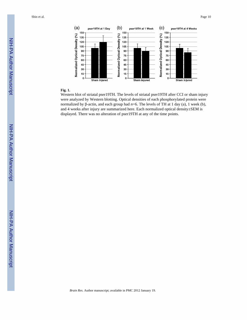
Fig. 1.Western blot of striatal pser19TH. The levels of striatal pser19TH after CCI or sham injurywere analyzed by Western blotting. Optical densities of each phosphorylated protein werenormalized by β-actin, and each group had n=6. The levels of TH at 1 day (a), 1 week (b),and 4 weeks after injury are summarized here. Each normalized optical density±SEM isdisplayed. There was no alteration of pser19TH at any of the time points.
Shin et al. Page 10
Brain Res. Author manuscript; available in PMC 2012 January 19.
NIH
-PA Author Manuscript
NIH
-PA Author Manuscript
NIH
-PA Author Manuscript
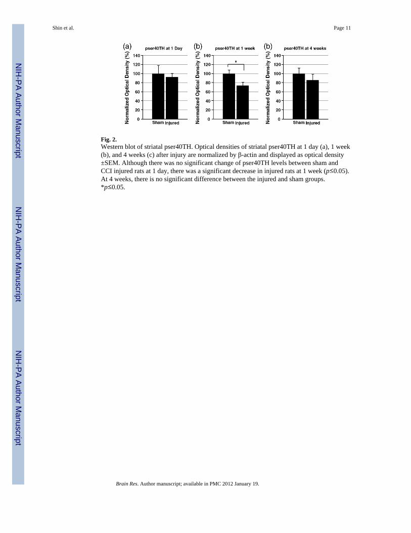
Fig. 2.Western blot of striatal pser40TH. Optical densities of striatal pser40TH at 1 day (a), 1 week(b), and 4 weeks (c) after injury are normalized by β-actin and displayed as optical density±SEM. Although there was no significant change of pser40TH levels between sham andCCI injured rats at 1 day, there was a significant decrease in injured rats at 1 week (p≤0.05).At 4 weeks, there is no significant difference between the injured and sham groups.*p≤0.05.
Shin et al. Page 11
Brain Res. Author manuscript; available in PMC 2012 January 19.
NIH
-PA Author Manuscript
NIH
-PA Author Manuscript
NIH
-PA Author Manuscript
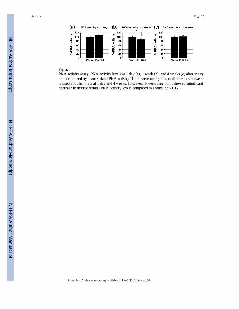
Fig. 3.PKA activity assay. PKA activity levels at 1 day (a), 1 week (b), and 4 weeks (c) after injuryare normalized by sham striatal PKA activity. There were no significant differences betweeninjured and sham rats at 1 day and 4 weeks. However, 1-week time point showed significantdecrease in injured striatal PKA activity levels compared to shams. *p≤0.05.
Shin et al. Page 12
Brain Res. Author manuscript; available in PMC 2012 January 19.
NIH
-PA Author Manuscript
NIH
-PA Author Manuscript
NIH
-PA Author Manuscript
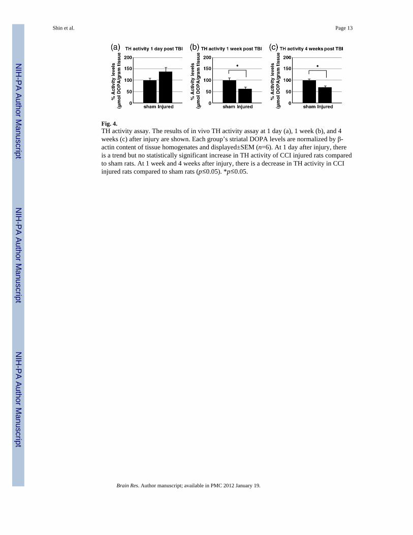
Fig. 4.TH activity assay. The results of in vivo TH activity assay at 1 day (a), 1 week (b), and 4weeks (c) after injury are shown. Each group’s striatal DOPA levels are normalized by β-actin content of tissue homogenates and displayed±SEM (n=6). At 1 day after injury, thereis a trend but no statistically significant increase in TH activity of CCI injured rats comparedto sham rats. At 1 week and 4 weeks after injury, there is a decrease in TH activity in CCIinjured rats compared to sham rats (p≤0.05). *p≤0.05.
Shin et al. Page 13
Brain Res. Author manuscript; available in PMC 2012 January 19.
NIH
-PA Author Manuscript
NIH
-PA Author Manuscript
NIH
-PA Author Manuscript
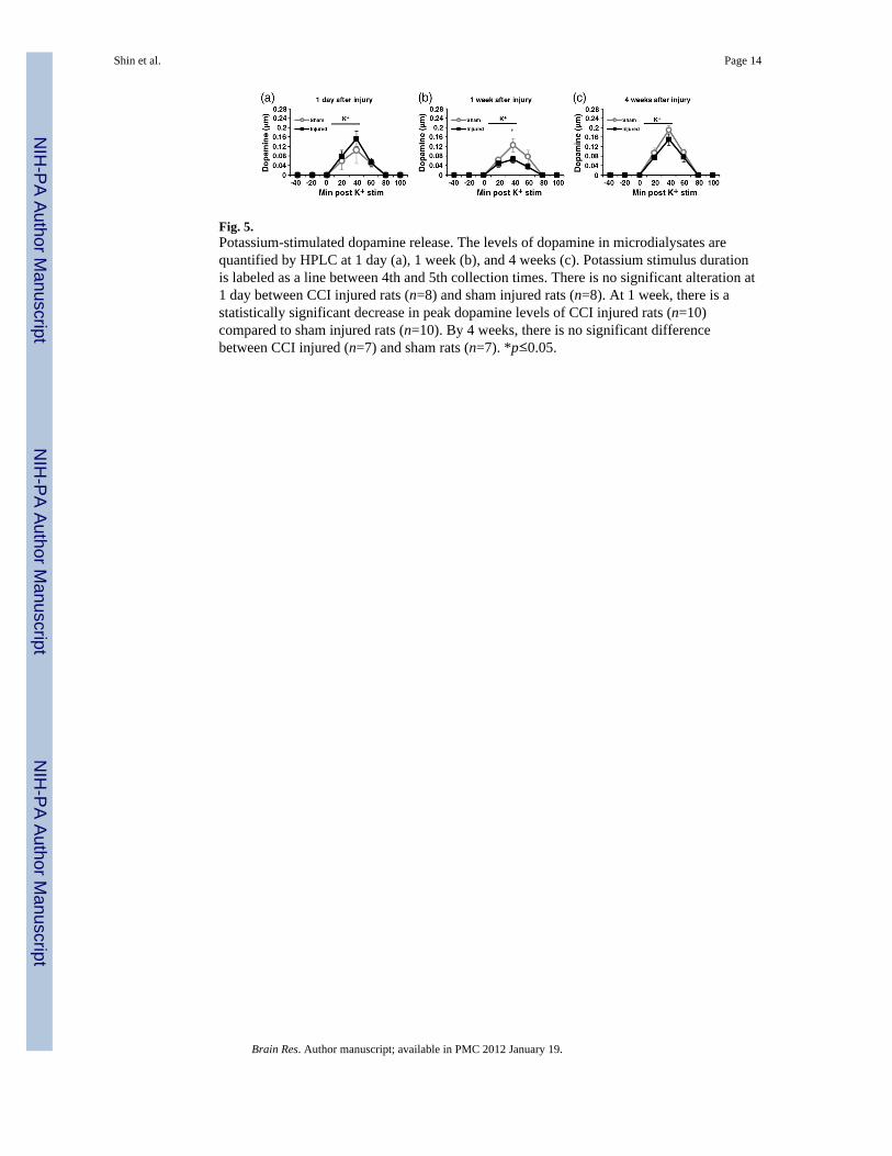
Fig. 5.Potassium-stimulated dopamine release. The levels of dopamine in microdialysates arequantified by HPLC at 1 day (a), 1 week (b), and 4 weeks (c). Potassium stimulus durationis labeled as a line between 4th and 5th collection times. There is no significant alteration at1 day between CCI injured rats (n=8) and sham injured rats (n=8). At 1 week, there is astatistically significant decrease in peak dopamine levels of CCI injured rats (n=10)compared to sham injured rats (n=10). By 4 weeks, there is no significant differencebetween CCI injured (n=7) and sham rats (n=7). *p≤0.05.
Shin et al. Page 14
Brain Res. Author manuscript; available in PMC 2012 January 19.
NIH
-PA Author Manuscript
NIH
-PA Author Manuscript
NIH
-PA Author Manuscript
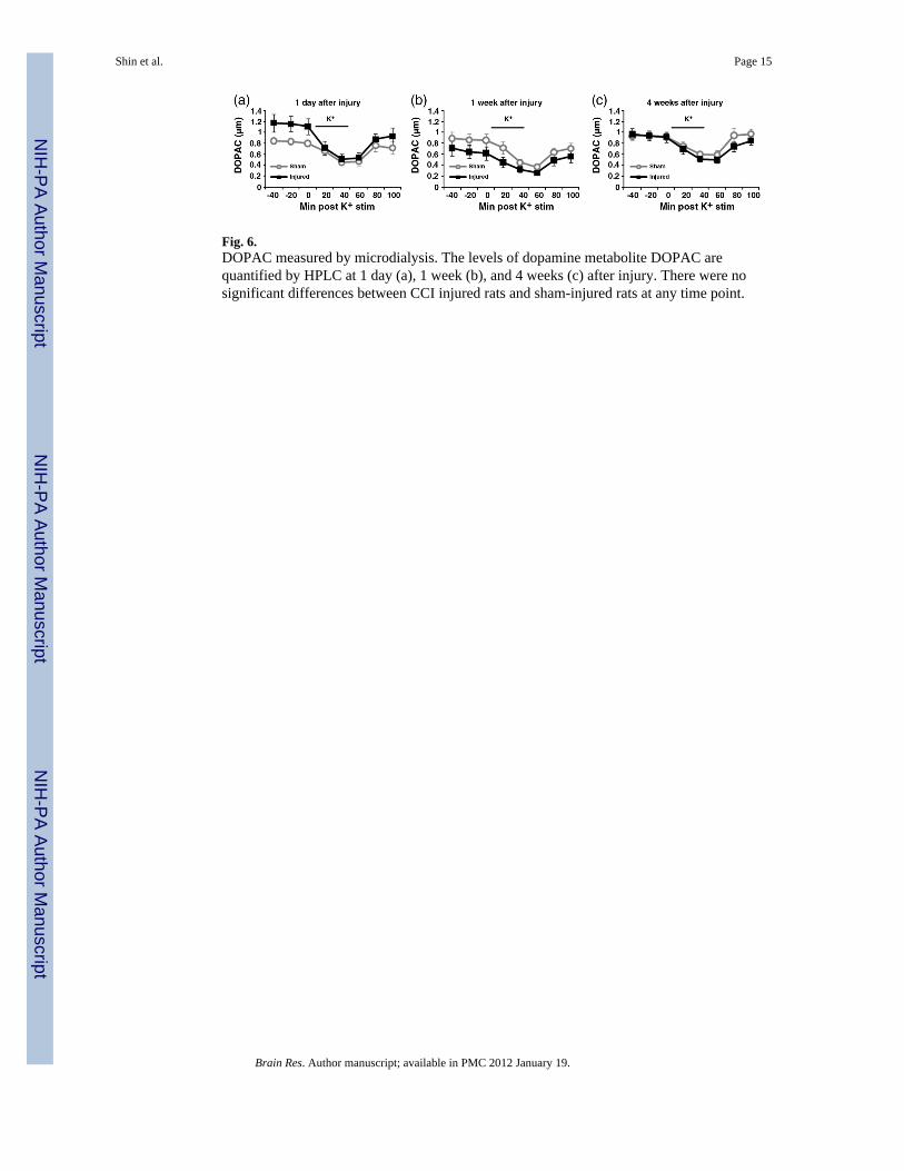
Fig. 6.DOPAC measured by microdialysis. The levels of dopamine metabolite DOPAC arequantified by HPLC at 1 day (a), 1 week (b), and 4 weeks (c) after injury. There were nosignificant differences between CCI injured rats and sham-injured rats at any time point.
Shin et al. Page 15
Brain Res. Author manuscript; available in PMC 2012 January 19.
NIH
-PA Author Manuscript
NIH
-PA Author Manuscript
NIH
-PA Author Manuscript
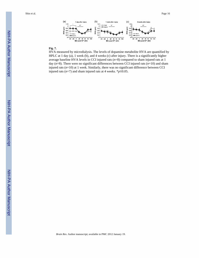
Fig. 7.HVA measured by microdialysis. The levels of dopamine metabolite HVA are quantified byHPLC at 1 day (a), 1 week (b), and 4 weeks (c) after injury. There is a significantly higheraverage baseline HVA levels in CCI injured rats (n=8) compared to sham injured rats at 1day (n=8). There were no significant differences between CCI injured rats (n=10) and shaminjured rats (n=10) at 1 week. Similarly, there was no significant difference between CCIinjured rats (n=7) and sham injured rats at 4 weeks. *p≤0.05.
Shin et al. Page 16
Brain Res. Author manuscript; available in PMC 2012 January 19.
NIH
-PA Author Manuscript
NIH
-PA Author Manuscript
NIH
-PA Author Manuscript
Copyright © 2022 FDOKUMEN