Neurons in the Thalamic CM-Pf Complex Supply Striatal ...
17
Neurons in the Thalamic CM-Pf Complex Supply Striatal Neurons With Information About Behaviorally Significant Sensory Events NAOYUKI MATSUMOTO, 1,3 TAKAFUMI MINAMIMOTO, 1 ANN M. GRAYBIEL, 2 AND MINORU KIMURA 1,3 1 Faculty of Health and Sport Sciences, Osaka University, Osaka 560-0043, Japan; 2 Department of Brain and Cognitive Sciences, MIT, Cambridge, Massachusetts 02139; and 3 Department of Physiology, Kyoto Prefectural University of Medicine, Kyoto 602-8566, Japan Received 1 May 2000; accepted in final form 2 November 2000 Matsumoto, Naoyuki, Takafumi Minamimoto, Ann M. Graybiel, and Minoru Kimura. Neurons in the thalamic CM-Pf complex supply striatal neurons with information about behaviorally significant sensory events. J Neurophysiol 85: 960 –976, 2001. The projection from the thalamic centre me ´dian–parafascicular (CM-Pf) complex to the caudate nucleus and putamen forms a massive striatal input system in primates. We examined the activity of 118 neurons in the CM and 62 neurons in the Pf nuclei of the thalamus and 310 tonically active neurons (TANs) in the striatum in awake behaving macaque monkeys and analyzed the effects of pharmacologic inactivation of the CM-Pf on the sensory responsiveness of the striatal TANs. A large proportion of CM and Pf neurons responded to visual (53%) and/or auditory beep (61%) or click (91%) stimuli presented in behavioral tasks, and many responded to unexpected auditory, visual, or somatosensory stimuli presented outside the task context. The neurons fell into two classes: those having short-latency facilitatory responses (SLF neurons, pre- dominantly in the Pf) and those having long-latency facilitatory re- sponses (LLF neurons, predominantly in the CM). Responses of both types of neuron appeared regardless of whether or not the sensory stimuli were associated with reward. These response characteristics of CM-Pf neurons sharply contrasted with those of TANs in the striatum, which under the same conditions responded preferentially to stimuli associated with reward. Many CM-Pf neurons responded to alerting stimuli such as unexpected handclaps and noises only for the first few times that they occurred; after that, the identical stimuli gradually became ineffective in evoking responses. Habituation of sensory responses was particularly common for the LLF neurons. Inactivation of neuronal activity in the CM and Pf by local infusion of the GABA A receptor agonist, muscimol, almost completely abolished the pause and rebound facilitatory responses of TANs in the striatum. Such injections also diminished behavioral responses to stimuli associated with reward. We suggest that neurons in the CM and Pf supply striatal neurons with information about behaviorally significant sensory events that can activate conditional responses of striatal neurons in combination with dopamine-mediated nigrostriatal inputs having mo- tivational value. INTRODUCTION The posterior intralaminar nuclei of the thalamus, the centre me ´dian (CM) and parafascicular nucleus (Pf), are small in rodents but are among the largest thalamic nuclei in primates, including humans. These nuclei (together called the CM-Pf complex) are classified as part of the ascending reticulo- thalamo-cortical activating system based on the work of Moruzzi and Magoun (1949), Jasper (1960), and subsequent investigators (see Hu et al. 1989; Mennemeier et al. 1997; Pare ´ et al. 1988; Steriade et al. 1994, 1997). The CM-Pf complex also receives massive inputs from the motor and premotor cortex and from the basal ganglia (Fenelon et al. 1991; Fran- c ¸ois et al. 1991; Macchi and Bentivoglio 1986; Nakano et al. 1990; Sadikot et al. 1992a; Smith and Parent 1986; Steriade et al. 1997). These nuclei do project to the neocortex, but the main outputs of the CM and Pf lead to the striatum (Jones 1997). The CM-Pf complex thus at once is associated with state-setting modulatory systems and with basal ganglia action- gating circuits. Positron emission tomography (PET) evidence supports a general activating function for the CM-Pf complex, in that the CM is specifically activated when subjects shift from a relaxed waking state to an alert state during performance of an atten- tion-demanding reaction time task (Kinomura et al. 1996). Nothing is yet known, however, about the functional activity of CM and Pf neurons in alert behaving monkeys, and it is not yet known what behaviorally significant effects CM-Pf neurons have on their main cellular targets, neurons in the striatum. In the experiments reported here, we approached these issues by recording the activity of single neurons in the CM and the Pf of macaque monkeys as they learned and performed sensori- motor tasks. We then tested for the effects of blocking CM-Pf activity on the response properties of striatal neurons recorded and on accompanying behavioral responses made in the same tasks. We focused the striatal recordings on the striatal neurons that have irregular tonic activity (the tonically active neurons called TANs). These are thought to be the cholinergic inter- neurons of the striatum (Aosaki et al. 1995; Kawaguchi 1992; Kimura et al. 1990; Wilson et al. 1990), which have been shown in anatomical experiments to receive a strong input from the CM-Pf complex (Lapper and Bolam 1992; Sidibe ´ and Smith 1999). We have earlier shown that TANs have the property of acquiring motivation-dependent responses to sen- sory stimuli as a result of reward-based sensorimotor condi- tioning (Aosaki et al. 1994a,b, 1995; Graybiel et al. 1994). By testing for the effects of CM-Pf inactivation on the acquired responses of striatal TANs, we tried to identify the contribution Address for reprint requests: M. Kimura, Dept. of Physiology, Kyoto Pre- fectural University of Medicine, Kawaramachi, Hirokoji Kyoto 602-8566, Japan (E-mail: [email protected]). The costs of publication of this article were defrayed in part by the payment of page charges. The article must therefore be hereby marked ‘‘advertisement’’ in accordance with 18 U.S.C. Section 1734 solely to indicate this fact. 960 0022-3077/01 $5.00 Copyright © 2001 The American Physiological Society www.jn.physiology.org
-
Upload
khangminh22 -
Category
Documents
-
view
2 -
download
0
Transcript of Neurons in the Thalamic CM-Pf Complex Supply Striatal ...

Neurons in the Thalamic CM-Pf Complex Supply Striatal NeuronsWith Information About Behaviorally Significant Sensory Events
NAOYUKI MATSUMOTO,1,3 TAKAFUMI MINAMIMOTO, 1 ANN M. GRAYBIEL,2 AND MINORU KIMURA 1,3
1Faculty of Health and Sport Sciences, Osaka University, Osaka 560-0043, Japan;2Department of Brain and CognitiveSciences, MIT, Cambridge, Massachusetts 02139; and3Department of Physiology, Kyoto Prefectural University of Medicine,Kyoto 602-8566, Japan
Received 1 May 2000; accepted in final form 2 November 2000
Matsumoto, Naoyuki, Takafumi Minamimoto, Ann M. Graybiel,and Minoru Kimura. Neurons in the thalamic CM-Pf complexsupply striatal neurons with information about behaviorally significantsensory events.J Neurophysiol85: 960–976, 2001. The projectionfrom the thalamic centre me´dian–parafascicular (CM-Pf) complex tothe caudate nucleus and putamen forms a massive striatal input systemin primates. We examined the activity of 118 neurons in the CM and62 neurons in the Pf nuclei of the thalamus and 310 tonically activeneurons (TANs) in the striatum in awake behaving macaque monkeysand analyzed the effects of pharmacologic inactivation of the CM-Pfon the sensory responsiveness of the striatal TANs. A large proportionof CM and Pf neurons responded to visual (53%) and/or auditory beep(61%) or click (91%) stimuli presented in behavioral tasks, and manyresponded to unexpected auditory, visual, or somatosensory stimulipresented outside the task context. The neurons fell into two classes:those having short-latency facilitatory responses (SLF neurons, pre-dominantly in the Pf) and those having long-latency facilitatory re-sponses (LLF neurons, predominantly in the CM). Responses of bothtypes of neuron appeared regardless of whether or not the sensorystimuli were associated with reward. These response characteristics ofCM-Pf neurons sharply contrasted with those of TANs in the striatum,which under the same conditions responded preferentially to stimuliassociated with reward. Many CM-Pf neurons responded to alertingstimuli such as unexpected handclaps and noises only for the first fewtimes that they occurred; after that, the identical stimuli graduallybecame ineffective in evoking responses. Habituation of sensoryresponses was particularly common for the LLF neurons. Inactivationof neuronal activity in the CM and Pf by local infusion of the GABAA
receptor agonist, muscimol, almost completely abolished the pauseand rebound facilitatory responses of TANs in the striatum. Suchinjections also diminished behavioral responses to stimuli associatedwith reward. We suggest that neurons in the CM and Pf supply striatalneurons with information about behaviorally significant sensoryevents that can activate conditional responses of striatal neurons incombination with dopamine-mediated nigrostriatal inputs having mo-tivational value.
I N T R O D U C T I O N
The posterior intralaminar nuclei of the thalamus, the centremedian (CM) and parafascicular nucleus (Pf), are small inrodents but are among the largest thalamic nuclei in primates,including humans. These nuclei (together called the CM-Pfcomplex) are classified as part of the ascending reticulo-
thalamo-cortical activating system based on the work ofMoruzzi and Magoun (1949), Jasper (1960), and subsequentinvestigators (see Hu et al. 1989; Mennemeier et al. 1997; Pare´et al. 1988; Steriade et al. 1994, 1997). The CM-Pf complexalso receives massive inputs from the motor and premotorcortex and from the basal ganglia (Fenelon et al. 1991; Fran-cois et al. 1991; Macchi and Bentivoglio 1986; Nakano et al.1990; Sadikot et al. 1992a; Smith and Parent 1986; Steriade etal. 1997). These nuclei do project to the neocortex, but themain outputs of the CM and Pf lead to the striatum (Jones1997). The CM-Pf complex thus at once is associated withstate-setting modulatory systems and with basal ganglia action-gating circuits.
Positron emission tomography (PET) evidence supports ageneral activating function for the CM-Pf complex, in that theCM is specifically activated when subjects shift from a relaxedwaking state to an alert state during performance of an atten-tion-demanding reaction time task (Kinomura et al. 1996).Nothing is yet known, however, about the functional activity ofCM and Pf neurons in alert behaving monkeys, and it is not yetknown what behaviorally significant effects CM-Pf neuronshave on their main cellular targets, neurons in the striatum. Inthe experiments reported here, we approached these issues byrecording the activity of single neurons in the CM and the Pfof macaque monkeys as they learned and performed sensori-motor tasks. We then tested for the effects of blocking CM-Pfactivity on the response properties of striatal neurons recordedand on accompanying behavioral responses made in the sametasks. We focused the striatal recordings on the striatal neuronsthat have irregular tonic activity (the tonically active neuronscalled TANs). These are thought to be the cholinergic inter-neurons of the striatum (Aosaki et al. 1995; Kawaguchi 1992;Kimura et al. 1990; Wilson et al. 1990), which have beenshown in anatomical experiments to receive a strong inputfrom the CM-Pf complex (Lapper and Bolam 1992; Sidibe´ andSmith 1999). We have earlier shown that TANs have theproperty of acquiring motivation-dependent responses to sen-sory stimuli as a result of reward-based sensorimotor condi-tioning (Aosaki et al. 1994a,b, 1995; Graybiel et al. 1994). Bytesting for the effects of CM-Pf inactivation on the acquiredresponses of striatal TANs, we tried to identify the contribution
Address for reprint requests: M. Kimura, Dept. of Physiology, Kyoto Pre-fectural University of Medicine, Kawaramachi, Hirokoji Kyoto 602-8566,Japan (E-mail: [email protected]).
The costs of publication of this article were defrayed in part by the paymentof page charges. The article must therefore be hereby marked ‘‘advertisement’’in accordance with 18 U.S.C. Section 1734 solely to indicate this fact.
960 0022-3077/01 $5.00 Copyright © 2001 The American Physiological Society www.jn.physiology.org
of intralaminar thalamic inputs to the motivation-dependentresponsiveness of a major class of striatal neurons.
Our findings suggest that a large majority of CM and Pfneurons respond with precisely timed modulations of theirdischarge rates to one or more modalities of sensory stimulihaving behavioral significance. We further demonstrate thatthe activity of CM-Pf neurons is required for expression of thesensory responses of TANs acquired through sensorimotorlearning. We suggest that information about behaviorally sig-nificant sensory events provided by the CM-Pf complex couldfunction in cooperation with dopamine-mediated signals trans-mitting motivational value to provide a basis for the action-selection functions of cortico-basal ganglia circuits.
M E T H O D S
Behavioral paradigms
Three macaque monkeys (Macaca fuscata: monkey TM,male, 6.5kg; monkey AK,female, 6.7 kg; andmonkey NA,female, 5.6 kg) wereused in this study. The experiments were carried out in compliancewith the guidelines for the care and use of experimental animals of thePhysiological Society of Japan. Monkeys were trained to sit in aprimate chair in a soundproof, electrically shielded room. Ambientillumination was controlled and was dim (monkeys AKandNA; 1.2cd/m2) or dark (monkey TM;0.15 cd/m2). A small panel (543 23 cm)was placed 50 cm in front ofmonkeys AKandNA,and 22 cm in frontof monkey TM(Fig. 1A). A light-emitting diode (LED) was attached
at the center of the panel. The LED could be illuminated (300 cd/m2)under computer control. Before conditioning, click noises made by asolenoid valve, beep sounds (1 kHz, 100 ms duration), flashes of theLED (100 ms duration), and drops of reward water on a spoon in frontof the monkey’s mouth were presented independently in random orderat a fixed time interval (7 s; Fig. 1B). Two tasks were used forbehavioral conditioning. One was the stimulus with reward (WR)task, in which the solenoid clicks were followed by reward waterdelivered 200 ms later. The second task was the stimulus withoutreward (WOR) task, in which clicks, beeps, and LED flashes werepresented without reward (Fig. 1B). The three types of sensory stimuliwere presented separately in blocks of 20–30 trials, except in specialtests inmonkeys TMand AK. In each block of trials, the stimulioccurred at variable intertrial intervals ranging from 5 to 12 s. Thestimuli appeared in random order inmonkey NA.In monkey NA,to testthe somatosensory responses of CM-Pf neurons, tactile stimulationwas applied manually to the neck, shoulder, back, or hands by meansof a stimulus probe.
Surgery
All surgeries were carried out under sterile conditions with themonkeys under deep pentobarbital sodium anesthesia. Anesthesia wasinduced with ketamine hydrochloride (6 mg/kg im) and pentobarbitalsodium (Nembutal, 27.5 mg/kg ip), and supplemental Nembutal (10mg/kg/2 h, im) was given as needed. Before behavioral training, fourhead restraint bolts and two stainless steel recording chambers wereimplanted with stereotaxic guidance on the skull of each monkey. Onechamber, for recording neuronal activity in the striatum, was placed
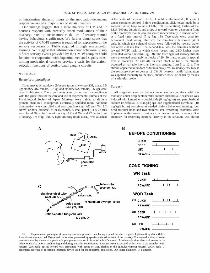
FIG. 1. Experimental paradigm.A: monkeys sat in a primate chair facing a panel on which a green light-emitting diode (LED,2 cm diam) was attached. Beeps and clicks were presented by speakers placed in front of the monkey. For reward, a drop of waterwas delivered by means of a peristaltic pump onto a spoon in front of animal’s mouth.B: schematic time charts of events in thebehavioral tasks before conditioning and during and after conditioning. Rewards were associated with clicks in the stimulus-with-reward (WR) task, but no reward was associated with beeps or LED flashes in the stimulus-without-reward (WOR) task.C:schematic drawing of recording-injection device used for the muscimol injections. OD, outer diameter; D, diameter.
961ROLE OF PROJECTIONS OF CM-PF THALAMUS TO THE STRIATUM

laterally at a 45° angle inmonkeys TMandAK to minimize damage tointernal capsule fibers by electrode penetrations. The second chamber,for recording neuron activity in the thalamus, was mounted horizon-tally for monkeys TMandAK but was tilted laterally by 5° formonkeyNA. The centers of the thalamic recording chambers were adjustedaccording to Horsley-Clark stereotaxic coordinates: lateral5 4 mmand anterior5 13 mm formonkey TM,lateral5 3 mm and anterior510 mm formonkey AK,and lateral5 2 mm and anterior5 10 mm inmonkey NA.
Recordings
Single neuron activity was recorded extracellularly with glass-insulated elgiloy microelectrodes or epoxy-coated tungsten microelec-trodes (Frederic Hair and Co., 26-10-2L or 25-10-2L) with an exposedtip of 15–60mm and with an impedance of 0.5–1.5 MV. The elec-trodes were inserted through the implanted recording chamber andadvanced by means of an oil-drive micromanipulator (Narishige,MO-95). Neuronal activity recorded by the microelectrodes was am-plified and displayed on an oscilloscope with conventional electro-physiological techniques. Band-pass filters (50 Hz to 3 kHz band-pass
with a 6-dB/octave rolloff) were used. Action potentials of singleneurons were isolated by the use of either a time-amplitude windowdiscriminator or a spike sorter with a template matching algorithm(Alpha Omega, MSD4), and the onset times of the action potentialswere recorded on a laboratory computer (NEC9801RA, 9821Bf)together with onset and offset times of stimulus and behavioral eventsoccurring during behavioral tasks. The licking movements that oc-curred during consumption of water reward were monitored by apressure sensor on the spoon on which a drop of water was delivered.These analog signals were also fed to the computer through the A-Dconverter interface at a sampling rate of 100 Hz. The responses ofneurons were defined in perievent time histograms of neuronal im-pulse discharges as increases or decreases of discharge rate after abehavioral event relative to discharges prior to the event if the changesachieved a significance level ofP , 0.05 by a two-tailed Wilcoxontest (Kimura 1986).
Microstimulation
The effects of microstimulation were examined during micro-electrode penetrations through the thalamus made to map thelocation of the CM-Pf complex. Electrical current pulses (40 pulses
FIG. 2. Histological reconstructions of the electrode tracks in the thalamus of 2 monkeys (TM andAK) in which the effects ofthalamic microstimulation were examined. Sites at which leg, arm, and orofacial movements were evoked are marked by‚, ●, andh, respectively. The locations at which current pulse trains at 100mA evoked no responses are marked by a dash. Stars indicatelocations where the electrolytic lesion marks were made. The coronal sections shown are roughly 1 mm apart. ApproximateHorsley-Clarke coordinates are indicated above, and section numbers below. Cd N., caudate nucleus; MD, mediodorsal nucleus;VLc, ventrolateral nucleus pars caudalis; VLps, ventrolateral nucleus pars postrema; VPI, ventral posterior inferior nucleus; VPLo,ventral posterolateral nucleus pars oralis; VPLc, ventral posterolateral nucleus pars caudalis; VPM, ventral posteromedial nucleus;VPMpc, ventral posteromedial nucleus pars parvocellularis; GMpc, medial geniculate nucleus pars parvocellularis; CL, centro-lateral nucleus; CM, centromedian nucleus; Pf, parafascicular nucleus; LP, lateral posterior nucleus; Pul, pulvinar.
962 N. MATSUMOTO, T. MINAMIMOTO, A. M. GRAYBIEL, AND M. KIMURA

with 0.2 ms duration at 3-ms intervals) were passed through therecording microelectrode at stimulus intensities of,100 mA.The microelectrode served as the cathode. Stimulus-inducedmovements of the body were carefully observed by two experi-menters as the current pulse trains were delivered at a repetitionrate of 0.5 Hz.
Injection of muscimol into the CM-Pf complex in thethalamus
To inhibit neuronal activity in the CM-Pf complex, we injected theGABAA receptor agonist, muscimol, locally into the CM-Pf complex
of monkey TM.We used a stainless steel injection cannula (300mm,OD) through which a teflon-coated tungsten wire (70-mm coateddiameter, A-M Systems) had been threaded so that its cut tip pro-truded 0.7–1.0 mm beyond the tip of the cannula (Fig. 1C). Thecannula was connected by teflon tubing (800mm OD) to a 10-mlHamilton microsyringe. A guide tube (650mm, OD) was fixed to themicrodrive, and the recording-injection device was placed inside theguide tube. Once the guide tube had been lowered through the duramater into the brain, the recording-injection device was advanced20–23 mm from the tip of the guide tube to reach the CM-Pf complex.The CM-Pf complex was located by recording neuronal activity in thethalamus through the tungsten wire electrode while advancing the
FIG. 3. Photomicrographs of coronalsections through the right thalamus ofmon-key NA. A: Nissl-staining.B: staining withthe acetylthiocholine method to demonstrateacetylcholinesterase (AChE) activity. Starsindicate electrolytic lesion made in the lat-eral part of CM. Scale bar: 1 mm. s-Pf,subparafascicular nucleus; other abbrevia-tions, as in Fig. 2.
963ROLE OF PROJECTIONS OF CM-PF THALAMUS TO THE STRIATUM

recording-injection device. Muscimol (Sigma, 1mg/1 ml saline, pH7.3) was injected at a rate of 0.2ml/min for total amounts of 1–3ml.The activity of neurons at the injection site stopped dischargingimmediately after injection of muscimol. The recording-injection de-vice was removed after recording neuronal activity in the CM-Pfcomplex and in the striatum, in which conventional elgiloy electrodeswere placed. Experiments involving muscimol injection were sepa-rated by at least 3–4 days to allow recovery from the effects of themuscimol injections.
Injection of retrograde tracer into the striatum
After recordings of neuronal activity in the CM-Pf complex andstriatum had been completed inmonkey TM,we injected into thestriatum the beta-subunit of cholera toxin (CTB, Sigma) as a retro-grade tracer. The CTB was prepared as a saturated solution by adding0.5 mg of CTB to 20ml of saline, stirring vigorously, and letting theprecipitate settle for 5 min before filling the microsyringe (Flahertyand Graybiel 1993). CTB was injected by means of a 1-ml Hamiltonmicrosyringe (needle, 700mm OD) that was attached to the micro-manipulator (Narishige, MO-95) and inserted through the striatalrecording chamber. Based on recordings of neuronal activity in the
striatum made with elgiloy microelectrodes, we injected CTB atstriatal sites where the activity of TANs had previously been identi-fied. Three injections were made, at two sites in the putamen and onesite in the caudate nucleus (see Fig. 13A). A total 0.05ml CTB wasinjected at each site.
Histology
At the end of all recording experiments, small electrolytic lesionswere made at several locations along selected electrode tracks both inthe CM-Pf complex and in the striatum. Direct anodal current (20mA)was passed for 30 s through either elgiloy or tungsten microelectrodes.
Four days after injection of CTB,monkey TMwas deeply anesthe-tized with Nembutal (60 mg/kg ip) and was perfused transcardiallywith 4% paraformaldehyde in 0.1 M phosphate buffer. Coronal 40-mm-thick sections through the thalamus and striatum were stainedwith cresylecht violet. In addition, CTB was demonstrated immuno-histochemically in 30 sections of interest separated by 200mm.Sections were incubated with polyclonal antiserum against the beta-subunit of CTB (List Biolabs; 1:2,000 dilution) for 2 days, thenincubated with a biotinylated secondary antibody, stained with theDAB-avidin-biotin peroxidase technique (Vector Labs), mounted, de-
FIG. 4. Sensory responses of 2 types of thalamic CM-Pf neurons and a striatal tonically active neuron (TAN) recorded afterbehavioral conditioning on the WR and WOR tasks. Spike rasters and accompanying histograms are aligned at the time ofpresentation of the sensory stimuli indicated.A: representative activity of a CM neuron with long-latency facilitation followingstimulus presentation (LLF neuron).B: activity of a Pf neuron showing short-latency facilitation after stimulus presentation (SLFneuron).C: representative activity of a striatal TAN recorded in the putamen. Note that thalamic responses occur in both WR andWOR tasks, whereas the TAN response occurs only in the WR task.
964 N. MATSUMOTO, T. MINAMIMOTO, A. M. GRAYBIEL, AND M. KIMURA
hydrated, and coverslipped.Monkey AK was perfused with 10%formaldehyde, andmonkey NAwas perfused with 4% paraformalde-hyde after completion of the physiological experiments. Coronal50-mm-thick sections through the striatum and CM-Pf complex ofboth hemispheres were stained with cresylecht violet or with thethiocholine method (monkey NA) to demonstrate acetylcholinesterase(AChE) activity (Graybiel and Berson 1980; Hardy et al. 1976).
R E S U L T S
Altogether, 136 recording tracks were made in the thalamus,and 98 tracks were made in the striatum (Fig. 2). In the striatalrecordings, we analyzed the activity of TANs identified on thebasis of their low (2–10 spikes/s) spontaneous discharge ratesand the broad waveforms of their extracellularly recordedaction potentials (Alexander and DeLong 1985; Aosaki et al.1994b; Apicella et al. 1991; Kimura et al. 1984; Raz et al.1996). To identify neuronal activity in the CM-Pf complex, wecarried out microstimulation mapping experiments andmapped neuronal responses elicited in the behavioral tasks.These stimulation and recording maps were combined withpostmortem histological reconstructions of the electrode tracksin each monkey (Figs. 2 and 3). The CM and Pf were identifiedin Nissl-stained sections based on the histological criteria ofOlszewski (1952) and Jones (1997) and, in addition, inmonkeyNA, on the basis of AChE staining. We recorded the activitiesof 208 TANs in the striatum, 36 neurons in the CM, and 10neurons in the Pf on the left side inmonkey TM;102 TANs and24 neurons in the CM and 23 neurons in the Pf on the right sidein monkey AK;and 60 neurons in the CM and 29 neurons in thePf bilaterally inmonkey NA.
Microstimulation mapping in the thalamus
Microelectrode penetrations were made over a broad medio-lateral extent of the thalamus (Fig. 2). In these penetrations, werecorded the activity of thalamic neurons while advancing theelectrode and delivered current pulses for microstimulationwhile withdrawing the electrode. The effects of microstimula-tion were examined systematically at 0.5- to 1.0-mm intervals.Consistent with previous reports (Buford et al. 1996; Vitek et
TABLE 1. Multimodal properties of CM-Pf neuron activity
Neurons
LLF SLF
Monkey NA
Auditory 1 tactile 1 visual 14 (47) 3 (34)Auditory 1 tactile 11 (36) 4 (44)Auditory 1 visual 2 (7) 1 (11)Tactile 1 visual 1 (3) 0Auditory 2 (7) 0Visual 0 1 (11)Total 30 9
Monkeys TM and AK
Auditory 1 visual 43 (66) 19 (38)Auditory 21 (32) 29 (58)Visual 1 (2) 2 (4)Total 65 50
Figures indicate number of neurons responsive to either auditory or visual ortactile stimuli and corresponding percentages (in parentheses). Tactile re-sponses were examined in onlymonkey NA.CM-Pf, centre me´dian–parafas-cicular; LLF, long-latency facilitatory; SLF, short-latency facilitatory.
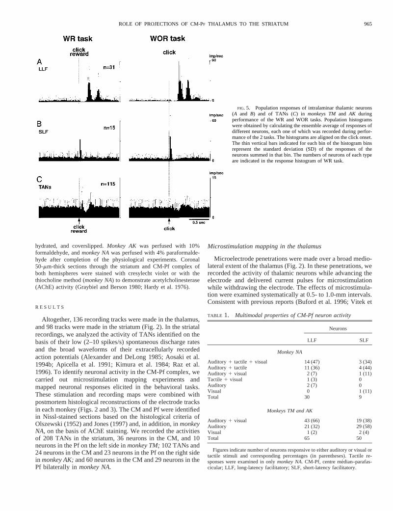
FIG. 5. Population responses of intralaminar thalamic neurons(A and B) and of TANs (C) in monkeys TMand AK duringperformance of the WR and WOR tasks. Population histogramswere obtained by calculating the ensemble average of responses ofdifferent neurons, each one of which was recorded during perfor-mance of the 2 tasks. The histograms are aligned on the click onset.The thin vertical bars indicated for each bin of the histogram binsrepresent the standard deviation (SD) of the responses of theneurons summed in that bin. The numbers of neurons of each typeare indicated in the response histogram of WR task.
965ROLE OF PROJECTIONS OF CM-PF THALAMUS TO THE STRIATUM
al. 1996), clear effects of microstimulation were observed inthe laterally located motor nuclei of the thalamus, the ventralposterolateral nucleus (VPL) and the ventral posteromedialnucleus (VPM), indicative of a leg-lateral, face-medial soma-totopy. By contrast, in the more medially located thalamicnuclei, including the CM-Pf complex, the mediodorsal nucleus(MD) and the centrolateral nucleus (CL), we observed nostimulation-induced bodily movements. Marker lesions (seeFig. 3) allowed verification of the stimulation sites.
Activation patterns of CM and Pf neurons
By recording neuronal activity as the microstimulation elec-trodes were advanced vertically through the thalamus, we wereable to identify patterns of activity characteristic of CM and Pfneurons. We found the CM and Pf approximately 3–6 mmbelow the surface of the thalamus, with the medial part of thecomplex ,3 mm lateral from the midline. The recordingmicroelectrodes passed through the MD before reaching theCM and Pf. Neurons in the CM and Pf had lower firing rates(4.16 3.3 spikes/s; mean6 SD) than those in the MD (7.264.0 spikes/s), and small-amplitude spikes. The CM-Pf neuronshad characteristic grouped, repetitive discharges. These prop-erties meant that special care was necessary to isolate theactivity of single CM and Pf neurons, but they helped inon-line recognition of the nuclei as recording electrodes en-tered or left the CM and Pf.
Very high percentages of neurons recorded in the CM-Pfcomplex (97%, 177/182 neurons examined) exhibited activitychanges as the monkeys performed the behavioral tasks. Twoclasses of neurons were identified in the CM and Pf, based ontheir different task-related activity patterns. One type showedlong-latency increases in firing rate both after clicks (latency227 6 30 ms,n 5 40) in the WR task and after presentationsof clicks (latency 2336 29 ms,n 5 93), beeps (latency 251657 ms,n 5 70), and LED flashes (latency 2816 56 ms,n 558) in the WOR task (Fig. 4A). The background discharge rateof these neurons was 4.46 3.6 spikes/s. Many neurons of thistype showed characteristic single or periodic repeating activa-tions after the sensory stimuli, as illustrated in Fig. 4A. Forpurposes of classification, we refer to these neurons as thelong-latency facilitation (LLF) type.
The second subpopulation of CM-Pf neurons responded withshort-latency single burst discharges after the clicks (latency24 6 9 ms,n 5 23) in the WR task and clicks (latency 30613 ms,n 5 51), beeps (latency 446 37 ms,n 5 33), and LEDflashes (latency 906 31 ms,n 5 26) in the WOR task (Fig.4B). The background discharge rate of these neurons was 3.562.5 spikes/s. We call this type of CM-Pf neuron the short-latency facilitation (SLF) type. In the example shown in Fig.4B, the neuron responded briskly with a single burst to clicksboth in the WR and in the WOR tasks and beeps in the WORtask, but had only a small, single burst in response to LEDflashes in the WOR task. For comparison, the activity of astriatal TAN recorded during the WR and WOR tasks isillustrated in Fig. 4C. The TAN showed brief initial activationat a latency of 696 30 ms (n 5 34) followed by a pause in itstonic discharges (latency 1316 45 ms,n 5 74) and reboundfacilitation (latency 2546 54 ms, n 5 54) after the clickfollowed by reward in the WR task, but it showed no signifi-cant responses to beeps or to LED flashes in the WOR task.
The differential responses of LLF and SLF neurons in theCM-Pf complex ofmonkeys TMandAK are illustrated in Fig.5, which shows population response histograms for the twotypes of neuron and, for comparison, the population responsesof TANs in the striatum in the WR and WOR tasks. Thepopulation response histograms for the LLF type (Fig. 5A)shows two or three discrete, periodic burst discharges (period;200 ms) after clicks in both the WR and WOR tasks. As apopulation, this class of neurons had long but similar latenciesof activation after the clicks in the two tasks. However, the twoto three periodic burst discharges after the clicks were mainlyobserved inmonkey TM,whereas single burst discharges afterthe clicks were the dominant responses inmonkey AK(and alsoin monkey NA,not shown). Although the activation of theseneurons in response to the clicks occurred at a long latency,this long-latency activation was preceded by a suppression ofdischarges at a short latency (506 27 ms). The SLF neurons(Fig. 5B) exhibited responses that were tightly locked to theclicks in both the WR and the WOR tasks.
As shown in Fig. 5,A and B, both LLF and SLF neuronsshowed responses not only to auditory but also to visualstimuli. In monkey NA,in which the responsiveness of 30 LLF
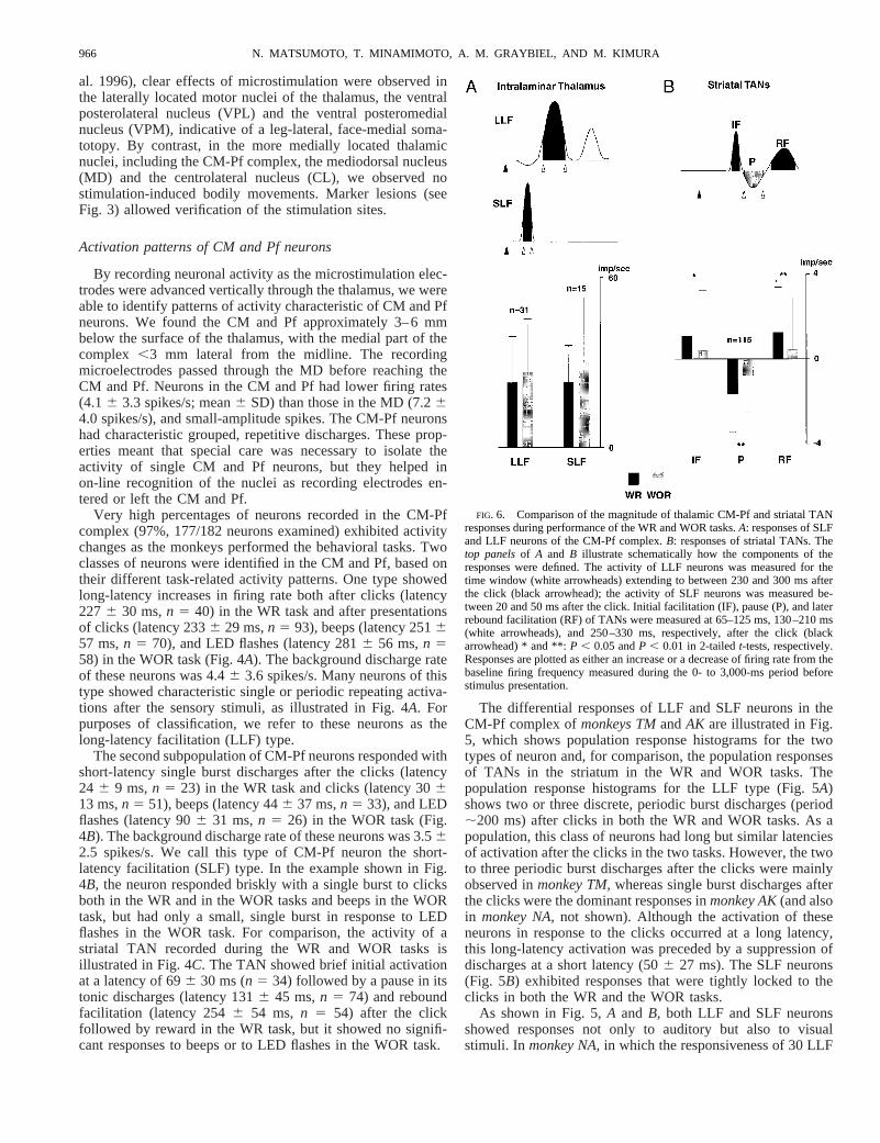
FIG. 6. Comparison of the magnitude of thalamic CM-Pf and striatal TANresponses during performance of the WR and WOR tasks.A: responses of SLFand LLF neurons of the CM-Pf complex.B: responses of striatal TANs. Thetop panelsof A and B illustrate schematically how the components of theresponses were defined. The activity of LLF neurons was measured for thetime window (white arrowheads) extending to between 230 and 300 ms afterthe click (black arrowhead); the activity of SLF neurons was measured be-tween 20 and 50 ms after the click. Initial facilitation (IF), pause (P), and laterrebound facilitation (RF) of TANs were measured at 65–125 ms, 130–210 ms(white arrowheads), and 250–330 ms, respectively, after the click (blackarrowhead) * and **:P , 0.05 andP , 0.01 in 2-tailedt-tests, respectively.Responses are plotted as either an increase or a decrease of firing rate from thebaseline firing frequency measured during the 0- to 3,000-ms period beforestimulus presentation.
966 N. MATSUMOTO, T. MINAMIMOTO, A. M. GRAYBIEL, AND M. KIMURA

and 9 SLF to auditory (click and beep), visual (LED), andsomesthetic (tactile) stimuli was examined, nearly all LLF(93%) and SLF (89%) neurons showed multimodal responses(Table 1). Inmonkeys TMandAK, in which auditory and visualstimuli were used, about half of the LLF and SLF neuronsresponded to both auditory and visual stimuli, and the otherhalf responded either to auditory or visual stimulus (Table 1).
In the striatum, TANs (n 5 115) showed responses to theclicks followed by reward (Fig. 5C, WR task). The responsesto the reward-associated clicks consisted of three consecutivecomponents, an initial short-latency facilitation (IF), a pause(P), and a later rebound facilitation (RF). However, when theclick sounds were not associated with reward (WOR task), themagnitudes of these characteristic responses decreased mark-edly (Fig. 5C).
To determine whether the neuronal responses of the CM-Pfneurons to the click stimuli were different in the WR and WORtasks, we compared the neuronal responses in the two condi-tions quantitatively. We did the same for the striatal TANsrecorded. Figure 6 shows the response magnitudes of SLF andLLF neurons in the thalamus (Fig. 6A) and TANs in thestriatum (Fig. 6B). Increases and decreases in firing rate rela-tive to baseline levels are plotted for each neuronal type. In thethalamus, the SLF and LLF neurons showed no significantdifference in click responses in the WR and WOR tasks (Fig.6A, P5 0.32 for the LLF,P 5 0.42 for the SLF, paired 2-tailedt-tests). By contrast, all three components of the responses ofstriatal TANs (IF, P, and RF) were significantly larger in theWR task than in the WOR task (Fig. 6B, P , 0.05 for the IF,P , 0.01 for the P and RF, paired 2-tailedt-tests).
Next, we examined the onset latencies of the click responsesof individual neurons in the WR and WOR tasks (Fig. 7).
Variations in the onset latency of facilitatory responses of theLLF and SLF neurons in the WR task (Fig. 7A) were notsignificantly different from those in the WOR task (P 5 0.63for the LLF, P 5 0.28 for the SLF, Siegel-Turkey test). Bycontrast, the variations in the latency distributions of the IF andP responses of TANs in WOR condition were larger than thosein WR condition (Fig. 7B; P , 0.02 for IF,P , 0.0001 for P,Siegel-Turkey test). The RF responses of the TANs were notsignificantly different (P 5 0.08, Siegel-Turkey test). Thusthere was a contrast between the CM-Pf neurons of both LLFand SLF types, which showed similar latencies of response toclicks whether they were associated with reward or not, and thestriatal TANs, which showed much more sharply timed re-sponse latencies to reward-associated clicks.
Table 2 summarizes the responsiveness of the two types ofintralaminar neurons to beep, LED, tactile, and click stimuli inthe WOR and WR conditions. More than 90% of both LLF andSLF neurons responded consistently to the click noise not onlyin WOR but also in WR conditions inmonkeys TMand AK,and values were similar for the responses ofmonkey NA,whichwas tested only in the WOR task. By contrast, there wereconsiderable differences among the three monkeys in the re-sponsiveness of the intralaminar neurons to the beeps and LEDflashes. The SLF and LLF neurons inmonkey TMshowedmuch higher responsiveness to these stimuli than did intralami-nar neurons in the other two monkeys. These differences mayreflect differences in the conditions under which the stimuliwere presented to the monkeys. Formonkey TM,the stimuliwere presented in much darker ambient light conditions thanfor monkeys AKand NA, and the LED was much closer tomonkey TM(22 cm) than to the other two monkeys (50 cm, see
FIG. 7. Onset latencies of click responses ofthalamic CM-Pf neurons and striatal TANs dur-ing WR and WOR tasks.A: SLF and LLF neu-rons in the intralaminar thalamus.B: striatalTANs. Both SLF and LLF neurons showed sim-ilar distributions of response latencies (LLF;P 5 0.63, SLF;P 5 0.28, Siegel-Turkey test).By contrast, TANs showed significant differ-ences in their response latencies in the WR andWOR tasks (P , 0.02 for IF,P , 0.0001 for P,P 5 0.08 for RF). Abbreviations are the same asin Fig. 6.
967ROLE OF PROJECTIONS OF CM-PF THALAMUS TO THE STRIATUM

METHODS). Thus the stimuli might have had stronger alerting ororienting effects onmonkey TM.
Additional characteristics of the sensory responsiveness of neu-rons in CM and Pf became evident in tests of their responsiveness,which were performed for most of the neurons recorded. Consid-erable numbers of both LLF and SLF neurons responded tounexpected stimuli such as handclaps and knocks on the door orwalls of the room in which the monkey was sitting. In manyinstances, the neurons responded to such alerting stimuli only forthe first several times that they occurred; after that, the samestimuli gradually became ineffective in evoking responses. To testmore systematically for habituation of such responses, we com-pared the responsiveness of 26 CM and Pf neurons of both SLFand LLF types in two sets of trials: trials in which clicks, beeps,and LED flashes were presented in random order in a 75-trial-longblock (Fig. 8,A andB), and trials in which only clicks appeared,repeatedly, in a 25-trial-long block (Fig. 8,C andD). We foundthat 13 of 16 LLF neurons examined exhibited habituation, asevident by comparing the population histograms in Fig. 8,A andC, and the raster plots of Fig. 8,B andD. Smaller numbers ofshort-latency facilitation neurons (5 of 10) showed the habituationresponses.
Sudden taps to the skin also evoked brisk responses fromboth CM and Pf neurons. We did not identify the receptive
fields of the somatosensory responses, but the fields seemedlarge because taps to the neck, shoulder, back, or hands of theanimal were similarly effective in evoking responses in most ofthe neurons. We did not explore the entire body surface andexamined tactile responses only inmonkey NA.
Locations of task-related neuronal activity in the thalamus
We recorded thalamic neuronal activity during the presen-tation of sensory stimuli in the WR and WOR tasks not only inthe CM-Pf complex, but also in other nuclei, including the MD,CL, VPL pars oralis (VPLo), and VPL pars caudalis (VPLc).Recording sites at which we identified LLF and SLF neuronswere located within the CM and Pf. Neurons with activityrelated to the orofacial movements made to consume rewardwater, or related to spontaneous limb movements, were ob-served in the VPLo and VPLc.
There was a clear tendency for the LLF and SLF neurons to bein separate locations within the CM-Pf complex (Fig. 9). It wasrare to record both types of neuron in a single vertical electrodetrack; single penetrations tended to isolate neurons of either theLLF or the SLF type. LLF neurons were almost exclusively foundin the CM, whereas SLF neurons were predominantly found in thePf and the medial part of CM (Fig. 9).
TABLE 2. Responsiveness of intralaminar neurons to sensory stimuli
Stimuli
Conditions
WOR WR
Beep LED Click Tactile Click
Short-latency facilitation typeMonkey TM 6/6 (100.0) 5/6 (83.3) 3/3 (100.0) 8/8 (100.0)Monkey AK 12/26 (46.2) 7/25 (28.0) 22/26 (84.6) 15/16 (93.8)Monkey NA 15/32 (46.9) 14/32 (43.8) 26/32 (81.3) 8/10 (80.0)
Long-latency facilitation typeMonkey TM 31/32 (96.9) 21/22 (95.5) 26/26 (100.0) 36/38 (94.7)Monkey AK 12/17 (70.6) 8/16 (50.0) 16/17 (94.1) 4/5 (80.0)Monkey NA 27/57 (47.4) 29/57 (50.9) 51/57 (89.5) 22/23 (95.7)
Figures indicate number of neurons (responsive/examined) and corresponding percentages (in parentheses). Inmonkey NA,responses to click in WR conditionwere not examined. WOR, without reward; WR, with reward; LED, light-emitting diode.
FIG. 8. Habituation of sensory responses of intralaminarthalamic neurons. In 26 neurons recorded in CM and Pf, neuralresponses were 1st examined in a 75-trial-long block in whichbeeps, clicks, and LED flashes appeared in a random order withequal probabilities and at equal intervals (A andB). The neuralactivity shown is centered at the onset of the 25 presentationsof the click that occurred during a 75-trial-long block for boththe population response histogram (A) and the raster display(B). The same neurons were then examined in a 25-trial-longblock in which only clicks were delivered and the activity wasagain displayed by centering on click onsets (C and D). Thehistograms inA andC are ensemble averages of the activitiesof 16 LLF neurons. The thin vertical bars indicate standarddeviation (SD) of responses. The raster displays inB and Dshow impulse discharges in chronological order with the 1sttrial at thetop and the last trial at thebottom.
968 N. MATSUMOTO, T. MINAMIMOTO, A. M. GRAYBIEL, AND M. KIMURA

Inactivation of the CM and Pf nuclei in the thalamusmarkedly reduces the sensory responses of TANs in thestriatum
To determine whether the neurons of the CM-Pf complexsupply task-dependent input to striatal TANs during perfor-mance of the WR and WOR tasks, we identified TANs inmonkey TMas it performed the behavioral tasks and comparedthe TANs’ activity before and after blocking neural activity inCM and Pf by local infusion of the GABAA receptor agonist,muscimol (Fig. 10A).
We injected 1–3ml of muscimol (1mg/1 ml saline, pH 7.3)into the CM-Pf complex in eight experiments (Fig. 9). Beforeinjection of muscimol, we confirmed that the tip of the injec-tion cannula was in the CM or Pf by recording neuronalactivity through the tungsten electrode protruding from the tipof the cannula (Fig. 10A). Figure 10B shows a populationresponse histogram of TANs recorded before muscimol injec-tion, with the histograms centered at the time of presentation ofclicks in the WR task. Clear-cut initial activation followed bysuppression and subsequent facilitation of discharges is evi-dent. The number of TANs responsive to the clicks in the WR
task was much lower in TANs following injection of muscimol(Fig. 10D), and the population histogram of activity of theseTANs (Fig. 10D) showed a much smaller response after theclick, mainly an initial facilitation.
Quantitative analysis of these responses (Fig. 11) indicatedthat the main effects of the muscimol injection into the CM-Pfcomplex was on the amplitudes of the pause (P) and reboundfacilitation (RF; Fig. 11A), both of which were significantlysmaller after muscimol injection than before it (P , 0.0001 forthe P and RF, 2-tailedt-test). The initial facilitation response(IF), by contrast, showed only a tendency to decrease withouta statistically significant decline (P 5 0.33). There was also aclear change in the distribution of onset latencies of the TANresponses. After muscimol injection into the CM-Pf, the vari-ance of the onset latency of pause (P; Fig. 11B) and reboundfacilitation (data not shown) became significantly larger (P ,0.002 for the P,P , 0.05 for the RF, Siegel-Turkey test). Thelatency of the initial facilitation (IF) did not change (P 5 0.3).
These strong effects of inactivation of the CM and Pf com-plex on the responses of striatal TANs continued for more than5 h. The population histogram in Fig. 10D is based on the
FIG. 9. Sites at which short-latency and long-latency facilitatory responses were recorded in the CM and Pf of all monkeys.Recording sites were reconstructed from brain sections postmortem and are shown on reconstructed electrode tracks. Red barsindicate SLF neurons, and blue bars indicate LLF neurons. Sites of injection of muscimol and saline in the CM and Pf inmonkeyTM are indicated by filled black and green circles, respectively. FR, fasciculus retroflexus; other abbreviations, same as in Fig. 2.
969ROLE OF PROJECTIONS OF CM-PF THALAMUS TO THE STRIATUM

activity of TANs recorded between 5 and 234 min after mus-cimol injection. Figure 10E illustrates a representative rasterplot of the activity of a single TAN during the WR taskrecorded from 82 to 87 min after muscimol injection. Re-sponses to the clicks were undetectable following the musci-mol injection. Figure 10 also shows that the muscimol injec-tions into CM and Pf did not have significant effects on thebackground firing rate or discharge pattern of the TANs. Be-fore muscimol injection, the background discharge rate of the121 TANs recorded was 4.26 1.3 impulses/s (Fig. 10B),whereas after injection the firing rate was 4.06 1.4 impulses/s(49 TANs, Fig. 10D).
To test whether the effects of muscimol injection into theCM and Pf resulted from mechanical damage to the CM-Pf ornearby neurons, we injected the same amount of physiologicalsaline (3ml) into the same part of the CM-Pf complex in whichmuscimol had been injected in a previous experiment, and thenrecorded the activity of 38 TANs during the WR task (Fig. 9).There was a tendency for reduction of the pause response (P 50.04, 2-tailedt-test), but the magnitude of the rebound facili-tation (RF) was not significantly different from that beforesaline injection (P . 0.99 for RF, 2-tailedt-test). The magni-tudes of the pause and rebound facilitatory responses aftermuscimol injection were significantly smaller than those aftersaline injection (P , 0.05 for P,P , 0.01 for RF, 2-tailedt-tests). One example of the activity of a TAN after saline
FIG. 11. Responsiveness of striatal TANs to the reward-associated clickbefore and after muscimol injection into the CM-Pf complex.A: initial facil-itation (IF), pause (P), and later rebound facilitation (RF) measured as anincrease or decrease of firing rate of TANs relative to background firing rate,before and after the muscimol injection into the CM-Pf complex. **,P ,0.001, 2-tailedt-test.B: distribution of onset latencies of pause responses to theclick in the WR task before and after muscimol injection into the CM-Pfcomplex. The variance of the latency distribution for the pause response aftermuscimol injection was significantly larger than the variance of the responsesbefore muscimol injection (P , 0.002, Siegel-Turkey test).
FIG. 10. Effects of inactivation of neuronal activity inCM and Pf on the activity of TANs recorded concurrentlyduring performance of the WR task.A: schematic illustrationof the experimental setup.B: population response of TANs tothe clicks associated with reward prior to muscimol injectioninto the CM-Pf complex. Numbers above the histogramindicate total number of neurons sampled; thin vertical barsindicate SDs.C: an example of TAN responses to the clicksafter saline injection into the CM-Pf complex. The TANcontinued to respond to the reward-associated clicks.D:population response of TANs to the clicks after muscimolinjection into the CM-Pf complex.E: an example of theactivity of a TAN recorded after muscimol was injected intothe CM-Pf complex. The TAN showed no response to thereward-associated clicks.
970 N. MATSUMOTO, T. MINAMIMOTO, A. M. GRAYBIEL, AND M. KIMURA
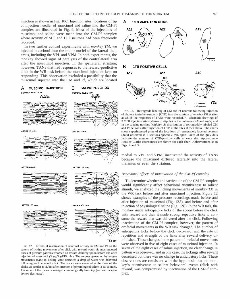
injection is shown in Fig. 10C. Injection sites, locations of tipof injection needle, of muscimol and saline into the CM-Pfcomplex are illustrated in Fig. 9. Most of the injections ofmuscimol and saline were made into the CM-Pf complexwhere activity of SLF and LLF neurons had been frequentlyrecorded.
In two further control experiments withmonkey TM,weinjected muscimol into the motor nuclei of the lateral thal-amus, including the VPL and VPM. In both experiments, themonkey showed signs of paralysis of the contralateral armafter the muscimol injection. In the ipsilateral striatum,however, TANs that had responses to the reward-predictiveclick in the WR task before the muscimol injection kept onresponding. This observation excluded a possibility that themuscimol injected into the CM and Pf, which are located
medial to VPL and VPM, inactivated the activity of TANsbecause the muscimol diffused laterally into the lateralthalamus or even the striatum.
Behavioral effects of inactivation of the CM-Pf complex
To determine whether an inactivation of the CM-Pf complexwould significantly affect behavioral attentiveness to salientstimuli, we analyzed the licking movements ofmonkey TMinthe WR task before and after muscimol injection. Figure 12shows examples of the pressure recordings made before andafter injection of muscimol (Fig. 12A), and before and afterinjection of physiological saline (Fig. 12B). In the WR task, themonkey made anticipatory licks of the spoon before the clickwith reward and then it made strong, repetitive licks to con-sume the reward that was delivered after the click. Followinginactivation of the CM-Pf complex, however, the pattern oforofacial movements in the WR task changed. The number ofanticipatory licks before the click decreased, and the rate ofrepetition and strength of the licks after reward delivery di-minished. These changes in the pattern of orofacial movementswere observed in five of eight cases of muscimol injection. Inseven of the eight cases of saline injection, no clear change inpattern was observed, and in one case, the lickings after rewarddecreased but there was no change in anticipatory licks. Theseobservations are consistent with the hypothesis that the mon-key’s attentiveness to salient behavioral events (click withreward) was compromised by inactivation of the CM-Pf com-plex.
FIG. 12. Effects of inactivation of neuronal activity in CM and Pf on thepattern of licking movements after click with reward water.A: superimposedtraces of pressure patterns recorded on reward-delivery spoon before and afterinjection of muscimol (3mg/3 ml/15 min). The torques generated by tonguemovements made in licking were detected; a drop of water was deliveredfollowing each solenoid click. The traces were centered at the time of theclicks.B: similar toA, but after injection of physiological saline (3ml/15 min).The order of the traces is arranged chronologically fromtop (earliest trace) tobottom(last trace).
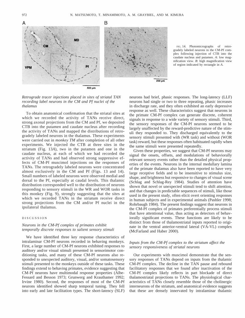
FIG. 13. Retrograde labeling of CM and Pf neurons following injectionof cholera toxin beta-subunit (CTB) into the striatum ofmonkey TMat sitesat which the responses of TANs were recorded.A: schematic drawings of3 CTB injection sites (shown in stipple) in the putamen (left andright) andin the caudate nucleus (middle). B: distribution of retrogradely labeled CMand Pf neurons after injection of CTB at the sites shown above. The chartsshow superimposed plots of the locations of retrogradely labeled neurons(dots) observed in 3 sections spaced 2 mm apart. Sizes of the gray dotsindicate the number of CTB-positive cells at each site. ApproximateHorsley-Clarke coordinates are shown for each chart. Abbreviations as inFigs. 2 and 3.
971ROLE OF PROJECTIONS OF CM-PF THALAMUS TO THE STRIATUM
Retrograde tracer injections placed in sites of striatal TANrecording label neurons in the CM and Pf nuclei of thethalamus
To obtain anatomical confirmation that the striatal sites atwhich we recorded the activity of TANs receive direct,strong axonal projections from the CM and Pf, we depositedCTB into the putamen and caudate nucleus after recordingthe activity of TANs and mapped the distributions of retro-gradely labeled neurons in the thalamus. These experimentswere carried out inmonkey TMafter completion of all otherexperiments. We injected the CTB at three sites in thestriatum (Fig. 13A), two in the putamen and one in thecaudate nucleus, at each of which we had recorded theactivity of TANs and had observed strong suppressive ef-fects of CM-Pf muscimol injections on the responses ofTANs. The retrogradely labeled neurons were concentratedalmost exclusively in the CM and Pf (Figs. 13 and 14).Small numbers of labeled neurons were observed medial anddorsal to the Pf, especially at caudal levels. This thalamicdistribution corresponded well to the distribution of neuronsresponding to sensory stimuli in the WR and WOR tasks inthis monkey (Fig. 9), strongly suggesting that the sites atwhich we recorded TANs in the striatum receive directstrong projections from the CM and/or Pf nuclei in theipsilateral thalamus.
D I S C U S S I O N
Neurons in the CM-Pf complex of primates exhibittemporally discrete responses to salient sensory stimuli
We have identified three key response characteristics ofintralaminar CM-Pf neurons recorded in behaving monkeys.First, a large number of CM-Pf neurons exhibited responses toauditory and/or visual stimuli presented in sensorimotor con-ditioning tasks, and many of these CM-Pf neurons also re-sponded to unexpected auditory, visual, and/or somatosensorystimuli presented to the monkeys outside of these tasks. Thesefindings extend to behaving primates, evidence suggesting thatCM-Pf neurons have multimodal response properties (Albe-Fessard and Besson 1973; Grunwerg and Krauthamer 1992;Irvine 1980). Second, the responses of most of the CM-Pfneurons identified showed sharp temporal tuning. They fellinto early and late facilitation types. The short-latency (SLF)
neurons had brief, phasic responses. The long-latency (LLF)neurons had single or two to three repeating, phasic increasesin discharge rate, and they often exhibited an early depressiveresponse as well. These characteristics suggest that neurons inthe primate CM-Pf complex can generate discrete, coherentsignals in response to a wide variety of sensory stimuli. Third,the sensory responses of the CM-Pf neurons seemed to belargely unaffected by the reward-predictive nature of the stim-uli they responded to. They discharged equivalently to thesensory stimuli presented with (WR task) and without (WORtask) reward, but these responses often habituated rapidly whenthe same stimuli were presented repeatedly.
Given these properties, we suggest that CM-Pf neurons maysignal the onsets, offsets, and modulations of behaviorallyrelevant sensory events rather than the detailed physical prop-erties of the events. Neurons in the internal medullary laminaof the primate thalamus also have been reported to have verylarge receptive fields and to be insensitive to stimulus size,shape, and brightness but responsive to changes of visual scene(Schlag and Schlag-Rey 1984). Studies of attention haveshown that novel or unexpected stimuli tend to shift attention,and that changes in predictable sequences of stimuli, like thoseused in the present study, often elicit overt orienting responsesin human subjects and in experimental animals (Pashler 1998;Rohrbaugh 1984). The present findings suggest that neurons inthe CM-Pf complex of primates preferentially process stimulithat have attentional value, thus acting as detectors of behav-iorally significant events. These functions are likely to bedistinct from those of thalamostriatal inputs reported to origi-nate in the ventral anterior-ventral lateral (VA-VL) complex(McFarland and Haber 2000).
Inputs from the CM-Pf complex to the striatum affect thesensory responsiveness of striatal neurons
Our experiments with muscimol demonstrate that the sen-sory responses of TANs depend on inputs from the thalamicCM-Pf complex. The decline in the TAN pause and reboundfacilitatory responses that we found after inactivation of theCM-Pf complex likely reflects in part blockade of directthalamostriatal projections to TANs. The physiological char-acteristics of TANs closely resemble those of the cholinergicinterneurons of the striatum, and anatomical evidence suggeststhat these are densely innervated by intralaminar thalamic
FIG. 14. Photomicrographs of retro-gradely labeled neurons in the CM-Pf com-plex following injection of CTB into thecaudate nucleus and putamen.A: low mag-nification view.B: high magnification viewof region indicated by rectangle inA.
972 N. MATSUMOTO, T. MINAMIMOTO, A. M. GRAYBIEL, AND M. KIMURA

inputs from the CM-Pf complex (Kawaguchi 1992; Lapper andBolam 1992; Sidibe´ and Smith 1999). Neurons in CM and Pfalso project to other interneurons in the striatum, however, aswell as to medium spiny projection neurons there, especiallythose of the direct pathway (Fenelon et al. 1991; Franc¸ois et al.1991; Nakano et al. 1990; Sadikot et al. 1992a,b; Sidibe´ andSmith 1999; Smith and Parent 1986). The effects of the mus-cimol injections could thus in part reflect blockade of indirectthalamostriatal projections from CM-Pf neurons to striatal pro-jection neurons or interneurons that in turn project to TANs.We favor this view because of evidence that local intrastriatalblockade of GABAA transmission in the striatum can abolishthe pause response of TANs at the site of the infusion (Wa-tanabe and Kimura 1998).
Indirect thalamo-cortico-striatal connections might also con-tribute to the decline in TAN responses observed after CM-Pfinactivation, but many more CM-Pf neurons project to thestriatum than to the neocortex (Jones 1997; Macchi and Ben-tivoglio 1986; Sadikot et al. 1992b), and few CM-Pf neuronshave branched axonal projections to both striatum and neocor-tex (Deschenes et al. 1996). It is unlikely, therefore that thenearly complete abolition of TAN responses that we foundresulted mainly from inactivation of an indirect thalamo-cor-tico-striatal pathway. This conclusion is consistent with earlierwork in acute preparations demonstrating that the multimodalresponses of striatal neurons survive lesions of the neocortex(Albe-Fessard et al. 1960; Rogers and McKenzie 1973). Otherpossible indirect routes from CM-Pf to the striatum includeconnections via the subthalamic nucleus (Feger et al. 1994;Sadikot et al. 1992a) and subthalamo-pallido-striatal connec-tions (Sidibeand Smith 1996; Smith et al. 1998).
Learning circuits in the striatum can be modulated bythalamic inputs from the CM-Pf complex
The sensory responsiveness of TANs can be strongly mod-ulated by sensorimotor conditioning (Aosaki et al. 1994b), aprocess that depends on dopamine-containing nigrostriatal in-put (Aosaki et al. 1994a; Apicella et al. 1997). The TANs ofthe striatum thus appear to be part of learning circuits in thebasal ganglia (Graybiel et al. 1994). The experiments we reporthere suggest that these learning circuits are strongly modulatedby thalamic inputs from the CM-Pf complex. Most of theCM-Pf neurons we recorded responded to sensory stimuliwhether or not they were associated with reward, whereas amajority of the TANs responded to sensory stimuli presented inthe same experiment only after the monkeys had learned toassociate the stimuli with rewards. We examined reward-de-pendency in a smaller number of CM-Pf neurons than TANs.The differences in reward-dependency between the CM-Pfneurons and TANs could be significant, however, because theactivity of CM-Pf neurons was more influenced by other en-vironmental events, such as unexpected noises, than was that ofthe TANs recorded under the same environmental conditions.CM-Pf neurons responded consistently to the click of thesolenoid valve without reward when the clicks, beeps, andLED flashes appeared at random order, but the responseshabituated when the no-reward click appeared repeatedly.CM-Pf neurons showed no sign of habituation to the clickfollowed by reward. The reward may enhance the attentional
importance of otherwise habituating stimuli and thus enhancethe responsiveness of CM-Pf neurons.
These findings suggest a model in which TAN-based learn-ing circuits in the striatum receive inputs both from the CM-Pfcarrying sensory signals with attentional and orienting valueand inputs from the substantia nigra pars compacta carryingreward-related information (Schultz 1998). The integration ofthese extensive inputs in turn may be influenced by, or becontrolled by intrinsic GABAergic striatal neurons (Watanabeand Kimura 1998).
The responses of TANs tend to be triphasic, with an early,phasic activation followed by a prolonged decrease in firingrate and then a postpause facilitation. All three of these com-ponents show learning-dependent changes during sensorimotorconditioning (Aosaki et al. 1994b, 1995). The inactivation ofCM-Pf neurons induced by injection of muscimol sharplydecreased the TANs’ pause response and subsequent reboundfacilitation, but did not abolish the short-latency facilitation ofthe TANs. The facilitatory responses of the LLF neurons in thethalamus were too late (;220–280 ms) to affect the pauseresponses of the TANs (;130 ms), but these LLF neurons didhave early suppressive responses (;50 ms) that could haveaffected the pause and rebound phases. The early facilitation ofthe thalamic SLF neurons (;25–90 ms) clearly could haveaffected these responses as well, through excitatory thalamo-striatal projections (Wilson et al. 1983), but this effect wouldrequire a sign reversal, possibly via other striatal neuronsacting via GABAA receptors.
We observed a striking predominance of LLF neurons in theCM, which projects to the putamen, and a relative predomi-nance of SLF neurons in the Pf, which projects to the caudatenucleus (Sadikot et al. 1992a). TAN responses in both striatalnuclei were affected by the CM-Pf inactivation. TAN re-sponses in different striatal regions have overlapping latencies(Aosaki et al. 1995), but as a group, the pause responses ofTANs in the caudate nucleus to click stimuli occurred earlier(116.76 40.3,n 5 12) than did those in the putamen (138.2635.6,n 5 109). The CM and Pf nuclei both receive inputs fromsome regions of the brain stem, including the midbrain retic-ular formation, the superior colliculus, and the pedunculopon-tine nucleus. Other inputs to these nuclei, however, are knownto differ. The CM receives basal ganglia inputs from theinternal pallidum, but the Pf receives inputs from the substantianigra pars reticulata. Cortical inputs to these thalamic nucleialso are different (Ku¨nzle 1977, 1978). These differences couldcontribute to the latency differences we observed. LLF neuronshave consistent suppressive sensory responses that have shortlatencies compared with those of the facilitatory responses ofSLF neurons. Sensory stimuli might drive SLF neuronsthrough excitatory circuits, but drive CM neurons throughinhibitory-excitatory circuits in the thalamus.
The short-latency initial facilitatory response of TANs (;70ms) showed at most only a small decrease during inactivationof the CM-Pf complex. This early response also remains afterdamage to the dopamine-containing inputs to the striatum,despite almost complete loss of the pause and succeedingfacilitation of activity that normally follow (Aosaki et al.1994a). Thus inactivation of the CM-Pf complex and damageto the dopamine-containing inputs have similar suppresiveeffects on the activity of TANs in the striatum. One possiblesource for the short-latency initial facilitatory response is input
973ROLE OF PROJECTIONS OF CM-PF THALAMUS TO THE STRIATUM

from the neocortex. Thomas et al. (2000) have described cor-tical inputs to the distal dendrites of cholinergic interneurons inthe monkey. One of major effects of nigrostriatal dopaminedepletion is a neglect of stimuli appearing on the side con-tralateral to the depletion (Kato et al. 1995; Matsumoto et al.1999; Miyashita et al. 1995). Blockade of thalamostriatal sig-nals after dopamine depletion in the striatum might be respon-sible for the loss of attentiveness to contralateral events in bothmonkey and human. In the thalamus, contralateral visual ne-glect is observed after thalamic lesions involving the internalmedullary lamina (Orem et al. 1973; Watson and Heilman1979) and impairment of attentional orienting has been ob-served after lesion of reticular nucleus (Weese et al. 1999). Inthe present study, lowered attentiveness after inactivation ofthe CM-Pf complex was suggested by behavioral evidence forreduced licking response to click stimuli associated with forth-coming rewards.
If, as evidence suggests, the TANs are cholinergic interneu-rons (Aosaki et al. 1995), the TANs could be integrating inputsfrom at least four sources: the substantia nigra pars compacta,the CM-Pf complex of the thalamus, local neurons of thestriatum, and the neocortex.
Thalamo-striatal loop circuits may critically influenceaction-selection functions of cortico-basal ganglia circuits
The CM and Pf have long been considered as “loop nuclei”of the basal ganglia, because they project to the striatum andreceive inputs from the internal segment of the globus pallidus(GPi) and substantia nigra pars reticulata (SNr) (Graybiel andRagsdale 1979; Parent and Hazrati 1995a,b). Compared toother basal ganglia loop nuclei, the CM and Pf are unique inhaving the striatum as their principal output targets. ThusPf-caudate nucleus-SNr-Pf and CM-putamen-GPi-CM circuitsmay be true “internal loops” of the basal ganglia, operating inparallel with cortico-basal ganglia loops but having a majorpoint of access to cortico-basal ganglia circuits at the level ofthe striatum.
Our findings suggest two possible functions for these inter-nal thalamo-striatal loops. One is to supply the striatum withattention-gated multimodal sensory information. Given the lowmodality specificity and the rapid habituation of the CM-Pfresponses, it seems likely that this input could provide thestriatum (and cortico-basal ganglia circuits) with informationabout the appearance, disappearance, or change of attention-demanding, behaviorally significant events. This view expandson earlier observations on activity in the intralaminar thalamus(Kinomura et al. 1996) and evidence that unilateral sensoryneglect can result from striatal dysfunction (Denny-Brown andYanagisawa 1976; Ljungberg and Ungerstedt 1976; Vargo andMarshall 1996). Multiple brain stem inputs to the intralaminarthalamus could contribute to such functional characteristics ofCM-Pf neurons. Candidates include inputs from the midbrainreticular formation (McCormick and Bal 1994; Moruzzi andMagoun 1949; Steriade et al. 1993), the deep layers of thesuperior colliculus (Ichinohe and Shoumura 1998; Royce et al.1991), the pedunculopontine nucleus (Aizawa et al. 1999;Curro-Dossi et al. 1991; Isaacson and Tanaka 1986), and thelocus coeruleus (Royce et al. 1991; Usher et al. 1999).
A second function of thalamo-striatal loops suggested by ourfindings is a contribution to functions of the basal ganglia
related to the selection of forthcoming actions (Boussaoud andKermadi 1997; Cools 1980; Fukai and Tanaka 1997; Graybiel1998; Graybiel and Kimura 1995; Hikosaka et al. 1989; Juept-ner et al. 1997; Kimura et al. 1993; Mink 1996). The GABAer-gic projection neurons of the GPi and SNr send strong, tonicsuppressive inputs to target neurons in the VA-VL nuclei of thethalamus through their high-frequency discharges, and theseGPi and SNr neurons are under the control of the striatum,which sends them direct suppressive inputs and indirect facili-tatory inputs via the subthalamic loop. This characteristic op-ponent-circuit design has suggested the view that the basalganglia act broadly to inhibit competing behavioral mecha-nisms that would otherwise interfere with intended actions and,simultaneously, to remove focally inhibition of the desiredbehavior so as to allow the selected action to proceed (Fukaiand Tanaka 1997; Mink 1996). Selections of actions are madeon the basis of particular behavioral contexts. An adequateprogram for action can be selected if a particular behavioralcontext triggers function in the relevant circuit. Thus a crucialcondition for adequate operation of these circuits is the pres-ence of context-dependent signals indicating when and how toactivate ensembles of output neurons in the striatum. Informa-tion about behaviorally significant sensory events originatingin the CM-Pf complex could provide such signals. StriatalTANs, as points of convergence of this information with do-pamine-mediated nigrostriatal signals having motivationalvalue, could operate to bias the action-selection functions ofcortico-basal ganglia circuits.
This study was supported by research grants from the Ministry of EducationJapan (07408035, 08279106, JSPS-RFTF96L00201) and Special CoordinationFund to Brain Science to M. Kimura, by Grant-in-Aid for Encouragement ofYoung Scientists from the Ministry of Education, Japan (09780770) to N.Matsumoto, and by National Institute of Neurological Disorders and StrokeJavits Award NS-25529 to A. M. Graybiel.
REFERENCES
AIZAWA H, KOBAYASHI Y, YAMAMOTO M, AND ISA T. Injection of nicotine intothe superior colliculus facilitates occurrence of express saccades in mon-keys.J Neurophysiol82: 1642–1646, 1999.
ALBE-FESSARD D AND BESSON J. Convergent thalamic and cortical projec-tions—the non-specific system. In:Handbook of Sensory Physiology. So-matosensory System,edited by Iggo A. New York: Springer Verlag, 1973,vol. 2, p. 489–560.
ALBE-FESSARD D, OSWALDO E, AND ROCHA-MIRANDA C. Activites evoqueesdans le noyau caude du chat en reponsea des types divers d’afferences. I.Etude macrophysiologique.Electroencephalogr Clin Neurophysiol12: 405–420, 1960.
ALEXANDER GE AND DELONG MR. Microstimulation of the primate neostria-tum. I. Somatotopic organization of striatal microexcitable zones and theirrelation to neuronal response properties.J Neurophysiol53: 1417–1430,1985.
AOSAKI T, GRAYBIEL AM, AND KIMURA M. Effect of the nigrostriatal dopaminesystem on acquired neural responses in the striatum of behaving monkeys.Science265: 412–415, 1994a.
AOSAKI T, KIMURA M, AND GRAYBIEL AM. Temporal and spatial characteris-tics of tonically active neurons of the primate’s striatum.J Neurophysiol73:1234–1252, 1995.
AOSAKI T, TSUBOKAWA H, ISHIDA A, WATANABE K, GRAYBIEL AM, AND
KIMURA M. Responses of tonically active neurons in the primate’s striatumundergo systematic changes during behavioral sensory-motor conditioning.J Neurosci14: 3969–3984, 1994b.
APICELLA P, LEGALLET E, AND TROUCHEE. Responses of tonically dischargingneurons in the monkey striatum to primary rewards delivered during differ-ent behavioral states.Exp Brain Res116: 456–466, 1997.
974 N. MATSUMOTO, T. MINAMIMOTO, A. M. GRAYBIEL, AND M. KIMURA

APICELLA P, SCARNATI E, AND SCHULTZ W. Tonically discharging neurons ofmonkey striatum respond to preparatory and rewarding stimuli.Exp BrainRes85: 388–392, 1991.
BOUSSAOUD D AND KERMADI I. The primate striatum: neuronal activity inrelation to spatial attention versus motor preparation.Eur J Neurosci9:2152–2168, 1997.
BUFORD JA, INASE M, AND ANDERSON ME. Contrasting locations of pallidal-receiving neurons and microexcitable zones in primate thalamus.J Neuro-physiol75: 1105–1116, 1996.
COOLS AR. Role of the neostriatal dopaminergic activity in sequencing andselecting behavioural strategies: facilitation of processes involved in select-ing the best strategy in a stressful situation.Behav Brain Res1: 361–378,1980.
CURRODOSSIPARE D AND STERIADE M. Short-lasting nicotinic and long-lastingmuscarinic depolarizing responses of thalamocortical neurons to stimulationof mesopontine cholinergic nuclei.J Neurophysiol65: 393–406, 1991.
DENNY-BROWN D AND YANAGISAWA N. The role of the basal ganglia in theinitiation of movement. In:The Basal Ganglia,edited by Yahr MD. NewYork: Raven, 1976, p. 115–149.
DESCHENESM, BOURASSAJ,AND PARENT A. Striatal and cortical projections ofsingle neurons from the central lateral thalamic nucleus in the rat.Neuro-science72: 679–687, 1996.
FEGERJ, BEVAN M, AND CROSSMANAR. The projections from the parafascicu-lar thalamic nucleus to the subthalamic nucleus and the striatum arise fromseparate neuronal populations: a comparison with the corticostriatal andcorticosubthalamic efferents in a retrograde fluorescent double-labelingstudy.Neuroscience60: 125–132, 1994.
FENELON G, FRANCOIS C, PERCHERONG, AND YELNICK J. Topographic distri-bution of the neurons of the central complex (centremedian-parafascicularcomplex) and of other thalamic neurons projecting to the striatum in ma-caques.Neuroscience45: 495–510, 1991.
FLAHERTY AW AND GRAYBIEL AM. Output architecture of the primate puta-men.J Neurosci13: 3222–3237, 1993.
FRANCOIS C, PERCHERONG, PARENT A, SADIKOT AF, FENELON G, AND YELNICK
J. Topography of the projection from the central complex of the thalamus tothe sensorimotor striatal territory in monkeys.J Comp Neurol305: 17–34,1991.
FUKAI T AND TANAKA S. A simple neural network exhibiting selective activa-tion of neuronal ensembles: from winner-take-all to winner-share-all.NeuralComput9: 77–97, 1997.
GRAYBIEL AM. The basal ganglia and chunking of action repertoires.Neuro-biol Learn Mem70: 119–136, 1998.
GRAYBIEL AM, A OSAKI T, FLAHERTY A, AND KIMURA M. The basal ganglia andadaptive motor control.Science265: 1826–1831, 1994.
GRAYBIEL AM AND BERSON DM. Histochemical identification and afferentconnections of subdivisions in the lateralis posterior-pulvinar complex andrelated thalamic nuclei in the cat.Neuroscience5: 1175–1238, 1980.
GRAYBIEL AM AND KIMURA M. Adaptive neural networks in the basal ganglia.In: Models of Information Processing in the Basal Ganglia,edited by HoukJC, Davis JL, and Beiser DG. Cambridge, MA: MIT Press, 1995, p.103–116.
GRAYBIEL AM AND RAGSDALE CW JR. Fiber connections of the basal ganglia.In: Development and Chemical Specificity of Neurons,edited by Cuenod M,Kreutzberg GW, and Bloom FE. Amsterdam: Elsevier/North-Holland Bio-medical, 1979, vol. 51, p. 239–283.
GRUNWERG BS AND KRAUTHAMER GM. Sensory responses of intralaminarthalamic neurons activated by the superior colliculus.Exp Brain Res88:541–550, 1992.
HARDY H, HEIMER L, SWITZER R, AND WATKINS D. Simultaneous demonstra-tion of horseradish peroxidase and acetylcholinesterase.Neurosci Lett3:1–5, 1976.
HIKOSAKA O, SAKAMOTO M, AND USUI S. Functional properties of monkeycaudate neurons. II. Visual and auditory responses.J Neurophysiol61:799–813, 1989.
HU B, STERIADE M, AND DESCHENES M. The effects of brainstem peribrachialstimulation on perigeniculate neurons: the blockage of spindle waves.Neu-roscience31: 1–12, 1989.
ICHINOHE N AND SHOUMURA K. A di-synaptic projection from the superiorcolliculus to the head of the caudate nucleus via the centromedian-parafas-cicular complex in the cat: an anterograde and retrograde labeling study.Neurosci Res32: 295–303, 1998.
IRVINE DR. Acoustic properties of neurons in posteromedial thalamus of cat.J Neurophysiol43: 395–408, 1980.
ISAACSON LG AND TANAKA D. Cholinergic and non-cholinergic projectionsfrom the canine pontomesencephalic tegmentum (Ch5 area) to the caudalintralaminar thalamic nuclei.Exp Brain Res62: 179–188, 1986.
JASPERHH. Unspecific thalamocortical relations. In:Handbook of Physiology.Neurophysiology.Washington, DC: Am. Physiol. Soc., 1960, vol. 2, p.1307–1321.
JONES EG. Thalamic organization and chemical anatomy. In:Thalamus. Or-ganization and Function,edited by Steriade M, Jones EG, and McCormickCD. Amsterdam: Elsevier, 1997, vol. I, p. 31–174.
JUEPTNERM, FRITH CD, BROOKS DJ, FRACKOWIAK RS, AND PASSINGHAM RE.Anatomy of motor learning. II. Subcortical structures and learning by trialand error.J Neurophysiol77: 1325–1337, 1997.
KATO M, MIYASHITA N, HIKOSAKA O, MATSUMURA M, USUI S, AND KORI A.Eye movements in monkeys with local dopamine depletion in the caudatenucleus. I. Deficits in spontaneous saccades.J Neurosci15: 912–927, 1995.
KAWAGUCHI Y. Large aspiny cells in the matrix of the rat neostriatum in vitro:physiological identification, relation to the compartments and excitatorypostsynaptic currents.J Neurophysiol67: 1669–1682, 1992.
KIMURA M. The role of primate putamen neurons in the association of sensorystimulus with movement.Neurosci Res3: 436–443, 1986.
KIMURA M, AOSAKI T, AND ISHIDA A. Neurophysiological aspects of differen-tial roles of the putamen and caudate nucleus in voluntary movement.AdvNeurol 60: 62–70, 1993.
KIMURA M, KATO M, AND SHIMAZAKI H. Physiological properties of projectionneurons in the monkey striatum to the globus pallidus.Exp Brain Res82:672–676, 1990.
KIMURA M, RAJKOWSKI J, AND EVARTS EV. Tonically discharging putamenneurons exhibit set dependent responses.Proc Natl Acad Sci USA81:4998–5001, 1984.
KINOMURA S, LARSSONJ, GULYAS B, AND ROLAND PE. Activation by attentionof the human reticular formation and thalamic intralaminar nuclei.Science271: 512–515, 1996.
KUNZLE H. Projections from the primary somatosensory cortex to basal gangliaand thalamus in the monkey.Exp Brain Res30: 481–492, 1977.
KUNZLE H. An autoradiographic analysis of the efferent connections frompremotor and adjacent prefrontal regions (areas 6 and 9) inMacaca fascicu-laris. Brain Behav Evol15: 185–234, 1978.
LAPPERS AND BOLAM JP. Input from the frontal cortex and the parafascicularnucleus to cholinergic interneurons in the dorsal striatum of the rat.Neuro-science51: 533–545, 1992.
LJUNGBERG T AND UNGERSTEDT U. Sensory inattention produced by 6-hy-droxydopamine–induced degeneration of ascending dopamine neurons inthe brain.Exp Neurol53: 585–600, 1976.
MACCHI G AND BENTIVOGLIO M. The thalamic intralaminar nuclei and thecerebral cortex. In:Cerebral Cortex,edited by Jones E and Peters A. NewYork: Plenum, 1986, vol. 5, p. 355–401.
MATSUMOTO N, HANAKAWA T, MAKI S, GRAYBIEL AM, AND KIMURA M. Roleof nigrostriatal dopamine system in learning to perform sequential motortasks in a predictive manner.J Neurophysiol82: 978–998, 1999.
MCCORMICK D AND BAL T. Sensory gating mechanisms of the thalamus.CurrOpin Neurobiol4: 550–556, 1994.
MCFARLAND NR AND HABER SN. Convergent inputs from thalamic motornuclei and frontal cortical areas to the dorsal striatum in the primate.J Neurosci20: 3798–3813, 2000.
MENNEMEIER M, CROSSONB, WILLIAMSON D, NADEAU S, FENNELL E, VALEN-STEIN E, AND HEILMAN K. Tapping, talking and the thalamus: possibleinfluence of the intralaminar nuclei on basal ganglia function.Neuropsy-chologia35: 183–193, 1997.
MINK JN. The basal ganglia: focused selection and inhibition of competingmotor programs.Prog Neurobiol50: 381–425, 1996.
MIYASHITA N, HIKOSAKA O, AND KATO M. Visual hemineglect induced byunilateral striatal dopamine deficiency in monkeys.Neuroreport6: 1257–1260, 1995.
MORUZZI G AND MAGOUN HW. Brain stem reticular formation and the activa-tion of EEG.Electroencephalogr Clin Neurophysiol1: 455–473, 1949.
NAKANO K, HASEGAWA Y, TOKUSHIGE A, NAKAGAWA S, KAYAHARA T, AND
MIZUNO N. Topographical projections from the thalamus, the subthalamicnucleus and pedunculopontine tegmental nucleus to the striatum in theJapanese monkeyMacaca fuscata. Brain Res537: 54–68, 1990.
OLSZEWSKI J. The Thalamus of Macaca mulatta: An Atlas for Use With theStereotaxic Instrument. Basel: Karger, 1952.
OREM J, SCHLAG-REY M, AND SCHLAG J. Unilateral visual neglect and thalamicintralaminar lesions in the cat.Exp Neurol40: 784–797, 1973.
975ROLE OF PROJECTIONS OF CM-PF THALAMUS TO THE STRIATUM

PARE D, SMITH Y, PARENT A, AND STERIADE M. Projections of brainstem corecholinergic and non-cholinergic neurons of cat to intralaminar and reticularthalamic nuclei.Neuroscience25: 69–86, 1988.
PARENT A AND HAZRATI LN. Functional anatomy of the basal ganglia. I. Thecortico-basal ganglia-thalamo-cortical loop.Brain Res Rev20: 91–127, 1995a.
PARENT A AND HAZRATI LN. Functional anatomy of the basal ganglia. II. Theplace of subthalamic nucleus and external pallidum in basal ganglia cir-cuitry. Brain Res Rev20: 128–154, 1995b.
PASHLER HH. The Psychology of Attention.Cambridge, MIT Press, 1998, p.88–250.
RAZ A, FEINGOLD A, ZELANSKAYA V, VAADIA E, AND BERGMAN H. Neuronalsynchronization of tonically active neurons in the striatum of normal andparkinsonian primates.J Neurophysiol76: 2083–2088, 1996.
ROGERSDK AND MCKENZIE JS. Regional differences within the caudate nucleusfor suppression of extraleminiscal thalamic units.Brain Res56: 345–349, 1973.
ROHRBAUGH JW. The orienting reflex. In:Varieties of Attention,edited byParasuraman R and Davies DR. New York: Academic, 1984, p. 323–373.
ROYCE GJ, BROMLEY S, AND GRACCO C. Subcortical projections to the centro-median and parafascicular thalamic nuclei in the cat.J Comp Neurol306:129–155, 1991.
SADIKOT AF, PARENT A, AND FRANCOIS C. Efferent connections of the centro-median and parafascicular thalamic nuclei in the squirrel monkey: A PHA-Lstudy of subcortical projections.J Comp Neurol315: 137–159, 1992a.
SADIKOT AF, PARENT A, SMITH Y, AND BOLAM JP. Efferent connections of thecentromedian and parafascicular thalamic nuclei in the squirrel monkey: alight and electron microscopic study of the thalamostriatal projection inrelation to striatal heterogeneity.J Comp Neurol320: 228–242, 1992b.
SCHLAG J AND SCHLAG-REY M. Visuomotor functions of central thalamus inmonkey II. Unit activity related to visual events, targeting, and fixation.J Neurophysiol51: 1175–1196, 1984.
SCHULTZ W. Predictive reward signal of dopamine neurons.J Neurophysiol80:1–27, 1998.
SIDIBE M AND SMITH Y. Differential synaptic innervation of striatofugal neuronsprojecting to the internal or external segments of the globus pallidus by thalamicafferents in the squirrel monkey.J Comp Neurol365: 445–465, 1996.
SIDIBE M AND SMITH Y. Thalamic inputs to striatal interneurons in monkeys:synaptic organization and co-localization of calcium binding proteins.Neu-roscience89: 1189–1208, 1999.
SMITH Y, BEVAN M, SHINK E, AND BOLAM JP. Microcircuitry of the direct andindirect pathways of the basal ganglia.Neuroscience86: 353–387, 1998.
SMITH Y AND PARENT A. Differential connections of the caudate nucleus andputamen in the squirrel monkey (Saimiri sciureus). Neuroscience18: 347–371, 1986.
STERIADE M, CONTRERAS D, AND AMZICA F. Synchronized sleep oscillationsand their paroxysmal developments.Trends Neurosci17: 199–208, 1994.
STERIADE M, JONESEG,AND MCCORMICK CD. Thalamus. Intralaminar nuclei.Amsterdam: Elsevier, 1997, vol. 1, p. 55–73.
STERIADE M, MCCORMICK DA, AND SEJNOWSKI TJ. Thalamocortical oscilla-tions in the sleeping and aroused brain.Science262: 679–685, 1993.
THOMAS TM, SMITH Y, LEVEY AI, AND HERSCH SM. Cortical inputs toM2-immunoreactive striatal interneurons in rat and monkey.Synapse37:252–261, 2000.
USHER M, COHEN JD, SERVAN SCHREIBER D, RAJKOWSKI J, AND ASTON JONES
G. The role of locus coeruleus in the regulation of cognitive performance.Science283: 549–554, 1999.
VARGO JM AND MARSHALL JF. Frontal cortex ablation reversibly decreasesstriatal zif/268 and junB expression: temporal correspondence with sensoryneglect and its spontaneous recovery.Synapse22: 291–303, 1996.
VITEK JL, ASHEJ, DELONG MR, AND KANEOKE Y. Microstimulation of primatemotor thalamus: somatotopic organization and differential distribution ofevoked motor responses among subnuclei.J Neurophysiol75: 2486–2495,1996.
WATANABE K AND KIMURA M. Dopamine receptor-mediated mechanismsinvolved in the expression of learned activity of primate striatal neurons.J Neurophysiol79: 2568–2580, 1998.
WATSON RT AND HEILMAN KM. Thalamic neglect.Neurology29: 690–694,1979.
WEESEGD, PHILLIPS JM, AND BROWN VJ. Attentional orienting is impaired byunilateral lesions of the thalamic reticular nucleus in the rat.J Neurosci19:10135–10139, 1999.
WILSON CJ, CHANG HT, AND KITAI ST. Origins of post synaptic potentialsevoked in spiny neostriatal projection neurons by thalamic stimulation in therat. Exp Brain Res51: 217–226, 1983.
WILSON CJ, CHANG HT, AND KITAI ST. Firing patterns and synaptic potentialsof identified giant aspiny interneurons in the rat neostriatum.J Neurosci10:508–519, 1990.
976 N. MATSUMOTO, T. MINAMIMOTO, A. M. GRAYBIEL, AND M. KIMURA