Bogus Visual Feedback Alters Onset of Movement-Evoked Pain in People With Neck Pain
Upload
khangminh22Category
view
5download
0
Visual Evoked Potentials
Clinical applications
Armando Tello, M.D., Ph.D.Clinical Neurophysiology Department
Hospital EspañolMexico

Conflict of Interest
None

Outline
Anatomy
Physiology
Methods
Technical factors influencing results
Clinical ApplicationsSome examples
Conclusions

BackgroundAdrian and Matthews (Brain,1934), “The Berger rhythm: potential changes from the occipital lobes in man”First described miniature potentials of the occipital cortex when a patient was exposed to visual stimuli.
Halliday et al (Lancet, 1972), first to applied clinically pattern reversal VEPs in the diagnosis of patients of optic neuritis.Nowadays, PRVEP are an objective electrophysiological method to diagnose and monitor numerous ophthalmological and neurological diseases. They also are an objective way of study visual acuity and visual field in the assessment of cases having suspected multiple sclerosis (MS).
Flash VEP are useful in children which do not cooperate for the neurologic evaluation, and in cases of aggravationof ophthalmologic pathologies.
PVEPs useful to evaluate functionality of visual pathway, monitor diseases of the retina, optic nerve, visual tract, optical radiation and visual cortex.
Most of the cases in clinical practice, however, concentrate in pathologies of the Optic Nerve ( 90%).

Optic Nerve
a. Intraocular 1mm
b. Intraorbital 25mm
c. Intracanalicular 9mm
d. Intracranial 12-16mm

LGN
Contralateral optic radiations
Layers 1,4,6
Ipsilateral optic radiations
Layers 2,3,5

Occipital Cortex
Primary Visual Cortex (V1)
Superior Retina→ CuneusInferior Retina → Lingual Gyrus

Occipital Cortex
Secondary Visual Cortex (V2)
Dorsal Bundle: Posterior parietal cortex:Spatial and movement of objects (where)
Ventral Bundle: Inferotemporal cortex:Recognize faces, shapes, size, colors (what)
Posterior parietal area
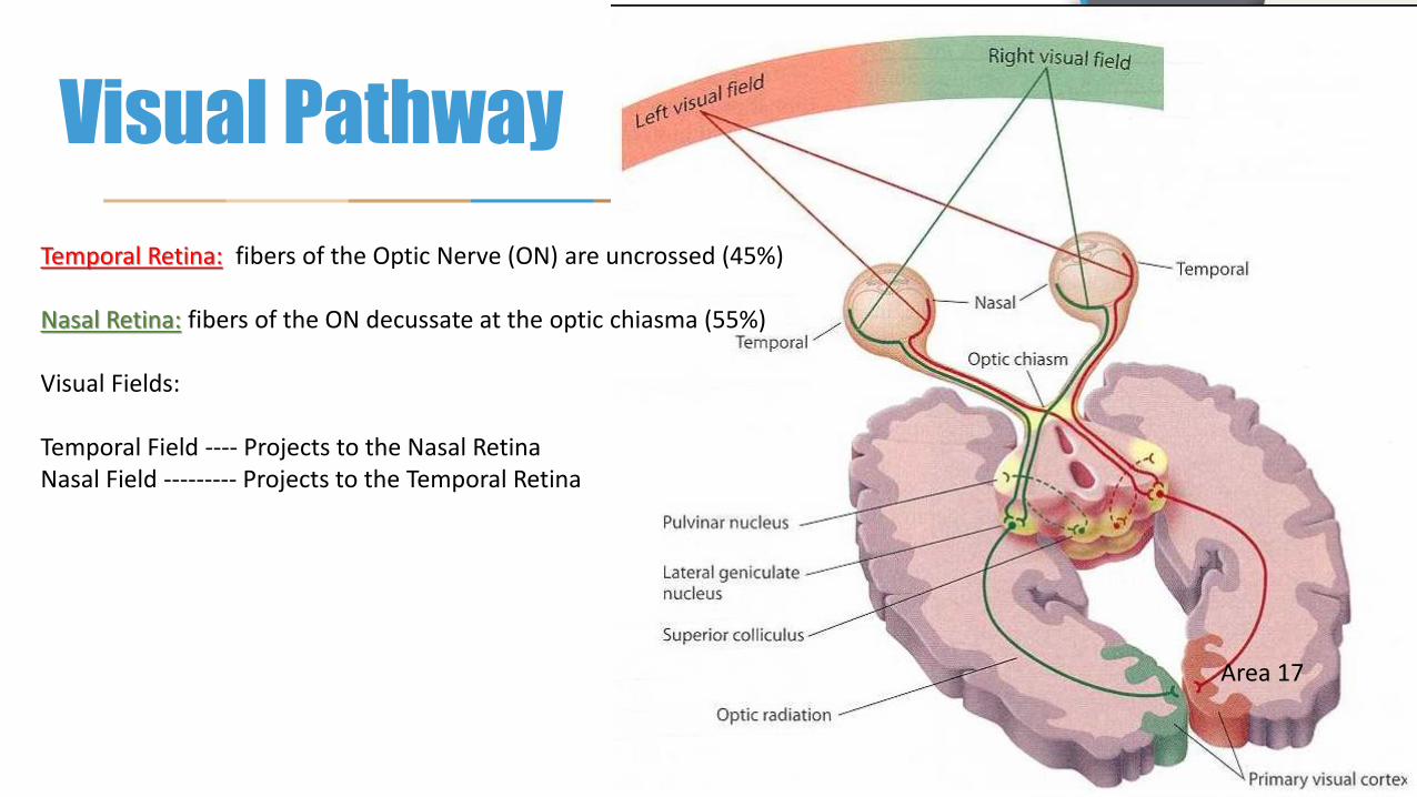
Visual Pathway
Temporal Retina: fibers of the Optic Nerve (ON) are uncrossed (45%)
Nasal Retina: fibers of the ON decussate at the optic chiasma (55%)
Visual Fields:
Temporal Field ---- Projects to the Nasal RetinaNasal Field --------- Projects to the Temporal Retina
Area 17

Principles of PRVEP
Pattern reversal VEP is a diagnostic tool, that examine conduction of the visual pathway
Checkerboard pattern reversal is the most widely used stimulus because of its relative
simplicity and reliability
Checks help to explore the function of the striate cortex (area 17) because confined spatial frequency
analyzers are likely present there

Types of Visual Stimulation
Checkerboard Pattern Reversal. Full, hemi field, quadrant
Googles (diffuse light, flash)

Shape of the VEPs
P100
N75
N145Absolute Latencies (ms)
N75, P100, N145
Amplitudes (µV)
N75-P100P100-N145

Methods70-100 cm from the screenConstant visual fixation to the center of the screenRR <4 HzSmaller check sizes stimulate fovea. Foveal fibers are the fastest fibers, thus latencies shortenStimulation with larger checks, stimulates peripheralfibers which are slower than foveal fibers
15´
30´
60´
120´
Latency
min of arc

Other types of Visual Stimulation
Multifocal visual evoked potentials
Pattern reversal Steady State visual evoked potentials

Montages
Queen Square SystemAmerican Clinical Neurophysiologic Society
The Queen Square System is superior to the IFCN because LO and RO are farther than O1, O2, thus larger signal

Montages
International 10-20 System
IFCN
Oz
As with any Evoked Potentials study, keep impedances below 5 K
Ʊ

Settings for PRVEP
✓Patient Position: Seated comfortably
✓Dim light room
✓Screen 70-100 cm in front of the eyes
✓Fix to the center of the screen (specially in hemifield stimulation)
✓Correct visual acuity with patient lenses (in case of using)
✓Monocular stimulation by covering non stimulating eye
✓Be sure the patient remains fully alert
✓In hemi visual stimulation, must focus in central spot

Factors affecting latency and amplitude
1. Sex: women have shorter latencies than males
2. Head size: the larger head the size, the longer the latency of the P100
3. Pupillary size: do not perform VEPs with dilated of constricted pupils (avoid anticholinergic or sympathetic substances)
4. Visual Acuity: always correct refraction errors

Recording and Stimulus
• Filters LFF: 0.2-1.0 Hz /HFF: 200-300 Hz
• Sweep 20-25ms/div, Sensitivity 5-10 µV/div
• Average 150-200 responses
• Polarity agreement not standardized
• Stim rate 1.3-2.7 (avoid integer of 60 Hz). Higher rates may increase latencies
• Brightness contrast= >0.5 (Lmax-Lmin/Lmax+Lmin)
L: Luminance measured by a photometer

How display monitors affects VEP latencies
• CRT: Cathode ray tube refreshes almost instantaneously.
• LCDs: the fastest refreshes at 2 ms, slower than CRT, despite better resolution.
• The slower the refreshing time, the longer the latency of VEPs. Some LCD refreshes at 30 ms and cause extreme P100 latency prolongation.
• Normative values, should be done with only one display. In case of upgrade monitors, run normative data again.

VEP analysis
• Wave recognition: N70, P100, N145 in midline and parasagittal electrodes
• N105 in MF-AU
• Latencies P100: 95-115 ms Interocular latency differences < 8 ms
• Amplitude N70, P100 and N145 (P100 Interocular amplitude difference < 2 SD)
• Shape “V” and “W”

Montages and shape of waves
ACNS Guidelines. JCN• Volume 23, Number 2, April 2006
It should be noted that up to 15% of cases, N100 may be absent in normal subjects

Influence of head size/Sex VEP Latencies
Gregori B, et al. Clin Neurophysiol. 2006 May;117(5):1154-7
AP diameter Glabella-Inion
PRVEPOz-Cz
Gregori et at, showed a clearcorrelation between head sizeand P100 Latency.ON the other hand, sex differences had not a strong correlation

Factors affecting VEP responses
• Age: Age decreases flicker sensitivity producing increments in latencies of Steady state VEPs. These age-related changes affect the magnocellular (M) but not the parvocellular (P) pathways, as shown by Brown, A et al*
* doi.org/10.3389/fnagi.2018.00430
Magnocellular Pathway
Parvocellular Pathway

INTERPRETATION • Latency P100
•→ Oz (MO)- Fz
• → At the maximal plateu
Mayo Clinic Normative P100 Values
Age Female Male
< 60 Y <115ms <120ms
> 60 Y <120ms <125ms
Interocular latency <10 ms
Side to side amplitude <50%
mean +3 SD
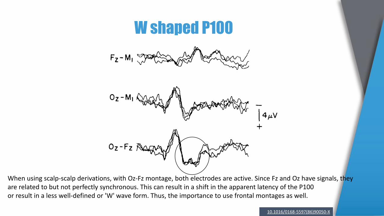
W shaped P100
When using scalp-scalp derivations, with Oz-Fz montage, both electrodes are active. Since Fz and Oz have signals, they are related to but not perfectly synchronous. This can result in a shift in the apparent latency of the P100 or result in a less well-defined or 'W' wave form. Thus, the importance to use frontal montages as well.
10.1016/0168-5597(86)90050-X

Paradoxical Responses
Nasal Retina
Decussation at the Chiasm
Because of the direction of the P100 dipole, the projection of the activated cortex, is reflected better in the contralateral electrode.

Specifics for Abnormalities1) Latency prolongation of P100
Monocular: pre-chiasmatic Binocular: pre-chiasmatic, chiasmatic, post-chiasmatic or technical error
2) Amplitude
Monocular: pre-chiasmatic or technical errorBinocular: pre-chiasmatic, chiasmatic, post-chiasmatic or technical error
3) Topography
Crossed asymmetry: pre-chiasmaticUncrossed asymmetry: post-chiasmatic (Cortex, optic radiation)
4) “W” Shape
Pre-chiasmaticTechnical, normal variation
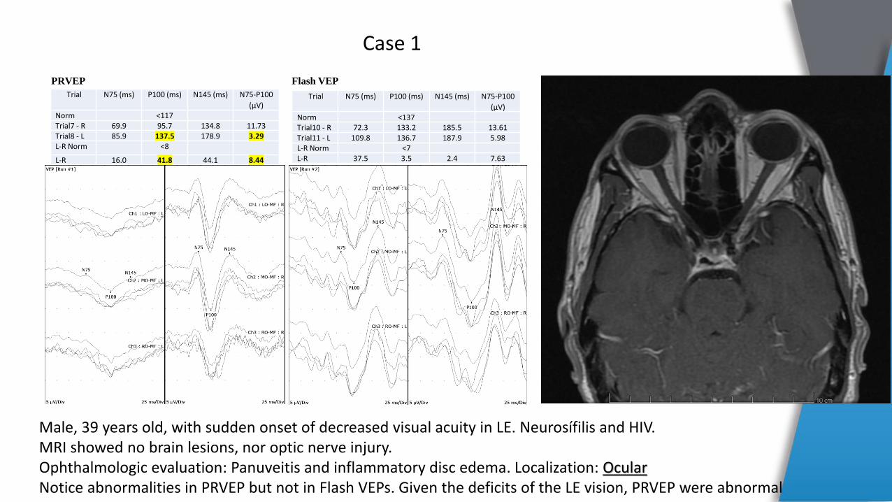
Trial N75 (ms) P100 (ms) N145 (ms) N75-P100
(µV)Norm <137Trial10 - R 72.3 133.2 185.5 13.61Trial11 - L 109.8 136.7 187.9 5.98L-R Norm <7L-R 37.5 3.5 2.4 7.63
Flash VEP
Trial N75 (ms) P100 (ms) N145 (ms) N75-P100
(µV)Norm <117Trial7 - R 69.9 95.7 134.8 11.73Trial8 - L 85.9 137.5 178.9 3.29L-R Norm <8
L-R 16.0 41.8 44.1 8.44
PRVEP
Male, 39 years old, with sudden onset of decreased visual acuity in LE. Neurosífilis and HIV.MRI showed no brain lesions, nor optic nerve injury. Ophthalmologic evaluation: Panuveitis and inflammatory disc edema. Localization: OcularNotice abnormalities in PRVEP but not in Flash VEPs. Given the deficits of the LE vision, PRVEP were abnormal
Case 1

Trial N75 (ms) P100 (ms) N145 (ms) N75-P100 (µV)Norm <117Trial4 - L 67.2 92.6 131.3 9.98Trial8 - R 75.4 110.9 147.7 3.38L-R Norm <8L-R 8.2 18.3 16.4 6.60
PRVEP
Female, 30 years old, with 8 days history of right ocular pain, decreased visual acuity, phosphenes, incapacity to distinguish colors. No previous medical history. MRI shows enhancement of right optic nerve at the intraorbital, intracanal, and intracranial segments.Localization: Pre-chiasmatic
Case 2
R R

Case 3
Trace N75 (ms) P100 (ms) N145 (ms) N75-P100 (µV)
Oz-Cz : R GAvg 101.2 131.6 182.4 0.39Oz-Cz : L GAvg 82.0 119.9 143.8 1.91L-R 19.2 11.7 38.6 1.52
PRVEP 30º
Female, 60 years old, bilateral restricted visual fields, predominantly on the RE. History of benign sellar tumor removed the previous year.Post-operative MRI shows intrasellar fluid collection, with chiasmatic luxation, predominantly towards the left side.Localization: Chiasmatic

Lesion on the left optical tract, RE stimulation
RO
MO
LO
N70
P100
N140
N140
P100
N70

Lesion on the left optical tract, LE stimulation
RO
MO
LO
N70
P100
N140
N140
P100
N70
Left cortex y deafferented, therefore there is no activity that projects to the ROUncrossed abnormality

Clinical Applications
1) Multiple Sclerosis: Optic Neuritis is the main reference to perform PRVEP. In the acute phase, when vision is lost, the PRVEP are lost, and as vision returns, amplitude of the P100 recover, but not latencies. In fact, even with normal vision, patients may present for long periods of time, normal P100morphology with prolonged latencies (like biological marker of ON) due to the demyelination process.
2) Tumors: Optic nerve tumors, affect morphology more importantly than latencies, since not all fibers of the ON are affected, and the remaining fibers can conduct the visual information. Larger tumors may blockcompletely the visual pathway.
3) Neuropathies: can cause amplitude changes as well as latency prolongation, depending on the type ofneuropathy: demyelinating Vs: axonal.
4) Metabolic Disorders: Leukodystrophies, Krabbe Disease and several other, may cause prechiasmaticor postchiasmatic lesions. Depending on the clinical stage, abnormalities will be different.
5) Conversive blindness: Caution should be taken with these patients, since they will not cooperate to fixate the checkerboard and cause low amplitude and increased latency. So, flash VEP can be used instead.
Prechiasmatic Lesions

Clinical Applications
1) Lesions on the Sella Turcica. Craniopharyngiomas, Pituitary Tumors: Compression of the optic chiasma produce distortion of the VEPs, mainly amplitude and less effects on latency. Monocular hemifield and full field stimulation may help to determine the localization, by observing “crossed asymmetry”.
Chiasmatic Lesions (crossed abnormalities)

Clinical Applications
1) Lesions on the posterior visual pathway (LGN, Cortex). Ischemic, demyelinating lesions, tumors, hemorrhagic, produce distortion of the VEPs, mainly amplitude and less effects on latency. Uncrossed asymmetry of the parasagittal P100 with monocular stimulation is typical. Binocular hemifield stimulation should be done to corroborate localization.
2) Neurodegenerative Disorders: Dementias (Alzheimer, Parkinson), metabolic disorders (leukodystrophies) may cause bilateral lesions. Most of these lesions, cause bilateral changes in amplitude, also latency prolongation may be seen. Specially because these patients do not cooperate in the test (lack of fixation on the screen).
Postchiasmatic Lesions (uncrossed asymmetries)

Clinical Applications
1) Refractive errors2) Glaucoma3) Amblyopia4) Disorders of the anterior chamber5) Various types of retinal pathologies
VEPs are not routinely applied to these pathologies, but when used, most of the times, changes in amplitudeand latencies can be seen, depending on the seriousness of the lesions.
Ophthalmologic Disorders

Conclusions
VEPs may provide important diagnostic information regarding the functional integrity of the visual system. Increments in latency with relatively preserved waveform morphology is a signal of a demyelinating pathology. Distortions of morphology with no changes in latency may traduce compression
VEPs help to confirm the presence of visual pathology or to detect subclinical asymptomatic involvement of the visual pathway
In healthy subjects, PRVEP latencies are influenced by stimulus-related variables such as luminance, spatial frequency, contrast. Other factors like fixation on the screen are of paramount importance
Pattern reversal is the favored stimulus for most clinical purposes
Flash VEPs are useful when poor optics, lack of cooperation or poor vision, malingering and patients with nystagmus
Utilization of central and parasagittal electrodes, are useful to localize lesions of the visual pathway

Conclusions
Normal VEP practically exclude abnormalities of the optic nerve
VEP is superior in cases of optic nerve and anterior chiasmatic lesions than MR, but the later is
clearly superior in retrochiasmatic diseases
VEP remain abnormal over long periods in patients with Optic Neuritis due to MS, despite
recovery of visual acuity
Objective and reproducible test for optic nerve function
Inexpensive as compared with to MRI
Under certain circumstances, may be helpful to positively establish optic nerve function in patients
with subjective complaint of visual loss
Copyright © 2022 FDOKUMEN