Dissecting the cellular contributions to early visual sensory processing deficits in schizophrenia...
9
Dissecting the cellular contributions to early visual sensory processing deficits in schizophrenia using the VESPA evoked response Edmund C. Lalor a,b,c , Sherlyn Yeap a,b , Richard B. Reilly a,c , Barak A. Pearlmutter d , John J. Foxe a,b,e, ⁎ a The Cognitive Neurophysiology Laboratory, St Vincent's Hospital Fairview, Dublin, Ireland b The Cognitive Neurophysiology Laboratory, Nathan S. Kline Institute for Psychiatric Research, Program in Cognitive Neuroscience and Schizophrenia, Orangeburg, New York 10962, USA c School of Mechanical, Electrical and Electronic Engineering, University College Dublin, Dublin, Ireland d The Hamilton Institute, National University of Ireland, Maynooth, Co. Kildare, Ireland e Program in Cognitive Neuroscience, Department of Psychology, City College of the City University of New York, 138th Street & Convent Avenue, New York, New York 10031, USA Received 8 March 2007; received in revised form 29 August 2007; accepted 6 September 2007 Available online 8 November 2007 Abstract Electrophysiological research has shown clear dysfunction of early visual processing mechanisms in patients with schizophrenia. In particular, the P1 component of the visual evoked potential (VEP) is substantially reduced in amplitude in patients. A novel visual evoked response known as the VESPA (Visual Evoked Spread Spectrum Analysis) was recently described. This response has a notably different scalp topography from that of the traditional VEP, suggesting preferential activation of a distinct subpopulation of cells. As such, this method constitutes a potentially useful candidate for investigating cellular contributions to early visual processing deficits. In this paper we compare the VEP and VESPA responses between a group of healthy control subjects and a group of schizophrenia patients. We also introduce an extension of the VESPA method to incorporate nonlinear processing in the visual system. A significantly reduced P1 component was found in patients using the VEP (with a large effect size; Cohen's d = 1.6), while there was no difference whatsoever in amplitude between groups for either the linear or nonlinear VESPA. This pattern of results points to a highly specific cellular substrate of early visual processing deficits in schizophrenia, suggesting that these deficits are based on dysfunction of magnocellular pathways with parvocellular processing remaining largely intact. © 2007 Elsevier B.V. All rights reserved. Keywords: EEG; Visual evoked potential; VESPA; Schizophrenia; P1 component 1. Introduction Visual evoked potential (VEP) studies have consis- tently demonstrated that patients with schizophrenia exhibit relatively severe deficits in early visual sensory processing, as indexed by a robust decrement in amplitude of the occipital P1 component (e.g., Foxe et al., 2001, 2005; Butler et al., 2001, 2007; Doniger et al., 2002; Available online at www.sciencedirect.com Schizophrenia Research 98 (2008) 256 – 264 www.elsevier.com/locate/schres ⁎ Corresponding author. The Cognitive Neurophysiology Laborato- ry, Nathan S. Kline Institute for Psychiatric Research, Program in Cognitive Neuroscience and Schizophrenia, 140 Old Orangeburg Road, Orangeburg, New York 10962, USA. Tel.: +1 845 398 6547; fax: +1 845 398 6545. E-mail addresses: [email protected] (E.C. Lalor), [email protected] (R.B. Reilly), [email protected] (B.A. Pearlmutter), [email protected] (S. Yeap), [email protected] (J.J. Foxe). 0920-9964/$ - see front matter © 2007 Elsevier B.V. All rights reserved. doi:10.1016/j.schres.2007.09.037
-
Upload
independent -
Category
Documents
-
view
0 -
download
0
Transcript of Dissecting the cellular contributions to early visual sensory processing deficits in schizophrenia...

Available online at www.sciencedirect.com
98 (2008) 256–264www.elsevier.com/locate/schres
Schizophrenia Research
Dissecting the cellular contributions to early visual sensory processingdeficits in schizophrenia using the VESPA evoked response
Edmund C. Lalor a,b,c, Sherlyn Yeap a,b, Richard B. Reilly a,c,Barak A. Pearlmutter d, John J. Foxe a,b,e,⁎
a The Cognitive Neurophysiology Laboratory, St Vincent's Hospital Fairview, Dublin, Irelandb The Cognitive Neurophysiology Laboratory, Nathan S. Kline Institute for Psychiatric Research,Program in Cognitive Neuroscience and Schizophrenia, Orangeburg, New York 10962, USA
c School of Mechanical, Electrical and Electronic Engineering, University College Dublin, Dublin, Irelandd The Hamilton Institute, National University of Ireland, Maynooth, Co. Kildare, Ireland
e Program in Cognitive Neuroscience, Department of Psychology, City College of the City University of New York,138th Street & Convent Avenue, New York, New York 10031, USA
Received 8 March 2007; received in revised form 29 August 2007; accepted 6 September 2007Available online 8 November 2007
Abstract
Electrophysiological research has shown clear dysfunction of early visual processing mechanisms in patients with schizophrenia.In particular, the P1 component of the visual evoked potential (VEP) is substantially reduced in amplitude in patients. A novel visualevoked response known as the VESPA (Visual Evoked Spread Spectrum Analysis) was recently described. This response has anotably different scalp topography from that of the traditional VEP, suggesting preferential activation of a distinct subpopulation ofcells. As such, this method constitutes a potentially useful candidate for investigating cellular contributions to early visual processingdeficits. In this paper we compare the VEP and VESPA responses between a group of healthy control subjects and a group ofschizophrenia patients.We also introduce an extension of the VESPAmethod to incorporate nonlinear processing in the visual system.A significantly reduced P1 component was found in patients using the VEP (with a large effect size; Cohen's d=1.6), while there wasno difference whatsoever in amplitude between groups for either the linear or nonlinear VESPA. This pattern of results points to ahighly specific cellular substrate of early visual processing deficits in schizophrenia, suggesting that these deficits are based ondysfunction of magnocellular pathways with parvocellular processing remaining largely intact.© 2007 Elsevier B.V. All rights reserved.
Keywords: EEG; Visual evoked potential; VESPA; Schizophrenia; P1 component
⁎ Corresponding author. The Cognitive Neurophysiology Laborato-ry, Nathan S. Kline Institute for Psychiatric Research, Program inCognitive Neuroscience and Schizophrenia, 140 Old OrangeburgRoad, Orangeburg, New York 10962, USA. Tel.: +1 845 398 6547;fax: +1 845 398 6545.
E-mail addresses: [email protected] (E.C. Lalor),[email protected] (R.B. Reilly), [email protected](B.A. Pearlmutter), [email protected] (S. Yeap),[email protected] (J.J. Foxe).
0920-9964/$ - see front matter © 2007 Elsevier B.V. All rights reserved.doi:10.1016/j.schres.2007.09.037
1. Introduction
Visual evoked potential (VEP) studies have consis-tently demonstrated that patients with schizophreniaexhibit relatively severe deficits in early visual sensoryprocessing, as indexed by a robust decrement in amplitudeof the occipital P1 component (e.g., Foxe et al., 2001,2005; Butler et al., 2001, 2007; Doniger et al., 2002;

257E.C. Lalor et al. / Schizophrenia Research 98 (2008) 256–264
Spencer et al., 2003; Schechter et al., 2005; Haenschelet al., in press). Concomitant structural deficits have alsobeen shown in the visual sensory pathways (Butler et al.,2006). Scalp topographies and source analysis havesuggested that these deficits may specifically reflectdysfunction of the dorsal visual stream while processingin the ventral stream remains relatively more intact (e.g.Foxe et al., 2001, 2005). It is also suggested that certainventral stream processes are contingent on inputs from thedorsal stream and as a result failure in these ‘higher-level’ventral stream processes may ultimately be a consequenceof these underlying dorsal stream deficits (Doniger et al.,2002; Foxe et al., 2005).
Further to the above findings, a substantial decrementin the P1 component was recently demonstrated in clin-ically unaffected first-degree relatives of schizophreniapatients (Yeap et al., 2006), establishing a possiblegenetic basis for the observed effects (see also Donohoeet al., in press). This points to the potential use of P1amplitude as an endophenotypic marker for schizo-phrenia and, as such, it may be a significant step in thequest for a diagnostic test facilitating early detection ofschizophrenia in high-risk individuals.
It would be of great benefit to this line of research tohave a more sensitive method for eliciting this deficit.One very promising candidate method, known as theVESPA technique (for Visual Evoked Spread SpectrumAnalysis), was recently described (Lalor et al., 2006).This method uses stimuli, the luminance or contrast ofwhich is rapidly and unobtrusively modulated by astochastic signal, enabling the estimation of the linearimpulse response of the visual system. The temporalprofile of these VESPAs is highly correlated with that oftransient VEPs evoked using standard, discrete stimuli.This includes a clearly defined and, hence, measurableP1 component. The rapidly estimable VESPA has beenshown to be superior to the VEP in terms of the amountof time necessary to obtain a response with a specificsignal-to-noise ratio. Furthermore, the method allowsfor a large degree of flexibility in design, not just interms of the parameters of the stimuli, as in VEP studies,but also in the characteristics of the modulating signal.
The topography of the VESPA is notably differentfrom that of the transient VEP. The abiding characteristicof the early VESPAmaps is a persistently delimited focusover midline occipital scalp without any evidence for thecharacteristic early bilateral spread over lateral occipitalscalp regions that is consistently seen for the standardVEP (e.g. Gomez-Gonzalez et al., 1994; Foxe andSimpson, 2002). This pattern suggests that the VESPAmay well have a distinct cellular activation pattern fromthat of the VEP, favoring midline structures such as striate
cortex and neighboring retinotopically mapped extra-striate regions, and perhaps also regions in the dorsalvisual stream, activation of which are known to producemidline scalp topographies (Clark and Hillyard 1996;Foxe and Simpson 2002). This suggests the VESPA as anexcellent candidate for further investigation of a dorsalstream based P1 deficit in schizophrenia.
For that reason, the aim of this paper is to compareVEPs and VESPA responses from schizophrenia patientsand healthy controls. Specifically, we examine the relativemagnitudes of the P1 components between groups forboth types of response. A direct comparison between theVEP and VESPA is complicated by the assumption oflinearity intrinsic to the VESPA estimation. In order toaddress this, we introduce a method for extending theVESPA analysis to higher orders and we expand ourcomparison between patients and controls to quadraticVESPA responses.
2. Materials and methods
2.1. Subjects
Written informed consent is obtained from 13(1 female) patients with DSM-IV diagnosis of schizo-phrenia. The Ethics Committee of St. Vincent's Hospitalapproved the experimental procedures. Patients wereaged 21 to 49 (mean±SD, 33.2±10.1 years) and had amean illness duration of 12.3 years (SD±9.5). Thesepatients had mean±SD scores on the Brief PsychiatricRating Scale and SANS of 33.1±5.8 and 22.2±17.7,respectively. Twelve of the patients were receivingantipsychotic medication at the time of testing with amean chlorpromazine equivalent dose of 406.71 mg/d(range, 50–1500 mg/d). The types of antipsychoticsincluded atypicals, typicals or a combination of both.One patient had ceased taking medication 5 monthsprior to testing and was medication-free at the time oftesting.
Control subjects were recruited from the St Vincent'sHospital staff community and through local recruitmentefforts in the hospital catchment area. This groupcomprised 11 (2 female) paid volunteers aged 19 to50 years (mean±SD, 26.5±8.7 years). The mean age ofpatients and controls did not differ significantly (t50=1.6,p=0.12). All of the 11 controls, and 12 of the 13 patientswere right-handed as assessed by the Edinburgh Hand-edness Inventory (Oldfield, 1971). None of the controlswere receiving any psychotropicmedication at the time oftesting. Also, all controls were free of any psychiatricillness or symptoms by self-report using criteria from theStructured Clinical Interview for DSM-III-R–Non-

Fig. 1. Stimuli used to elicit (a) the VEP — non-illusory arrangementfrom Kanizsa study (b) the VESPA— single checkerboard the contrastof which is rapidly modulated as in Lalor et al. (2006).
258 E.C. Lalor et al. / Schizophrenia Research 98 (2008) 256–264
Patient (SCID-NP), and all reported no history of alcoholor substance abuse.
2.2. Stimuli
VEPswere obtained during a study aimed at assessingschizophrenia patient deficits in visual binding processesusing Kanizsa illusory figures. Accordingly, twelvedifferent stimulus displays were presented, some con-taining Kanizsa illusions and others not. Specifically, theVEPs presented in the current study were obtained usinga layout consisting of three centrally presented, black“pacmen” (disks with missing sectors) elements, whosearrangement was such as to not give any illusory effect,as in Fig. 1(a).1 The array of elements subtendedmaximal visual angles of 2.28° horizontally andvertically and was presented for 400 ms. This allowedERP analysis to be performed for the first 400mswithoutcontamination of any visual offset response. The meaninter-stimulus-interval (ISI) between trials was 900 ms(ranging from 600–1200 ms). Thus, each trial had amean duration of 1300 ms. Note that this stimulusarrangement was chosen as it has previously been shownto elucidate a large VEP P1 deficit in patients withschizophrenia (Foxe et al., 2005; Spencer et al., 2003).
In the case of the VESPA, the stimulus consisted of acheckerboard pattern with equal numbers of black andwhite checks as in Fig. 1(b). Each check subtended avisual angle of 0.65° both horizontally and vertically,while the checkerboard as a whole subtended visualangles of 5.25° vertically and horizontally. The refreshrate of the monitor was set to 60 Hz and on every refreshthe contrast of the checkerboard patternwasmodulated bya stochastic signal with the mean luminance remainingconstant. The stochastic signals used had their powerdistributed uniformly between 0 and 30 Hz. See Laloret al. (2006) for details.
2.3. Experimental procedure
Each VEP experimental block consisted of, onaverage, 12.75 presentations of each of the 12 displaytypes in a random order. Subjects underwent 20 blocks,resulting in 255 presentations of each display type.During VEP runs a small fixation point was present in thecenter of the screen, on which subject were instructed tomaintain their gaze.
1 Although only the non-illusion inducing arrangement was used,the reader should note that the P1 component is entirely insensitive tothe presence or absence of illusory contours (Murray et al., 2002,2004, 2006).
Every subject underwent three VESPA runs of 200 seach. Subjects were instructed to maintain visual fixationon the center of the screen for the duration of each run.While abstaining from eye-blinks was not possible giventhe trial lengths, subjects were instructed to keep thenumber of eye-blinks to a minimum. A different modu-lating waveform was used for each run, although allwaveforms had identical statistics.
2.4. EEG acquisition and analysis
EEG data were recorded from 72 electrode positionsreferenced to location Fz, filtered over the range 0–134Hzand digitized at a rate of 512 Hz using the BioSemi ActiveTwo system. Subsequently, the EEG was digitally filteredwith a high-pass filter with passband above 2 Hz and −60dB response at 1 Hz and a low-pass filter with 0–35 Hzpassband and −50 dB response at 45 Hz.
VEPs were calculated by averaging time-lockedresponses to the presentations of the display type de-scribed earlier. A time window of 500 ms starting 100 mspre-stimulus was used. Any epochs where the EEGexceeded +/−120 μV were rejected, resulting in a meanrejection rate of 11%.
The VESPA is an estimate of the linear impulseresponse of the visual system (Lalor et al., 2006). It isbased on the assumption that the EEG response to astimulus, whose luminance or contrast is rapidly modu-lated by a stochastic signal, consists of a convolution ofthat signal with an unknown impulse response. Given theknown stimulus signal and the measured EEG, thisimpulse response, i.e., the VESPA, can be estimated usingthe method of linear least squares. In the present studyVESPAs were measured using a sliding window of500 ms of data starting 100 ms pre-stimulus.
It is possible that a VESPA founded on anassumption of linearity may not be sensitive to thedeficits apparent in the VEP. The method can, however,easily be extended to higher orders. For example, in thecase of a quadratic analysis, this is accomplished byincluding in the least squares estimation not only the
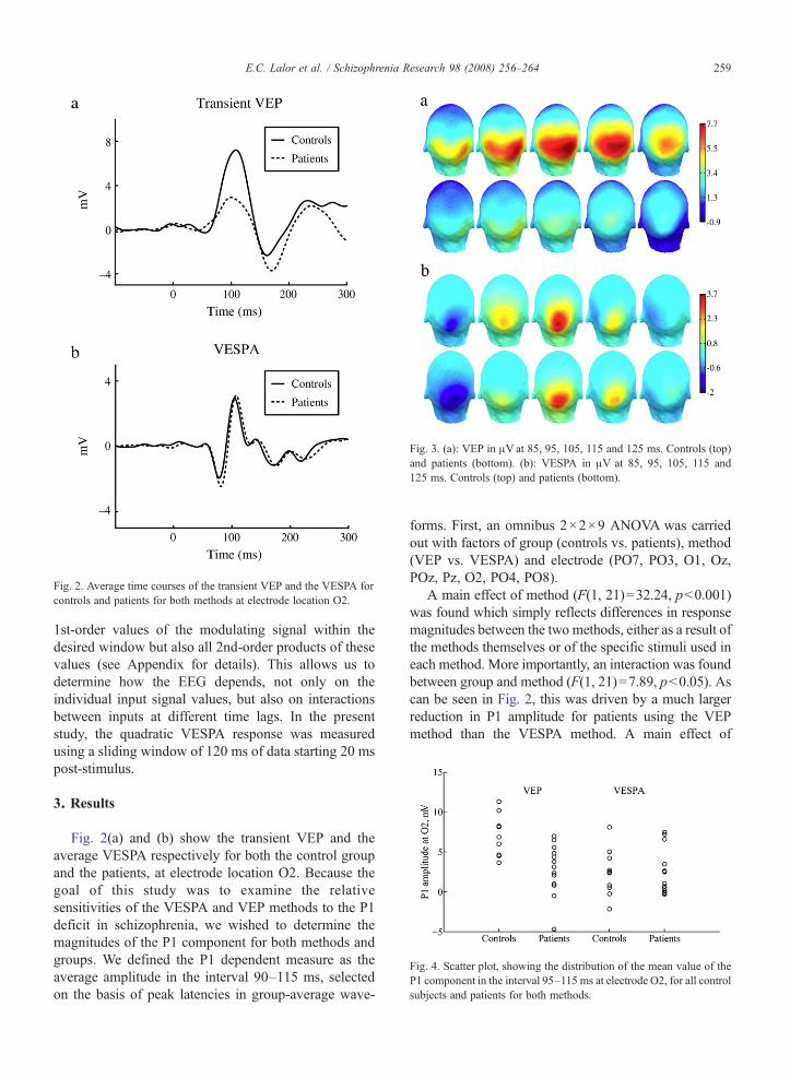
Fig. 3. (a): VEP in μVat 85, 95, 105, 115 and 125 ms. Controls (top)and patients (bottom). (b): VESPA in μV at 85, 95, 105, 115 and125 ms. Controls (top) and patients (bottom).
Fig. 2. Average time courses of the transient VEP and the VESPA focontrols and patients for both methods at electrode location O2.
259E.C. Lalor et al. / Schizophrenia Research 98 (2008) 256–264
r
Fig. 4. Scatter plot, showing the distribution of the mean value of theP1 component in the interval 95–115 ms at electrode O2, for all controlsubjects and patients for both methods.
1st-order values of the modulating signal within thedesired window but also all 2nd-order products of thesevalues (see Appendix for details). This allows us todetermine how the EEG depends, not only on theindividual input signal values, but also on interactionsbetween inputs at different time lags. In the presentstudy, the quadratic VESPA response was measuredusing a sliding window of 120 ms of data starting 20 mspost-stimulus.
3. Results
Fig. 2(a) and (b) show the transient VEP and theaverage VESPA respectively for both the control groupand the patients, at electrode location O2. Because thegoal of this study was to examine the relativesensitivities of the VESPA and VEP methods to the P1deficit in schizophrenia, we wished to determine themagnitudes of the P1 component for both methods andgroups. We defined the P1 dependent measure as theaverage amplitude in the interval 90–115 ms, selectedon the basis of peak latencies in group-average wave-
forms. First, an omnibus 2×2×9 ANOVA was carriedout with factors of group (controls vs. patients), method(VEP vs. VESPA) and electrode (PO7, PO3, O1, Oz,POz, Pz, O2, PO4, PO8).
A main effect of method (F(1, 21)=32.24, pb0.001)was found which simply reflects differences in responsemagnitudes between the two methods, either as a result ofthe methods themselves or of the specific stimuli used ineach method. More importantly, an interaction was foundbetween group and method (F(1, 21)=7.89, pb0.05). Ascan be seen in Fig. 2, this was driven by a much largerreduction in P1 amplitude for patients using the VEPmethod than the VESPA method. A main effect of

260 E.C. Lalor et al. / Schizophrenia Research 98 (2008) 256–264
electrode (F(8, 168)=10.29, pb0.001) reflected thetopographic specificity of the P1. A significant interactionbetween electrode and method (F(8, 168)=3.11, pb0.05)reflected the topographic differences between methodsevident in Fig. 3. There was no three-way interactionbetween group, method and electrode (F(8, 168)=1.57,pN0.1).
To examine the interaction between group andmethodfurther, planned t-tests were carried out for each methodseparately. We used the P1, averaged across electrodesO1, Oz and O2, as the dependent measure. A significantdifference was found between groups for the VEPmethod (t=3.5, pb0.005) whereas no difference wasfound between groups for the VESPA method (t=0.08,pN0.9). The Cohen's d effect size was calculated forthe VEP P1 and found to be 1.57. Figs. 4 and 5 providefurther illustration of the differing effects found using the
Fig. 5. Statistical cluster plot marking for all electrodes the time pointsat which the event-related potential differed significantly betweengroups on the basis of 2-tailed t-tests at an α level of 0.05. Whitedenotes nonsignificance while positive t values (ControlsNPatients)are marked on a green scale and negative t values (PatientsNControls)are marked in gold. Electrodes are ordered from the bottom, occipital(O), parietal (P), central (C), and frontal (F) proceeding in the anteriordirection in rows from left to right. In the case of the VEP, a cluster isseen over posterior sites in the P1 interval 90 to 120 ms as expectedfrom the results of the planned analysis of variance. No meaningfulclusters are seen for the VESPA.
Fig. 6. Grand average quadratic VESPAs at electrode location Oz forcontrols, patients and the difference between controls and patients.These plots indicate how strongly the EEG at a particular time pointdepends on the interaction between inputs at two previous time points.Prominent “P1” activity can be see around 100×100 ms for bothgroups with a negligible difference between groups.
VEP andVESPAmethods. Fig. 4 is a scatter plot showinghow the mean value of the P1 component in the interval95–115 ms at electrode O2 is distributed within thecontrol and patients groups for both methods. Thestandard deviations for the controls were 2.37 for theVEP and 2.76 for the VESPA and for the patients were3.21 for the VEP and 2.83 for the VESPA respectively.

261E.C. Lalor et al. / Schizophrenia Research 98 (2008) 256–264
Fig. 5 illustrates a pair of statistical cluster plots markingthe time points at which the VEP and VESPArespectively differ significantly between groups for allelectrodes. A large cluster is evident in the VEP plot overposterior sites in the P1 interval 90–120 ms. No suchcluster is evident in the VESPA plot.
Fig. 6 shows the average quadratic VESPA for bothgroups for electrode location Oz. Also plotted is thedifference between controls and patients. Statisticsconfirmed the lack of a between-group differenceevident from the figure. We defined the P1 dependentmeasure as the average amplitude in the interval 90–115 ms×90–115 ms. A 2-way ANOVAwas carried outusing this P1 measure, with factors of group (controlsvs. patients) and electrode (PO7, PO3, O1, Oz, POz, Pz,O2, PO4, PO8). No significant difference was foundbetween groups (F(1, 22)=0.197, pN0.6).
4. Discussion
Replicating the findings of earlier studies (Foxe et al.,2001, 2005; Butler et al., 2001, 2007; Doniger et al.,2002; Spencer et al., 2003; Schechter et al., 2005;Haenschel et al., in press), a substantial reduction in theamplitude of the P1 component of the transient VEP wasobserved for schizophrenia patients compared withhealthy controls. However, somewhat to our surprise,there was no difference whatsoever in VESPA P1amplitude between patients and controls, in either thelinear or quadratic case. This striking contrast inoutcomes between two essentially similar methodspoints to a highly specific disparity between the earlyvisual sensory processing systems of patients andcontrols. While the scalp topography of the VESPA(Fig. 3) suggests that it may preferentially activate thedorsal visual stream and given that a number of studieshave proposed that the VEP P1 deficit shown by patientswith schizophrenia may be due to specific dysfunctionof this stream (Doniger et al., 2002; Foxe et al., 2005),we had originally expected that the midline-focusedVESPA might prove to be more sensitive to this deficit.This is clearly not the case here. In what follows, weconsider a number of possible reasons for the lack of areduction in the amplitude of the VESPA P1.
One explanation concerns the subcortical source of thescalp VESPA and that of the P1 deficit in the VEP. Thehuman visual system consists of discrete subcorticalmagnocellular and parvocellular pathways that projectpreferentially to dorsal and ventral cortical streams. In ourprevious studies, we have consistently posited a magno-cellular basis for the observed VEP P1 deficits (e.g. Foxeet al., 2005; Butler et al., 2005, 2007; see also Kim et al.,
2005a). The VESPA scalpmaps of Fig. 3 suggest possiblepreferential stimulation of the dorsal stream and thus ofmagnocellular pathways. Therefore, the lack of a differ-ence in theVESPAbetween controls and patients suggeststhat either there is no dysfunction of the magnocellularsystem in schizophrenia or that the VESPA does notactually reflect activity of the magnocellular system.
Magnocellular and parvocellular cells differ not onlyanatomically, but also functionally, in terms of preferredstimuli. Parvocellular cells with their spectrally opponentnature are know to be less sensitive to luminance contrastthan magnocellular cells (Kaplan et al., 1990; Lee et al.,1990). While, the high contrast gain of cells in themagnocellular pathway might suggest that they may bemore sensitive to the contrast modulations of the VESPAstimulus, their response saturates at fairly low contrasts(10–15%; e.g., Baseler and Sutter 1997). Parvocellularneurons, meanwhile, have lower contrast gain, but do notsaturate (see Butler et al., 2007). Given that the stimulusdescribed in this study spends less than 2% of its timebelow 15% contrast (Lalor et al., 2006), it seemsreasonable to conclude that the VESPA may actuallyreflect mostly activity of parvocellular pathways.
The two pathways are also known to differ in theirresponse characteristics to the temporal frequencies ofstimuli. The commonly held belief is that magnocellularcells are more suited to high temporal frequency flicker(e.g., Kaplan and Benardete, 2001). As a result, it couldagain be concluded that the rapid modulation of theVESPA might preferentially activate that subsystem.However, both parvocellular and magnocellular cells inthe lateral geniculate nucleus (LGN) of the macaquehave been reported to respond best at temporalfrequencies in the range of 10–20 Hz (Hicks et al.,1983). More specifically, Derrington and Lennie (1984)found that parvocellular units were most sensitive tostimuli modulated at temporal frequencies close to10 Hz and magnocellular units to stimuli modulated atfrequencies nearer 20 Hz. They also reported that theloss of sensitivity as temporal frequency fell belowoptimum was more marked in magnocellular thanparvocellular units. These findings suggest that, whilemagnocellular cells are known to have a shorter latencyand more transient response to stimuli (Marrocco et al.,1982; Maunsell et al., 1999), parvocellular cells shouldhave no difficulty in following the 0–30 Hz frequencycontent of the VESPA stimulus.
A further property that differs between the twosubsystems is the linearity of their temporal response.While the parvocellular system is approximately linear,the temporal responses of magnocellular cells areparticularly nonlinear due to contrast gain control

Appendix A. Extension to quadratic VESPA estimation
As detailed in Lalor et al. (2006), we estimate thelinear VESPA as an n-dimensional vector w consistingof the sampled points of the response function
(1)w s0ð Þ;w s1ð Þ; N ;w sn�1ð Þð ÞT ;where n is the number of sampled points of the responsefunction that we wish to estimate. This is done by firstforming the n-dimensional vector xt consisting of thesampled points of the modulating stimulus
(2)
x t � t0ð Þ; x t � t0 þ 1ð Þð Þ; N ; x t � t0 þ n� 1ð Þð Þð ÞT ;
where t0 is the estimation window offset. The values ofxt are simply the normalized luminance or contrastvalues of the displayed stimulus.
We then solve for w using the equation,
(3)w ¼ hxtxTt þ kMi�1xtyt
where λ is a regularization parameter and M is a near-diagonal matrix.
In this paper, we expand the VESPA estimation to aquadratic model of how the EEG depends on the inputstimulus. This is accomplished by replacing Eq. (2) witha vector with n+n(n+1) /2 elements, where n is thewindow size, containing the n 1st-order elements asbefore, and the n(n+1) /2 2nd-order elements (allproducts of the form x(t− t0− i)x(t− t0− j) where
262 E.C. Lalor et al. / Schizophrenia Research 98 (2008) 256–264
(Kaplan and Benardete, 2001). The nonlinear nature ofthis system has been referred to in a recent study, whichinvestigated early-stage visual processing deficits inpatients with schizophrenia using the steady-state VEP(SSVEP; Kim et al., 2005b). This study has shownreductions in the second harmonics of the stimulusfrequency. Given that second harmonics are thought todepend preferentially on magnocellular pathways, thereduced harmonics are attributed to deficits in thosepathways. However, it was also pointed out that deficitsin nonlinear mechanisms present in cortex, which areimportant in producing responses at higher harmonics ortemporal frequencies, would also result in greaterattenuation of higher harmonic responses in patientsthan controls. For these reasons, it is clear that a linearVESPA simply may not be sensitive to the nonlinearsystems responsible for the generation of the P1 deficitin the transient VEP. In order to address this, we haveextended the VESPA method to a quadratic analysis inthis paper. The fact that no significant differences werefound between patients and controls for the quadraticVESPA lends further support to the notion that, if indeedmagnocellular dysfunction underlies the P1 deficit in theVEP, the stimulus used in this study was biased towardlinear parvocellular cell populations.
Another potential reason for the dramatic dissocia-tion between VEP and VESPA results stems from thedebate over whether the ERP in response to a stimulusconstitutes an evoked event or comes about throughinduced changes in ongoing brain dynamics. Whilemost ERP studies assume the former, some studies havesuggested that the ERP at least partly arises from thestimulus-induced phase-resetting of electrophysiologi-cal processes (e.g., Makeig et al., 2002; Hanslmayret al., 2007). While the VESPA does not rule out thenotion of an induced contribution to VEPs obtainedusing discrete stimuli, its continuous nature, which doesnot allow for any time-locked lower frequency phase-resetting of ongoing brain dynamics, clearly demon-strates that ERPs can be evoked. This leads to aconfound in the comparison between VEP and VESPAin that it is at least possible that the reduced VEP P1components displayed by the patients reflect dysfunc-tion of phase-resetting processes or ongoing oscillatoryactivity. In support of this notion, one recent studyproposed alpha band activity as the likely source of anearly induced ERP contribution (Hanslmayr et al., 2007)while various characteristics of alpha oscillations havebeen shown to differ in patients with schizophrenia,including lower peak frequency (Javitt, 1997) and lowerpower (Sponheim et al., 1994). The inconclusive (andsometimes contradictory) nature of studies attempting to
evaluate phase-resetting and the demonstration of thepurely evoked VESPA ERP itself lend support to studiespositing a predominant role for stimulus-evoked activityin sensory ERP generation (e.g., Shah et al., 2004).Therefore, attributing a divergence in results as dramaticas reported in this paper to deficiency in induced ERPgeneration seems, at best, speculative.
In summary, we have demonstrated a striking disparityin relative ERP responses between patients and controlsusing two different methods of visual stimulation. Thispoints to the highly specific nature of early visual deficitsin schizophrenia and speaks particularly to the notion thatthose deficits are based substantially on magnocellularstream dysfunction where activity of the parvocellularsystem is largely spared. While the VESPA as implemen-ted in this study was not sensitive to the mechanismsresponsible for a reduced P1 component in schizophrenia,the flexibility of themethod, in terms of the characteristicsof both the stimuli and the modulating signal, suggests itsutility as a method for further investigation of thosemechanisms.

0≤ i≤ j≤n). The quadratic VESPA w, of this samedimensionality can be solved using,
(4)w ¼ hxtxTt þ dIi�1xtyt
where δ is a different regularization parameter and I isthe identity matrix. In this study, δ=5×10⁻⁶ gave goodreduction in estimation error.
263E.C. Lalor et al. / Schizophrenia Research 98 (2008) 256–264
Role of funding sourceThis work was supported in part by a National Institute of Mental
Health (NIMH) Grant MH-65350 to J. J. Foxe, and by a Fund forDigital Research Programme grant from the Higher EducationalAuthority (HEA) of Ireland to R. B. Reilly. Neither the NIMH nor HEAhad any further role in study design; in the collection, analysis andinterpretation of data; in the writing of the report; or in the decision tosubmit the paper for publication.
ContributorsDr. Lalor designed the stimulus sequences, programmed all
paradigms, analyzed all data and wrote the first draft of the manuscript.Dr. Foxe designed the experimental protocol and edited multiple draftsof the manuscript. Dr. Yeap collected all data and tabulated patientdemographics, performed the clinical ratings and aided in analyses.Drs. Reilly and Pearlmutter provided critical input regardingdevelopment of the VESPA technique and its implementation andprovided comments on early drafts of the manuscript. All authorscontributed to and have approved the final manuscript. The principleinvestigator, Dr. Foxe, takes responsibility for the integrity of the dataand the accuracy of the data analysis, and attests that all authors had fullaccess to all the data in the study.
Conflict of interestDrs. Lalor, Pearlmutter, Reilly and Foxe are listed as inventors on a
patent application for the VESPA method. Dr. Yeap declares noconflict of interest.
AcknowledgementsThe authors are grateful to Dr. Simon P. Kelly for helpful
comments and discussion. The authors would also like to express theirsincere gratitude to the Chief Executive Officer at St. Vincent'sHospital, Mr. Edward Byrne and to the Director of Nursing, Mrs. PhilBourke, for their ongoing and essential support of the CognitiveNeurophysiology Laboratory.
References
Baseler, H.A., Sutter, E.E., 1997. M and P components of the VEP andtheir visual field distribution. Vis. Res. 37 (6), 675–690.
Butler, P.D., Schechter, I., Zemon, V., Schwartz, S.G., Greenstein, V.C.,Gordon, J., Schroeder, C.E., Javitt, D.C., 2001. Dysfunction ofearly-stage visual processing in schizophrenia. Am. J. Psychiatry158 (7), 1126–1133.
Butler, P.D., Zemon, V., Schechter, I., Saperstein, A.M., Hoptman, M.J.,Lim, K.O., Revheim, N., Silipo, G., Javitt, D.C., 2005. Early-stagevisual processing and cortical amplification deficits in schizophrenia.Arch. Gen. Psychiatry 62 (5), 495–504.
Butler, P.D., Hoptman, M.J., Nierenberg, J., Foxe, J.J., Javitt, D.C.,Lim, K.O., 2006. Visual white matter integrity in schizophrenia.Am. J. Psychiatry 163 (11), 2011–2013.
Butler, P.D., Martinez, A., Foxe, J.J., Kim, D., Zemon, V., Silipo, G.,Mahoney, J., Shpaner, M., Jalbrzikowski, M., Javitt, D.C., 2007.Subcortical visual dysfunction in schizophrenia drives secondarycortical impairments. Brain 130 (Pt 2), 417–430.
Clark, V.P., Hillyard, S.A., 1996. Spatial selective attention affectsextrastriate but not striate components of the visual evokedpotential. J. Cogn. Neurosci. 8, 387–402.
Derrington, A.M., Lennie, P., 1984. Spatial and temporal contrastsensitivities of neurones in lateral geniculate nucleus of macaque.J. Physiol. 357, 219–240.
Doniger,G.M., Foxe, J.J.,Murray,M.M.,Higgins,B.A., Javitt,D.C., 2002.Impaired visual object recognition and dorsal/ventral stream interactionin schizophrenia. Arch. Gen. Psychiatry 59 (11), 1011–1020.
Donohoe, G., Morris, D.W., De Sanctis, P., Magno, E., Montesi, J.L.,Garavan, H.P., Robertson, I.H., Javitt, D.C., Gill, M., Corvin, A.P.,Foxe, J.J., in press. Early visual processing deficits in dysbindin-associated schizophrenia. Biological Psychiatry. doi:10.1016/j.biopsych.2007.07.022.
Foxe, J.J., Simpson, G.V., 2002. Flow of activation from V1 to frontalcortex in humans. A framework for defining “early” visualprocessing. Exp. Brain Res. 142 (1), 139–150.
Foxe, J.J., Doniger, G.M., Javitt, D.C., 2001. Early visual processingdeficits in schizophrenia: impaired P1 generation revealed by high-density electrical mapping. NeuroReport 12 (17), 3815–3820.
Foxe, J.J., Murray, M.M., Javitt, D.C., 2005. Filling-in in schizophre-nia: a high density electrical mapping and source-analysisinvestigation of illusory contour processing. Cereb. Cortex 15(12), 1914–1927.
Gomez-Gonzalez, C.M., Clark, V.P., Fan, S., Luck, S.J., Hillyard, S.A.,1994. Sources of attention-sensitive visual event-related potentials.Brain Topogr. 7 (1), 41–51.
Haenschel, C., Bittner, R.A., Haertling, F., Rotarska-Jagiela, A.,Maurer, K., Singer, W., Linden, D.E.J., in press. Impaired early-stage visual processing contributes to working memory dysfunc-tion in adolescents with schizophrenia — a study with event-related potentials and functional magnetic resonance imaging.Arch Gen Psychiatry.
Hanslmayr, S., Klimesch, W., Sauseng, P., Gruber, W., Doppelmayr,M., Freunberger, R., Pecherstorfer, T., Birbaumer, N., 2007. Alphaphase reset contributes to the generation of ERPs. Cereb. Cortex17 (1), 1–8.
Hicks, T.P., Lee, B.B., Vidyasagar, T.R., 1983. The responses of cells inmacaque lateral geniculate nucleus to sinusoidal gratings. J. Physiol.337, 183–200.
Javitt, D.C., 1997. Psychophysiology of schizophrenia. Curr. Opin.Psychiatry 10 (1), 11–15.
Kaplan, E., Benardete, E., 2001. The dynamics of primate retinalganglion cells. Prog. Brain Res. 134, 17–34.
Kaplan, E., Lee, B.B., Shapley, R.M., 1990. New views of primateretinal function. In: Osborne, N., Chader, G. (Eds.), Progress inRetinal Research, vol. 9. Pergamon Press, Oxford, pp. 273–336.
Kim, D., Wylie, G., Pasternak, R., Butler, P.D., Javitt, D.C., 2005a.Magnocellular contributions to impaired motion processing inschizophrenia. Schizophr. Res. 82 (1), 1–8.
Kim, D., Zemon, V., Saperstein, A., Butler, P.D., Javitt, D.C., 2005b.Dysfunction of early-stage visual processing in schizophrenia:harmonic analysis. Schizophr. Res. 76 (1), 55–65.
Lalor, E.C., Pearlmutter, B.A., Reilly, R.B., McDarby, G., Foxe, J.J.,2006. The VESPA: a method for the rapid estimation of a visualevoked potential. NeuroImage 32 (4), 1549–1561.
Lee, B.B., Pokorny, J., Smith, V.C., Martin, P.R., Valberg, A., 1990.Luminance and chromatic modulation sensitivity of macaque

264 E.C. Lalor et al. / Schizophrenia Research 98 (2008) 256–264
ganglion cells and human observers. J. Opt. Soc. Am. A 7 (12),2223–2236.
Makeig, S., Westerfield, M., Jung, T.P., Enghoff, S., Townsend, J.,Courchesne, E., Sejnowski, T.J., 2002. Dynamic brain sources ofvisual evoked responses. Science 295 (5555), 690–694.
Marrocco, R.T., McClurkin, J.W., Young, R.A., 1982. Spatialsummation and conduction latency classification of cells of thelateral geniculate nucleus of macaques. J. Neurosci. 2 (9),1275–1291.
Maunsell, J.H.R., Ghose, G.M., Assad, J.A., McAdams, C.J.,Boudreau, C.E., Noerager, B.D., 1999. Visual response latenciesof magnocellular and parvocellular LGN neurons in macaquemonkeys. Vis. Neurosci. 16 (1), 1–14.
Murray,M.M.,Wylie, G.R., Higgins, B.A., Javitt, D.C., Schroeder, C.E.,Foxe, J.J., 2002. The spatiotemporal dynamics of illusory contourprocessing: combined high-density electrical mapping, sourceanalysis, and functional magnetic resonance imaging. J. Neurosci.22 (12), 5055–5073.
Murray, M.M., Foxe, D.M., Javitt, D.C., Foxe, J.J., 2004. Settingboundaries: brain dynamics of modal and amodal illusory shapecompletion in humans. J. Neurosci. 24 (31), 6898–6903.
Murray, M.M., Imber, M.L., Javitt, D.C., Foxe, J.J., 2006. Boundarycompletion is automatic and dissociable from shape discrimina-tion. J. Neurosci. 26 (46), 12043–12054.
Oldfield, R.C., 1971. The assessment and analysis of handedness: theEdinburgh inventory. Neuropsychologia 9 (1), 97–113.
Schechter, I., Butler, P.D., Zemon, V.M., Revheim, N., Saperstein, A.M.,Jalbrzikowski, M., Pasternak, R., Silipo, G., Javitt, D.C., 2005.Impairments in generation of early-stage transient visual evokedpotentials to magno- and parvocellular-selective stimuli in schizo-phrenia. Clin. Neurophysiol. 116 (9), 2204–2215.
Shah, A.S., Bressler, S.L., Knuth, K.H., Ding, M., Mehta, A.D.,Ulbert, I., Schroeder, C.E., 2004. Neural dynamics and thefundamental mechanisms of event-related brain potentials.Cereb. Cortex 14 (5), 476–483.
Spencer, K.M., Nestor, P.G., Niznikiewicz, M.A., Salisbury, D.F.,Shenton, M.E., McCarley, R.W., 2003. Abnormal neural synchro-ny in schizophrenia. J. Neurosci. 23 (19), 7407–7411.
Sponheim, S.R., Clementz, B.A., Iacono, W.G., Beiser, M., 1994.Resting EEG in first-episode and chronic schizophrenia. Psycho-physiology 31 (1), 37–43.
Yeap, S., Kelly, S.P., Sehatpour, P., Magno, E., Javitt, D.C., Garavan,H., Thakore, J.H., Foxe, J.J., 2006. Early visual sensory deficits asendophenotypes for schizophrenia: high-density electrical map-ping in clinically unaffected first-degree relatives. Arch. Gen.Psychiatry 63 (11), 1180–1188.