Are laser-evoked brain potentials modulated by attending to first or second pain?
11
Are laser-evoked brain potentials modulated by attending to first or second pain? A. Mouraux a, * , L. Plaghki b a Laboratoire de neurophysiologie (NEFY), Universite ´ catholique de Louvain, Belgium b Unite ´ de re ´adaptation (READ), Universite ´ catholique de Louvain, Belgium Received 17 February 2006; received in revised form 12 October 2006; accepted 18 October 2006 Abstract By co-activating Ad- and C-nociceptors, a brief and intense infrared laser stimulus may produce a double sensation referred to as ‘first’ and ‘second’ pain, evoke late laser-evoked brain potentials (LEPs) related to the processing of Ad-fiber input, but fails to evoke any consistent activity related to C-fibers. Yet, ultra-late LEPs may be recorded if C-nociceptors are activated in isolation. Bromm and Treede (1985) reported that if a subject selectively focused his attention towards second pain, co-activation of Ad- and C-noci- ceptors could elicit both late and ultra-late LEPs (Bromm B, Treede RD. Electroencephalogr Clin Neurophysiol 1985;61). They hypothesized that for C-fiber input to elicit consistent ultra-late LEPs, attention must be specifically focused towards that sensory channel. However, the significance of this study was limited by the fact it relied on results of a single subject and used latency-cor- rection algorithms. In an attempt to replicate these findings, this study examined LEPs recorded while trained subjects focused their attention either towards first pain or towards second pain. Whether or not subjects attended second pain, laser stimuli co-activating Ad- and C-nociceptors failed to elicit ultra-late LEPs, indicating that focusing attention towards C-fiber sensory input is not a suf- ficient condition for that input to evoke LEPs. However, selectively attending to first or second pain significantly modulated Ad-fiber related late LEPs. When subjects attended first pain, a late parietal P3 component was recorded, possibly related to task closure. When subjects attended second pain, a prefrontal positive enhancement of the P2 component was observed. Whether it could reflect brain processes related to response-inhibition is discussed. Ó 2006 International Association for the Study of Pain. Published by Elsevier B.V. All rights reserved. Keywords: Laser evoked potentials; Ad-fibers; C-fibers; Go/NoGo; Response inhibition 1. Introduction Allowing the synchronous and selective activation of Ad- and C-nociceptors, infrared laser stimulators have been extensively used to study time-locked nociception- related electrophysiological responses (for a review, see Treede et al., 2003). Provided that the peripheral conduc- tion distance is long, applying such a laser stimulus to the hairy skin can produce two temporally distinct sensa- tions, reminiscent of the ‘first’ and ‘second pain’ described by Lewis and Ponchin (1937). The first sensa- tion is often described as well localized, brief, and prick- ing. The second is often described as more diffuse and longer-lasting. Psychophysical and neurophysiological studies have shown that these two sensations are, respec- tively, related to the activation of Ad- and C-fiber affer- ents (Bromm and Treede, 1984; Willis, 1985; Price, 1996). Although subjects clearly report sensations related to the activation of both nociceptive afferents, laser-evoked brain potentials (LEPs) only reveal ‘late’ components whose latencies (160–390 ms; Bromm and Treede, 1984) are compatible with the faster conduction velocities of Ad-fibers. Several methods (reviewed in Plaghki and Mouraux, 2003) allow activating C-nociceptors in 0304-3959/$32.00 Ó 2006 International Association for the Study of Pain. Published by Elsevier B.V. All rights reserved. doi:10.1016/j.pain.2006.10.018 * Corresponding author. Tel.: +32 2 764 5467; fax: +32 2 764 5465. E-mail address: [email protected] (A. Mouraux). www.elsevier.com/locate/pain Pain 129 (2007) 321–331
Transcript of Are laser-evoked brain potentials modulated by attending to first or second pain?

www.elsevier.com/locate/pain
Pain 129 (2007) 321–331
Are laser-evoked brain potentials modulated by attendingto first or second pain?
A. Mouraux a,*, L. Plaghki b
a Laboratoire de neurophysiologie (NEFY), Universite catholique de Louvain, Belgiumb Unite de readaptation (READ), Universite catholique de Louvain, Belgium
Received 17 February 2006; received in revised form 12 October 2006; accepted 18 October 2006
Abstract
By co-activating Ad- and C-nociceptors, a brief and intense infrared laser stimulus may produce a double sensation referred to as‘first’ and ‘second’ pain, evoke late laser-evoked brain potentials (LEPs) related to the processing of Ad-fiber input, but fails to evokeany consistent activity related to C-fibers. Yet, ultra-late LEPs may be recorded if C-nociceptors are activated in isolation. Brommand Treede (1985) reported that if a subject selectively focused his attention towards second pain, co-activation of Ad- and C-noci-ceptors could elicit both late and ultra-late LEPs (Bromm B, Treede RD. Electroencephalogr Clin Neurophysiol 1985;61). Theyhypothesized that for C-fiber input to elicit consistent ultra-late LEPs, attention must be specifically focused towards that sensorychannel. However, the significance of this study was limited by the fact it relied on results of a single subject and used latency-cor-rection algorithms. In an attempt to replicate these findings, this study examined LEPs recorded while trained subjects focused theirattention either towards first pain or towards second pain. Whether or not subjects attended second pain, laser stimuli co-activatingAd- and C-nociceptors failed to elicit ultra-late LEPs, indicating that focusing attention towards C-fiber sensory input is not a suf-ficient condition for that input to evoke LEPs. However, selectively attending to first or second pain significantly modulated Ad-fiberrelated late LEPs. When subjects attended first pain, a late parietal P3 component was recorded, possibly related to task closure.When subjects attended second pain, a prefrontal positive enhancement of the P2 component was observed. Whether it could reflectbrain processes related to response-inhibition is discussed.� 2006 International Association for the Study of Pain. Published by Elsevier B.V. All rights reserved.
Keywords: Laser evoked potentials; Ad-fibers; C-fibers; Go/NoGo; Response inhibition
1. Introduction
Allowing the synchronous and selective activation ofAd- and C-nociceptors, infrared laser stimulators havebeen extensively used to study time-locked nociception-related electrophysiological responses (for a review, seeTreede et al., 2003). Provided that the peripheral conduc-tion distance is long, applying such a laser stimulus to thehairy skin can produce two temporally distinct sensa-tions, reminiscent of the ‘first’ and ‘second pain’
0304-3959/$32.00 � 2006 International Association for the Study of Pain. P
doi:10.1016/j.pain.2006.10.018
* Corresponding author. Tel.: +32 2 764 5467; fax: +32 2 764 5465.E-mail address: [email protected] (A. Mouraux).
described by Lewis and Ponchin (1937). The first sensa-tion is often described as well localized, brief, and prick-ing. The second is often described as more diffuse andlonger-lasting. Psychophysical and neurophysiologicalstudies have shown that these two sensations are, respec-tively, related to the activation of Ad- and C-fiber affer-ents (Bromm and Treede, 1984; Willis, 1985; Price, 1996).
Although subjects clearly report sensations related tothe activation of both nociceptive afferents, laser-evokedbrain potentials (LEPs) only reveal ‘late’ componentswhose latencies (�160–390 ms; Bromm and Treede,1984) are compatible with the faster conduction velocitiesof Ad-fibers. Several methods (reviewed in Plaghkiand Mouraux, 2003) allow activating C-nociceptors in
ublished by Elsevier B.V. All rights reserved.

322 A. Mouraux, L. Plaghki / Pain 129 (2007) 321–331
isolation. Avoiding the concomitant activation of Ad-nociceptors does not only result in the disappearance of‘first pain’ and of the late LEP. It also leads to the appear-ance of ‘ultra-late’ LEP components whose latencies(�750–1150 ms) are compatible with the much slowerconduction velocities of C-fibers (Bromm et al., 1983;Bragard et al., 1996; Magerl et al., 1999).
In most experimental studies, subjects are asked topay close attention to the upcoming stimulus. If thatstimulus co-activates Ad- and C-nociceptors, it is mostprobable that attention is primarily focused towardsAd-fiber elicited ‘first pain’ as this sensation does notonly precede but is also much more salient and intrusivethan the C-fiber elicited sensation of ‘second pain’ (Nah-ra and Plaghki, 2003). Accordingly, when subjects areasked to perform timed stimulus-detection tasks,response latencies reflect the peripheral conduction timesof Ad-fibers. In other words, detection-related processesmay end before C-fiber inputs even reach their corticaltargets. In contrast, when specific methods are used toselectively activate C-nociceptors, attention may entirelyfocus on the isolated sensation of ‘second pain’. Conse-quently, it could be hypothesized that a necessary condi-tion for C-fiber input to elicit ultra-late LEPs is thatattention is entirely focused onto that specific sensorychannel. In support of this hypothesis, Bromm and Tre-ede (1985) reported results suggesting that if a subjectfocuses his attention towards ‘second pain’, C-nociceptoractivation may elicit ultra-late LEPs, regardless ofwhether the stimulus co-activates Ad-nociceptors. How-ever, the fact that this observation was obtained from asingle subject, and that the identification of ultra-lateLEPs relied on the use of a latency-correction algorithm,limited its significance. Therefore, and to further explorethis finding, the present study compared LEP responseselicited by stimuli co-activating Ad- and C-nociceptorsafter subjects were trained to attend selectively to either‘first’ or ‘second’ pain. Indeed, a better characterizationof the conditions required for nociceptive input to triggerLEP responses may lead to a better understanding oftheir functional significance.
2. Methods
2.1. Subjects
Six subjects, aged between 23 and 28 years (3 females, 3males), took part in the study after giving their informed con-sent. Guidelines of the Ethics Committee of the Universitecatholique de Louvain were followed.
2.2. Nociceptive stimulus
Cutaneous heat stimuli were delivered by a CO2 laserdesigned and built in the Department of Physics at the Univer-site catholique de Louvain (Plaghki et al., 1994). The CO2 laser
system generated a highly collimated infrared beam (wave-length: 10.6 lm). Stimuli were applied to the dorsum of the leftfoot. The dorsum of the foot was chosen to maximize periph-eral conduction distance and therefore maximize the delaybetween the perception of Ad-fiber elicited ‘first pain’ and thatof C-fiber elicited ‘second pain’. Heat pulse duration was40 ms. Beam diameter at target site was 10 mm. Inter-trialinterval varied randomly between 30 and 40 s. To avoid noci-ceptor habituation or sensitization, stimulus target was slightlydisplaced from trial-to-trial. All equipment associated with theproduction of the stimulus was outside the visual field of thesubject who also wore muffler headphones.
2.3. Preliminary session
At first, and for each individual subject, the thermal activa-tion threshold of both Ad- and C-nociceptors was estimated.Reaction-time (RT) served as criterion. Indeed, in a previousstudy (Mouraux et al., 2003) it was shown that, as the conduc-tion velocities of Ad-fibers (�10 m/s) and C-fibers (�1 m/s)differ greatly, RT latencies may be used to determine whetherstimulus detection is elicited by Ad-fiber input (detection of‘first pain’) or C-fiber input (detection of ‘second pain’).Threshold estimates were obtained by means of two inter-leaved and accelerated adaptive staircases (details of the psy-chophysical method can be found in Mouraux and Plaghki,2000). The first staircase converged towards the absolute detec-tion threshold (probability p = 0.5 of detecting the stimulus).As the thermal activation threshold of C-warm receptors andC-nociceptors is known to be lower than that of Ad-nocicep-tors (Bromm and Treede, 1984; Bjerring and Arendt-Nielsen,1988; Nahra and Plaghki, 2003), the absolute detection thresh-old (average 7.6 ± 2.8 mJ/mm2) can be assumed to reflect thedetection threshold of C-fiber elicited sensations. The secondstaircase converged towards the detection threshold of Ad-fiber elicited ‘first pain’, defined as a detection withRT < 750 ms (probability p = 0.5 of detecting the stimuluswith RT < 750 ms). Indeed, if peripheral conduction distanceis taken into account, such RT latencies are only compatiblewith the greater conduction velocity of Ad-fibers (see alsoOpsommer et al., 1999). Using this arbitrary RT criterion,the average thermal activation threshold of Ad-nociceptorswas 10.0 ± 1.6 mJ/mm2.
For each subject, threshold estimates were used to definetwo different stimulus intensities. The first stimulus intensity(‘Ad+C’: 14.7 ± 3.1 mJ/mm2) exceeded the thermal activationthreshold of both Ad- and C-nociceptors. The second stimulusintensity (‘C’: 8.5 ± 1.8 mJ/mm2) was above the threshold ofC-fiber receptors but below that of Ad-fiber receptors.
2.4. Training session
Subjects were trained at identifying the perceptual corre-lates of C-fiber activation by exposing them to a series of ‘C’stimuli. Such as in the preliminary session, they were requestedto press a button as soon as detecting the stimulus. Further-more, after each trial, subjects were asked to quantify andqualitatively describe perceived sensations. Once subjects feltat ease with the detection of ‘second pain’ perceived in isola-tion, they were exposed to a series of ‘Ad+C’ stimuli. Subjectswere asked to ignore the first-occurring sensation of ‘first pain’

A. Mouraux, L. Plaghki / Pain 129 (2007) 321–331 323
and focus their attention towards the later-occurring sensationof ‘second pain’. Here again, the task consisted in detecting,quantifying, and qualitatively describing ‘second pain’. Sub-jects felt comfortable with the task after approximately 20trials.
2.5. Recording session
The recording session was composed of two distinct exper-imental conditions presented in six alternating blocks of tentrials (see also Fig. 1). The same stimulus intensity, supralimi-nal for Ad- and C-nociceptors (‘Ad+C’: 14.7 ± 3.1 mJ/mm2),was used in both experimental conditions. In blocks of the firstcondition (attend ‘first pain’), subjects were requested todetect, quantify, and qualitatively describe the sensation of‘first pain’ produced by the stimulus. In blocks of the secondexperimental condition (attend ‘second pain’), subjects wererequested to detect, quantify, and qualitatively describe thesensation of ‘second pain’ elicited by that same stimulus. Itwas hypothesized that subjects would selectively attend to
Fig. 1. In both experimental conditions, CO2 laser heat stimuli(14.7 mJ/mm2) co-activating Ad- and C-nociceptors were applied tothe dorsum of the left foot. Due to the different conduction velocitiesof both afferent fibers, the Ad-fiber elicited sensation of ‘first pain’preceded the C-fiber elicited sensation of ‘second pain’ by severalhundreds of milliseconds. (A) In the first experimental condition(attend ‘first pain’), subjects were requested to detect (press a buttonheld in the right hand upon perception), quantify (using a visualanalogue scale coding the intensity of the sensation), and qualitativelydescribe (by selecting an item out of a list of seven descriptors) thesensation of first pain. (B) In the second experimental condition,subjects were requested to detect, quantify, and qualitatively describethe sensation of second pain.
Ad-fiber sensory input in the first experimental condition whilethey would selectively attend to C-fiber sensory input in thesecond experimental condition.
2.6. Data acquisition
2.6.1. Reaction-time
A warning buzzer, transmitted through the earphones, sig-naled the beginning of each trial. The foreperiod between thatwarning signal and the onset of the laser stimulus varied atrandom between 2 and 5 s (rectangular distribution). Subjectswere instructed to push as fast as possible a micro-switch heldin the right hand when perceiving ‘first pain’ (first experimentalcondition) or ‘second pain’ (second experimental condition).RT was measured as the time between the opening of the lasershutter and the pressing of the micro-switch.
2.6.2. Intensity and quality of perception
After each trial, subjects were asked to quantify and quali-tatively describe the perceived sensation of ‘first pain’ (firstexperimental condition) or ‘second pain’ (second experimentalcondition). Intensity of perception was assessed using a 101point visual-analogue scale (VAS). Lower (VAS = 0) andupper (VAS = 100) extremities of a 15 cm vertical line werelabeled ‘no perception’ and ‘maximum pain’. An anchor atthe middle of the line (VAS = 50) marked the border betweennon-painful and painful domains of sensation. Quality of per-ception was assessed by asking subjects to choose one itemamong the following list of seven descriptors: ‘not perceived’,‘light touch’, ‘touch’, ‘tingling’, ‘warm’, ‘pricking’, and‘burning’.1
2.7. EEG acquisition and analysis
Subjects were instructed to keep eyes open upon hearing thewarning signal. EEG was recorded at 167 cps using 19Ag–AgCl electrodes placed on the scalp according to the Inter-national 10–20 system and referenced to linked earlobes.Ground was placed at the right wrist. Two electrodes, oneplaced at the upper left and the other at the lower right sideof the right eye, monitored ocular movements and eye blinks.EEG was amplified (gain: 1000; filter: 0.06–75 Hz) using a PL-EEG system (Walter Graphtek, Germany). Epochs extendedfrom 500 ms before to 3500 ms after stimulus onset. Afterbaseline-correction (reference interval: �500–0 ms), epochswere band-pass filtered (0.3–25 Hz FFT filter). Sweepscontaminated by electrooculogram artifacts were rejected byvisual inspection. Average waveforms were computed for eachsubject and experimental condition.
To assess the effect of the latency correction algorithmswhich have been used to reveal C-fiber related LEP componentsin a number of previous studies (Carmon et al., 1980; Brommand Treede, 1985, 1987; Purves and Boyd, 1993), the followingadditional average waveforms were computed. For each subjectand experimental condition, epochs were realigned using an
1 For French-speaking subjects, the descriptors were : ‘non percu’,‘effleurement’, ‘toucher’, ‘picotement’, ‘chaleur’, ‘piqure’, and‘brulure’.
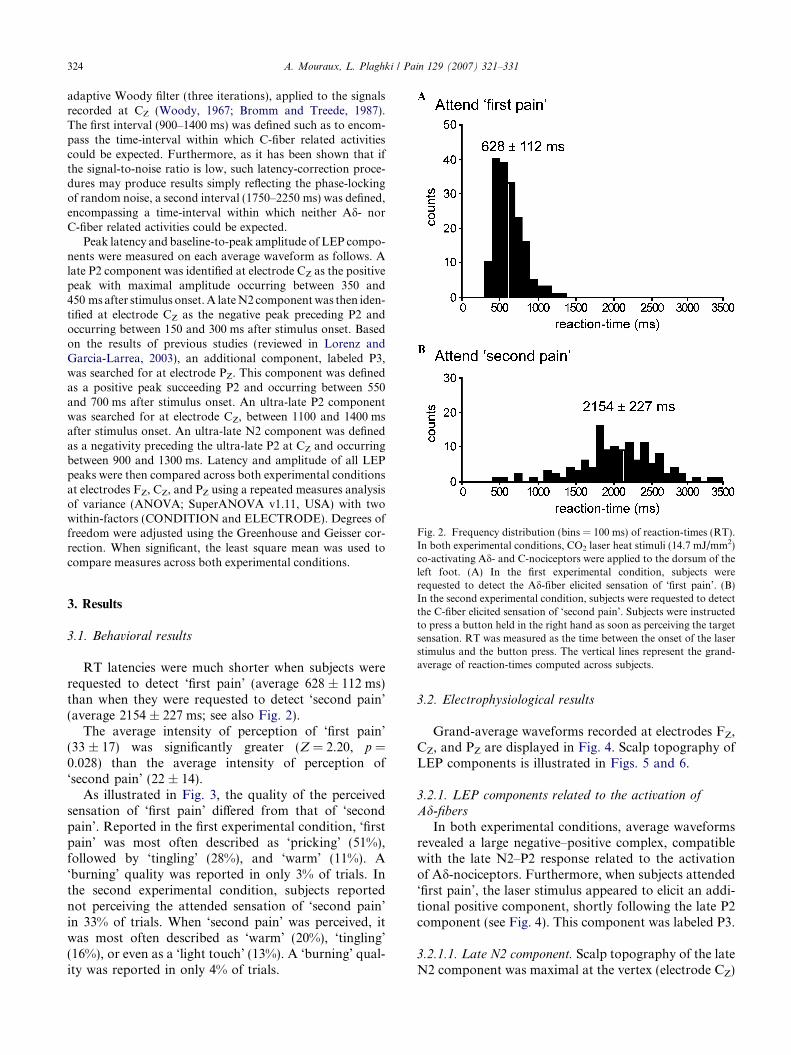
Fig. 2. Frequency distribution (bins = 100 ms) of reaction-times (RT).In both experimental conditions, CO2 laser heat stimuli (14.7 mJ/mm2)co-activating Ad- and C-nociceptors were applied to the dorsum of the
324 A. Mouraux, L. Plaghki / Pain 129 (2007) 321–331
adaptive Woody filter (three iterations), applied to the signalsrecorded at CZ (Woody, 1967; Bromm and Treede, 1987).The first interval (900–1400 ms) was defined such as to encom-pass the time-interval within which C-fiber related activitiescould be expected. Furthermore, as it has been shown that ifthe signal-to-noise ratio is low, such latency-correction proce-dures may produce results simply reflecting the phase-lockingof random noise, a second interval (1750–2250 ms) was defined,encompassing a time-interval within which neither Ad- norC-fiber related activities could be expected.
Peak latency and baseline-to-peak amplitude of LEP compo-nents were measured on each average waveform as follows. Alate P2 component was identified at electrode CZ as the positivepeak with maximal amplitude occurring between 350 and450 ms after stimulus onset. A late N2 component was then iden-tified at electrode CZ as the negative peak preceding P2 andoccurring between 150 and 300 ms after stimulus onset. Basedon the results of previous studies (reviewed in Lorenz andGarcia-Larrea, 2003), an additional component, labeled P3,was searched for at electrode PZ. This component was definedas a positive peak succeeding P2 and occurring between 550and 700 ms after stimulus onset. An ultra-late P2 componentwas searched for at electrode CZ, between 1100 and 1400 msafter stimulus onset. An ultra-late N2 component was definedas a negativity preceding the ultra-late P2 at CZ and occurringbetween 900 and 1300 ms. Latency and amplitude of all LEPpeaks were then compared across both experimental conditionsat electrodes FZ, CZ, and PZ using a repeated measures analysisof variance (ANOVA; SuperANOVA v1.11, USA) with twowithin-factors (CONDITION and ELECTRODE). Degrees offreedom were adjusted using the Greenhouse and Geisser cor-rection. When significant, the least square mean was used tocompare measures across both experimental conditions.
left foot. (A) In the first experimental condition, subjects wererequested to detect the Ad-fiber elicited sensation of ‘first pain’. (B)In the second experimental condition, subjects were requested to detectthe C-fiber elicited sensation of ‘second pain’. Subjects were instructedto press a button held in the right hand as soon as perceiving the targetsensation. RT was measured as the time between the onset of the laserstimulus and the button press. The vertical lines represent the grand-average of reaction-times computed across subjects.
3. Results
3.1. Behavioral results
RT latencies were much shorter when subjects wererequested to detect ‘first pain’ (average 628 ± 112 ms)than when they were requested to detect ‘second pain’(average 2154 ± 227 ms; see also Fig. 2).
The average intensity of perception of ‘first pain’(33 ± 17) was significantly greater (Z = 2.20, p =0.028) than the average intensity of perception of‘second pain’ (22 ± 14).
As illustrated in Fig. 3, the quality of the perceivedsensation of ‘first pain’ differed from that of ‘secondpain’. Reported in the first experimental condition, ‘firstpain’ was most often described as ‘pricking’ (51%),followed by ‘tingling’ (28%), and ‘warm’ (11%). A‘burning’ quality was reported in only 3% of trials. Inthe second experimental condition, subjects reportednot perceiving the attended sensation of ‘second pain’in 33% of trials. When ‘second pain’ was perceived, itwas most often described as ‘warm’ (20%), ‘tingling’(16%), or even as a ‘light touch’ (13%). A ‘burning’ qual-ity was reported in only 4% of trials.
3.2. Electrophysiological results
Grand-average waveforms recorded at electrodes FZ,CZ, and PZ are displayed in Fig. 4. Scalp topography ofLEP components is illustrated in Figs. 5 and 6.
3.2.1. LEP components related to the activation of
Ad-fibersIn both experimental conditions, average waveforms
revealed a large negative–positive complex, compatiblewith the late N2–P2 response related to the activationof Ad-nociceptors. Furthermore, when subjects attended‘first pain’, the laser stimulus appeared to elicit an addi-tional positive component, shortly following the late P2component (see Fig. 4). This component was labeled P3.
3.2.1.1. Late N2 component. Scalp topography of the lateN2 component was maximal at the vertex (electrode CZ)
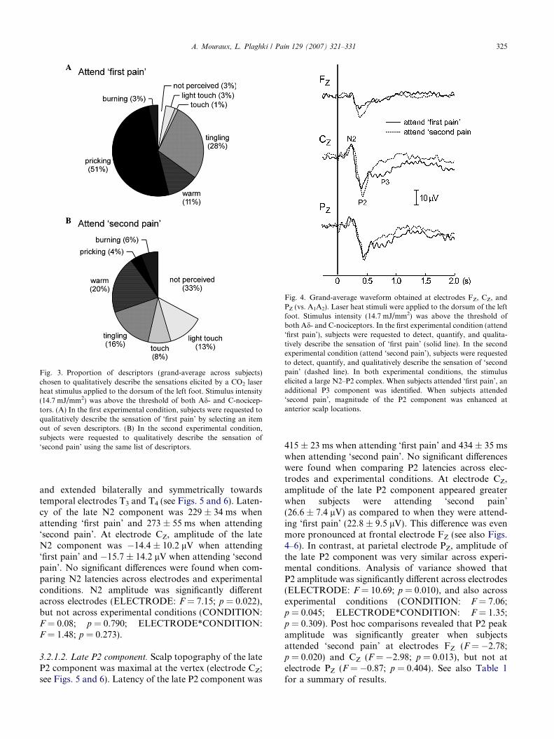
Fig. 3. Proportion of descriptors (grand-average across subjects)chosen to qualitatively describe the sensations elicited by a CO2 laserheat stimulus applied to the dorsum of the left foot. Stimulus intensity(14.7 mJ/mm2) was above the threshold of both Ad- and C-nocicep-tors. (A) In the first experimental condition, subjects were requested toqualitatively describe the sensation of ‘first pain’ by selecting an itemout of seven descriptors. (B) In the second experimental condition,subjects were requested to qualitatively describe the sensation of‘second pain’ using the same list of descriptors.
Fig. 4. Grand-average waveform obtained at electrodes FZ, CZ, andPZ (vs. A1A2). Laser heat stimuli were applied to the dorsum of the leftfoot. Stimulus intensity (14.7 mJ/mm2) was above the threshold ofboth Ad- and C-nociceptors. In the first experimental condition (attend‘first pain’), subjects were requested to detect, quantify, and qualita-tively describe the sensation of ‘first pain’ (solid line). In the secondexperimental condition (attend ‘second pain’), subjects were requestedto detect, quantify, and qualitatively describe the sensation of ‘secondpain’ (dashed line). In both experimental conditions, the stimuluselicited a large N2–P2 complex. When subjects attended ‘first pain’, anadditional P3 component was identified. When subjects attended‘second pain’, magnitude of the P2 component was enhanced atanterior scalp locations.
A. Mouraux, L. Plaghki / Pain 129 (2007) 321–331 325
and extended bilaterally and symmetrically towardstemporal electrodes T3 and T4 (see Figs. 5 and 6). Laten-cy of the late N2 component was 229 ± 34 ms whenattending ‘first pain’ and 273 ± 55 ms when attending‘second pain’. At electrode CZ, amplitude of the lateN2 component was �14.4 ± 10.2 lV when attending‘first pain’ and �15.7 ± 14.2 lV when attending ‘secondpain’. No significant differences were found when com-paring N2 latencies across electrodes and experimentalconditions. N2 amplitude was significantly differentacross electrodes (ELECTRODE: F = 7.15; p = 0.022),but not across experimental conditions (CONDITION:F = 0.08; p = 0.790; ELECTRODE*CONDITION:F = 1.48; p = 0.273).
3.2.1.2. Late P2 component. Scalp topography of the lateP2 component was maximal at the vertex (electrode CZ;see Figs. 5 and 6). Latency of the late P2 component was
415 ± 23 ms when attending ‘first pain’ and 434 ± 35 mswhen attending ‘second pain’. No significant differenceswere found when comparing P2 latencies across elec-trodes and experimental conditions. At electrode CZ,amplitude of the late P2 component appeared greaterwhen subjects were attending ‘second pain’(26.6 ± 7.4 lV) as compared to when they were attend-ing ‘first pain’ (22.8 ± 9.5 lV). This difference was evenmore pronounced at frontal electrode FZ (see also Figs.4–6). In contrast, at parietal electrode PZ, amplitude ofthe late P2 component was very similar across experi-mental conditions. Analysis of variance showed thatP2 amplitude was significantly different across electrodes(ELECTRODE: F = 10.69; p = 0.010), and also acrossexperimental conditions (CONDITION: F = 7.06;p = 0.045; ELECTRODE*CONDITION: F = 1.35;p = 0.309). Post hoc comparisons revealed that P2 peakamplitude was significantly greater when subjectsattended ‘second pain’ at electrodes FZ (F = �2.78;p = 0.020) and CZ (F = �2.98; p = 0.013), but not atelectrode PZ (F = �0.87; p = 0.404). See also Table 1for a summary of results.
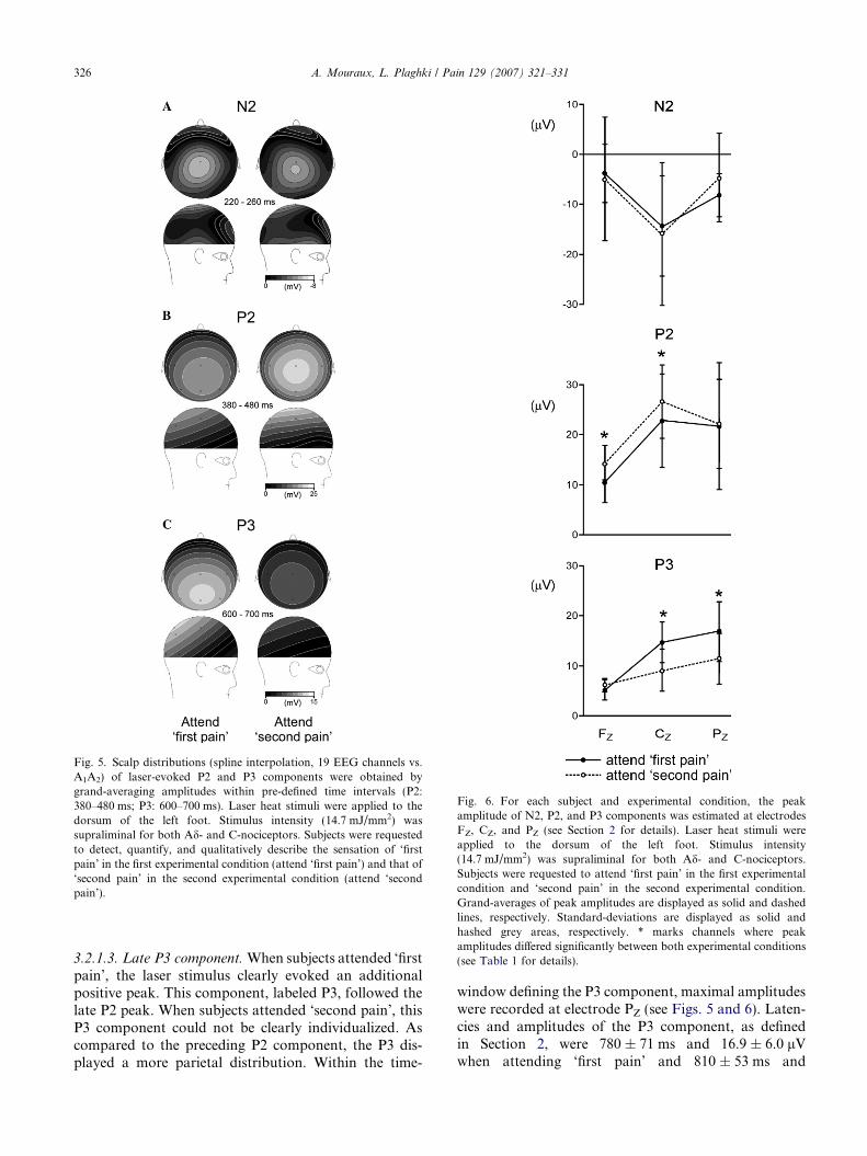
Fig. 6. For each subject and experimental condition, the peakamplitude of N2, P2, and P3 components was estimated at electrodesFZ, CZ, and PZ (see Section 2 for details). Laser heat stimuli wereapplied to the dorsum of the left foot. Stimulus intensity(14.7 mJ/mm2) was supraliminal for both Ad- and C-nociceptors.Subjects were requested to attend ‘first pain’ in the first experimentalcondition and ‘second pain’ in the second experimental condition.Grand-averages of peak amplitudes are displayed as solid and dashedlines, respectively. Standard-deviations are displayed as solid andhashed grey areas, respectively. * marks channels where peakamplitudes differed significantly between both experimental conditions(see Table 1 for details).
Fig. 5. Scalp distributions (spline interpolation, 19 EEG channels vs.A1A2) of laser-evoked P2 and P3 components were obtained bygrand-averaging amplitudes within pre-defined time intervals (P2:380–480 ms; P3: 600–700 ms). Laser heat stimuli were applied to thedorsum of the left foot. Stimulus intensity (14.7 mJ/mm2) wassupraliminal for both Ad- and C-nociceptors. Subjects were requestedto detect, quantify, and qualitatively describe the sensation of ‘firstpain’ in the first experimental condition (attend ‘first pain’) and that of‘second pain’ in the second experimental condition (attend ‘secondpain’).
326 A. Mouraux, L. Plaghki / Pain 129 (2007) 321–331
3.2.1.3. Late P3 component. When subjects attended ‘firstpain’, the laser stimulus clearly evoked an additionalpositive peak. This component, labeled P3, followed thelate P2 peak. When subjects attended ‘second pain’, thisP3 component could not be clearly individualized. Ascompared to the preceding P2 component, the P3 dis-played a more parietal distribution. Within the time-
window defining the P3 component, maximal amplitudeswere recorded at electrode PZ (see Figs. 5 and 6). Laten-cies and amplitudes of the P3 component, as definedin Section 2, were 780 ± 71 ms and 16.9 ± 6.0 lVwhen attending ‘first pain’ and 810 ± 53 ms and

Fig. 7. Laser heat stimuli were applied to the dorsum of the left foot.Stimulus intensity (14.7 mJ/mm2) was above the threshold of both Ad-and C-nociceptors. In the second experimental condition (attend‘second pain’), subjects were requested to detect, quantify, andqualitatively describe the sensation of ‘second pain’. As the C-fiberelicited sensation was not perceived in 33% of trials, separate grand-average waveforms (electrode CZ vs. A1A2) were computed based onwhether or not subjects effectively reported perceiving the sensation of‘second pain’ (filled line: ‘second pain’ perceived; dashed line: ‘secondpain’ not perceived). Note the marked similarity between bothwaveforms.
Table 1Paired comparisons of N2, P2, and P3 peak amplitudes
Peak amplitude (lV)a F p
Attend ‘first pain’ Attend ‘second pain’
N2 FZ �3.7 ± 6.0 �4.6 ± 12.7 0.62CZ �14.4 ± 10.2 �15.7 ± 14.4 1.83PZ �8.1 ± 4.5 �4.5 ± 9.1 �0.60
P2 FZ 10.4 ± 4.2 14.2 ± 3.6 �2.78 0.020*CZ 22.8 ± 9.5 26.6 ± 7.4 �2.98 0.014*PZ 21.7 ± 12.8 22.2 ± 9.1 �0.87 0.404
P3 FZ 5.1 ± 2.1 6.1 ± 1.4 0.39 0.704CZ 14.7 ± 4.2 9.0 ± 4.2 5.09 <0.001*PZ 16.9 ± 6.0 11.4 ± 5.2 3.73 0.004*
*p < 0.05; (least square mean between conditions).a Grand-average ± standard-deviation.
A. Mouraux, L. Plaghki / Pain 129 (2007) 321–331 327
11.4 ± 5.2 lV when attending ‘second pain’ (electrodePZ). No significant difference was found when comparingP3 latency across electrodes and experimental conditions.In contrast, P3 peak amplitude was significantly differentacross experimental conditions (CONDITION:F = 8.61; p = 0.032; ELECTRODE*CONDITION:F = 5.82; p = 0.022), but not across electrodes (ELEC-TRODE: F = 4.49; p = 0.059). Post hoc comparisonsrevealed that P3 peak amplitude was significantly greaterwhen subjects attended ‘first pain’ at electrodes PZ
(F = 3.72; p = 0.004) and CZ (F = 5.09; p < 0.001) butnot at electrode FZ (F = 0.39; p = 0.704). See also Table1 for a summary of results LEP components related to theactivation of C-fibers.
When no latency-correction algorithm was applied,none of the two experimental conditions revealed thepresence of an ultra-late LEP response related to thecentral processing of C-fiber input (see Fig. 4), and thiseven when excluding the trials where subjects had failedto detect the sensation of ‘second pain’. Indeed, as illus-trated in Fig. 7, the average waveforms obtained byselectively averaging epochs based on whether or notsubjects effectively detected the sensation of ‘secondpain’ were markedly similar.
When a latency-correction algorithm was appliedwithin the interval where C-fiber related activities wereexpected (900–1400 ms), average waveforms revealedcomponents whose morphology varied greatly acrosssubjects and experimental conditions. In some cases,these activities resembled ultra-late LEP-like compo-nents (see Fig. 8 for an example). Within that time-inter-val, the average latency of the most negative peak was1238 ± 163 ms when attending ‘first pain’ and1077 ± 68 ms when attending ‘second pain’ (electrodeCZ). The average latency of the most positive peakwas 1005 ± 69 ms when attending ‘first pain’ and1209 ± 215 ms when attending ‘second pain’. Averagepeak-to-peak amplitude was 29.2 ± 7.4 lV when
attending ‘first pain’ and 25.5 ± 4.2 lV when attending‘second pain’. None of these differences were significant.Furthermore, applying the same latency-correctionalgorithm within a time-interval where neither C-nor Ad-fiber evoked-potentials would be expected(1750–2250 ms) led to the enhancement of similarcomponents. Within that later time-frame, the averagelatency of the most negative peak was 1933 ± 68 mswhen attending ‘first pain’ and 2041 ± 61 ms whenattending ‘second pain’. The average latency of the mostpositive peak was 1980 ± 168 ms when attending ‘firstpain’ and 2045 ± 63 ms when attending ‘second pain’.The average peak-to-peak amplitude was 29.6 ± 9.2 lVwhen attending ‘first pain’ and 27.2 ± 4.9 lV whenattending ‘second pain’. None of these differences weresignificant.
4. Discussion
The laser stimulus could produce two temporally andqualitatively distinct sensations, respectively, related tothe activation of Ad- and C-nociceptors. In both exper-imental conditions, subjects performed a detection taskrelated to the perception of that laser stimulus. Howev-er, in the first condition, they were requested to selective-ly attend and respond to Ad-fiber evoked sensations(i.e., ‘first pain’) while in the second, they were requestedto selectively attend and respond to C-fiber evoked sen-sations (i.e. ‘second pain’).
The laser stimulus did not elicit reproducible, C-fiberrelated, ultra-late LEP components, and this even whensubjects selectively attended ‘second pain’. Therefore,the present study failed to replicate the findings reportedby Bromm and Treede (1985).

Fig. 8. A latency-correction algorithm, such as that used to revealC-fiber related LEP components in a number of previous studies(Carmon et al., 1980; Bromm and Treede, 1985, 1987; Purves andBoyd, 1993), was applied to results of the present study. In panel A, theuncorrected average waveforms (CZ vs. A1A2) obtained from onesubject and for both experimental conditions (attend ‘first pain’: solidline; attend ‘second pain’: dashed line) are displayed. Laser heat stimuliwere applied to the dorsum of the left foot. Stimulus intensity (14.7mJ/mm2) was well above the threshold of both Ad- and C-nociceptors.In panel B, epochs of that same subject were realigned using anadaptive Woody filter (three iterations), applied to signals recorded atCZ (Woody, 1967; Bromm and Treede, 1987). A time interval of900–1400 ms was chosen such as to encompass the time-interval withinwhich C-fiber related activities could be expected. In panel C, the sameprocedure was applied within another time interval (1750–2250 ms)where neither Ad- nor C-fiber related activities could be expected.These results illustrate that, if the signal-to-noise ratio is low, suchlatency-correction procedures may produce LEP-like waveformsreflecting the phase-locking of ongoing EEG activity unrelated to thestimulus and/or noise.
2 To render the evoking stimulus infrequent or unexpected, moststudies employ an ‘oddball paradigm’ where infrequent target stimuliare interspersed within a sequence of non-target frequent stimuli.However, several studies have shown that similar results may beobtained when target stimuli are presented in isolation, provided thatlong and varying inter-stimulus intervals are used.
328 A. Mouraux, L. Plaghki / Pain 129 (2007) 321–331
In contrast, selectively attending to ‘first’ or ‘second’pain significantly modulated late, Ad-fiber related, lateLEP components. Indeed, when subjects attended ‘firstpain’, the stimulus clearly elicited an additional parietalpositive component whereas when they attended ‘secondpain’, the magnitude of the laser-evoked P2 componentwas significantly enhanced at prefrontal recording sites.
4.1. Perception of first and second pain
When subjects were requested to detect ‘first pain’,detection latencies were much shorter than when theywere requested to detect ‘second pain’. This finding iscompatible with the fact that conduction velocities of
thinly myelinated Ad-fibers (�10 m/s) are much greaterthan that of unmyelinated C-fibers (�1 m/s).
Subjects consistently identified ‘first pain’ andpredominantly described the sensation as ‘pricking’. Incontrast, they were unable to detect ‘second pain’ inapproximately one-third of trials. When perceived,‘second pain’ was predominantly described as ‘warm’,tingling’, or even as a ‘light touch’. These results are verysimilar to that of a recent study that examined the per-ception of laser stimuli after selectively blocking periph-eral A-fibers (Nahra and Plaghki, 2003). Takentogether, these results suggest that the contribution ofC-fibers to the sensations produced by a brief and local-ized heat stimulus applied to the hairy skin is (1) verysparse and (2) not significantly altered by the concomi-tant activation of Ad-fibers.
4.2. Attending to first or second pain: electrophysiological
correlates
4.2.1. Late parietal positive component
When subjects selectively attended ‘first pain’, thelaser stimulus elicited an additional positive componentwhich closely followed the late N2 and P2 components.Latency and topography of that component was similarto that of the laser-evoked P3 (or P600) reported in anumber of studies (Towell and Boyd, 1993; Kandaet al., 1996; Siedenberg and Treede, 1996; Legrainet al., 2002; Lorenz and Garcia-Larrea, 2003). Investiga-tors have hypothesized that this component is related tothe P3b potential, which has been described in studiesinvolving other sensory modalities (Picton, 1992).Indeed, the amplitudes of both components are similarlyenhanced when the evoking stimulus is infrequent orunexpected,2 and when subjects are actively involvedin the detection of that stimulus. Furthermore, bothresponses display a similar parietal topography. Toexplain the functional significance of the P3b compo-nent, two leading hypotheses have been put forward.The ‘context updating’ hypothesis (Donchin and Coles,1988) proposes that the P3b reflects the updating ofworking memory which would follow the arrival ofrelevant sensory information. The ‘context closure’hypothesis (Desmedt, 1980; Verleger, 1988) proposesthat the P3b reflects processes occurring when expecta-tions are met, possibly related to the closure of informa-tion processing.
In a recent study examining the LEP componentselicited by two consecutively applied laser stimuli,

A. Mouraux, L. Plaghki / Pain 129 (2007) 321–331 329
Mouraux et al. (2004) provided experimental evidence insupport of the ‘context closure’ hypothesis. Indeed,although both stimuli were equally unexpected andtask-relevant, only the second stimulus, which endedthe subject’s expectations, elicited a consistent P3component.
The present study provides additional evidence indi-cating that the laser-evoked P3 reflects processes relatedto the ending of expectations. Indeed, while the laserstimulus elicited a clearly identifiable P3 when subjectswere selectively attending ‘first pain’ (i.e., when percep-tion of Ad-fiber evoked sensations terminated expecta-tions), it did not when subjects were selectivelyattending ‘second pain’ (i.e., when expectations endedonly after perceiving later-occurring C-fiber evokedsensations).
4.2.2. Late prefrontal positive component
When subjects were selectively attending andresponding to C-fiber elicited ‘second pain’, the magni-tude of the late P2 component, although related to thecentral-processing of Ad-fiber input, appeared signifi-cantly enhanced. The topographical distribution of thisenhancement was clearly different from that of the P2component, indicating that this modulation could notmerely result from an overall increase of the electro-cor-tical activity underlying the P2. The observed effect musttherefore have resulted either (1) from the modulation ofone or a subgroup of the cortical generators underlyingthe laser-evoked P2 or (2) from the occurrence (or non-occurrence) of an additional, temporally overlapping,electrophysiological component.
Correct detection of ‘second pain’ required subjectsto refrain from responding to the closely preceding sen-sation of ‘first pain’. Therefore, processes related toresponse inhibition could have contributed to theobserved modulation of Ad-fiber related responses.Indeed, a number of several studies have shown that ifsubjects are requested to withhold from responding toa certain stimulus (e.g., ‘go-nogo’ or ‘stop signal’ exper-imental paradigms3), that stimulus may elicit a series ofelectrophysiological components, which have beenhypothesized to reflect brain processes related toresponse-inhibition or conflict-monitoring (Simsonet al., 1977; Pfefferbaum et al., 1985; Falkenstein et al.,1995; Falkenstein et al., 1999; Nakata et al., 2004).For somatosensory stimuli (electrical stimulation of
3 In the ‘go-nogo’ task, subjects respond to one type of stimulus butwithhold their response to another type of stimulus. For example, theinstruction can be to respond to a high-pitched tone (‘go’ stimulus) butto withhold the response to a low-pitched tone (‘nogo’ stimulus). In the‘stop signal’ task, subjects perform a reaction-time task to a repeti-tively presented stimulus. This task is occasionally interrupted by astop signal, presented with a lower probability, and instructing subjectsto withhold their response.
the second or fifth left-hand digit), response-inhibitionhas been shown to induce an enhancement of the vertexnegativity (140–200 ms) but also to elicit a P3-likepositive component (referred to as ‘nogo-P3’). As com-pared to the P3b component, the nogo-P3 displays a sig-nificantly more anterior scalp topography (Nakataet al., 2004). Source analyses (Kiefer et al., 1998; Nie-uwenhuis et al., 2003; Bekker et al., 2005) have suggest-ed that the bulk of the electrophysiological activityassumed to be related to response-inhibition arises fromthe anterior cingulate cortex (ACC). Therefore, it maybe hypothesized that the anterior positive enhancementobserved in the present study reflected processes similarto that of the ‘nogo-P3’ described in other sensorymodalities.
Most studies involving laser stimulation require sub-jects to stand still and hold back the motor responseswhich are urged by the salient and nociceptive natureof the evoking stimulus. Whether processes related toresponse-inhibition contribute to the LEP waveformsrecorded under such experimental conditions shouldthus be considered. Source analyses (reviewed in Gar-cia-Larrea et al., 2003) have indicated that LEPs receivecontributions from several distinct sources, out of whichbilateral operculo-insular and ACC regions appear asmost consistent. As results of the present study suggestthat electrophysiological correlates of response-inhibi-tion may overlap the laser-evoked P2 component, andas previous studies have indicated that these activitiesmay originate from the ACC, it could be hypothesizedthat at least part of the ACC sources contributing toLEPs reflect processes which are in fact related toresponse-inhibition.
The P3b component elicited by infrequent task-rele-vant stimuli is usually preceded by a negative deflection,often referred to as N2b (Naatanen et al., 1982). As thisN2b overlaps the vertex P2 wave, it commonly entails adecrease of its measured amplitude. Therefore, an addi-tional hypothesis to be considered is that the apparentresponse enhancement of the LEP P2 component whenattending ‘second pain’ resulted from the non-occur-rence of this negative deflection, overlapping the LEPP2 component when attending ‘first pain’. However,contradicting this hypothesis is the fact that no similardecrease of the LEP P2 component was observed in pre-vious studies examining Ad-fiber responses elicited with-in an oddball paradigm (e.g., Legrain et al., 2002).
4.2.3. Ultra-late LEP
Results of the present study did not confirm thehypothesis postulated by Bromm and Treede (1985).Indeed, laser stimuli co-activating Ad- and C-nocicep-tors failed to elicit reproducible C-fiber LEPs both whensubjects were attending ’first pain’ and when they wereattending ‘second pain’. Therefore, it appears that train-ing subjects to focus their attention towards C-fiber

330 A. Mouraux, L. Plaghki / Pain 129 (2007) 321–331
sensory input is not a sufficient condition for that inputto elicit consistent ultra-late LEP responses.
To identify C-fiber related responses, Bromm andTreede used a latency-correction algorithm which aimedat aligning hypothesized ultra-late LEP components pri-or to computing the average waveform. Several studieshave now shown that if C-fibers are activated in isola-tion, the stimulus elicits ultra-late LEP componentswhich may be clearly identified without the use of suchalgorithms. Applying a similar adaptive Woody filterprocedure to the present data led to the enhancementof waveforms which, in some cases, resembled ultra-lateLEP components (see Fig. 8 for an example). However,the fact that (1) the morphology of these componentsvaried greatly across subjects and experimental condi-tions, (2) such components were revealed both whensubjects attended ‘first pain’ and when subjects attended‘second pain’, and (3) applying the same algorithm with-in a later time-window led to the enhancement of similarcomponents, at latencies where neither C- nor Ad-fiberrelated activities could be expected, constitute clear indi-cations that these activities resulted from the phase-lock-ing of EEG activity which was unrelated to the stimulus.
Acknowledgements
We thank Prof. A. Fayt and Ir. P. Stouffs for conceiv-ing and building the CO2 laser skin stimulator.
References
Bekker EM, Kenemans JL, Verbaten MN. Source analysis of the N2in a cued Go/NoGo task. Brain Res Cogn Brain Res 2005;22:221–31.
Bjerring P, Arendt-Nielsen L. Argon laser induced single corticalresponses: a new method to quantify pre-pain and pain percep-tions. J Neurol Neurosurg Psychiatry 1988;51:43–9.
Bragard D, Chen AC, Plaghki L. Direct isolation of ultra-late (C-fibre)evoked brain potentials by CO2 laser stimulation of tiny cutaneoussurface areas in man. Neurosci Lett 1996;209:81–4.
Bromm B, Neitzel H, Tecklenburg A, Treede RD. Evoked cerebralpotential correlates of C-fibre activity in man. Neurosci Lett1983;43:109–14.
Bromm B, Treede RD. Nerve fibre discharges, cerebral potentials andsensations induced by CO2 laser stimulation. Hum Neurobiol1984;3:33–40.
Bromm B, Treede RD. Evoked cerebral potential changes accompa-nying attention shifts between first and second pain. Electroencep-halogr Clin Neurophysiol 1985;61.
Bromm B, Treede RD. Human cerebral potentials evoked by CO2laser stimuli causing pain. Exp Brain Res 1987;67:153–62.
Carmon A, Friedman Y, Coger R, Kenton B. Single trial analysis ofevoked potentials to noxious thermal stimulation in man. Pain1980;8:21–32.
Desmedt JE. P300 in serial tasks: an essential post-decision closuremechanism. Prog Brain Res 1980;54:682–6.
Donchin E, Coles MGH. Is the P300 component a manifestation ofcontext updating? Behav Brain Sci 1988;11:357–74.
Falkenstein M, Hoormann J, Hohnsbein J. ERP components in Go/Nogo tasks and their relation to inhibition. Acta Psychol (Amst)1999;101:267–91.
Falkenstein M, Koshlykova NA, Kiroj VN, Hoormann J, HohnsbeinJ. Late ERP components in visual and auditory Go/Nogo tasks.Electroencephalogr Clin Neurophysiol 1995;96:36–43.
Garcia-Larrea L, Frot M, Valeriani M. Brain generators of laser-evoked potentials: from dipoles to functional significance. Neuro-physiol Clin 2003;33:279–92.
Kanda M, Fujiwara N, Xu X, Shindo K, Nagamine T, Ikeda A,Shibasaki H. Pain-related and cognitive components of somato-sensory evoked potentials following CO2 laser stimulation in man.Electroencephalogr Clin Neurophysiol 1996;100:105–14.
Kiefer M, Marzinzik F, Weisbrod M, Scherg M, Spitzer M. The timecourse of brain activations during response inhibition: evidencefrom event-related potentials in a go/no go task. Neuroreport1998;9:765–70.
Legrain V, Guerit JM, Bruyer R, Plaghki L. Attentional modulation ofthe nociceptive processing into the human brain: selective spatialattention, probability of stimulus occurrence, and target detectioneffects on laser evoked potentials. Pain 2002;99:21–39.
Lewis T, Ponchin EE. The double pain response of the human skin to asingle stimulus. Clin Sci 1937;3:67–76.
Lorenz J, Garcia-Larrea L. Contribution of attentional and cognitivefactors to laser evoked brain potentials. Neurophysiol Clin2003;33:293–301.
Magerl W, Ali Z, Ellrich J, Meyer RA, Treede RD. C- and A delta-fiber components of heat-evoked cerebral potentials in healthyhuman subjects. Pain 1999;82:127–37.
Mouraux A, Guerit JM, Plaghki L. Refractoriness cannot explain whyC-fiber laser-evoked brain potentials are recorded only if concom-itant Adelta-fiber activation is avoided. Pain 2004;112:16–26.
Mouraux A, Guerit JM, Plaghki L. Non-phase locked electroenceph-alogram (EEG) responses to CO2 laser skin stimulations mayreflect central interactions between A[delta]- and C-fibre afferentvolleys. Clin Neurophysiol 2003;114:710–22.
Mouraux A, Plaghki L. Fonctions psychometriques et methodespsychophysiques adaptatives pour l’etude de la douleur In: SFD-SOFRED Ed. Evaluation de la douleur experimentale chezl’homme et l’animal. Paris, 2000.
Naatanen R, Simpson M, Loveless NE. Stimulus deviance and evokedpotentials. Biol Psychol 1982;14:53–98.
Nahra H, Plaghki L. The effects of A-fiber pressure block onperception and neurophysiological correlates of brief non-painfuland painful CO2 laser stimuli in humans. Eur J Pain2003;7:189–99.
Nakata H, Inui K, Nishihira Y, Hatta A, Sakamoto M, Kida T,Wasaka T, Kakigi R. Effects of a go/nogo task on event-relatedpotentials following somatosensory stimulation. Clin Neurophysiol2004;115:361–8.
Nieuwenhuis S, Yeung N, van den Wildenberg W, Ridderinkhof KR.Electrophysiological correlates of anterior cingulate function in ago/no-go task: effects of response conflict and trial type frequency.Cogn Affect Behav Neurosci 2003;3:17–26.
Opsommer E, Masquelier E, Plaghki L. Determination of nerveconduction velocity of C-fibres in humans from thermal thresholdsto contact heat (thermode) and from evoked brain potentials toradiant heat (CO2 laser). Neurophysiol Clin 1999;29:411–22.
Pfefferbaum A, Ford JM, Weller BJ, Kopell BS. ERPs to responseproduction and inhibition. Electroencephalogr Clin Neurophysiol1985;60:423–34.
Picton TW. The P300 wave of the human event-related potential. JClin Neurophysiol 1992;9:456–79.
Plaghki L, Delisle D, Godfraind JM. Heterotopic nociceptive condi-tioning stimuli and mental task modulate differently the perceptionand physiological correlates of short CO2 laser stimuli. Pain1994;57:181–92.

A. Mouraux, L. Plaghki / Pain 129 (2007) 321–331 331
Plaghki L, Mouraux A. How do we selectively activate skinnociceptors with a high power infrared laser? Physiologyand biophysics of laser stimulation. Neurophysiol Clin 2003;33:269–77.
Price DD. Selective activation of A-delta and C nociceptive affer-ents by different parameters of nociceptive heat stimulation: atool for analysis of central mechanisms of pain. Pain 1996;68:1–3.
Purves AM, Boyd SG. Time-shifted averaging for laserevoked potentials. Electroencephalogr Clin Neurophysiol 1993;88:118–22.
Siedenberg R, Treede RD. Laser-evoked potentials: exogenous andendogenous components. Electroencephalogr Clin Neurophysiol1996;100:240–9.
Simson R, Vaughan Jr HG, Ritter W. The scalp topography ofpotentials in auditory and visual Go/NoGo tasks. Electroencep-halogr Clin Neurophysiol 1977;43:864–75.
Towell AD, Boyd SG. Sensory and cognitive components of the CO2laser evoked cerebral potential. Electroencephalogr Clin Neuro-physiol 1993;88:237–9.
Treede RD, Lorenz J, Baumgartner U. Clinical usefulness of laser-evoked potentials. Neurophysiol Clin 2003;33:303–14.
Verleger R. Event-related brain potentials and cognition: a critique ofthe context updating hypothesis and an alternative interpretation.Behav Brain Sci 1988;11.
Willis WD. The pain system. New York: Karger; 1985.Woody C. Characterization of an adaptive filter for the analysis of
variable latency neuroelectric signals. Med Biol Eng 1967;5:539–53.


![[- 200 [ PROVIDING MODULATED COMMUNICATION SIGNALS ]](https://static.fdokumen.com/doc/165x107/6328adc85c2c3bbfa804c60f/-200-providing-modulated-communication-signals-.jpg)