Intensity-modulated radiation therapy for head and neck cancer
12
DOI: 10.1634/theoncologist.12-5-555 2007;12;555-564 Oncologist den Weyngaert and Dirk Van Gestel Vincent Grégoire, Wilfried De Neve, Avraham Eisbruch, Nancy Lee, Danielle Van Intensity-Modulated Radiation Therapy for Head and Neck Carcinoma This information is current as of May 28, 2007 http://www.TheOncologist.com/cgi/content/full/12/5/555 located on the World Wide Web at: The online version of this article, along with updated information and services, is rights reserved. Print ISSN: 1083-7159. Online ISSN: 1549-490X. Blackwell Street, Suite 260, Durham, North Carolina, 27701. © 2007 by AlphaMed Press, all ® is owned, published, and trademarked by AlphaMed Press, 318 The Oncologist annually. ® has been continuously published since 1995. The Journal is published 12 times The Oncologist entrusted with the care of adult or pediatric cancer patients. gynecologic, and surgical oncologists and is designed specifically for the busy practitioner ® is devoted to medical and practice issues for medical, hematological, radiation, The Oncologist by on May 28, 2007 www.TheOncologist.com Downloaded from
-
Upload
independent -
Category
Documents
-
view
1 -
download
0
Transcript of Intensity-modulated radiation therapy for head and neck cancer

DOI: 10.1634/theoncologist.12-5-555 2007;12;555-564 Oncologist
den Weyngaert and Dirk Van Gestel Vincent Grégoire, Wilfried De Neve, Avraham Eisbruch, Nancy Lee, Danielle Van
Intensity-Modulated Radiation Therapy for Head and Neck Carcinoma
This information is current as of May 28, 2007
http://www.TheOncologist.com/cgi/content/full/12/5/555located on the World Wide Web at:
The online version of this article, along with updated information and services, is
rights reserved. Print ISSN: 1083-7159. Online ISSN: 1549-490X. Blackwell Street, Suite 260, Durham, North Carolina, 27701. © 2007 by AlphaMed Press, all
® is owned, published, and trademarked by AlphaMed Press, 318The Oncologistannually. ® has been continuously published since 1995. The Journal is published 12 timesThe Oncologist
entrusted with the care of adult or pediatric cancer patients.gynecologic, and surgical oncologists and is designed specifically for the busy practitioner
® is devoted to medical and practice issues for medical, hematological, radiation,The Oncologist
by on May 28, 2007
ww
w.TheO
ncologist.comD
ownloaded from
Intensity-Modulated Radiation Therapy for Head and NeckCarcinoma
VINCENT GRÉGOIRE,a WILFRIED DE NEVE,b AVRAHAM EISBRUCH,c NANCY LEE,d
DANIELLE VAN DEN WEYNGAERT,e DIRK VAN GESTELe
aRadiation Oncology Department and Center for Molecular Imaging and Experimental Radiotherapy,Université Catholique de Louvain, St-Luc University Hospital, Brussels, Belgium; bRadiation Oncology
Department, Ghent University Hospital, Gent, Belgium; cDepartment of Radiation Oncology, University ofMichigan, Ann Arbor, Michigan, USA; dDepartment of Radiation Oncology, Memorial Sloan-Kettering
Cancer Center, New York, New York, USA; eDepartment of Radiotherapy, Middelheim General Hospital,Antwerp, Belgium
Key Words. Radiotherapy • Head and neck cancer • IMRT
LEARNING OBJECTIVES
After completing this course, the reader will be able to:
1. Outline innovations related to targeted radiation therapy.
2. Describe trials proving an advantage using IMRT.
3. Assess treatment planning modalities and how IMRT fields are designed.
4. Evaluate when there is a toxicity advantage for IMRT.
5. Discern when conventional radiotherapy should be used instead of IMRT.
Access and take the CME test online and receive 1 AMA PRA Category 1 Credit™ at CME.TheOncologist.comCMECME
ABSTRACT
Intensity-modulated radiation therapy (IMRT) forhead and neck tumors refers to a new approach thataims at increasing the radiation dose gradient betweenthe target tissues and the surrounding normal tissues atrisk, thus offering the prospect of increasing the locore-gional control probability while decreasing the compli-cation rate. As a prerequisite, IMRT requires a properselection and delineation of target volumes. For the lat-ter, recent data indicate the potential of functional im-aging to complement anatomic imaging modalities.Nonrandomized clinical series in paranasal sinuses and
pharyngolaryngeal carcinoma have shown that IMRTwas able to achieve a very high rate of locoregional con-trol with less morbidity, such as dry-eye syndrome, xe-rostomia, and swallowing dysfunction. The promisingresults of IMRT are likely to be achieved when manytreatment conditions are met, for example, optimal se-lection and delineation of the target volumes and organsat risk, appropriate physical quality control of the irra-diation, and accurate patient setup with the use of on-board imaging. Because of the complexity of the varioustasks, it is thus likely that these conditions will only be
Correspondence: Vincent Grégoire, M.D., Ph.D., Radiation Oncology Department, St-Luc University Hospital, 10 Avenue Hippocrate,B-1200 Brussels, Belgium. Telephone: 32-2-764-94-43; Fax: 32-2-764-94-25; e-mail: [email protected] Received De-cember 17, 2005; accepted for publication February 22, 2007. ©AlphaMed Press 1083-7159/2007/$30.00/0 doi: 10.1634/theoncolo-gist.12-5-555
TheOncologist®
Head and Neck Cancers
The Oncologist 2007;12:555–564 www.TheOncologist.com
by on May 28, 2007
ww
w.TheO
ncologist.comD
ownloaded from
met in institutions having large patient throughput andexperience with IMRT. Therefore, patient referral to
those institutions is recommended. The Oncologist 2007;12:555–564
Disclosure of potential conflicts of interest is found at the end of this article.
INTRODUCTION
During the second half of last century, key technological in-novations have tremendously modified the daily practice ofradiotherapy, leading to substantial improvements in treat-ment delivery and outcome. The introduction of linear ac-celerators (linacs) in the early 1950s, the increasing use ofcomputed tomography (CT) scanning for target volume de-lineation from the 1980s on, and more recently, in the late1990s, the availability of advanced treatment planning sys-tems together with multileaf collimators have progressivelycontributed to a more targeted and conformed dose deliv-ery, that is, a better dose distribution within the target vol-umes while sparing surrounding normal tissues. In addition,linacs are nowadays equipped with electronic portal imag-ing devices for verification of patient positioning, thus en-abling a better conformity between the planned dose and thedose that is actually delivered. All these technical innova-tions have led on the one hand to delivering a much higherdose to the target volumes, thus possibly increasing localtumor control, while on the other hand minimizing the un-due dose delivered to the surrounding normal tissues, thuspossibly decreasing treatment morbidity.
The introduction of inverse treatment planning systemswith intensity modulation, that is, intensity-modulated ra-diation therapy (IMRT), over the last few years has broughtanother refinement in the ballistics of dose delivery, en-abling further improvement in dose delivery and treatmentoutcome. IMRT is not only a technique for delivering opti-mized nonuniform beam intensities to a target volume, butit also provides a new approach to the whole treatment pro-cedure from patient immobilization to beam delivery. Im-plementation of IMRT thus requires a knowledge of setupuncertainties, adequate selection and delineation of targetvolumes based on optimal imaging modalities, appropriatespecification and dose prescription regarding dose-volumeconstraints, and ad hoc quality control of both the clinicaland physical aspects of the whole procedure.
In head and neck (HN) malignancies, IMRT appears tobe more and more commonly used for routine treatment, es-pecially in the U.S. [1]. However, only a few randomizedtrials have demonstrated its superiority over conventionaltreatments [2] and only a few meaningful retrospectivestudies are available that demonstrate its potential and itspossible drawbacks. The most convincing data on the supe-
rior therapeutic gain achievable with IMRT are from tu-mors close to the base of the skull, such as nasopharyngealand sinonasal cancers, for which a higher rate of local con-trol and a lower incidence of complications have been re-ported in comparison with standard two-dimensional (2D)techniques in retrospective comparisons [3, 4]. A substan-tially lower rate of late radiation-induced toxicity, such asxerostomia, has also been extensively documented follow-ing the use of IMRT for pharyngolaryngeal squamous cellcarcinomas (SCCs) [2, 5–8]. A few retrospective studieshave also reported that, despite the high conformality indose distribution, geographical miss is rather uncommon inIMRT for pharyngolaryngeal tumors, provided that an ad-equate selection of target volumes is made [9–12].
Although IMRT shows promise as a radiation procedureaimed at increasing therapeutic gain, in the HN area, it stillpresents a number of challenges and avenues that have yetto be fully explored. This article presents an overview of thepresent clinical data supporting the use of IMRT for HN tu-mors.
CHALLENGE OF SELECTION AND DELINEATION OF
THE TARGET VOLUMES
Among the various steps of the IMRT process, target vol-ume selection and delineation represent, without a doubt,the most dramatic changes in clinical approach comparedwith the former 2D treatment of HN tumors. As IMRT al-lows highly conformal dose distribution to target volumesof almost any shape, the adequate selection and delineationof these volumes become of critical importance. An ad-aptation of the target volumes used in the 2D approach, inwhich selection and delineation were determined moreby technical limitations than by oncological consider-ations and patient anatomy, would likely result in no ben-efit in terms of dose sparing to nontarget tissues. On theother hand, an excessively restrictive selection and de-lineation of target volumes could easily jeopardize theclinical impact of the highly conformal dose distribu-tions produced.
Over the past few years, several authors have made rec-ommendations for a more precise selection of the clinicaltarget volume (CTV) for both primary tumors and necknodes [10, 13, 14]. It is beyond the scope of this review ar-ticle to discuss these recommendations at length. For pri-mary tumors, the issue of selection is briefly discussed in
556 IMRT in HN Cancer
by on May 28, 2007
ww
w.TheO
ncologist.comD
ownloaded from

the following sections dealing with the various HN sites.For the neck, comprehensive review of clinical, radiologi-cal, and pathological data on node distribution for the var-ious sites within the HN area supports the concept ofselective treatment according to nodal stage. In a nutshell,for N0 and N1 patients, selective irradiation of level II–IVor level I–III can be recommended for oropharyngeal, hy-popharyngeal, and laryngeal primaries, or for oral cavityprimaries, respectively. For nasopharyngeal tumors and forpatients with a neck staged as higher than N2a, comprehen-sive irradiation of all neck levels is recommended.
Guidelines for the delineation of the various node levelsin the neck have also been proposed, and a series of consen-sus recommendations endorsed by major North American(Radiation Therapy Oncology Group [RTOG]) and Euro-pean (Danish Head and Neck Cancer, European Organiza-tion for Research and Treatment of Cancer, Grouped’Oncologie Radiothérapie Tête et Cou) cooperativegroups has been elaborated for the N0 neck [15, 16]. For thenode-positive and the postoperative neck, further recom-mendations have been recently proposed [17, 18]. It is be-yond the scope of this article to discuss theserecommendations at length, but the main philosophy ofthese new additional guidelines was to include extra nodalregions (i.e., retrostyloid area and subclavicular fossa)and/or extra structures (e.g., muscles or gland) found to beat risk for microscopic tumor infiltration.
Another important issue regarding target volume delin-eation is the choice of the optimal imaging modality usedfor planning purposes. CT images are typically used be-cause they allow dose calculation with corrections for tissuedensity inhomogeneity. Contrast medium should be rou-tinely used to allow a much better contrast between normaltissues and tumors, especially because recent data haveshown that contrast medium does not substantially influ-ence dose optimization (C. Clark, The Royal MarsdenHospital, London, personal communication). For naso-pharyngeal carcinoma, however, magnetic resonance imag-ing (MRI) has been shown to be superior to CT in reducinginterobserver variability [19, 20]. This advantage of MRIwas not observed in oropharyngeal, laryngeal, and hypo-pharyngeal tumors [21]. A few groups also investigated therole of functional imaging with fluorodeoxyglucose-positron emission tomography (FDG-PET) to delineategross tumor volume (GTV) [22, 23]. Daisne et al. [22] as-sessed the accuracy of FDG-PET in comparison with CTfor GTV delineation in oropharyngeal, hypopharyngeal,and laryngeal SCC using three-dimensional (3D) registra-tion of the various imaging modalities. In a subset of laryn-geal tumors, the imaging modalities could even becompared with the actual surgical specimen taken as a
“gold standard.” FDG-PET demonstrated higher accuracyin delineating GTV, with a statistically significant smallertarget volume. Interestingly, this difference in GTV delin-eation translated into a difference in CTV and planning tar-get volume (PTV) delineation, which in turn translated intoa difference in dose distribution; the dose was much moreconformed when FDG-PET was used as the primary imag-ing modality [24].
Although pretreatment contrast-enhanced CT still pres-ently remains the standard technique used in treatmentplanning for HNSCC, it is not unlikely that in the near fu-ture the introduction of additional modalities may also beconsidered, not only before treatment, but also possiblyduring treatment to adapt the GTV to the extent of tumorregression. Furthermore, for PET imaging, tracers otherthan FDG for targeting biological pathways for radiation re-sponse (e.g., proliferation, hypoxia) are under evaluation[25]. The functional assessment of tumor biology may con-tribute to the delineation of sub-GTV that could benefitfrom a heterogeneous dose distribution [26]. Although verychallenging and extremely promising, these approachesstill need thorough validation before they can be introducedinto routine clinical practice [27].
CLINICAL EVIDENCE FOR THE USE OF IMRT IN
HN TUMORS
IMRT for Nasopharyngeal CarcinomaBecause the nasopharynx is situated near numerous criticalnormal organs, that is, the brain stem and optic chiasm,IMRT is ideal in its attempt to deliver an adequate dose tothe gross tumor while sparing these surrounding normal tis-sues. IMRT plans involving the nasopharynx require con-touring the gross disease, which includes the primary grosstumor as well as any gross nodal disease. Subclinical nodalCTV involves bilateral coverage of neck nodal levels delin-eated up to the base of skull to include the retropharyngealnodes [4]. For node-negative patients, levels II–V should becontoured. For node-positive cases, inclusion of level IBappears reasonable, although convincing data are missing.In addition to the important at-risk nodal regions, the CTVshould also include the entire nasopharynx, clivus, base ofskull, pterygoid fossa, parapharyngeal space, inferior sphe-noid sinus, and posterior third of the nasal cavity and max-illary sinuses (ensuring adequate margin to the ptery-gopalatine fossae). The CTV that encompass the aboveregions can be modified according to tumor (T) stage. Amargin around all CTVs should be added to account for pa-tient motion and setup errors, that is, the PTV. This marginis typically 5–6 mm, unless daily imaging and position cor-
557Grégoire, De Neve, Eisbruch et al.
www.TheOncologist.com
by on May 28, 2007
ww
w.TheO
ncologist.comD
ownloaded from
rection are being made, in which case it may be reduced toa minimum of 3 mm [28].
At Memorial Sloan-Kettering Cancer Center, the imple-mentation of IMRT for nasopharyngeal cases has resultedin significant improvements over the traditional and 3D-conformal radiotherapy (3D-CRT) plans. First, there is bet-ter coverage of the retropharynx, base of skull, and medialaspects of the nodal volumes [29]. Xia et al. [30] comparedIMRT treatment plans with conventional treatment plansfor locally advanced nasopharyngeal cancers. They con-cluded that IMRT provides better tumor target coveragewith significantly better sparing of sensitive normal tissuestructures in the treatment of locally advanced nasopharyn-geal carcinoma. Another dose distribution study, by Kam etal. [31, 32] from Hong Kong, compared IMRT with 2D-radiotherapy (2D-RT) and 3D-CRT treatment plans. Threepatients with different stages, including T1N0, T2bN2, andT4N2, were compared. In all stages, IMRT was noted tohave significant dose distribution advantages. In early-stage disease, it provided better parotid gland and temporo-mandibular joint sparing. In locally advanced disease, itoffered better tumor coverage and normal organ sparingand permitted room for dose escalation.
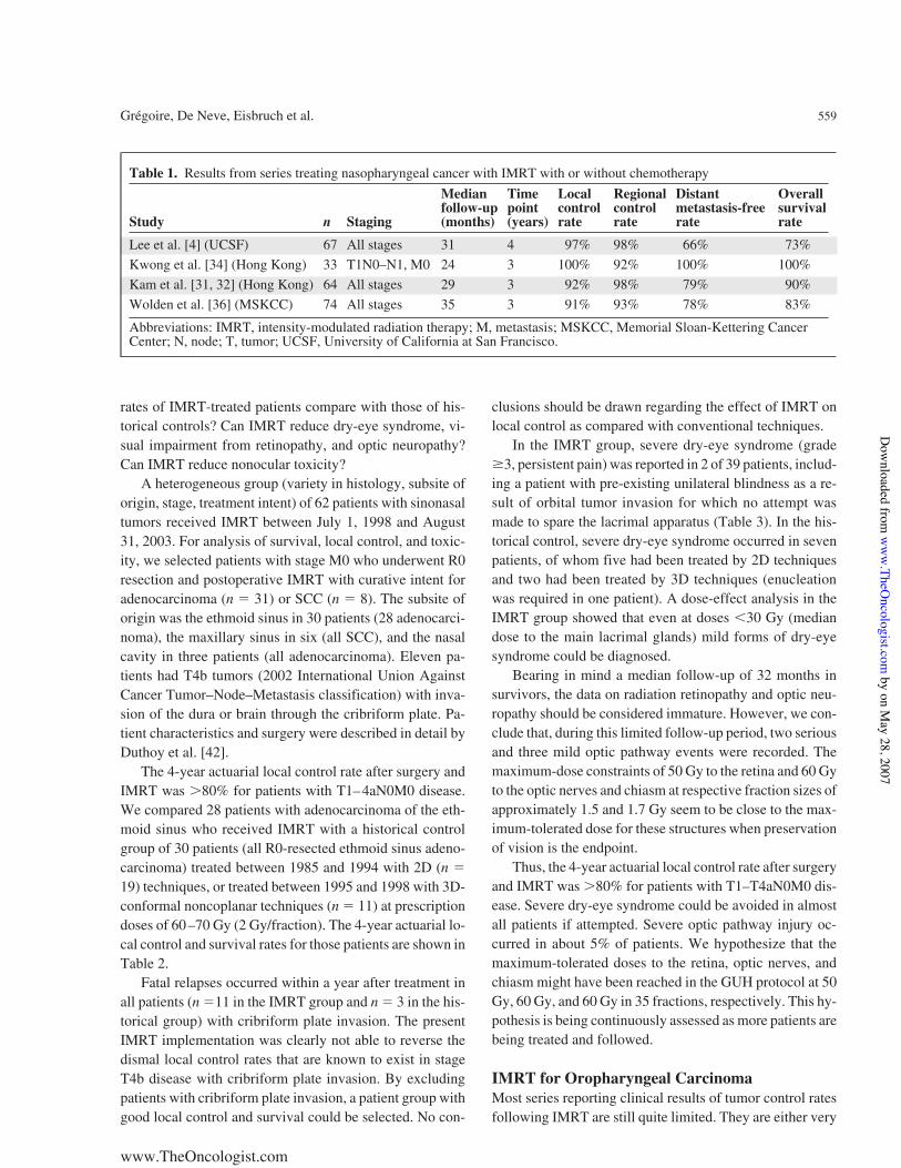
The dose distribution advantages seen with IMRT fornasopharyngeal cancers have also translated into excellentclinical outcomes. At the most recent American Society ofClinical Oncology meeting, Kam et al. [33] presented phaseIII evidence showing the advantage of IMRT in terms ofimprovement in xerostomia when compared with conven-tional radiotherapy. These data add to the data published byPow et al. [2] demonstrating a significant improvement inquality of life in patients with nasopharyngeal cancer ran-domized to IMRT compared with those randomized to con-ventional radiotherapy. In terms of tumor control, the mostmature data on local progression-free rates using IMRT fornasopharyngeal cancer come from the University of Cali-fornia at San Francisco (UCSF) (Table 1). With a medianfollow-up of 31 months, the 4-year local progression-freerate was 97% while the 4-year regional progression-freerate was 98% [4]. Bucci et al. [34] have recently updated theUCSF experience and included more patients (n ! 118).Excellent locoregional progression-free rates were seen.Other important studies have recently emerged out of HongKong and are also detailed in Table 1. Although single-institution studies show superb outcomes in terms of sali-vary preservation as well as locoregional control of disease,distant metastasis remains a significant issue in all these se-ries [4, 34–36]. Efforts focused on decreasing the rates ofdistant metastasis using targeted biologic agents are underway by the RTOG.
An RTOG phase II trial using IMRT with or without
chemotherapy for all localized nasopharyngeal cancer hascompleted accrual and awaits maturity of follow-up data.The protocol is an important one, because it tests whetherthe ability to achieve excellent control rates in nasopharyn-geal cancer patients treated with IMRT can be reproducedin a multi-institutional setting.
The reirradiation of nasopharyngeal cancer has beencommonly used and IMRT offers tremendous dose distri-bution advantages. Reirradiation is more feasible withIMRT than with conventional techniques for the many rea-sons stated above. Lu et al. [37] have recently reported ontheir experience with reirradiation using IMRT for recur-rent nasopharyngeal cancer. Acute toxicity of the skin, mu-cosa, and salivary glands was acceptable according toRTOG criteria. Tumor necrosis was seen toward the end ofIMRT in 14 patients (28.6%). At a median follow-up of 9months, the locoregional control rate was 100%. Althoughlonger follow-up is necessary, the preliminary toxicity andlocal control data for these recurrent cases are promising[37].
IMRT for Nasal and Paranasal SinusesBy their location, sinonasal tumors are surrounded by crit-ical structures, including the frontal and temporal lobes ofthe brain, pituitary gland and brainstem, lacrimal glands,eyes, optic nerves, and chiasm. Using conventional radio-therapy techniques, the lacrimal apparatus and the opticpathway structures (retina, optic nerves, chiasm) often re-ceived doses equal to the target prescription dose. Conven-tional radiation therapy for sinonasal cancer resulted insignificant ocular toxicity [35, 38–39]. Local control ratesof 70%–90% in stages T1–T2 and "50% in stages T3–T4were achieved with prescription doses of 56–75 Gy [40].IMRT allows selective underdosage of organs at risk bycreating concave dose distributions around the optic path-way structures together with steep cranial, lateral, and cau-dal gradients outside the PTV to spare the lacrimalapparatus and the central nervous system. Hypothetically,selective underdosage could decrease toxicity at unchangedtarget prescription doses.
It is unlikely that answers with level I evidence (evi-dence generated from randomized clinical trials) willemerge in the foreseeable future because of the rarity of thedisease. A PubMed search on November 14, 2005 usingIMRT and “paranasal sinus” as key words yielded 23 pub-lications. From these, 17 were on IMRT planning or tech-nical issues. Of the six clinical publications, three reportedon #11 patients, one from UCSF [41] and two from GhentUniversity Hospital (GUH) [3, 42].
Using the patient database at GUH, we try to answer thefollowing questions: How do local control and survival
558 IMRT in HN Cancer
by on May 28, 2007
ww
w.TheO
ncologist.comD
ownloaded from
rates of IMRT-treated patients compare with those of his-torical controls? Can IMRT reduce dry-eye syndrome, vi-sual impairment from retinopathy, and optic neuropathy?Can IMRT reduce nonocular toxicity?
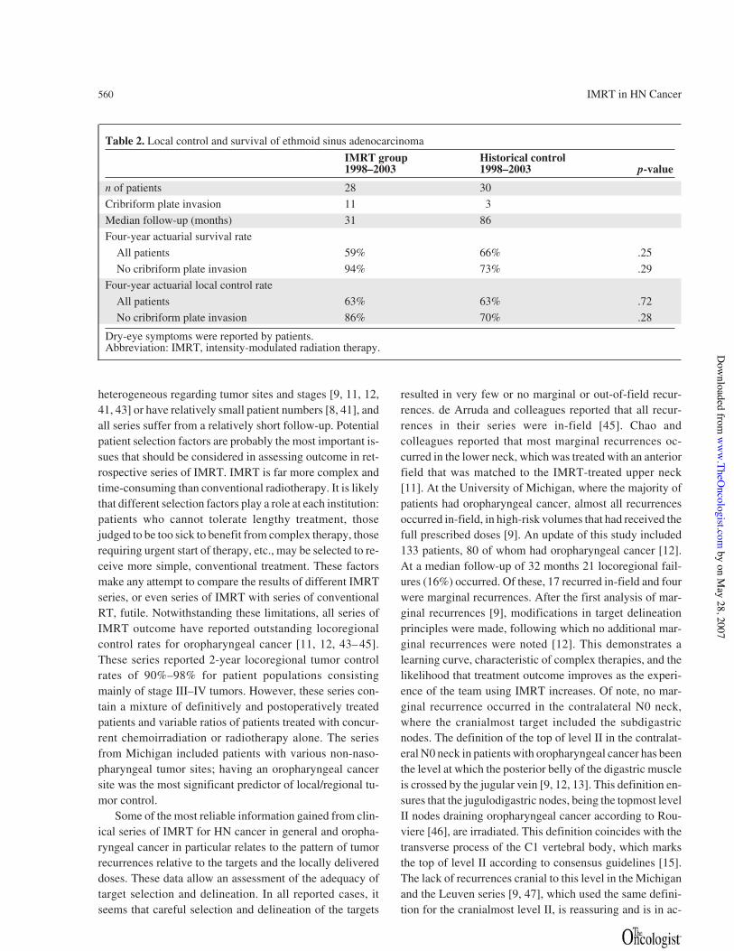
A heterogeneous group (variety in histology, subsite oforigin, stage, treatment intent) of 62 patients with sinonasaltumors received IMRT between July 1, 1998 and August31, 2003. For analysis of survival, local control, and toxic-ity, we selected patients with stage M0 who underwent R0resection and postoperative IMRT with curative intent foradenocarcinoma (n ! 31) or SCC (n ! 8). The subsite oforigin was the ethmoid sinus in 30 patients (28 adenocarci-noma), the maxillary sinus in six (all SCC), and the nasalcavity in three patients (all adenocarcinoma). Eleven pa-tients had T4b tumors (2002 International Union AgainstCancer Tumor–Node–Metastasis classification) with inva-sion of the dura or brain through the cribriform plate. Pa-tient characteristics and surgery were described in detail byDuthoy et al. [42].
The 4-year actuarial local control rate after surgery andIMRT was #80% for patients with T1–4aN0M0 disease.We compared 28 patients with adenocarcinoma of the eth-moid sinus who received IMRT with a historical controlgroup of 30 patients (all R0-resected ethmoid sinus adeno-carcinoma) treated between 1985 and 1994 with 2D (n !19) techniques, or treated between 1995 and 1998 with 3D-conformal noncoplanar techniques (n ! 11) at prescriptiondoses of 60–70 Gy (2 Gy/fraction). The 4-year actuarial lo-cal control and survival rates for those patients are shown inTable 2.
Fatal relapses occurred within a year after treatment inall patients (n !11 in the IMRT group and n ! 3 in the his-torical group) with cribriform plate invasion. The presentIMRT implementation was clearly not able to reverse thedismal local control rates that are known to exist in stageT4b disease with cribriform plate invasion. By excludingpatients with cribriform plate invasion, a patient group withgood local control and survival could be selected. No con-
clusions should be drawn regarding the effect of IMRT onlocal control as compared with conventional techniques.
In the IMRT group, severe dry-eye syndrome (grade!3, persistent pain) was reported in 2 of 39 patients, includ-ing a patient with pre-existing unilateral blindness as a re-sult of orbital tumor invasion for which no attempt wasmade to spare the lacrimal apparatus (Table 3). In the his-torical control, severe dry-eye syndrome occurred in sevenpatients, of whom five had been treated by 2D techniquesand two had been treated by 3D techniques (enucleationwas required in one patient). A dose-effect analysis in theIMRT group showed that even at doses "30 Gy (mediandose to the main lacrimal glands) mild forms of dry-eyesyndrome could be diagnosed.
Bearing in mind a median follow-up of 32 months insurvivors, the data on radiation retinopathy and optic neu-ropathy should be considered immature. However, we con-clude that, during this limited follow-up period, two seriousand three mild optic pathway events were recorded. Themaximum-dose constraints of 50 Gy to the retina and 60 Gyto the optic nerves and chiasm at respective fraction sizes ofapproximately 1.5 and 1.7 Gy seem to be close to the max-imum-tolerated dose for these structures when preservationof vision is the endpoint.
Thus, the 4-year actuarial local control rate after surgeryand IMRT was #80% for patients with T1–T4aN0M0 dis-ease. Severe dry-eye syndrome could be avoided in almostall patients if attempted. Severe optic pathway injury oc-curred in about 5% of patients. We hypothesize that themaximum-tolerated doses to the retina, optic nerves, andchiasm might have been reached in the GUH protocol at 50Gy, 60 Gy, and 60 Gy in 35 fractions, respectively. This hy-pothesis is being continuously assessed as more patients arebeing treated and followed.
IMRT for Oropharyngeal CarcinomaMost series reporting clinical results of tumor control ratesfollowing IMRT are still quite limited. They are either very
Table 1. Results from series treating nasopharyngeal cancer with IMRT with or without chemotherapy
Study n Staging
Medianfollow-up(months)
Timepoint(years)
Localcontrolrate
Regionalcontrolrate
Distantmetastasis-freerate
Overallsurvivalrate
Lee et al. [4] (UCSF) 67 All stages 31 4 97% 98% 66% 73%Kwong et al. [34] (Hong Kong) 33 T1N0–N1, M0 24 3 100% 92% 100% 100%Kam et al. [31, 32] (Hong Kong) 64 All stages 29 3 92% 98% 79% 90%Wolden et al. [36] (MSKCC) 74 All stages 35 3 91% 93% 78% 83%
Abbreviations: IMRT, intensity-modulated radiation therapy; M, metastasis; MSKCC, Memorial Sloan-Kettering CancerCenter; N, node; T, tumor; UCSF, University of California at San Francisco.
559Grégoire, De Neve, Eisbruch et al.
www.TheOncologist.com
by on May 28, 2007
ww
w.TheO
ncologist.comD
ownloaded from
heterogeneous regarding tumor sites and stages [9, 11, 12,41, 43] or have relatively small patient numbers [8, 41], andall series suffer from a relatively short follow-up. Potentialpatient selection factors are probably the most important is-sues that should be considered in assessing outcome in ret-rospective series of IMRT. IMRT is far more complex andtime-consuming than conventional radiotherapy. It is likelythat different selection factors play a role at each institution:patients who cannot tolerate lengthy treatment, thosejudged to be too sick to benefit from complex therapy, thoserequiring urgent start of therapy, etc., may be selected to re-ceive more simple, conventional treatment. These factorsmake any attempt to compare the results of different IMRTseries, or even series of IMRT with series of conventionalRT, futile. Notwithstanding these limitations, all series ofIMRT outcome have reported outstanding locoregionalcontrol rates for oropharyngeal cancer [11, 12, 43– 45].These series reported 2-year locoregional tumor controlrates of 90%–98% for patient populations consistingmainly of stage III–IV tumors. However, these series con-tain a mixture of definitively and postoperatively treatedpatients and variable ratios of patients treated with concur-rent chemoirradiation or radiotherapy alone. The seriesfrom Michigan included patients with various non-naso-pharyngeal tumor sites; having an oropharyngeal cancersite was the most significant predictor of local/regional tu-mor control.
Some of the most reliable information gained from clin-ical series of IMRT for HN cancer in general and oropha-ryngeal cancer in particular relates to the pattern of tumorrecurrences relative to the targets and the locally delivereddoses. These data allow an assessment of the adequacy oftarget selection and delineation. In all reported cases, itseems that careful selection and delineation of the targets
resulted in very few or no marginal or out-of-field recur-rences. de Arruda and colleagues reported that all recur-rences in their series were in-field [45]. Chao andcolleagues reported that most marginal recurrences oc-curred in the lower neck, which was treated with an anteriorfield that was matched to the IMRT-treated upper neck[11]. At the University of Michigan, where the majority ofpatients had oropharyngeal cancer, almost all recurrencesoccurred in-field, in high-risk volumes that had received thefull prescribed doses [9]. An update of this study included133 patients, 80 of whom had oropharyngeal cancer [12].At a median follow-up of 32 months 21 locoregional fail-ures (16%) occurred. Of these, 17 recurred in-field and fourwere marginal recurrences. After the first analysis of mar-ginal recurrences [9], modifications in target delineationprinciples were made, following which no additional mar-ginal recurrences were noted [12]. This demonstrates alearning curve, characteristic of complex therapies, and thelikelihood that treatment outcome improves as the experi-ence of the team using IMRT increases. Of note, no mar-ginal recurrence occurred in the contralateral N0 neck,where the cranialmost target included the subdigastricnodes. The definition of the top of level II in the contralat-eral N0 neck in patients with oropharyngeal cancer has beenthe level at which the posterior belly of the digastric muscleis crossed by the jugular vein [9, 12, 13]. This definition en-sures that the jugulodigastric nodes, being the topmost levelII nodes draining oropharyngeal cancer according to Rou-viere [46], are irradiated. This definition coincides with thetransverse process of the C1 vertebral body, which marksthe top of level II according to consensus guidelines [15].The lack of recurrences cranial to this level in the Michiganand the Leuven series [9, 47], which used the same defini-tion for the cranialmost level II, is reassuring and is in ac-
Table 2. Local control and survival of ethmoid sinus adenocarcinoma
IMRT group1998–2003
Historical control1998–2003 p-value
n of patients 28 30Cribriform plate invasion 11 3Median follow-up (months) 31 86Four-year actuarial survival rate
All patients 59% 66% .25No cribriform plate invasion 94% 73% .29
Four-year actuarial local control rateAll patients 63% 63% .72No cribriform plate invasion 86% 70% .28
Dry-eye symptoms were reported by patients.Abbreviation: IMRT, intensity-modulated radiation therapy.
560 IMRT in HN Cancer
by on May 28, 2007
ww
w.TheO
ncologist.comD
ownloaded from

cordance with the observations of Rouviere [46] and withthe consensus recommendations [15]. Two marginal recur-rences in the Michigan series were noted in the lateral ret-ropharyngeal nodes (both occurred in patients withoropharyngeal cancer), in which the cranialmost extent ofthe retropharyngeal nodal targets was defined at the top ofC1, according to the observations of Rouviere [46] of thelocations of the lateral retropharyngeal nodes. Followingthese observations, we currently define the retropharyngealnodes through the base of the skull. Interesting observationsabout marginal or out-of-field recurrences were reported inthe series from the University of Iowa [43]. They found sev-eral cases of recurrence in the contralateral level I in pa-tients with lateral oral cavity and oropharyngeal cancer,underscoring the need to include these nodes in locoregion-ally advanced oral cancer. As more data about marginal re-currences are accumulated, a better understanding andhigher certainty are expected to be gained in defining thetargets, which will lead to better tumor control and to anability to further spare noninvolved tissues.
IMRT for Laryngeal CarcinomaComparative dose distribution studies have shown thatIMRT can improve the target dose homogeneity in laryn-geal and hypopharyngeal SCC while reducing the dose tonormal tissues at risk [48–50]. Clinical data on IMRT forlaryngeal and hypopharyngeal SCC are, however, quitescarce and typically include patients with definitive andpostoperative irradiation with or without induction or con-comitant chemotherapy. Chao et al. [11] and Dawson et al.[9] reported very limited series with "10 patients in eachlocation, from which definitive conclusions cannot bedrawn. Yao et al. [43] reported on a series of 33 patientswith laryngeal SCC; the 2-year locoregional control ratereached 85%, which was significantly lower than that ob-served in oropharyngeal tumors [43]. Patient selection biascannot, however, be ruled out. A recent study reported thefeasibility of a dose-escalation trial in laryngeal SCC usingIMRT [51].
LESS NORMAL TISSUE MORBIDITY WITH IMRTSeveral clinical studies assessed the utility of IMRT in pa-rotid salivary gland sparing and in reducing xerostomia. Atthe University of Michigan, the partial parotid gland dosesand volumes following multisegmental IMRT were corre-lated with selective salivary output from each parotid gland[5]. It was found that the output related to the mean doses tothe glands. The large majority of the glands that received amean dose #26 Gy did not produce measurable saliva anddid not recover, whereas glands that received lower meandoses produced variable salivary output that increased overtime. One year after radiotherapy, parotid glands that re-ceived a moderate dose (mean dose, 17–26 Gy) recovered,on average, to the pretreatment salivary production levels[6]. When the doses to the parotid glands were very low, asin cases of unilateral neck radiotherapy in which the con-tralateral glands received mean doses "10 Gy, an “over-compensation” of the damage to the ipsilateral glands wasnoted in the second year post-therapy. In particular, the sal-ivary flow rates from the contralateral glands exceeded, onaverage, their pretreatment flow rates. This finding moti-vates reducing the doses to the salivary glands to as low lev-els as possible.
Data on dose response in the parotid glands are accumu-lating [10, 52–55]. The common finding with all these datais that a relationship seems to exist between the mean dosesto the glands and their residual salivary output. It is appar-ent from the studies presented in the review that very dif-ferent mean doses have been reported as thresholds beyondwhich functional deficit occurs. These doses are in therange of 20 Gy to almost 40 Gy. What is the reason for thisdiscrepancy? Several explanations are possible. One expla-nation relates to different methodologies in assessing sali-vary flow. Some studies used selective parotid outflowmeasurements, others used whole mouth saliva, assumingno contribution from the submandibular glands (which hadreceived high doses) and no contribution from the minorsalivary glands (whose output is relatively low), and somestudies used scintigraphy techniques to assess parotid gland
Table 3. Ocular toxicity after radiotherapy for ethmoid tumors
Radiationtechnique n
Dry-eyeDrop in vision<2/10Grade 1 Grade 2 >Grade 3
IMRT 39 2 3 2 22D 19 ? ? 5 03D 11 ? ? 2 2
Abbreviations: 2D, two-dimensional; 3D, three-dimensional; IMRT, intensity-modulated radiation therapy.
561Grégoire, De Neve, Eisbruch et al.
www.TheOncologist.com
by on May 28, 2007
ww
w.TheO
ncologist.comD
ownloaded from
function. Beyond this confounding factor, there are clinicalfactors, beyond radiation dose, that affect the salivary out-put but have not been taken into account in most of thesestudies. These factors include dehydration, common in pa-tients receiving HN radiotherapy, and various medicinesfound to significantly affect salivary flow rates [5, 6]. Anadditional important factor is one reported recently by in-vestigators from the University Hospital in Groningen, TheNetherlands [56]. They irradiated different parts of rat sal-ivary glands using high-precision proton radiation andfound regional differences in dose–salivary production re-lationships. Such regional differences are likely to exist inthe human parotid glands, and they may be the reason fordifferent results found by researchers using different radio-therapy techniques, which produce different dose distribu-tions within the glands. Further research into intraparotidregional differences in sensitivity to radiation is requiredfor a better understanding of radiation limits. Regardless ofthe dose threshold, it is now apparent that spared glands notonly partly retain the salivary output, but the output in-creases over time through at least 2 years after radiotherapy[6], compared with generally no improvement over timefollowing standard radiotherapy, in which most of the pa-rotid glands receive full radiotherapy doses.
Once a reasonable preservation of the salivary output isachieved, can we expect similar improvements in patient-reported xerostomia symptoms? The correlation betweensalivary output from the major salivary glands and xerosto-mia symptoms is significant, but is not very high. Part of thereason for this less-than-straightforward relationship lies inthe contribution of the minor salivary glands. These glandsare scattered throughout the oral cavity, notably on the sur-face of the palate, and produce much of the salivary mucin.It has been found that the dose delivered to the oral cavity,serving as a surrogate for the residual function of the minorsalivary glands (whose output cannot be measured), was anindependent factor in patient-reported xerostomia after ra-diotherapy, similar to the importance of doses to the majorsalivary glands [6]. An optimal practice is to add sparing ofthe oral cavity as an objective in radiotherapy planning, inaddition to sparing of the parotid glands. An analysis at theUniversity of Michigan of a validated patient-reported xe-rostomia questionnaire demonstrated that xerostomia im-proved significantly over time, in tandem with an increasein saliva production [6]. Two years following irradiation,xerostomia reported by patients receiving parotid-sparingbilateral neck radiation was only slightly worse than that inpatients receiving unilateral neck radiotherapy. Investiga-tors at UCSF also reported an improvement to mild or noxerostomia during the second year after IMRT for nasopha-ryngeal cancer [41, 57]. It is apparent from all of these stud-
ies that the partial sparing of the salivary glands, madepossible by IMRT, achieves tangible gains both in the re-tention of salivary production and in the symptoms of xe-rostomia.
Another issue in the efforts to reduce xerostomia byIMRT is the importance of the submandibular glands.These glands lie anterior to the level II lymph node targetsin the neck. It is not possible to spare a substantial amountof these glands while treating both sides of the neck (whichis required for all advanced HN cancer), resulting in nomeasurable salivary output from these glands after radia-tion [6]. Whether the use of technology like proton beamswould help is not yet known. Canadian investigators whomoved one submandibular gland to the submental space,away from the radiotherapy fields, reported an impressivereduction in xerostomia [58]. However, this technique hasnot yet gained broad acceptance, as submandibular lymphnodes are so closely related to the gland, or even into thegland, that transposition may not only spare the organ atrisk but also the target tissue.
Additional potential functional gains from IMRT com-pared with conventional radiotherapy include swallowingand speech measures, reported to be superior using IMRTcompared with standard radiotherapy [59]. These potentialbenefits may translate into improvements in broad aspectsof quality of life [60]. Thus, IMRT of HN cancer mayachieve broad improvements in quality of life rather than belimited to improvements in xerostomia alone. Efforts toidentify the structures whose damage causes long-term dys-phagia and aspiration, and to determine IMRT strategiesthat may spare these structures without underdosing the tar-gets, have been reported [61]. An assessment of the clinicaleffect of these efforts is ongoing. In a recent study, how-ever, a dosimetric comparison showed a much higher doseto the larynx when the entire neck was irradiated withIMRT than with a classical three-field setup with an ante-rior field shielding the midline structures [62]. To avoidsuch a perverse effect of IMRT, the laryngeal and hypopha-ryngeal structures have to be delineated as organs at risk,and assigned with a dose-volume constraint.
CONCLUSION
The data accumulated so far tend to indicate that this newtechnique increases the therapeutic ratio by decreasingtreatment morbidity such as xerostomia. Whether all pa-tients with HNSCC will benefit from IMRT is still un-known. We hypothesize that patients with very earlydisease (e.g., stage I tumors) or very extended disease (e.g.,stage IVb tumors) will have less benefit from this tech-nique, the former because a very high therapeutic ratio is
562 IMRT in HN Cancer
by on May 28, 2007
ww
w.TheO
ncologist.comD
ownloaded from
already achieved and the latter because extended target vol-umes will need to be irradiated.
The promising results of IMRT can, however, beachieved only when all treatment conditions are met, for ex-ample, optimal selection and delineation of the target vol-umes and organs at risk, appropriate physical qualitycontrol of the irradiation, and accurate patient setup withthe use of onboard imaging or more advanced imaging likeCT during therapy [28]. Because of the complexity of the
various tasks, it is thus likely that these conditions will bemet only in institutions having a large patient throughput.Therefore, patient referral to those institutions with experi-ence in treating many patients with IMRT of HN cancer isrecommended.
DISCLOSURE OF POTENTIAL CONFLICTS
OF INTEREST
The authors indicate no potential conflicts of interest.
REFERENCES
1 Mell LK, Roeske JC, Mundt AJ. A survey of intensity-modulated radiationtherapy use in the United States. Cancer 2003;98:204–211.
2 Pow EH, Kwong DL, McMillan AS et al. Xerostomia and quality of lifeafter intensity-modulated radiotherapy vs. conventional radiotherapy forearly-stage nasopharyngeal carcinoma: Initial report on a randomized con-trolled clinical trial. Int J Radiat Oncol Biol Phys 2006;66:981–991.
3 Claus F, Boterberg T, Ost P et al. Short term toxicity profile for 32 sinonasalcancer patients treated with IMRT. Can we avoid dry eye syndrome? Ra-diother Oncol 2002;64:205–208.
4 Lee N, Xia P, Quivey JM et al. Intensity-modulated radiotherapy in thetreatment of nasopharyngeal carcinoma: An update of the UCSF experi-ence. Int J Radiat Oncol Biol Phys 2002;53:12–22.
5 Eisbruch A, Ten Haken RK, Kim HM et al. Dose, volume, and functionrelationships in parotid salivary glands following conformal and intensity-modulated irradiation of head and neck cancer. Int J Radiat Oncol Biol Phys1999;45:577–587.
6 Eisbruch A, Kim HM, Terrell JE et al. Xerostomia and its predictors fol-lowing parotid-sparing irradiation of head-and-neck cancer. Int J RadiatOncol Biol Phys 2001;50:695–704.
7 Chao KS, Majhail N, Huang CJ et al. Intensity-modulated radiation therapyreduces late salivary toxicity without compromising tumor control in pa-tients with oropharyngeal carcinoma: A comparison with conventionaltechniques. Radiother Oncol 2001;61:275–280.
8 Henson BS, Inglehart MR, Eisbruch A et al. Preserved salivary output andxerostomia-related quality of life in head and neck cancer patients receivingparotid-sparing radiotherapy. Oral Oncol 2001;37:84–93.
9 Dawson LA, Anzai Y, Marsh L et al. Patterns of local-regional recurrencefollowing parotid-sparing conformal and segmental intensity-modulatedradiotherapy for head and neck cancer. Int J Radiat Oncol Biol Phys 2000;46:1117–1126.
10 Chao KS, Wippold FJ, Ozyigit G et al. Determination and delineation ofnodal target volumes for head-and-neck cancer based on patterns of failurein patients receiving definitive and postoperative IMRT. Int J Radiat OncolBiol Phys 2002;53:1174–1184.
11 Chao KS, Ozyigit G, Tran BN et al. Patterns of failure in patients receivingdefinitive and postoperative IMRT for head-and-neck cancer. Int J RadiatOncol Biol Phys 2003;55:312–321.
12 Eisbruch A, Marsh LH, Dawson LA et al. Recurrences near base of skullafter IMRT for head-and-neck cancer: Implications for target delineation inhigh neck and for parotid gland sparing. Int J Radiat Oncol Biol Phys 2004;59:28–42.
13 Eisbruch A, Foote RL, O’Sullivan B et al. Intensity-modulated radiationtherapy for head and neck cancer: Emphasis on the selection and delinea-tion of the targets. Semin Radiat Oncol 2002;12:238–249.
14 Grégoire V, Coche E, Cosnard G et al. Selection and delineation of lymphnode target volumes in head and neck conformal radiotherapy. Proposal forstandardizing terminology and procedure based on the surgical experience.Radiother Oncol 2000;56:135–150.
15 Grégoire V, Levendag P, Ang KK et al. CT-based delineation of lymphnode levels and related CTVs in the node negative neck: DAHANCA,EORTC, GORTEC, RTOG consensus guidelines. Radiother Oncol 2003;69:227–236.
16 Levendag P, Braaksma M, Coche E et al. Rotterdam and Brussels CT-basedneck nodal delineation compared with the surgical levels as defined by theAmerican Academy of Otolaryngology-Head and Neck Surgery. Int J Ra-diat Oncol Biol Phys 2004;58:113–123.
17 Apisarnthanarax S, Elliott DD, El-Naggar AK, et al. Determining optimalclinical target volume margins in head-and-neck cancer based on micro-scopic extracapsular extension of metastatic neck nodes. Int J Radiat OncolBiol Phys 2006;64:678–683.
18 Gregoire V, Eisbruch A, Hamoir M et al. Proposal for the delineation of thenodal CTV in the node-positive and the post-operative neck. Radiother On-col 2006;79:15–20.
19 Rasch C, Keus R, Pameijer FA et al. The potential impact of CT-MRImatching on tumor volume delineation in advanced head and neck cancer.Int J Radiat Oncol Biol Phys 1997;39:841–848.
20 Steenbakkers R, Duppen J, Fitton I et al. Observer variation in delineationof nasopharyngeal carcinoma for radiotherapy, A 3-D analysis. Int J RadiatOncol Biol Phys 2004;60(suppl):S160–S161.
21 Geets X, Daisne JF, Arcangeli S et al. Inter-observer variability in the de-lineation of pharyngo-laryngeal tumor, parotid glands and cervical spinalcord: Comparison between CT-scan and MRI. Radiother Oncol 2005;77:25–31.
22 Daisne JF, Duprez T, Weynand B et al. Tumor volume in pharyngolaryn-geal squamous cell carcinoma: Comparison at CT, MR imaging, and FDGPET and validation with surgical specimen. Radiology 2004;233:93–100.
23 Paulino AC, Koshy M, Howell R et al. Comparison of CT- and FDG-PET-defined gross tumor volume in intensity-modulated radiotherapy for head-and-neck cancer. Int J Radiat Oncol Biol Phys 2005;61:1385–1392.
24 Geets X, Daisne JF, Tomsej M, et al. Impact of the type of imaging modalityon target volumes delineation and dose distribution in pharyngo-laryngealsquamous cell carcinoma: Comparison between pre- and per-treatmentstudies. Radiother Oncol 2006;78:291–297.
25 Chao KS, Bosch WR, Mutic S et al. A novel approach to overcome hypoxictumor resistance: Cu-ATSM-guided intensity-modulated radiation therapy.Int J Radiat Oncol Biol Phys 2001;49:1171–1182.
26 Ling CC, Humm J, Larson S et al. Towards multidimensional radiotherapy(MD-CRT): Biological imaging and biological conformality. Int J RadiatOncol Biol Phys 2000;47:551–560.
563Grégoire, De Neve, Eisbruch et al.
www.TheOncologist.com
by on May 28, 2007
ww
w.TheO
ncologist.comD
ownloaded from
27 Frank SJ, Chao KS, Schwartz DL et al. Technology insight: PET andPET/CT in head and neck tumor staging and radiation therapy planning.Nat Clin Pract Oncol 2005;2:526–533.
28 Feng M, Eisbruch A. Future issues in highly conformal radiotherapy forhead and neck cancer. J Clin Oncol 2007;25:1009–1013.
29 Hunt MA, Hsiung CY, Spirou SV et al. Evaluation of concave dose distri-butions created using an inverse planning system. Int J Radiat Oncol BiolPhys 2002;54:953–962.
30 Xia P, Lee N, Liu YM et al. A study of planning dose constraints for treat-ment of nasopharyngeal carcinoma using a commercial inverse treatmentplanning system. Int J Radiat Oncol Biol Phys 2004;59:886–896.
31 Kam MK, Chau RM, Suen J et al. Intensity-modulated radiotherapy in na-sopharyngeal carcinoma: Dosimetric advantage over conventional plansand feasibility of dose escalation. Int J Radiat Oncol Biol Phys 2003;56:145–157.
32 Kam MK, Teo PM, Chau RM et al. Treatment of nasopharyngeal carci-noma with intensity-modulated radiotherapy: The Hong Kong experience.Int J Radiat Oncol Biol Phys 2004;60:1440–1450.
33 Kam MK, Leung SF, Zee B et al. Impact of intensity-modulated radiother-apy (IMRT) on salivary gland function in early-stage nasopharyngeal car-cinoma (NPC) patients: A prospective randomized study. J Clin Oncol2005;23(suppl 1):500S.
34 Bucci M, Xia P, Lee N et al. Intensity modulated radiation therapy for car-cinoma of the nasopharynx: An update of the UCSF experience. Int J RadiatOncol Biol Phys 2004;60:S317–S318.
35 Kwong DL, Pow EH, Sham JS et al. Intensity-modulated radiotherapy forearly-stage nasopharyngeal carcinoma: A prospective study on disease con-trol and preservation of salivary function. Cancer 2004;101:1584–1593.
36 Wolden SL, Chen WC, Pfister DG et al. Intensity-modulated radiation ther-apy (IMRT) for nasopharynx cancer: Update of the Memorial Sloan-Kettering experience. Int J Radiat Oncol Biol Phys 2006;64:57–62.
37 Lu TX, Mai WY, Teh BS et al. Initial experience using intensity-modulatedradiotherapy for recurrent nasopharyngeal carcinoma. Int J Radiat OncolBiol Phys 2004;58:682–687.
38 Parsons JT, Bova FJ, Fitzgerald CR et al. Severe dry-eye syndrome follow-ing external beam irradiation. Int J Radiat Oncol Biol Phys 1994;30:775–780.
39 Parsons JT, Bova FJ, Mendenhall WM et al. Response of the normal eye tohigh dose radiotherapy. Oncology (Williston Park) 1996;10:837–847; dis-cussion 847–848, 851, 852.
40 Dulguerov P, Jacobsen MS, Allal AS et al. Nasal and paranasal sinus car-cinoma: Are we making progress? A series of 220 patients and a systematicreview. Cancer 2001;92:3012–3029.
41 Lee N, Xia P, Fischbein NJ et al. Intensity-modulated radiation therapy forhead-and-neck cancer: The UCSF experience focusing on target volumedelineation. Int J Radiat Oncol Biol Phys 2003;57:49–60.
42 Duthoy W, Boterberg T, Claus F et al. Postoperative intensity-modulatedradiotherapy in sinonasal carcinoma: Clinical results in 39 patients. Cancer2005;104:71–82.
43 Yao M, Dornfeld KJ, Buatti JM et al. Intensity-modulated radiation treat-ment for head-and-neck squamous cell carcinoma–the University of Iowaexperience. Int J Radiat Oncol Biol Phys 2005;63:410–421.
44 Butler EB, Teh BS, Grant WH 3rd et al. SMART (simultaneous modulatedaccelerated radiation therapy) boost: A new accelerated fractionationschedule for the treatment of head and neck cancer with intensity modulatedradiotherapy. Int J Radiat Oncol Biol Phys 1999;45:21–32.
45 de Arruda FF, Puri DR, Zhung J et al. Intensity-modulated radiation therapy
for the treatment of oropharyngeal carcinoma: The Memorial Sloan-Ket-tering Cancer Center experience. Int J Radiat Oncol Biol Phys 2006;64:363–373.
46 Rouviere H. Lymphatic Systems of the Head and Neck. Ann Arbor, MI:Edwards Brothers, 1938.
47 Bussels B, Maes A, Hermans R et al. Recurrences after conformal parotid-sparing radiotherapy for head and neck cancer. Radiother Oncol 2004;72:119–127.
48 Braaksma MM, Wijers OB, Levendag PC et al. Optimisation of conformalradiation therapy by intensity modulation: Cancer of the larynx and salivarygland function. Radiother Oncol 2003;66:291–302.
49 Clark CH, Bidmead AM, Nutting CM et al. Intensity-modulated radiother-apy improves target coverage, spinal cord sparing and allows dose escala-tion in patients with locally advanced cancer of the larynx. Radiother Oncol2004;70:189–198.
50 Penagaricano JA, Ratanatharathorn V, Papanikolaou N et al. Intensity-modulated radiation therapy reduces the dose to normal tissue in T2N0M0squamous cell carcinoma of the glottic larynx. Med Dosim 2004;29:254–257.
51 Guerrero Urbano MT, Clarck C, Nutting C et al. Results of intensity mod-ulated radiotherapy (IMRT) in laryngeal and hypopharyngeal cancer: Adose escalation study. Eur J Cancer Suppl 2005;3:287.
52 Roesink JM, Konings AW, Terhaard CH et al. Preservation of the rat pa-rotid gland function after radiation by prophylactic pilocarpine treatment:Radiation dose dependency and compensatory mechanisms. Int J RadiatOncol Biol Phys 1999;45:483–489.
53 Maes A, Weltens C, Flamen P et al. Preservation of parotid function withuncomplicated conformal radiotherapy. Radiother Oncol 2002;63:203–211.
54 Bussels B, Maes A, Flamen P et al. Dose-response relationships within theparotid gland after radiotherapy for head and neck cancer. Radiother Oncol2004;73:297–306.
55 Munter MW, Karger CP, Hoffner SG et al. J. Evaluation of salivary glandfunction after treatment of head-and-neck tumors with intensity-modulatedradiotherapy by quantitative pertechnetate scintigraphy. Int J Radiat OncolBiol Phys 2004;58:175–184.
56 Konings AW, Coppes RP, Vissink A. On the mechanism of salivary glandradiosensitivity. Int J Radiat Oncol Biol Phys 2005;62:1187–1194.
57 Sultanem K, Shu HK, Xia P et al. Three-dimensional intensity-modulatedradiotherapy in the treatment of nasopharyngeal carcinoma: The Universityof California-San Francisco experience. Int J Radiat Oncol Biol Phys 2000;48:711–722.
58 Jha N, Seikaly H, Harris J et al. Prevention of radiation induced xerostomiaby surgical transfer of submandibular salivary gland into the submentalspace. Radiother Oncol 2003;66:283–289.
59 Mittal BB, Pauloski BR, Haraf DJ et al. Swallowing dysfunction–preven-tative and rehabilitation strategies in patients with head-and-neck cancerstreated with surgery, radiotherapy, and chemotherapy: A critical review. IntJ Radiat Oncol Biol Phys 2003;57:1219–1230.
60 Lin A, Kim HM, Terrell JE et al. Quality of life following parotid-sparingIMRT of head and neck cancer: A prospective longitudinal study. Int J Ra-diat Oncol Biol Phys 2003;57:61–70.
61 Eisbruch A, Schwartz M, Rasch C et al. Dysphagia and aspiration after che-moradiotherapy for head-and-neck cancer: Which anatomic structures areaffected and can they be spared by IMRT? Int J Radiat Oncol Biol Phys2004;60:1425–1439.
62 Amdur RJ, Li JG, Liu C et al. Unnecessary irradiation in the IMRT era.Head Neck 2004;26:257–263; discussion 263–264.
564 IMRT in HN Cancer
by on May 28, 2007
ww
w.TheO
ncologist.comD
ownloaded from

DOI: 10.1634/theoncologist.12-5-555 2007;12;555-564 Oncologist
den Weyngaert and Dirk Van Gestel Vincent Grégoire, Wilfried De Neve, Avraham Eisbruch, Nancy Lee, Danielle Van
Intensity-Modulated Radiation Therapy for Head and Neck Carcinoma
This information is current as of May 28, 2007
& ServicesUpdated Information
http://www.TheOncologist.com/cgi/content/full/12/5/555including high-resolution figures, can be found at:
by on May 28, 2007
ww
w.TheO
ncologist.comD
ownloaded from


![[- 200 [ PROVIDING MODULATED COMMUNICATION SIGNALS ]](https://static.fdokumen.com/doc/165x107/6328adc85c2c3bbfa804c60f/-200-providing-modulated-communication-signals-.jpg)