Helminth infections: soil-transmitted helminth infections and schistosomiasis
Upload
independentCategory
view
1download
0
Current Reviews
Deep Neck Infections
Silvio Marra, MD, and Andrew J. Hotaling, MD
The occurrence of deep neck infections has been declining since the advent of antibiotic therapy. However, they do still occur and represent challenging diagnostic and treat- ment problems.
A discussion of deep neck infections man- dates a detailed discussion of the anatomic planes and spaces in the neck where such infections occur. Because both the fascial lay- ers and the planes they form are described in a variety of ways, we attempt to standardize terminology to help simplify the descriptive process.
After describing the anatomy, a discussion of the deep cervical spaces will follow. Then, the presentation, signs, symptoms, and treat- ment of specific deep neck infections will be discussed.
ANATOMY
Introduction of Terminology
There are two major fascial layers of the head and neck, the superficial and the deep fascial layers, and one minor layer, the vis- ceral layer (Fig 1). The superficial fascia is the subcutaneous tissue of the head and neck that encloses both the voluntary muscles of facial expression and the platysma muscle in the neck. The deep fascia is divided into three layers: the anterior, middle, and posterior lay- ers. In addition, the posterior layer of the deep
From the Department of Ctolaryngology-Loyola Univer- sity Medical Center, Head and Neck Surgery, Maywood, IL.
Address reprint requests to Andrew J. Hotaling, MD, Department of Otolaryngology-Head and Neck Surgery, Loyola University Medical Center, 2160 S First Ave, Maywood, IL 60153.
Copyright 0 1996 by W.B. Saunders Company 0196-0709/96/i 705-0002$5.00/O
fascia splits after attaching to the transverse vertebral processes into two separate layers, the more anterior alar layer and the more posterior prevertebral layer. A separate layer of tissue surrounds the esophagus, trachea, and thyroid glands, which will be referred to as the visceral fascia. Table 1 outlines the cervical fascia as they will be referred to in this discussion. Table 2 provides synonyms for the cervical fascia terminology used here.
The spaces formed by these layers can be divided into those above the hyoid bone and those below the hyoid bone. Tables 3 and 4 outline the cervical fascial spaces. The spaces are listed in Table 3 as they appear from superficial to deep, and Table 4 lists common synonyms. Above the level of the hyoid, the following spaces are formed by the division of the anterior layer of the deep fascia: the space of the body of the mandible, the submaxillary gland space, the masticator space, and the parotid space.
Moving medially, the peripharyngeal spaces represent the second layer of fascial spaces, including the lateral pharyngeal space, the retropharyngeal space, and the submandibu- lar space. The peripharyngeal spaces all lie deep to the anterior layer of the deep cervical fascia. Two remaining spaces lie above the level of the hyoid. The danger space, so named because it allows access into the mediastinum, is the space formed by the separation of the alar and prevertebral fascia (Fig 1). The other space is the prevertebral space, which lies posterior to the danger space.
The retropharyngeal, danger, and preverte- bra1 spaces are all continuous below and above the level of the hyoid. The pretracheal space, which lies below the hyoid, is the space occupied by connective tissue that surrounds the trachea and lies anterior to the esophagus.
American Journal of Otolaryngology, Vol 17, No 5 (September-October), 1996: pp 287-298 287
288 MARRA AND HOTALING
TABLE 2. Synonyms of Cervical Fascia
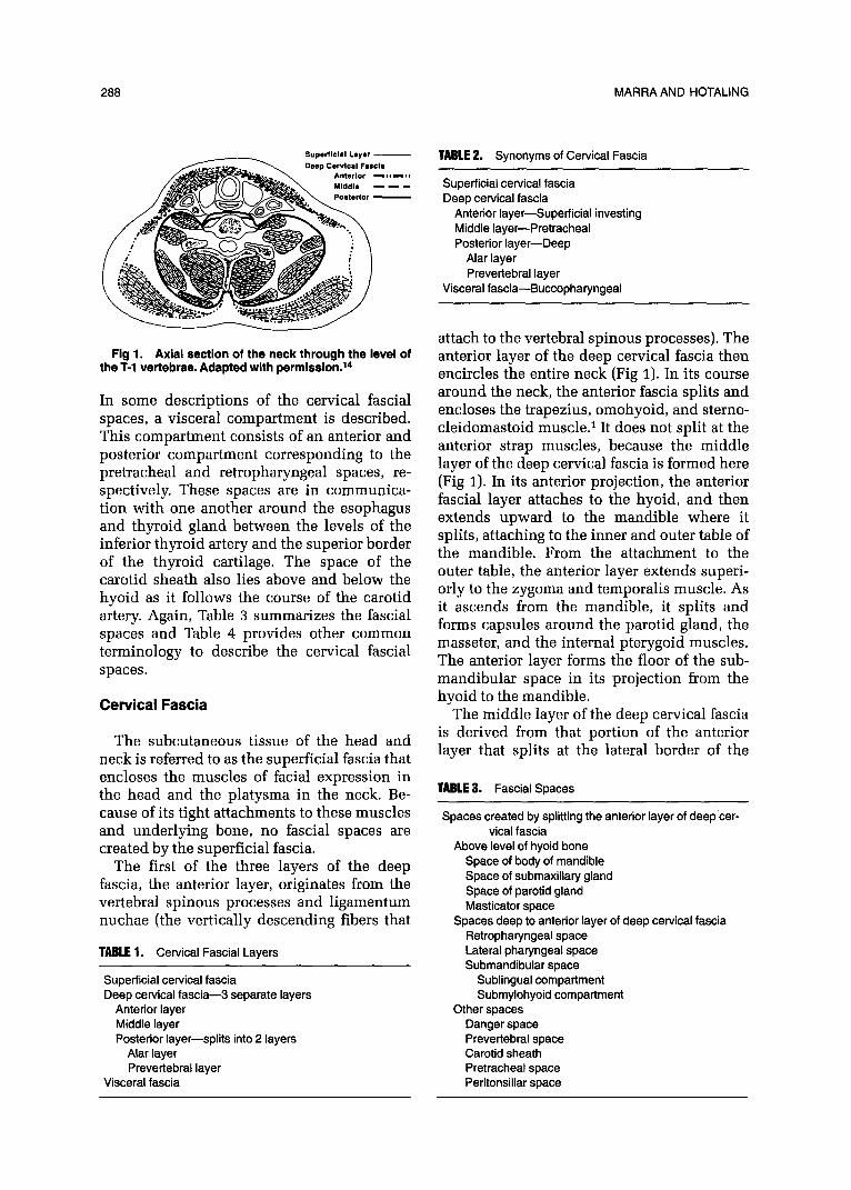
Fig 1. Axial section of the neck through the level of the T-l vertebrae. Adapted with permission.14
In some descriptions of the cervical fascial spaces, a visceral compartment is described. This compartment consists of an anterior and posterior compartment corresponding to the pretracheal and retropharyngeal spaces, re- spectively. These spaces are in communica- tion with one another around the esophagus and thyroid gland between the levels of the inferior thyroid artery and the superior border of the thyroid cartilage. The space of the carotid sheath also lies above and below the hyoid as it follows the course of the carotid artery. Again, Table 3 summarizes the fascial spaces and Table 4 provides other common terminology to describe the cervical fascial spaces.
Cervical Fascia
The subcutaneous tissue of the head and neck is referred to as the superficial fascia that encloses the muscles of facial expression in the head and the platysma in the neck. Be- cause of its tight attachments to these muscles and underlying bone, no fascial spaces are created by the superficial fascia.
The first of the three layers of the deep fascia, the anterior layer, originates from the vertebral spinous processes and l igamentum nuchae (the vertically descending fibers that
TABLE 1. Cervical Fascial Layers
Superficial cervical fascia Deep cervical fascia-3 separate layers
Anterior layer Middle layer Posterior layer-splits into 2 layers
Alar layer Prevertebral layer
Visceral fascia
Superficial cervical fascia Deep cervical fascia
Anterior layer-Superficial investing Middle layer-Pretracheal Posterior layer-Deep
Alar layer Prevertebral layer
Visceral fascia-Buccopharyngeal
attach to the vertebral spinous processes). The anterior layer of the deep cervical fascia then encircles the entire neck (Fig 1). In its course around the neck, the anterior fascia splits and encloses the trapezius, omohyoid, and sterno- cleidomastoid musc1e.l It does not split at the anterior strap muscles, because the m iddle layer of the deep cervical fascia is formed here (Fig 1). In its anterior projection, the anterior fascial layer attaches to the hyoid, and then extends upward to the mandible where it splits, attaching to the inner and outer table of the mandible. From the attachment to the outer table, the anterior layer extends superi- orly to the zygoma and temporalis muscle. As it ascends from the mandible, it splits and forms capsules around the parotid gland, the masseter, and the internal pterygoid muscles. The anterior layer forms the floor of the sub- mandibular space in its projection from the hyoid to the mandible.
The m iddle layer of the deep cervical fascia is derived from that portion of the anterior layer that splits at the lateral border of the
TABLE 3. Fascial Spaces
Spaces created by splitting the anterior layer of deep cer- vical fascia
Above level of hyoid bone Space of body of mandible Space of submaxillary gland Space of parotid gland Masticator space
Spaces deep to anterior layer of deep cervical fascia Retropharyngeal space Lateral pharyngeal space Submandibular space
Sublingual compartment Submylohyoid compartment
Other spaces Danger space Prevertebral space Carotid sheath Pretracheal space Peritonsillar space

DEEP NECK INFECTIONS 289
TABLE 4. Synonyms of Cervical Spaces
Spaces formed by splitting anterior layer Space of parotid gland Space of submaxillary gland Space of body of mandible Masticator space-Masseteric, mandibulopterygoid,
temporal pouch Space deep to the anterior layer of the deep cervical
fascia Retropharyngeal space+Retrovisceral space, retro-
esophageal visceral compartment (posterior) Lateral pharyngeal space-Parapharyngeal, peripha-
ryngeal, pharyngomaxillary, pterygopharyngeal pterygomandibular, pharyngomasticatory
Anterior-Prestyloid Posterior-Poststyloid
Submandibular space Sublingual-Floor of mouth Submylohyoid-Submaxillary, submandibular
Other spaces Danger space Prevertebral space-Paravertebral space Carotid sheath Pretracheal space-Visceral compartment (anterior) Peritonsillar space
strap muscles and travels posteriorly to the strap muscles (Fig 1). Thus, it lies posterior to the strap muscles and anterior to the trachea and thyroid. The middle layer has as its supe- rior extent the hyoid bone, to which it at- taches; inferiorly, the middle layer joins the fibrous pericardium (Fig 2).
The posterior layer also originates from the vertebral spinous processes and encircles the
Fig 2. Sagital section illustrating the anterior, middle, and the posterior layers of the deep cervical fascia. Adapted with permission.14
neck. It is not limited superiorly by the hyoid bone and, in fact, extends to the base of skull. It lies deep to the trapezius muscle and en- closes the vertebral muscles (Fig 1). Laterally, it attaches to the transverse processes of the vertebrae. As it continues anteriorly from this attachment, the posterior fascia split into two separate layers: the alar fascia and the prever- tebral fascia. The alar fascia is the more ante- rior of these two; the prevertebral fascia is posterior and covers the longus colli muscle (Fig 1).
Separate from the superficial and deep cervi- cal fascial layers is the visceral fascia. The visceral fascia has been referred to as the connective tissue coat that surrounds the esophagus, thyroid gland, and trachea [Note: The visceral fascia is distinct from the visceral compartment). Above the level of the hyoid, the visceral fascia is often referred to as the buccopharyngeal fascia. It is intimately associ- ated with these structures and forms no real spaces deep to these structures.
Fascial Spaces
Having described the various fascial layers, a description of the spaces they create follows, The spaces will be described in the order of their anatomic presentation from external to internal. Table 3 describes the spaces as they will be identified in this paper. Table 4 lists other commonly used nomenclature for these spaces. The contents of these spaces can be found in Table 5.
The space of the body of the mandible is formed by the splitting of the anterior layer of the deep cervical fascia in its attachment to the inner and outer table of the mandible. It is actually a potential space, because no soft tissue lies within this plane. It is limited anteriorly by the attachment of the anterior belly of the digastric to the mandible and posteriorly by the internal pterygoid.
Both the parotid space and the space of the submaxillary gland are also formed by the splitting of the anterior layer. These spaces are filled with glands and associated nodes. These glands are intimately adherent to their sur- rounding fascial layers. The lateral pharyngeal space lies deep to the parotid space and may become involved with infection if there is

MARRA AND HOTALING 290
TABLE 5. Major Contents of Cervical Spaces
Anterior layer spaces Space of parotid gland-Parotid gland, facial nerve,
lymph nodes Space of submaxillary gland-Submaxillary gland,
lymph nodes Masticator space-Muscles (temporal, masseter,
internal pterygoid) internal maxillary artery, man- dibular nerve
Space of body of mandible-Potential space Peripharyngeal spaces
Retropharyngeal space-Loose connective tissue, lymph nodes
Lateral pharyngeal space Anterior-Connective tissue, lymph nodes Posterior-Cranial nerves IX, X, XII; carotid sheath,
cervical sympathetics Submandibular space
Sublingual-Sublingual gland, lingual nerve and artery, genioglossus, geniohyoid muscle, subman- dibular duct
Submylohyoid-Digastric muscle Other
Danger space-Loose connective tissue Prevertebral space-Longus colli muscle Carotid Sheath-Internal jugular, carotid artery, cranial
nerve X Pretracheal space-Connective tissue Peritonsillar space-Faucial tonsils
medial spread from the deep lobe of the pa- rotid gland.
The masticator space is also formed by the splitting of the anterior layer to enclose the ramus of mandible, masseter, internal ptery- goid, and lower portion of the temporalis muscle. This space contains the majority of branches of the mandibular nerve and the internal maxillary artery.
Moving medially, the peripharyngeal spaces form a ring around the pharynx and include the retropharyngeal, lateral pharyngeal, and the submandibular spaces. These spaces all lie deep to the anterior layer of the deep fascia. All of these spaces are in relative communica- tion with one another via the muscles and vessels that pass through them.
The retropharyngeal space lying behind the pharynx and esophagus extends superiorly to the base of skull and inferiorly to the bifurca- tion of the trachea. It has a m idline raphe that separates two chains of lymph nodes that lie within its space, just off m idline. The m idline raphe is formed by the attachment of the superior constrictor muscle to the alar layer of the deep cervical fascia.
The lateral pharyngeal space, which is con-
tiguous with the retropharyngeal space later- ally, also extends to the base of skull. The lateral pharyngeal space does not extend infe- rior to the hyoid bone. It has been described as conical in shape, with its base the petrous portion of the temporal bone and its apex at the hyoid bone. It has as its medial extent the lateral pharyngeal wall and is bounded later- ally by the mandible, parotid gland, and the internal pterygoid muscle (Note: The lateral pharyngeal wall does not include the tonsils). It is divided into an anterior and posterior compartment by the styloid process and its musculature. The anterior compartment con- tains fat and connective tissue, whereas the posterior compartment contains cranial nerves IX, X, and XII; the cervical sympathetic chain; the internal jugular vein; and the common carotid artery.
The submandibular space is the most ante- rior portion of the peripharyngeal spaces and is bounded superiorly by the lingual mucosa and inferiorly by the anterior layer of the deep fascia. This space is divided by the mylohyoid muscle into sublingual and submaxillary por- tions that are continuous posteriorly around the free margin of the mylohyoid muscle. The sublingual portion contains the submandibu- lar duct, lingual nerve, and hypoglossal nerve, whereas the submaxillary portion contains the anterior belly of the digastric muscle.
The danger space is that space between the alar and prevertebral fascia. It extends from the base of skull to the diaphragm. It is a potential space that can become involved with infection extending anteriorly from the prever- tebral space or posteriorly from the retropha- ryngeal space and provides another route for spread of infection into the mediastinum.
The pretracheal space is the most anterior of the fascial spaces below the hyoid bone. The fascial spaces below the hyoid also include the retropharyngeal, danger, and prevertebral spaces. The pretracheal space has as its ante- rior border the m iddle layer of the deep fascia and is bounded posteriorly by the esophagus. It has as its superior extent the hyoid bone, and, inferiorly, it extends to the level of the fourth thoracic vertebra along the arch of the aorta.
The carotid sheath, which encloses the ca- rotid artery, the internal jugular vein, and the vagus nerve, forms a potential space for its

DEEP NECK INFECTIONS 291
contents that may be susceptible to infection. It is formed by a combination of all three parts of the deep cervical fascia. Its anteromedial wall is formed by the anterior layer (the por- tion deep to the sternocleidomastoid muscle (SCM) and its posterior wall is formed by a lamina that forms from the anterior layer before it splits off the middle layer. Of ana- tomic and surgical interest is the fact that the cervical sympathetic chain lies posteromedial to the carotid sheath but superficial to the posterior fascial layer. It should also be noted that the carotid sheath traverses the posterior compartment of the lateral pharyngeal space and descends into the mediastinum. This has been termed by MosherZ as the Lincoln’s High- way of the neck.
MICROBIOLOGY
In the preantibiotic era, the organism most often found in deep neck abscesses was Staphylococcus aureus. Since the advent of antibiotics, aerobic Streptococcal species and non-Streptococcal anaerobes have become the bacteria predominately responsible for deep neck infections. As a rule, these bacteria repre- sent components of oral flora that become virulent when normal mucosal barriers are broken. At present, the most commonly iso- lated anaerobes include Peptostreptococcus, Fusobacterium (predominantly nucleatum), and Bacteroides (mainly melaninogenicus).3
Classically, the Streptococcus species are divided into the hemolytic and nonhemolytic strains. The hemolytic strains are predomi- nately the group A species. The nonhemolytic strains are usually classified as alpha and gamma species, with the alpha species further subdivided into group D or nongroup D subspe- cies, In odontogenic infections, the most com- mon of the Streptococcal species is the alpha nonhemolytic Streptococcus, whereas infec- tions of pharyngeal origin have the hemolytic group A strains more commonly isolated.
A review of neck abscesses by Tom and Rice4 indicates the frequent occurrence of the Streptococcal species. In 27 peripharyngeal space infections, 32 Streptococcal species were isolated. The species most commonly isolated were the alpha nongroup D strains, with 13 positive cultures and the group A beta-hemo- lytic strain with seven positive cultures. Of the
anaerobic species, the most common were Bacteroides (11) and Peptostreptococcus (7).
The anaerobe Eikenella corrodens is becom- ing increasingly isolated in head and neck infection and is frequently resistant to clinda- mycin, which is often used to treat head and neck infections.5
The gram-negative rods are rarely found in isolates of head and neck infections. They may be found in patients who are debilitated, el- derly, diabetic, and/or immunocompromised.3
DIAGNOSIS AND TREATMENT OF SPECIFIC SPACE INFECTION
This section will review the presentation and treatment of the deep neck infections. The discussion will be limited to those spaces which classify as deep neck infections: the peripharyngeal spaces and the prevertebral space.
General Considerations
The prerequisites for successful manage- ment of deep neck infections include proper diagnosis and treatment, emphasizing control of the airway, effective antibiotic therapy, and timely surgical intervention.
There are a multitude of signs and symp- toms, that may be present in a patient with a deep neck infection. Most commonly, these patients will present with generalized symp- toms, including fever, chills, malaise, and loss of appetite. Localizing symptoms will include odynophagia, dysphagia, sore throat, neck stiff- ness, neck pain, trismus, and voice changes. Signs include neck swelling, elevation of the floor of mouth, drooling, diaphoresis, elevated temperature, and bulging of the pharyngeal wall. Classic descriptions of pharyngeal wall bulging is midline for prevertebral space infec- tions and unilateral for retropharyngeal space infections, because of the midline raphe formed by the superior constrictor muscle, which prevents retropharyngeal space involvement crossing the midline.
In addition to a “hot potato” voice, airway signs include dyspnea, stridor, and shortness of breath. These signs are seen in advanced disease, and immediate steps to secure the airway are required to prevent airway obstruc- tion or respiratory arrest.

292 MARRA AND HOTALING
Any bleeding, ranging from ecchymosis of the neck skin to frank blood from the ear, nose, or mouth, may represent a sentinel or herald bleed, resulting from infectious involvement of the carotid artery with a potentially lethal outcome.
In evaluating the patient in whom a neck abscess is suspected, an orderly work-up should be followed. A brief history and physi- cal begins the initial evaluation of the patient. If there is no immediate threat to the patient’s airway, a more leisurely history may be ob- tained. Physical examination should include a complete head and neck examination, with particular attention to the airway and to the intraoral exam, specifically inspecting the floor of mouth, oropharynx, and pharyngeal wall. Attention should also be directed behind the palatopharyngeal fold, where a bulge may denote a lateral pharyngeal space infection. Once the history and physical have been ac- complished, pertinent radiographic studies can be obtained.
When surgical drainage is performed, it should be emphasized that with any aspira- tion or drainage of an abscess, aerobic and anaerobic cultures are needed to guide further therapy. A Gram’s stain may aid in initial antibiotic selection. Accurate anaerobic cul- tures are often difficult to obtain and require adherence to strict anaerobic sampling tech- nique. If an abscess cavity is opened, a portion of the wall can be sent to exclude the possibil- ity of an abscess secondary to tumor.
High-dose penicillin continues to be the antibiotic therapy of choice. Third generation ’ cephalosporins have also become popular re- cently. The development of clindamycin resis- tance argues against single antibiotic treat- ment with this drug.6
Radiology
Numerous radiographic techniques are avail- able to evaluate deep neck infections. These include chest x-ray, lateral neck x-ray, ultraso- nography, computed tomography (CT) scan and magnetic resonance imaging (MRI), and bone scan.
The simplest and most readily available study is the lateral cervical x-ray. It is appli- cable in those cases in which there is suspi- cion of retropharyngeal or prevertebral space
involvement. These lateral cervical films are interpreted by measuring the distance from the anterior aspect of the vertebral body to the air column of the posterior pharyngeal wall. At the level of C-2, the normal distance may be as wide as 7 mm in both adults and children. At the level of C-6, the normal distance may be as much as 22 mm in adults and 14 mm in children, It is important to obtain these films in inspiration as artifact can be generated if an expiratory film is taken. Expiratory films may falsely exaggerate the normal soft tissue dis- tance which is measured. A 33% false nega- tive rate has been reported for lateral neck films, and clinical judgement should deter- mine the need for further imaging (Fig 3).’
Ultrasound has been advocated by some to help detect abscess cavities, but the anatomic detail provided by the CT scan makes this modality superior in confirming information not only about the presence of an abscess, but the surrounding anatomic detail. Bone scan can verify osteomyelitis, secondary to a prever- tebral abscess.
Both CT and MRI may provide information about the anatomic detail of the head and neck. Currently, CT scanning is usually more readily available, less expensive, and can effec- tively localize abscesses in the head and neck.
Fig 3. Lateral neck film illustrating increased soft tissue column representing a retropharyngeal space abcess in a 4-year-old patient. The arrow shows the increased soft tissue width.

DEEP NECK INFECTIONS
MRI often has the disadvantage of not being in the immediate hospital complex, requiring transportation of the patient away from the facilities and personnel needed for rapid inter- vention. Thus, the transport presents a poten- tial hazard to patients with deep neck abscess, because their condition may deteriorate rap- idly. The use of CT or MRI is not done in an acute situation in which there is a potential respiratory complication of deep neck infec- tion. These complications mandate immediate surgical attention and sho.uld not wait for imaging. However, in those cases in which the patient is stable and in no respiratory distress, such imaging may provide precise detail of the anatomic location and extent of an abscess cavity.
In evaluating patients with deep neck infec- tions, the chest x-ray may provide information as to the possibility of complications resulting from deep neck infections, such as mediasti- nal extension, lung abscess, and pyopneumo- thorax. The presence of an infiltrate on chest x-ray can suggest aspiration, and it may docu- ment mediastinal involvement necessitating aggressive management of a patient.
Submandibular Space
Since the advent of antibiotics, the subman- dibular space has become the most common of the deep neck abscesses.6 Submandibular space infections can involve either of the two sub- compartments, the sublingual or the submylo- hyoid. Because these two subcompartments
TABLE 6. Clinical Features of Deep Neck Infections
communicate posteriorly around the mylohy- oid muscle, they are often both involved. Ludwig’s angina is the name for the prototypi- cal infection of the submandibular space infec- tion and, by definition, requires that both compartments be involved.2
Odontogenic infection represents the source of submandibular space infection in 70% to 85% of cases.7 Other reported causes include laceration of the floor of mouth, mandible fracture, tumor, sialoadenitis, and lymphadeni- tis. The most frequent isolate responsible for submandibular space infection is the alpha hemolytic Streptococcus7
Typical patients are between ages of 20 and 40 and are otherwise healthy. There is a 3:2 male preponderance. 3 Patients will present with pain in the oral cavity, anterior neck stiffness, drooling, and dysphagia. Table 6 provides an overview of the common present- ing findings of the submandibular space infec- tion as well as comparing these findings to the signs present in the other deep neck infec- tions. Because of the underlying strength of the anterior layer of the deep fascia that forms the inferior aspect of the submandibular space, external swelling is limited. The flexible tis- sue in the floor of mouth can be distended superiorly and resulting in swelling and eleva- tion of the floor of mouth, with resultant anterior and/or posterior tongue displace- ment.
Examination usually shows an enlarged, swollen tongue that protrudes anteriorly and a swollen, indurated, elevated, and erythema-
Complications
Spaces Dyspnea Pain Location Trismus Swelling Dysphagia
Submandibular Present Minimal Floor of mouth
Lateral Pharyngeal Anterior Severe Prominent Anterior oropharynx Posterior Minimal Minimal Posterior oropharynx
behind posterior pharyn- geal pillar
Retropharyngeal Present Minimal
Prevertebral Present None
Posterior pharynx, off mid- line
Posterior pharynx, midline
Present Present Asphyxias, tongue necrosis aspiration pneumonia, lung abscess
Present Occasional - Present Severe Carotid artery erosion,
internal jugular throm- bosis, lateral and cav- ernous sinus thrombosis
Present Present Mediastinitis, pyopneumo- thorax bronchial erosion
Present Occasional -
Data from Megran.15

294 MARRA AND HOTALING
tous floor of mouth. The suprahyoid neck is tender with woody induration. Fluctuance is rarely found. Trismus is usually not detected and suggests lateral pharyngeal space involve- ment if present. The patient’s voice, if any can be generated, has a “hot potato” quality. Sys- temic signs of fever and chills are also present. If tachypnea or stridor is present, these signs herald impending airway obstruction and man- date immediate attention.
Complications of a submandibular space infection include complications that occur when infection leaves the confines of the space and those related to complications from the space itself. The buccopharyngeal gap, created by the styloglossus muscle as it leaves the submandibular space between the supe- rior and middle constrictor muscles, serves as an avenue for infection to exit the submandibu- lar space and enter the lateral pharyngeal space. The most common cause of death in patients with Ludwig’s angina is asphyxia, secondary to upper airway obstruction. Other complications include tongue necrosis, aspira- tion pneumonia, and lung abscess.
The three components of treating the sub- mandibular space infection (or any of the deep neck infections) include airway protection, antibiotic therapy, and surgical intervention. Because asphyxia is the number one cause of death in Ludwig’s angina, protection of the airway is of paramount concern. A number of methods have been used to manage the airway in these patients including observation, oral intubation, fiberoptic intubation, cricothy- rotomy, and tracheotomy.’
Initial observation in a select group of pa- tients is possible. In Patterson’s series’ of 20 patients, only seven required airway control. The other patients were managed with observa- tion. However, observation is possible only in an intensive care unit when the progression of infection does not require surgical drainage. The airway should be controlled before the advent of stridor or cyanosis.
Antibiotics are the mainstay of therapy. High-dose intravenous penicillin is still the drug of choice until culture results are avail- able, because most oral flora remain suscep- tible to penicillin. However, organisms with antibiotic resistance are increasing in preva- lence, and if necessary, additional anaerobic coverage with metronidazole or clindamycin
should be added.l As stated above, clindamy- tin therapy alone is discouraged because of the increasing presence of resistant Eikenella species.3
Those patients who fail to respond to antibi- otic therapy or who progress rapidly require surgical intervention. Drainage can be intra- oral or by external incision. Intraoral drainage via incision in the anterior aspect of the floor of mouth is indicated only in uncomplicated submandibular space infections limited to the sublingual compartment. Otherwise, an exter- nal approach is recommended.
The external submental incision is dia- grammed in Figure 4. This incision is made only after the airway has been adequately controlled. Despite reports of other modalities being used to control the airway, tracheotomy remains the method of choice and gold stan- dard.
The landmark provided by the mandible is often obscured by swelling, so a guage of four fingerwidths below the auricle is used to start the submaxillary incision at the anterior as- pect of the SCM. The submaxillary incision should follow a line below and parallel to the body of the mandible, so the marginal man- dibular nerve is avoided. The submental inci- sion is carried through the anterior fascia and is continued across midline to the midportion of the contralateral submandibular gland. Af- ter the anterior fascia has been transected, the mylohyoid is entered in the midline with blunt dissection. Care should be taken to not penetrate the oral mucosa, violation of which could result in a orocutaneous fistula. Drains
Fig 4. External approaches to the deep neck spaces.

DEEP NECK INFECTIONS 295
are placed through the mylohyoid, and the incision can be loosely closed. Postoperative care would include wet-to-dry dressings on the wound. Decompressing the abscess and intravenous antibiotics will resolve most infec- tions within 7 to 10 days.
Lateral Pharyngeal Space
A number of causes for infection into the lateral pharyngeal space exist. These include lateral extension of infection from a peritonsil- lar abscess, posterior extension from the sub- mandibular space, anterior extension from retropharyngeal space infection, and medial extension from a deep lobe parotid abscess. Penetrating trauma may also lead to infection of this space, but the most common source is extension of infection from the masticator spacee2 It should be noted that a peritonsillar abscess does not involve the lateral pharyn- geal space. The peritonsillar space is medial to the lateral pharyngeal space and is separated from the lateral pharyngeal space by the supe- rior constrictor muscle. Only with lateral exten- sion does a peritonsillar abscess involve the lateral pharyngeal space.
When involved with infection, the anterior and posterior compartments of the lateral pha- ryngeal space have different presentations be- cause of their varying content. The anterior compartment contains only fat, connective tissue, lymph nodes, and muscle, whereas the posterior compartment contains the carotid sheath and cranial nerves IX, X, and XII. The typical anterior compartment infection presents with complaints of pain, dysphagia, and trismus. Systemic signs of fever and chills are also common. Usually an infection presents 7 to 10 days after an otherwise uncomplicated tonsillitis or pharyngitis. The four classic signs of the presentation for an anterior compart- ment infection include trismus, swelling at the angle of the mandible, medial bulging of the pharyngeal wall, and signs of systemic toxicity. Although an abscess may be present, fluctuance is often absent.
In contrast, infection of the posterior space typically presents without trismus and with- out localizing signs, This portion of the space does not contain the pterygoid musculature; thus, trismus is uncommon. In addition, the posterior presentation is difficult to detect on
intraoral examination. Systemic signs of fever and toxicity are present, and infections of this space are often labelled as fevers of unknown origin. The medial bulging of the pharyngeal wall seen with anterior compartment involve- ment is not present, and swelling, if present, is usually behind the palatopharyngeal arch and thus is often missed on examination.
The carotid sheath lies within the posterior compartment of the lateral pharyngeal space; thus, vascular complications may arise from aggressive or untreated infections of this area. The most common vascular complication is suppurative internal jugular vein thrombosis with a preantibiotic era mortality rate of 60%.’ With current antibiotic therapy, the mortality has been reduced. With the use of intravenous antibiotics in a patient who has already had surgical drainage of the lateral pharyngeal space, suppuration of the internal jugular can be managed without the sacrifice of the vein if the patient is stable.8 Complications related to internal jugular vein thrombosis include bac- teremia, septic pulmonary emboli, lateral and cavernous sinus thrombosis, and metastatic abscess formation.g
The most devastating vascular complication is carotid artery erosion. Despite the advent of antibiotics, these patients experience a 20% to 40% mortality. Most of these patients suc- cumb to asphyxia, although some die of acute exsanguination. The most frequently effected vessel is the internal carotid (62%), followed by the external carotid (25%), with the com- mon carotid making up the remainder.lO A carotid artery bleed may be heralded by a number of signs, including recurrent, small hemorrhages from the mouth, nose, or ear, hematoma in the surrounding tissues, the on- set of shock, a protracted clinical course, an ipsilateral Horner’s syndrome (ptosis, meiosis, and anhydrosis), unexplained IX to XII cranial neuropathies and persistent peritonsillar swell- ing despite resolution of the abscess.‘l
The treatment of the lateral space infection is similar to that of the submandibular space infection in that airway control, antibiotics, and surgical drainage form the keystones of therapy. One advantage to the lateral pharynx infection is that the airway is managed more easily. If tracheotomy is required, one is less likely to have midline swelling interfering with the identification of surgical landmarks.

296 MARRA AND HOTALING
Antibiotic therapy is similar to that for Ludwig’s angina, except that anaerobes play a greater role in infection of this space, requir- ing the addition of clindamycin or metronida- zole to high-dose penicillin.
The surgical approaches to the lateral pha- ryngeal space vary, depending on whether a complication is expected. Surgical drainage should not be undertaken during the initial cellulitic phase because this will not lead to an accelerated resolution. Intraoral drainage should be condemned because of the presence of the carotid sheath within the lateral pharyn- geal space.
In those cases in which no complications are anticipated (carotid artery erosion, inter- nal jugular vein thrombosis, mediastinal spread, and so forth), the submaxillary (Bat- son) approach can be used (Fig 4).l An inci- sion is made 3 to 4 fingerwidths below the auricle along the anterior border of the SCM. This incision is carried horizontally to the level of the m id-portion of the submandibular gland. The incision should be carried through the anterior fascia, and the angular band of fascia, which connects the parotid and sub- mandibular gland, should be identified. Blunt dissection should be carried out through this fascia in a superomedial direction underneath the angle of the mandible to enter into the lateral pharyngeal space. This should be opened and drained. The incision can then be loosely approximated.
The anterior sternocleidomastoid (Mosher) approach (Fig 4) is the alternate approach that should be used when operating on a patient in whom access to the carotid artery may be required.lJ The initial incision is a vertical limb, which is placed 3 to 4 fingerwidths below the auricle along the anterior boarder of the SCM. This incision is made without the head being rotated to prevent placing the SCM over the carotid sheath. The incision is taken through the anterior cervical fascia, and care- ful dissection is carried out to identify the carotid sheath, which is opened. The common carotid is isolated with a loose ligature placed around it. A submaxillary incision is then made at the superior aspect of the vertical incision along the anterior border of the sterno- cleidomastoid muscle (Fig 4). It is taken hori- zontally across to the anterior portion of the submandibular gland. The inferior aspect of
the submandibular gland fascia is dissected, allowing for elevation of the submandibular gland anteriorly. The lateral space is then entered and drained. Again, the incision may be loosely closed with a drain in place. If there is high suspicion for carotid artery erosion, the ligature around the common carotid may be left as a safety measure until the infection has reso1ved.l
Retropharyngeal Space
The retropharyngeal space has two chains of lymphatics that lie just off m idline. Usually these are involuted by the age of 5, which accounts for the rarity of retropharyngeal infec- tion occurring as a result of suppuration of these nodes after this age. Other sources of retropharyngeal infection include: penetrating or blunt trauma, instrumentation (ie, esopha- goscopy), intubation, placement of feeding tube, infection from other spaces (including posterior spread from the lateral pharyngeal space), and anterior spread of the prevertebral space infection.
Early in the presentation of a retropharyn- geal space infection, diagnosis may be diffi- cult, especially in children. Initially, the swell- ing is located superiorly and may be m istaken in children for adenoidal hypertrophy and inflammation. Sore throat is not a typical complaint, but a child may refuse food intake. Trismus is m inimal and, if present, should alert to the possibility of further extension to the lateral pharyngeal space. Patients have slight elevation of temperature. A “hot potato” voice, when present, is secondary to supraglot- tic swelling. When the swelling is below the nasopharynx, a mass along the posterior pha- ryngeal wall will be seen off the m idline (Fig 5). In addition, cervical adenopathy, irritabil- ity and nuchal rigidity may be present in children. If there is severe respiratory distress, mediastinal extension should be considered.
Gianoli et all2 recently reviewed the chang- ing etiology of retropharyngeal space infection and noted that traumatic causes of retropharyn- geal space infection had become as common as the classically described suppuration of the retropharyngeal nodes. As a result, their series had an older age of presentation than the typically described child less than 5 years of age. A review of the m icrobiology from this

DEEP NECK INFECTIONS 297
Fig 5. CT scan illustrating a retropharyngeal space abcess in a 4-year-old patient. Note that the abcess is off midline. The arrow denotes the abcess capsule.
study did not show continued predominance of Streptococcal species, which were not speci- fied, and a trend toward anaerobic organ- isms.12
The retropharyngeal space has communica- tion with the anterior and posterior aspects of the superior mediastinum. The anterior medi- astinum is accessible by the pretracheal space, as reviewed in the anatomic discussion, and the posterior aspect of the superior mediasti- num is accessible via the retropharyngeal space to the level of the tracheal bifurcation. Conse- quently, complications of infection of this space include mediastinitis, pyopneumotho- rax, bronchial erosion, and purulent pericardi- tis. In addition, other complications unrelated to mediastinal spread include rupture with aspiration, empyema, and pneumonia.
In those patients who develop mediastinitis, there is a high mortality rate of 40% to SO%, usually from overwhelming sepsis. In early mediastinitis, a cervicomediastinal approach can be taken, but in advanced cases, thora- cotomy provides the only hope for salvage.13
Treatment for uncomplicated cases involves both intravenous antibiotics and surgical drain- age. Transoral drainage can be carried out in these infections, because the vascular sheath
lies in the lateral space. However, if the lateral space becomes involved, an external approach is necessary.
The transoral approach should be initiated with the patient in a Trendelenburg position (as for adenoidectomy). Topical anesthesia may be used in cooperative patients. Initial exploration of a lateralized posterior pharyn- geal wall mass should be performed with needle aspiration, both to confirm the pres- ence of purulence and to rule out the presence of blood. The presence of blood within the cavity suggests carotid erosion and dictates an external approach. Once needle aspiration has confirmed the presence of pus, the cavity should be incised with a vertical incision and opened widely with blunt dissection using a hemostat.
If an external approach is necessary, the anterior sternocleidomastoid incision should be used (Fig 4). An incision is made parallel to the anterior border of the sternocleidomastoid muscle at the level of the hyoid and carried inferiorly to the cricoid. In those cases in which there is inferior dissection of the retro- pharyngeal abscess, the inferior extent of the neck incision can then be extended to the level of the clavicle. Adequate drainage can be obtained without thoracotomy to the level of T-4. The initial incision is then carried through the platysma and anterior layer of the deep cervical fascia. The sternocleidomastoid is lateralized, and the carotid sheath is identi- fied. Mobilizing the carotid sheath medially, one will come to the prevertebral fascia and enter the retropharyngeal space. With medial retraction of the larynx, trachea, and thyroid gland, the abscess cavity is exposed, opened, and drained.
Prevertebral Space
The classic infection of this space is Potts abscess resulting from tuberculous infection of the vertebral body. Other causes of preverte- bra1 space infection include posterior exten- sion from retropharyngeal space infection trav- eling through the danger space to the prevertebral space, trauma, previous surgery (ie, anterior cervical fusion), and extension of cervical osteomyelitis.
These infections typically present in an indolent fashion. If present in the high cervi-

298 MARRA AND HOTALING
cal area, they may be visible intraorally as a midline bulge. These patients may have only minimal complaints including pain in the back of the neck and shoulder, which are aggravated with swallowing. Other symptoms, including dysphagia and respiratory compro- mise, may or may not be present.
Complications from infectious involvement of this space include extension into the danger and retropharyngeal spaces with their atten- dant complications, as well as potential de- struction of vertebral bodies with neurological sequelae from either subluxation or epidural compression.
The principles of treatment are similar to the infections of the other spaces and involve airway control if indicated, antibiotics, and surgical drainage. Antibiotic therapy should be guided towards Staphylococcus oureus, the most common organism. The classic Pott’s abscess is caused by mycobacterium and should be considered in those cases in which treatment is refractory to the usual antibiotics.
The surgical approach is guided by the patient’s symptomatology. If respiratory com- promise is imminent, then the airway must be secured. Again, tracheotomy provides the saf- est manner in controlling the airway. The choice for internal versus external drainage depends on the location of the infection. If accessible intraorally, usually from Cl to C3, prevertebral infections may be drained in a similar fashion to retropharyngeal abscess. The limitation of the transoral approach may be the difficulty in achieving adequate removal of bone.
Two external approaches can be used to drain these abscesses. The anterior sternoclei- domastoid approach or an approach that uses ‘an incision posterior to the sternocleidomas- toid muscle (Fig 4). The dissection is carried to the prevertebral fascia, because it covers the scalene musculature and enters into the retro- pharyngeal space. Further medial dissection at the level of the abscess should permit one to enter through the prevertebral and alar fascia to open the abscess and provide for drainage. The importance of obtaining a specimen for Gram’s stain and culture to guide further anti- biotic coverage cannot be overemphasized.
CONCLUSION
An overview of the anatomy of deep neck infections has been provided, describing the cervical fascial layers and their fascial spaces. Standard terminology (Tables l-3) has been used. In addition, the presentation and man- agement of the deep neck infections has been detailed, including discussion of the peripha- ryngeal spaces and the prevertebral space.
REFERENCES
1. Brown DF, Richtsmeier WJ: Infections of the deep fascial spaces of the head and neck. Continuing Education Program, American Academy of Otolaryngology Head and Neck Surgery Foundation, 1987
2. Mosher HP: The submaxillary fossa approach to deep pus in the neck. Tram Am Acad Ophthalmol Oto- laryngol34:19-36,1929
3. Blomquist IK, Bayer AS: Life threatening deep fas- cial space infections of the head and neck. Infect Dis Clin North Am 2:737-764,1988
4. Tom MB, Rice DH: Presentation and management of neck abscess: A retrospective analysis. Laryngoscope 98: 877-880,1988
5. Tami TA, Parker GS: Clinical notes: Eikenella cor- rodens: An emerging pathogen in head and neck infec- tions. Arch Otolaryngol110:752-754,1984
6. Beck AL: The influence of the chemotherapeutic and antibiotic drugs on the incidence and course of deep neck infections. Ann Otol Rhino1 Laryngol61:515-532,1952
7. Patterson HC, Kelly JH, Stroone M: Ludwig’s angina: An update. Laryngoscope 92:370-377,1982
8. Yau PC, Norante JD: Thrombophlebitis of the inter- nal jugular vein secondary to pharyngitis. Arch Otolaryn- go1106:507-508,198O
9. Lemierre A: On certain septicaemas due to anaerobic organisms. Lancet 2:701-703,1936
10. Salinger S, Pearlman SJ: Hemorrhage from pharyn- geal and peritonsillar abscesses. Arch Otolaryngol18:464- 509,1933
11. Blum DJ, McCaffrey TV: Original articles: Septic necrosis of the internal carotid artery. A complication of peritonsillar abscess. Otolaryngol Head Neck Surg 91:114- 118,1983
12. Gianoli GJ, EspinolaTE, Guarisco, JL, et al: Retropha- ryngeal space infection changing trends. Otolaryngol Head Neck Surgery 165:92-loo,1993
13. Estrera AS, Landa MJ, Grisham SM, et al: Descend- ing necrotizing mediastinitis. Surg Gynecol Obstet 157:545- 552,1983
14. Hollinshead WH: Anatomy for Surgeons, Volume 1, The Head and Neck. Philadelphia, PA, Lippincott-Raven, 1954
15. Megran DW: Odonotogenic infections. Pediatr In- fect Dis 3:257-265,1984
Copyright © 2022 FDOKUMEN