By Mo3ath Head & Neck Practical Anatomy - KSUMSC
43
-
Upload
khangminh22 -
Category
Documents
-
view
1 -
download
0
Transcript of By Mo3ath Head & Neck Practical Anatomy - KSUMSC

1 By Mo3ath Head & Neck Practical Anatomy
1 صفحة
2 By Mo3ath Head & Neck Practical Anatomy
2 صفحة
To 428 With love .. ღ
Mo3ath Al-Saiady
3 By Mo3ath Head & Neck Practical Anatomy
3 صفحة
Thanks to :
Amani ALbijadi For her helping in preparation this handout
Sara ALHasani For her revesion to this handout
Saad AlQahtani For his supervising on the team's work
Ahmed AlMazrou
Group A leader
Also thanks to anatomy's DVD heros:
Bilal Marwa (428 Godfather )
Yahya Asiri (Best penguin ever )
Mohammed Aba- Alhasan
And finally to the best anatomy leader :
Mohammed Al-Otibi

4 By Mo3ath Head & Neck Practical Anatomy
4 صفحة
Practical ► 1 ◄
✖ Skull & Scalp ✖

5 By Mo3ath Head & Neck Practical Anatomy
5 صفحة
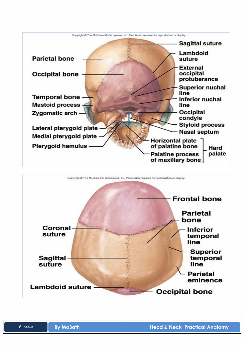
6 By Mo3ath Head & Neck Practical Anatomy
6 صفحة
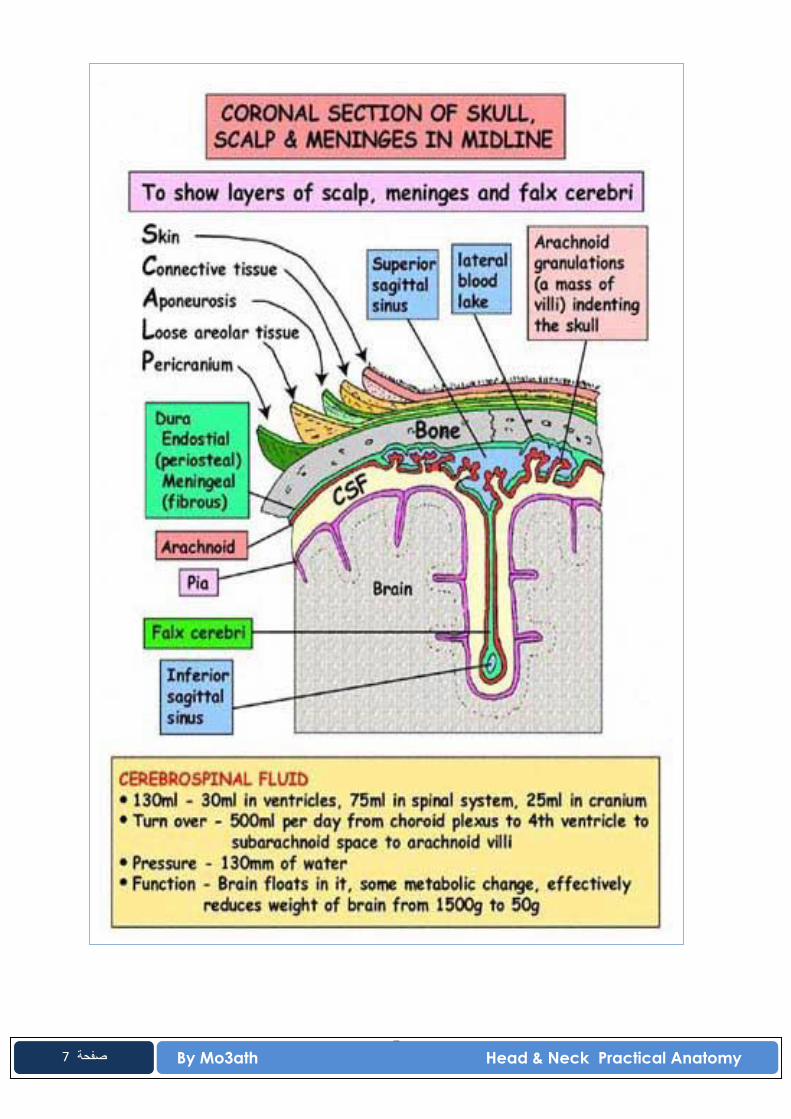
7 By Mo3ath Head & Neck Practical Anatomy
7 صفحة
8 By Mo3ath Head & Neck Practical Anatomy
8 صفحة
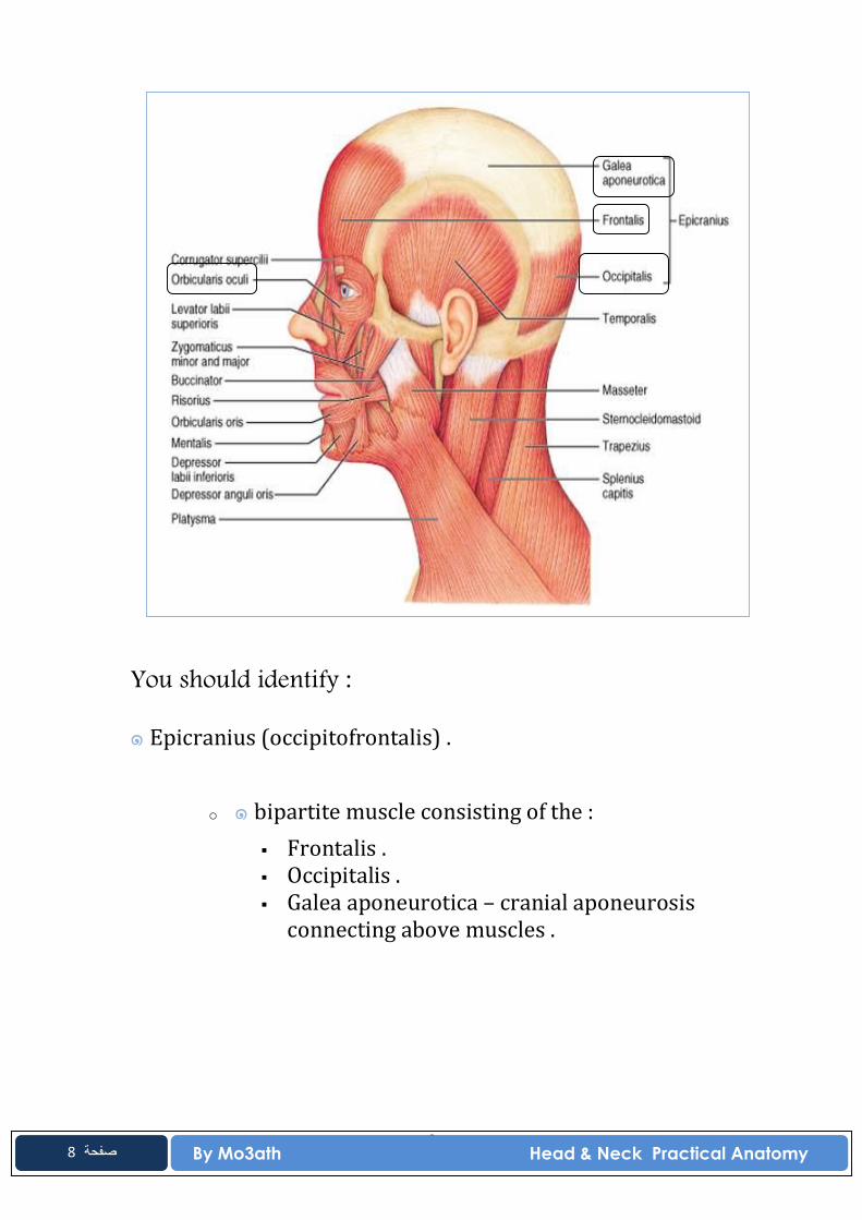
You should identify :
๑ Epicranius (occipitofrontalis) .
o ๑ bipartite muscle consisting of the : Frontalis . Occipitalis . Galea aponeurotica – cranial aponeurosis
connecting above muscles .

9 By Mo3ath Head & Neck Practical Anatomy
9 صفحة
Practical ► 2 ◄
✖ Face & Parotid Gland ✖
MUSCLES OF THE FACE
You should identify :
๑ orbicularis oculi.
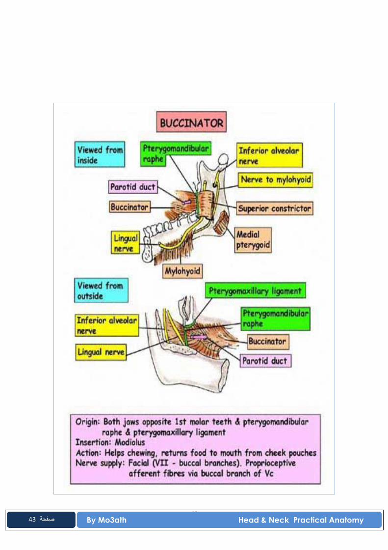
๑ buccinator .

10 By Mo3ath Head & Neck Practical Anatomy
10 صفحة
๑ orbicularis oris.
MOTOR INNERVATION TO THE FACE
The motor innervation to the muscles of facial expression is Cranial Nerve VII (Facial)
The Facial nerve divides into 5 major divisions :
T -- temporal Z -- zygomatic B -- buccal M -- mandibular C -- cervical
ⒽⒾⓃⓉ
In the exam , you will not be asked to write the divisions' names of facial nerve .
11 By Mo3ath Head & Neck Practical Anatomy
11 صفحة
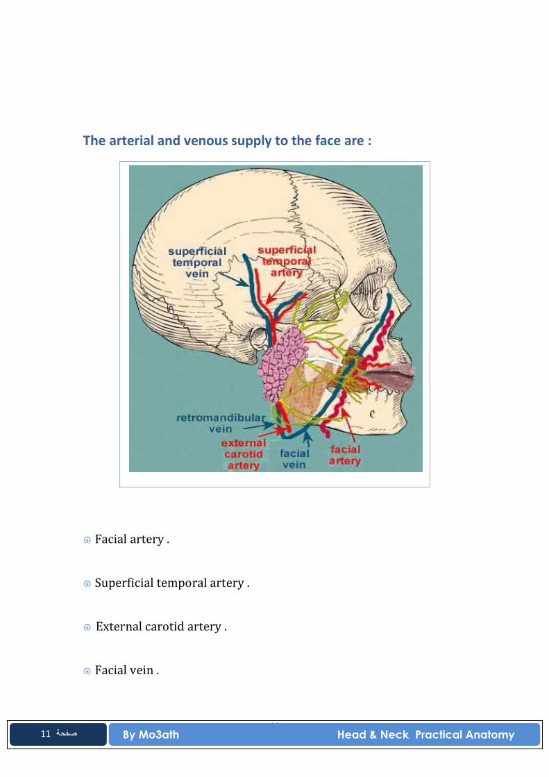
The arterial and venous supply to the face are :
๑ Facial artery .
๑ Superficial temporal artery .
๑ External carotid artery .
๑ Facial vein .
12 By Mo3ath Head & Neck Practical Anatomy
12 صفحة
๑ Superficial temporal vein .
๑ Retromandibular vein .
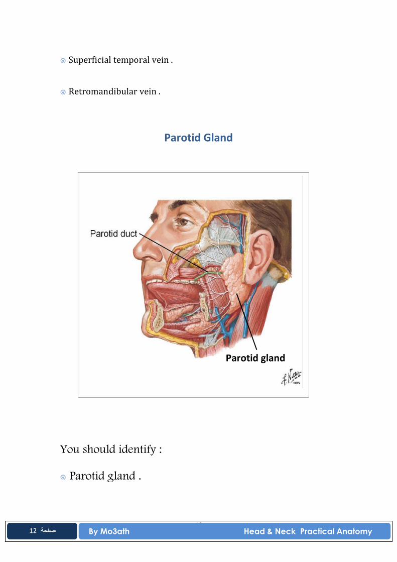
Parotid Gland
You should identify :
๑ Parotid gland .
Parotid gland
13 By Mo3ath Head & Neck Practical Anatomy
13 صفحة
๑ Parotid duct .
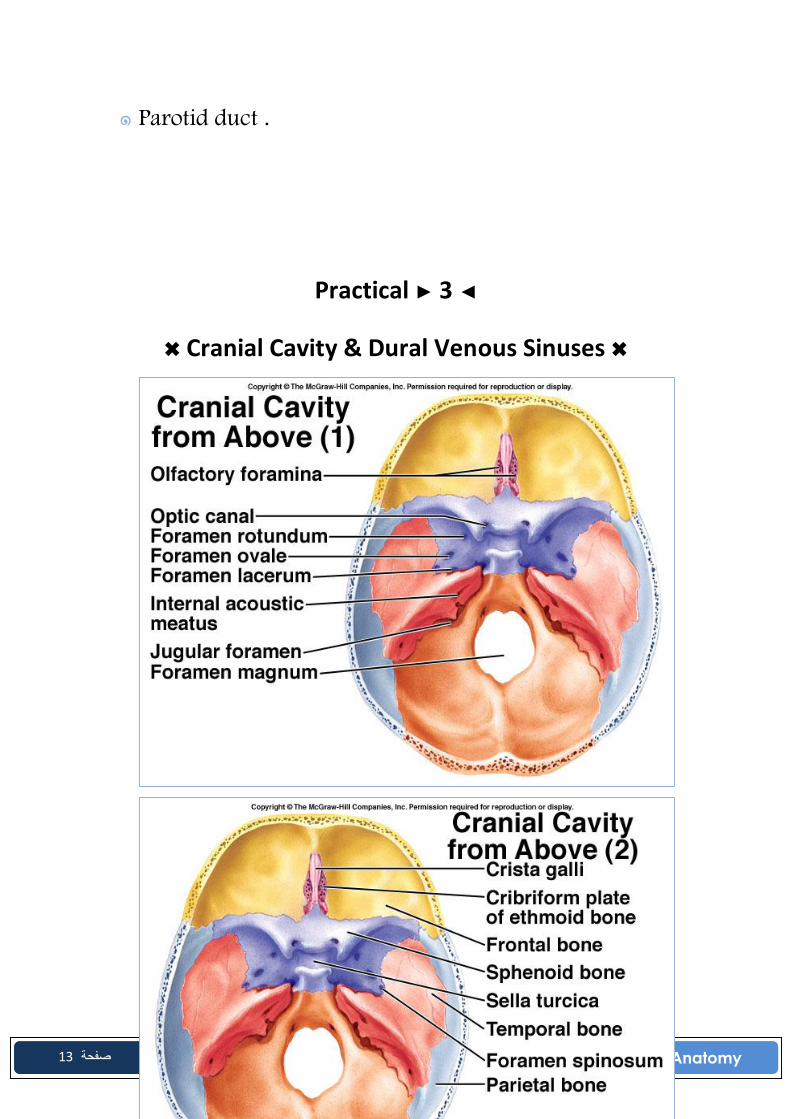
Practical ► 3 ◄
✖ Cranial Cavity & Dural Venous Sinuses ✖

14 By Mo3ath Head & Neck Practical Anatomy
14 صفحة
15 By Mo3ath Head & Neck Practical Anatomy
15 صفحة
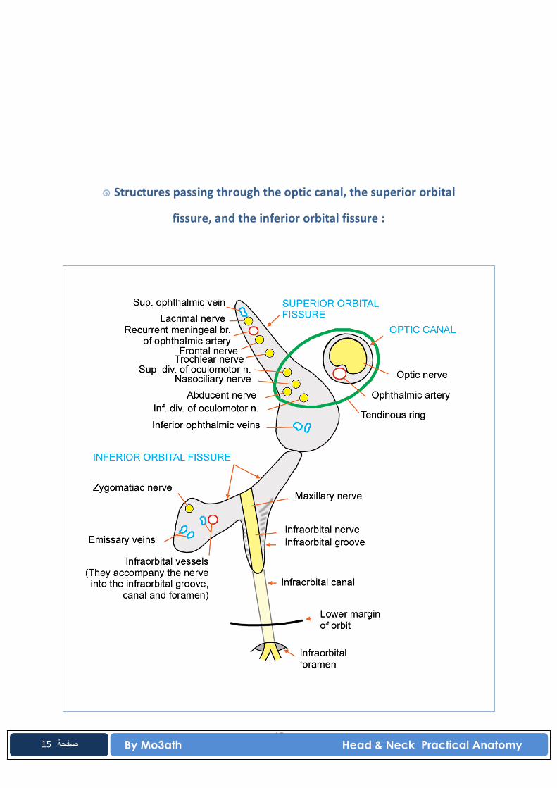
๑ Structures passing through the optic canal, the superior orbital
fissure, and the inferior orbital fissure :
16 By Mo3ath Head & Neck Practical Anatomy
16 صفحة
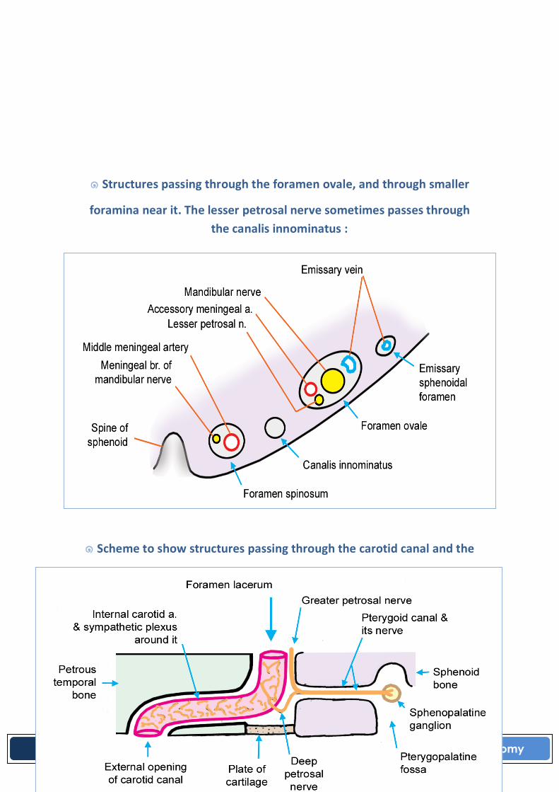
๑ Structures passing through the foramen ovale, and through smaller
foramina near it. The lesser petrosal nerve sometimes passes through the canalis innominatus :
๑ Scheme to show structures passing through the carotid canal and the
foramen lacerum:
17 By Mo3ath Head & Neck Practical Anatomy
17 صفحة
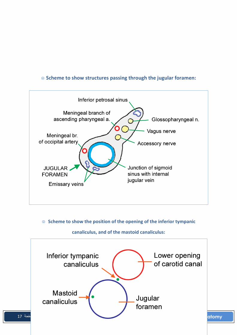
๑ Scheme to show structures passing through the jugular foramen:
๑ Scheme to show the position of the opening of the inferior tympanic
canaliculus, and of the mastoid canaliculus:

18 By Mo3ath Head & Neck Practical Anatomy
18 صفحة
๑ Scheme to show the arrangement of structures passing
through the foramen magnum:
19 By Mo3ath Head & Neck Practical Anatomy
19 صفحة
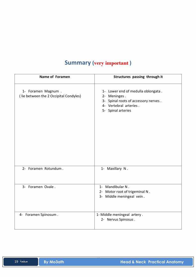
Summary (very important )
Name of Foramen Structures passing through it
1- Foramen Magnum .
( lie between the 2 Occipital Condyles)
1- Lower end of medulla oblongata . 2- Meninges . 3- Spinal roots of accessory nerves . 4- Vertebral arteries . 5- Spinal arteries
2- Foramen Rotundum .
1- Maxillary N .
3- Foramen Ovale . 1- Mandibular N . 2- Motor root of trigeminal N . 3- Middle meningeal vein .
4- Foramen Spinosum . 1- Middle meningeal artery . 2- Nervus Spinosus .

20 By Mo3ath Head & Neck Practical Anatomy
20 صفحة
5- Foramen Lacerum . 1- Internal carotid artery . 2- Meningeal br. Of ascending pharyngeal
artery .
6- Optic Foramen . Optic nerve . Ophthalmic artery .

21 By Mo3ath Head & Neck Practical Anatomy
21 صفحة
* Occulomotor Nerve 3rd * Trochlear Nerve 4th * Abducent Nerve 6th * Opthalmic veins
* Cranial nerves 9th, 10th , 11th * Internal Jugular vein * Inferior petrosal Sinus
* Facial Nerve 7th ( medial) * Vestibulocochlear 8th ( lateral) * Internal Auditory Vessels
* Hypoglossal Nerve 12th
* Internal Carotid Artery * Internal Carotid Sympathetic Plexus * Deep Petrosal Nerve
* Facial Nerve 7th * Stylomastoid Artery
* Infra orbital Nerve and Vessels
* Olfactory Nerves
(7) Superior Orbital Fissure
(8) Jugular Foramen
(9) Internal Auditory Meatus
(10) Hypoglossal Canal ( Anterior Condylar Foramen)
(11) Carotid Canal
(12) Stylomastoid Foramen
(13) Inferior Orbital Fissure
(14) Foramina Of Cribriform Plate Of Ethmoid
22 By Mo3ath Head & Neck Practical Anatomy
22 صفحة
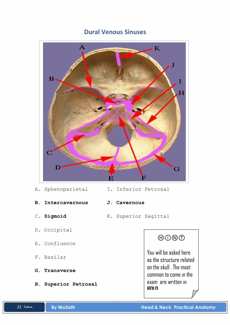
Dural Venous Sinuses
A. Sphenoparietal I. Inferior Petrosal B. Intercavernous J. Cavernous C. Sigmoid K. Superior Sagittal D. Occipital E. Confluence F. Basilar G. Transverse H. Superior Petrosal
ⒽⒾⓃⓉ
You will be asked here as the structure related on the skull . The most common to come in the exam are written in BOLD.
23 By Mo3ath Head & Neck Practical Anatomy
23 صفحة
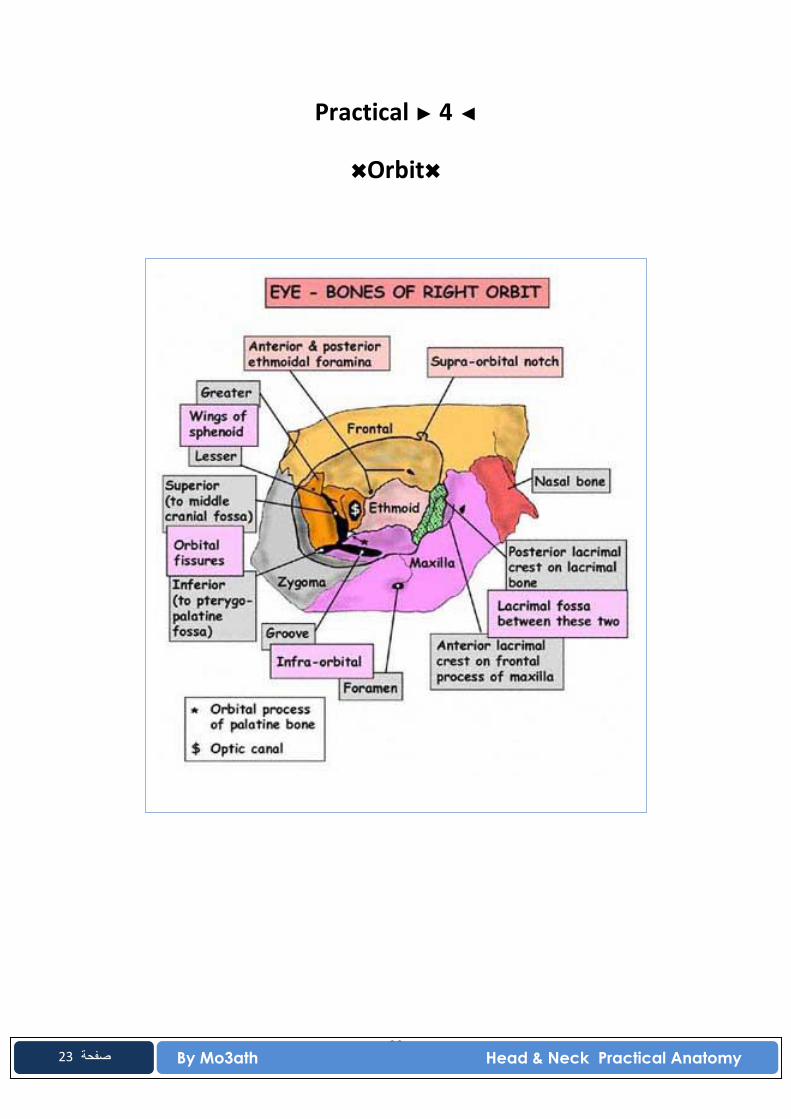
Practical ► 4 ◄
✖Orbit✖
24 By Mo3ath Head & Neck Practical Anatomy
24 صفحة
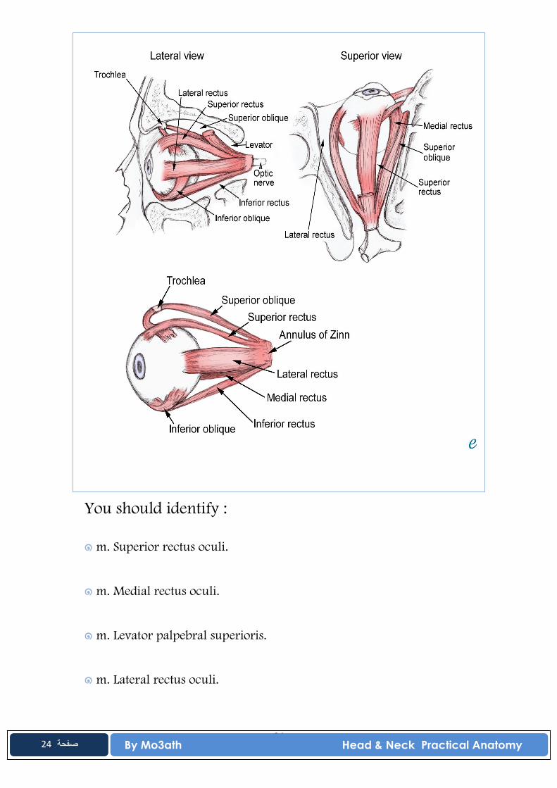
You should identify :
๑ m. Superior rectus oculi.
๑ m. Medial rectus oculi.
๑ m. Levator palpebral superioris.
๑ m. Lateral rectus oculi.
25 By Mo3ath Head & Neck Practical Anatomy
25 صفحة
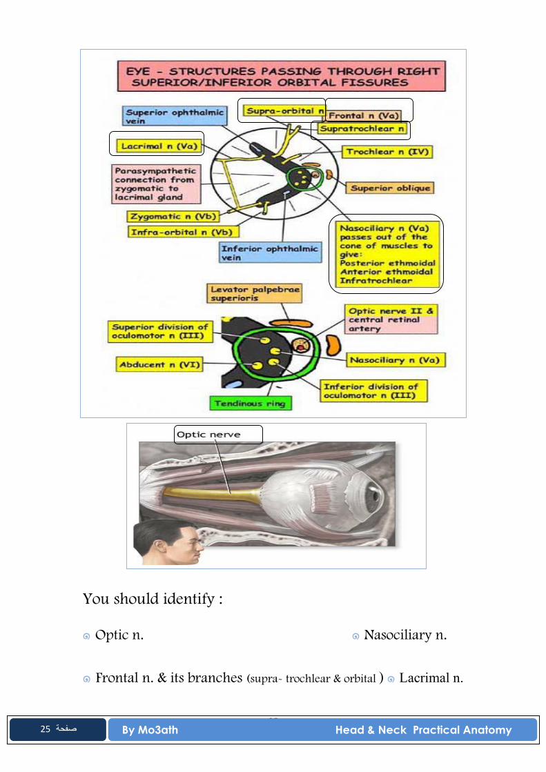
You should identify :
๑ Optic n. ๑ Nasociliary n.
๑ Frontal n. & its branches (supra- trochlear & orbital ) ๑ Lacrimal n.

26 By Mo3ath Head & Neck Practical Anatomy
26 صفحة
You should identify :
๑ Opthalmic a.
27 By Mo3ath Head & Neck Practical Anatomy
27 صفحة
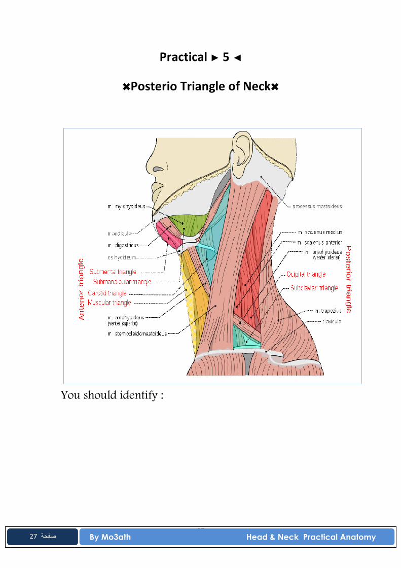
Practical ► 5 ◄
✖Posterio Triangle of Neck✖
You should identify :
28 By Mo3ath Head & Neck Practical Anatomy
28 صفحة
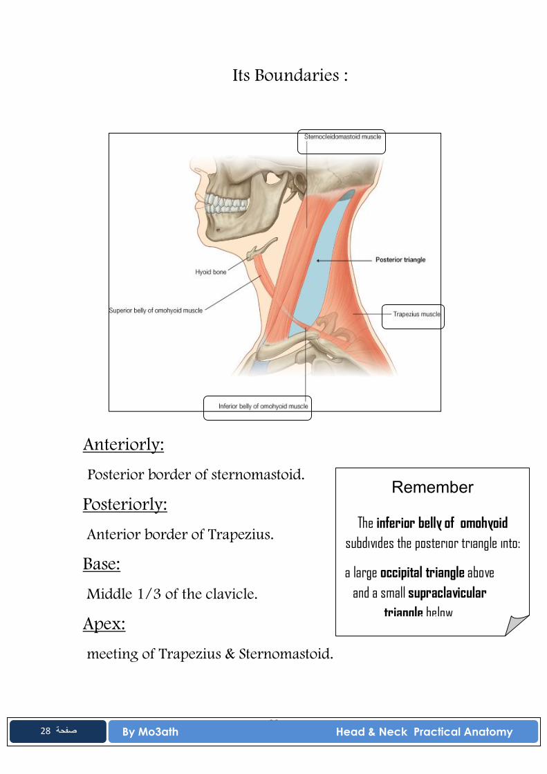
Its Boundaries :
Anteriorly:
Posterior border of sternomastoid. Posteriorly: Anterior border of Trapezius.
Base: Middle 1/3 of the clavicle.
Apex: meeting of Trapezius & Sternomastoid.
Remember
The inferior belly of omohyoid subdivides the posterior triangle into:
a large occipital triangle above and a small supraclavicular
triangle below.
29 By Mo3ath Head & Neck Practical Anatomy
29 صفحة
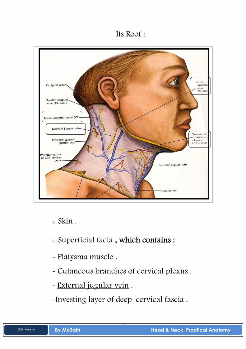
Its Roof :
๑ Skin .
๑ Superficial facia , which contains :
- Platysma muscle .
- Cutaneous branches of cervical plexus .
- External jugular vein .
-Investing layer of deep cervical fascia .
30 By Mo3ath Head & Neck Practical Anatomy
30 صفحة
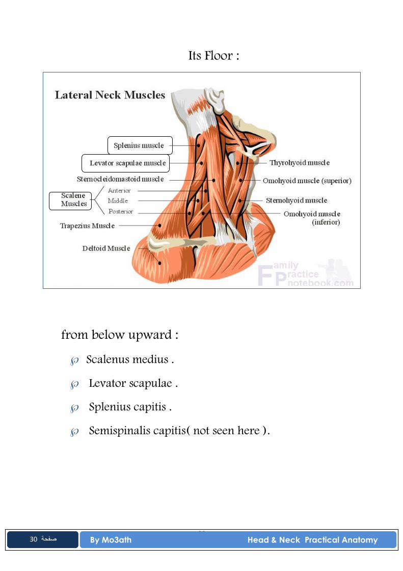
Its Floor :
from below upward :
Scalenus medius .
Levator scapulae .
Splenius capitis .
Semispinalis capitis( not seen here ).
31 By Mo3ath Head & Neck Practical Anatomy
31 صفحة
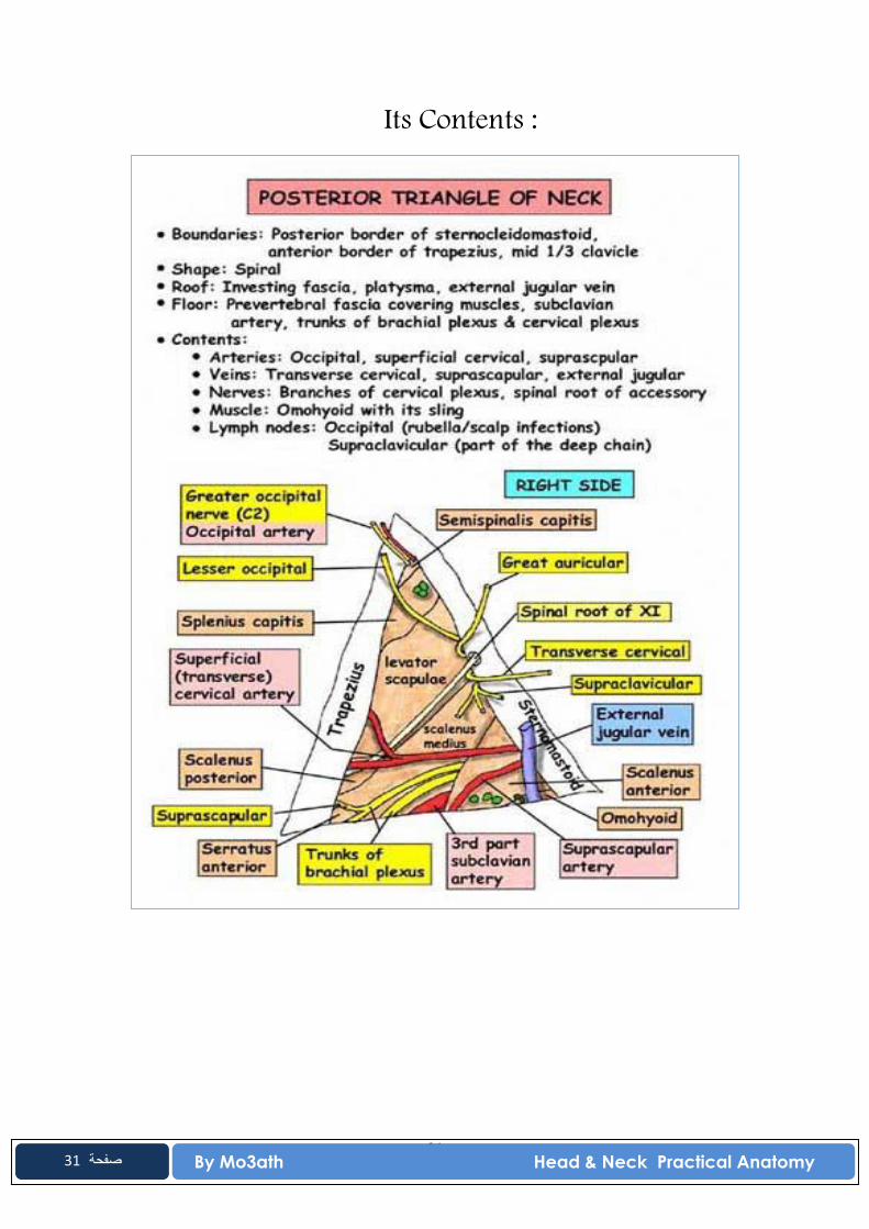
Its Contents :

32 By Mo3ath Head & Neck Practical Anatomy
32 صفحة
Practical ► 6 ◄
✖Anterior Triangle of Neck✖
You should identify :
Its Boundries :
• Anteriorly:
Median plane.
• Posteriorly:
Anterior border of sternomastoid.
• Superiorly:
33 By Mo3ath Head & Neck Practical Anatomy
33 صفحة
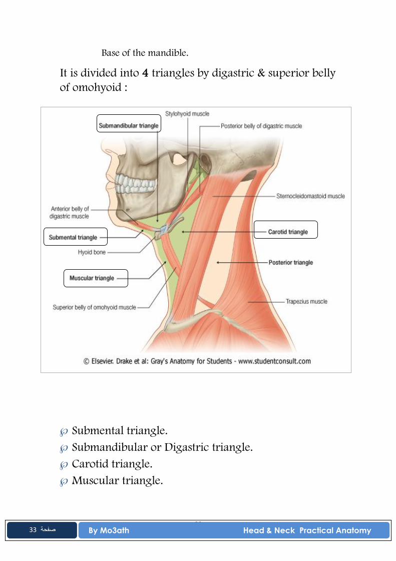
Base of the mandible.
It is divided into 4 triangles by digastric & superior belly of omohyoid :
Submental triangle. Submandibular or Digastric triangle. Carotid triangle. Muscular triangle.
34 By Mo3ath Head & Neck Practical Anatomy
34 صفحة
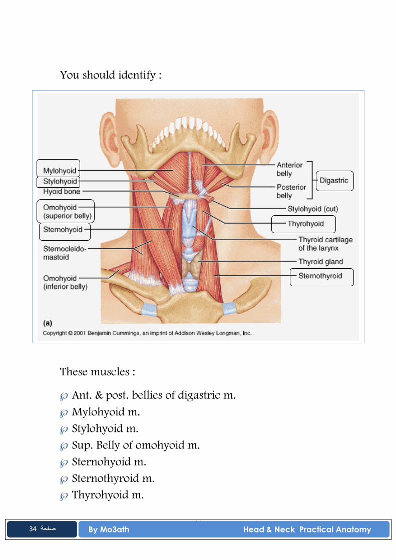
You should identify :
These muscles :
Ant. & post. bellies of digastric m. Mylohyoid m. Stylohyoid m. Sup. Belly of omohyoid m. Sternohyoid m. Sternothyroid m. Thyrohyoid m.

35 By Mo3ath Head & Neck Practical Anatomy
35 صفحة
These vessels :
Internal jugular v.
36 By Mo3ath Head & Neck Practical Anatomy
36 صفحة
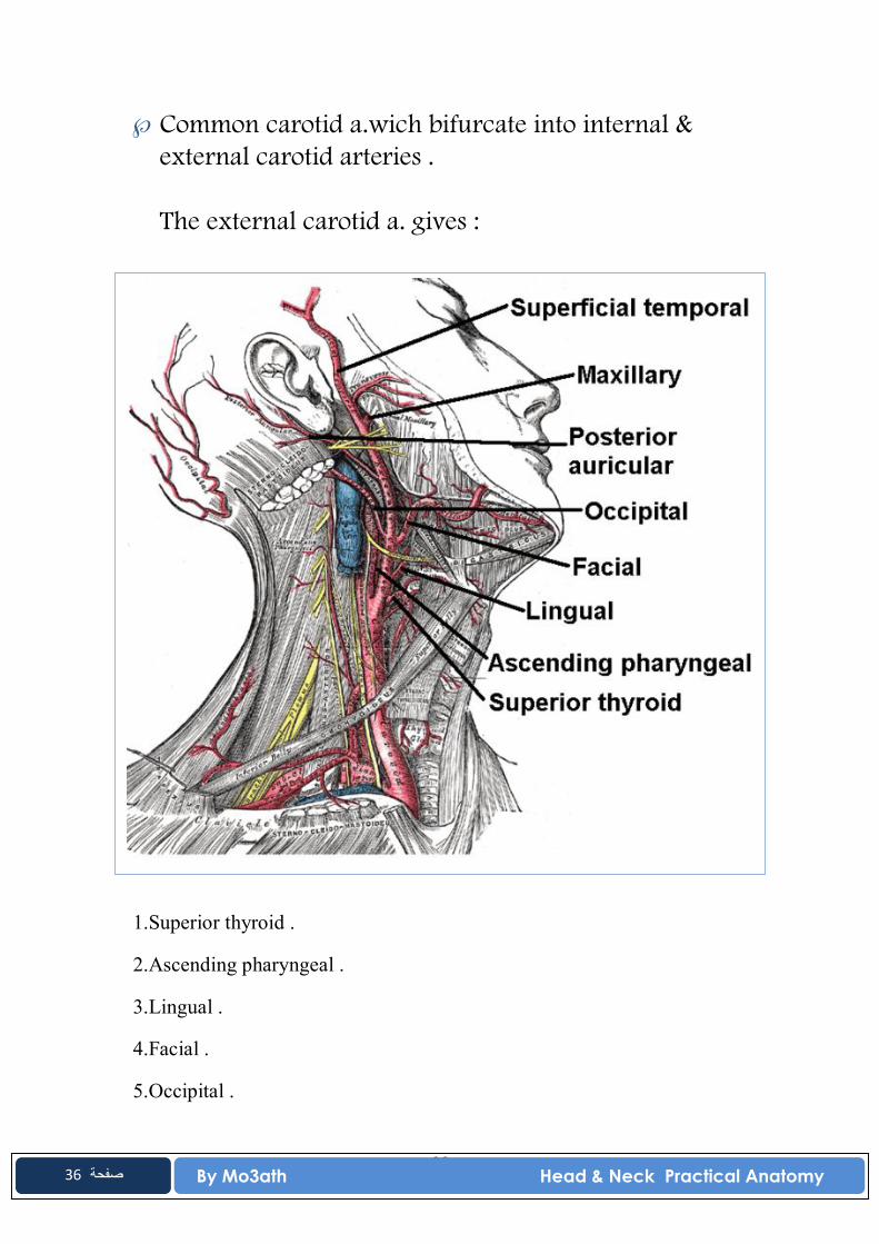
Common carotid a.wich bifurcate into internal & external carotid arteries . The external carotid a. gives :
1.Superior thyroid .
2.Ascending pharyngeal .
3.Lingual .
4.Facial .
5.Occipital .
37 By Mo3ath Head & Neck Practical Anatomy
37 صفحة
6.Posterior auricular .
7.Superficial temporal .
8.Maxillary .
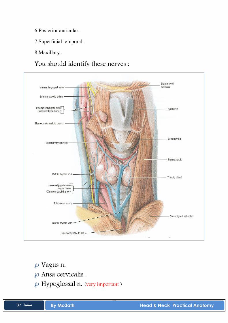
You should identify these nerves :
Vagus n. Ansa cervicalis . Hypoglossal n. (very important )
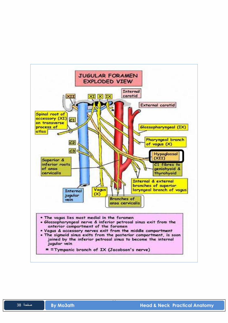
38 By Mo3ath Head & Neck Practical Anatomy
38 صفحة
39 By Mo3ath Head & Neck Practical Anatomy
39 صفحة
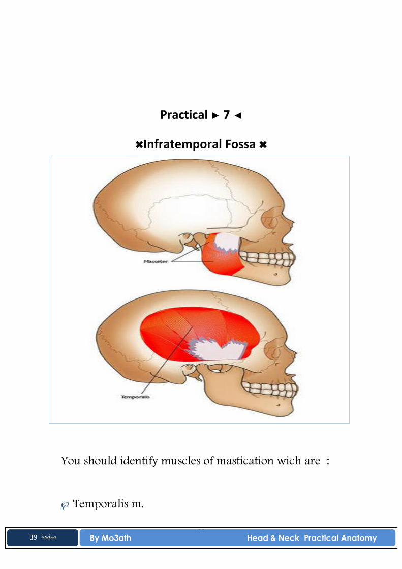
Practical ► 7 ◄
✖Infratemporal Fossa ✖
You should identify muscles of mastication wich are :
Temporalis m.
40 By Mo3ath Head & Neck Practical Anatomy
40 صفحة
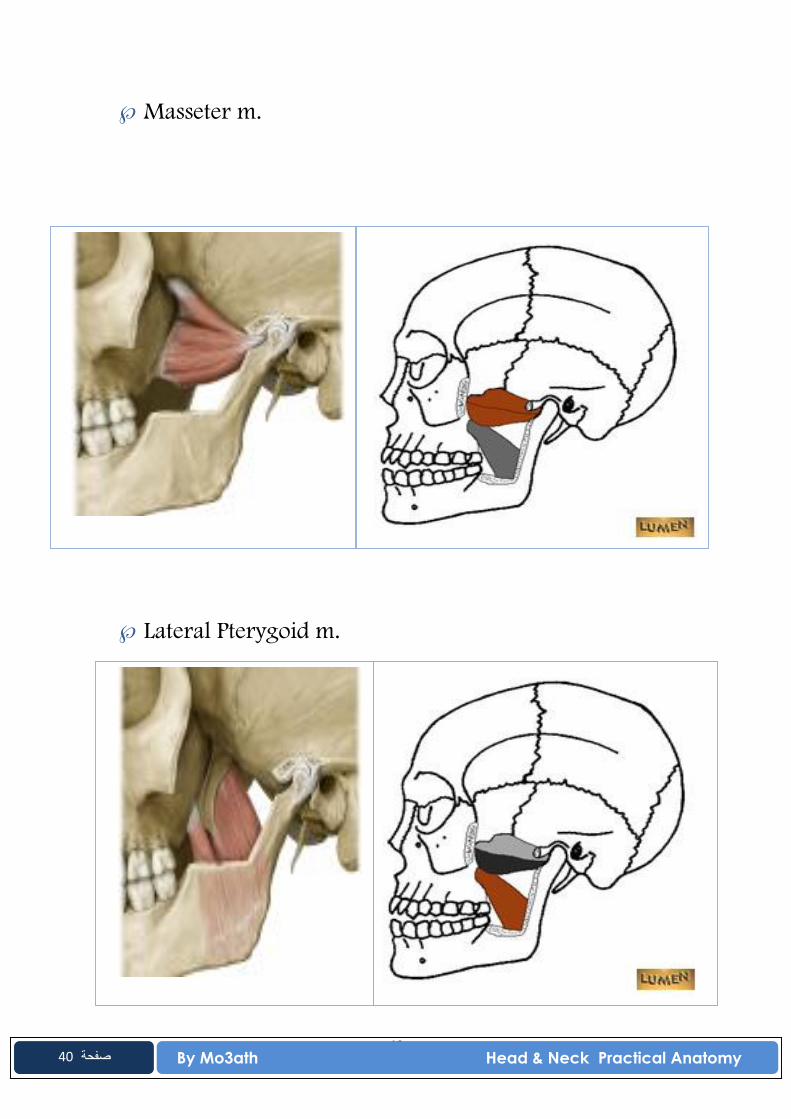
Masseter m.
Lateral Pterygoid m.
41 By Mo3ath Head & Neck Practical Anatomy
41 صفحة
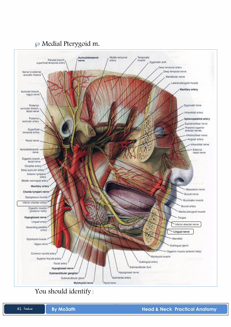
Medial Pterygoid m.
You should identify :
42 By Mo3ath Head & Neck Practical Anatomy
42 صفحة
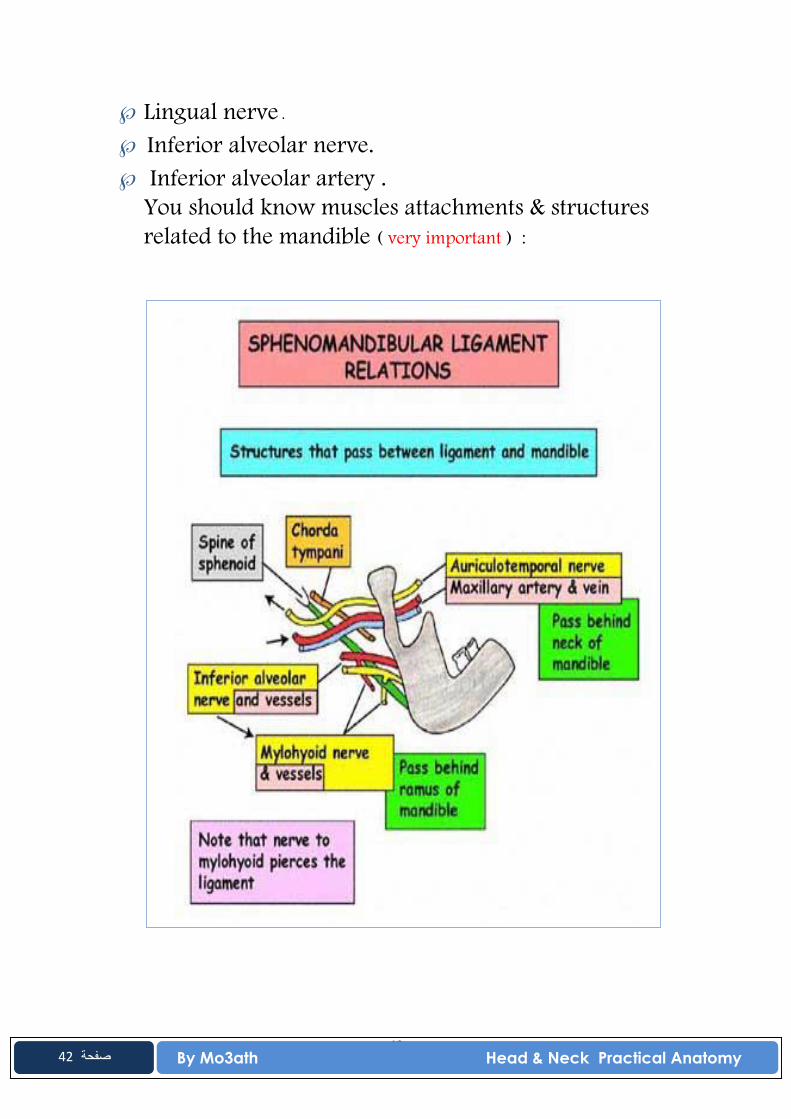
Lingual nerve . Inferior alveolar nerve. Inferior alveolar artery .
You should know muscles attachments & structures related to the mandible ( very important ) :
43 By Mo3ath Head & Neck Practical Anatomy
43 صفحة