Bogus Visual Feedback Alters Onset of Movement-Evoked Pain in People With Neck Pain
20
For Review Only Bogus visual feedback alters movement-evoked pain onset in people with neck pain Journal: Psychological Science Manuscript ID: PSCI-14-1100.R1 Manuscript Type: Research article Date Submitted by the Author: 30-Sep-2014 Complete List of Authors: Harvie, Daniel; The University of South Australia, The Sansom Institute for Health Research Broecker, Markus; The University of South Australia, The Wearable Computer Lab Smith, Ross; The University of South Australia, The Wearable Computer Lab Meulders, Ann; University of Leuven, Research Group on Health Psychology, Department of Psychology Madden, Victoria; The University of South Australia, The Sansom Institute for Health Research Moseley, G; The University of South Australia, The Sansom Institute for Health Research; Neuroscience Research Australia, ; PainAdelaide, Keywords: Perception, Virtual Reality, Threat, Motion Perception Manuscript under review for Psychological Science
Transcript of Bogus Visual Feedback Alters Onset of Movement-Evoked Pain in People With Neck Pain

For Review O
nly
Bogus visual feedback alters movement-evoked pain onset
in people with neck pain
Journal: Psychological Science
Manuscript ID: PSCI-14-1100.R1
Manuscript Type: Research article
Date Submitted by the Author: 30-Sep-2014
Complete List of Authors: Harvie, Daniel; The University of South Australia, The Sansom Institute for Health Research Broecker, Markus; The University of South Australia, The Wearable Computer Lab Smith, Ross; The University of South Australia, The Wearable Computer Lab
Meulders, Ann; University of Leuven, Research Group on Health Psychology, Department of Psychology Madden, Victoria; The University of South Australia, The Sansom Institute for Health Research Moseley, G; The University of South Australia, The Sansom Institute for Health Research; Neuroscience Research Australia, ; PainAdelaide,
Keywords: Perception, Virtual Reality, Threat, Motion Perception
Manuscript under review for Psychological Science

For Review O
nly
Running head: VISUAL FEEDBACK ALTERS MOVEMENT-EVOKED PAIN
1
Bogus visual feedback alters movement-evoked pain onset in people with neck pain
Daniel S. Harvie, Markus Broecker, Ross T. Smith, Ann Meulders, Victoria J. Madden, G.
Lorimer Moseley
University of South Australia, Australia
Author Note
Daniel S. Harvie, Victoria J. Madden of the Samson Institute for Health Research, University
of South Australia, and PainAdelaide, Adelaide, Australia; Markus Broeker, Ross Smith of
The Wearable Computer Lab, University of South Australia, Australia; Ann Meulders of the
Research Group on Health Psychology, Department of Psychology, University of Leuven,
Belgium and Center for Excellence on Generalization Research in Health and
Psychopathology, University of Leuven, Belgium; G. Lorimer Moseley of the Samson
Institute for Health Research, University of South Australia, PainAdelaide, and Neuroscience
Research Australia, Australia.
G. Lorimer Moseley is supported by an NHMRC Principal Research Fellowship ID
1061279. This study supported by NHMRC Grant ID 1047317. Ann Meulders is supported
by a postdoctoral research grant of the Research Foundation-Flanders, Belgium (FWO-
Vlaanderen) Grant ID 12E3714N and an EFIC-Grünenthal research grant E-G-G ID
169518451. The authors wish to thank Julie Peacock, Jonathon Schubert, William Kuang,
Ellie Magarey (Marion Physiotherapy), Di Wilson, Peter Roberts, Eva Boesch and Toby
Moen (Roberts Physiotherapy), Jason Collins and Glen Kocher (Northcare Physiotherapy) for
assistance with recruitment and Anthony Ikiosoglous for assistance with blinded data
extraction.
Page 1 of 19 Manuscript under review for Psychological Science
123456789101112131415161718192021222324252627282930313233343536373839404142434445464748495051525354555657585960

For Review O
nly
Running head: VISUAL FEEDBACK ALTERS MOVEMENT-EVOKED PAIN
2
Correspondence concerning this article can be addressed to Lorimer Moseley, University of South Australia, GPO Box 2471, Adelaide, South Australia 5001.
E-mail: [email protected]
Abstract
Pain is a protective perceptual response shaped by contextual, psychological and sensory
inputs that suggest danger to the body. Sensory cues suggesting that a body part is moving
towards a painful position, may credibly signal danger and thereby modulate pain. In this
experiment, we used virtual reality to investigate whether manipulating visual-proprioceptive
cues could alter movement-evoked pain, in 24 people with neck pain. We hypothesised that
pain would occur at a lesser degree of head rotation when visual feedback overstated true
rotation, and at a greater degree of rotation when visual feedback understated true rotation.
Our hypothesis was clearly supported: when vision overstated the amount of rotation, pain
occurred at 7% less rotation, and when vision understated rotation, pain occurred at 6% more
rotation. We conclude that visual-proprioceptive information modulates movement-evoked
pain threshold. This suggests that stimuli that become associated with pain, can themselves
trigger pain.
Keywords: pain, perception, virtual reality, redirected walking, illusions, body
representation, movement, multisensory processing.
Page 2 of 19Manuscript under review for Psychological Science
123456789101112131415161718192021222324252627282930313233343536373839404142434445464748495051525354555657585960

For Review O
nly
Running head: VISUAL FEEDBACK ALTERS MOVEMENT-EVOKED PAIN
3
Bogus visual feedback alters movement-evoked pain onset in people with neck pain
Introduction
Over the past three decades, the multidimensional nature of pain and nociception has been
elucidated by research revealing that many factors, from sensory (Moseley & Arntz, 2007),
cognitive (Wiech, Ploner, & Tracey, 2008; Brooks & Tracey 2005), and emotional (Wiech &
Tracey, 2009; Brooks & Tracey 2005) domains, modulate pain. Critically, non-nociceptive
somatosensory information can both modulate and evoke pain (Acerra & Moseley, 2005;
Derbyshire, 2004; Arntz & Claassens, 2004), suggesting that pain is an ‘information-evoked’
response. The sensory cues capable of contributing to pain have not been well explored,
although some authors suggest that any information that leads the brain to conclude that the
body is in danger may evoke pain (Merksey & Bogduk, 1994; Moseley, 2003; Price, 1999;
Arntz & Claassens, 2004).
This suggestion is not surprising, given what is known about other perceptual
domains, where sensory elements combine into meaningful wholes. When perceiving a table,
for example, we are not aware of individual colours, edges and shapes, but rather the unified
whole (Goldstein, 2013; Hochstein & Ahissar, 2002; Weiten, 2007). There is ample
psychological research investigating the principles underlying this sensory integration,
particularly with respect to vision (Wagemans et al., 2012), but such principles have received
scant attention in the study of pain.
Pain serves a protective function, so it is natural to think that non-nociceptive sensory
information might help to determine whether pain is an appropriate perceptual response. For
example, nociceptive input from a small laceration may evoke pain only after visual
Page 3 of 19 Manuscript under review for Psychological Science
123456789101112131415161718192021222324252627282930313233343536373839404142434445464748495051525354555657585960

For Review O
nly
Running head: VISUAL FEEDBACK ALTERS MOVEMENT-EVOKED PAIN
4
information is added. Experimentally, non-nociceptive cues contingently paired with
nociceptive input (through classical conditioning) modulate the pain evoked by subsequent
nociceptive stimulation (Atlas, Bolger, Lindquist, & Wager, 2010; Keltner et al., 2006;
Koyama, McHaffie, Laurienti, & Coghill, 2005). It is assumed that, after such pairing, the
non-nociceptive cues become signals of body-related threat and thus join the suite of
information used by the brain to determine whether pain is an appropriate protective
perceptual response. This view is supported by experimental evidence showing that the pain
evoked by a nociceptive stimulus is affected by the meaning of both the nociceptive stimulus
and other non-nociceptive stimuli that are presented at the same time (Arntz & Claassens,
2004; Moseley & Arntz, 2007).
Very few studies have investigated the relationship between non-nociceptive
information and clinical pain. One study showed that people with complex regional pain
syndrome (CRPS) experienced pain when given visual input (via a mirror) suggesting touch,
despite the absence of actual touch (Acerra & Moseley, 2005). However, this procedure did
not evoke pain in a group with non-CRPS neuropathic hand pain (Krämer, Seddigh, Moseley,
& Birklein, 2008). One might therefore suggest that non-nociceptive cues are only important
in certain conditions. But if pain is ‘information-evoked’ — as we contend that it is — then a
threshold of relevant information, nociceptive and/or non-nociceptive, must be reached in
order to evoke pain. This idea is untested, but if relevant non-nociceptive input does affect
the amount of additional input required to evoke pain, it would effectively constitute a change
in pain threshold. One type of input that might have such influence is proprioceptive
information about specific movements and body positions. This is especially likely when a
vulnerable body part needs to be protected. In the instance of a neck injury, for example,
specific proprioceptive information might predict nociceptive stimulation and thus contribute
to defensive responses including pain.
Page 4 of 19Manuscript under review for Psychological Science
123456789101112131415161718192021222324252627282930313233343536373839404142434445464748495051525354555657585960

For Review O
nly
Running head: VISUAL FEEDBACK ALTERS MOVEMENT-EVOKED PAIN
5
In the present study, we tested the hypothesis that proprioceptive information might
contribute to pain by examining how altering visual-proprioceptive feedback during neck
rotation affects the range of motion to the onset of pain (movement-evoked pain threshold) in
longstanding neck pain sufferers. Using the ‘information-based’ view of pain, we
hypothesised that pain would occur earlier when visual-proprioceptive information overstated
real-world rotation, and later when visual-proprioceptive information understated real-world
rotation.
Methods
Participants
Twenty four individuals (6 males; mean age ± SD = 45 ± 15 years) volunteered to
participate in this study. Sample size was determined a priori to enable detection of a small-
medium effect size (f = 0.2) with 80% power, conservatively assuming a 60% correlation
among repeated measures. The average duration of complaint was 11 years (SD = 11; range:
2 months to 45 years) and the participant’s physiotherapists described their pain conditions as
resulting primarily from posture/tension/repeated strain (n=9), whiplash (n=7), degeneration
(n=5), trauma (n=2) and scoliosis (n=1). Participants were mildly to moderately disabled
(Neck Pain Disability Index score ± SD = 29% ± 13%). Participants were recruited through
local physiotherapy clinics and were reimbursed AU$20 for their participation. Participants
were excluded if they had pain-free neck rotation, were unable to tolerate repeated rotation to
the first onset of pain, had severely impaired vision, were under the age of 18 or if their
physiotherapist had identified significant neurological impairments such as sensory or motor
deficits and easily provoked, constant or progressive upper limb dysaesthesia. The protocol
Page 5 of 19 Manuscript under review for Psychological Science
123456789101112131415161718192021222324252627282930313233343536373839404142434445464748495051525354555657585960

For Review O
nly
Running head: VISUAL FEEDBACK ALTERS MOVEMENT-EVOKED PAIN
6
was approved by the Human Research Ethics Committee of the University of Suth Australia
(Protocol number: 31537).
Stimulus material and apparatus
A VR technique known as ‘re-directed walking’ modulates visual-proprioceptive
feedback by tracking real-world movement and then feeding this back into the virtual
environment in under- or over-stated form, creating the illusion of more or less movement
than is actually happening. Within certain limits, participants remain unaware of the
manipulation (Steinicke, Bruder, Jerald, Frenz, & Lappe, 2008). An Oculus Rift VR Head
Mounted Display (HMD) designed for immersive VR environments was used. The HMD
shown in Figure 1 displayed a virtual world and recorded head movement using internal
gyroscopes. Customised software was used to apply the selected rotation gains and map each
of six scenes to the virtual template. The six scenes included four outdoor scenes (a park, a
mountain, a countryside and church grounds) and two indoor scenes (a dining room and a
living room).
Page 6 of 19Manuscript under review for Psychological Science
123456789101112131415161718192021222324252627282930313233343536373839404142434445464748495051525354555657585960

For Review O
nly
Running head: VISUAL FEEDBACK ALTERS MOVEMENT-EVOKED PAIN
7
Fig 1. Set-up of the VR experiment showing a supportive chair with trunk fixation,
headphones, head mounted display and the 360o (cylindrical) virtual template which
accommodated the six virtual scenes.
Experimental design
The study had a within-subject, randomised, double-blinded, repeated-measures
design. The range of neck rotation to the first onset of pain was quantified in three conditions
where virtual rotation was (a) 20% less than (rotation gain = 0.8), (b) equal to (rotation gain =
1) or (c) 20% greater than (rotation gain = 1.2) actual physical rotation. The order of the
three conditions was counterbalanced among participants, creating six possible orders. Four
participants were assigned to each of these counterbalancing orders according to a pre-
randomised order. Participants were blinded to the experimental manipulation and study
purpose, and the files relating to the three VR conditions were coded, thereby blinding the
experimenter to the order of conditions. The measurement of neck rotation was automated
and data were only extracted after all the data had been collected (A.I.).
Measurement
Axial neck rotation to the onset of pain was measured in degrees. Because
participants stopped at the first onset of pain in each trial, the range of motion to onset of pain
was defined as the peak rotation for each trial. This measure was extracted from each
automated trial output by a blinded assessor (A.I.).
Protocol
In order to prevent changes in postural alignment and to isolate neck movement,
subjects were seated in a supportive chair with the torso fixed in place by a seatbelt at the
level of the shoulders (see Figure 1). A laser pointer was fixed to the HMD and used to mark
Page 7 of 19 Manuscript under review for Psychological Science
123456789101112131415161718192021222324252627282930313233343536373839404142434445464748495051525354555657585960

For Review O
nly
Running head: VISUAL FEEDBACK ALTERS MOVEMENT-EVOKED PAIN
8
the starting position on the wall as a physical reference point for ‘zeroing’ of the gyroscopes
between measures and conditions. Participants wore white noise-emitting headphones to
counter any incidental noise, which might inform head orientation and disrupt the illusion.
For each of the three conditions, participants were asked to rotate slowly to the left,
stopping at the very first onset of pain, and then to return to the centre. Once the head had
returned to centre, the next trial was loaded and the task repeated to the right. Each condition
consisted of six left-rotation and six right-rotation measures. After each condition,
participants were asked to rate the average pain intensity experienced, for each rotation
direction to allow for subsequent assessment of any overall differences in pain intensity
between conditions. A 0-10 numerical rating scale (NRS) was used with anchors 0 = no pain
and 10 = the worst imaginable pain. To minimise the possibility that subjects would become
aware of the different rotation settings and thus directly compare them, each condition was
separated by a three-minute interval. In addition, six changing VR scenes acted as a decoy
from the actual study purpose and reduced the risk of participants’ anchoring their rotation to
a previous visual cue within a VR scene. In order to assess blinding, participants were asked
on completion of the experiment if they noticed anything different between the three
conditions.
Manipulation check 1: Setting boundaries for altered visual-proprioceptive feedback
VR operates by tracking real-world changes in orientation and applying this to the
virtual world being experienced via the HMD. The ‘rotation gain’ (the factor by which real
rotation is translated to virtual rotation) can be manipulated such that virtual and physical
rotation differ. In order to blind participants to this manipulation, the upper and lower limits
of the rotation gain were based on the results of a pilot study. During this pilot study, an
independent cohort of nine healthy participants (7 males; Mage= 32 years, SDage= 12) were
Page 8 of 19Manuscript under review for Psychological Science
123456789101112131415161718192021222324252627282930313233343536373839404142434445464748495051525354555657585960

For Review O
nly
Running head: VISUAL FEEDBACK ALTERS MOVEMENT-EVOKED PAIN
9
presented with a range of rotation gain settings and asked to rotate their heads. They were to
indicate when a difference between real and virtual rotation occurred by judging the observed
rotation as slower than, equal to, or faster than their true physical rotation. We aimed to
determine the rotation gain at which participants were more likely to judge the virtual and
real rotation to be equal than they were to judge them to be different. As shown in Figure 2,
the rotation gains that corresponded to these points were 0.72 and 1.18. As a result, our
experimental gain settings were chosen to fall between 0.8 and 1.2, and additional controls
were implemented to ensure that participants in the main study remained blinded.
Fig 2. The percentage of rotation trials rated as slow, equal or fast for each rotation gain
setting. * indicates the range of rotation gain settings where virtual and real-world movement
speeds were perceived as being equal more often than faster or slower.
Manipulation check 2: Reliability and validity of measurement
The reliability of the gyroscopic measurement of rotation was tested by attaching the
HMD to a mechanical (goniometric) arm and testing its ability to repeatedly and accurately
0
10
20
30
40
50
60
70
80
0.6 0.7 0.8 0.9 1.0 1.1 1.2 1.3
Slow
Equal
Fast
Rotation gain
% o
f ro
tati
on
s ra
ted
slo
w,
eq
ua
l a
nd
fa
st
*
Page 9 of 19 Manuscript under review for Psychological Science
123456789101112131415161718192021222324252627282930313233343536373839404142434445464748495051525354555657585960

For Review O
nly
Running head: VISUAL FEEDBACK ALTERS MOVEMENT-EVOKED PAIN
10
measure three set angles (20o, 40o, and 60o). Initial observation of repeated measures
revealed that while the measurement was highly precise over a small number of trials, it
gradually drifted with more trials. Therefore, a protocol was developed which required the
virtual compass to be ‘zeroed’ every 5 trials. To further prevent accumulation of error, the
programme was refreshed between conditions. This protocol was found to have a correlation
with actual rotation of r = .994, and a high degree of precision for repeated measures at
20o(M = 19.8o, SD = 0.8o), 40o (M = 39.6o, SD = 0.3o) and 60o (M = 59.1o, SD = 2.5o).
Data extraction and statistical analysis overview
In order to test the main hypothesis that visual information that overstates or
understates true rotation can affect movement-evoked pain, we compared the range of motion
to first onset of pain between the three conditions, using repeated-measures ANOVA with
Bonferroni-corrected pairwise comparisons. In order to account for between-subject
differences in range of motion, data for each participant were transformed to ‘a proportion of
the average range of motion demonstrated in the neutral condition’. Alpha was set at p <
0.05 and the effect sizes partial eta squared (���) and Cohen’s d were interpreted with respect
to Cohen’s guidelines (0.01 = small, 0.059 = medium and 0.138 = large; and 0.2 = small, 0.5
= medium, 0.8 = large respectively) (Cohen, 1988). As a manipulation check, each
participant’s average movement-evoked pain for each condition was normalised to a
proportion of their average across conditions. Normalised pain ratings were then compared
between conditions using repeated-measures ANOVA.
Page 10 of 19Manuscript under review for Psychological Science
123456789101112131415161718192021222324252627282930313233343536373839404142434445464748495051525354555657585960

For Review O
nly
Running head: VISUAL FEEDBACK ALTERS MOVEMENT-EVOKED PAIN
11
Results
Primary outcome: Range of motion to pain onset
The repeated-measures ANOVA revealed a large overall effect of visual-
proprioceptive feedback (Condition) on pain-free range, F(2, 135) = 18.35, p < .001, ��� =
0.214. All pairwise comparisons were significant (p < .014). When vision understated true
rotation, there was a medium effect toward increased range of motion to the first onset of
pain, p = .014, d = 0.65; and when vision overstated true rotation, there was a large effect,
this time, toward decreased range of motion to the first onset of pain, p = .005, d = 0.83. That
is, the visual feedback was able to delay the onset of pain by 6% (CI 2-11%) or advance it by
7% (CI 4-11%), showing an overall manipulation of 13%.
Feedback suggests
less movement
Feedback matches
movement
Feedback suggests
more movement
Me
an
ra
ng
e o
f m
oti
on
to
pa
in o
nse
t
(95
% C
I)
0.90
0.95
1.00
1.05
1.10
Page 11 of 19 Manuscript under review for Psychological Science
123456789101112131415161718192021222324252627282930313233343536373839404142434445464748495051525354555657585960
For Review O
nly
Running head: VISUAL FEEDBACK ALTERS MOVEMENT-EVOKED PAIN
12
Fig. 4. Mean (circle) and 95% confidence interval (error bars) for the range of motion to
first onset of pain presented as a proportion of the mean range of rotation for the neutral
condition.
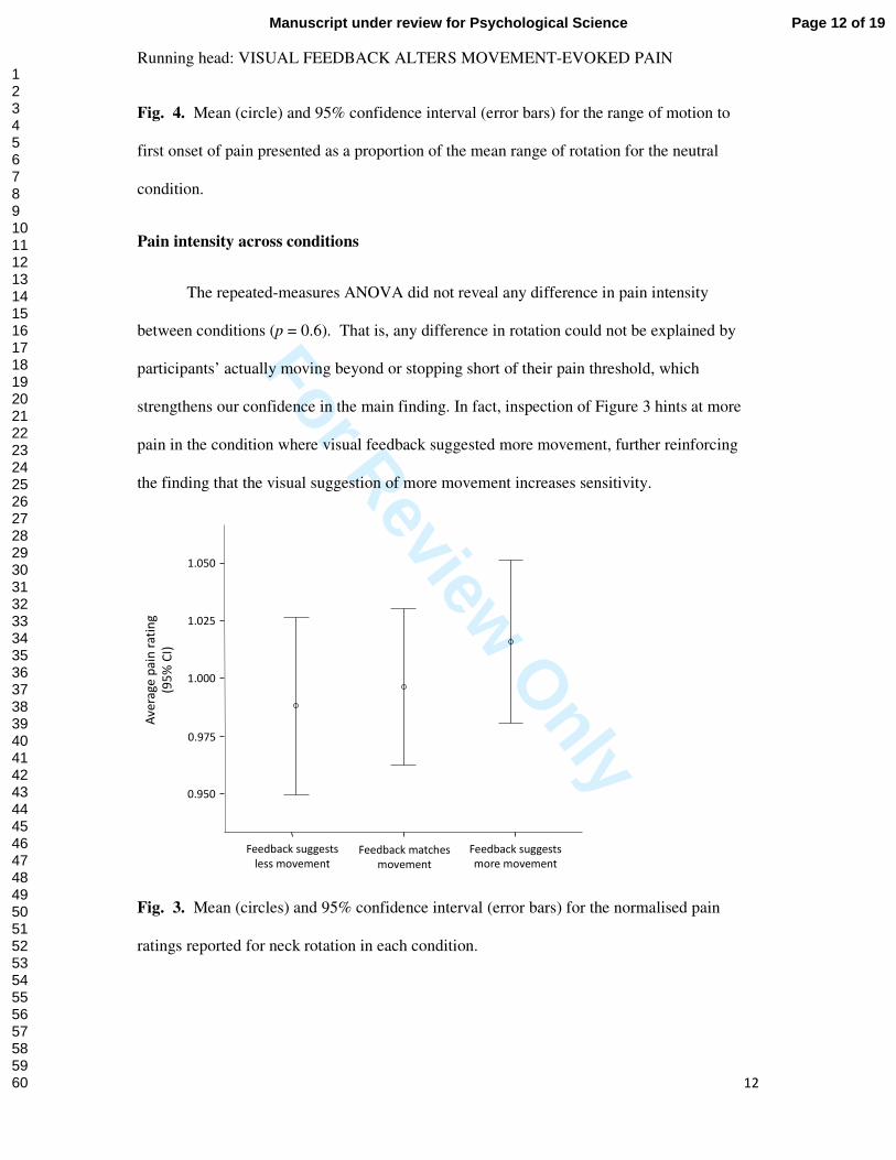
Pain intensity across conditions
The repeated-measures ANOVA did not reveal any difference in pain intensity
between conditions (p = 0.6). That is, any difference in rotation could not be explained by
participants’ actually moving beyond or stopping short of their pain threshold, which
strengthens our confidence in the main finding. In fact, inspection of Figure 3 hints at more
pain in the condition where visual feedback suggested more movement, further reinforcing
the finding that the visual suggestion of more movement increases sensitivity.
Fig. 3. Mean (circles) and 95% confidence interval (error bars) for the normalised pain
ratings reported for neck rotation in each condition.
Feedback suggests
less movement
Feedback matches
movement
Feedback suggests
more movement
Av
era
ge
pa
in r
ati
ng
(95
% C
I)
0.950
0.975
1.000
1.025
1.050
Page 12 of 19Manuscript under review for Psychological Science
123456789101112131415161718192021222324252627282930313233343536373839404142434445464748495051525354555657585960

For Review O
nly
Running head: VISUAL FEEDBACK ALTERS MOVEMENT-EVOKED PAIN
13
Discussion
We examined how altering visual-proprioceptive feedback during neck rotation affects the
range of motion to the onset of pain (the movement-evoked pain threshold) in longstanding
neck pain sufferers. Using the ‘information-based’ view of pain, we hypothesised that pain
would occur at a lesser degree of head rotation when visual-proprioceptive information
overstated true rotation, and at a greater degree when visual-proprioceptive information
understated true rotation. The hypothesis was clearly supported – visual feedback that
overstated neck rotation resulted in pain onset occurring at a lesser degree of movement
(reduced pain threshold), whereas visual feedback that understated neck rotation resulted in
pain onset occurring at a greater degree of movement (increased pain threshold). The finding
that visual-proprioceptive cues may contribute to a pain-evoking sensory suite is particularly
relevant because chronic pain is most commonly provoked by particular movements and
body positions.
Our results appear consistent with the view of pain as the perceptual result of the
brain’s inference that body tissue is in danger (Merksey & Bogduk, 1994; Moseley, 2003;
Price, 1999; Arntz & Claassens, 2004) and corroborate related evidence of the relationship
between experienced pain intensity and cues that imply threat to tissue (Arntz & Claassens,
2004; Atlas & Wagner 2012; Moseley & Arntz, 2007; Wiech et al., 2010). For example, a
noxious cold stimulus evokes more pain if it is accompanied by a red light than if it is
accompanied by a blue one (Moseley & Arntz, 2007); a noxious laser stimulus evokes more
pain, and different cortical activation, if it is delivered to an area of skin thought by the
participant to be ‘thinner than normal’ than if it is delivered to skin thought to be normal
(Wiech et al., 2010). Such examples offer compelling evidence that pain can be modulated if
there is credible evidence of tissue danger, even if that evidence is not from the nociceptive
Page 13 of 19 Manuscript under review for Psychological Science
123456789101112131415161718192021222324252627282930313233343536373839404142434445464748495051525354555657585960

For Review O
nly
Running head: VISUAL FEEDBACK ALTERS MOVEMENT-EVOKED PAIN
14
domain. The current results offer a new direction however, because they show a shift in pain
threshold, rather than an upregulation of pain. This is important because most examples of
amplification of pain can be explained by enhanced senstivity within the nociceptive system
– most notably termed ‘central sensitisation’. We contend that our results, of a reduced pain
threshold to movement, are very unlikely due to central sensitisation because that would be
more likely to manifest in the opposite result – greater pain increase in response to greater
magnitude of movement. We contend that the most obvious explanation for the current
results is associative learning. That is, neck rotation involves a suite of motor, visual and
proprioceptive processes that, for the person with neck pain, becomes associated with
nociceptive input, such that the non-nociceptive aspects of the sensorial suite are sufficient to
trigger pain with or without the nociceptive component.
That the effect clearly relies on visual input triggering some sort of ‘threat threshold’,
does not exclude the possibility that non-cortical mechanisms are involved. Most obvious
here is the descending modulatory system, whereby primarily brainstem nuclei exert both
inhibitory and facilitatory influences over dorsal horn neurones (see Woolf & Slater, 2006 for
review). According to modern models of pain, however, the evaluative processes that
subserve descending modulation are grounded in those that subserve the production of pain
itself. That is, pain can be considered to reflect the perceived need to protect body tissue;
descending modulation can be considered a ‘correction’ of spinal nociceptor activity (i.e. the
‘danger message’) so as to bring it into line with the brain’s evaluation of true danger (see
Moseley, 2007; Butler, 2013; Fields, 2006). This understanding is analogous to that applied
to motor control, whereby motor commands are corrected according to somatosensory and
visual feedback (Sperry, 1950; Von Holst, 1950). Those models are also relevant here
because it is also possible that the bogus visual feedback modulates proprioceptive sensitivity
(Gandevia, Refshauge & Collins, 2002), which in turn may modulate nociceptive input.
Page 14 of 19Manuscript under review for Psychological Science
123456789101112131415161718192021222324252627282930313233343536373839404142434445464748495051525354555657585960

For Review O
nly
Running head: VISUAL FEEDBACK ALTERS MOVEMENT-EVOKED PAIN
15
Notably, the pain advancing effect of overstating rotation was greater than the pain
delaying effect of understating rotation, indicating that visual feedback had a greater ability to
advance than delay pain. This finding fits with the greater potential cost of a perceptual error
that delays the onset of pain, which is consistent with inferential perceptual models (i.e.
Bayesian) that include a cost function (Feldman, 2013; Tabor, Catley, Gandevia, Thacker, &
Moseley, 2013).
Although the current work was experimental in nature, it raises intriguing potential
clinical implications. First, our results clearly suggest a rethink of how we interpret simple
clinical tests such as movement-evoked pain. Such tests are widely held to reflect sensitivity
of tissues and nociceptive pathways – repeatable and stable movement-evoked pain
thresholds are considered to be consistent with a primary nociceptive driver of pain – the
presence of tissue pathology (Jones & Rivett, 2004). However, our results suggest that this is
a naïve perspective. As such, it is not unreasonable to suggest that a VR set-up such as that
used here might play a role in the assessment of people in pain, to identify and quantify the
role of non-nociceptive somatosensory cues in pain and impairment. Second, if cues
signalling danger amplify or indeed trigger pain, then these cues present a novel target for
therapy. One way to extinguish the effect of such cues on pain might be to experientially
dissociate them from pain. For example, the re-directed walking techniques used in this
study can provide the illusory experience of large movements, but limit real-world movement
and pain. This idea might also be relevant to the use of visual illusory movements
administered by a mirror – common interventions for chronic limb pain conditions including
phantom limb pain and complex regional pain syndrome (Moseley, 2004; Moseley, 2006;
Ezendam, Bongers & Jannik, 2009; Bowering et al., 2012; Daly & Bialocerkowski, 2009) use
mirror-therapy, but the idea of distorting the feedback as a method of disentangling non-
nociceptive movement-related cues appears to have not been considered.
Page 15 of 19 Manuscript under review for Psychological Science
123456789101112131415161718192021222324252627282930313233343536373839404142434445464748495051525354555657585960

For Review O
nly
Running head: VISUAL FEEDBACK ALTERS MOVEMENT-EVOKED PAIN
16
The relationship observed here, between potentially threatening information and
movement-evoked pain, might also provide insight as to why cognitive and behavioural
interventions, such as education and exposure, which target perceived threat/pain-related fear,
also positively alter the relationship between movement and pain (Moseley, 2004; Vlaeyen,
de Jong, Geilen, Heuts, & van Breukelen, 2002). Whilst education, for example, may aim to
convince a patient that their pain is not a direct correlate of tissue stress, demonstrating this
with real-time evidence that their pain depends on visually encoded movement not actual
movement, may have therapeutic power.
Further research using the current framework might exploit more immersive and
multisensory VR, which may enable the delivery of more convincing and multimodal illusory
evidence of danger to the body. Further studies could also investigate how visual-
proprioceptive and other cues might acquire the ability to modulate/mediate pain (i.e. through
associative learning) as well as investigate why the effect might persist or over-generalise,
and how it might be extinguished. Disentangling pain from nociception is a challenge that
has been identified in experimental and cognitive psychology research (Moseley, 2012), but
the methodology used here lays a platform from which this challenge might be taken on.
Conclusion
In people with neck pain, when visual-proprioceptive feedback overstates true neck
rotation, neck pain occurs at a lesser degree of head rotation. When visual-proprioceptive
feedback understates true neck rotation, neck pain occurs later. We conclude, then, that
visual-proprioceptive information modulates pain threshold during head rotation in people
with neck pain. This has broad implications for our view of pain as an ‘information-evoked’
response and supports further investigation of non-nociceptive contributions to long-standing
Page 16 of 19Manuscript under review for Psychological Science
123456789101112131415161718192021222324252627282930313233343536373839404142434445464748495051525354555657585960

For Review O
nly
Running head: VISUAL FEEDBACK ALTERS MOVEMENT-EVOKED PAIN
17
pain. Furthermore, the methodology outlined here presents a new method for theoretical and
experimental interrogation of pain, and raises the possibility of novel assessment and
therapeutic applications.
Author contributions
D.S.H and G.L.M. developed the study concept, contributed to design, data analysis,
interpretation and write-up. D.S.H. collected the data. V.J.M. contributed to study design and
write-up. M.B. and R.T.S contributed to study design and provided technical expertise for the
VR set-up. A.M. contributed to interpretation and write-up. All authors approved the final
version of the manuscript for submission.
References
Acerra, N. E., & Moseley, G. L. (2005). Dysynchiria: watching the mirror image of the unaffected limb elicits pain on the affected side. Neurology, 65(5), 751-753.
Arntz, A., & Claassens, L. (2004). The meaning of pain influences its experienced intensity. Pain,
109(1-2), 20-25. Atlas, L. Y., Bolger, N., Lindquist, M. A., & Wager, T. D. (2010). Brain mediators of predictive cue
effects on perceived pain. The Journal of Neuroscience, 30(39), 12964-12977. Atlas, L. Y., & Wager, T. D. (2012). How expectations shape pain. Neuroscience Letters, 520(2),
140-148. Bowering, K. J., O'Connell, N. E, Tabor, A., Catley, M. J., Leake, H. B., Moseley, G. L., & Stanton,
T. R. (2013). The effects of graded motor imagery and its components on chronic pain: a systematic review and meta-analysis. The Journal of Pain, 14(1), 3-13.
Butler, D. S., & Moseley, G. L.. (2013). Explain Pain:(Revised and Updated: Noigroup Publications. Carruthers, G. (2008). Types of body representation and the sense of embodiment. Consciousness and
cognition, 17(4), 1302-1316. Cohen, J. (1988). Statistical power analysis for the behavioral sciencies. London, UK: Routledge. Daly, A. E., & Bialocerkowski, A. E. (2009). Does evidence support physiotherapy management of
adult Complex Regional Pain Syndrome Type One? A systematic review. European Journal
of Pain, 13(4), 339-353. Ezendam, D., Bongers, R. M., & Jannink, M. J. A. (2009). Systematic review of the effectiveness of
mirror therapy in upper extremity function. Disability & Rehabilitation, 31(26), 2135-2149. Feldman, J. (2013). Bayesian models of perceptual organization. Handbook of perceptual
organization.
Page 17 of 19 Manuscript under review for Psychological Science
123456789101112131415161718192021222324252627282930313233343536373839404142434445464748495051525354555657585960

For Review O
nly
Running head: VISUAL FEEDBACK ALTERS MOVEMENT-EVOKED PAIN
18
Fields, H. L., Basbaum, A. I., & Heinricher, M. M. (2006). Wall and Melzack's Textbook of Pain. SB
McMahon and M Koltzenburg. Wall and Melzack's Textbook of Pain. UK: Elsevier Churchill
Livingstone, 125-130. Gandevia, S. C., Refshauge, K. M., & Collins, D. F. (2002). Proprioception: peripheral inputs and
perceptual interactions Sensorimotor Control of Movement and Posture (pp. 61-68): Springer. Goldstein, E B. (2014). Sensation and perception (6th ed.). Belmont, CA: Wadsworth-Cengage
Learning. Hochstein, S., & Ahissar, M. (2002). View from the top: Hierarchies and reverse hierarchies in the
visual system. Neuron, 36(5), 791-804. Jones, M. A., & Rivett, D. A. (2004). Chapter 1 - Introduction to clinical reasoning. In M. A. J. A.
Rivett (Ed.), Clinical Reasoning for Manual Therapists (pp. 3-24). Oxford: Butterworth-Heinemann.
Keltner, J. R., Furst, A., Fan, C. R., Rick, I. B., & Fields, H. L. (2006). Isolating the modulatory effect of expectation on pain transmission: a functional magnetic resonance imaging study. The
Journal of neuroscience, 26(16), 4437-4443. Koyama, T., McHaffie, J. G., Laurienti, P. J., & Coghill, R. C. (2005). The subjective experience of
pain: where expectations become reality. Proceedings of the National Academy of Sciences of
the United States of America, 102(36), 12950-12955. Krämer, H. H., Seddigh, S., Moseley, G. L., & Birklein, F. (2008). Dysynchiria is not a common
feature of neuropathic pain. European Journal of Pain, 12(1), 128-131. Merksey, H., & Bogduk, N. (1994). Classification of chronic pain. Seattle: International Association
for the Study of Pain Press, 194, 210. Moseley, G. L. (2007). Reconceptualising pain according to its underlying biology. Phys. Ther. Rev,
12, 169-178. Moseley, G. L. (2006). Graded motor imagery for pathologic pain A randomized controlled trial.
Neurology, 67(12), 2129-2134. Moseley, G. L., Gallace, A., & Spence, C. (2012). Bodily illusions in health and disease:
physiological and clinical perspectives and the concept of a cortical ‘body matrix’. Neuroscience & Biobehavioral Reviews, 36(1), 34-46.
Moseley, G. L. (2004). Graded motor imagery is effective for long-standing complex regional pain syndrome: a randomised controlled trial. Pain, 108(1), 192-198.
Moseley, G. L. (2007). Reconceptualising pain according to its underlying biology. Phys. Ther. Rev,
12, 169-178. Moseley, G. L. (2004). Evidence for a direct relationship between cognitive and physical change
during an education intervention in people with chronic low back pain. European Journal of
Pain, 8(1), 39-45. Moseley, G. L., & Arntz, A. (2007). The context of a noxious stimulus affects the pain it evokes.
Pain, 133(1), 64-71. Moseley, G. L., Gallace, A., & Spence, C. (2012). Bodily illusions in health and disease:
Physiological and clinical perspectives and the concept of a cortical ‘body matrix’. Neuroscience & Biobehavioral Reviews, 36(1), 34-46.
Moseley, G. L. (2003). A pain neuromatrix approach to patients with chronic pain. Manual therapy,
8(3), 130-140. Mountcastle, V. B. (2005). The sensory hand: neural mechanisms of somatic sensation. Cambridge,
MA: Harvard University Press. Price, Donald D. (1999). Psychological mechanisms of pain and analgesia. Seattle, WA: IASP press. Ro, T., Wallace, R., Hagedorn, J., Farnè, A., & Pienkos, E. (2004). Visual enhancing of tactile
perception in the posterior parietal cortex. Journal of Cognitive Neuroscience, 16(1), 24-30. Steinicke, F., Bruder, G., Jerald, J., Frenz, H., & Lappe, M. (2008). Analyses of human sensitivity to
redirected walking. Paper presented at the Proceedings of the 2008 ACM symposium on Virtual reality software and technology.
Sperry, R. W. (1950). Neural basis of the spontaneous optokinetic response produced by visual inversion. Journal of comparative and physiological psychology, 43(6), 482.
Page 18 of 19Manuscript under review for Psychological Science
123456789101112131415161718192021222324252627282930313233343536373839404142434445464748495051525354555657585960

For Review O
nly
Running head: VISUAL FEEDBACK ALTERS MOVEMENT-EVOKED PAIN
19
Tabor, A., Catley, M. J., Gandevia, S., Thacker, M. A., & Moseley, G. L. (2013). Perceptual bias in pain: a switch looks closer when it will relieve pain than when it won’t. Pain.
Tracey, Irene. (2010). Getting the pain you expect: mechanisms of placebo, nocebo and reappraisal effects in humans. Nat Med, 16(11), 1277-1283.
Vlaeyen, J. W.S., de Jong, J., Geilen, M., Heuts, P. H. T. G., & van Breukelen, G. (2002). The treatment of fear of movement/(re) injury in chronic low back pain: further evidence on the effectiveness of exposure in vivo. The Clinical journal of pain, 18(4), 251-261.
Von Holst, E. (1950). Die Arbeitsweise des Statolithen-apparates bei Fischen. Zeitschrift für
vergleichende Physiologie, 32(1-2), 60-120. Vos, T., Flaxman, A. D., Naghavi, M., Lozano, R., Michaud, C., Ezzati, M., . . . Aboyans, V. (2013).
Years lived with disability (YLDs) for 1160 sequelae of 289 diseases and injuries 1990–2010: a systematic analysis for the Global Burden of Disease Study 2010. The Lancet, 380(9859), 2163-2196.
Wagemans, J., Elder, J. H., Kubovy, M., Palmer, S. E., Peterson, M. A., Singh, M., & von der Heydt, R. (2012). A century of Gestalt psychology in visual perception: I. Perceptual grouping and figure–ground organization. Psychological bulletin, 138(6), 1172.
Weiten, W. (2007). Psychology: Themes and Variations: Themes And Variations: Cengage Learning. Wiech, K., Lin, Chia-shu, B., Kay H., Bingel, U., Ploner, M., & Tracey, I. (2010). Anterior insula
integrates information about salience into perceptual decisions about pain. The Journal of
Neuroscience, 30(48), 16324-16331. Wiech, K., Ploner, M., & Tracey, I. (2008). Neurocognitive aspects of pain perception. Trends in
cognitive sciences, 12(8), 306-313. Wiech, K., & Tracey, I. (2009). The influence of negative emotions on pain: behavioral effects and
neural mechanisms. Neuroimage, 47(3), 987-994. Woolf, C. J., & Salter, M. W. (2006). Plasticity and pain: role of the dorsal horn. Wall and Melzack's
textbook of pain, 5, 91-105. Yolton, J. W. (1996). Perception & reality: A history from Descartes to Kant: Cornell University
Press Ithaca.
Page 19 of 19 Manuscript under review for Psychological Science
123456789101112131415161718192021222324252627282930313233343536373839404142434445464748495051525354555657585960