Theoretical Investigation of Stresses and Displacement in RC ...
Microbiology (1 998), 144,3335-3342 Printed in Crreiit Britain
The transcriptional regulator gene phrR in Sinorhizobiurn rneliloti WSM419 is regulated by low pH and other stresses
Wayne G. Reeve, Ravi P. Tiwari, Cheryl M. Wong, Michael J. Dilworth and Andrew R. Glennt
Author for correspondence: Michael J. Dilworth. Tel: +61 8 9360 2112. Fax: +61 8 9360 6486. e-mail : [email protected]
Centre for Rhizobium Studies, Division of Science, Murdoch University, Murdoch, WA 61 50, Australia
The phrR gene in Sinorhizobium meliloti (previously known as Rhizobium meliloti) WSM419, directly downstream from actA, is induced by low pH or certain stresses (e.g. high concentrations of Zn2+, Cu2+, H202 or ethanol), but not in stationary phase or by other stresses (e.g. phosphate limitation, elevated temperature, high concentrations of sucrose or iron). A DNA fragment containing the wild-type phrR gene could not be cloned and inverse PCR was therefore used to amplify a 3-5 kb BamHl fragment containing phrR from the mutant 5. meliloti TG2-6 (actA::TnS). DNA fragments from a BamHI/Sall digest of the amplified product were cloned into pUK2l and sequenced. The phrR open reading frame contiguous to actA appears to code for a 15.2 kDa protein showing significant identity with the proteins encoded by y4wC and y4aM in Rhizobium sp. NGR234. Al l three proteins resemble transcriptional regulators in containing a DNA-binding helix-turn-helix motif similar to that reported for URF4 in Rhodospirillum rubrum and repressors in col i phage.
~ Keywords : acidity, stress, acid-inducible gene, regulator, DNA-binding domain
INTRODUCTION
The survival or growth of bacteria can be adversely affected by low p H and cells need to adapt to a changing environment to survive and be competitive. An under- standing of the response of bacteria to low p H is particularly relevant to interactions between bacteria and their hosts. Bacteria of medical importance must survive harsh acidic environments such as those en- countered in the stomach or macrophage phagolyso- somes (Mekalanos, 1992 ; Foster, 1995) while agri- culturally important bacteria like root nodule bacteria must first survive in acidic soils before they can successfully invade legume roots (Munns, 1986). The relatively tight regulation of the cytoplasmic pH (Booth, 1985) suggests that there are mechanisms allowing cells to combat proton influx. The mechanisms allowing cells
t Present address: Vice Chancellery, University of Tasmania, Hobart, Tasmania, Australia.
Abbreviations: ATR, acid-tolerance response; ONP, o-nitrophenol ; pNP, p-nitrophenol.
The GenBank accession number for the sequence of the phrR gene is L13845.
to survive and grow at low p H are not yet defined, although a number of processes have been proposed to be involved for the enterics, including cytoplasmic buffering (Booth, 1985; Krulwich et al., 1985), DNA repair (Foster, 1995), ion cycling (Booth, 1985), pH amelioration (Huang et al., 1986; Slonczewski et al., 1987; Stim & Bennet, 1993) and proton translocation (Shibata et al., 1992; White et al., 1992). Among the stress-inducible genetic systems is the ‘ acid habituation’ or ‘ acid-tolerance response ’ (ATR) identi- fied in Escherichia coli (Goodson & Rowbury, 1989) and Salmonella typhimurium (Foster, 1991), and more recently found in Aeromonas hydrophila (Karem et al., 1994), Listeria monocytogenes (Farber & Pagatto, 1992) and various species of root nodule bacteria (O’Hara & Glenn, 1994). The ATR is an important response that enables cells exposed to mildly acid p H to cope more effectively with subsequent more severe low-pH con- ditions than cells previously grown at neutral pH. In the enterobacteria, exposure to mildly acidic conditions induces the synthesis of 51 acid-shock proteins (ASPS) (Lee et al., 1995) and provides cross-protection to heat, osmotic and oxidative challenge (Leyer & Johnson, 1993 ; Lee et al., 1995).
0002-2740 0 1998 SGM 3335
W. G. R E E V E a n d O T H E R S
Agriculture in Western Australia is heavily dependent upon the saprophytic competence of root nodule bac- teria, the persistence of which between seasons is very much dependent on the pH of the soil and its rate of acidification. Production from medic-based pastures is markedly decreased in soils of low pH. The acid sensitivity of the (Sino) rhixobium-Medicago symbiosis results from the inability of many of the standard rhizobial inoculants to survive and grow in acid soils. The discovery of natural variants from the south Mediterranean which perform well in such moderately acid soils has allowed stable medic pastures to be developed on more than 1 million ha of soils that were previously unable to sustain them. The isolation of the superior Mediterranean strains raises questions as to why these strains are more acid tolerant in field conditions than others and about how such strains cope with acid stress.
Several genes involved in acid tolerance in Sino- rhizobium meliloti (previously known as Rhixobium meliloti) WSM419 have been identified by Tn5 muta- genesis (O’Hara et al., 1989; Tiwari et al., 1992). The process by which this strain senses and/or responds to low pH appears to involve a signal transduction system (Acts, a histidine protein kinase ‘sensor’, and ActR, its cognate regulator) ; insertional inactivation of either component results in an acid-sensitive phenotype (Tiwari et al., 1996b). It is anticipated that signal processing via Acts-ActR will result in altered gene expression in response to low pH. Similarly, the presence of an intact actA gene, which may code for a lipid acyl transferase, has also been shown to be essential (Tiwari et al., 1996a). Studies using l ac2 fusions have revealed that actA (Tiwari et al., 1996a), actR and acts (Tiwari et al., 1996b) are constitutively expressed. By analogy to the enteric organisms, the existence of an ATR system in root nodule bacteria (O’Hara & Glenn, 1994) suggests that synthesis of new proteins should be induced under acidic conditions.
This paper reports the first finding of a pH-regulated gene (designated phrR, for pH regulated) in S. meliloti WSM419. This gene, located downstream from the actA gene, encodes a putative repressor protein PhrR and is induced by low pH and other environmental stresses.
METHODS
Bacterial strains, plasmids and media. Bacterial strains and plasmids are described in Table 1. Strains of Sinorhizobium were grown at 28 “C in JMM minimal medium (O’Hara et al., 1989). Escherichia coli strains were grown in Luria-Bertani (LB) medium at 37°C. Media were supplemented with the following concentrations of antibiotics (pg ml-l) : ampicillin (loo), chloramphenicol (20), gentamicin (40), kanamycin (50), or tetracycline (20).
DNA preparation and manipulation. The plasmid DNA isolation, transformation and manipulation techniques used have been described earlier (Sambrook et al., 1989; Inoue et al., 1990). Restriction and modification enzymes were pur- chased from either Life Technology or Boehringer Mannheim.
Genomic DNA was isolated from Sinorbizobiurn cultures as described earlier (Reeve et al., 1997). DNA sequencing and analysis. The techniques used have been described earlier (Tiwari et al., 1996a, b). Inverse PCR. Genomic DNA from strain TG2-6 was restriction digested using BamHI and the mixture of fragments religated at low concentration (1 pg ml-l). The ligated mixture was ethanol precipitated and the DNA dissolved to give a concentration of 10 pg ml-’. The circular DNA molecules generated by ligation were linearized by HilzdIII. Two primers, one in the phrR region (PHR-187 primer; 5’ CGC AGA TAC TCA ATG TGC C 3’) and another from the Tn5 sequence (TN5-2770 primer ; 5’ AGG TCA CTG CGT GGA TGG AG 37, were used to amplify a 3.5 kb fragment using the method of Rich & Willis (1990). The amplified DNA was run on a 1 ‘/o gel and the 3.5 kb PCR product was purified using Agarase (Boehringer Mannheim). Reconstruction of phrR. The BamHI fragment of pRT206-6 (the clone containing the reconstructed ac tA; Tiwari et al., 1996a) was cloned into the BamHI site of pUK21 to construct pRT206-18. Plasmid pRT206- 16, containing the SalI clone from the PCR product of TG2-6 DNA, was partially cut by SalI, blunted and ligated to a BarnHI-blunted digest of QSm interposon to construct pRT206-23. The SalI-KpnI fragment from pRT206-23 was cloned into the SalIIKpnI sites of pRT206-18 to construct pRT206-24. This plasmid contains a reconstructed wild-type phrR gene that was verified by DNA sequence analysis. To facilitate mobilization of this plasmid into Sinorbizobiurn, the BamHI fragment of pRT206-24 was cloned into the BglII site of the broad-host-range vector pMP220 (Spaink et al., 1987) to construct pRT206-25.
Inactivation of phrR. T o facilitate marker exchange, the plasmid antibiotic-resistance marker of pRT206-24 was changed from kanamycin to ampicillin by subcloning the BamHI fragment of pRT206-24 into pGEM7Zf( - ) to con- struct pRT206-24G. The QKm resistance interposon was cloned into the ClaI site in the 0.7 kb EcoRI-SalI fragment to construct pRTES1. pRT206-24G was cut with SalI, blunted and then the DNA cleaved with EcoRI and ligated with pRTES1, which was cut with HindIII, blunted and then cut by EcoRI. The new clone pRT206-27 had the interposon at the tenth codon of phrR.
pRT206-24G was cut by SalI, blunted and ligated with HindIII-blunted nKm interposon to construct pRT206-28, placing the interposon at codon 126 of phrR. A BamHI fragment of either pRT206-27 or pRT206-28 was cloned into the BglII site of pMP220 to construct pRT206-27M and pRT206-28M, respectively. The latter plasmids were mobilized into WSM419 using pRK2013 as a helper. The incompatible plasmid pPHl JI (Beringer et al., 1978) was mobilized into tetracycline- and kanamycin-resistant clones and transconjugants were selected on gentamicin and kana- mycin. Genomic DNA was extracted from tetracycline- sensitive isolates and hybridized with DIG-labelled pESl to confirm that marker exchange had occurred.
Nodulation tests. Cultures of S. meliloti WSM419 or RTlO were inoculated onto germinated seedlings of Medicago murex or M . sativa as described by Reeve et al. (1997). Nodules isolated from 4-week-old plants were sterilized, crushed in sterile saline and the suspension replica patched onto solid medium in the presence and absence of kanamycin. Reporter gene fusions. Reporter gene fusions were con- structed in wide-host-range IncPl plasmids containing a
3336
Low-pH-regulated gene phrR
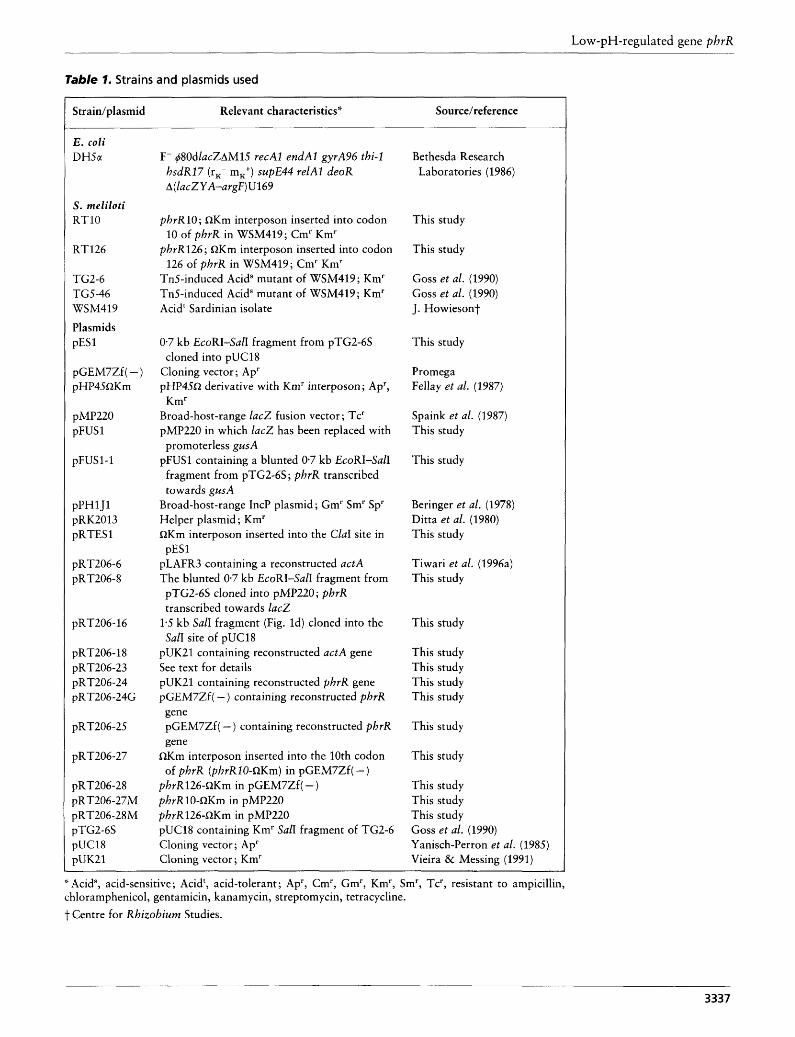
Table 1. Strains and plasmids used
Strain/ plasmid Relevant characteristics* Source/reference
E. coli DH5u
S . meliloti RTlO
RT126
TG2-6 TGS-46 WSM419 Plasmids pES 1
pGEM7Zf( - ) pHP45QKm
pMP220 pFUS 1
pFUS1-1
pPHl J1 pRK2013 pRTESl
pRT206-6 pRT206-8
pRT206-16
pRT206-18 pRT206-23 pRT206-24 pR T206-24G
pRT206-25
pRT206-27
pRT206-28 pRT206-27M pRT206-28M pTG2-6S pUC18 pUK21
F- @OdlacZAMlS recAl endAl gyrA96 thi-1 hsdR17 (rx- mK+) supE44 relAl deoR A( lacZYA-argF)U169
phrR10; QKm interposon inserted into codon
phrR126; QKm interposon inserted into codon
Tn5-induced Acids mutant of WSM419; Km' Tn5-induced Acids mutant of WSM419 ; Km' Acidt Sardinian isolate
10 of phrR in WSM419; Cm' Km'
126 of phrR in WSM419; Cm' Km'
0.7 kb EcoRI-SalI fragment from pTG2-6S cloned into pUC18
Cloning vector ; Ap' pHP45R derivative with Km' interposon ; Ap',
Broad-host-range lacZ fusion vector; Tc' pMP220 in which lacZ has been replaced with
promoterless gusA pFUSl containing a blunted 0.7 kb EcoRI-SalI
fragment from pTG2-6S ; phrR transcribed towards gusA
Broad-host-range IncP plasmid; Gm' Sm' Sp' Helper plasmid; Km' QKm interposon inserted into the ClaI site in
pLAFR3 containing a reconstructed actA The blunted 0.7 kb EcoRI-SalI fragment from
Km'
pESl
pTG2-6S cloned into pMP220; phrR transcribed towards lac2
1.5 kb SalI fragment (Fig. Id) cloned into the SalI site of pUC18
pUK21 containing reconstructed actA gene See text for details pUK21 containing reconstructed phrR gene pGEM7Zf( - ) containing reconstructed phrR gene pGEM7Zf ( - ) containing reconstructed phrR gene
of phrR (phrR10-QKm) in pGEM7Zf( - ) QKm interposon inserted into the 10th codon
phrR126-QKm in pGEM7Zf( - ) phvR10-QKm in pMP220 phrR126-QKm in pMP220 pUC18 containing Km' SalI fragment of TG2-6 Cloning vector ; Ap' Cloning vector ; Km'
Bethesda Research Laboratories (1986)
This study
This study
Goss et al. (1990) Goss et al. (1990) J. Howiesont
This study
Promega Fellay et al. (1987)
Spaink et al. (1987) This study
This study
Beringer et al. (1978) Ditta et al. (1980) This study
Tiwari et al. (1996a) This study
This study
This study This study This study This study
This study
This study
This study This study This study Goss et al. (1990) Yanisch-Perron et al. (1985) Vieira & Messing (1991)
'- Acid", acid-sensitive; Acidt, acid-tolerant ; Ap', Cm', Gm', Km', Sm', Tc', resistant to ampicillin, chloramphenicol, gentamicin, kanamycin, streptomycin, tetracycline. t Centre for Rhizobium Studies.
3337
W. G. R E E V E a n d OTHERS
promoterless lac2 (pMP220) or gusA (pFUS1). The 0.7 kb EcoRI-SafI fragment from S. meliloti TG2-6 (containing the phrR promoter) was cloned from pTG2-6S (Goss et al., 1990) as an EcoRI fragment into the EcoRI site of pMP220 (Spaink et al., 1987) or pFUSl to construct pRT206-8 or pFUS1-1, respectively. Expression studies. Exponential cultures, grown in JMM broth at pH 7.0 containing chloramphenicol (for WSM419) or tetracycline (for WSM419[pFUSl, pFUS1-1, pMP220 or pRT206-24]), were centrifuged (3024 g, 5 min ; Beckman Avanti J25I centrifuge) and the cell pellet resuspended in normal saline (0*89'/0, w/v, NaCl). An aliquot of cells was used to inoculate flasks containing JMM at a range of pH values (5*8-7.0), and JMM at pH 5.8 or 7.0 with or without addition of 20-100 pM CuSO,, 0.8 '/o (v/v) ethanol, 0.1- 100 pM FeCI,, 200-800 pM H,O,, 0-300 pM potassium phos- phate, 12.5% (w/v) sucrose and 10-40pM ZnSO,. The cultures were then incubated at 28 "C (unless otherwise indicated) on a gyratory shaker set at 250 r.p.m. and harvested at an OD,,, between 0.2 and 0.5. Stationary-phase cells that had grown to a constant density were harvested after S d incubation in JMM at pH 7.0. For the time-course experiment, cells grown in JMM at pH 7.0 were centrifuged and resuspended in JMM at pH 5.8 to provide an initial OD,,, of 0.3.
Enzyme assays. Exponential-phase cultures (OD,,, 0.2-0.5) were centrifuged and resuspended into cold normal saline to an OD,,, of approximately 0.6. Three replicates per strain were performed. Protein concentration was measured using a Bio-Rad protein assay kit. P-Gaiactosidase activity. /I-Galactosidase was quantified as de- scribed by Miller (1972) and the specific activity was expressed as nmol o-nitrophenol (ONP) produced min-' (mg protein)-' at 28 "C. P-Glucuronidase activity. A SO-200 pl aliquot of cells was mixed with 790 p1 buffer (SO mM sodium phosphate, 50 mM DTT and 1 mM EDTA; pH 7) and saline was added if required to produce a final volume of 990 pl. For a blank, 200 p1 saline was used instead of cells. One drop of toluene was added to each tube, the mixture vortexed and the tubes then incubated at 37 "C for 30 min with the lids off to remove toluene. Tubes were equilibrated at 28 "C for 5 min before the assay was started. A 10 p1 aliquot of p-nitrophenyl /I-D-glucuronide (pNPG; 35 mg ml-l) was added to start the reaction. T o terminate the reaction, a 200 pl aliquot from each tube was removed and added to 700 plO.46 M Na,CO, at three different time points. These tubes were centrifuged for 1 min at room temperature to pellet cell debris. The absorbance of the supernatant was read at 405 nm. p-Glucuronidase specific activity was expressed as nmol p-nitrophenol (pNP) produced min-' (mg protein)-' at 28 "C.
RESULTS AND DISCUSSION
Studies on the actA region
Previous studies of the actA gene in S. meliloti WSM419 showed that it was essential for acid tolerance and constitutively expressed (Tiwari et al., 1996a). However, when a 0.7kb EcoRI-SalI fragment containing an incomplete ORF downstream from actA (Tiwari et al., 1996a) was cloned into the broad-host-range vector pMP220, a fivefold induction of this gene occurred in cells grown at pH 5.8 relative to pH 7.0 (see Table 2). It was therefore termed a phr (pH regulated) gene.
Further studies on this low-pH-regulated gene were hindered initially by our inability to clone the complete gene. The DNA region containing actA was extensively restriction mapped (Fig. l a ) using information derived from Southern hybridization studies (Tiwari et al., 1992), and clones isolated from either TG2-6 (Goss et al., 1990), or WSM419 (Tiwari et al., 1996a). The restriction map located a BamHI site 4.5 kb from the site of Tn5 insertion in TG2-6 (Fig. l a ) . Attempts to clone the 6.3 kb BamHI fragment (containing actA and the phr gene) from strain WSM419 using a subgenomic library were unsuccessful (Tiwari et al., 1992), suggesting that a gene located on this fragment may be toxic to E. coli. A 7.5 kb BamHI fragment from TG2-6 (Fig. l a ) containing the phr gene could not be cloned into either pUC18 or pBR322 using the kanamycin-resistance marker of TnS as a positive selection for incorporation of the insert. DNA downstream of actA could not be identified from a library of WSM419, which is known to overrepresent other genes (Tiwari et al., 1996a). These studies, together with the finding that actA could be recon- structed (Tiwari et al., 1996a), narrow the toxic region to within a 4.2 kb EcoRI-BamHI fragment (Fig. l a ) .
Cloning of the phr gene
An inverse-PCR strategy was used to amplify a DNA fragment containing the phr gene (Fig. l b ) from TG2-6 genomic DNA using PHR-187 and TN5-2770 primers. The 3-5 kb PCR product was subsequently purified from an agarose gel and used directly for sequencing (Fig. Ic). Restriction digestion with SalI showed three sites in this fragment (Fig. Id) and the two SalI fragments were cloned into the SalI site of pUC18. Sequencing of the PCR product using PHR-187 primer and sequencing of the two SalI clones consolidated the order of SalI fragments in this 3.5 kb fragment. Restriction mapping of the 1.5 kb SalI fragment identified two SphI sites (Fig. Id), which were used to obtain double-stranded DNA sequence of the phr gene.
Nucleotide sequence data
Analysis of 988 bp of DNA sequence derived from an EcoRI-SphI DNA fragment revealed an open reading frame (phrR) that started 140 bases downstream of the stop codon of actA (Tiwari et al., 1996a). The region upstream of the start codon of phrR contains a putative RBS. The gene phrR encodes a predicted 15.2 kDa protein product containing 139 amino acids. The complete nucleotide and annotated protein sequence is available from GenBank (accession number L1384S).
A database search with PhrR detected 50.7% identity over a 138 amino acid overlap with the hypothetical 16-5 kDa Y4wC protein of Rhizobium sp. NGR234. Another match of 49.6 % identity over a 131 amino acid overlap occurred with the hypothetical Y4aM protein from the same organism. Although the latter two proteins are encoded by genes located on the symbiotic megaplasmid, the phrR locus is positioned on the
3338

Low-pH-regulated gene phrR
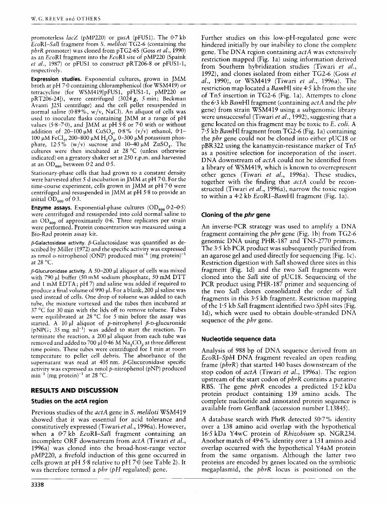
I _J
TG2-6 genomic DNA
Bn S I
The 3.5 kb PCR product was used for sequencing and cloning.
* Cut by BurnHI
B
Fig. 1. Inverse-PCR strategy to reconstruct the phrR gene. The actA gene region (a) of 5. meliloti TG2-6 (Tiwari et a/., 1996a) was digested with BamHl (b), ligated and cut by HindIII (c). The 3-5 kb DNA fragment (d) was amplified, restriction mapped and used for sequencing. The 1.0 and 1.5 kb Sall fragments were cloned into pGEM7Zf( -) to construct pRT206-15 and pRT206-16, respectively. Restriction sites are as follows : B, BamHI; Bg, Bglll; E, EcoRl; Ev, EcoRV; H, Hindlll; S, Sall; Sm, Smal; Sp, Sphl. The Sphl sites have been partially restriction mapped and therefore marked with an asterisk (*).
DNA binding domain
Fig. 2. Alignment of PhrR with three other DNA-binding proteins as compared earlier by Falk & Wagner (1978). The underlined region corresponds to the DNA-binding domain. Sm, Sinorhizobium meliloti WSM419; Rr, Rhodospirillurn rubrum; 434, bacteriophage 434; P22, bacteriophage P22.
chromosome of S. meliloti WSM419 (Tiwari et al., 1992, 1996a). The proteins encoded by y4wC and y4aM have been classified as putative transcriptional regulators (Freiberg et al., 1997) on the basis of their identity with the DNA-binding domain of a protein encoded by a gene URF4 in the ATPase region of Rhodospirillum rubrum (Falk & Walker, 1988). The PhrR protein has 535% identity in a 71 amino acid overlap which spans the DNA-binding domain of the URF4 protein. The PhrR and URF4 DNA-binding domains are similar to those reported for the phage 434 Cro and P22 C2 repressor proteins (Fig. 2). The phr gene thus appears to code for a repressor protein that binds to DNA. For this reason this gene has been termed phrR for pH-regulated Regulator.
Inactivation of phrR
The phrR gene was inactivated by inserting the lnKm interposon at codon 10 or codon 126. The two mutants (RT10 and RT126) created by the inactivation of phrR
were as acid tolerant as the parental strain WSM419, suggesting that, in contrast to actA, the phrR gene is not essential for growth at low pH. The phrR gene is not required for successful invasion of Medicago since the mutant RTlO produced normal nodules on both Medicago murex and M. satiua. Kanamycin-resistant cells were recovered from the nodules of plants inocu- lated with RT10. No nodules were found on uninocu- lated seedlings.
Expression of phrR
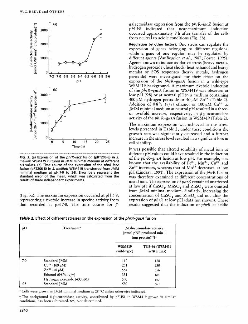
The phrR-lac2 fusion (pRT206-8) or the phrR-gusA fusion (pFUS1-1) was mobilized into S. meliloti WSM419 to study factors controlling the expression of phrR.
Regulation by pH. The phrR-lac2 fusion in S. meliloti WSM419 was expressed at low levels [approx. 500 nmol min-l (mg protein)-'] between pH 7-0 and pH 6-2, but increased significantly as the pH fell to 5-8
3339
W. G. R E E V E a n d OTHERS
3000
2000
1000
0 0 7.2 7.0 6.8 6.6 6.4 6.2 6.0 5.8 5.6
PH
Y ~ o ~ , l I I 1 l 0 5 10 15 20 25
Time (h)
Fig. 3. (a) Expression of the phrR-lacZ fusion (pRT206-8) in 5. meliloti WSM419 cultured in JMM minimal medium at different pH values. (b) Time course of the expression of the phrR-lacZ fusion (pRT206-8) in 5. rneliloti WSM419 transferred from JMM minimal medium a t pH 7.0 to 5.8. Error bars represent the standard error of the mean, which was calculated from the results of three independent experiments.
(Fig. 3a). The maximum expression occurred at pH 5.8, representing a fivefold increase in specific activity from that recorded at pH7-0. The time course for p-
galactosidase expression from the phrR-lacZ fusion at pH 5-8 indicated that near-maximum induction occurred approximately 8 h after transfer of the cells from neutral to acidic conditions (Fig. 3b). Regulation by other factors. One stress can regulate the expression of genes belonging to different regulons, while a gene of one regulon may be regulated by different agents (VanBogelen et al., 1987; Foster, 1995). Agents known to induce oxidative stress (heavy metals, hydrogen peroxide), heat shock (heat, ethanol and heavy metals) or SOS responses (heavy metals, hydrogen peroxide) were investigated for their effect on the expression of the phrR-gusA fusion in a wild-type WSM419 background. A maximum fivefold induction of the phrR-gusA fusion in WSM419 was observed at low pH (5.8) or at neutral pH in a medium containing 400 pM hydrogen peroxide or 40 pM Zn2+ (Table 2 ) . Addition of 0.8% (v/v) ethanol or 100 pM Cu2+ to JMM minimal medium at neutral pH resulted in a three- or twofold increase, respectively, in @-glucuronidase activity of the phrR-gusA fusion in WSM419 (Table 2). The maximum expression was achieved at the stress levels presented in Table 2; under these conditions the growth rate was significantly decreased and a further increase in the stress level resulted in a significant loss in cell viability. It was possible that altered solubility of metal ions at different pH values could have resulted in the induction of the phrR-gusA fusion at low pH. For example, it is known that the availability of Fe3+, Mn2+, Cu2+ and Zn2+ increases, whereas that of Mo2+ decreases, at low pH (Lindsay, 1991). The expression of the phrR fusion was therefore examined at different concentrations of metal ions. The expression of phrR remained unaffected at low pH if CuSO,, MnSO, and ZnSO, were omitted from JMM minimal medium. Similarly, increasing the concentration of CuSO, and ZnSO, did not alter the expression of phrR at low pH (data not shown). These results suggested that the induction of phrR at acidic
Table 2. Effect of different stresses on the expression of the phrR-gusA fusion
PH Treatment* B-Glucuronidase activity [nmol pNP produced min-'
(mg protein)-']t
WSM419 TG5-46 (WSM419 (wild-type) actR : : Tn5)
7-0 Standard JMM Cu2+ (100 pM) Zn2+ (40 pM) Ethanol (0.8 '/o, V/V)
Hydrogen peroxide (400 pM) 5-8 Standard JMM
110 25 1 554 33 1 590 580
128 230 556 ND
ND 561
'' Cells were grown in JMM minimal medium at 28 "C unless otherwise indicated. t The background p-glucuronidase activity, contributed by pFUSl in WSM419 grown in similar conditions, has been subtracted. ND, Not determined.
3340
Low-pH-regulated gene pbrR
values was not due to altered solubility of these metal ions in the range of concentrations tested. Furthermore, induction at low pH does not appear to be associated with an increase in Fe3+ solubility because no effect was observed on the expression of the phrR-gusA fusion when the concentration of iron was elevated from 1 to 100 pM in neutral-pH medium.
The increase in p-glucuronidase activity of the phrR- gusA fusion by certain stresses is not the result of an increase in the mean generation time of the cells per se, since no significant change in P-glucuronidase activity was observed in stationary-phase cells or in exponential- phase cells exposed to phosphate depletion (3 pM potassium phosphate), a high sucrose concentration (12*So/o, w/v) or elevated temperature (38 "C). It is unusual that phrR is up-regulated in response to ethanol but not heat. In contrast, 1.5 proteins of E. coli were up- regulated not only in response to heat but also to ethanol (VanBogelen et al., 1987). The DNA sequence that includes the phrR promoter does not contain the consensus heat-shock promoter sequence [ - 35 TCN- CCCTTGAA (13-15 bp) CCCCATTTA - 10 region] ; this is consistent with the lack of up-regulation of phrR by heat.
The significant increase in P-glucuronidase activity by low pH and certain other environmental factors suggests the involvement of a regulatory system controlling expression of phrR. One possible regulatory circuit could involve the ActS/R two-component signal trans- duction system (Tiwari et al., 1996b) in controlling the expression of phrR in response to pH. Expression of the latter gene was therefore examined in both the wild-type (WSM419) and an actR (regulator) mutant (TG5-46). Since there was no significant difference in phrR expression between an actR and wild-type background, it appears that the regulatory system for phrR expression does not require actR.
These data show that, in addition to the genes like actA, acts and actR that are absolutely essential for growth of S. meliloti at low pH, there is another class of genes which, while not essential for growth, appear to be induced by exposure to low pH. In the case of the phrR gene, other environmental stresses also cause some degree of induction. Regulons responsive to a particular environmental stress can be coordinately expressed by a common regulatory gene (Gottesman, 1984). Expression of phrR is up-regulated by exposure of cells to different stresses. PhrR is a putative repressor that may be part of a cascade that regulates gene expression in response to different stimuli. The demonstration that acid-inducible proteins exist in this strain opens the way for further studies on the nature and role of such proteins in the response of WSM419 to acid stress.
ACKNOWLEDGEMENTS
This work was generously supported by the Australian Research Council and the Grains Research & Development Corporation.
REFERENCES
Beringer, J. E., Beynon, J. L., Buchanan-Wollaston, A. V. & Johnston, A. W. B. (1978). Transfer of the drug-resistance trans- poson Tn5 to Rhizobium. Nature 276, 633-634. Bethesda Research Laboratories (1986). BRL pUC host: E. coli DH5aTM competent cells. Focus 8, 9. Booth, 1. R. (1985). Regulation of cytoplasmic pH in bacteria. Microbiol Rev 49,359-378. Ditta, G., Stanfield, S., Corbin, D. & Helinski, D. R. (1980). Broad host range DNA cloning system for Gram-negative bacteria : construction of a gene bank of Rhizobium meliloti. Proc Natl Acad Sci USA 77,7347-7351. Falk, G. & Walker, 1. E. (1988). DNA sequence of a gene cluster coding for subunits of the F, membrane sector of ATP synthase in Rhodospirillum rubrum. Biochem 3 254, 109-122. Farber, J. M. & Pagatto, F. (1992). The effect of acid shock on the heat resistance of Listeria monocytogenes. Lett Appl Microbiol
Fellay, R., Frey, J. & Krisch, H. (1987). Interposon mutagenesis of soil and water bacteria : a family of DNA fragments designed for in vitro insertional mutagenesis of Gram-negative bacteria. Gene
Foster, 1. W. (1991). Salmonella acid shock proteins are required for the adaptive acid tolerance response. J Bacteriol 173, 6896-6902. Foster, J. W. (1995). Low pH adaptation and the acid tolerance response of Salmonella typhimurium. Crit Rev Microbiol 21,
Freiberg, C., Fellay, R., Bairoch, A., Broughton, W. J., Rosenthal, A. & Perret, X. (1997). Molecular basis of symbiosis between Rhizobium and legumes. Nature 387, 394-401. Goodson, N. Y. & Rowbury, R. 1. (1989). Habituation to normally lethal acidity by prior growth of Escherichia coli at a sub-lethal acid pH value. Lett Appl Microbiol8, 77-79. Goss, T. G., O'Hara, G. W., Dilworth, M. J. & Glenn, A. R. (1990). Cloning, characterization, and complementation of lesions caus- ing acid-sensitivity in Tn5-induced mutants of Rhizobium mefifoti WSM419. 3 Bacteriol 172, 5173-5179. Gottesman, 5. (1984). Bacterial regulation : global regulatory networks. Annu Rev Genet 18,415-441. Huang, L., Forsberg, C. W. & Gibbins, L. N. (1986). Influence of external pH and fermentation products on Clostridium aceto- butylicum intracellular pH and cellular distribution of fermen- tation products. Appl Environ Microbiol 51, 1230-1234. Inoue, H., Nojima, H. & Okayarna, H. (1990). High efficiency transformation of Escherichia coli with plasmids. Gene 96,23-28. Karem, K. L., Foster, J. W. & Bej, A. K. (1994). Adaptive acid tolerance response (ATR) in Aeromonas hydrophila. Micro- biology 140, 1731-1736. Krulwich, T. A., Angus, R., Scheier, M. & Guffanti, A. A. (1985). Buffering capacity of bacilli that grow in different pH ranges. J Bacteriol 162, 768-772. Lee, 1. S., Lin, J. S., Hall, H. K., Bearson, B. & Foster, J. W. (1995). The stationary-phase sigma factor sigma S (rpoS) is required for a sustained acid tolerance response in virulent Salmonella typhimurium. Mol Microbiol 17, 155-167. Leyer, G. 1. & Johnson, E. A. (1993). Acid adaptation induces cross-protection against environmental stresses in Salmonella typhimurium. Appl Environ Microbiol59, 1842-1847. Lindsay, W. L. (1 991). Inorganic equilibria affecting micro-
15, 197-201.
52, 147-154.
215-237.
3341
W. G. R E E V E a n d OTHERS
nutrients in soils. In Micronutrients in Agriculture, pp. 89-112. Edited by J. J. Mortvedt, F. R. Cox, L. M. Shuman & R. M. Welch. Madison: Soil Science Society of America. Mekalanos, J. 1. (1992). Environmental signals controlling ex- pression of virulence determinants in bacteria. ] Bacteriol 174,
Miller, J. H. (1 972). Experiments in Molecular Genetics. New York: Cold Spring Harbor Laboratory. Munns, D. N. (1986). Acid soil tolerance in legumes and rhizobia. In Advances in Plant Nutrition, pp. 63-91. Edited by B. Tinker & A. Lauchlis. New York : Praeger Scientific. O'Hara, G. W. & Glenn, A. R. (1994). The adaptive acid tolerance response in root nodule bacteria and Escherichia coli. Arch Microbiol 161, 286-292. O'Hara, G. W., Goss, T. J., Dilworth, M. J. & Glenn, A. R. (1989). Maintenance of intracellular p H and acid tolerance in Rhizobium meliloti. Appl Environ Microbiol 55, 1870-1876. Reeve, W. G., Tiwari, R. P., Dilworth, M. 1. & Glenn, A. R. (1997). A helicase gene (helo) in Rhizobium meliloti WSM419. FEMS Microbiol Lett 153, 43-49. Rich, 1. 1. & Willis, D. K. (1990). A single oligonucleotide can be used to rapidly isolate DNA sequences flanking a transposon Tn.5 insertion by the polymerase chain reaction. Nucleic Acids Res 18, 6673-6676. Sambrook, J., Fritsch, E. F. & Maniatis, T. (1989). Molecular Cloning: a Laboratory Manual, 2nd edn. Cold Spring Harbor, NY: Cold Spring Harbor Laboratory. Shibata, C., Ehara, T., Tomura, K., Igarashi, K. & Kobayashi, H. (1 992). Gene structure of Enterococcus hirae (Streptococcus faecalis) FIFO-ATPase, which functions as a regulator of cyto- plasmic pH. J Bacteriol 174, 6117-6124. Slonczewski, J. L., Gonzalez, T. N., Bartholomew, F. M. & Holt, N. 1. (1987). Mud-directed lacZ fusions regulated by low pH in Escherichia coli. J Bacteriol 169, 3001-3006.
1-7.
Spaink, H. P., Robert, J. H., Okker, C. A., Wijffelman, E. P. & Lugtenberg, B. J. J. (1987). Promoters in the nodulation region of the Rhizobium leguminosarum Sym plasmid pRLl JI. Plant Mol Biol 9, 27-39. Stim, K. P. & Bennett, G. N. (1993). Nucleotide sequence of the adi gene, which encodes the biodegradative acid-induced arginine decarboxylase of Escherichia coli. J Bacteriol 175, 1221-1234. Tiwari, R. P., Reeve, W. G. & Glenn, A. R. (1992). Mutations conferring acid sensitivity in the acid tolerant strains Rhizobium meliloti WSM419 and Rhizobium leguminosarum biovar viciae WSM710. FEMS Microbiol Lett 100, 107-1 12. Tiwari, R. P., Reeve, W. G., Dilworth, M. 1. &Glenn, A. R. (1996a). An essential role for actA in acid-tolerance of Rhizobium meliloti. Microbiology 142, 601-610. Tiwari, R. P., Reeve, W. G., Dilworth, M. J. & Glenn, A. R. (1996b). Acid tolerance in Rhizobium meliloti strain WSM419 involves a two-component sensor-regulator system. Microbiology 142,
VanBogelen, R. A., Kelley, P. M. & Neidhardt, F. C. (1987). Differential induction of heat shock, SOS, and oxidation stress regulons and accumulation of nucleotides in Escherichia coli. J Bacteriol 169, 26-32. Vieira, J. & Messing, J. (1991). New pUC-derived cloning vectors with different selectable markers and DNA replication origins. Gene 100, 189-194. White, S., Tuttle, F. E., Blankenhorn, D., Dosch, D. C. & Slonczewski, 1. L. (1992). pH dependence and gene structure of inaA in Escherichia coli. J Bacteriol 174, 1537-1543. Yanisch-Perron, C., Vieira, 1. & Messing, 1. (1985). Improved M13 phage cloning vectors and host strains : nucleotide sequences of the M13mp18 and pUC19 vectors. Gene 33, 103-119.
1693-1704.
Received 10 June 1998; revised 2 September 1998; accepted 4 September 1998.
3342
Copyright © 2022 FDOKUMEN