Bifunctional Ligands that Target Cells Displaying the αvβ3 Integrin
Upload
washingtonCategory
view
2download
0
The structure of a receptor with two associating transmembranedomains on the cell surface: integrin αIIbβ3
Jieqing Zhu1,2, Bing-Hao Luo2,3, Patrick Barth2,4, Jack Schonbrun4,5, David Baker4, andTimothy A. Springer1,6
1 The Immune Disease Institute and Department of Pathology, Harvard Medical School, 200 Longwood Ave,Boston, MA 02115
4 Department of Biochemistry and Howard Hughes Medical Institute, University of Washington, Seattle, WA98195
AbstractStructures of intact receptors with single-pass transmembrane (TM) domains are essential tounderstand how extracellular and cytoplasmic domains regulate association and signaling throughTM domains. A chemical and computational method to determine structures of the membrane regionsof such receptors on the cell surface is developed here and validated with glycophorin. An integrinheterodimer structure reveals association over most of the lengths of the α and β TM domains, andthat the principles governing association of hetero and homo TM dimers differ. A turn at the Gly ofthe juxtamembrane (JM) GFFKR motif caps the α TM helix, and brings the two Phe of GFFKR intothe α/β interface. A JM Lys residue in β also has an important role in the interface. The structure isincompatible with previous NMR JM/cytoplasmic complex structures, and together with NMRstructures of isolated α and β TM domains, shows how TM association/dissociation regulates integrinsignaling. A joint ectodomain and membrane structure shows that substantial flexibility between theextracellular and TM domains is compatible with TM signaling.
IntroductionMembrane proteins with a single transmembrane (TM) domain per monomer are important intransmitting signals between the extracellular and cytoplasmic environments. For example,signal transmission in receptor kinases generally involves ligand-triggered association ofmonomeric single-TM proteins into homodimers or heterodimers (Zhang et al., 2006).
Integrins, adhesion receptors that transmit signals bidirectionally across membranes, areanother heavily studied class of signaling receptors (Luo et al., 2007; Wegener and Campbell,2008). Integrins have two subunits which constitutively heterodimerize through their large,ligand-binding extracellular domain. Reversible association through the single-pass α and βsubunit TM domains mediates TM signaling. The cytoplasmic domains are short and can bindcytoskeletal proteins.
6Corresponding author: E-mail: [email protected] contribution.3Current address: Department of Biological Sciences, 202 Life Sciences Building, Louisiana State University, Baton Rouge, LA 708035Current address: Spotfire, 212 Elm Street, Somerville, MA 02144Publisher's Disclaimer: This is a PDF file of an unedited manuscript that has been accepted for publication. As a service to our customerswe are providing this early version of the manuscript. The manuscript will undergo copyediting, typesetting, and review of the resultingproof before it is published in its final citable form. Please note that during the production process errors may be discovered which couldaffect the content, and all legal disclaimers that apply to the journal pertain.
NIH Public AccessAuthor ManuscriptMol Cell. Author manuscript; available in PMC 2009 October 24.
Published in final edited form as:Mol Cell. 2009 April 24; 34(2): 234–249. doi:10.1016/j.molcel.2009.02.022.
NIH
-PA Author Manuscript
NIH
-PA Author Manuscript
NIH
-PA Author Manuscript

Structures are known in both active and inactive conformations for receptor kinase extracellularand cytoplasmic domain fragments (Zhang et al., 2006), and for integrin extracellular ligand-binding fragments (Luo et al., 2007) and cytoplasmic domains (Vinogradova et al., 2002).However, none of these structures include TM domains, and how signals are transmittedbetween these domains and across the membrane is largely unknown. In contrast, structuralwork on signaling proteins that span the membrane six or more times, such as channels and G-protein coupled receptors, is far more advanced.
Thus far, our understanding of how single-TM proteins associate in the membrane is confinedto a limited number of experimental studies on isolated TM domains. Structures of theconstitutively associating TM domains of the erythrocyte glycoprotein glycophorin A (GPA)have been solved in detergent and in pelleted vesicles (MacKenzie et al., 1997; Smith et al.,2001). A constitutively disulfide-linked TM dimer was also structurally defined in detergentby NMR (Call et al., 2006). Bicelles, made of a mixture of lipids and short-chain, detergent-like lipids, are closer mimics of bilayers than detergents. Two further dimeric TM fragmentstructures (Bocharov et al., 2008a; Bocharov et al., 2008b), and monomeric αIIb and β3 TMdomain structures (Lau et al., 2008a; Lau et al., 2008b), have recently been determined inbicelles. However, since regulated TM domain association is driven by domains outside themembrane, structures of such associated TM domain fragments beg the question of whetherthey are physiologically relevant, and if so, whether they correspond to resting or activeconformations.
The biological importance of integrins, and the tractability of studying regulated TM andcytoplasmic domain association in the absence of ligand, in contrast to most other signalingreceptors, has made them an attractive model system for studying association of their TM andcytoplasmic domains (Wegener and Campbell, 2008). In integrins, the α and β subunit TMdomains associate in the resting state, driven by the close proximity of the C-termini of α andβ subunit ectodomains in the bent conformation. TM and cytoplasmic domain separation,induced by binding of the β subunit cytoplasmic domain through talin to the actin cytoskeleton,drives integrin extension, and shifts the ligand-binding α/β headpiece to an open, high-affinityconformation (Luo et al., 2007; Wegener and Campbell, 2008; Zhu et al., 2008). Disulfidecross-linking of portions of the TM domains in the outer membrane leaflet demonstrateshelicity and the approximate orientations between the αIIb and β3 helices in the resting state,and an absence of association in the active state (Luo et al., 2004). Various TM domainorientations have been proposed based on these data or mutagenesis (Gottschalk, 2005; Li etal., 2005; Partridge et al., 2005). A comprehensive NMR complex structure showed how theintegrin cytoplasmic domains associated, and provided a model for integrin activation byshowing that the cytoskeletal protein talin dissociated the complex (Vinogradova et al.,2002). However, different α/β complex structures, or a lack of association have also beendescribed (Ulmer et al., 2001; Vinogradova et al., 2004; Weljie et al., 2002). A unique featureof integrins is a highly conserved GFFKR motif following the α subunit TM domain. Mutationof any of the residues in this motif activates integrins by destabilizing association of αand βsubunit TM domains (Hughes et al., 1996; O’Toole et al., 1994).
Disulfide cross-linking has been used to obtain structural information about membraneproteins, especially to probe TM domain organization and conformational change in bacterialchemoreceptors (Bass et al., 2007). However, development of this technique has not proceededto the level of describing three dimensional structures. Rosetta uses knowledge-based as wellas physicochemical potentials and an efficient method of sampling conformational space tofind low energy structures. Furthermore, the potentials in Rosetta can be combined withexperimental restraints (Das and Baker, 2008).
Zhu et al. Page 2
Mol Cell. Author manuscript; available in PMC 2009 October 24.
NIH
-PA Author Manuscript
NIH
-PA Author Manuscript
NIH
-PA Author Manuscript

Here, we use disulfide-based distance restraints with Rosetta Membrane (Barth et al., 2007;Barth et al., 2009) to characterize the structure of the TM and juxtamembrane (JM) domainsin intact integrins on the cell surface. The juxtamembrane GFFKR motif in and ajuxtamembrane Lys in β form an important part of the intersubunit interface. The conclusionsare supported by mutational studies. Joining an αIIbβ3 ectodomain crystal structure to themembrane structure yields insights about integrin orientation on the cell surface, and the findingthat ectodomain flexibility is compatible with transmembrane signaling.
ResultsMethod Development
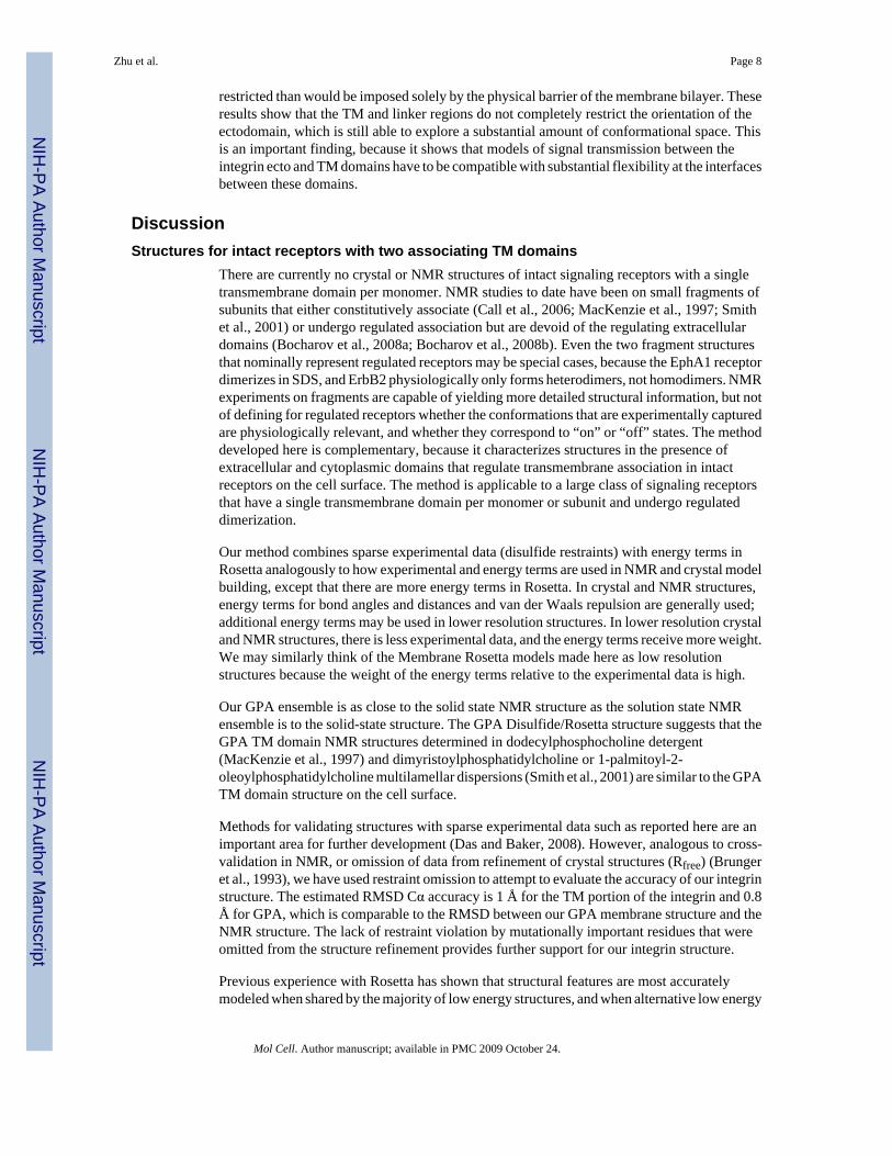
We extended previous work on disulfide crosslinking of the first 9 integrin TM residues (Luoet al., 2004) across the membrane and into the cytoplasm. Residues in the JM and TM segmentsof αIIb, β3, and GPA (Fig. 1A) were individually mutated to cysteine. αIIb and β3 mutants wereco-expressed in 293T transfectants. Cells were treated with or without Cu (II)-o-phenanthroline(Cu-phenanthroline) to catalyze disulfide bond formation, quenched with N-ethylmaleimide,and lysed with detergent. [35S] integrins were immunoprecipitated and subjected tononreducing SDS-PAGE (Fig. 1B–D), and disulfide-linked heterodimer was quantitated as %of total integrin. Disulfide bond formation within the first few TM residues was not increasedfurther by Cu-phenanthroline (Fig. 1B). Disulfide formation in this region and external to themembrane was also quantitated in redox buffers and after reduction with dithiothreitol followedby Cu-phenanthroline (Methods and Fig. S1). By the seventh TM residue, disulfide formationwas almost completely Cu-phenanthroline dependent (Fig. 1C) (Luo et al., 2004), whereas bythe tenth TM residue, Cu-phenanthroline alone had little effect (Fig. 1D). However, disulfidecross-linking in the middle and cytoplasmic side of the membrane was made possible by freeze-thawing cells, which enables access of Cu-phenanthroline and oxygen to the otherwisereducing cytoplasmic environment (Fig. 1D and E). Freeze-thawing had no effect oncrosslinking of more exofacial residues (Fig. S2). Cysteines near the membrane/cytoplasminterface may be palmitylated and thus unavailable for crosslinking (Kovalenko, 2004).Blocking palmitylation with 2-bromopalmitate (2-BP) markedly enhanced cross-linkingefficiency (Fig. 1E).
Standard cross-linking conditions thus employed 15 μg/ml 2-BP, a 1.5h pulse with [35S]methionine and cysteine, a 17h chase, freeze-thaw, and treatment with Cu-phenanthroline for10 min at 0°C. Kinetics showed that cross-linking was maximal at 10 min., even with cysteinepairs showing only partial cross-linking (Fig. S3). In cysteine crosslinking, it is thought that athiol peroxide (S-OH) equivalent to sulfenic acid is the first oxidation product, and thatnucleophilic attack on the oxidized cysteine by a second, unoxidized cysteine SH group resultsin disulfide formation, with release of water. We hypothesize that crosslinking by the second,unoxidized cysteine is in competition with oxidation of the second cysteine, which wouldprevent crosslinking to the first cysteine and account for the lack of further crosslinking after10 min. The dependence of disulfide formation on kinetics of oxidation of the first cysteine,and its limitation by the kinetics of oxidation of the second cysteine, may make the extent ofcross-linking relatively insensitive to kinetic differences in SH oxidation at different depthswithin the membrane, because the first and second cysteines must be nearby and at similardepths. This hypothesis agrees with the lack of correlation between depth in the membrane andextent of crosslinking (Fig. 2).
Dimerization through the GPA TM domains is constitutive, and is not dependent on theextracellular, O-glycosylated domain (MacKenzie et al., 1997). The αIIb and β3 ectodomainswere fused to GPA residues 71–131, comprising the TM and cytoplasmic domains, or GPAresidues 60–131, which include an additional 11 extracellular residues (Fig. 1F). The use ofαIIb GPA/β3 GPA heterodimers enabled measurement of crosslinking not only between
Zhu et al. Page 3
Mol Cell. Author manuscript; available in PMC 2009 October 24.
NIH
-PA Author Manuscript
NIH
-PA Author Manuscript
NIH
-PA Author Manuscript

identical GPA TM residues (e.g. 80 with 80), but also between nonidentical residues (e.g. 80with 81). Compared to wild-type αIIbβ3, the αIIbβ3/GPA 60–131 heterodimer was constitutivelyactive, whereas αIIbβ3/GPA 71–131 was resistant to activation (Fig. 1G). These results areconsistent with the importance of close association between the C-termini of integrin α and βsubunit ectodomains in stabilizing the resting state (Luo et al., 2007), and stronger associationbetween the GPA than the integrin TM domains. The αIIbβ3/GPA60–131 and αIIbβ3/GPA71–131 chimeras gave essentially identical disulfide crosslinking (Figure 1H), showing that GPATM domain association was unaffected by the length of GPA extracellular domain includedin the chimera. Crosslinking between non-identical GPA transmembrane residues was alsoindependent of the integrin subunit fusion partner (Fig. S4).
Crosslinking and RestraintsThe integrin and GPA TM domains show α-helical periodicity with crosslinking peaks every3–4 residues, suggesting helix against helix packing (Fig. 2A–C). Similar periodic peaks inthe β3 cytoplasmic domain at residues I719, D723, F727, and F730 (Fig. 2A) suggest that thisregion is α-helical as well, and interacts with the Lys and Arg residues of the GFFKR motifand several following residues in the α subunit.
The GPA crosslinking data fit the GPA NMR structure well (Fig. 2D) with the assumption thatmaximal crosslinking of 100% would be seen with maximal contact between Cys Cα atoms atthe van der Waals distance of 3.6 Å, and that crosslinking would linearly fall to 0% when thisdistance was increased to 10 Å (solid line, Fig. 2D). Because distance is not the only factorthat could affect crosslinking efficiency, it was important not to over-restrain the distancebetween crosslinked residues. Therefore, an upper distance bound was chosen, below whichno penalty would be applied (dashed lines, Fig. 2D and E). Restraints were used only for residuepairs with ≥20% crosslinking (Fig. 2DE) and only for residues that did not activate ligandbinding by αIIbβ3 when individually mutated to cysteine (see below). Furthermore, tocompensate for inherent flexibility or structural perturbations introduced by cysteinesubstitution, the lowest crosslinking value within two residues was subtracted prior to restraintcalculation (Fig. S5).
Structure refinementRosetta has an efficient method for sampling structural diversity by assembling 3 and 9-residuefragments from the protein data bank into protein structures. Fragments are selected that aresimilar in sequence to the target sequence, and are enriched for (but do not all have) thepredicted secondary structure of the target structure. Fragments are selected from high-resolution crystal structures irrespective of whether they are membrane or water-solubleproteins. Structures are built using a Monte Carlo fragment insertion protocol, minimizing anenergy function that favors hydrophobic burial, strand pairing, and other low-resolutionproperties of protein structures. A subsequent high-resolution refinement stage optimizes vander Waals packing, hydrogen bonding, and solvation interactions (Methods).
Membrane Rosetta uses a model of a membrane with three slabs, a central hydrophobic slabcorresponding to the lipid alkyl groups, and two intermediate slabs corresponding to theinterface regions between the alkyl groups and the polar headgroups. Both the low resolution(Barth et al., 2009) and high resolution (Barth et al., 2007) potentials differ from water-solubleproteins in these slabs, with exposure of non-polar groups favored rather than disfavored. Thepotentials for the head group regions and the external regions are the same as for water-solubleproteins. The embedding (depth and orientation) of proteins in the membrane slab is optimizedto give the lowest energy. Disulfide distance restraints are incorporated as an additionalpotential term used to evaluate model energy; a distance greater than the upper bound shown
Zhu et al. Page 4
Mol Cell. Author manuscript; available in PMC 2009 October 24.
NIH
-PA Author Manuscript
NIH
-PA Author Manuscript
NIH
-PA Author Manuscript

in Fig. 2DE raises energy. This energy term is evaluated in every Monte Carlo step, and thusaffects the course of model building.
Building the integrin TM and juxtamembrane domains was done in several stages, as detailedin Methods and the Supplement. In the final stage, all models with the 10% lowest energy wereclustered by structural similarity (Fig. S6, S7). The vast majority of low energy structures havesimilar structural features (Fig. S7). Structure assessment below uses the structures at the centerof the largest (top) GPA and integrin clusters (referred to as final structures), the structuralensembles within the top clusters (Table 1), and the centers of the top five clusters (Fig. S7).
Structure AssessmentCross-validation with omitted restraints is used for the validation of crystal and NMR structures(Brunger et al., 1993). GPA and integrin models generated without restraints were far from theGPA NMR and integrin final models (Fig. 3 and S8). When 12.5% restraints were used, modelswere much closer. When 25 and 50% restraints were used, the models approached a plateauin Cα RMSD for integrin of 1 Å for TM and 1.8 Å for TM + cytoplasmic domains and 0.8 Åfor GPA (Fig. 3A,C,E). This provides one estimate of structure accuracy. Crystal structureaccuracy is evaluated by omitting data from refinement, and checking the agreement betweenthe omitted data and the final structure (Rfree). Similarly, we checked structures for violationsof restraints that were not used in structure generation (Fig. 3B,D,E). When 50% of restraintswere used in structure generation, there was little violation of the other 50% of omittedrestraints (Fig. 3B,D,E). These results suggest that at least for the regions of the structures thatare well covered by restraints, i.e. the TM regions, the structures match the actual structurewell.
The solid state GPA NMR structure, which fits the solution state restraints better than thesolution NMR structure (Smith et al., 2001), fits the solution state NMR and Disulfide/Rosettastructures, respectively, with Cα RMSD of 0.97±0.10 and 0.96±0.15 Å (see Table 1B for moredetails). These results show that the Disulfide/Rosetta structure is as similar to the solid stateNMR structure as the latter is to the solution state NMR structure. The similarities between theDisulfide/Rosetta and NMR structures extend to details including sidechain rotamers (Fig. 3Gand H). For example, sidechains in cross-linking peaks have identical rotamers, except forLeu-90 and Ile-91 in one of the two Disulfide/Rosetta TM helices (Fig. 3H), which in contrastto the solid state NMR structure, were not constrained to be symmetric.
The final αIIbβ3 membrane structure showed no restraint violation (Fig. 2E) and the ensembleshowed little restraint violation (Table 1A). Furthermore, crosslinking data from activatingcysteine mutations that were omitted from structure calculation also did not violate restraintsand trended well with the idealized crosslinking-distance relationship (Fig. 2E and Table S1).The apparent lack of effect on crosslinking by these activating mutations may result from theuse of 0°C and 37°C in crosslinking and activation assays, respectively.
αIIbβ3 Membrane structure and comparison to mutational resultsAll low energy αIIbβ3 structures have the same interhelical interface, and almost all have theGly of GFFKR in a left-handed helix conformation that caps the αIIb α-helix and brings thetwo Phe of GFFKR into the interface with the β3 α-helix (Fig. 4A–E and S7A, B). These keyfeatures are seen in all 5 top clusters with clustering at 2.0 Å (Fig. S7A) and 4 of 5 of the topclusters at 3.0 Å (Fig. S7B). The sole exception, Cluster 3 with clustering at 3.0 Å (Fig. S7B),has the highest disulfide restraint violation (Fig. S6B), and can be ruled out because it isincompatible with mutational data described below that show that the αIIb GFFKR motif andβ3 Lys-716 have an important role in the αIIb/β3 interface. The β3 TM α-helix continues as α-
Zhu et al. Page 5
Mol Cell. Author manuscript; available in PMC 2009 October 24.
NIH
-PA Author Manuscript
NIH
-PA Author Manuscript
NIH
-PA Author Manuscript
helix into the cytoplasm (Fig. 4B), and in some clusters the cytoplasmic portion of the β3 TMα-helix is divided into two segments separated by a turn near Glu-726 (Fig. S7)
The integrin TM domains associate with a right-handed crossing angle of −37° compared to−41 to −45° in GPA (Table 1) (MacKenzie et al., 1997;Smith et al., 2001). The most prominentfeatures of α-helical surfaces are ridges formed by the sidechains of every fourth residue insequence (i+4n), and grooves in between (Fig. 4F). In a common packing mode betweenadjacent α-helices, observed for α-helices that cross at angles of −50° ± 20°, a ridge formedby residues i+4n on one helix fits into a groove on another helix between the ridges formed byresidues j+4n and j+1+4n (Chothia et al., 1981). This has been termed 4-4 packing. The αIIband β3 TM α-helices show precisely this packing mode (Fig. 4F and G). The ridge is on αIIb,and is formed by residues W968, G972, G976, and L980. The groove is on β3 between residuesV696 and L697, V700 and M701, I704 and L705; G708 and L709 (Fig. 4G). Residues incrosslinking peaks are found at the interface (Fig. 4C, D, and G). Since the ridges and groovesspiral on the side of the cylindrical α-helix, they are convex. The G972-XXX-G976 motif inαIIb decreases the height of the ridge at the center of the packing interface, enabling the twohelical cylinders to pack closer together.
In the NMR and cell surface GPA structures, the TM helices pack most closely together at aGXXXG motif (MacKenzie et al., 1997) (Fig. 3H and 4I). Two differences in interhelicalorientation with integrins are apparent when the grooves in β3 and one of the GPA monomersare superimposed (Fig. 4I). 1) Although packing in GPA resembles ridge in groove (MacKenzieet al., 1997), the two low ridges bearing G79 and G80 move closer to one another than inintegrin (horizontally in Fig. 4H), providing more separation from the high ridges bearing V80and V84. 2) The ridge in the integrin slides in its groove relative to GPA (vertically in Fig.4G), so that the spacing of residues in the interface differs (Fig. 4G–H).
Mutational studies are in excellent agreement with the inter-helical integrin interface identifiedhere. Cysteine and leucine scanning show that the most activating, and hence structurallydisruptive mutations within the TM domains, are of the three interfacial Gly residues, αIIbGly-972 and Gly-976, and β3 Gly-708 (Fig. 5A) (Luo et al., 2005). By contrast, mutations ofαIIb Gly-975 and β3 Gly-702, which are not interfacial (Fig. 4D and G), are not activating (Fig.5A). These results correlate with the observations that the interfacial, but not the non-interfacialGly residues, are invariantly small (Gly, Ala, or Ser) residues in integrin TM domains (Fig.S10).
The GFFKR motif has a structural role in αIIbβ3 association consonant with its long-knownfunctional importance. The αIIb TM α-helix extends beyond the 23-residue TM hydrophobicsegment through Lys-989 and Val-990. However, at Gly-991 of the GFFKR motif, thebackbone adopts a left-handed rather than right-handed α-helical conformation (Fig. 4E). Glyin left-handed conformation at the C-termini of α-helices is the most common C-cap motif,and is responsible for the marked over-representation of Gly at the +1 position following α-helices (Richardson and Richardson, 1989). Almost all (91%) low energy models, and all butone outlier cluster (Fig. S7), have a turn at the Gly (Fig. S11). Many of these models, includingthe final structure, have Phi/Psi angles for this Gly that are disallowed for non-Gly residues(Fig. S11), potentially explaining why mutation of this residue is so destabilizing. The turn atthe Gly brings the two Phe residues of the GFFKR motif into the interface between the αIIband β3 TM α-helices (Fig. 4E). The αIIb and β3 TM α-helices are further apart near this interfacethan anywhere else in the membrane, and Phe-993 in particular occupies space between thetwo helices and is central in the interface. Phe-993 contacts on the β3 subunit Leu-712 and isnearby the aliphatic portion of Lys-716. In the αIIb subunit, Phe-993 abuts Met-987 andPhe-992. Both Phe sidechains orient toward the hydrophobic core of the membrane. A meander
Zhu et al. Page 6
Mol Cell. Author manuscript; available in PMC 2009 October 24.
NIH
-PA Author Manuscript
NIH
-PA Author Manuscript
NIH
-PA Author Manuscript

in the αIIb cytoplasmic domain containing the Lys and Arg of the GFFKR motif and three moreC-terminal residues abuts the β3 cytoplasmic α-helix.
Mutational data support the juxtamembrane αIIb structure. Cysteine substitutions in the GFFKRmotif are activating (Fig. 5A), emphasizing the important role of this motif in thejuxtamembrane interface between the αIIb and β3 subunits. Mutation of FF of GFFKR to AA,YY, WW, or LL demonstrated that even the aromatic substitutions YY and WW are activating(Fig. 5B). These results are consistent with the important packing role, particularly of Phe-993,in the αIIbβ3 interface, and not with the general role that aromatic residues, particularly Trp,often play in the hydrophobic/polar membrane interface.
In the β3 juxtamembrane domain, residues Lys-716, Ile-719, and Asp-723 are in the interfacewith the αIIb GFFKR motif (Fig. 2A and 4E). Remarkably, the Lys-716 ε-amino grouphydrogen bonds to the αIIb backbone (Fig. 4E). Hydrogen bonds to either or both of the αIIbPhe-992 and Lys-994 carbonyls, with Phe-992 predominating, are found in 31% of all lowenergy models, 65% of the cluster 1 ensemble, and in 5 of 9 of the cluster center models shownin Fig. S7 that have the turn at Gly-991.
Cysteine scanning of β3 showed that the only activating mutation in the juxtamembrane/cytoplasmic domain is K716C (Fig. 5A). Furthermore, this mutation is more activating thanthe single other activating Cys mutation identified, G708C in the TM domain. These resultsconfirm the important structural role of Lys-716 in the αIIbβ3 interface. β3 Lys-716 was alsomutated to Ser, Arg, Glu, Leu, and Pro. All substitutions were activating except for Arg (Fig.5B), the only other sidechain which could form a similar hydrogen bond to an αIIb backbonecarbonyl oxygen, and is found at this position in integrin β subunits (Fig. S10). Mutationshowed that in contrast to Arg, polar (Ser, Glu) and aliphatic (Leu) sidechains could notsubstitute for Lys-716. These findings lend credence to the hydrogen bond between the β3Lys-716 sidechain and the αIIb backbone.
Arg-995 of the αIIb GFFKR motif is nearby both β3 Asp-723 and Glu-726 (Fig. 4E), consistentwith the proposal based on mutational studies of a salt bridge between Arg-995 and Asp-723(Hughes et al., 1996). However, the sidechains of Arg-995 and Asp-723 sample a largeconformational space, and are within hydrogen-bonding distance in only 30% of the cluster 1structural ensemble and 16% of low energy structures (Table S2).
We have consistently observed little effect of mutation of β3 Asp-723, the putative salt-bridgepartner of αIIb Arg-995 in either 293T transfectants (Fig. 5A, B) or transient CHO-K1transfectants (Fig. 5C). Since Asp-723 mutants have been found to be active in stable CHOtransfectants (Hughes et al., 1996), this may reflect differences in level of cell surface densityor in assay sensitivity.
Structure of the full-length αIIbβ3 integrinModels of the entire receptor were generated by assembling the structures of the TM+cytoplasmic domains with the ectodomain crystal structures (Zhu et al., 2008). Redox bufferdisulfide restraints were used in the 6-residue αIIb and 4-residue β3 linkers between thesesegments (Fig. 2A,B and Table S3B). Restraints were validated based on crosslinking betweennearby residues defined in αIIbβ3 and αVβ3 crystal structures; because of flexibility in crystalstructures of the β3 tail domain (Zhu et al., 2008), restraints were loosened relative to the TMregion (Fig. S12). The linkers were modeled while simultaneously optimizing the rigid-bodyorientation of the ectodomain with respect to the membrane. Two long, disordered loops at themembrane-proximal base of the αIIb calf-2 domain were included as flexible loops in modelsbecause they may impact orientation on the cell surface. The lowest energy models exhibit adiversity of ectodomain orientations (Fig. 5A). However, the range of orientations was more
Zhu et al. Page 7
Mol Cell. Author manuscript; available in PMC 2009 October 24.
NIH
-PA Author Manuscript
NIH
-PA Author Manuscript
NIH
-PA Author Manuscript
restricted than would be imposed solely by the physical barrier of the membrane bilayer. Theseresults show that the TM and linker regions do not completely restrict the orientation of theectodomain, which is still able to explore a substantial amount of conformational space. Thisis an important finding, because it shows that models of signal transmission between theintegrin ecto and TM domains have to be compatible with substantial flexibility at the interfacesbetween these domains.
DiscussionStructures for intact receptors with two associating TM domains
There are currently no crystal or NMR structures of intact signaling receptors with a singletransmembrane domain per monomer. NMR studies to date have been on small fragments ofsubunits that either constitutively associate (Call et al., 2006; MacKenzie et al., 1997; Smithet al., 2001) or undergo regulated association but are devoid of the regulating extracellulardomains (Bocharov et al., 2008a; Bocharov et al., 2008b). Even the two fragment structuresthat nominally represent regulated receptors may be special cases, because the EphA1 receptordimerizes in SDS, and ErbB2 physiologically only forms heterodimers, not homodimers. NMRexperiments on fragments are capable of yielding more detailed structural information, but notof defining for regulated receptors whether the conformations that are experimentally capturedare physiologically relevant, and whether they correspond to “on” or “off” states. The methoddeveloped here is complementary, because it characterizes structures in the presence ofextracellular and cytoplasmic domains that regulate transmembrane association in intactreceptors on the cell surface. The method is applicable to a large class of signaling receptorsthat have a single transmembrane domain per monomer or subunit and undergo regulateddimerization.
Our method combines sparse experimental data (disulfide restraints) with energy terms inRosetta analogously to how experimental and energy terms are used in NMR and crystal modelbuilding, except that there are more energy terms in Rosetta. In crystal and NMR structures,energy terms for bond angles and distances and van der Waals repulsion are generally used;additional energy terms may be used in lower resolution structures. In lower resolution crystaland NMR structures, there is less experimental data, and the energy terms receive more weight.We may similarly think of the Membrane Rosetta models made here as low resolutionstructures because the weight of the energy terms relative to the experimental data is high.
Our GPA ensemble is as close to the solid state NMR structure as the solution state NMRensemble is to the solid-state structure. The GPA Disulfide/Rosetta structure suggests that theGPA TM domain NMR structures determined in dodecylphosphocholine detergent(MacKenzie et al., 1997) and dimyristoylphosphatidylcholine or 1-palmitoyl-2-oleoylphosphatidylcholine multilamellar dispersions (Smith et al., 2001) are similar to the GPATM domain structure on the cell surface.
Methods for validating structures with sparse experimental data such as reported here are animportant area for further development (Das and Baker, 2008). However, analogous to cross-validation in NMR, or omission of data from refinement of crystal structures (Rfree) (Brungeret al., 1993), we have used restraint omission to attempt to evaluate the accuracy of our integrinstructure. The estimated RMSD Cα accuracy is 1 Å for the TM portion of the integrin and 0.8Å for GPA, which is comparable to the RMSD between our GPA membrane structure and theNMR structure. The lack of restraint violation by mutationally important residues that wereomitted from the structure refinement provides further support for our integrin structure.
Previous experience with Rosetta has shown that structural features are most accuratelymodeled when shared by the majority of low energy structures, and when alternative low energy
Zhu et al. Page 8
Mol Cell. Author manuscript; available in PMC 2009 October 24.
NIH
-PA Author Manuscript
NIH
-PA Author Manuscript
NIH
-PA Author Manuscript

structures can also be ruled out by experimental data (Das and Baker, 2008). Consequently,we have focused not on a single structure, but on the overall characteristics of the 10% lowestenergy structures. We have only emphasized conclusions when they were validated by amajority of low energy structures, and when alternatives could be ruled out. The most criticalexample of this is the GFFKR motif. We obtained restraints between the αIIb and β3 TM helicespreceding GFFKR, and between the helical JM portion of β3 and the K and R residues ofGFFKR and several following residues. Thus, low energy structures of the GFF moiety had tobe found by Rosetta that were consistent with these restraints. The structure of the GFF moietyillustrates one of the strengths of Rosetta, which uses a large library of fragments from high-resolution structures. The most common C-cap motif for α-helices involves a Gly in left handedhelical conformation (Richardson and Richardson, 1989), and the backbone hydrogen bondsin this motif include the first F in GFFKR (Fig. 4E). Thus, this conformation was readily found,and 91% of low energy structures were built including this motif. The other 9% of structurescontinued in right-handed α-helical conformation through the G, and do not have GFF inαIIb and K716 in β3 in the interface. These structures can be ruled out, because mutationalstudies show that GFF in αIIb and K716 in β3 have a critical role in αIIbβ3 association.Interestingly, the only cluster lacking the turn at the G showed an unnatural, continuouslycurved TM β3 α-helix, as a consequence of satisfying disulfide restraints between αIIb and β3residues on either side of the GFF motif (Fig. S7B), presenting yet another criterion fordiscarding this model. We therefore conclude that both the TM and JM portions of ourαIIbβ3 membrane structure are strongly supported.
The integrin TM interfaceOur study definitively identifies the interface between the αIIb and β3 TM α-helices. Previousdisulfide crosslinking of the first 9 residues of the TM domains established only theapproximate orientation between their helices, and did not include the second G of the GXXXGmotif (Luo et al., 2004). Based on that data, approximate orientations between the αIIb andβ3 TM helices (Lau et al., 2008a) or detailed models (Gottschalk, 2005) have subsequentlybeen presented. As an example of the lack of sufficiency of the previous data, the detailedmodel has an Cα RMSD of 2.7 Å over 44 TM residues, and does not follow the ridge-in-groovepacking we describe in the inner half of the membrane (Gottschalk, 2005). Subsequent to (Luoet al., 2004), another group used a different approach, and produced two models thatsuperimpose on our structure over 44 TM residues with Cα RMSD of 6.8 and 8.0 Å (Partridgeet al., 2005). Thus, there has previously been no consensus on the orientation between theαIIb and β3 TM domains.
Compared to previous homodimer TM structures, the integrin TM heterodimer structureillustrates the important principle that ridge in groove packing in heterodimers is notconstrained by symmetry, enabling packing of the ridge at intermediate positions in the groovenot allowed in symmetric homodimers (vertically in Fig. 4G and H). This finding is likely toreflect general differences between homodimeric and heterodimeric TM domain association.Many single-span receptors, such as the EGFR/ErbB family, can associate either as homo orheterodimers. ErbB family heterodimers have an important role in signaling, and their TMdomains associate more stably than the corresponding homodimers (Duneau et al., 2007). Itwill be interesting to compare homo and heterodimers within a single receptor family todetermine whether differences in packing modes between homo and heterodimers regulatesthe stability or signaling of TM domains.
While association between GPA TM domains is constitutive and occurs even in SDS,association between the integrin TM domains is regulated and labile to detergents (Li et al.,2001; MacKenzie et al., 1997). The finding here that integrin ectodomain fusion directly to theGPA TM/cytoplasmic domains in the αIIbβ3/GPA 71–131 chimera results in resistance to
Zhu et al. Page 9
Mol Cell. Author manuscript; available in PMC 2009 October 24.
NIH
-PA Author Manuscript
NIH
-PA Author Manuscript
NIH
-PA Author Manuscript

activation is compatible with the greater stability of the GPA dimer. This stability is not relatedto surface area buried in the interface, which with a 1.4 Å radius probe is 780 Å2 for GPA,compared to 1050 Å2 for the TM domain portion of integrin and 1300 Å2 when the membrane-embedded JM portion is included. The β-branched residues, Ile, Thr, and Val, that only haveone available rotamer in α-helices, are present in the GPA TM domain interface. Thisminimizes loss of entropy when these residues are transferred from the lipid environment to aprotein interface (Liu et al., 2003; MacKenzie et al., 1997). By contrast, integrins have asubstantial number of interfacial Leu and Met residues, for which the conformational entropyloss upon TM helix-helix association will be much greater. Furthermore, hydrogen-bondedThr residues stabilize the GPA but not integrin interface (MacKenzie et al., 1997; Smith et al.,2001).
The JM interface and comparison of associated and dissociated integrin subunitsThe juxtamembrane αIIbβ3 structure is most unusual. The αIIb α-helix is terminated by a turnat the Gly of GFFKR motif that brings its Phe residues into the interface with the β3 subunitand partially embeds them in the hydrophobic portion of the membrane bilayer. Furthermore,a Lys residue immediately following the β3 TM hydrophobic segment also participates in theinterface with αIIb. These key structural findings are all supported by mutational studies. Ourstudy demonstrates the importance of juxtamembrane segments in dimerization. Previoushomodimeric NMR structures of associating TM domains (Bocharov et al., 2008a; Bocharovet al., 2008b; Call et al., 2006; MacKenzie et al., 1997; Smith et al., 2001) show differentpacking modes between two TM α-helices, but do not contain additional segments thatcontribute to packing within the membrane. JM amphipathic segments intervene between TMdomains and intracellular signaling domains in many receptors, including the epidermal growthfactor receptor, and thus may be of key importance in signal transduction (McLaughlin et al.,2005). The demonstration here that the integrin α subunit GFFKR motif is an amphipathic, JMsegment that directly participates in the association between the α and β TM domains raisesthe intriguing possibility that amphipathic JM segments in other receptors may participate indimerization interactions between their TM domains.
Careful studies by Ulmer et al. have described the NMR structures of isolated αIIb and β3 TM/cytoplasmic domain fragments in bicelles (Lau et al., 2008a; Lau et al., 2008b). In contrast tofindings that similar αIIb and β3 constructs formed homomultimers in detergents (Li et al.,2001), Ulmer and coworkers found that these subunits were monomeric in the more bilayer-like bicelle environment. Comparisons between the isolated peptide and membrane complexstructures show a remarkable similarity in the αIIb structures in the GFFKR motif (Fig. 6B–D); it should be noted that no information from the monomer structures was used in thecalculation of the αIIbβ3 complex structure. The backbone conformation is identical throughthe GFFKR motif to Lys-994, which is the last structured residue in the bicelle αIIb structure(Lau et al., 2008a). The same hydrogen bonds that support the turn at Gly-991, between thebackbone O atom of Met-987 and N atoms of Gly-991 and Phe-992, are found in bothstructures, and in general, in the C-cap motif with Gly in left-handed helical conformation(Richardson and Richardson, 1989). Moreover, Phe-993, which is central in the αIIbβ3 complexstructure, and has a well-defined rotamer in the αIIb structure, has the same rotamer in thecomplex structure (Fig. 6B–D).
The isolated αIIb and β3 TM domain structures provide the basis for comparisons to our complexstructure. There is no precedent for association between TM monomers, one of which has asignificant juxtamembrane segment that associates with the α-helical TM segment. It wasunknown whether the αIIb juxtamembrane segment would alter conformation after association.Indeed, there was good reason to expect that the conformation of the αIIb GFFKR segmentwould alter after association with β3, because as pointed out (Lau et al., 2008a), residues in
Zhu et al. Page 10
Mol Cell. Author manuscript; available in PMC 2009 October 24.
NIH
-PA Author Manuscript
NIH
-PA Author Manuscript
NIH
-PA Author Manuscript

this motif were found to be in α-helical conformation in an αIIbβ3 complex NMR structurecontaining the entire GFFKR motif and two preceding residues (Vinogradova et al., 2002).Thus, it is surprising that the only significant difference between the structured portions of thepeptide and complex structures is a difference in tilt between the TM and JM segments. Thistilt brings Phe-993 in monomeric αIIb 2 Å closer to Val-984 to pack against it in van der Waalscontact (Fig. 6D). In the αIIbβ3 complex, β3 Leu-712 contacts αIIb Val-984, and there is notroom for Phe-993 to come as close (Fig. 6C). Thus, some conformational adjustments mustoccur between the associated and dissociated states of the αIIb subunit.
Another interesting juxtamembrane feature is the sidechain-backbone intersubunit hydrogenbond donated by the β3 Lys-716 sidechain. A caveat is that this hydrogen bond appears in only65% of structures in the ensemble. However, the presence of such a bond is strongly supportedby the ability of Arg but not other amino acids to substitute for Lys. This bond is to the αIIbbackbone near the end of the GFFKR motif, and thus stabilizes both αβ association and GFFKRconformation. In our structure, Lys-716 is by far the most important β3 residue for interactionwith αIIb in the JM region, and it is fitting that β3 Lys-716 and the αIIb GFFKR motif, whichinteract with one another, are the sites where mutations are most disruptive to αβ interaction.Arg and Lys are abundant at the boundary between TM and cytoplasmic domains where theydetermine the polarity of membrane insertion by the positive-inside rule (von Heijne, 1992);thus, while Lys and Arg are of general importance, a specific role for β3 Lys-716 wasunanticipated.
Comparison to previous complex structuresSurprisingly, our results disagree with a prominent NMR structure of the αIIbβ3 JM/cytoplasmiccomplex (Vinogradova et al., 2002). There is no similarity whatsoever in the interfaces betweenthe NMR complex structure in aqueous media and our cell surface complex structure (Fig.S13A). The previous comprehensive study showed that mutations that activate integrinsdisrupted the αIIbβ3 complex, and that the talin head domain bound to the β3 JM/cytoplasmicfragment, and disrupted its complex with the αIIb JM/cytoplasmic fragment (Vinogradova etal., 2002). Other NMR studies have yielded conflicting results. Refinement by one group didnot converge on a single NMR structure, but on two different structures (Weljie et al., 2002),neither of which is in agreement with that of (Vinogradova et al., 2002) or described here (Fig.S13B,C). Furthermore, the same αIIb JM/cytoplasmic fragments that associated in aqueousmedia were found not to associate in the presence of dodecylphosphocholine (Vinogradova etal., 2004). Moreover, a lack of complex formation has been reported even with proximityenforced by fusion of αIIb and β3 JM/cytoplasmic fragments to coiled-coils (Ulmer et al.,2001).
In retrospect, perhaps it is not surprising that none of the NMR structures resemble theDisulfide/Rosetta structure (Fig. S13). The two Phe residues of the GFFKR motif pack in thehydrophobic lipid environment, and in an interface against the sidechains of the α and β TMhelices, all of which are missing in the discrepant structures. The lack of appropriate structureof the juxtamembrane/cytoplasmic segments when isolated from the TM segments, and thelack of association between the α and β TM segments in intact cells when the GFFKR motifis deleted (Luo et al., 2004), demonstrate cooperativity between these segments for folding andassembly, consistent with their intimate interaction in the Disulfide/Rosetta structure.
Integrin structure on the cell surfaceUsing a crystal structure of the αIIbβ3 extracellular domain (Zhu et al., 2008), the αIIbβ3membrane structure, and disulfide restraints on the short extracellular linkers, we characterizedthe structure of intact αIIbβ3 on the cell surface. Multiple low-energy orientations between theextracellular and transmembrane domains were found. This finding suggests that the
Zhu et al. Page 11
Mol Cell. Author manuscript; available in PMC 2009 October 24.
NIH
-PA Author Manuscript
NIH
-PA Author Manuscript
NIH
-PA Author Manuscript

orientation of integrins on the cell surface is dynamic rather than fixed, and that the 6-residueαIIb and 4-residue β3 linkers between the extracellular and TM domains are flexible. The lackof a fixed structure for this region is consistent with marked variation in the sequence andlength of these linkers, particularly among integrin subunits. For example, the αV subunit,which also associates with β3, has an 8-residue QPAPMPVP linker compared to the 6-residueEERAIP linker in αIIb.
In contrast to our results, a cryoEM structure of native αIIbβ3 in octylglucoside detergentsuggested a defined, extended orientation between the extracellular and TM domains (Adairand Yeager, 2002). However, the cryoEM extracellular domain structure does not agree withthat determined in crystals or negative stain (Zhu et al., 2008) and it has been suggested thatthe cryoEM structure may have averaged over bent and extended integrin conformations (Luoet al., 2007).
Our finding that limited but not extensive flexibility between the extracellular and TM domainis compatible with signal transduction has important implications. Flexibility and variation inthe ectodomain-TM domain linkers are compatible with activation by large-scale movementssuch as separation in the plane of the membrane between the TM domains (Luo et al., 2004),but not with small-scale twisting or pistoning motions with which the linkers could comply.On the other hand, extreme flexibility in the linkers decouples extracellular domain activationfrom the TM domains, as shown here by the αIIbβ3/GPA 60–131 chimera and previously byinsertion of artificial linkers (Xiong et al., 2003). Decoupling likely arises because long flexiblelinkers enable integrin ectodomain extension and activation while the TM domains remainassociated.
Inside-out activation of integrinsLigand binding by integrins can be activated by binding of the actin cytoskeleton to the βcytoplasmic domain. Two classes of cytoskeleton-associated proteins mediate this linkage,talin and kindlins (Moser et al., 2008; Wegener and Campbell, 2008). Talin and kindlin bindthrough their FERM domains to NPXY motifs at β3 residues 744–747 and 756–759,respectively. Talin also binds to more membrane-proximal β3 regions, including Phe-727 andPhe-730 (Wegener et al., 2007). The Disulfide/Rosetta structure shows that these residues arein the β3 α-helix and face the αIIb cytoplasmic tail. Superposition of the talin β3 and αIIbβ3complexes demonstrates that talin clashes with αIIb residues that interact with the β3 α-helix,and immediately follow the GFFKR motif, i.e. αIIb residues N996-P998 (Fig. 6E). However,the position of the segment with talin-binding residues Phe-727 and Phe-730 is not well defined,since some clusters show two helices in the β3 cytoplasmic domain separated by a turn nearGlu-726 (Fig. S7A and B)). Furthermore, the talin complex structure shows a loss of helicityin β3 N-terminal to His-722, and isolated β3 in bicelles loses α-helicity after residue His-722(Lau et al., 2008b). Thus, it is plausible either that talin binding to β3 would perturb itsassociation with αIIb, or that flexibility in the β3 α-helix between its sites of association withαIIb and talin would eliminate clashes in a talin/αIIbβ3 complex. Since the evidence that talininterferes with association between αIIb and β3 comes from an NMR study on a complex thatour results suggest is non-physiologic, the mechanism by which talin activates integrinsrequires further structural study.
Once integrins bind through talins or kindlins to actin filaments, lateral force exerted by thecytoskeleton will favor an extended integrin conformation with the hybrid domain in β swungout in the direction of the pulling force in the open conformation with high affinity for ligand(Zhu et al., 2008). This mechanism can explain activation by talin as well as by kindlins, whichbind to an NPXY motif that is distal from the αIIb/β3 interface (Moser et al., 2008). Lateralforce would tilt the dissociated β3 TM domain in the plane of the membrane. The long
Zhu et al. Page 12
Mol Cell. Author manuscript; available in PMC 2009 October 24.
NIH
-PA Author Manuscript
NIH
-PA Author Manuscript
NIH
-PA Author Manuscript

continuous α-helix seen in the dissociated β3 TM domain is well suited to tilting (Lau et al.,2008b).
We have described the structure of an intact receptor containing two TM domains on the cellsurface. The results shed important light on the structural basis for transmembrane signalingthrough integrins, demonstrate that the principles differ for association between heterodimericand homodimeric TM domains, and demonstrate that JM domains can have an important rolein TM domain association. The method can be extended to many other important classes ofcell surface receptors with two associating TM domains, and yield information on howregulated changes in the association state of the TM domains transmits signals between theextracellular and intracellular environments.
MethodsDisulfide crosslinking and immunoprecipitation
293T transfectants were pretreated with 15 μg/ml of 2-BP for 1 hour, labeled with [35S]cysteine/methionine (10mCi/ml, PerkinElmer Life Science) for 1.5 h, and chased for 17 h.Cells were detached and kept intact or broken by freeze-thaw, treated on ice with 200 μMCuSO4/1000 μM o-phenanthroline for 10 min., with 10 mM N-ethylmaleimide for 10 min, andlysed in 2% Triton X-100. Lysate supernatants were immunoprecipitated with anti-β3 mAbAP3 and protein G agarose, and subjected to nonreducing 7.5% SDS-PAGE. Radioactivitywas quantitated with a PhosphorImager. Crosslinking efficiency was quantitated as disulfide-linked heterodimer as % of heterodimer plus αIIb and β3 monomers. For constitutivelycrosslinked residues, crosslinking was also measured after 1 h in 5 mM cysteamine/1 mMcystamine redox buffer and after DTT treatment followed by Cu-phenanthroline. Details arein the Supplement.
Membrane structure generationThe energy terms used in Membrane Rosetta (Barth et al., 2007; Barth et al., 2009) include aLennard-Jones potential, a backbone torsional term, a knowledge-based pairwise interactionterm between amino acid residues, a penalty term for the disulfide restraints that is proportionalto distance beyond the upper bound, an implicit Lazaridis-Karplus solvation term for theaqueous and hydrophobic environments (with the hydrophobic potential based on experimentaltransfer free energies from water to cyclohexane), and an orientation-dependent hbonding term.Models are first built with centroids for sidechains, and then refined in all-atom mode. The allatom mode membrane environment has an inner, slab-shaped, 24-Å thick isotropic,hydrophobic phase representing the lipid alkyl groups, outer isotropic phases representing theheadgroup and membrane-external regions, and in between, two 10-Å thick, anisotropic phasesrepresenting the interface region where alkyl, acyl, and glycerol groups are located. Thecentroid membrane environment is similar, but replaces each anisotropic slab with threeisotropic slabs. The solvation and hbond potentials differ in the hydrophobic and aqueousenvironments, and are interpolated in the interface region. The hbond potential includes weakCαH-O bonds and bifurcated hbonds. The position of models in the membrane slab is variedto find the lowest energy embedding.
Structures were built in several stages. First, twelve-residue ideal helices were docked. Then,all but two residues of each helix were discarded, and chain-growing with fragment assemblywas used to build longer structures that could in principle have any secondary structure. A totalof two (GPA) or three (integrin) chain growing stages were used, alternating in N to C and Cto N direction, and previously built residues were discarded in each stage, so that no remnantfrom the first one (GPA) or two stages (integrin) were present in the final structures (Fig. S6).Approximately 10,000 models were generated in each stage. Disulfide restraints were used as
Zhu et al. Page 13
Mol Cell. Author manuscript; available in PMC 2009 October 24.
NIH
-PA Author Manuscript
NIH
-PA Author Manuscript
NIH
-PA Author Manuscript

part of the total energy function in each Monte Carlo step within each chain-growing stage.Furthermore, between each stage, the 100 models used as seeds for the next stage were selectedby the independent criteria of low restraint violation and low energy (in this case, energy didnot include the restraint violation penalty). The stages are summarized in more detail in Fig.S6 and Supplementary Materials. Structures have been deposited in the protein databank ascodes ……
Modeling the intact integrin in the membrane bilayerTo provide starting TM structures, one additional stage of chain-growing was performed fromthe TM regions to grow the linker regions, using redox buffer disulfide restraints for the linkerregion, and distance restraints for the two C-terminal residues present in αIIb and β3 in thecrystal structure (Zhu et al., 2008). After joining these TM structures to the crystal structure,the two linkers were simultaneously remodeled with Membrane Rosetta using the linkerdisulfide restraints to find low energy orientations with respect to the membrane(Supplementary Materials). Two long, membrane-proximal loops in the calf-2 domain that aredisordered in the crystal structure were included in the models.
Ligand binding assayLigand mimetic IgM PAC-1 (Becton Dickinson, San Jose, CA) and FITC-labeled humanfibrinogen (Enzyme Research Laboratories, South Bend, IN) binding to transfected cells wasas described (Luo et al., 2004).
Supplementary MaterialRefer to Web version on PubMed Central for supplementary material.
AcknowledgmentsWe thank Junichi Takagi and Guo-hui Li for early help with the project. Supported by NIH grant HL-48675and HHMI.
ReferencesAdair BD, Yeager M. Three-dimensional model of the human platelet integrin αIIbβ3 based on electron
cryomicroscopy and x-ray crystallography. Proc Natl Acad Sci USA 2002;99:14059–14064. [PubMed:12388784]
Barth P, Schonbrun J, Baker D. Toward high-resolution prediction and design of transmembrane helicalprotein structures. Proc Natl Acad Sci USA 2007;104:15682–15687. [PubMed: 17905872]
Barth P, Wallner BDB. Prediction of membrane protein structures with complex topologies using limitedconstraints. Proc Natl Acad Sci USA 2009;106:1409–1414. [PubMed: 19190187]
Bass RB, Butler SL, Chervitz SA, Gloor SL, Falke JJ. Use of site-directed cysteine and disulfide chemistryto probe protein structure and dynamics: applications to soluble and transmembrane receptors ofbacterial chemotaxis. Methods Enzymol 2007;423:25–51. [PubMed: 17609126]
Bocharov EV, Mayzel ML, Volynsky PE, Goncharuk MV, Ermolyuk YS, Schulga AA, Artemenko EO,Efremov RG, Arseniev AS. Spatial structure and pH-dependent conformational diversity of dimerictransmembrane domain of the receptor tyrosine kinase EphA1. J Biol Chem. 2008a
Bocharov EV, Mineev KS, Volynsky PE, Ermolyuk YS, Tkach EN, Sobol AG, Chupin VV, KirpichnikovMP, Efremov RG, Arseniev AS. Spatial structure of the dimeric transmembrane domain of the growthfactor receptor ErbB2 presumably corresponding to the receptor active state. J Biol Chem 2008b;283:6950–6956. [PubMed: 18178548]
Brunger AT, Clore GM, Gronenborn AM, Saffrich R, Nilges M. Assessing the quality of solution nuclearmagnetic resonance structures by complete cross-validation. Science (New York, NY) 1993;261:328–331.
Zhu et al. Page 14
Mol Cell. Author manuscript; available in PMC 2009 October 24.
NIH
-PA Author Manuscript
NIH
-PA Author Manuscript
NIH
-PA Author Manuscript

Call ME, Schnell JR, Xu C, Lutz RA, Chou JJ, Wucherpfennig KW. The structure of the zetazetatransmembrane dimer reveals features essential for its assembly with the T cell receptor. Cell2006;127:355–368. [PubMed: 17055436]
Chothia C, Levitt M, Richardson D. Helix to helix packing in proteins. J Mol Biol 1981;145:215–250.[PubMed: 7265198]
Das R, Baker D. Macromolecular modeling with rosetta. Annu Rev Biochem 2008;77:363–382.[PubMed: 18410248]
Duneau JP, Vegh AP, Sturgis JN. A dimerization hierarchy in the transmembrane domains of the HERreceptor family. Biochemistry 2007;46:2010–2019. [PubMed: 17253768]
Gottschalk KE. A coiled-coil structure of the αIIbβ3 integrin transmembrane and cytoplasmic domainsin its resting state. Structure 2005;13:703–712. [PubMed: 15893661]
Hughes PE, Diaz-Gonzalez F, Leong L, Wu C, McDonald JA, Shattil SJ, Ginsberg MH. Breaking theintegrin hinge. J Biol Chem 1996;271:6571–6574. [PubMed: 8636068]
Kovalenko OV, Yang X, Kolesnikova TV, Hemler ME. Evidence for specific tetraspanin homodimers:inhibition of palmitoylation makes cysteine residues available for cross-linking. Biochem J2004;377:407–417. [PubMed: 14556650]
Lau TL, Dua V, Ulmer TS. Structure of the integrin alphaIIb transmembrane segment. J Biol Chem 2008a;283:16162–16168. [PubMed: 18417472]
Lau TL, Partridge AW, Ginsberg MH, Ulmer TS. Structure of the integrin beta3 transmembrane segmentin phospholipid bicelles and detergent micelles. Biochemistry 2008b;47:4008–4016. [PubMed:18321071]
Li R, Babu CR, Lear JD, Wand AJ, Bennett JS, Degrado WF. Oligomerization of the integrin aIIbb3:Roles of the transmembrane and cytoplasmic domains. Proc Natl Acad Sci USA 2001;98:12462–12467. [PubMed: 11606749]
Li W, Metcalf DG, Gorelik R, Li R, Mitra N, Nanda V, Law PB, Lear JD, Degrado WF, Bennett JS. Apush-pull mechanism for regulating integrin function. Proc Natl Acad Sci USA 2005;102:1424–1429.[PubMed: 15671157]
Liu W, Crocker E, Siminovitch DJ, Smith SO. Role of side-chain conformational entropy intransmembrane helix dimerization of glycophorin A. Biophys J 2003;84:1263–1271. [PubMed:12547806]
Luo BH, Carman CV, Springer TA. Structural basis of integrin regulation and signaling. Annu Rev Imm2007;25:619–647.
Luo BH, Carman CV, Takagi J, Springer TA. Disrupting integrin transmembrane domainheterodimerization increases ligand binding affinity, not valency or clustering. Proc Natl Acad SciUSA 2005;102:3679–3684. [PubMed: 15738420]
Luo BH, Springer TA, Takagi J. A specific interface between integrin transmembrane helices and affinityfor ligand. PLoS Biol 2004;2:776–786.
MacKenzie KR, Prestegard JH, Engelman DM. A transmembrane helix dimer: structure and implications.Science (New York, NY 1997;276:131–133.
McLaughlin S, Smith SO, Hayman MJ, Murray D. An electrostatic engine model for autoinhibition andactivation of the epidermal growth factor receptor (EGFR/ErbB) family. J Gen Physiol 2005;126:41–53. [PubMed: 15955874]
Moser M, Nieswandt B, Ussar S, Pozgajova M, Fassler R. Kindlin-3 is essential for integrin activationand platelet aggregation. Nature medicine 2008;14:325–330.
O’Toole TE, Katagiri Y, Faull RJ, Peter K, Tamura R, Quaranta V, Loftus JC, Shattil SJ, Ginsberg MH.Integrin cytoplasmic domains mediate inside-out signal transduction. J Cell Biol 1994;124:1047–1059. [PubMed: 7510712]
Partridge AW, Liu S, Kim S, Bowie JU, Ginsberg MH. Transmembrane domain packing stabilizesintegrin aIIbb3 in the low affinity state. J Biol Chem 2005;280:7294–7200. [PubMed: 15591321]
Richardson, JS.; Richardson, DC. Principles and Patterns of Protein Conformation. In: Fasman, GD.,editor. Prediction of Protein Structure and the Principles of Protein Conformation. New York: PlenumPress; 1989. p. 1-98.
Zhu et al. Page 15
Mol Cell. Author manuscript; available in PMC 2009 October 24.
NIH
-PA Author Manuscript
NIH
-PA Author Manuscript
NIH
-PA Author Manuscript

Smith SO, Song D, Shekar S, Groesbeek M, Ziliox M, Aimoto S. Structure of the transmembrane dimerinterface of glycophorin A in membrane bilayers. Biochemistry 2001;40:6553–6558. [PubMed:11380249]
Ulmer TS, Yaspan B, Ginsberg MH, Campbell ID. NMR analysis of structure and dynamics of thecytosolic tails of integrin alpha IIb beta 3 in aqueous solution. Biochemistry 2001;40:7498–7508.[PubMed: 11412103]
Vinogradova O, Vaynberg J, Kong X, Haas TA, Plow EF, Qin J. Membrane-mediated structuraltransitions at the cytoplasmic face during integrin activation. Proc Natl Acad Sci USA2004;101:4094–4099. [PubMed: 15024114]
Vinogradova O, Velyvis A, Velyviene A, Hu B, Haas TA, Plow EF, Qin J. A structural mechanism ofintegrin αIIbβ3 “inside-out” activation as regulated by its cytoplasmic face. Cell 2002;110:587–597.[PubMed: 12230976]
von Heijne G. Membrane protein structure prediction. Hydrophobicity analysis and the positive-insiderule. J Mol Biol 1992;225:487–494. [PubMed: 1593632]
Wegener KL, Campbell ID. Transmembrane and cytoplasmic domains in integrin activation and protein-protein interactions (review). Mol Membr Biol 2008;25:376–387. [PubMed: 18654929]
Wegener KL, Partridge AW, Han J, Pickford AR, Liddington RC, Ginsberg MH, Campbell ID. Structuralbasis of integrin activation by talin. Cell 2007;128:171–182. [PubMed: 17218263]
Weljie AM, Hwang PM, Vogel HJ. Solution structures of the cytoplasmic tail complex from plateletαIIb- and β3-subunits. Proc Natl Acad Sci USA 2002;99:5878–5883. [PubMed: 11983888]
Xiong YM, Chen J, Zhang L. Modulation of CD11b/CD18 adhesive activity by its extracellular,membrane-proximal regions. J Immunol 2003;171:1042–1050. [PubMed: 12847278]
Zhang X, Gureasko J, Shen K, Cole PA, Kuriyan J. An allosteric mechanism for activation of the kinasedomain of epidermal growth factor receptor. Cell 2006;125:1137–1149. [PubMed: 16777603]
Zhu J, Luo BH, Xiao T, Zhang C, Nishida N, Springer TA. Structure of a Complete Integrin Ectodomainin a Physiologic Resting State and Activation and Deactivation by Applied Forces. Mol Cell2008;32:849–861. [PubMed: 19111664]
Zhu et al. Page 16
Mol Cell. Author manuscript; available in PMC 2009 October 24.
NIH
-PA Author Manuscript
NIH
-PA Author Manuscript
NIH
-PA Author Manuscript
Figure 1. Disulfide crosslinking in native cell membraneA. Sequences of GPA and αIIbβ3 integrin TM and cytoplasmic domains. Numbers in red showpositions tested in panels B–E. B–E. Disulfide crosslinking of αIIbβ3 with indicated cysteinemutations in 293T transfectants, with or without Cu-phenanthroline, freeze-thaw, and 2-BPtreatment as indicated. Immunoprecipitated 35S-labeled material was subjected to nonreducingSDS-PAGE and autoradiography. Positions of αIIb (α), β3 (β) and αIIbβ3 heterodimer (α-β)are shown. F. Integrin αIIbβ3/GPA chimeras. G. Ligand binding by chimeras. Binding of ligandmimetic PAC-1 (IgM) (upper panel) or FITC-labeled fibrinogen (Fg) (lower panel) to 293Ttransfectants was measured in the presence of 1mM Ca2+ or 1mM Mn2+ plus 10 μg/mlactivating mAb PT25-2. Binding is expressed as mean fluorescence intensity (MFI) of Fg orPAC-1 relative to MFI of Cy3-labled anti- β3 mAb AP3. H. αIIbβ3/GPA60–131 (closed
Zhu et al. Page 17
Mol Cell. Author manuscript; available in PMC 2009 October 24.
NIH
-PA Author Manuscript
NIH
-PA Author Manuscript
NIH
-PA Author Manuscript

symbols) and αIIbβ3/GPA71–131 (open symbols) chimeras show similar disulfidecrosslinking.
Zhu et al. Page 18
Mol Cell. Author manuscript; available in PMC 2009 October 24.
NIH
-PA Author Manuscript
NIH
-PA Author Manuscript
NIH
-PA Author Manuscript
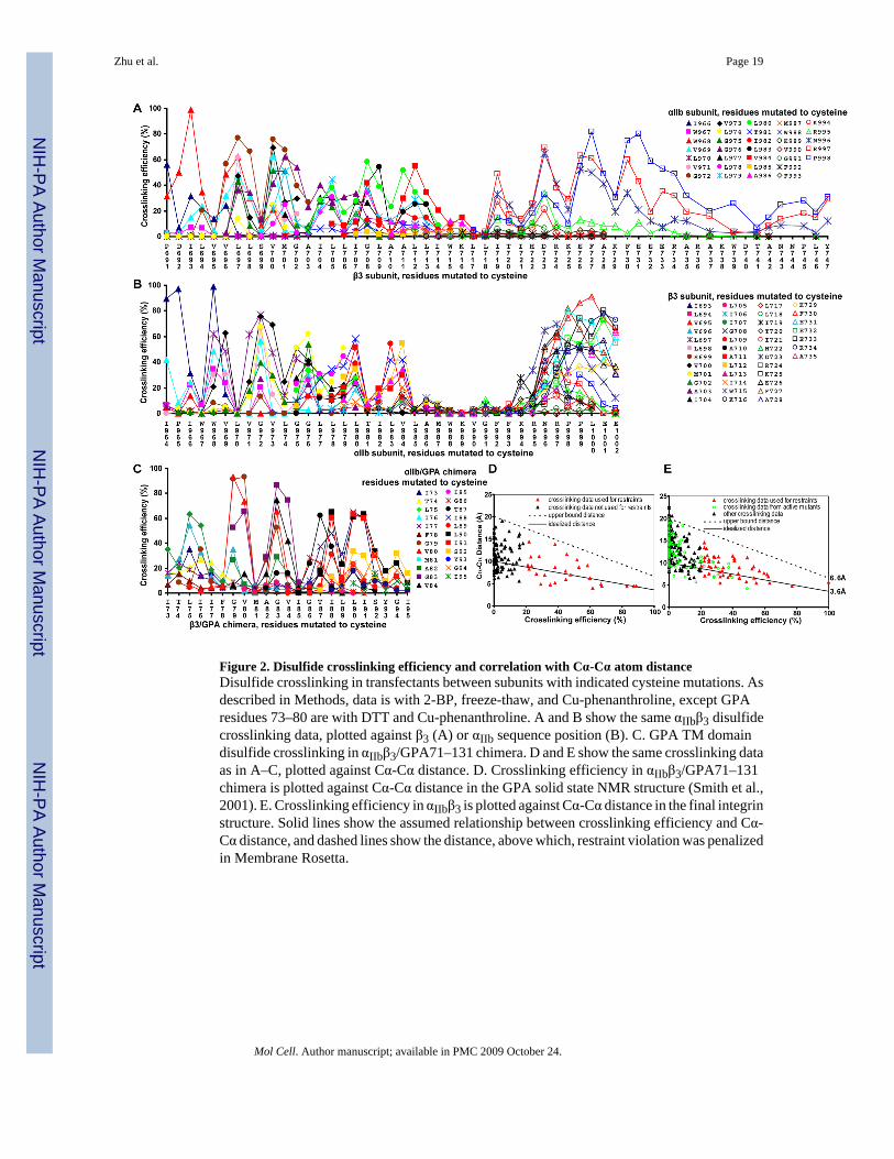
Figure 2. Disulfide crosslinking efficiency and correlation with Cα-Cα atom distanceDisulfide crosslinking in transfectants between subunits with indicated cysteine mutations. Asdescribed in Methods, data is with 2-BP, freeze-thaw, and Cu-phenanthroline, except GPAresidues 73–80 are with DTT and Cu-phenanthroline. A and B show the same αIIbβ3 disulfidecrosslinking data, plotted against β3 (A) or αIIb sequence position (B). C. GPA TM domaindisulfide crosslinking in αIIbβ3/GPA71–131 chimera. D and E show the same crosslinking dataas in A–C, plotted against Cα-Cα distance. D. Crosslinking efficiency in αIIbβ3/GPA71–131chimera is plotted against Cα-Cα distance in the GPA solid state NMR structure (Smith et al.,2001). E. Crosslinking efficiency in αIIbβ3 is plotted against Cα-Cα distance in the final integrinstructure. Solid lines show the assumed relationship between crosslinking efficiency and Cα-Cα distance, and dashed lines show the distance, above which, restraint violation was penalizedin Membrane Rosetta.
Zhu et al. Page 19
Mol Cell. Author manuscript; available in PMC 2009 October 24.
NIH
-PA Author Manuscript
NIH
-PA Author Manuscript
NIH
-PA Author Manuscript

Figure 3. Estimation of structure accuracy and validation of the GPA cell surface structureA–F. Effect of restraint omission. Models were generated using different subsets of 12.5, 25,or 50% of the total restraints as described in the Supplement. A, C and E. Models werecompared over Cα atoms to the final structure made with all restraints, and in the case of GPA,also to the GPA NMR structure. By definition, models made with 100% of restraints areidentical to the final structure. B, D, and F. Models generated with a subset of restraints werescored for violation of the omitted restraints, i.e., those not used in model generation. The perresidue RMS distance violation, i.e., distance above the upper bound (see Fig. 2D and E), isshown. How well the models satisfy omitted restraints is a measure of model accuracy similarto the Rfree value in crystallography. G. The ensemble of 20 GPA Disulfide/Rosetta structures,showing all heavy atoms (green) superimposed on the solid state NMR structure (red). H.
Zhu et al. Page 20
Mol Cell. Author manuscript; available in PMC 2009 October 24.
NIH
-PA Author Manuscript
NIH
-PA Author Manuscript
NIH
-PA Author Manuscript
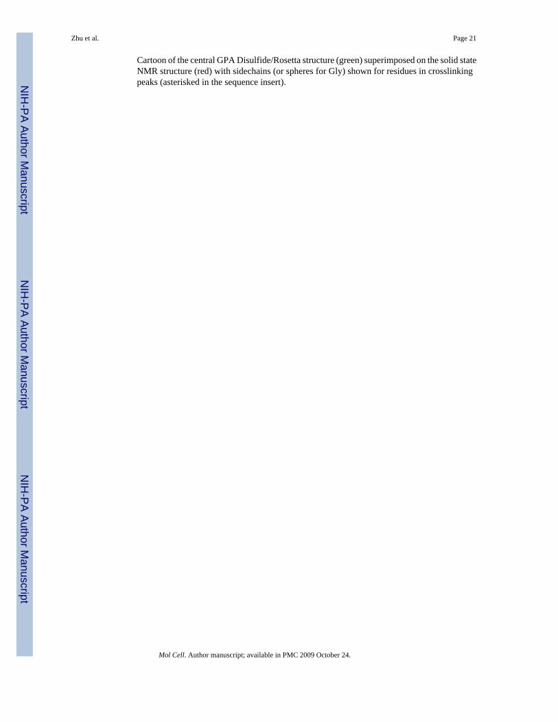
Cartoon of the central GPA Disulfide/Rosetta structure (green) superimposed on the solid stateNMR structure (red) with sidechains (or spheres for Gly) shown for residues in crosslinkingpeaks (asterisked in the sequence insert).
Zhu et al. Page 21
Mol Cell. Author manuscript; available in PMC 2009 October 24.
NIH
-PA Author Manuscript
NIH
-PA Author Manuscript
NIH
-PA Author Manuscript

Figure 4. Structure of the membrane region of an integrinA. The disulfide/Rosetta structural ensemble superimposed on the 46-residue TM segments.B. The cluster-center structure. C. The sequences, with residues in crosslinking peaksasterisked. D. Transparent molecular surfaces for αIIb and β3 TM segments with all sidechainsshown. In A–D, sidechains and Gly Cα spheres in the crosslinking peaks in the 23 residuehydrophobic segment are shown in red or green. Residues in the cytoplasmic juxtamembraneinterface region (18 to 12 Å from the membrane center) are cyan. E. A blow-up of the lowerTM, JM, and cytoplasmic segments. Hydrogen bonds are dashed green lines. Nitrogens,oxygens, and sulfurs are blue, red and yellow, respectively. F–H. Crick helical-net diagrams(Chothia et al., 1981). Cylindrical helical surfaces are cut at one position along thecircumference, unrolled, and aligned at the helical interfaces. I. Superposition of the integrin
Zhu et al. Page 22
Mol Cell. Author manuscript; available in PMC 2009 October 24.
NIH
-PA Author Manuscript
NIH
-PA Author Manuscript
NIH
-PA Author Manuscript

and GPA TM helices, on residues that form the two ridges forming the groove in β3 and oneGPA monomer in which the GXXXG motifs nestle.
Zhu et al. Page 23
Mol Cell. Author manuscript; available in PMC 2009 October 24.
NIH
-PA Author Manuscript
NIH
-PA Author Manuscript
NIH
-PA Author Manuscript

Figure 5. Ligand binding by αIIbβ3 integrin mutantsA. Binding of ligand-mimetic PAC-1 IgM by cysteine-scanning mutants in αIIb (upper panel)and β3 (lower panel). Results in absence (Ca) and presence of activation (Mn/PT25-2) areshown, along with wild-type (WT) and GFFKR/GAAKR mutant controls. Leucine-scanningresults from a previous study (Luo et al., 2005) are also plotted. B and C. Effects of othermutations in 293T (B) and CHO-K1 (C) transient transfectants.
Zhu et al. Page 24
Mol Cell. Author manuscript; available in PMC 2009 October 24.
NIH
-PA Author Manuscript
NIH
-PA Author Manuscript
NIH
-PA Author Manuscript
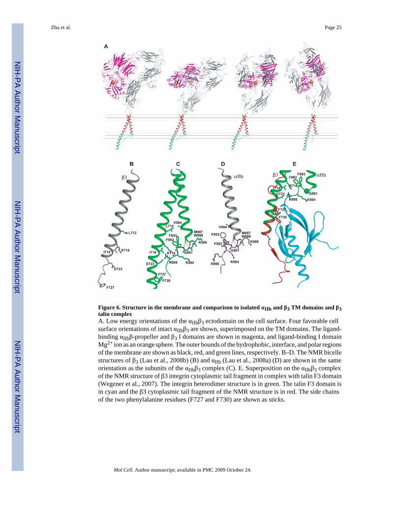
Figure 6. Structure in the membrane and comparison to isolated αIIb and β3 TM domains and β3talin complexA. Low energy orientations of the αIIbβ3 ectodomain on the cell surface. Four favorable cellsurface orientations of intact αIIbβ3 are shown, superimposed on the TM domains. The ligand-binding αIIbβ-propeller and β3 I domains are shown in magenta, and ligand-binding I domainMg2+ ion as an orange sphere. The outer bounds of the hydrophobic, interface, and polar regionsof the membrane are shown as black, red, and green lines, respectively. B–D. The NMR bicellestructures of β3 (Lau et al., 2008b) (B) and αIIb (Lau et al., 2008a) (D) are shown in the sameorientation as the subunits of the αIIbβ3 complex (C). E. Superposition on the αIIbβ3 complexof the NMR structure of β3 integrin cytoplasmic tail fragment in complex with talin F3 domain(Wegener et al., 2007). The integrin heterodimer structure is in green. The talin F3 domain isin cyan and the β3 cytoplasmic tail fragment of the NMR structure is in red. The side chainsof the two phenylalanine residues (F727 and F730) are shown as sticks.
Zhu et al. Page 25
Mol Cell. Author manuscript; available in PMC 2009 October 24.
NIH
-PA Author Manuscript
NIH
-PA Author Manuscript
NIH
-PA Author Manuscript

NIH
-PA Author Manuscript
NIH
-PA Author Manuscript
NIH
-PA Author Manuscript
Zhu et al. Page 26Ta
ble
1
Tab
le 1
A. E
xper
imen
tal r
estr
aint
s and
stru
ctur
e st
atis
tics
Exp
erim
enta
l res
trai
nts
GPA
Inte
grin
Num
ber o
f res
idue
s46
90 (E
C,1
2; T
M, 4
6; C
T, 3
2)a
Num
ber o
f res
train
ts54
(sym
met
rized
)48
(TM
, 33;
CT,
15)
a
Stru
ctur
e st
atis
tics
GPA
(20
mod
els)
Inte
grin
(52
mod
els)
TM (4
6 re
sidu
es)
TM +
CT
(78
resi
dues
)b
Cα-
RM
SD (Å
) to
clus
ter c
ente
r mod
el0.
68±0
.23
1.94
±0.4
2
Dis
tanc
e re
stai
nts R
MS
viol
atio
n (Å
)0.
01±0
.02
(0.0
0)c
0.06
±0.0
4 (0
.00)
c
Ang
le b
etw
een
TM h
elix
axe
s (°)
−45.
4±1.
8 (−
43.1
)c−3
7.5±
3.1
(−36
.9)c
Dis
tanc
e be
twee
n TM
hel
ix a
xes (
Å)
7.3±
0.49
(7.1
)c8.
2±0.
77 (7
.8)c
RM
SDs f
rom
idea
lized
geo
met
ry
Bon
ds (Å
)0.
0067
±0.0
080.
012±
0.00
5
Ang
les (
°)0.
28±0
.001
50.
57±0
.10
Ram
acha
ndra
n st
atis
ticsd
Ram
acha
ndra
n fa
vore
d10
0.0%
99.8
±0.4
%
Ram
acha
ndra
n ou
tlier
s0.
0%0.
02±0
.15%
PDB
cod
e
Tab
le 1
B. C
ompa
riso
n of
solu
tion
NM
R st
ruct
ure
(1A
FO) a
nd d
isul
fide/
RO
SET
TA
GPA
stru
ctur
es to
solid
stat
e N
MR
stru
ctur
e
I73-
I95
(46
resi
dues
)I7
6-I8
8 (2
6 re
sidu
es)
Ato
ms
Cα
Bac
kbon
eA
ll he
avy
Cα
Bac
kbon
eA
ll he
avy
RM
SD (Å
)
1AFO
(20
mod
els)
0.97
±.0.
101.
14±0
.11
1.69
±0.0
80.
67±0
.10
0.64
±0.0
91.
09±0
.18
Dis
ulfid
e/R
OSE
TTA
(20
mod
els)
0.96
±0.1
51.
09±0
.13
1.59
±0.1
90.
61±0
.12
0.59
±0.1
21.
27±0
.20
Mol Cell. Author manuscript; available in PMC 2009 October 24.

NIH
-PA Author Manuscript
NIH
-PA Author Manuscript
NIH
-PA Author Manuscript
Zhu et al. Page 27
Tab
le 1
B. C
ompa
riso
n of
solu
tion
NM
R st
ruct
ure
(1A
FO) a
nd d
isul
fide/
RO
SET
TA
GPA
stru
ctur
es to
solid
stat
e N
MR
stru
ctur
e
I73-
I95
(46
resi
dues
)I7
6-I8
8 (2
6 re
sidu
es)
Ato
ms
Cα
Bac
kbon
eA
ll he
avy
Cα
Bac
kbon
eA
ll he
avy
RM
SD (Å
)
Larg
est c
lust
er c
ente
r mod
el0.
710.
841.
280.
440.
430.
91
a EC, e
xtra
cellu
lar r
egio
n; T
M, t
rans
mem
bran
e re
gion
; CT,
juxt
amem
bran
e an
d cy
topl
asm
ic re
gion
.
b Onl
y TM
(I96
6-W
988
of α
IIb
subu
nit a
nd I6
93-W
715
of β
3 su
buni
t) an
d C
T (K
989-
P998
of α
IIb
subu
nit a
nd K
716-
A73
7 of
β3
subu
nit)
resi
dues
are
incl
uded
.
c Val
ues a
re fo
r the
clu
ster
cen
ter s
truct
ure.
d Ram
acha
ndra
n st
atis
tics w
ere
mea
sure
d by
Mol
Prob
ity [D
avis
, 200
7 #1
8419
].
Mol Cell. Author manuscript; available in PMC 2009 October 24.
Copyright © 2022 FDOKUMEN