Size-exclusion chromatography can identify faster-associating protein complexes and evaluate design...
8
BioMed Central Page 1 of 8 (page number not for citation purposes) BMC Research Notes Open Access Short Report Size-exclusion chromatography can identify faster-associating protein complexes and evaluate design strategies Chad L Mayer 1,2 , W Kalani Snyder 1 , Monika A Swietlicka 1 , Andrew D VanSchoiack 1 , Chad R Austin 1,3 and Benjamin J McFarland* 1 Address: 1 Department of Chemistry and Biochemistry, Seattle Pacific University, 3307 Third Avenue West, Seattle, WA 98119-1997, USA, 2 Current address : Seattle Biomedical Research Institute, Seattle, WA 98109, USA and 3 Current address : Dept. of Microbiology, University of Colorado – Denver, Aurora, CO 80045, USA Email: Chad L Mayer - [email protected]; W Kalani Snyder - [email protected]; Monika A Swietlicka - [email protected]; Andrew D VanSchoiack - [email protected]; Chad R Austin - [email protected]; Benjamin J McFarland* - [email protected] * Corresponding author Abstract Background: We previously developed a set of rationally designed mutant MICA protein ligands for the NKG2D immunoreceptor in which MICA was mutated at residues that do not contact NKG2D. Some of these MICA mutants, predicted by RosettaDesign to be destabilized, bound NKG2D with affinities enhanced by more than an order of magnitude when evaluated by surface plasmon resonance (SPR). Findings: Small-zone size-exclusion chromatography (SEC) detected persistent high-affinity MICA mutant-NKG2D complexes in solution as early-eluting peaks. The SEC binding assay used standard protein purification instrumentation to evaluate complex stability, qualitatively paralleled the SPR results, and successfully discriminated among complexes that differed only in on-rates. We used the SEC binding assay, along with SPR, to assess the results of a follow-up design strategy targeting the non-interfacial redesigned region. Both SEC and SPR agreed that these mutations did not enhance affinity as much as previous mutants. When the SEC binding assay was run in 1 M urea, only the highest affinity complex was detected. Conclusion: This SEC binding assay provides a correlation with SPR results for protein complex affinities, detecting changes in complex on-rates, and tunable to lower sensitivity with 1 M urea. The SEC binding assay is complementary to other protein design evaluation methods, can be adapted to the undergraduate research laboratory, and may provide additional structural information about changes in hydrodynamic radii from elution times. Our assay allowed us to conclude that further alteration of MICA at non-contacting residues is unlikely to further enhance NKG2D affinity. Background Several protein design algorithms have been produced over the past decade for rationally altering and optimizing the cores of proteins, protein-ligand interfaces, and pro- tein-protein interfaces for structural and therapeutic application [1], including RosettaDesign [2]. Protein Published: 15 July 2009 BMC Research Notes 2009, 2:135 doi:10.1186/1756-0500-2-135 Received: 15 June 2009 Accepted: 15 July 2009 This article is available from: http://www.biomedcentral.com/1756-0500/2/135 © 2009 McFarland et al; licensee BioMed Central Ltd. This is an Open Access article distributed under the terms of the Creative Commons Attribution License (http://creativecommons.org/licenses/by/2.0 ), which permits unrestricted use, distribution, and reproduction in any medium, provided the original work is properly cited.
Transcript of Size-exclusion chromatography can identify faster-associating protein complexes and evaluate design...
BioMed CentralBMC Research Notes
ss
Open AcceShort ReportSize-exclusion chromatography can identify faster-associating protein complexes and evaluate design strategiesChad L Mayer1,2, W Kalani Snyder1, Monika A Swietlicka1, Andrew D VanSchoiack1, Chad R Austin1,3 and Benjamin J McFarland*1Address: 1Department of Chemistry and Biochemistry, Seattle Pacific University, 3307 Third Avenue West, Seattle, WA 98119-1997, USA, 2Current address : Seattle Biomedical Research Institute, Seattle, WA 98109, USA and 3Current address : Dept. of Microbiology, University of Colorado – Denver, Aurora, CO 80045, USA
Email: Chad L Mayer - [email protected]; W Kalani Snyder - [email protected]; Monika A Swietlicka - [email protected]; Andrew D VanSchoiack - [email protected]; Chad R Austin - [email protected]; Benjamin J McFarland* - [email protected]
* Corresponding author
AbstractBackground: We previously developed a set of rationally designed mutant MICA protein ligandsfor the NKG2D immunoreceptor in which MICA was mutated at residues that do not contactNKG2D. Some of these MICA mutants, predicted by RosettaDesign to be destabilized, boundNKG2D with affinities enhanced by more than an order of magnitude when evaluated by surfaceplasmon resonance (SPR).
Findings: Small-zone size-exclusion chromatography (SEC) detected persistent high-affinity MICAmutant-NKG2D complexes in solution as early-eluting peaks. The SEC binding assay used standardprotein purification instrumentation to evaluate complex stability, qualitatively paralleled the SPRresults, and successfully discriminated among complexes that differed only in on-rates. We usedthe SEC binding assay, along with SPR, to assess the results of a follow-up design strategy targetingthe non-interfacial redesigned region. Both SEC and SPR agreed that these mutations did notenhance affinity as much as previous mutants. When the SEC binding assay was run in 1 M urea,only the highest affinity complex was detected.
Conclusion: This SEC binding assay provides a correlation with SPR results for protein complexaffinities, detecting changes in complex on-rates, and tunable to lower sensitivity with 1 M urea.The SEC binding assay is complementary to other protein design evaluation methods, can beadapted to the undergraduate research laboratory, and may provide additional structuralinformation about changes in hydrodynamic radii from elution times. Our assay allowed us toconclude that further alteration of MICA at non-contacting residues is unlikely to further enhanceNKG2D affinity.
BackgroundSeveral protein design algorithms have been producedover the past decade for rationally altering and optimizing
the cores of proteins, protein-ligand interfaces, and pro-tein-protein interfaces for structural and therapeuticapplication [1], including RosettaDesign [2]. Protein
Published: 15 July 2009
BMC Research Notes 2009, 2:135 doi:10.1186/1756-0500-2-135
Received: 15 June 2009Accepted: 15 July 2009
This article is available from: http://www.biomedcentral.com/1756-0500/2/135
© 2009 McFarland et al; licensee BioMed Central Ltd. This is an Open Access article distributed under the terms of the Creative Commons Attribution License (http://creativecommons.org/licenses/by/2.0), which permits unrestricted use, distribution, and reproduction in any medium, provided the original work is properly cited.
Page 1 of 8(page number not for citation purposes)
BMC Research Notes 2009, 2:135 http://www.biomedcentral.com/1756-0500/2/135
design schemes require a step for screening a set of candi-date proteins. Size-exclusion chromatography (SEC) is anoption for design evaluation if a protein's size is changedsignificantly or a larger, high-affinity protein-protein com-plex is formed. For protein-protein complexes, the SECcolumn can be saturated with protein for a large-zoneassay [3], or a small plug of protein can be injected onto acolumn for a small-zone assay [4,5]. Small-zone tech-niques have been used for the MICA-NKG2D protein-pro-tein interaction we investigate in this paper using small(analytical) amounts of protein and detecting the pres-ence or absence of persistent complexes [6], as a purifica-tion step for MICA-NKG2D complex crystallization [7],and for diverse ends with other proteins [8-10].
SEC has the technical advantages of preparative scale,speed and cost-effectiveness relative to binding tech-niques such as surface plasmon resonance (SPR). In addi-tion, persistence of a complex through an SEC columnimplies that the proteins physically adhered or equili-brated quickly enough to effectively adhere on a scale ofminutes to hours, even as unbound molecules were parti-tioned away by the action of the column. This physicalevidence that the protein-protein complex has been main-tained over a long time shows that the complex has a sig-nificant "residence time," a concept which has proveduseful for small-molecule drugs [11] and may be usefulfor finding or characterizing potent designed-proteindrugs or receptor-ligand complexes [12].
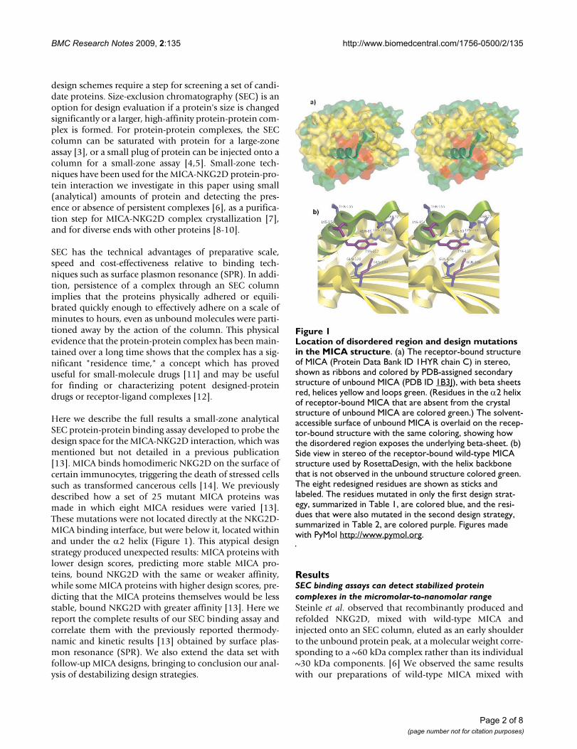
Here we describe the full results a small-zone analyticalSEC protein-protein binding assay developed to probe thedesign space for the MICA-NKG2D interaction, which wasmentioned but not detailed in a previous publication[13]. MICA binds homodimeric NKG2D on the surface ofcertain immunocytes, triggering the death of stressed cellssuch as transformed cancerous cells [14]. We previouslydescribed how a set of 25 mutant MICA proteins wasmade in which eight MICA residues were varied [13].These mutations were not located directly at the NKG2D-MICA binding interface, but were below it, located withinand under the α2 helix (Figure 1). This atypical designstrategy produced unexpected results: MICA proteins withlower design scores, predicting more stable MICA pro-teins, bound NKG2D with the same or weaker affinity,while some MICA proteins with higher design scores, pre-dicting that the MICA proteins themselves would be lessstable, bound NKG2D with greater affinity [13]. Here wereport the complete results of our SEC binding assay andcorrelate them with the previously reported thermody-namic and kinetic results [13] obtained by surface plas-mon resonance (SPR). We also extend the data set withfollow-up MICA designs, bringing to conclusion our anal-ysis of destabilizing design strategies.
ResultsSEC binding assays can detect stabilized protein complexes in the micromolar-to-nanomolar rangeSteinle et al. observed that recombinantly produced andrefolded NKG2D, mixed with wild-type MICA andinjected onto an SEC column, eluted as an early shoulderto the unbound protein peak, at a molecular weight corre-sponding to a ~60 kDa complex rather than its individual~30 kDa components. [6] We observed the same resultswith our preparations of wild-type MICA mixed with
Location of disordered region and design mutations in the MICA structureFigure 1Location of disordered region and design mutations in the MICA structure. (a) The receptor-bound structure of MICA (Protein Data Bank ID 1HYR chain C) in stereo, shown as ribbons and colored by PDB-assigned secondary structure of unbound MICA (PDB ID 1B3J), with beta sheets red, helices yellow and loops green. (Residues in the α2 helix of receptor-bound MICA that are absent from the crystal structure of unbound MICA are colored green.) The solvent-accessible surface of unbound MICA is overlaid on the recep-tor-bound structure with the same coloring, showing how the disordered region exposes the underlying beta-sheet. (b) Side view in stereo of the receptor-bound wild-type MICA structure used by RosettaDesign, with the helix backbone that is not observed in the unbound structure colored green. The eight redesigned residues are shown as sticks and labeled. The residues mutated in only the first design strat-egy, summarized in Table 1, are colored blue, and the resi-dues that were also mutated in the second design strategy, summarized in Table 2, are colored purple. Figures made with PyMol http://www.pymol.org.
Page 2 of 8(page number not for citation purposes)
BMC Research Notes 2009, 2:135 http://www.biomedcentral.com/1756-0500/2/135
Page 3 of 8(page number not for citation purposes)
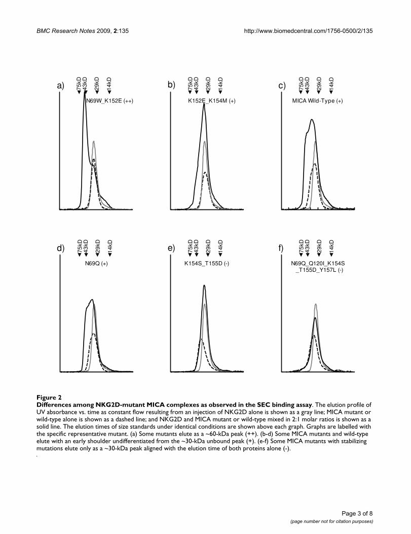
Differences among NKG2D-mutant MICA complexes as observed in the SEC binding assayFigure 2Differences among NKG2D-mutant MICA complexes as observed in the SEC binding assay. The elution profile of UV absorbance vs. time as constant flow resulting from an injection of NKG2D alone is shown as a gray line; MICA mutant or wild-type alone is shown as a dashed line; and NKG2D and MICA mutant or wild-type mixed in 2:1 molar ratios is shown as a solid line. The elution times of size standards under identical conditions are shown above each graph. Graphs are labelled with the specific representative mutant. (a) Some mutants elute as a ~60-kDa peak (++). (b-d) Some MICA mutants and wild-type elute with an early shoulder undifferentiated from the ~30-kDa unbound peak (+). (e-f) Some MICA mutants with stabilizing mutations elute only as a ~30-kDa peak aligned with the elution time of both proteins alone (-).
N69W_K152E (++)
a) 75kD
14kD
29kD
43kD
K152E_K154M (+)
b) 75kD
14kD
29kD
43kD
MICA Wild-Type (+)
75kD
14kD
29kD
43kDc)
N69Q (+)
d) 75kD
14kD
29kD
43kD
K154S_T155D (-)
e) 75kD
14kD
29kD
43kD
N69Q_Q120I_K154S _T155D_Y157L (-)
f) 75kD
14kD
29kD
43kD

BMC Research Notes 2009, 2:135 http://www.biomedcentral.com/1756-0500/2/135
Page 4 of 8(page number not for citation purposes)
Comparison of SPR and SEC binding assays for evaluation of redesigned MICA proteinsFigure 3Comparison of SPR and SEC binding assays for evaluation of redesigned MICA proteins. Each MICA mutant from Table 1 is organized according to the results of the SEC binding assay and graphed against a different parameter. The average of each category is shown as a vertical line. (a) SEC results compared to RosettaDesign score [13] shows that negative Rosetta-Design scores (predicted stabilized) only have binding results similar to or worse than wild-type, while positive RosettaDesign scores (predicted destabilized) include all 5 MICA mutants found to bind more tightly than wild-type. SEC results compared to SPR results [13] from (b) equilibrium and (c-d) kinetic analysis show similar correlations between equilibrium affinity and on-rate with the highest-affinity mutants by SEC, while no correlation with off-rate is seen because the set of mutants does not vary significantly in this parameter. Observed kon and koff rates from single-step kinetic fits for mutants predicted to be stabi-lized, and from two-step kinetic fits for mutants predicted to be destabilized (k+1 and k-2, respectively). (P values for the differ-ence between ++ and + complexes: t-test P = 0.0013 for Figure 3a; 0.0006 for Figure 3b; 0.0001 for Figure 3c; and 0.6 for Figure 3d.)
-9 -8 -7 -6 -5 -4 -3 -2 -1 0 1 2 3 4 5 6
-
+
++a)
Design score relative to wild-type
SE
C b
ind
ing
-9.0 -8.5 -8.0 -7.5 -7.0 -6.5 -6.0
-
+
++b)
G(equilibr ium)
SE
C b
ind
ing
1 10 100 1000
-
+
++c)
log (kon, obs)
SE
C b
ind
ing
1 10 100 1000
-
+
++d)
log (koff,obs)
SE
C b
ind
ing

BMC Research Notes 2009, 2:135 http://www.biomedcentral.com/1756-0500/2/135
homodimeric NKG2D (Figure 2c), and repeated theseconditions for our set of redesigned MICA mutants (Fig-ure 2). Some MICA mutants with high design scores (pre-dicting MICA destabilization) mixed in a 1:2 molar ratiowith NKG2D eluted as a separate, early ~60 kDa peak (Fig-ure 2a). When this early peak was collected and analyzedby reducing SDS-PAGE, protein bands were observed atthe molecular weights corresponding to both MICA (30kDa) and NKG2D (15 kDa). Other MICA mutants mixedwith NKG2D eluted as early shoulders to the 30-kDa peak(Figure 2b and 2d), or as symmetrical ~30-kDa peaks (Fig-ure 2e and 2f). A qualitative correlation can be observedbetween SPR equilibrium ΔG of binding and SEC bindassay results: Observation of an early shoulder corre-sponds to a low-micromolar range of affinity like wild-type MICA binding NKG2D. A differentiated early peakcorresponds to high-nanomolar binding, as withMIC_N69W_K152E, and the absence of an early peak cor-responds to low-to-mid-micromolar binding, as withMIC_N69Q_Q120I_K154S_T155D_Y157L.
SEC binding assays can detect differences in protein-protein on-ratesThe SPR study showed that some high design scores areassociated with high affinity, and the SEC binding assayleads to the same conclusion (Figure 3). Equilibriumaffinity agrees with the sets of protein binding strengthsdescribed by the SEC binding assay. Our design strategyaltered non-contacting residues that did not change theoff-rates, but did cause a large difference in on-rates. TheSEC binding assay detected this, discriminating amongcomplexes that had significantly varying on-rates, and theoff-rates for this set of proteins were so similar that theywere not a significant factor.
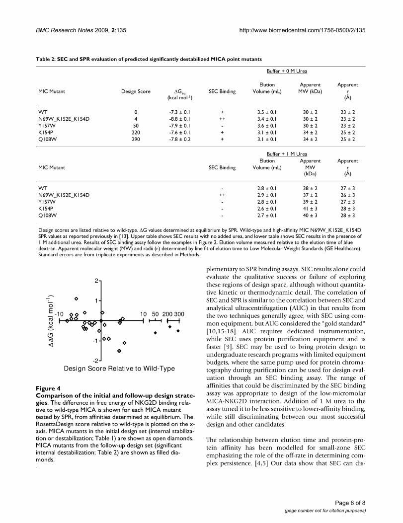
Rationally designed MICA destabilization at single non-contacting residues does not enhance NKG2D bindingA plot of NKG2D affinity vs. design score for the initial setof 25 MICA mutants produced previously shows that anarea of design space was unfilled because the initial designstrategy was biased toward stabilizing mutations and noMICA mutant was destabilized by more than 6 units. (Fig-ure 4) After we found that mild MICA destabilizationincreased NKG2D affinity by more than an order of mag-nitude (Table 1), we then designed three more destabiliz-ing mutants at the same locations to confirm if we werepast the point of "diminishing returns," at whichincreased MICA destabilization would destabilize thecomplex with NKG2D. Using RosettaDesign, non-cysteine residues were modeled at residues 108, 154, and157. Point mutations predicted to provide a range of largedisruptions to the wild-type structure were selected fromthe candidate designs produced by Rosetta, and preparedas previously reported [13]. In the SEC binding assay,none bound NKG2D better than wild-type MICA (Table2). SPR equilibrium analysis showed their affinities werenot significantly enhanced (Figure 4).
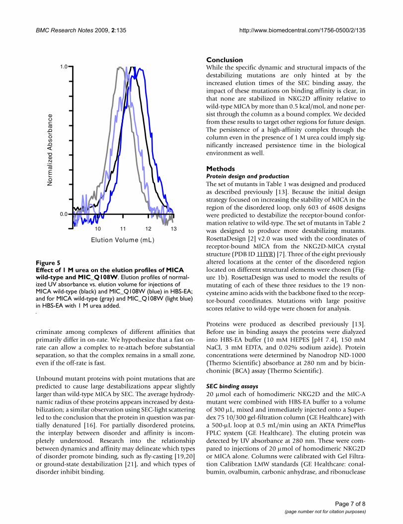
Destabilized mutants elute early in analytical SECIn the SEC binding assay, the peak maximum of theunbound MICA mutant would decrease, moving from 3.5mL for mutants with design scores of 50 or below to 3.1mL for higher design scores (Figure 5), increasing appar-ent hydrodynamic radius from 23 Å to 25 Å (Table 2).When the column was saturated with buffer containing 1M urea, the two mutants with the largest predicted desta-bilizations continued to elute early (Figure 5). The early-eluting shoulders for wild-type and the point-destabilizedmutants were no longer observed. High-affinityMICN69W_K152E_K154D when mixed with NKG2D stilleluted in 1 M urea as an early peak (Table 2).
DiscussionIn this study SEC formed the basis of a simple bindingassay to assess protein-protein interaction strength, com-
Table 1: SEC evaluation of predicted stabilized or mildly destabilized MICA mutants
MIC Mutant Design Score SEC Binding
N69Q_Q108L_Q120I_K154S_T155D -8.1 +N69Q_Q120I_K154S_T155D_Y157L -7.1 -N69Q_D72F_K154S_T155D -7.1 +N69Q_Q120I_K154S_T155D -6 +N69Q_D72F_Q108L_K152V_Y157L -5.9 +K152V_K154S_T155D_Y157L -5.8 +N69Q_K154D -4.3 +K154S_T155D -4 -K152V_K154S_T155D -3.9 -N69Q_D72F_Q108L -3.9 +N69Q_D72F -3.6 +N69Q -2.8 +K152V_Y157L -2.2 +K154D -1.5 +Q108L -0.5 +Wild-type 0 +D72W 0.3 -Q120I 0.7 +Q120I_K154M 0.8 n/aN69W 1.8 ++N69W_K152E_K154S 4.2 ++N69W_K152E_K154D 4.3 ++K152E_K154M 4.4 +N69W_D72F_K152E 4.5 ++N69W_D72W_K152E 5.1 +N69W_K152E 5.5 ++
Design scores are listed relative to wild-type. Results of SEC binding assay follow the examples in Figure 2; n/a, not applicable because unbound mutant did not elute from column due to non-specific binding.
Page 5 of 8(page number not for citation purposes)
BMC Research Notes 2009, 2:135 http://www.biomedcentral.com/1756-0500/2/135
plementary to SPR binding assays. SEC results alone couldevaluate the qualitative success or failure of exploringthese regions of design space, although without quantita-tive kinetic or thermodynamic detail. The correlation ofSEC and SPR is similar to the correlation between SEC andanalytical ultracentrifugation (AUC) in that results fromthe two techniques generally agree, with SEC using com-mon equipment, but AUC considered the "gold standard"[10,15-18]. AUC requires dedicated instrumentation,while SEC uses protein purification equipment and isfaster [9]. SEC may be used to bring protein design toundergraduate research programs with limited equipmentbudgets, where the same pump used for protein chroma-tography during purification can be used for design eval-uation through an SEC binding assay. The range ofaffinities that could be discriminated by the SEC bindingassay was appropriate to design of the low-micromolarMICA-NKG2D interaction. Addition of 1 M urea to theassay tuned it to be less sensitive to lower-affinity binding,while still discriminating between our most successfuldesign and other candidates.
The relationship between elution time and protein-pro-tein affinity has been modelled for small-zone SECemphasizing the role of the off-rate in determining com-plex persistence. [4,5] Our data show that SEC can dis-
Table 2: SEC and SPR evaluation of predicted significantly destabilized MICA point mutants
Buffer + 0 M Urea
Elution Apparent ApparentMIC Mutant Design Score ΔGeq
(kcal mol-1)SEC Binding Volume (mL) MW (kDa) r
(Å)
WT 0 -7.3 ± 0.1 + 3.5 ± 0.1 30 ± 2 23 ± 2N69W_K152E_K154D 4 -8.8 ± 0.1 ++ 3.4 ± 0.1 30 ± 2 23 ± 2Y157W 50 -7.9 ± 0.1 - 3.6 ± 0.1 30 ± 2 23 ± 2K154P 220 -7.6 ± 0.1 + 3.1 ± 0.1 34 ± 2 25 ± 2Q108W 290 -7.8 ± 0.2 + 3.1 ± 0.1 34 ± 2 25 ± 2
Buffer + 1 M UreaElution Apparent Apparent
MIC Mutant SEC Binding Volume (mL) MW(kDa)
r(Å)
WT - 2.8 ± 0.1 38 ± 2 27 ± 3N69W_K152E_K154D ++ 2.9 ± 0.1 37 ± 2 26 ± 3Y157W - 2.8 ± 0.1 39 ± 2 27 ± 3K154P - 2.6 ± 0.1 41 ± 3 28 ± 3Q108W - 2.7 ± 0.1 40 ± 3 28 ± 3
Design scores are listed relative to wild-type. ΔG values determined at equilibrium by SPR. Wild-type and high-affinity MIC N69W_K152E_K154D SPR values as reported previously in [13]. Upper table shows SEC results with no added urea, and lower table shows SEC results in the presence of 1 M additional urea. Results of SEC binding assay follow the examples in Figure 2. Elution volume measured relative to the elution time of blue dextran. Apparent molecular weight (MW) and radii (r) determined by line fit of elution time to Low Molecular Weight Standards (GE Healthcare). Standard errors are from triplicate experiments as described in Methods.
Comparison of the initial and follow-up design strategiesFigure 4Comparison of the initial and follow-up design strate-gies. The difference in free energy of NKG2D binding rela-tive to wild-type MICA is shown for each MICA mutant tested by SPR, from affinities determined at equilibrium. The RosettaDesign score relative to wild-type is plotted on the x-axis. MICA mutants in the initial design set (internal stabiliza-tion or destabilization; Table 1) are shown as open diamonds. MICA mutants from the follow-up design set (significant internal destabilization; Table 2) are shown as filled dia-monds.
-10 10
-2
-1
1
2
50 200 300
Design Score Relative to Wild-Type
G (
kcal
mo
l-1)
Page 6 of 8(page number not for citation purposes)
BMC Research Notes 2009, 2:135 http://www.biomedcentral.com/1756-0500/2/135
criminate among complexes of different affinities thatprimarily differ in on-rate. We hypothesize that a fast on-rate can allow a complex to re-attach before substantialseparation, so that the complex remains in a small zone,even if the off-rate is fast.
Unbound mutant proteins with point mutations that arepredicted to cause large destabilizations appear slightlylarger than wild-type MICA by SEC. The average hydrody-namic radius of these proteins appears increased by desta-bilization; a similar observation using SEC-light scatteringled to the conclusion that the protein in question was par-tially denatured [16]. For partially disordered proteins,the interplay between disorder and affinity is incom-pletely understood. Research into the relationshipbetween dynamics and affinity may delineate which typesof disorder promote binding, such as fly-casting [19,20]or ground-state destabilization [21], and which types ofdisorder inhibit binding.
ConclusionWhile the specific dynamic and structural impacts of thedestabilizing mutations are only hinted at by theincreased elution times of the SEC binding assay, theimpact of these mutations on binding affinity is clear, inthat none are stabilized in NKG2D affinity relative towild-type MICA by more than 0.5 kcal/mol, and none per-sist through the column as a bound complex. We decidedfrom these results to target other regions for future design.The persistence of a high-affinity complex through thecolumn even in the presence of 1 M urea could imply sig-nificantly increased persistence time in the biologicalenvironment as well.
MethodsProtein design and productionThe set of mutants in Table 1 was designed and producedas described previously [13]. Because the initial designstrategy focused on increasing the stability of MICA in theregion of the disordered loop, only 603 of 4608 designswere predicted to destabilize the receptor-bound confor-mation relative to wild-type. The set of mutants in Table 2was designed to produce more destabilizing mutants.RosettaDesign [2] v2.0 was used with the coordinates ofreceptor-bound MICA from the NKG2D-MICA crystalstructure (PDB ID 1HYR) [7]. Three of the eight previouslyaltered locations at the center of the disordered regionlocated on different structural elements were chosen (Fig-ure 1b). RosettaDesign was used to model the results ofmutating of each of these three residues to the 19 non-cysteine amino acids with the backbone fixed to the recep-tor-bound coordinates. Mutations with large positivescores relative to wild-type were chosen for analysis.
Proteins were produced as described previously [13].Before use in binding assays the proteins were dialyzedinto HBS-EA buffer (10 mM HEPES [pH 7.4], 150 mMNaCl, 3 mM EDTA, and 0.02% sodium azide). Proteinconcentrations were determined by Nanodrop ND-1000(Thermo Scientific) absorbance at 280 nm and by bicin-choninic (BCA) assay (Thermo Scientific).
SEC binding assays20 μmol each of homodimeric NKG2D and the MIC-Amutant were combined with HBS-EA buffer to a volumeof 300 μL, mixed and immediately injected onto a Super-dex 75 10/300 gel-filtration column (GE Healthcare) witha 500-μL loop at 0.5 mL/min using an AKTA PrimePlusFPLC system (GE Healthcare). The eluting protein wasdetected by UV absorbance at 280 nm. These were com-pared to injections of 20 μmol of homodimeric NKG2Dor MICA alone. Columns were calibrated with Gel Filtra-tion Calibration LMW standards (GE Healthcare: conal-bumin, ovalbumin, carbonic anhydrase, and ribonuclease
Effect of 1 M urea on the elution profiles of MICA wild-type and MIC_Q108WFigure 5Effect of 1 M urea on the elution profiles of MICA wild-type and MIC_Q108W. Elution profiles of normal-ized UV absorbance vs. elution volume for injections of MICA wild-type (black) and MIC_Q108W (blue) in HBS-EA; and for MICA wild-type (gray) and MIC_Q108W (light blue) in HBS-EA with 1 M urea added.
10 11 12 13-0.1
0.0
0.1
0.2
0.3
0.4
0.5
0.6
0.7
0.8
0.9
1.0
Elution Volume (mL)
No
rmal
ized
Ab
sorb
ance
Page 7 of 8(page number not for citation purposes)

BMC Research Notes 2009, 2:135 http://www.biomedcentral.com/1756-0500/2/135
A, and blue dextran for void volume determination; alsoSigma: cytochrome c). Several individual proteins andmixed receptor-ligand complexes were injected three ormore times onto the same column, resulting in variationsin elution volume of no more than 0.1 mL. Fractions werecollected using the AKTA Prime fraction collector for anal-ysis by reducing analytical SDS-PAGE using CoomassieBlue staining. Observed variations in elution volume fortriplicate experiments with urea in the buffer were as listedin Table 2.
SPR binding assaysFor the first set of mutants (Table 1), determination ofNKG2D-mutant MICA kinetics and thermodynamics bySPR was previously described [13]. NKG2D affinities forthe second set of MICA mutants (Table 2) were deter-mined by equilibrium binding analysis as used in the pre-vious study. (Fast kinetics precluded the use of kineticfits.) Standard errors reported in the data tables resultfrom triplicate (or more) experiments.
Competing interestsThe authors declare that they have no competing interests.
Authors' contributionsCLM and WKS carried out SEC binding assays. WKS andBJM carried out SPR binding assays. CLM, WKS, MAS,ADV, and CRA designed, made and purified mutant pro-teins. All authors participated in interpretation of thedata. CLM, WKS, CRA and BJM drafted the manuscript. Allauthors have read and approved the final manuscript.
AcknowledgementsWe thank Derek Wood and Kathryn Houmiel (Seattle Pacific University) for technical assistance and early evaluation of this research as student presentations, Tanja Kortemme (UCSF) for the initial design with Rosetta, and Roland Strong (Fred Hutchinson Cancer Research Center) for instru-ment use. This research was funded by National Institutes of Health Grant R15 AI058972.
References1. Kortemme T, Baker D: Computational design of protein-pro-
tein interactions. Curr Opin Chem Biol 2004, 8(1):91-7.2. Kortemme T, Baker D: A simple physical model for binding
energy hot spots in protein-protein complexes. Proceedings ofthe National Academy of Sciences of the United States of America 2002,99(22):14116-14121.
3. Winzor DJ: Quantitative characterization of ligand binding bychromatography. In Protein-Ligand Interactions: Hydrodynamics andCalorimetry: A Practical Approach Edited by: Harding SE, Chowdhry BZ.Oxford: Oxford University Press; 2001:47-74.
4. Stevens FJ: Analysis of protein-protein interaction by simula-tion of small-zone size exclusion chromatography. Stochas-tic formulation of kinetic rate contributions to observedhigh-performance liquid chromatography elution character-istics. Biophys J 1989, 55(6):1155-1167.
5. Wilton R, Myatt EA, Stevens FJ: Analysis of protein-protein inter-actions by simulation of small-zone gel filtration chromatog-raphy. In Protein-Protein Interactions: Methods and Protocols Volume261. Edited by: Fu H. Totowa, NJ: Humana Press Inc; 2004:137-154.
6. Steinle A, Li P, Morris DL, Groh V, Lanier LL, Strong RK, Spies T:Interactions of human NKG2D with its ligands MICA, MICB,
and homologs of the mouse RAE-1 protein family. Immunoge-netics 2001, 53(4):279-287.
7. Li P, Morris DL, Willcox BE, Steinle A, Spies T, Strong RK: Complexstructure of the activating immunoreceptor NKG2D and itsMHC class I-like ligand MICA. Nat Immunol 2001, 2(5):443-451.
8. Franzini M, Bramanti E, Ottaviano V, Ghiri E, Scatena F, Barsacchi R,Pompella A, Donato L, Emdin M, Paolicchi A: A high performancegel filtration chromatography method for gamma-glutamyl-transferase fraction analysis. Anal Biochem 2008, 374(1):1-6.
9. le Maire M, Arnou B, Olesen C, Georgin D, Ebel C, Moller JV: Gelchromatography and analytical ultracentrifugation to deter-mine the extent of detergent binding and aggregation, andStokes radius of membrane proteins using sarcoplasmicreticulum Ca2+-ATPase as an example. Nat Protoc 2008,3(11):1782-95.
10. Gralle M, Oliveira CL, Guerreiro LH, McKinstry WJ, Galatis D, Mas-ters CL, Cappai R, Parker MW, Ramos CH, Torriani I, et al.: Solutionconformation and heparin-induced dimerization of the full-length extracellular domain of the human amyloid precursorprotein. J Mol Biol 2006, 357(2):493-508.
11. Copeland RA, Pompliano DL, Meek TD: Drug-target residencetime and its implications for lead optimization. Nat Rev DrugDiscov 2006, 5(9):730-9.
12. Tummino PJ, Copeland RA: Residence time of receptor-ligandcomplexes and its effect on biological function. Biochemistry2008, 47(20):5481-5492.
13. Lengyel CS, Willis LJ, Mann P, Baker D, Kortemme T, Strong RK,McFarland BJ: Mutations designed to destabilize the receptor-bound conformation increase MICA-NKG2D associationrate and affinity. J Biol Chem 2007, 282(42):30658-30666.
14. Strong RK, McFarland BJ: NKG2D and Related Immunorecep-tors. Adv Protein Chem 2004, 68:281-312.
15. Berkowitz SA: Role of analytical ultracentrifugation in assess-ing the aggregation of protein biopharmaceuticals. AAPS J2006, 8(3):E590-605.
16. Philo JS: Is any measurement method optimal for all aggre-gate sizes and types? AAPS J 2006, 8(3):E564-71.
17. Gualfetti PJ, Iwakura M, Lee JC, Kihara H, Bilsel O, Zitzewitz JA, Mat-thews CR: Apparent radii of the native, stable intermediatesand unfolded conformers of the alpha-subunit of tryptophansynthase from E. coli, a TIM barrel protein. Biochemistry 1999,38(40):13367-13378.
18. Gabrielson JP, Brader ML, Pekar AH, Mathis KB, Winter G, CarpenterJF, Randolph TW: Quantitation of aggregate levels in a recom-binant humanized monoclonal antibody formulation by size-exclusion chromatography, asymmetrical flow field flowfractionation, and sedimentation velocity. J Pharm Sci 2007,96(2):268-79.
19. Hoffman RM, Blumenschein TM, Sykes BD: An interplay betweenprotein disorder and structure confers the Ca2+ regulationof striated muscle. J Mol Biol 2006, 361(4):625-33.
20. Shoemaker BA, Portman JJ, Wolynes PG: Speeding molecular rec-ognition by using the folding funnel: the fly-casting mecha-nism. Proceedings of the National Academy of Sciences of the UnitedStates of America 2000, 97(16):8868-8873.
21. Horn JR, Kraybill B, Petro EJ, Coales SJ, Morrow JA, Hamuro Y, Kos-siakoff AA: The role of protein dynamics in increasing bindingaffinity for an engineered protein-protein interaction estab-lished by H/D exchange mass spectrometry. Biochemistry 2006,45(28):8488-8498.
Page 8 of 8(page number not for citation purposes)
http://www.ncbi.nlm.nih.gov/entrez/query.fcgi?cmd=Retrieve&db=PubMed&dopt=Abstract&list_uids=2765653
http://www.ncbi.nlm.nih.gov/entrez/query.fcgi?cmd=Retrieve&db=PubMed&dopt=Abstract&list_uids=2765653